First published NMR (MRI) image of the brain: A shadow of the brain
|
|
|
- Rosa Copeland
- 5 years ago
- Views:
Transcription
1 Neuroimaging Structural magnetic resonance imaging measuring volumes of brain and brain regions Functional MRI measuring brain activity during cognitive tasks Diffusion MRI measuring motion of water molecules along diffusion gradients Positron emission tomography -- measuring metabolic processes, including changes in metabolism during cognitive tasks

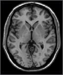
2 First published NMR (MRI) image of the brain: 1980 A shadow of the brain

3 Images collected in 1985 Multiple sclerosis Clinical trials for MRI began in 1983, FDA approved two years later, in Sensitivity of MRI to MS lesions compared to CT: 10 to 1
 6 4 2 0 1 READ PRI_SAM PRI_REV Left occipital lobe Ryan &")
4 First human functional MRI paper: Bandettini, Wong, et al. (1992) READ PRI_SAM PRI_REV Left occipital lobe Ryan & Schnyer, 2005
5 Functional image properties What does it measure (light transmittance, quantity of a specific material) Contrast sensitivity the smallest difference in quantity that is measurable, resulting in a difference in image intensity Spatial resolution the ability to distinguish changes in signal across different spatial locations Temporal resolution the sampling rate, or how fast you can detect a change in signal
 (B) Two MRI s (same image) at different contrast sensitivities, with greater signal intensity differences across gray white matter boundaries (A) compared")
6 1.4 Contrast and contrast-to-noise in MR images. (A) (B) Two MRI s (same image) at different contrast sensitivities, with greater signal intensity differences across gray white matter boundaries (A) compared to image (B). Contrast to noise ratio magnitude of intensity differences divided by background signal variance

7 1.4 Contrast and contrast-to-noise in MR images. In MRI, contrast also refers to sensitivity to a specific physical property of the nuclei For example, two images that are differentially sensitive to two properties of relaxation rates of hydrogen, T1 (A) and T2 (B).
8 Functional contrast to noise Low fcnr time High fcnr The ability to detect a signal change against a background of noise, or variance in signal The variance may be due to measurement error, or physiological noise

9 Spatial resolution Pixel: Smallest element in a 2D image in-plane resolution Voxel: 3D sample from which signal is collected and averages 1mm 1mm 5-10 mm Voxel size: In-plane resolution x section thickness

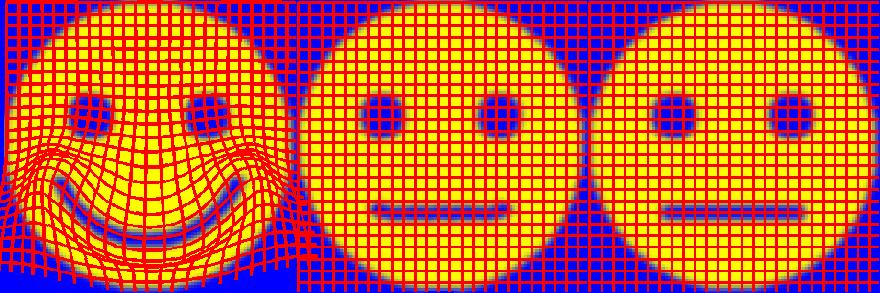
10 1.6 The human brain at different spatial resolutions. MRI images at various spatial resolutions. Note resolutions 1.5mm or smaller appear similar to us. A. 8mm B. 4mm C. 2mm D. 1.5mm E. 1mm
11 Anatomical: 1 x 1 x 5mm Functional: 3.4 x 3.4 x 5mm Functional images are lower resolution (larger voxels) and also have lower CNR because of the way the images are collected (echo-planar).
12 Temporal resolution of images Sampling rate: The frequency in time with which a measurement is made MRI can sample as quickly as 30 msecs Temporal resolution: The ability to distinguish changes in an image across time. Two limits to temporal resolution: Nyquist frequency a fundamental rule that a signal must be sampled twice as frequently as the fastest change in the signal that you wish to measure Signal frequency fast sampling does not matter if the signal change is slow (hemodynamic response is 12 secs)
 that was placed in a magnetic field.")
13 1.11 Nobel laureates Felix Bloch (A) and Edward Purcell (B) shared the 1952 prize in Physics. Felix Bloch and Edward Purcell: Nobel Prize in Physics, Purcell measured magnetic resonance in a block of material (paraffin wax) that was placed in a magnetic field. Purcell did the same with a container of water, devising a method that is identical to the basic MRI system: A static magnetic field, a transmit EM coil, and a coil for detecting emitted energy.
14 Magentic resonance: Resonant frequency the frequency at which a particular molecule precesses or spins like a top around its axis. AKA Larmor frequency Energy at that frequency will be absorbed ( excitation ). Once the energy source is removed, the molecule will return back to its normal resting state, giving off energy ( relaxation ). Magnetic resonance measureable energy emitted during relaxation.
15 MRI Signal: Step 1: Atoms with an uneven number of protons act as dipoles in a strong static magnetic field, they will align with the field and precess around that axis. B 0 Resting state
16 MRI Signal: Step 2: Apply energy pulse (normally in the radio frequency range) at the resonant frequency of the molecule the energy will be absorbed. B 0 RF Excitation state
17 Relaxation state MRI Signal: Step 3: Turn off the RF pulse, and the molecule gives off the absorbed energy over time (relaxation rate), which can be measured with an RF receiver coil. This is magnetic resonance. B 0 RF receiver coil
18 MRI Signal: Molecule of interest: Hydrogen Why hydrogen? Lots of it in the brain (water) Differs in densities across tissue types (least in white matter, more in gray matter, most in CSF) Also differs in the strength of bonds (water is freely diffusing in CSF, but more tightly bound in fatty tissue such as myelin) Both these properties will affect the relaxation rate how fast the water molecule returns to its low energy state
.")
19 1.14 The first MR image of the human body. Raymond Damadian, 1977 First cross-sectional image of the human body. Damadian showed that magnetic resonance of water differed depending on the type of biological tissue in which it was bound (Science, 1971). He built the first large-bore magnet called Indomitable, producing a cross-sectional image of the human body composed of 106 voxels. Each voxel was obtained separately, by moving the person s position slightly. Total imaging time was 4 hours.
 applied gradients to the static magetic field so that the field strength differed depending on the spatial location.")
20 1.13 Nobel laureates Paul Lauterbur (A) and Peter Mansfield (B). Paul Lauterbur and Peter Mansfield, Nobel Prize in Medicine, 2003 Lauterbur (1976) applied gradients to the static magetic field so that the field strength differed depending on the spatial location. The resonant frequency of hydrogen would therefore differ across spatial locations. The amount of energy emitted at a given frequency would determine where it was located in 2D space. Peter Mansfield (1976) found a more efficient way of collecting the signal, by applying a single EM pulse, and then acquiring signal continuously while you changed the spatial gradients. Then the complex signal could be reconstructed with Fourier analysis.
21 Components of MRI scanner: Static magnetic field Transmit radiofrequency coil Receiver radiofrequency coil Gradient coils (z, x, y) Shimming coils (1 st, 2 nd, 3 rd order)
22 Static magnetic field: Electromagnet solenoid with current that is maintained by supercooling, creates a magnetic field perpendicular to the axis of the coil. Housed in a vaccum chamber (dewar). Field strength proportional to the diameter of the coil and the strength of the current (nonlinearly related).
23 Radiofrequency coils Transmit coil: Electromagnetic coil used to generate oscillating energy (radiofrequency range) at the resonant frequency of a sample being measured (excitation). Receive coil: EM coil used to measure energy emitted by a sample as it returns to its lower energy state (relaxation) once the excitation pulse is turned off.

24 Surface coil, simple inductor-capacitor circuit used to produce strong magnetic field over a limited region of brain. Volume or birdcage coil, used to produce consistent images across the whole brain. Contains both transmit and receive RF coils.
 Surface coil obtains inductor strong local and resistor signal but generates limited area. B) Volume coil obtains an oscillating relatively magnetic uniform field.")
25 Volume coil is a Helmholtz pair design. Amount of energy that can be transmitted or received depends on distance between Rapid charge/discharge the coil and the between sample. A) Surface coil obtains inductor strong local and resistor signal but generates limited area. B) Volume coil obtains an oscillating relatively magnetic uniform field. signal at expense of local strength.
26 Gradient and shim coils Gradient coils superimpose small and consistent variations in the strength of the static magnetic field Used for spatial localization of the signal (more soon.) Three directions, x, y, z Shim coils small EM coils that are used to keep the static field homogeneous These are adjusted for each subject in the scanner, since each person s head will distort the field differently


27 Measuring hippocampal volumes: Predicts AD in patients who already have mild cognitive impairments.

28 Voxel based morphometry Traditional ROI based morphometry is time consuming, observer dependent, provides measures of large areas and implies a priori hypotheses regarding the structures to assess. VBM is an automated method that lets you test for volume differences across the entire brain, voxel by voxel. Examines local volume differences in different tissue types that may be independent of larger volumetric differences in gross anatomy. A voxel by voxel statistical analysis is used to detect regional differences in the amount of gray and white matter between populations.

29 12/15/2004 Division of Psychiatric Neuroimaging The Johns Hopkins School of Medicine

30 Segmentation Based on image intensity distributions

31 Segmentation use of priori information Tissue probability maps (TPMs) are used in addition to intensity information to enhance segmentation. T1 GM WM CSF Mask for cleaning up non brain
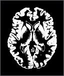
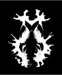
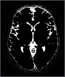
32 Segmentation Example final maps T1 gray white CSF


33 Modulation Corrects for distortions and changes in volume induced by nonlinear normalization. Analogy: as we blow up a balloon, the surface becomes thinner. Likewise, as we expand a brain area it s volume is reduced. Without modulation Source Template Modulated

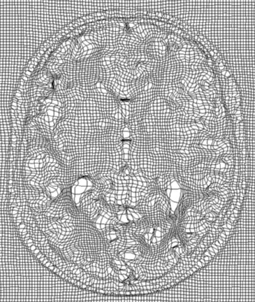
34 Deformation Field Original Warped Template Deformation field

35 Modulation Effect of modulating segmented images. The Jacobian determinant in the center represents the volume changes due to non-linear spatial normalization. These volume changes are used to modulate the segmentation result on the left and the modulated image is shown on the right side.

36 Advantages Automated: fast and not subject to individual bias. Able to examine regions that are not anatomically well defined. Able to see the whole brain rather than choosing specific regions. Can be normalized for overall differences in brain volume, but also small regional variations in volume which will otherwise add variance to regional measurements.
 Stroke patient vs.")
37 Measuring stroke (Shen et al. 2007) Stroke patient vs. controls


38 Correlation between BMI and Gray Matter Volume Decline Volume with Increasing BMI Increase Volume with Increasing BMI
39 fmri: What is it? Measures changes in signal intensity that arise from oxygenated blood in a region. Good things: High resolution, fast scanning time, non-invasive Not so good things: Very low signal to noise ratio, sensitive to motion, susceptibility artifacts
40 Functional MRI signal MRI signal is dependent on field strength of the magnet and the properties of the tissue. Also dependent upon changes in local environment Paramagnetic substances (such as deoxyhemoglobin) will lead to loss of local signal on T2* weighted image.
41 Functional MRI signal Local neuronal activity Increased local metabolic rate Increased blood flow Increased oxygenated hemoglobin Uptake of O 2 less than supply Surplus oxygenated hemoglobin Decreased concentrations of deoxyhemoglobin Increased local fmri T2* signal
42 BOLD Contrast Resting state arterial HbO 2 Hb signal venous Stimulated state signal
43 Determining Activation: A subtraction measure Subject is scanned at rest (R) HbO 2 Hb signal Subject is scanned during cognitive task (C) signal Regions of activity are determined the differences between scan R from scan C.

44 Caveats regarding fmri: Tertiary measure of neuronal activity. Very small signal changes, on order of 1 to 2%. Signal change predominates in region of large draining veins, not gray matter, and may vary in locality. Extremely sensitive to motion. Hemodynamic response is delayed msec scan, but sec response.
45 Anatomical: 1 x 1 x 5mm Functional: 3.4 x 3.4 x 5mm Dealing with low signal strengths: Larger voxel size increases signal to noise (sensitivity), but results in lower resolution images.
46 Susceptibility: Regions near transitions between brain and air (sinuses) will cause signal dropoff.
47 Motion: time

48 The hemodynamic response takes time, even for a single, fast behavioral response
49 Simple fmri experiment: Word identification 20secs: Word/Nonword R/L button press 20 secs: XXXX/OOOO R/L button press Word/Nonword X/O
50 Statistical analysis: Mean[W/N], Mean[X/O] Paired t-test = Pearson r (function 0,1) Alternatively, convolved function HRD with 0,1 TIMEPT: TIMEPT:
51 An immediate problem of experiment-wise error Mean[W/N] > Mean[X/O] Paired t-test or correlation p(voxel) <.05,.01, etc How many tests? Volume: 5mm section, 64x64, 20 sections 81,920 voxels x 60% (brain) Total tests: 49,152
52 Statistical assumptions: Independence across voxels: Critical issue in dealing with multiple comparisons and assessing true p value What is the true dependence across voxels? Functional fields: Regions of cortex work in concert; the degree of independence is an empirical question Distribution of vessels: BOLD may be correlated across regions due to shared blood flow
53 Approaches: Smoothing: SPM imposes a covariance structure across all voxels, in order to estimate the change in degrees of freedom. Downside is that covariance is not uniform. Upside is that smoothing deals with left-overmotion, and some partial voluming. Then apply FDR or FWE correction based on the known DF. Clustering: Independent tests on each voxel, but estimate the probability by chance alone that voxels will occur side by side.
54 At p =.05, 2048 voxels, chance alone = 102 Chance that 12 cluster continguously? Much less
55 ROI Another approach: Region of interest analysis Identifying a priori based on anatomy or prior research the regions to analyse -- greatly reduces experimentwise error rate.
56 Types of designs: Blocked -- easy to set up, but limited Factorial designs: Measuring interaction effects [A+B+X] - [B+X] = [A+X] - [X] Cognitive conjunctions: Varying the control conditions to identify common cognitive processes [A+B] - [B] compared to [A+C] - [C] Parametric designs: Varying a parameter within a given variable, example, reaction time differences or confidence judgements
57 Blocked designs -- easy to set up, but limited Can t randomize trials Anticipation of effects (blocks of Yes vs No) Cannot remove incorrect responses But, greater power to detect changes in signal Why? Signal amplitude increase is (somewhat) additive) Insensitive to the shape of the HDR a bonus How do you increase power in a blocked design? Allowing activation to return to baseline Switching as often as possible
 Dale & Buckner, 1997 One, two, or three stimuli presented at ISI of 2 or 5 sec.")
, subtraction of the one trial HDR results in subsequent responses that")
58 8.20 Linear addition of hemodynamic responses to individual stimulus events. (Part 1) Dale & Buckner, 1997 One, two, or three stimuli presented at ISI of 2 or 5 sec. Generally linear and additive respones, especially with ISI=5. Note that for 2 sec data (shown here), subtraction of the one trial HDR results in subsequent responses that are smaller amplitude and delayed. Not a perfectly linear system, particularly at short intervals.


59 Varying stimuli presented from 1 to 32 A. Standard HDR form B-C Other possible HDR forms
60 Event related designs Simplest case: 1...(12-18secs) etc Faster presentation with jitter x...x x...x..1.x.x x.. etc
61 Good things about event-related designs: Trials can be regrouped in various ways, based on condition, subject s responses, reaction times, etc Random presentation decreases anticipatory effects Presentation and responses can be self-paced, more like typical cognitive experimental designs Difficulties: Dependencies amongst trials, e.g., yes/no recognition: Item...Decision Response Problem: If two regions are hypothesized to play different roles in the decision vs response components of the task, how can you separate them?


62 Diffusion MRI: Over time, molecules within gases or liquids will move freely through the medium via Brownian motion MRI diffusion-weighted gradient causes changes in the MR signal that are dependent upon the amplitude and direction of diffusion
 Where movement is relatively unrestricted, ADC will be high and FA will be low Where")
63 Vectors indicate simplified versions of the random walk path of the molecules Isotropic diffusion no restrictions in the direction of movement, measured as the apparent diffusion coefficient (ADC) Anistropic diffusion movement is restricted in one or more directions, measured as fractional anisotropy (FA) Where movement is relatively unrestricted, ADC will be high and FA will be low Where movement is relatively restricted (e.g., in a myelinated axon), ADC will be lower and FA will be high


64 Changes with Age 3T, 25 directions, 2 B0, 2 averages 29 years 75 years 92 years

65 Eigen-system Analysis of Diffusion Tensor z z z y y y x x x z y x z y x z y x zz yz xz yz yy xy xz xy xx V V V V V V V V V V V V V V V V V V D D D D D D D D D D Major Eigen-Value : Major Eigen-Vector: 1 T z y x V V V ,2,3 2 1,2, i i i i FA D


66 Field of Diffusion Tensor Ellipsoids Whole Slice W.Zhan et. al. Local Structures


67 Fiber Assignment by Continuous Tracking FACT Algorithm Xue R., et.al., Magn. Reson. Med. 42 (1999),


 Blue Z")
")
68 Diffusion tensor MRI -- Tractography Red movement along the X axis (right to left) Green movement along the Y axis (anterior to posterior) Blue movement along the Z axis (superior to inferior)

69 Positron Emission Tomography: Measuring brain metabolism via radioactive tracers
 ultrasound X-ray (CT) E hc m mm m")
70 Television Radio, Radar Visible photons X-ray Gamma ray Non-ionizing radiation Ionizing radiation PET SPECT MRI X-ray (film) ultrasound X-ray (CT) E hc m mm m nm
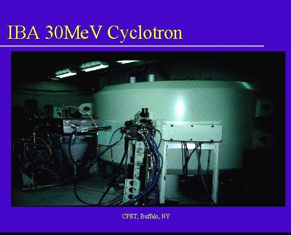
71 Positron emission tomography Cyclotron creates an isotope, where extra protons are added to the nucleus, creating instability. Isotope is connected to the compound of interest (such as oxygen or glucose) and injected. As the molecule decays, it emits a positron which is annihilated when it collides with an electron. Annihilation event releases energy (photons) that can be measured with detectors.
72
73 Annihilation: Decay via positron emission electron/positron annihilation annihilation photon g Conservation of momentum: Before: system at rest; momentum ~ 0 annihilation photon E - P + After: two photons created; must have same energy and travel in opposite direction. g
74 Emits gamma ray (two photons), travelling a path 180 degrees from the site of annihilation. Sufficient energy in gamma rays to increase probability of passing out of brain without attentuation. Scatter (how far the positron moves away from molecule) is 2 mm or less.
75 LOR determination Determining the line along which the two annihilation photons travel, known as the Line of Response or LOR, is a prerequisite step of any PET imaging modality and requires: Event detection (did an event occur?) Event positioning (where did it occur?) Coincidence determination (did two events occur in a straight line?) Physics of PET; photon detection - I
 * * detector Physics of PET; photon detection -")
76 Annihilation detection detector block (8x8 detectors) * line of response (LOR) * * detector Physics of PET; photon detection - I

77 Coincident detection Scintillating crystal detectors in circumferential arrays, measure coincident events only. Essentially counts coincident events, assumes a line of events (180 degrees).
78 Tomographic problem, reconstruction using back-projection
79 Parameters affecting image quality Sensitivity (SNR) or number of detectable counts: Dependent upon dose, scan length, kinetics of tracer, efficiency and number of detectors Spatial resolution Dependent upon resolution of detectors, small detector elements (bounded by the scatter at annihilation and the tracer kinetics)
80 Reconstruction Quantitative if corrections made for: 1. Gamma ray attenuation (1 in 5 from center of brain versus 4 in 5 at edge of brain). 2. Random incidences -- two unrelated gamma rays strike detectors simulataneously. 3. Scattered events - scattering (deflection) through tissue of gamma ray but still detected, thus incorrect position. 4. Differential efficiency of each detector, measured using uniform radiation source. 5. Dead time -- at high count rates, electronics limit the number of events countable.
81 PET tracers: 1. Oxygen - HL is 1.5 mins. [15O]-labeled water and oxygen used in quantification of oxygen consumption. 2. Carbon - HL is 10.0 mins. [11C]-labeled cocaine used to measure responses of dopamine D2 receptors during acute and chronic drug use. 3. Flourine - HL is 109 mins. [18F]-2-deoxyglucose (FDG) most often used in activation studies. Also used to label L-Dopa and fluoroethylspiperone which bind to D2 dopamine receptors.
: Regions of brain with reduced rates of glucose metabolism in 37 patients")
82 Preclinical detection of Alzheimer s disease: Reiman et al. (1996): Regions of brain with reduced rates of glucose metabolism in 37 patients with early stage probable AD.
 and their relation to patients with probable AD (purple); Reiman et al.")
83 Regions of brain with reduced rates of glucose metabolism in 11 e4 homozygotes (light blue) and their relation to patients with probable AD (purple); Reiman et al., 1996.
84 Unique uses of PET: Dopamine uptake using [18F]-Fluoroethylspiperone
 in the")
 and with Alzheimer's disease")

85 Comparing the absorption of PIB (Pittsburg Imaging Compound) in the brains of subjects without dementia (left) and with Alzheimer's disease (right). This compound binds to the proteins contained in beta amyloid plaques Plaques & Tangles
86 Neuroimaging methods: Provide many ways to measure structure and function of the human brain. What do you have to know to use them? -- how they work: physics -- how to analyse them: statistics -- how to interpret them: physiology -- how to apply them: cognitive neuroscience
Functional image properties
 Neuroimaging Structural magnetic resonance imaging measuring volumes of brain and brain regions Functional MRI measuring brain activity during cognitive tasks Diffusion MRI measuring motion of water molecules
Basic MRI physics and Functional MRI
 Basic MRI physics and Functional MRI Gregory R. Lee, Ph.D Assistant Professor, Department of Radiology June 24, 2013 Pediatric Neuroimaging Research Consortium Objectives Neuroimaging Overview MR Physics
EL-GY 6813/BE-GY 6203 Medical Imaging, Fall 2016 Final Exam
 EL-GY 6813/BE-GY 6203 Medical Imaging, Fall 2016 Final Exam (closed book, 1 sheets of notes double sided allowed, no calculator or other electronic devices allowed) 1. Ultrasound Physics (15 pt) A) (9
Contrast Mechanisms in MRI. Michael Jay Schillaci
 Contrast Mechanisms in MRI Michael Jay Schillaci Overview Image Acquisition Basic Pulse Sequences Unwrapping K-Space Image Optimization Contrast Mechanisms Static and Motion Contrasts T1 & T2 Weighting,
Technical University of Denmark
 Technical University of Denmark Page 1 of 10 pages Written test, 12 December 2012 Course name: Introduction to medical imaging Course no. 31540 Aids allowed: None. Pocket calculator not allowed "Weighting":
Magnetic resonance imaging MRI
 Magnetic resonance imaging MRI Introduction What is MRI MRI is an imaging technique used primarily in medical settings that uses a strong magnetic field and radio waves to produce very clear and detailed
MRI Physics I: Spins, Excitation, Relaxation
 MRI Physics I: Spins, Excitation, Relaxation Douglas C. Noll Biomedical Engineering University of Michigan Michigan Functional MRI Laboratory Outline Introduction to Nuclear Magnetic Resonance Imaging
Introduction to the Course and the Techniques. Jeffry R. Alger, PhD Ahmanson-Lovelace Brain Mapping Center Department of Neurology
 Introduction to the Course and the Techniques Jeffry R. Alger, PhD Ahmanson-Lovelace Brain Mapping Center Department of Neurology (jralger@ucla.edu) CTSI Neuroimaging April 2013 Rationale for the Course
Professor Stuart Bunt 217
 Professor Stuart Bunt 217 Traditional Anatomy Phrenology, the study of bumps on the skull. Measuring brain weights and size (still being done..see the fuss about Einstein s brain). Little link between
Radionuclide Imaging MII Positron Emission Tomography (PET)
") Radionuclide Imaging MII 3073 Positron Emission Tomography (PET) Positron (β + ) emission Positron is an electron with positive charge. Positron-emitting radionuclides are most commonly produced in cyclotron
Radionuclide Imaging MII 3073 Positron Emission Tomography (PET) Positron (β + ) emission Positron is an electron with positive charge. Positron-emitting radionuclides are most commonly produced in cyclotron
2015 U N I V E R S I T I T E K N O L O G I P E T R O N A S
 Multi-Modality based Diagnosis: A way forward by Hafeez Ullah Amin Centre for Intelligent Signal and Imaging Research (CISIR) Department of Electrical & Electronic Engineering 2015 U N I V E R S I T I
DEVIL PHYSICS THE BADDEST CLASS ON CAMPUS IB PHYSICS
 DEVIL PHYSICS THE BADDEST CLASS ON CAMPUS IB PHYSICS TSOKOS OPTION I-2 MEDICAL IMAGING Reading Activity Answers IB Assessment Statements Option I-2, Medical Imaging: X-Rays I.2.1. I.2.2. I.2.3. Define
Introduction to MRI Acquisition
 Introduction to MRI Acquisition James Meakin FMRIB Physics Group FSL Course, Bristol, September 2012 1 What are we trying to achieve? 2 What are we trying to achieve? Informed decision making: Protocols
ELG7173 Topics in signal Processing II Computational Techniques in Medical Imaging
 ELG7173 Topics in signal Processing II Computational Techniques in Medical Imaging Topic #1: Intro to medical imaging Medical Imaging Classifications n Measurement physics Send Energy into body Send stuff
Physics and Brain Imaging
 Physics and Brain Imaging Nuclear Magnetic Resonance (NMR) Magnetic Resonance Imaging (MRI) Functional MRI (fmri) Talk at Quarknet FSU Summer Workshop, July 24, 2017 Per Arne Rikvold Leonardo da Vinci
The Basics of Magnetic Resonance Imaging
 The Basics of Magnetic Resonance Imaging Nathalie JUST, PhD nathalie.just@epfl.ch CIBM-AIT, EPFL Course 2013-2014-Chemistry 1 Course 2013-2014-Chemistry 2 MRI: Many different contrasts Proton density T1
Outline. Superconducting magnet. Magnetic properties of blood. Physiology BOLD-MRI signal. Magnetic properties of blood
 Magnetic properties of blood Physiology BOLD-MRI signal Aart Nederveen Department of Radiology AMC a.j.nederveen@amc.nl Outline Magnetic properties of blood Moses Blood oxygenation BOLD fmri Superconducting
A Brief Introduction to Medical Imaging. Outline
 A Brief Introduction to Medical Imaging Outline General Goals Linear Imaging Systems An Example, The Pin Hole Camera Radiations and Their Interactions with Matter Coherent vs. Incoherent Imaging Length
Introduction to Magnetic Resonance Imaging (MRI) Pietro Gori
 Pietro Gori") Introduction to Magnetic Resonance Imaging (MRI) Pietro Gori Enseignant-chercheur Equipe IMAGES - Télécom ParisTech pietro.gori@telecom-paristech.fr September 20, 2017 P. Gori BIOMED 20/09/2017 1 / 76
Introduction to Magnetic Resonance Imaging (MRI) Pietro Gori Enseignant-chercheur Equipe IMAGES - Télécom ParisTech pietro.gori@telecom-paristech.fr September 20, 2017 P. Gori BIOMED 20/09/2017 1 / 76
Diffusion Weighted MRI. Zanqi Liang & Hendrik Poernama
 Diffusion Weighted MRI Zanqi Liang & Hendrik Poernama 1 Outline MRI Quick Review What is Diffusion MRI? Detecting Diffusion Stroke and Tumor Detection Presenting Diffusion Anisotropy and Diffusion Tensor
Technical University of Denmark
 Technical University of Denmark Page 1 of 11 pages Written test, 9 December 2010 Course name: Introduction to medical imaging Course no. 31540 Aids allowed: none. "Weighting": All problems weight equally.
Magnetic Resonance Imaging. Qun Zhao Bioimaging Research Center University of Georgia
 Magnetic Resonance Imaging Qun Zhao Bioimaging Research Center University of Georgia The Nobel Prize in Physiology or Medicine 2003 "for their discoveries concerning magnetic resonance imaging" Paul C.
A. I, II, and III B. I C. I and II D. II and III E. I and III
 BioE 1330 - Review Chapters 7, 8, and 9 (Nuclear Medicine) 9/27/2018 Instructions: On the Answer Sheet, enter your 2-digit ID number (with a leading 0 if needed) in the boxes of the ID section. Fill in
Introduction to Biomedical Imaging
 Alejandro Frangi, PhD Computational Imaging Lab Department of Information & Communication Technology Pompeu Fabra University www.cilab.upf.edu MRI advantages Superior soft-tissue contrast Depends on among
Year 12 Notes Radioactivity 1/5
 Year Notes Radioactivity /5 Radioactivity Stable and Unstable Nuclei Radioactivity is the spontaneous disintegration of certain nuclei, a random process in which particles and/or high-energy photons are
11/10/2014. Chapter 1: Introduction to Medical Imaging. Projection (Transmission) vs. Emission Imaging. Emission Imaging
 vs. Emission Imaging. Emission Imaging") Chapter 1: Introduction to Medical Imaging Overview of Modalities Properties of an Image: Limitations on Information Content Contrast (both object & image): Brightness difference Sharpness (blur): Smallest
Chapter 1: Introduction to Medical Imaging Overview of Modalities Properties of an Image: Limitations on Information Content Contrast (both object & image): Brightness difference Sharpness (blur): Smallest
MEDICAL EQUIPMENT: NUCLEAR MEDICINE. Prof. Yasser Mostafa Kadah
 MEDICAL EQUIPMENT: NUCLEAR MEDICINE Prof. Yasser Mostafa Kadah www.k-space.org Recommended Textbook Introduction to Medical Imaging: Physics, Engineering and Clinical Applications, by Nadine Barrie Smith
Magnetic Resonance Imaging
 Magnetic Resonance Imaging History Nuclear magnetic resonance was first described by Isidor Rabi in 1938 - Columbia University, New York City, (Nobel Prize Nobel Prize in Physics 1944) 1946 - Edward Mills
Nuclear Medicine Intro & Physics from Medical Imaging Signals and Systems, Chapter 7, by Prince and Links
 Nuclear Medicine Intro & Physics from Medical Imaging Signals and Systems, Chapter 7, by Prince and Links NM - introduction Relies on EMISSION of photons from body (versus transmission of photons through
Advanced Topics and Diffusion MRI
 Advanced Topics and Diffusion MRI Slides originally by Karla Miller, FMRIB Centre Modified by Mark Chiew (mark.chiew@ndcn.ox.ac.uk) Slides available at: http://users.fmrib.ox.ac.uk/~mchiew/teaching/ MRI
Diffusion Tensor Imaging (DTI): An overview of key concepts
: An overview of key concepts") Diffusion Tensor Imaging (DTI): An overview of key concepts (Supplemental material for presentation) Prepared by: Nadia Barakat BMB 601 Chris Conklin Thursday, April 8 th 2010 Diffusion Concept [1,2]:
Diffusion Tensor Imaging (DTI): An overview of key concepts (Supplemental material for presentation) Prepared by: Nadia Barakat BMB 601 Chris Conklin Thursday, April 8 th 2010 Diffusion Concept [1,2]:
Principles of MRI EE225E / BIO265. Instructor: Miki Lustig UC Berkeley, EECS
 Principles of MRI EE225E / BIO265 Instructor: Miki Lustig UC Berkeley, EECS Today... Administration http://inst.eecs.berkeley.edu/~ee225e/sp16/ Intro to Medical Imaging and MRI Medical Imaging (Before
Radioisotopes and PET
 Radioisotopes and PET 1 Radioisotopes Elements are defined by their number of protons, but there is some variation in the number of neutrons. Atoms resulting from this variation are called isotopes. Consider
Functional Neuroimaging with PET
 Functional Neuroimaging with PET Terry Oakes troakes@wisc.edu W.M.Keck Lab for Functional Brain Imaging and Behavior Seeing the Brain Just look at it! Anatomic Images (MRI) Functional Images PET fmri (Just
Tomography is imaging by sections. 1
 Tomography is imaging by sections. 1 It is a technique used in clinical medicine and biomedical research to create images that show how certain tissues are performing their physiological functions. 1 Conversely,
ENG4BF3 Medical Image Processing
 ENG4BF3 Medical Image Processing Medical Imaging Modalities Imaging in Medical Sciences Imaging is an essential aspect of medical sciences for visualization of anatomical structures and functional or metabolic
Master s Program in Medical Physics. Physics of Imaging Systems Basic Principles of Magnetic Resonance Imaging I. Prof. Dr. Lothar Schad.
 1 12/9/2008 Page 1 Master s Program in Medical Physics Physics of Imaging Systems Basic Principles of Magnetic Resonance Imaging I Chair in Faculty of Medicine Mannheim University of Heidelberg Theodor-Kutzer-Ufer
MEDICAL IMAGING. METHODS OF MODERN IMAGING, BASED ON ELECTRO-MAGNETIC RADIATION (radiowaves, infrared radiation, X-rays, γ-rays ) AND ULTRASOUND
 AND ULTRASOUND") MEDICAL IMAGING MEDICAL IMAGING METHODS OF MODERN IMAGING, BASED ON ELECTRO-MAGNETIC RADIATION (radiowaves, infrared radiation, X-rays, γ-rays ) AND ULTRASOUND MEDICAL IMAGING RADIOLOGY NUCLEAR MEDICINE
MEDICAL IMAGING MEDICAL IMAGING METHODS OF MODERN IMAGING, BASED ON ELECTRO-MAGNETIC RADIATION (radiowaves, infrared radiation, X-rays, γ-rays ) AND ULTRASOUND MEDICAL IMAGING RADIOLOGY NUCLEAR MEDICINE
Outlines: (June 11, 1996) Instructor:
 Instructor:") Magnetic Resonance Imaging (June 11, 1996) Instructor: Tai-huang Huang Institute of Biomedical Sciences Academia Sinica Tel. (02) 2652-3036; Fax. (02) 2788-7641 E. mail: bmthh@ibms.sinica.edu.tw Reference:
Magnetic Resonance Imaging (June 11, 1996) Instructor: Tai-huang Huang Institute of Biomedical Sciences Academia Sinica Tel. (02) 2652-3036; Fax. (02) 2788-7641 E. mail: bmthh@ibms.sinica.edu.tw Reference:
The physics of medical imaging US, CT, MRI. Prof. Peter Bogner
 The physics of medical imaging US, CT, MRI Prof. Peter Bogner Clinical radiology curriculum blocks of lectures and clinical practice (7x2) Physics of medical imaging Neuroradiology Head and neck I. Head
Structure of Biological Materials
 ELEC ENG 3BA3: Structure of Biological Materials Notes for Lecture #19 Monday, November 22, 2010 6.5 Nuclear medicine imaging Nuclear imaging produces images of the distribution of radiopharmaceuticals
Introduction to Medical Imaging. Medical Imaging
 Introduction to Medical Imaging BME/EECS 516 Douglas C. Noll Medical Imaging Non-invasive visualization of internal organs, tissue, etc. I typically don t include endoscopy as an imaging modality Image
Nuclear Magnetic Resonance Imaging
 Nuclear Magnetic Resonance Imaging Simon Lacoste-Julien Electromagnetic Theory Project 198-562B Department of Physics McGill University April 21 2003 Abstract This paper gives an elementary introduction
Basis of MRI Contrast
 Basis of MRI Contrast MARK A. HORSFIELD Department of Cardiovascular Sciences University of Leicester Leicester LE1 5WW UK Tel: +44-116-2585080 Fax: +44-870-7053111 e-mail: mah5@le.ac.uk 1 1.1 The Magnetic
Chapter 1 Introduction
 Chapter 1 Introduction A journey of a thousand miles must begin with a single step. LaoZi Tomography is an important area in the ever-growing field of imaging science. The term tomos (rofio
β and γ decays, Radiation Therapies and Diagnostic, Fusion and Fission Final Exam Surveys New material Example of β-decay Beta decay Y + e # Y'+e +
 β and γ decays, Radiation Therapies and Diagnostic, Fusion and Fission Last Lecture: Radioactivity, Nuclear decay Radiation damage This lecture: nuclear physics in medicine and fusion and fission Final
This Week. 3/23/2017 Physics 214 Summer
 This Week Atoms and nuclei What are we made of? The periodic table Why does it stop? How were the elements made? Radioactive decay Useful but can be toxic Discovery of X Rays: Cathode Rays and TV sets
 www.aask24.com www.aask24.com www.aask24.com P=Positron E= Emission T=Tomography Positron emission or beta plus decay (+ ) is a particular type of radioactive decay, in which a proton inside a radionuclide
Nuclear Reactions A Z. Radioactivity, Spontaneous Decay: Nuclear Reaction, Induced Process: x + X Y + y + Q Q > 0. Exothermic Endothermic
 Radioactivity, Spontaneous Decay: Nuclear Reactions A Z 4 P D+ He + Q A 4 Z 2 Q > 0 Nuclear Reaction, Induced Process: x + X Y + y + Q Q = ( m + m m m ) c 2 x X Y y Q > 0 Q < 0 Exothermic Endothermic 2
Nuclear Medicine RADIOPHARMACEUTICAL CHEMISTRY
 Nuclear Medicine RADIOPHARMACEUTICAL CHEMISTRY An alpha particle consists of two protons and two neutrons Common alpha-particle emitters Radon-222 gas in the environment Uranium-234 and -238) in the environment
The physics US and MRI. Prof. Peter Bogner
 The physics US and MRI Prof. Peter Bogner Sound waves mechanical disturbance, a pressure wave moves along longitudinal wave compression rarefaction zones c = nl, (c: velocity, n: frequency, l: wavelength
AQA Physics /7408
 AQA Physics - 7407/7408 Module 10: Medical physics You should be able to demonstrate and show your understanding of: 10.1 Physics of the eye 10.1.1 Physics of vision The eye as an optical refracting system,
Cambridge University Press MRI from A to Z: A Definitive Guide for Medical Professionals Gary Liney Excerpt More information
 Main glossary Aa AB systems Referring to molecules exhibiting multiply split MRS peaks due to spin-spin interactions. In an AB system, the chemical shift between the spins is of similar magnitude to the
Magnetic Resonance Imaging. Pål Erik Goa Associate Professor in Medical Imaging Dept. of Physics
 Magnetic Resonance Imaging Pål Erik Goa Associate Professor in Medical Imaging Dept. of Physics pal.e.goa@ntnu.no 1 Why MRI? X-ray/CT: Great for bone structures and high spatial resolution Not so great
Nuclear Magnetic Resonance Imaging
 Nuclear Magnetic Resonance Imaging Jeffrey A. Fessler EECS Department The University of Michigan NSS-MIC: Fundamentals of Medical Imaging Oct. 20, 2003 NMR-0 Background Basic physics 4 magnetic fields
Introduction to the Physics of NMR, MRI, BOLD fmri
 Pittsburgh, June 13-17, 2011 Introduction to the Physics of NMR, MRI, BOLD fmri (with an orientation toward the practical aspects of data acquisition) Pittsburgh, June 13-17, 2001 Functional MRI in Clinical
Imaging Brain Structure and Function
 Imaging Brain Structure and Function Thomas J. Grabowski, Jr., MD Professor, Radiology and Neurology (joint) Director, UW Integrated Brain Imaging Center Director, UW Alzheimer s Disease Research Center
Field trip: Tuesday, Feb 5th
 Pulse Sequences Field trip: Tuesday, Feb 5th Hardware tour of VUIIIS Philips 3T Meet here at regular class time (11.15) Complete MRI screening form! Chuck Nockowski Philips Service Engineer Reminder: Project/Presentation
Radioisotopes in action. Diagnostic application of radioisotopes. Steps of diagnostic procedure. Information from various medical imaging techniques
 Radioisotopes in action Diagnostic application of radioisotopes Steps of diagnostic procedure - Radioactive material introduced into the patient - Distribution and alteration of activity is detected -
NMR/MRI examination (8N080 / 3F240)
") NMR/MRI examination (8N080 / 3F240) Remarks: 1. This test consists of 3 problems with at total of 26 sub-questions. 2. Questions are in English. You are allowed to answer them in English or Dutch. 3. Please
NMR/MRI examination (8N080 / 3F240) Remarks: 1. This test consists of 3 problems with at total of 26 sub-questions. 2. Questions are in English. You are allowed to answer them in English or Dutch. 3. Please
22.56J Noninvasive Imaging in Biology and Medicine Instructor: Prof. Alan Jasanoff Fall 2005, TTh 1-2:30
 22.56J Noninvasive Imaging in Biology and Medicine Instructor: Prof. Alan Jasanoff Fall 2005, TTh 1-2:30 Sample problems HW1 1. Look up (e.g. in the CRC Manual of Chemistry and Physics www.hbcpnetbase.com)
The Physics in Psychology. Jonathan Flynn
 The Physics in Psychology Jonathan Flynn Wilhelm Wundt August 16, 1832 - August 31, 1920 Freud & Jung 6 May 1856 23 September 26 July 1875 6 June Behaviorism September 14, 1849 February 27, 1936 August
2. Which of the following statements help(s) to explain why gas can fill the vessel containing it completely while liquid cannot?
 to explain why gas can fill the vessel containing it completely while liquid cannot?") Name: Class: ( ) There are 30 questions. Time Allowed: 45 min 1. Kinetic theory explains the behaviour of a substance in terms of the behaviour of the molecules in it. Which of the following is/are the
Name: Class: ( ) There are 30 questions. Time Allowed: 45 min 1. Kinetic theory explains the behaviour of a substance in terms of the behaviour of the molecules in it. Which of the following is/are the
6: Positron Emission Tomography
 6: Positron Emission Tomography. What is the principle of PET imaging? Positron annihilation Electronic collimation coincidence detection. What is really measured by the PET camera? True, scatter and random
1st Faculty of Medicine, Charles University in Prague Center for Advanced Preclinical Imaging (CAPI)
") Radioation Resolution and Sensitivity Nuclear Imaging PET + SPECT Radioactive Decay (EC,Ɣ), (β -,Ɣ), (I.T.,Ɣ) β + Projection imaging collimator needed one angular view Projection imaging coincidence imaging,
Radioation Resolution and Sensitivity Nuclear Imaging PET + SPECT Radioactive Decay (EC,Ɣ), (β -,Ɣ), (I.T.,Ɣ) β + Projection imaging collimator needed one angular view Projection imaging coincidence imaging,
Introduction to functional MRI in humans. Michael Hallquist University of Pittsburgh
 Introduction to functional MRI in humans Michael Hallquist University of Pittsburgh Goals of human neuroimaging Localization of brain function (mapping) Understanding large-scale functional integration
BMB 601 MRI. Ari Borthakur, PhD. Assistant Professor, Department of Radiology Associate Director, Center for Magnetic Resonance & Optical Imaging
 BMB 601 MRI Ari Borthakur, PhD Assistant Professor, Department of Radiology Associate Director, Center for Magnetic Resonance & Optical Imaging University of Pennsylvania School of Medicine A brief history
Properties of the nucleus. 9.1 Nuclear Physics. Isotopes. Stable Nuclei. Size of the nucleus. Size of the nucleus
 Properties of the nucleus 9. Nuclear Physics Properties of nuclei Binding Energy Radioactive decay Natural radioactivity Consists of protons and neutrons Z = no. of protons (tomic number) N = no. of neutrons
RADIOLOGIV TECHNOLOGY 4912 COMPREHENSEIVE REVIEW/MRI WORSHEET #1- PATIENT CARE AND SAFETY/PHYSICAL PRINCIPLES
 RADIOLOGIV TECHNOLOGY 4912 COMPREHENSEIVE REVIEW/MRI WORSHEET #1- PATIENT CARE AND SAFETY/PHYSICAL PRINCIPLES 1. What are potential consequences to patients and personnel should there be a release of gaseous
III. Proton-therapytherapy. Rome SB - 2/5 1
 Outline Introduction: an historical review I Applications in medical diagnostics Particle accelerators for medicine Applications in conventional radiation therapy II III IV Hadrontherapy, the frontier
Lecture PowerPoint. Chapter 31 Physics: Principles with Applications, 6 th edition Giancoli
 Lecture PowerPoint Chapter 31 Physics: Principles with Applications, 6 th edition Giancoli 2005 Pearson Prentice Hall This work is protected by United States copyright laws and is provided solely for the
Magnetic Resonance Imaging (MRI)
") Magnetic Resonance Imaging Introduction The Components The Technology (MRI) Physics behind MR Most slides taken from http:// www.slideworld.org/ viewslides.aspx/magnetic- Resonance-Imaging- %28MRI%29-MR-Imaging-
Magnetic Resonance Imaging Introduction The Components The Technology (MRI) Physics behind MR Most slides taken from http:// www.slideworld.org/ viewslides.aspx/magnetic- Resonance-Imaging- %28MRI%29-MR-Imaging-
Nuclear Physics and Astrophysics
 Nuclear Physics and Astrophysics PHY-302 Dr. E. Rizvi Lecture 24 Medical Imaging Effects of Radiation We now know what radiation is But what does it mean for our bodies? Radioactivity is quantified in
Introduction to Nuclear Magnetic Resonance Spectroscopy
 Introduction to Nuclear Magnetic Resonance Spectroscopy Dr. Dean L. Olson, NMR Lab Director School of Chemical Sciences University of Illinois Called figures, equations, and tables are from Principles
Basics of Diffusion Tensor Imaging and DtiStudio
 Basics of Diffusion Tensor Imaging and DtiStudio DTI Basics 1 DTI reveals White matter anatomy Gray matter White matter DTI uses water diffusion as a probe for white matter anatomy Isotropic diffusion
Medical Biophysics II. Final exam theoretical questions 2013.
 Medical Biophysics II. Final exam theoretical questions 2013. 1. Early atomic models. Rutherford-experiment. Franck-Hertz experiment. Bohr model of atom. 2. Quantum mechanical atomic model. Quantum numbers.
How is it different from conventional MRI? What is MR Spectroscopy? How is it different from conventional MRI? MR Active Nuclei
 What is MR Spectroscopy? MR-Spectroscopy (MRS) is a technique to measure the (relative) concentration of certain chemical or biochemical molecules in a target volume. MR-Spectroscopy is an in vivo (in
Wednesday 23 January 2013 Afternoon
 Wednesday 23 January 2013 Afternoon A2 GCE PHYSICS A G485/01 Fields, Particles and Frontiers of Physics *G411600113* Candidates answer on the Question Paper. OCR supplied materials: Data, Formulae and
Introduction to SPECT & PET TBMI02 - Medical Image Analysis 2017
 Introduction to SPECT & PET TBMI02 - Medical Image Analysis 2017 Marcus Ressner, PhD, Medical Radiation Physicist, Linköping University Hospital Content What is Nuclear medicine? Basic principles of Functional
醫用磁振學 MRM 擴散張量影像 擴散張量影像原理. 本週課程內容 MR Diffusion 擴散張量造影原理 擴散張量造影應用 盧家鋒助理教授國立陽明大學生物醫學影像暨放射科學系
 本週課程內容 http://www.ym.edu.tw/~cflu 擴散張量造影原理 擴散張量造影應用 醫用磁振學 MRM 擴散張量影像 盧家鋒助理教授國立陽明大學生物醫學影像暨放射科學系 alvin4016@ym.edu.tw MRI The Basics (3rd edition) Chapter 22: Echo Planar Imaging MRI in Practice, (4th edition)
Part III Minor Option in Medical Physics 2018 Examples Sheet
 Part III Minor Option in Medical Physics 2018 Examples Sheet Any errors or comments should be addressed sent to: seb53@cam.ac.uk URLs that may be useful: Stanford Event Generation Simulator: http://tinyurl.com/pkg476r
MRI Fundamentals. Class II (MR Principles)
") MRI Fundamentals Class II (MR Principles) 1 Requirements for MRI Human body (Patient) Strong magnetic field (Magnet) External Radio Frequency source (RF Pulse) 2 3 Body Composition The molecular composition
MRI Fundamentals Class II (MR Principles) 1 Requirements for MRI Human body (Patient) Strong magnetic field (Magnet) External Radio Frequency source (RF Pulse) 2 3 Body Composition The molecular composition
Fundamental MRI Principles Module Two
 Fundamental MRI Principles Module Two 1 Nuclear Magnetic Resonance There are three main subatomic particles: protons neutrons electrons positively charged no significant charge negatively charged Protons
Diffusion imaging of the brain: technical considerations and practical applications
 Diffusion imaging of the brain: technical considerations and practical applications David G. Norris FC Donders Centre for Cognitive Neuroimaging Nijmegen Sustaining the physiologist in measuring the atomic
Introduction to MRI. Spin & Magnetic Moments. Relaxation (T1, T2) Spin Echoes. 2DFT Imaging. K-space & Spatial Resolution.
 Spin Echoes. 2DFT Imaging. K-space & Spatial Resolution.") Introduction to MRI Spin & Magnetic Moments Relaxation (T1, T2) Spin Echoes 2DFT Imaging Selective excitation, phase & frequency encoding K-space & Spatial Resolution Contrast (T1, T2) Acknowledgement:
Introduction to MRI Spin & Magnetic Moments Relaxation (T1, T2) Spin Echoes 2DFT Imaging Selective excitation, phase & frequency encoding K-space & Spatial Resolution Contrast (T1, T2) Acknowledgement:
Lecture PowerPoints. Chapter 31 Physics: Principles with Applications, 7th edition Giancoli
 Lecture PowerPoints Chapter 31 Physics: Principles with Applications, 7th edition Giancoli This work is protected by United States copyright laws and is provided solely for the use of instructors in teaching
Basic physics of nuclear medicine
 Basic physics of nuclear medicine Nuclear structure Atomic number (Z): the number of protons in a nucleus; defines the position of an element in the periodic table. Mass number (A) is the number of nucleons
This Week. 7/20/2016 Physics 214 Spring
 This Week Atoms and nuclei What are we made of? The periodic table Why does it stop? How were the elements made? Radioactive decay Useful but can be toxic Discovery of X Rays: Cathode Rays and TV sets
Lecture 12 February 11, 2016
 MATH 262/CME 372: Applied Fourier Analysis and Winter 2016 Elements of Modern Signal Processing Lecture 12 February 11, 2016 Prof. Emmanuel Candes Scribe: Carlos A. Sing-Long, Edited by E. Bates 1 Outline
MRS: IN VIVO SPECTROSCOPIC IMAGING MAIN POINTS
 MRS: IN VIVO SPECTROSCOPIC IMAGING MAIN POINTS 1. A MR spectrum can identify many metabolites other than water by: Locating the peak(s) determined by a characteristic chemical shift (ppm) resulting from
Chapter 2 PET Imaging Basics
 Chapter 2 PET Imaging Basics Timothy G. Turkington PET Radiotracers Positron emission tomography (PET) imaging is the injection (or inhalation) of a substance containing a positron emitter, the subsequent
Properties of the nucleus. 8.2 Nuclear Physics. Isotopes. Stable Nuclei. Size of the nucleus. Size of the nucleus
 Properties of the nucleus 8. Nuclear Physics Properties of nuclei Binding Energy Radioactive decay Natural radioactivity Consists of protons and neutrons Z = no. of protons (Atomic number) N = no. of neutrons
Applications of Spin Echo and Gradient Echo: Diffusion and Susceptibility Contrast
 Applications of Spin Echo and Gradient Echo: Diffusion and Susceptibility Contrast Chunlei Liu, PhD Department of Electrical Engineering & Computer Sciences and Helen Wills Neuroscience Institute University
Chapter 16 Nuclear Chemistry. An Introduction to Chemistry by Mark Bishop
 Chapter 16 Nuclear Chemistry An Introduction to Chemistry by Mark Bishop Chapter Map Nuclides Nuclide = a particular type of nucleus, characterized by a specific atomic number and nucleon number Nucleon
Outline Chapter 14 Nuclear Medicine
 Outline Chapter 14 uclear Medicine Radiation Dosimetry I Text: H.E Johns and J.R. Cunningham, The physics of radiology, 4 th ed. http://www.utoledo.edu/med/depts/radther Introduction Detectors for nuclear
MRI Physics II: Gradients, Imaging. Douglas C. Noll, Ph.D. Dept. of Biomedical Engineering University of Michigan, Ann Arbor
 MRI Physics II: Gradients, Imaging Douglas C., Ph.D. Dept. of Biomedical Engineering University of Michigan, Ann Arbor Magnetic Fields in MRI B 0 The main magnetic field. Always on (0.5-7 T) Magnetizes
CT-PET calibration : physical principles and operating procedures F.Bonutti. Faustino Bonutti Ph.D. Medical Physics, Udine University Hospital.
 CT-PET calibration : physical principles and operating procedures Faustino Bonutti Ph.D. Medical Physics, Udine University Hospital Topics Introduction to PET physics F-18 production β + decay and annichilation
Tissue Characteristics Module Three
 Tissue Characteristics Module Three 1 Equilibrium State Equilibrium State At equilibrium, the hydrogen vector is oriented in a direction parallel to the main magnetic field. Hydrogen atoms within the vector
Computational Brain Anatomy
 Computational Brain Anatomy John Ashburner Wellcome Trust Centre for Neuroimaging, 12 Queen Square, London, UK. Overview Voxel-Based Morphometry Morphometry in general Volumetrics VBM preprocessing followed
Tomography and Reconstruction
 Tomography and Reconstruction Lecture Overview Applications Background/history of tomography Radon Transform Fourier Slice Theorem Filtered Back Projection Algebraic techniques Measurement of Projection