Basic MRI physics and Functional MRI
|
|
|
- Posy Joseph
- 6 years ago
- Views:
Transcription

1 Basic MRI physics and Functional MRI Gregory R. Lee, Ph.D Assistant Professor, Department of Radiology June 24, 2013 Pediatric Neuroimaging Research Consortium
2 Objectives Neuroimaging Overview MR Physics Spins Excitation Relaxation Image Contrast Structural Imaging BOLD fmri
3 Three Approaches to Functional Neuroimaging Monitor brain activity directly EEG MEG Monitor local cerebral metabolic rate PET SPECT Monitor local CBF and CBV PET fmri Slide Courtesy of Scott Holland
4 MR Physics: History Nuclear magnetic resonance (NMR) refers to the absorption and re-emission of electromagnetic radiation by nuclei in a magnetic field. Discovered independently by Felix Bloch and Edward Purcell in (Nobel Prize in 1952). Initially used in the fields of physics and chemistry to study molecular structure (i.e. NMR Spectroscopy) 1973: Paul Lauterbur forms the first MR image using linear gradients. 1980s: MRI developed commercially
5 MR Physics: Spin Spin is an intrinsic form of angular momentum carried by elementary particles and atomic nuclei. It is a fundamental property of nature such as electrical charge or mass. Particles with spin can possess a magnetic dipole moment, µ. All nuclei with an odd number of protons and/or neutrons have a non-zero magnetic moment. Examples of NMR-active nuclei present in the human body are: 1 H, 13 C, 14 N, 17 O, 19 F, 23 Na, 31 P. 1 H is by far the most abundant and therefore easy to detect.

6 MR Physics: Spin Think of each hydrogen proton as a tiny bar magnet. 1 gram of your body has ~ 6x10 22 protons. In the absence of an external field, these are randomly oriented and there is no net magnetic moment. In the presence of an external magnetic field, spins can either align with or against the applied field. Image Source: More spins align with the applied field, leading to a net magnetic moment in the sample.
7 MR Physics: Precession A spin has both magnetization (M) and angular momentum (L): = An applied magnetic field (B 0 ) exerts a force on the magnetization, leading to a torque (T) = = This can be rewritten as the Bloch equation: =
8 MR Physics: Precession The Bloch equation: = States that the magnetization will precess about the applied magnetic field at a frequency: = This frequency is referred to as the Larmor frequency.
9 MR Physics: Precession A precessing magnetic moment, will induce an electric current in a MRI receiver coil (via Faraday s Law). The received signal will oscillate at the Larmor frequency. For a 3T magnet, the Larmor frequency for 1 H is approximately MHz.
10 MR Physics: Excitation In MRI, the net magnetic moment is initially aligned with B 0. However, we need to make it perpendicular to B0 so that it will precess and can be detected by our receiver coil. This is done via the application of RF energy at the Larmor frequency in a plane perpendicular to B 0. This RF field is typically applied at a strength of approximately 10µT for a duration of ms.
11 MR Physics: Excitation Summary of Basic NMR Experiment: 1.) Place spins in static magnetic field (B 0 ) to create a net magnetic moment. 2.) Apply RF excitation to tip spins into the transverse plane where they will precess. 3.) Acquire MR signal.
12 MR Physics: Relaxation Following excitation, the spin system will relax back to its equilibrium state. There are two primary components of relaxation: Longitudinal recovery (T 1 recovery) Decay of transverse magnetization (T 2 decay)
13 MR Physics: T 1 Relaxation T 1 relaxation is often referred to as spin-lattice relaxation and corresponds to the spin giving up energy into the surrounding molecular matrix as heat. T1 determines the rate of recovery of longitudinal magnetization (Mz). Typically 1-3 seconds. =( ) = 0 / + (1 / )
14 MR Physics: T 1 Relaxation (Recovery)
15 MR Physics: T 2 Relaxation T 2 relaxation is often referred to as spin-spin relaxation. This term describes the decay of the transverse magnetization (M xy ), with time constant T 2. This decay is due to phase incoherence among spins caused by random field fluctuations caused by intermolecular interactions. In brain tissue T 2 is typically tens of milliseconds. In general, T2 <= T1. = = 0 /
16 MR Physics: T 2 Relaxation (Decay)
17 Example with same T2, different ρ ρ= spin density


18 MR Images of Brain Anatomy T 1 -weighted coronal image T 1,CSF >> T 1 GM > T 1,WM Images Courtesy of Scott Holland T 2 -weighted transverse image T 2,CSF >> T 2 GM> >T 2,WM
19 MR Physics: T 2* Relaxation T 2* decay is like T 2 decay, but includes the decay from macroscopic static magnetic field inhomogeneity as well. 1 = Gradient echo images are sensitive to T2* while spin echo images are sensitive to T2.

20 MRI Hardware Image Source: slide from Tor Wager
21 MRI Hardware Image Source: slide from Doug Noll


22 MR Physics: Gradients MR systems are equipped with three sets of linear field gradients: G x, G y, G z. G x : G y : Application of these gradients leads to a linear modulation of the z-component of the B 0 field along either the x, y, or z spatial axis. By the Larmor relation, = ( ) In the presence of a gradient, frequency is position-dependent Image Source: slide from Doug Noll
23 MR Physics: Gradients An example of two narrow samples placed under a linear gradient field. The Fourier transform is the mathematical tool which allows us to transform a timecourseinto its separate frequency components.
24 MR Physics: Gradients Gradients are also used for slice selection. RF pulses have a finite bandwidth that can be mapped to a spatial band by use of a gradient pulse during RF excitation:
25 Conventional Proton MRI yields images of anatomy MRI Signal: ~ (,, ) = proton density = spin-lattice relaxation time = spin-spin relaxation time Differences in,, between tissues produce contrast in conventional MRI.,, are influenced by pathology.

26 MR Images of Brain Anatomy T 1 -weighted images

27 MR Images of Brain Anatomy T 2 -weighted images

28 MR Images of Brain Anatomy CSF GM WM Segmentation Example
29 fmri = functional Magnetic Resonance Imaging BOLD = blood oxygenation level dependent


30 How does fmri work? Brain stimulation Local neuronal activity Local increase in blood flow Decrease in deoxyhemoglobin MRI pixel intensity change Slide Courtesy of Scott Holland
31 Blood Oxygenation Level Dependent MRI Hemoglobin has different magnetic properties depending on whether or not it is oxygenated. The magnetic field in a given tissue is modulated by the magnetic susceptibility, : = 1+ Water, oxygenated blood and other brain tissues have approximately the same magnetic susceptibility. However, deoxygenated hemoglobin has unpaired electrons and is paramagnetic. Deoxygenated blood has a susceptibility of 2.3x10⁻⁶ relative to water. (Room air has a relative susceptibility of 9.4x10⁻⁶ ).
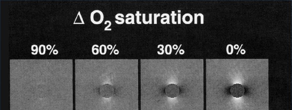
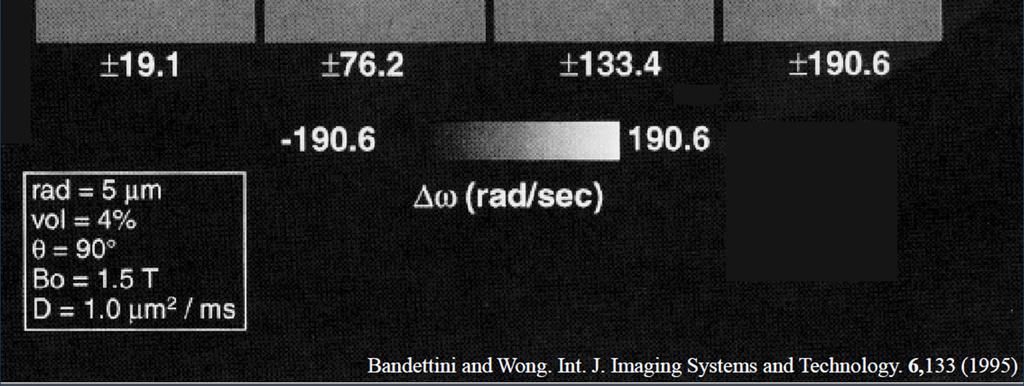
32 Blood Oxygenation Level Dependent MRI
33 Blood Oxygenation Level Dependent MRI As shown in the previous slide, deoxyhemoglobincauses local disturbances in the magnetic field that will lead to signal loss in T2* weighted images. This signal loss will be proportional to the concentration of deoxyhb. The total amount of deoxyhbis proportional to Hct* V * (1-Y) where Hctis hematocrit, V is blood volume, and Y is blood oxygenation.
34 Blood Oxygenation Level Dependent MRI Image From: KR Thulborn. NeuroImage 62: (2012)

35 Blood Oxygenation Level Dependent MRI Souce: Ogawa et. al. PNAS 1992


36 Slide Courtesy of Scott Holland
37 Mechanism of BOLD Functional MRI Brain activity Oxygen consumption Cerebral blood flow - + Oxyhemoglobin Deoxyhemoglobin T2* MRI signal intesity Adapted From Slide by of Scott Holland
38 Blood Oxygenation Level Dependent MRI Brain activation causes local increase in blood flow. Relatively tight coupling between neuronal activity and local CBF, both spatially and temporally. Thompson, J.K., Peterson, M.R., Freeman, RD, Single- Neuron Activity and the Tissue Oxygenation in the Cerebral Cortex. Science, 299: , Single neuron spike rate increase accompanied by immediate decrease in tissue oxygenation suggesting that high-resolution fmri may be used to localize activity in neurons. Slide Courtesy of Scott Holland
39 Blood Oxygenation Level Dependent MRI Magneticresonance imaging is sensitive to changes in the magnetic properties of blood. BOLD-related signal changes are small (1-5%). Noise in fmri images is typically around 0.5-1% For this reason, we cannot usually detect activation in a single image. Instead, signal changes are detected statistically by time-series analysis of many images. (i.e. we look for regions where the signal time course is highly correlated to the experimental paradigm) Slide Courtesy of Scott Holland
40 Hemodynamic Response to Brain Activity BOLD effect is not instantaneous Initial dip or undershoot of BOLD signal ~ 1 s Due to lag in CBF relative to O 2 demand Delay between activity and BOLD response ~ 4-6 s Decay of BOLD signal after activity stops ~ 6-8 s BOLD fmri is good for measuring sustained localized cortical activity but too slow to get dynamic information. Slide Courtesy of Scott Holland
41 Hemodynamic Response Function (HRF) Response to Single Short Stimulus Time (sec) Slide Courtesy of Scott Holland
42 Statistic Post-Processing BOLD signal is small 1-2% at 1.5T 2-4% at 3.0T Statistical methods are needed To detect activation above noise To determine significance of activation To assign probabilities to activation Statistical parameter mapping T-test Cross-correlation methods General linear model ICA/PCA Slide Courtesy of Scott Holland
43 Block Periodic fmri Paradigms Alternating blocks of behavior Stimulus (30 seconds typical) Control (30 seconds typical) Complete paradigm contains multiple blocks 5 x {Stimulation/Control} Increases Statistical Power Total acquisition time per task = 5 min. 30 sec. Control task minimizes incidental activation Control for sensory stimuli Control for attention Distract from target behavior Slide Courtesy of Scott Holland
44 Blood Oxygenation Level Dependent MRI A BOLD MRI acquisition is typically performed as a series of 2D slices which cover the whole brain. TE ~=T2* to optimize BOLD contrast. Longer TE -> greater signal loss near air-tissue interfaces. Echo-Planer Imaging (EPI) allows acquisition at a rate of approximately 50 ms/ slice. Whole brain in ~1-2 s. Typically use 3-5 mm voxels to enable single-shot EPI and to have adequate SNR. skh qc1.no6


45 Pixel Intensity Time Course with Periodic Stimulation and Rest REST 1 ACTIVE 1 REST 2 ACTIVE 2 REST 3 30 sec 30 sec 30 sec 30 sec 30 sec random short random short random tones story tones story tones Stimulus Stimulus Stimulus Rest Rest Rest Rest time Slide Courtesy of Scott Holland
46 fmri of Passive Story Listening Task 35 y.o. Female Listening to 3T 102 Normalized Activation skh qc1.no6 Slide Courtesy of Scott Holland Time (sec) Time course in temporo-parietal language cortex, Wernicke s area.
MRI Physics I: Spins, Excitation, Relaxation
 MRI Physics I: Spins, Excitation, Relaxation Douglas C. Noll Biomedical Engineering University of Michigan Michigan Functional MRI Laboratory Outline Introduction to Nuclear Magnetic Resonance Imaging
Introduction to MRI Acquisition
 Introduction to MRI Acquisition James Meakin FMRIB Physics Group FSL Course, Bristol, September 2012 1 What are we trying to achieve? 2 What are we trying to achieve? Informed decision making: Protocols
Introduction to MRI. Spin & Magnetic Moments. Relaxation (T1, T2) Spin Echoes. 2DFT Imaging. K-space & Spatial Resolution.
 Spin Echoes. 2DFT Imaging. K-space & Spatial Resolution.") Introduction to MRI Spin & Magnetic Moments Relaxation (T1, T2) Spin Echoes 2DFT Imaging Selective excitation, phase & frequency encoding K-space & Spatial Resolution Contrast (T1, T2) Acknowledgement:
Introduction to MRI Spin & Magnetic Moments Relaxation (T1, T2) Spin Echoes 2DFT Imaging Selective excitation, phase & frequency encoding K-space & Spatial Resolution Contrast (T1, T2) Acknowledgement:
The NMR Inverse Imaging Problem
 The NMR Inverse Imaging Problem Nuclear Magnetic Resonance Protons and Neutrons have intrinsic angular momentum Atoms with an odd number of proton and/or odd number of neutrons have a net magnetic moment=>
Introduction to Biomedical Imaging
 Alejandro Frangi, PhD Computational Imaging Lab Department of Information & Communication Technology Pompeu Fabra University www.cilab.upf.edu MRI advantages Superior soft-tissue contrast Depends on among
Outline. Superconducting magnet. Magnetic properties of blood. Physiology BOLD-MRI signal. Magnetic properties of blood
 Magnetic properties of blood Physiology BOLD-MRI signal Aart Nederveen Department of Radiology AMC a.j.nederveen@amc.nl Outline Magnetic properties of blood Moses Blood oxygenation BOLD fmri Superconducting
EL-GY 6813/BE-GY 6203 Medical Imaging, Fall 2016 Final Exam
 EL-GY 6813/BE-GY 6203 Medical Imaging, Fall 2016 Final Exam (closed book, 1 sheets of notes double sided allowed, no calculator or other electronic devices allowed) 1. Ultrasound Physics (15 pt) A) (9
The Basics of Magnetic Resonance Imaging
 The Basics of Magnetic Resonance Imaging Nathalie JUST, PhD nathalie.just@epfl.ch CIBM-AIT, EPFL Course 2013-2014-Chemistry 1 Course 2013-2014-Chemistry 2 MRI: Many different contrasts Proton density T1
Nuclear Magnetic Resonance Imaging
 Nuclear Magnetic Resonance Imaging Jeffrey A. Fessler EECS Department The University of Michigan NSS-MIC: Fundamentals of Medical Imaging Oct. 20, 2003 NMR-0 Background Basic physics 4 magnetic fields
Physics and Brain Imaging
 Physics and Brain Imaging Nuclear Magnetic Resonance (NMR) Magnetic Resonance Imaging (MRI) Functional MRI (fmri) Talk at Quarknet FSU Summer Workshop, July 24, 2017 Per Arne Rikvold Leonardo da Vinci
Fundamental MRI Principles Module 2 N. Nuclear Magnetic Resonance. X-ray. MRI Hydrogen Protons. Page 1. Electrons
 Fundamental MRI Principles Module 2 N S 1 Nuclear Magnetic Resonance There are three main subatomic particles: protons positively charged neutrons no significant charge electrons negatively charged Protons
Medical Imaging Physics Spring Quarter Week 9-1
 Medical Imaging Physics Spring Quarter Week 9-1 NMR and MRI Davor Balzar balzar@du.edu www.du.edu/~balzar Intro MRI Outline NMR & MRI Guest lecturer fmri Thursday, May 22 Visit to CUHSC It s not mandatory
Nuclear Magnetic Resonance Imaging
 Nuclear Magnetic Resonance Imaging Simon Lacoste-Julien Electromagnetic Theory Project 198-562B Department of Physics McGill University April 21 2003 Abstract This paper gives an elementary introduction
Fundamental MRI Principles Module Two
 Fundamental MRI Principles Module Two 1 Nuclear Magnetic Resonance There are three main subatomic particles: protons neutrons electrons positively charged no significant charge negatively charged Protons
Introduction to Magnetic Resonance Imaging (MRI) Pietro Gori
 Pietro Gori") Introduction to Magnetic Resonance Imaging (MRI) Pietro Gori Enseignant-chercheur Equipe IMAGES - Télécom ParisTech pietro.gori@telecom-paristech.fr September 20, 2017 P. Gori BIOMED 20/09/2017 1 / 76
Introduction to Magnetic Resonance Imaging (MRI) Pietro Gori Enseignant-chercheur Equipe IMAGES - Télécom ParisTech pietro.gori@telecom-paristech.fr September 20, 2017 P. Gori BIOMED 20/09/2017 1 / 76
Tissue Characteristics Module Three
 Tissue Characteristics Module Three 1 Equilibrium State Equilibrium State At equilibrium, the hydrogen vector is oriented in a direction parallel to the main magnetic field. Hydrogen atoms within the vector
Field trip: Tuesday, Feb 5th
 Pulse Sequences Field trip: Tuesday, Feb 5th Hardware tour of VUIIIS Philips 3T Meet here at regular class time (11.15) Complete MRI screening form! Chuck Nockowski Philips Service Engineer Reminder: Project/Presentation
Magnetic Resonance Imaging. Pål Erik Goa Associate Professor in Medical Imaging Dept. of Physics
 Magnetic Resonance Imaging Pål Erik Goa Associate Professor in Medical Imaging Dept. of Physics pal.e.goa@ntnu.no 1 Why MRI? X-ray/CT: Great for bone structures and high spatial resolution Not so great
BMB 601 MRI. Ari Borthakur, PhD. Assistant Professor, Department of Radiology Associate Director, Center for Magnetic Resonance & Optical Imaging
 BMB 601 MRI Ari Borthakur, PhD Assistant Professor, Department of Radiology Associate Director, Center for Magnetic Resonance & Optical Imaging University of Pennsylvania School of Medicine A brief history
G Medical Imaging. Outline 4/13/2012. Physics of Magnetic Resonance Imaging
 G16.4426 Medical Imaging Physics of Magnetic Resonance Imaging Riccardo Lattanzi, Ph.D. Assistant Professor Department of Radiology, NYU School of Medicine Department of Electrical and Computer Engineering,
How is it different from conventional MRI? What is MR Spectroscopy? How is it different from conventional MRI? MR Active Nuclei
 What is MR Spectroscopy? MR-Spectroscopy (MRS) is a technique to measure the (relative) concentration of certain chemical or biochemical molecules in a target volume. MR-Spectroscopy is an in vivo (in
Physics of MR Image Acquisition
 Physics of MR Image Acquisition HST-583, Fall 2002 Review: -MRI: Overview - MRI: Spatial Encoding MRI Contrast: Basic sequences - Gradient Echo - Spin Echo - Inversion Recovery : Functional Magnetic Resonance
Lecture 12 February 11, 2016
 MATH 262/CME 372: Applied Fourier Analysis and Winter 2016 Elements of Modern Signal Processing Lecture 12 February 11, 2016 Prof. Emmanuel Candes Scribe: Carlos A. Sing-Long, Edited by E. Bates 1 Outline
Chapter 14:Physics of Magnetic Resonance
 Chapter 14:Physics of Magnetic Resonance Slide set of 141 slides based on the chapter authored by Hee Kwon Song of the publication (ISBN 978-92-0-131010-1): Diagnostic Radiology Physics: A Handbook for
Contrast Mechanisms in MRI. Michael Jay Schillaci
 Contrast Mechanisms in MRI Michael Jay Schillaci Overview Image Acquisition Basic Pulse Sequences Unwrapping K-Space Image Optimization Contrast Mechanisms Static and Motion Contrasts T1 & T2 Weighting,
Introduction to the Physics of NMR, MRI, BOLD fmri
 Pittsburgh, June 13-17, 2011 Introduction to the Physics of NMR, MRI, BOLD fmri (with an orientation toward the practical aspects of data acquisition) Pittsburgh, June 13-17, 2001 Functional MRI in Clinical
MRI in Review: Simple Steps to Cutting Edge Part I
 MRI in Review: Simple Steps to Cutting Edge Part I DWI is now 2 years old... Mike Moseley Radiology Stanford DWI, b = 1413 T2wt, 28/16 ASN 21 San Francisco + Disclosures: Funding NINDS, NCRR, NCI 45 minutes
Introduction to functional MRI in humans. Michael Hallquist University of Pittsburgh
 Introduction to functional MRI in humans Michael Hallquist University of Pittsburgh Goals of human neuroimaging Localization of brain function (mapping) Understanding large-scale functional integration
The Theory of Nuclear Magnetic Resonance Behind Magnetic Resonance Imaging. Catherine Wasko Physics 304 Physics of the Human Body May 3, 2005
 The Theory of Nuclear Magnetic Resonance Behind Magnetic Resonance Imaging Catherine Wasko Physics 304 Physics of the Human Body May 3, 2005 Magnetic resonance imaging (MRI) is a tool utilized in the medical
Topics. The concept of spin Precession of magnetic spin Relaxation Bloch Equation. Bioengineering 280A Principles of Biomedical Imaging
 Bioengineering 280A Principles of Biomedical Imaging Fall Quarter 2006 MRI Lecture 1 Topics The concept of spin Precession of magnetic spin Relaxation Bloch Equation 1 Spin Intrinsic angular momentum of
2015 U N I V E R S I T I T E K N O L O G I P E T R O N A S
 Multi-Modality based Diagnosis: A way forward by Hafeez Ullah Amin Centre for Intelligent Signal and Imaging Research (CISIR) Department of Electrical & Electronic Engineering 2015 U N I V E R S I T I
Magnetic Resonance Imaging (MRI)
") Magnetic Resonance Imaging Introduction The Components The Technology (MRI) Physics behind MR Most slides taken from http:// www.slideworld.org/ viewslides.aspx/magnetic- Resonance-Imaging- %28MRI%29-MR-Imaging-
Magnetic Resonance Imaging Introduction The Components The Technology (MRI) Physics behind MR Most slides taken from http:// www.slideworld.org/ viewslides.aspx/magnetic- Resonance-Imaging- %28MRI%29-MR-Imaging-
Principles of Magnetic Resonance Imaging
 Principles of Magnetic Resonance Imaging Hi Klaus Scheffler, PhD Radiological Physics University of 1 Biomedical Magnetic Resonance: 1 Introduction Magnetic Resonance Imaging Contents: Hi 1 Introduction
Physical fundamentals of magnetic resonance imaging
 Physical fundamentals of magnetic resonance imaging Stepan Sereda University of Bonn 1 / 26 Why? Figure 1 : Full body MRI scan (Source: [4]) 2 / 26 Overview Spin angular momentum Rotating frame and interaction
Magnetic Resonance Imaging in a Nutshell
 Magnetic Resonance Imaging in a Nutshell Oliver Bieri, PhD Department of Radiology, Division of Radiological Physics, University Hospital Basel Department of Biomedical Engineering, University of Basel,
Basic p rinciples COPYRIGHTED MATERIAL. Introduction. Atomic s tructure
 1 Basic p rinciples Introduction 1 Atomic structure 1 Motion in the atom 2 MR active nuclei 2 The hydrogen nucleus 4 Alignment 4 Precession 8 The Larmor equation 9 Introduction The basic principles of
Introduction to NMR! Ravinder Reddy!
 Introduction to NMR! Ravinder Reddy! Brief History of NMR! First detection of NMR! MSNMR! FT NMR! 2D NMR! 2D-NMR and protein structure! Development of MRI! Outline! Concept of SPIN! Spin angular momentum!
FREQUENCY SELECTIVE EXCITATION
 PULSE SEQUENCES FREQUENCY SELECTIVE EXCITATION RF Grad 0 Sir Peter Mansfield A 1D IMAGE Field Strength / Frequency Position FOURIER PROJECTIONS MR Image Raw Data FFT of Raw Data BACK PROJECTION Image Domain
Chemistry 431. Lecture 23
 Chemistry 431 Lecture 23 Introduction The Larmor Frequency The Bloch Equations Measuring T 1 : Inversion Recovery Measuring T 2 : the Spin Echo NC State University NMR spectroscopy The Nuclear Magnetic
Biomedical Imaging Magnetic Resonance Imaging
 Biomedical Imaging Magnetic Resonance Imaging Charles A. DiMarzio & Eric Kercher EECE 4649 Northeastern University May 2018 Background and History Measurement of Nuclear Spins Widely used in physics/chemistry
MRI Physics II: Gradients, Imaging. Douglas C. Noll, Ph.D. Dept. of Biomedical Engineering University of Michigan, Ann Arbor
 MRI Physics II: Gradients, Imaging Douglas C., Ph.D. Dept. of Biomedical Engineering University of Michigan, Ann Arbor Magnetic Fields in MRI B 0 The main magnetic field. Always on (0.5-7 T) Magnetizes
COPYRIGHTED MATERIAL. Production of Net Magnetization. Chapter 1
 Chapter 1 Production of Net Magnetization Magnetic resonance (MR) is a measurement technique used to examine atoms and molecules. It is based on the interaction between an applied magnetic field and a
MR Fundamentals. 26 October Mitglied der Helmholtz-Gemeinschaft
 MR Fundamentals 26 October 2010 Mitglied der Helmholtz-Gemeinschaft Mitglied der Helmholtz-Gemeinschaft Nuclear Spin Nuclear Spin Nuclear magnetic resonance is observed in atoms with odd number of protons
Part III: Sequences and Contrast
 Part III: Sequences and Contrast Contents T1 and T2/T2* Relaxation Contrast of Imaging Sequences T1 weighting T2/T2* weighting Contrast Agents Saturation Inversion Recovery JUST WATER? (i.e., proton density
Chapter 7. Nuclear Magnetic Resonance Spectroscopy
 Chapter 7 Nuclear Magnetic Resonance Spectroscopy I. Introduction 1924, W. Pauli proposed that certain atomic nuclei have spin and magnetic moment and exposure to magnetic field would lead to energy level
SENSE & SUSCEPTIBILITY: RESPIRATION-RELATED SUSCEPTIBILITY EFFECTS AND THEIR INTERACTIONS WITH PARALLEL IMAGING. John Sexton.
 SENSE & SUSCEPTIBILITY: RESPIRATION-RELATED SUSCEPTIBILITY EFFECTS AND THEIR INTERACTIONS WITH PARALLEL IMAGING By John Sexton Thesis Submitted to the Faculty of the Graduate School of Vanderbilt University
The physics US and MRI. Prof. Peter Bogner
 The physics US and MRI Prof. Peter Bogner Sound waves mechanical disturbance, a pressure wave moves along longitudinal wave compression rarefaction zones c = nl, (c: velocity, n: frequency, l: wavelength
NMR, the vector model and the relaxation
 NMR, the vector model and the relaxation Reading/Books: One and two dimensional NMR spectroscopy, VCH, Friebolin Spin Dynamics, Basics of NMR, Wiley, Levitt Molecular Quantum Mechanics, Oxford Univ. Press,
Principles of MRI. Vinyl Record. Last time: Today: Homework Due tonight! EE225E / BIO265. Transforms a temporal signal to a spatial signal
 What is this? ` Principles of MRI Lecture 05 EE225E / BIO265 Instructor: Miki Lustig UC Berkeley, EECS The first NMR spectrum of ethanol 1951. 1 2 Today Last time: Linear systems, Fourier Transforms, Sampling
2.1.1 A Brief History of NMR The conception of NMR sprouted after the Pauli s prediction of nuclear spin in
 CHAPTER--2 BASICS OF NMR IMAGING AND SPECTROSCOPY 2.1 Introduction 2.1.1 A Brief History of NMR The conception of NMR sprouted after the Pauli s prediction of nuclear spin in 1924. Later Gorter (1936)
Magnetic resonance imaging MRI
 Magnetic resonance imaging MRI Introduction What is MRI MRI is an imaging technique used primarily in medical settings that uses a strong magnetic field and radio waves to produce very clear and detailed
On Signal to Noise Ratio Tradeoffs in fmri
 On Signal to Noise Ratio Tradeoffs in fmri G. H. Glover April 11, 1999 This monograph addresses the question of signal to noise ratio (SNR) in fmri scanning, when parameters are changed under conditions
Basis of MRI Contrast
 Basis of MRI Contrast MARK A. HORSFIELD Department of Cardiovascular Sciences University of Leicester Leicester LE1 5WW UK Tel: +44-116-2585080 Fax: +44-870-7053111 e-mail: mah5@le.ac.uk 1 1.1 The Magnetic
Introduction of Key Concepts of Nuclear Magnetic Resonance
 I have not yet lost that sense of wonder, and delight, that this delicate motion should reside in all ordinary things around us, revealing itself only to those who looks for it. E. M. Purcell, Nobel Lecture.
Sketch of the MRI Device
 Outline for Today 1. 2. 3. Introduction to MRI Quantum NMR and MRI in 0D Magnetization, m(x,t), in a Voxel Proton T1 Spin Relaxation in a Voxel Proton Density MRI in 1D MRI Case Study, and Caveat Sketch
Magnetic Resonance Imaging
 http://www.qldxray.com.au/filelibrary/mri_cardiovascular_system_ca_0005.jpg Magnetic Resonance Imaging 1 Overview 1. The magnetic properties of nuclei, and how they behave in strong magnetic fields. 2.
Introduction to the Course and the Techniques. Jeffry R. Alger, PhD Ahmanson-Lovelace Brain Mapping Center Department of Neurology
 Introduction to the Course and the Techniques Jeffry R. Alger, PhD Ahmanson-Lovelace Brain Mapping Center Department of Neurology (jralger@ucla.edu) CTSI Neuroimaging April 2013 Rationale for the Course
The Physical Basis of Nuclear Magnetic Resonance Part I ESMRMB. Jürgen R. Reichenbach
 The Physical Basis of Nuclear agnetic Resonance Part I Jürgen R. Reichenbach odule 1 October 17, 216 Outline of odule Introduction Spin and magnetic moment Spin precession, Larmor frequency agnetic properties
MRI Physics (Phys 352A)
") MRI Physics (Phys 352A) Manus J. Donahue: mj.donahue@vanderbilt.edu Department of Radiology, Neurology, Physics, and Psychiatry Office: Vanderbilt University Institute of Imaging Science (VUIIS) AAA-3115
MRI Physics (Phys 352A) Manus J. Donahue: mj.donahue@vanderbilt.edu Department of Radiology, Neurology, Physics, and Psychiatry Office: Vanderbilt University Institute of Imaging Science (VUIIS) AAA-3115
NMR Spectroscopy Laboratory Experiment Introduction. 2. Theory
 1. Introduction 64-311 Laboratory Experiment 11 NMR Spectroscopy Nuclear Magnetic Resonance (NMR) spectroscopy is a powerful and theoretically complex analytical tool. This experiment will introduce to
V27: RF Spectroscopy
 Martin-Luther-Universität Halle-Wittenberg FB Physik Advanced Lab Course V27: RF Spectroscopy ) Electron spin resonance (ESR) Investigate the resonance behaviour of two coupled LC circuits (an active rf
ELECTRON SPIN RESONANCE & MAGNETIC RESONANCE TOMOGRAPHY
 ELECTRON SPIN RESONANCE & MAGNETIC RESONANCE TOMOGRAPHY 1. AIM OF THE EXPERIMENT This is a model experiment for electron spin resonance, for clear demonstration of interaction between the magnetic moment
A Hands on Introduction to NMR Lecture #1 Nuclear Spin and Magnetic Resonance
 A Hands on Introduction to NMR 22.920 Lecture #1 Nuclear Spin and Magnetic Resonance Introduction - The aim of this short course is to present a physical picture of the basic principles of Nuclear Magnetic
HST.583 Functional Magnetic Resonance Imaging: Data Acquisition and Analysis Fall 2008
 MIT OpenCourseWare http://ocw.mit.edu HST.583 Functional Magnetic Resonance Imaging: Data Acquisition and Analsis Fall 2008 For information about citing these materials or our Terms of Use, visit: http://ocw.mit.edu/terms.
Topics. Spin. The concept of spin Precession of magnetic spin Relaxation Bloch Equation
 Bioengineering 280A Principles of Biomedical Imaging Fall Quarter 2005 MRI Lecture 1 Topics The concept of spin Precession of magnetic spin Relaation Bloch Equation Spin Intrinsic angular momentum of elementary
The physics of medical imaging US, CT, MRI. Prof. Peter Bogner
 The physics of medical imaging US, CT, MRI Prof. Peter Bogner Clinical radiology curriculum blocks of lectures and clinical practice (7x2) Physics of medical imaging Neuroradiology Head and neck I. Head
Master s Program in Medical Physics. Physics of Imaging Systems Basic Principles of Magnetic Resonance Imaging I. Prof. Dr. Lothar Schad.
 1 12/9/2008 Page 1 Master s Program in Medical Physics Physics of Imaging Systems Basic Principles of Magnetic Resonance Imaging I Chair in Faculty of Medicine Mannheim University of Heidelberg Theodor-Kutzer-Ufer
Bioengineering 278" Magnetic Resonance Imaging" Winter 2010" Lecture 1! Topics:! Review of NMR basics! Hardware Overview! Quadrature Detection!
 Bioengineering 278" Magnetic Resonance Imaging" Winter 2010" Lecture 1 Topics: Review of NMR basics Hardware Overview Quadrature Detection Boltzmann Distribution B 0 " = µ z $ 0 % " = #h$ 0 % " = µ z $
Spin Dynamics Basics of Nuclear Magnetic Resonance. Malcolm H. Levitt
 Spin Dynamics Basics of Nuclear Magnetic Resonance Second edition Malcolm H. Levitt The University of Southampton, UK John Wiley &. Sons, Ltd Preface xxi Preface to the First Edition xxiii Introduction
4/4/11. Particles possess intrinsic angular momentum. Spin angular momentum is quantized (it can only take on discrete values)
") For the completely filled shells, subshell (4d 10 ) the orbital magnetic momentum is zero; for the 5s orbital M L is also zero. Hypothesis: the argent atom possesses no magnetic momentum >> they move in
For the completely filled shells, subshell (4d 10 ) the orbital magnetic momentum is zero; for the 5s orbital M L is also zero. Hypothesis: the argent atom possesses no magnetic momentum >> they move in
RADIOLOGIV TECHNOLOGY 4912 COMPREHENSEIVE REVIEW/MRI WORSHEET #1- PATIENT CARE AND SAFETY/PHYSICAL PRINCIPLES
 RADIOLOGIV TECHNOLOGY 4912 COMPREHENSEIVE REVIEW/MRI WORSHEET #1- PATIENT CARE AND SAFETY/PHYSICAL PRINCIPLES 1. What are potential consequences to patients and personnel should there be a release of gaseous
Outlines: (June 11, 1996) Instructor:
 Instructor:") Magnetic Resonance Imaging (June 11, 1996) Instructor: Tai-huang Huang Institute of Biomedical Sciences Academia Sinica Tel. (02) 2652-3036; Fax. (02) 2788-7641 E. mail: bmthh@ibms.sinica.edu.tw Reference:
Magnetic Resonance Imaging (June 11, 1996) Instructor: Tai-huang Huang Institute of Biomedical Sciences Academia Sinica Tel. (02) 2652-3036; Fax. (02) 2788-7641 E. mail: bmthh@ibms.sinica.edu.tw Reference:
NMR BMB 173 Lecture 16, February
 NMR The Structural Biology Continuum Today s lecture: NMR Lots of slides adapted from Levitt, Spin Dynamics; Creighton, Proteins; And Andy Rawlinson There are three types of particles in the universe Quarks
Topics. The History of Spin. Spin. The concept of spin Precession of magnetic spin Relaxation
 Topics Bioengineering 280A Principles of Biomedical Imaging Fall Quarter 2008 MRI Lecture 1 The concept of spin Precession of magnetic spin Relaation Spin The History of Spin Intrinsic angular momentum
With that first concept in mind, it is seen that a spinning nucleus creates a magnetic field, like a bar magnet
 NMR SPECTROSCOPY This section will discuss the basics of NMR (nuclear magnetic resonance) spectroscopy. Most of the section will discuss mainly 1H or proton spectroscopy but the most popular nuclei in
MRI in Practice. Catherine Westbrook MSc, DCRR, CTC Senior Lecturer Anglia Polytechnic University Cambridge UK. John Talbot MSc, DCRR
 MRI in Practice Third edition Catherine Westbrook MSc, DCRR, CTC Senior Lecturer Anglia Polytechnic University Cambridge UK and Carolyn Kaut RothRT(R) (MR) (CT) (M) (CV) Fellow SMRT (Section for Magnetic
Introductory MRI Physics
 C HAPR 18 Introductory MRI Physics Aaron Sodickson EXRNAL MAGNETIC FIELD, PROTONS AND EQUILIBRIUM MAGNETIZATION Much of the bulk of the magnetic resonance imaging (MRI) scanner apparatus is dedicated to
Relaxation times in nuclear magnetic resonance
 Relaxation times in TEP Related topics Nuclear spins, atomic nuclei with a magnetic moment, precession movement of the nuclear spins, Landau-Lifshitz equation, Bloch equation, magnetisation, resonance
VIII. NUCLEAR MAGNETIC RESONANCE (NMR) SPECTROSCOPY
 SPECTROSCOPY") 1 VIII. NUCLEAR MAGNETIC RESONANCE (NMR) SPECTROSCOPY Molecules are extremely small entities; thus, their direct detection and direct investigation is still almost impossible. For the detection and detailed
1 VIII. NUCLEAR MAGNETIC RESONANCE (NMR) SPECTROSCOPY Molecules are extremely small entities; thus, their direct detection and direct investigation is still almost impossible. For the detection and detailed
M R I Physics Course. Jerry Allison Ph.D., Chris Wright B.S., Tom Lavin B.S., Nathan Yanasak Ph.D. Department of Radiology Medical College of Georgia
 M R I Physics Course Jerry Allison Ph.D., Chris Wright B.S., Tom Lavin B.S., Nathan Yanasak Ph.D. Department of Radiology Medical College of Georgia M R I Physics Course Spin Echo Imaging Hahn Spin Echo
Principles of Nuclear Magnetic Resonance Microscopy
 Principles of Nuclear Magnetic Resonance Microscopy Paul T. Callaghan Department of Physics and Biophysics Massey University New Zealand CLARENDON PRESS OXFORD CONTENTS 1 PRINCIPLES OF IMAGING 1 1.1 Introduction
Chem 325 NMR Intro. The Electromagnetic Spectrum. Physical properties, chemical properties, formulas Shedding real light on molecular structure:
 Physical properties, chemical properties, formulas Shedding real light on molecular structure: Wavelength Frequency ν Wavelength λ Frequency ν Velocity c = 2.998 10 8 m s -1 The Electromagnetic Spectrum
Lecture 02 Nuclear Magnetic Resonance Spectroscopy Principle and Application in Structure Elucidation
 Application of Spectroscopic Methods in Molecular Structure Determination Prof. S. Sankararaman Department of Chemistry Indian Institution of Technology Madras Lecture 02 Nuclear Magnetic Resonance Spectroscopy
NMR and MRI : an introduction
 Intensive Programme 2011 Design, Synthesis and Validation of Imaging Probes NMR and MRI : an introduction Walter Dastrù Università di Torino walter.dastru@unito.it \ Introduction Magnetic Resonance Imaging
Nuclear Magnetic Resonance (NMR)
") Nuclear Magnetic Resonance (NMR) Nuclear Magnetic Resonance (NMR) The Nuclear Magnetic Resonance Spectroscopy (NMR) is one of the most important spectroscopic methods to explore the structure and dynamic
Nuclear Magnetic Resonance (NMR) Nuclear Magnetic Resonance (NMR) The Nuclear Magnetic Resonance Spectroscopy (NMR) is one of the most important spectroscopic methods to explore the structure and dynamic
NUCLEAR MAGNETIC RESONANCE. The phenomenon of nuclear magnetic resonance will be used to study magnetic moments of nuclei.
 14 Sep 11 NMR.1 NUCLEAR MAGNETIC RESONANCE The phenomenon of nuclear magnetic resonance will be used to study magnetic moments of nuclei. Theory: In addition to its well-known properties of mass, charge,
Tissue Parametric Mapping:
 Tissue Parametric Mapping: Contrast Mechanisms Using SSFP Sequences Jongho Lee Department of Radiology University of Pennsylvania Tissue Parametric Mapping: Contrast Mechanisms Using bssfp Sequences Jongho
Master of Science Thesis. Development of a phantom for optimisation and quality control in functional MRI (fmri) Anders Nilsson
 Anders Nilsson") Master of Science Thesis Development of a phantom for optimisation and quality control in functional MRI (fmri) Anders Nilsson Supervisor: Johan Olsrud, PhD Medical Radiation Physics Clinical Sciences,
Master of Science Thesis Development of a phantom for optimisation and quality control in functional MRI (fmri) Anders Nilsson Supervisor: Johan Olsrud, PhD Medical Radiation Physics Clinical Sciences,
Part II: Magnetic Resonance Imaging (MRI)
") Part II: Magnetic Resonance Imaging (MRI) Contents Magnetic Field Gradients Selective Excitation Spatially Resolved Reception k-space Gradient Echo Sequence Spin Echo Sequence Magnetic Resonance Imaging
Part II: Magnetic Resonance Imaging (MRI) Contents Magnetic Field Gradients Selective Excitation Spatially Resolved Reception k-space Gradient Echo Sequence Spin Echo Sequence Magnetic Resonance Imaging
Me myself and MRI: adventures in not understanding nuclear physics.
 Me myself and MRI: adventures in not understanding nuclear physics. Thomas E. Gladwin August 28, 2007 Contents 1 Introduction 2 2 Nuclei 2 2.1 Precession............................... 2 2.2 Spin-up and
Introduction to Nuclear Magnetic Resonance Spectroscopy
 Introduction to Nuclear Magnetic Resonance Spectroscopy Dr. Dean L. Olson, NMR Lab Director School of Chemical Sciences University of Illinois Called figures, equations, and tables are from Principles
Magnetic Resonance Imaging
 Magnetic Resonance Imaging History Nuclear magnetic resonance was first described by Isidor Rabi in 1938 - Columbia University, New York City, (Nobel Prize Nobel Prize in Physics 1944) 1946 - Edward Mills
Bloch Equations & Relaxation UCLA. Radiology
 Bloch Equations & Relaxation MRI Systems II B1 I 1 I ~B 1 (t) I 6 ~M I I 5 I 4 Lecture # Learning Objectives Distinguish spin, precession, and nutation. Appreciate that any B-field acts on the the spin
NMR Spectroscopy of Polymers
 UNESCO/IUPAC Course 2005/2006 Jiri Brus NMR Spectroscopy of Polymers Brus J 1. part At the very beginning the phenomenon of nuclear spin resonance was studied predominantly by physicists and the application
Imagent for fnirs and EROS measurements
 TECHNICAL NOTE Imagent for fnirs and EROS measurements 1. Brain imaging using Infrared Photons Brain imaging techniques can be broadly classified in two groups. One group includes the techniques that have
Magnetic Resonance Imaging. Qun Zhao Bioimaging Research Center University of Georgia
 Magnetic Resonance Imaging Qun Zhao Bioimaging Research Center University of Georgia The Nobel Prize in Physiology or Medicine 2003 "for their discoveries concerning magnetic resonance imaging" Paul C.
NMR/MRI examination (8N080 / 3F240)
") NMR/MRI examination (8N080 / 3F240) Remarks: 1. This test consists of 3 problems with at total of 26 sub-questions. 2. Questions are in English. You are allowed to answer them in English or Dutch. 3. Please
NMR/MRI examination (8N080 / 3F240) Remarks: 1. This test consists of 3 problems with at total of 26 sub-questions. 2. Questions are in English. You are allowed to answer them in English or Dutch. 3. Please
Contents. Introduction The General Linear Model. General Linear Linear Model Model. The General Linear Model, Part I. «Take home» message
 DISCOS SPM course, CRC, Liège, 2009 Contents The General Linear Model, Part I Introduction The General Linear Model Data & model Design matrix Parameter estimates & interpretation Simple contrast «Take
Functional magnetic resonance imaging
 University of Ljubljana Faculty of Mathematics and Physics Department of Physics Seminar I b - 2nd year, Second cycle degree Functional magnetic resonance imaging Author: Patricia Cotič Supervisor: Assoc.
Functional Magnetic Resonance Imaging (FMRI) is an imaging technique for
 is an imaging technique for") Chapter 2 Principles of FMRI Functional Magnetic Resonance Imaging (FMRI) is an imaging technique for examining brain function. Since its first appearance in 1991 (Belliveau et al.[8]) the use of FMRI
Chapter 2 Principles of FMRI Functional Magnetic Resonance Imaging (FMRI) is an imaging technique for examining brain function. Since its first appearance in 1991 (Belliveau et al.[8]) the use of FMRI