Introduction to MRI Acquisition
|
|
|
- Elvin Patterson
- 5 years ago
- Views:
Transcription
1 Introduction to MRI Acquisition James Meakin FMRIB Physics Group FSL Course, Bristol, September
2 What are we trying to achieve? 2
3 What are we trying to achieve? Informed decision making: Protocols need to be tailored to the problem (Motion? Effect size? Area of activation?) Learning some physics will make this less daunting 2
4 What are we trying to achieve? Informed decision making: Protocols need to be tailored to the problem (Motion? Effect size? Area of activation?) Learning some physics will make this less daunting A common language: Explain your needs to physicists/radiographers Understand their response There is a LOT of jargon, but you can master it! 2
5 MRI Physics Today: Basics of (nuclear) Magnetic Resonance Image Formation Functional MRI The BOLD effect Acquisition and artefacts 3
6 Nuclear Spin spin H 1 Some elementary particles (eg Hydrogen) exhibit spin Appear to rotate about an axis Charge + spin = magnetic moment 4
7 Nuclear Spin spin N H 1 S Some elementary particles (eg Hydrogen) exhibit spin Appear to rotate about an axis Charge + spin = magnetic moment 4

8 Nuclear Spin spin N H 1 S Some elementary particles (eg Hydrogen) exhibit spin Appear to rotate about an axis Charge + spin = magnetic moment 4
9 Magnetic Fields (B) No Field 5
10 Magnetic Fields (B) What happens when you place a bunch of nuclei with spin into a magnetic field? No Field 5
11 Magnetic Fields (B) What happens when you place a bunch of nuclei with spin into a magnetic field? Main B Field 5

12 Magnetic Fields (B) What happens when you place a bunch of nuclei with spin into a magnetic field? Main B Field On average, they ll tend to align with the field (a net magnetic moment) 5

13 Precession A force (Gravity or B Field) tries to tilt the spinning object But because of spin, the axis precesses instead of tilting 6
14 Excitation B 0 courtesy of William Overall ω 0 = γb 0 Energy pulse tips magnetisation away from B0...if energy rotates at resonant frequency: RF pulse! 7
15 Excitation B 0 courtesy of William Overall ω 0 = γb 0 Energy pulse tips magnetisation away from B0...if energy rotates at resonant frequency: RF pulse! 7
16 Precession B 0 courtesy of William Overall ω 0 = γb 0 Once excited, magnetisation precesses at resonance frequency 8
17 Precession B 0 courtesy of William Overall ω 0 = γb 0 Once excited, magnetisation precesses at resonance frequency 8
18 Signal detection B 0 Changing magnetic field induces current in wire Precessing magnetisation detected with coil Can only detect component in transverse (xy) plane courtesy of William Overall 9
19 Signal detection B 0 Changing magnetic field induces current in wire Precessing magnetisation detected with coil Can only detect component in transverse (xy) plane courtesy of William Overall 9
20 Magnetic Resonance ω 0 = γb 0 Magnetic: external field (B0) magnetises sample Usually detect hydrogen protons in water Potentially any element with spin (1 H, 19 F, 31 P...) Resonance: magnetization has characteristic frequency Also called the Larmour frequency For protons, resonance frequency is in RF range Proportional to the strength of the magnetic field the spin is in 10
21 Relaxation B 0 courtesy of William Overall Magnetization relaxes back into alignment with B0 Speed of relaxation has time constants: T1 and T2 11

22 Relaxation B 0 courtesy of William Overall Magnetization relaxes back into alignment with B0 Speed of relaxation has time constants: T1 and T2 11
23 Relaxation: T1 and T2 M xy Time RF pulse Signal decays according to T2 in transverse plane 12
")
24 Echo time (TE) & T 2 contrast Signal Gray matter White matter Echo time (TE) 13
25 MRI Physics Today: Basics of (nuclear) Magnetic Resonance Image Formation Functional MRI The BOLD effect Acquisition and artefacts 14
26 Making an image B 0 magnet Differentiate between signal from different locations 15
27 Making an image B 0 G magnet Differentiate between signal from different locations Add a spatially varying magnetic field gradient (G) Field varies linearly along one direction Gradient field adds to or subtracts from B0 15
28 Precession B 0 courtesy of William Overall ω 0 = γ(b 0 +ΔB) Resonance frequency is proportional to total field 16
29 Magnetic gradients Higher field Higher frequency B 0 Lower field Lower frequency 17

30 Magnetic gradients Higher field Higher frequency B 0 Lower field Lower frequency Protons at each position precess at different frequencies RF coil hears all of the protons at once Distinguish material at a given position by selectively listening to that frequency 17
31 Decoding Frequency: The Fourier Transform Expresses a function of time as a function of frequency Imagine an orchestra: you differentiate between different instruments based on their frequency 18
 Violins Frequency / Hz")
32 The Fourier Transform Fourier Amplitude transform Time / s Fourier transform Amplitude Bass Cellos (loudest) Violins Frequency / Hz 19
33 Spatial frequencies Image Space 20
34 Spatial frequencies Image Space Fourier Transform 20
35 Spatial frequencies Image Space Fourier Transform K-Space Brightness = How much of this spatial frequency is in your image 20
36 Spatial frequencies Low Spatial Frequencies Image Space Fourier Transform K-Space Brightness = How much of this spatial frequency is in your image 20

37 Spatial frequencies Low Spatial Frequencies Image Space Fourier Transform K-Space Brightness = How much of this spatial frequency is in your image 20
38 Spatial frequencies Image Space Fourier Transform K-Space Brightness = How much of this spatial frequency is in your image 20
39 Spatial frequencies Image Space Fourier Transform K-Space Brightness = How much of this spatial frequency is in your image High Spatial Frequencies 20

40 Spatial frequencies Image Space Fourier Transform K-Space Brightness = How much of this spatial frequency is in your image High Spatial Frequencies 20
41 Spatial frequencies Image Space Fourier Transform K-Space Brightness = How much of this spatial frequency is in your image 20
42 Spatial frequencies Image Space Fourier Transform K-Space Delete some spatial frequencies in the LR direction Brightness = How much of this spatial frequency is in your image 20
43 Spatial frequencies Image Space Fourier Transform K-Space Delete some spatial frequencies in the LR direction Brightness = How much of this spatial frequency is in your image 20
44 Spatial frequencies Image Space Fourier Transform K-Space Delete some spatial frequencies in the LR direction Brightness = How much of this spatial frequency is in your image 20


45 2D k-space describes contribution of each spatial frequency (8,1) x (0,4) x x ky=0 (2,1) kx=0 21
46 What does this have to do with MRI? Remember, we detect all excited protons in the object at the same time They re resonating at different frequencies due to the gradients We acquire the data in k-space! We then fill k-space & Fourier transform it to get the image 22
47 Simple MRI pulse sequence RF Acq ➀ ➁ ➂ ➃ ➄ TE TR ➀ Excite magnetization (transmit RF pulse) ➁ Wait for time TE ( echo time ) ➂ Acquire signal from transverse magnetization (Mxy) ➃ Wait until time TR ( repetition time ) ➄ Repeat from ➀ 23
48 Linescan (2DFT) Acquisition kx ky Acquire one line after each excitation Useful for structural images (minimal artefacts) 24
49 Linescan (2DFT) Acquisition kx ky Acquire one line after each excitation Useful for structural images (minimal artefacts) 24
50 Echo-planar Imaging (EPI) Acquisition kx ky Acquire all of k-space in a single shot Used for FMRI, diffusion imaging 25

51 Echo-planar Imaging (EPI) Acquisition kx ky Acquire all of k-space in a single shot Used for FMRI, diffusion imaging 25
52 Signal-to-noise ratio (SNR) Signal-to-noise ratio: describes signal robustness All else being equal, we want to maximise SNR!! high SNR low SNR SNR = Signal σ noise 26
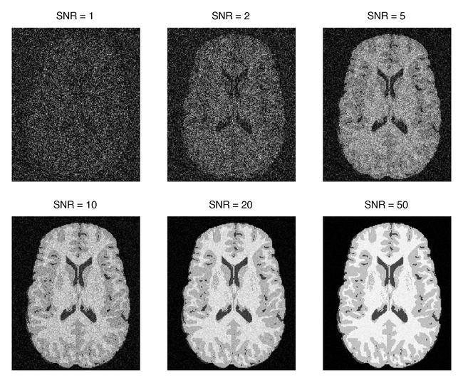
53 Signal-to-noise ratio (SNR) 27
54 Protocol choices affecting SNR... RF receive coil & field strength Timing: TE & TR Voxel volume Scan duration Anything affecting signal!!! 28

55 Protocol choices affecting SNR... RF receive coil & field strength Timing: TE & TR Voxel volume Scan duration Anything affecting signal!!! 28

56 What affects noise? Acquisition time σnoise scan time Longer acquisition less noise higher SNR SNR improves with the square root of scan time 29

57 What affects noise? Acquisition time σnoise scan time Longer acquisition less noise higher SNR SNR improves with the square root of scan time 29
58 What affects signal? Voxel volume? 8x SNR Larger voxels have signal from more tissue! Signal proportional to voxel volume 2x2x2mm has 8x higher SNR than 1x1x1mm! 30
59 Averaging to achieve high resolution? 8x SNR 31

60 Averaging to achieve high resolution? 8x SNR Can we recover lost SNR by averaging? Yes! But it requires a 64-fold increase in scan time! 31
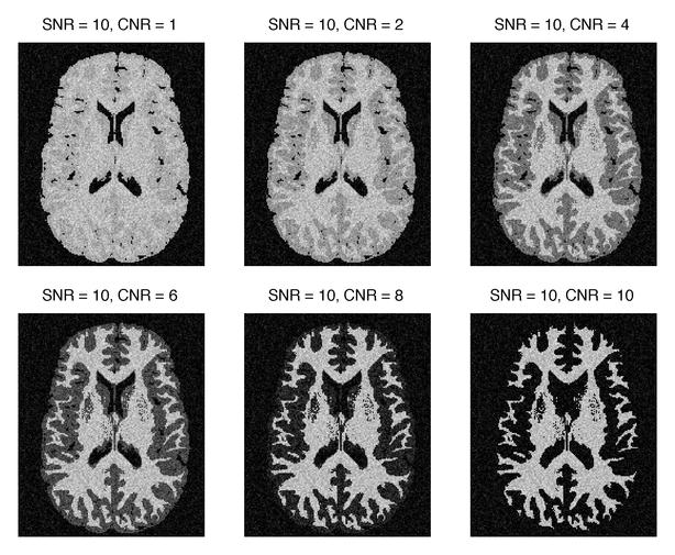
61 Contrast-to-noise ratio (CNR) 32
62 MRI Physics Today: Basics of (nuclear) Magnetic Resonance Image Formation Functional MRI The BOLD effect Acquisition and artefacts 33
 Deoxyhemoglobin: paramagnetic")
63 Deoxyhemoglobin is the source of FMRI signal Oxyhemoglobin: diamagnetic (same as tissue) Deoxyhemoglobin: paramagnetic (magnetic) 34
64 The BOLD Effect [ Ogawa et al, 1990 ] Blood Oxygenation Level Dependent (BOLD) effect 35
![The BOLD Effect [ Ogawa et al, 1990 ]](/docs-images/90/103718905/images/65-1.jpg "imaging voxel Blood Oxygenation Level")

65 The BOLD Effect [ Ogawa et al, 1990 ] imaging voxel Blood Oxygenation Level Dependent (BOLD) effect Creates a range of frequencies in imaging voxel 35
66 36

67 36
68 Vascular Response to Activation neuron capillary HbO dhb HbO 2 2 dhb dhb HbO HbO 2 2 dhb HbO 2 HbO HbO dhb 2 2 HbO 2 HbO 2 HbO 2 = oxyhemoglobin dhb = deoxyhemoglobin 37
69 Vascular Response to Activation neuron capillary HbO dhb HbO 2 2 dhb dhb HbO HbO 2 2 dhb HbO 2 HbO HbO dhb 2 2 HbO 2 HbO 2 HbO 2 = oxyhemoglobin dhb = deoxyhemoglobin 37
70 Vascular Response to Activation neuron capillary HbO dhb HbO 2 2 dhb dhb HbO HbO 2 2 dhb HbO 2 HbO HbO dhb 2 2 HbO 2 HbO 2 O 2 metabolism dhb HbO 2 = oxyhemoglobin dhb = deoxyhemoglobin 37
71 Vascular Response to Activation neuron capillary HbO 2 dhb dhb dhb dhb HbO HbO 2 2 dhb dhb HbO dhb 2 dhb dhb HbO 2 O 2 metabolism dhb HbO 2 = oxyhemoglobin dhb = deoxyhemoglobin 37
72 Vascular Response to Activation neuron capillary HbO 2 HbO 2 HbO 2 HbO 2 HbO dhb 2 HbO 2 dhb dhb dhb dhb HbO 2 HbO 2 HbO 2 HbO HbO HbO 2 2 HbO 2 dhb dhb 2 HbO HbO dhb 2 HbO HbO 2 2 HbO 2 dhb 2 HbO 2 O 2 metabolism blood flow dhb HbO 2 HbO 2 = oxyhemoglobin dhb = deoxyhemoglobin 37
73 Vascular Response to Activation neuron capillary HbO 2 HbO 2 HbO 2 HbO HbO 2 2 HbO dhb 2 HbO 2 dhb dhb dhb dhb HbO 2 HbO 2 HbO 2 HbO HbO HbO 2 2 HbO 2 dhb dhb 2 HbO HbO dhb 2 HbO HbO 2 2 HbO 2 dhb 2 HbO 2 HbO 2 HbO 2 O 2 metabolism blood flow blood volume dhb HbO 2 HbO 2 HbO 2 = oxyhemoglobin dhb = deoxyhemoglobin 37
74 Vascular Response to Activation neuron capillary HbO 2 HbO 2 HbO 2 HbO HbO 2 2 HbO dhb 2 HbO 2 dhb dhb dhb dhb HbO 2 HbO 2 HbO 2 HbO HbO HbO 2 2 HbO 2 dhb dhb 2 HbO HbO dhb 2 HbO HbO 2 2 HbO 2 dhb 2 HbO 2 HbO 2 HbO 2 O 2 metabolism blood flow blood volume dhb HbO 2 HbO 2 HbO 2 = oxyhemoglobin dhb = deoxyhemoglobin [dhb] 37
75 BOLD Contrast O 2 use blood flow blood volume [dhb] BOLD signal rest [dhb] [dhb] active Echo time (T E, ms) Signal increases during activation (less decay) Signal change for longer delay (TE) Typically, 1 5% signal change 38
76 BOLD Contrast O 2 use blood flow blood volume [dhb] BOLD signal rest difference (contrast) active Echo time (T E, ms) Signal increases during activation (less decay) Signal change for longer delay (TE) Typically, 1 5% signal change 38
77 BOLD Contrast 1.0 O 2 use blood flow blood volume [dhb] BOLD signal optimal range rest difference (contrast) active Echo time (T E, ms) Signal increases during activation (less decay) Signal change for longer delay (TE) Typically, 1 5% signal change 38

78 BOLD signal and field strength (B0) SNR and BOLD increase with field strength Image artefacts worse at higher field strength 3T is currently a good tradeoff of signal vs artefacts 39
79 Sources of BOLD Signal Blood flow Neuronal activity Metabolism [dhb] BOLD signal Blood volume Indirect measure of activity (via metabolism!) Subject s physiological state & pathology can change neurovascular coupling, muddying interpretation 40
80 Hemodynamic response function (HRF) on Stimulus timing off time 41
81 Hemodynamic response function (HRF) on Stimulus timing off time Vascular response to activity is delayed & blurred 41
82 Hemodynamic response function (HRF) on Stimulus timing BOLD response off time Vascular response to activity is delayed & blurred 41

83 Hemodynamic response function (HRF) on Stimulus timing BOLD response off time Vascular response to activity is delayed & blurred Described by hemodynamic response function Limits achievable temporal resolution Must be included in signal model 41
84 What is required of the scanner? image 1 23 TR Typical stimulus lasts 1 30 s Rapid imaging: one image every few seconds Anatomical images take minutes to acquire! Acquire single-shot images (e.g., EPI) 42
85 Typical* FMRI Parameters * Typical, not fixed!! Parameter Value Relevant points T E (echo time) T R (repeat time) Matrix size / Resolution Scan duration 1.5T: 60 ms 3.0T: ms 7.0T: ms 1 4 s 64x64 / 2-3 mm 2-60 mins Determines functional contrast, set T2* HRF blurring < 1s; Poor resolution > 6s Limited by distortion, SNR, FOV Lower limit: sensitivity Upper limit: compliance 43
 Noise: signal fluctuations leading to less robust detection with respect to")
86 Confounds: Noise time Purely random noise (example: thermal ) Structured noise (example: physiological ) Noise: signal fluctuations leading to less robust detection with respect to statistical measures 44


87 Confounds: Artefacts Dropout Distortion Ghosting Artefacts: systematic errors that interfere with interpretability of data/images 45
 for")
88 Source of signal dropout BOLD contrast is based on signal dephasing BOLD imaging requires long delay (T E ) for contrast 46

89 Source of signal dropout sinus Dephasing also occurs near air-tissue boundaries Sensitivity to BOLD effect reduces near air-tissue boundaries 47

90 BOLD Signal Dropout Short TE Long TE Dephasing near air-tissue boundaries (e.g., sinuses) BOLD contrast coupled to signal loss ( black holes ) Air-tissue effect is often larger than BOLD effect surrounding vessels! 48

91 Image distortion field offset local warping Field map EPI We think frequency maps to spatial location... So errors in frequency cause spatial mis-localization! 49
92 Non-BOLD fmri BOLD depends on CBF, CBV, CMRO2 Consider looking at these variables separately for longitudinal studies: CBF - Arterial Spin Labeling (ASL) CBV - Vascular Space Occupancy (VASO) CMRO2 - Calibrated BOLD 50
93 Final Thoughts 51
94 Final Thoughts Learn how different experimental parameters affect SNR and image artefacts 51
95 Final Thoughts Learn how different experimental parameters affect SNR and image artefacts Tradeoffs: you can t get something for nothing, but you do have options 51
96 Final Thoughts Learn how different experimental parameters affect SNR and image artefacts Tradeoffs: you can t get something for nothing, but you do have options Get to know a physicist/radiographer: get help setting up study protocols, show them your artefacts 51
97 Final Thoughts Learn how different experimental parameters affect SNR and image artefacts Tradeoffs: you can t get something for nothing, but you do have options Get to know a physicist/radiographer: get help setting up study protocols, show them your artefacts Quality assurance: always look at your data, even if you are running a well-tested protocol 51
98 Acknowledgements Karla Miller for slides Previous years lecture (and more) available at PractiCal fmri (UC Berkeley) Animations: Spinbench 52
99 Thank you! 53
Basic MRI physics and Functional MRI
 Basic MRI physics and Functional MRI Gregory R. Lee, Ph.D Assistant Professor, Department of Radiology June 24, 2013 Pediatric Neuroimaging Research Consortium Objectives Neuroimaging Overview MR Physics
Advanced Topics and Diffusion MRI
 Advanced Topics and Diffusion MRI Slides originally by Karla Miller, FMRIB Centre Modified by Mark Chiew (mark.chiew@ndcn.ox.ac.uk) Slides available at: http://users.fmrib.ox.ac.uk/~mchiew/teaching/ MRI
Field trip: Tuesday, Feb 5th
 Pulse Sequences Field trip: Tuesday, Feb 5th Hardware tour of VUIIIS Philips 3T Meet here at regular class time (11.15) Complete MRI screening form! Chuck Nockowski Philips Service Engineer Reminder: Project/Presentation
EL-GY 6813/BE-GY 6203 Medical Imaging, Fall 2016 Final Exam
 EL-GY 6813/BE-GY 6203 Medical Imaging, Fall 2016 Final Exam (closed book, 1 sheets of notes double sided allowed, no calculator or other electronic devices allowed) 1. Ultrasound Physics (15 pt) A) (9
Contrast Mechanisms in MRI. Michael Jay Schillaci
 Contrast Mechanisms in MRI Michael Jay Schillaci Overview Image Acquisition Basic Pulse Sequences Unwrapping K-Space Image Optimization Contrast Mechanisms Static and Motion Contrasts T1 & T2 Weighting,
Introduction to MRI. Spin & Magnetic Moments. Relaxation (T1, T2) Spin Echoes. 2DFT Imaging. K-space & Spatial Resolution.
 Spin Echoes. 2DFT Imaging. K-space & Spatial Resolution.") Introduction to MRI Spin & Magnetic Moments Relaxation (T1, T2) Spin Echoes 2DFT Imaging Selective excitation, phase & frequency encoding K-space & Spatial Resolution Contrast (T1, T2) Acknowledgement:
Introduction to MRI Spin & Magnetic Moments Relaxation (T1, T2) Spin Echoes 2DFT Imaging Selective excitation, phase & frequency encoding K-space & Spatial Resolution Contrast (T1, T2) Acknowledgement:
Outline. Superconducting magnet. Magnetic properties of blood. Physiology BOLD-MRI signal. Magnetic properties of blood
 Magnetic properties of blood Physiology BOLD-MRI signal Aart Nederveen Department of Radiology AMC a.j.nederveen@amc.nl Outline Magnetic properties of blood Moses Blood oxygenation BOLD fmri Superconducting
Introduction to the Physics of NMR, MRI, BOLD fmri
 Pittsburgh, June 13-17, 2011 Introduction to the Physics of NMR, MRI, BOLD fmri (with an orientation toward the practical aspects of data acquisition) Pittsburgh, June 13-17, 2001 Functional MRI in Clinical
FREQUENCY SELECTIVE EXCITATION
 PULSE SEQUENCES FREQUENCY SELECTIVE EXCITATION RF Grad 0 Sir Peter Mansfield A 1D IMAGE Field Strength / Frequency Position FOURIER PROJECTIONS MR Image Raw Data FFT of Raw Data BACK PROJECTION Image Domain
Introduction to Biomedical Imaging
 Alejandro Frangi, PhD Computational Imaging Lab Department of Information & Communication Technology Pompeu Fabra University www.cilab.upf.edu MRI advantages Superior soft-tissue contrast Depends on among
MRI Physics II: Gradients, Imaging. Douglas C. Noll, Ph.D. Dept. of Biomedical Engineering University of Michigan, Ann Arbor
 MRI Physics II: Gradients, Imaging Douglas C., Ph.D. Dept. of Biomedical Engineering University of Michigan, Ann Arbor Magnetic Fields in MRI B 0 The main magnetic field. Always on (0.5-7 T) Magnetizes
On Signal to Noise Ratio Tradeoffs in fmri
 On Signal to Noise Ratio Tradeoffs in fmri G. H. Glover April 11, 1999 This monograph addresses the question of signal to noise ratio (SNR) in fmri scanning, when parameters are changed under conditions
NMR/MRI examination (8N080 / 3F240)
") NMR/MRI examination (8N080 / 3F240) Remarks: 1. This test consists of 3 problems with at total of 26 sub-questions. 2. Questions are in English. You are allowed to answer them in English or Dutch. 3. Please
NMR/MRI examination (8N080 / 3F240) Remarks: 1. This test consists of 3 problems with at total of 26 sub-questions. 2. Questions are in English. You are allowed to answer them in English or Dutch. 3. Please
Tissue Characteristics Module Three
 Tissue Characteristics Module Three 1 Equilibrium State Equilibrium State At equilibrium, the hydrogen vector is oriented in a direction parallel to the main magnetic field. Hydrogen atoms within the vector
MRI Physics (Phys 352A)
") MRI Physics (Phys 352A) Manus J. Donahue: mj.donahue@vanderbilt.edu Department of Radiology, Neurology, Physics, and Psychiatry Office: Vanderbilt University Institute of Imaging Science (VUIIS) AAA-3115
MRI Physics (Phys 352A) Manus J. Donahue: mj.donahue@vanderbilt.edu Department of Radiology, Neurology, Physics, and Psychiatry Office: Vanderbilt University Institute of Imaging Science (VUIIS) AAA-3115
HST.583 Functional Magnetic Resonance Imaging: Data Acquisition and Analysis Fall 2008
 MIT OpenCourseWare http://ocw.mit.edu HST.583 Functional Magnetic Resonance Imaging: Data Acquisition and Analysis Fall 2008 For information about citing these materials or our Terms of Use, visit: http://ocw.mit.edu/terms.
Physics of MR Image Acquisition
 Physics of MR Image Acquisition HST-583, Fall 2002 Review: -MRI: Overview - MRI: Spatial Encoding MRI Contrast: Basic sequences - Gradient Echo - Spin Echo - Inversion Recovery : Functional Magnetic Resonance
Magnetic Resonance Imaging. Pål Erik Goa Associate Professor in Medical Imaging Dept. of Physics
 Magnetic Resonance Imaging Pål Erik Goa Associate Professor in Medical Imaging Dept. of Physics pal.e.goa@ntnu.no 1 Why MRI? X-ray/CT: Great for bone structures and high spatial resolution Not so great
Introduction to Magnetic Resonance Imaging (MRI) Pietro Gori
 Pietro Gori") Introduction to Magnetic Resonance Imaging (MRI) Pietro Gori Enseignant-chercheur Equipe IMAGES - Télécom ParisTech pietro.gori@telecom-paristech.fr September 20, 2017 P. Gori BIOMED 20/09/2017 1 / 76
Introduction to Magnetic Resonance Imaging (MRI) Pietro Gori Enseignant-chercheur Equipe IMAGES - Télécom ParisTech pietro.gori@telecom-paristech.fr September 20, 2017 P. Gori BIOMED 20/09/2017 1 / 76
How is it different from conventional MRI? What is MR Spectroscopy? How is it different from conventional MRI? MR Active Nuclei
 What is MR Spectroscopy? MR-Spectroscopy (MRS) is a technique to measure the (relative) concentration of certain chemical or biochemical molecules in a target volume. MR-Spectroscopy is an in vivo (in
Outlines: (June 11, 1996) Instructor:
 Instructor:") Magnetic Resonance Imaging (June 11, 1996) Instructor: Tai-huang Huang Institute of Biomedical Sciences Academia Sinica Tel. (02) 2652-3036; Fax. (02) 2788-7641 E. mail: bmthh@ibms.sinica.edu.tw Reference:
Magnetic Resonance Imaging (June 11, 1996) Instructor: Tai-huang Huang Institute of Biomedical Sciences Academia Sinica Tel. (02) 2652-3036; Fax. (02) 2788-7641 E. mail: bmthh@ibms.sinica.edu.tw Reference:
MRI Physics I: Spins, Excitation, Relaxation
 MRI Physics I: Spins, Excitation, Relaxation Douglas C. Noll Biomedical Engineering University of Michigan Michigan Functional MRI Laboratory Outline Introduction to Nuclear Magnetic Resonance Imaging
Functional Magnetic Resonance Imaging (FMRI) is an imaging technique for
 is an imaging technique for") Chapter 2 Principles of FMRI Functional Magnetic Resonance Imaging (FMRI) is an imaging technique for examining brain function. Since its first appearance in 1991 (Belliveau et al.[8]) the use of FMRI
Chapter 2 Principles of FMRI Functional Magnetic Resonance Imaging (FMRI) is an imaging technique for examining brain function. Since its first appearance in 1991 (Belliveau et al.[8]) the use of FMRI
Exam 8N080 - Introduction to MRI
 Exam 8N080 - Introduction to MRI Friday April 10 2015, 18.00-21.00 h For this exam you may use an ordinary calculator (not a graphical one). In total there are 5 assignments and a total of 50 points can
K-space. Spin-Warp Pulse Sequence. At each point in time, the received signal is the Fourier transform of the object s(t) = M( k x
 = M( k x") Bioengineering 280A Principles of Biomedical Imaging Fall Quarter 2015 MRI Lecture 4 k (t) = γ 2π k y (t) = γ 2π K-space At each point in time, the received signal is the Fourier transform of the object
Bioengineering 280A Principles of Biomedical Imaging Fall Quarter 2015 MRI Lecture 4 k (t) = γ 2π k y (t) = γ 2π K-space At each point in time, the received signal is the Fourier transform of the object
Magnetic Resonance Imaging (MRI)
") Magnetic Resonance Imaging Introduction The Components The Technology (MRI) Physics behind MR Most slides taken from http:// www.slideworld.org/ viewslides.aspx/magnetic- Resonance-Imaging- %28MRI%29-MR-Imaging-
Magnetic Resonance Imaging Introduction The Components The Technology (MRI) Physics behind MR Most slides taken from http:// www.slideworld.org/ viewslides.aspx/magnetic- Resonance-Imaging- %28MRI%29-MR-Imaging-
Technical University of Denmark
 Technical University of Denmark Page 1 of 10 pages Written test, 12 December 2012 Course name: Introduction to medical imaging Course no. 31540 Aids allowed: None. Pocket calculator not allowed "Weighting":
The NMR Inverse Imaging Problem
 The NMR Inverse Imaging Problem Nuclear Magnetic Resonance Protons and Neutrons have intrinsic angular momentum Atoms with an odd number of proton and/or odd number of neutrons have a net magnetic moment=>
G Medical Imaging. Outline 4/13/2012. Physics of Magnetic Resonance Imaging
 G16.4426 Medical Imaging Physics of Magnetic Resonance Imaging Riccardo Lattanzi, Ph.D. Assistant Professor Department of Radiology, NYU School of Medicine Department of Electrical and Computer Engineering,
EE225E/BIOE265 Spring 2013 Principles of MRI. Assignment 9 Solutions. Due April 29th, 2013
 EE5E/BIOE65 Spring 013 Principles of MRI Miki Lustig This is the last homework in class. Enjoy it. Assignment 9 Solutions Due April 9th, 013 1) In class when we presented the spin-echo saturation recovery
MRI in Review: Simple Steps to Cutting Edge Part I
 MRI in Review: Simple Steps to Cutting Edge Part I DWI is now 2 years old... Mike Moseley Radiology Stanford DWI, b = 1413 T2wt, 28/16 ASN 21 San Francisco + Disclosures: Funding NINDS, NCRR, NCI 45 minutes
Master of Science Thesis. Development of a phantom for optimisation and quality control in functional MRI (fmri) Anders Nilsson
 Anders Nilsson") Master of Science Thesis Development of a phantom for optimisation and quality control in functional MRI (fmri) Anders Nilsson Supervisor: Johan Olsrud, PhD Medical Radiation Physics Clinical Sciences,
Master of Science Thesis Development of a phantom for optimisation and quality control in functional MRI (fmri) Anders Nilsson Supervisor: Johan Olsrud, PhD Medical Radiation Physics Clinical Sciences,
MRI in Practice. Catherine Westbrook MSc, DCRR, CTC Senior Lecturer Anglia Polytechnic University Cambridge UK. John Talbot MSc, DCRR
 MRI in Practice Third edition Catherine Westbrook MSc, DCRR, CTC Senior Lecturer Anglia Polytechnic University Cambridge UK and Carolyn Kaut RothRT(R) (MR) (CT) (M) (CV) Fellow SMRT (Section for Magnetic
Physical fundamentals of magnetic resonance imaging
 Physical fundamentals of magnetic resonance imaging Stepan Sereda University of Bonn 1 / 26 Why? Figure 1 : Full body MRI scan (Source: [4]) 2 / 26 Overview Spin angular momentum Rotating frame and interaction
Apodization. Gibbs Artifact. Bioengineering 280A Principles of Biomedical Imaging. Fall Quarter 2013 MRI Lecture 5. rect(k x )
") Bioengineering 280A Principles of Biomedical Imaging Fall Quarter 2013 MRI Lecture 5 GE Medical Systems 2003 Gibbs Artifact Apodization rect(k ) Hanning Window h(k )=1/2(1+cos(2πk ) 256256 image 256128
Bioengineering 280A Principles of Biomedical Imaging Fall Quarter 2013 MRI Lecture 5 GE Medical Systems 2003 Gibbs Artifact Apodization rect(k ) Hanning Window h(k )=1/2(1+cos(2πk ) 256256 image 256128
M R I Physics Course. Jerry Allison Ph.D., Chris Wright B.S., Tom Lavin B.S., Nathan Yanasak Ph.D. Department of Radiology Medical College of Georgia
 M R I Physics Course Jerry Allison Ph.D., Chris Wright B.S., Tom Lavin B.S., Nathan Yanasak Ph.D. Department of Radiology Medical College of Georgia M R I Physics Course Spin Echo Imaging Hahn Spin Echo
Basis of MRI Contrast
 Basis of MRI Contrast MARK A. HORSFIELD Department of Cardiovascular Sciences University of Leicester Leicester LE1 5WW UK Tel: +44-116-2585080 Fax: +44-870-7053111 e-mail: mah5@le.ac.uk 1 1.1 The Magnetic
Topics. The concept of spin Precession of magnetic spin Relaxation Bloch Equation. Bioengineering 280A Principles of Biomedical Imaging
 Bioengineering 280A Principles of Biomedical Imaging Fall Quarter 2006 MRI Lecture 1 Topics The concept of spin Precession of magnetic spin Relaxation Bloch Equation 1 Spin Intrinsic angular momentum of
SENSE & SUSCEPTIBILITY: RESPIRATION-RELATED SUSCEPTIBILITY EFFECTS AND THEIR INTERACTIONS WITH PARALLEL IMAGING. John Sexton.
 SENSE & SUSCEPTIBILITY: RESPIRATION-RELATED SUSCEPTIBILITY EFFECTS AND THEIR INTERACTIONS WITH PARALLEL IMAGING By John Sexton Thesis Submitted to the Faculty of the Graduate School of Vanderbilt University
Introductory MRI Physics
 C HAPR 18 Introductory MRI Physics Aaron Sodickson EXRNAL MAGNETIC FIELD, PROTONS AND EQUILIBRIUM MAGNETIZATION Much of the bulk of the magnetic resonance imaging (MRI) scanner apparatus is dedicated to
Bioengineering 278" Magnetic Resonance Imaging" " Winter 2011" Lecture 9! Time of Flight MRA!
 Bioengineering 278" Magnetic Resonance Imaging" " Winter 2011" Lecture 9 Motion Encoding using Longitudinal Magnetization: Magnetic Resonance Angiography Time of Flight Contrast Enhanced Arterial Spin
Biomedical Imaging Magnetic Resonance Imaging
 Biomedical Imaging Magnetic Resonance Imaging Charles A. DiMarzio & Eric Kercher EECE 4649 Northeastern University May 2018 Background and History Measurement of Nuclear Spins Widely used in physics/chemistry
The physics US and MRI. Prof. Peter Bogner
 The physics US and MRI Prof. Peter Bogner Sound waves mechanical disturbance, a pressure wave moves along longitudinal wave compression rarefaction zones c = nl, (c: velocity, n: frequency, l: wavelength
NMR and MRI : an introduction
 Intensive Programme 2011 Design, Synthesis and Validation of Imaging Probes NMR and MRI : an introduction Walter Dastrù Università di Torino walter.dastru@unito.it \ Introduction Magnetic Resonance Imaging
Principles of Magnetic Resonance Imaging
 Principles of Magnetic Resonance Imaging Hi Klaus Scheffler, PhD Radiological Physics University of 1 Biomedical Magnetic Resonance: 1 Introduction Magnetic Resonance Imaging Contents: Hi 1 Introduction
Part III: Sequences and Contrast
 Part III: Sequences and Contrast Contents T1 and T2/T2* Relaxation Contrast of Imaging Sequences T1 weighting T2/T2* weighting Contrast Agents Saturation Inversion Recovery JUST WATER? (i.e., proton density
Principles of MRI. Vinyl Record. Last time: Today: Homework Due tonight! EE225E / BIO265. Transforms a temporal signal to a spatial signal
 What is this? ` Principles of MRI Lecture 05 EE225E / BIO265 Instructor: Miki Lustig UC Berkeley, EECS The first NMR spectrum of ethanol 1951. 1 2 Today Last time: Linear systems, Fourier Transforms, Sampling
BME I5000: Biomedical Imaging
 BME I5000: Biomedical Imaging Lecture 9 Magnetic Resonance Imaging (imaging) Lucas C. Parra, parra@ccny.cuny.edu Blackboard: http://cityonline.ccny.cuny.edu/ 1 Schedule 1. Introduction, Spatial Resolution,
Medical Imaging Physics Spring Quarter Week 9-1
 Medical Imaging Physics Spring Quarter Week 9-1 NMR and MRI Davor Balzar balzar@du.edu www.du.edu/~balzar Intro MRI Outline NMR & MRI Guest lecturer fmri Thursday, May 22 Visit to CUHSC It s not mandatory
Chapter 14:Physics of Magnetic Resonance
 Chapter 14:Physics of Magnetic Resonance Slide set of 141 slides based on the chapter authored by Hee Kwon Song of the publication (ISBN 978-92-0-131010-1): Diagnostic Radiology Physics: A Handbook for
Fundamental MRI Principles Module 2 N. Nuclear Magnetic Resonance. X-ray. MRI Hydrogen Protons. Page 1. Electrons
 Fundamental MRI Principles Module 2 N S 1 Nuclear Magnetic Resonance There are three main subatomic particles: protons positively charged neutrons no significant charge electrons negatively charged Protons
The physics of medical imaging US, CT, MRI. Prof. Peter Bogner
 The physics of medical imaging US, CT, MRI Prof. Peter Bogner Clinical radiology curriculum blocks of lectures and clinical practice (7x2) Physics of medical imaging Neuroradiology Head and neck I. Head
Measuring cerebral blood flow and other haemodynamic parameters using Arterial Spin Labelling MRI. David Thomas
 Measuring cerebral blood flow and other haemodynamic parameters using Arterial Spin Labelling MRI David Thomas Principal Research Associate in MR Physics Leonard Wolfson Experimental Neurology Centre UCL
HY Ιατρική Απεικόνιση. Διδάσκων: Kώστας Μαριάς
 HY 571 - Ιατρική Απεικόνιση Διδάσκων: Kώστας Μαριάς 11. MRI Τ1,Τ2, PD and physiological parameter imaging Summary and Clarifications Resonance is referred to as the property of an atom to absorb energy
Introduction to functional MRI in humans. Michael Hallquist University of Pittsburgh
 Introduction to functional MRI in humans Michael Hallquist University of Pittsburgh Goals of human neuroimaging Localization of brain function (mapping) Understanding large-scale functional integration
Relaxation times in nuclear magnetic resonance
 Relaxation times in TEP Related topics Nuclear spins, atomic nuclei with a magnetic moment, precession movement of the nuclear spins, Landau-Lifshitz equation, Bloch equation, magnetisation, resonance
RAD229: Midterm Exam 2015/2016 October 19, Minutes. Please do not proceed to the next page until the exam begins.
 RAD229: Midterm Exam 2015/2016 October 19, 2015 ---- 75 Minutes Name: Student ID: General Instructions: 1. Write your name legibly on this page. 2. You may use notes including lectures, homework, solutions
Me myself and MRI: adventures in not understanding nuclear physics.
 Me myself and MRI: adventures in not understanding nuclear physics. Thomas E. Gladwin August 28, 2007 Contents 1 Introduction 2 2 Nuclei 2 2.1 Precession............................... 2 2.2 Spin-up and
Blood Water Dynamics
 Bioengineering 208 Magnetic Resonance Imaging Winter 2007 Lecture 8 Arterial Spin Labeling ASL Basics ASL for fmri Velocity Selective ASL Vessel Encoded ASL Blood Water Dynamics Tissue Water Perfusion:
Nuclear Magnetic Resonance Imaging
 Nuclear Magnetic Resonance Imaging Jeffrey A. Fessler EECS Department The University of Michigan NSS-MIC: Fundamentals of Medical Imaging Oct. 20, 2003 NMR-0 Background Basic physics 4 magnetic fields
The Basics of Magnetic Resonance Imaging
 The Basics of Magnetic Resonance Imaging Nathalie JUST, PhD nathalie.just@epfl.ch CIBM-AIT, EPFL Course 2013-2014-Chemistry 1 Course 2013-2014-Chemistry 2 MRI: Many different contrasts Proton density T1
Tissue Parametric Mapping:
 Tissue Parametric Mapping: Contrast Mechanisms Using SSFP Sequences Jongho Lee Department of Radiology University of Pennsylvania Tissue Parametric Mapping: Contrast Mechanisms Using bssfp Sequences Jongho
Fundamental MRI Principles Module Two
 Fundamental MRI Principles Module Two 1 Nuclear Magnetic Resonance There are three main subatomic particles: protons neutrons electrons positively charged no significant charge negatively charged Protons
Spatial encoding in Magnetic Resonance Imaging. Jean-Marie BONNY
 Spatial encoding in Magnetic Resonance Imaging Jean-Marie BONNY What s Qu est an image ce qu une? image? «a reproduction of a material object by a camera or a related technique» Multi-dimensional signal
MRI at a Glance. Blackwell Science CATHERINE WESTBROOK. MSC DCRR CTC Director of Training and Education Lodestone Patient Care Ltd
 MRI at a Glance MRI at a Glance CATHERINE WESTBROOK MSC DCRR CTC Director of Training and Education Lodestone Patient Care Ltd Blackwell Science 2002 by Blackwell Science Ltd, a Blackwell Publishing Company
Lab 2: Magnetic Resonance Imaging
 EE225E/BIOE265 Spring 2013 Principles of MRI Miki Lustig Developed by: Galen Reed and Miki Lustig Lab 2: Magnetic Resonance Imaging Introduction In this lab, we will get some hands-on experience with an
Sketch of the MRI Device
 Outline for Today 1. 2. 3. Introduction to MRI Quantum NMR and MRI in 0D Magnetization, m(x,t), in a Voxel Proton T1 Spin Relaxation in a Voxel Proton Density MRI in 1D MRI Case Study, and Caveat Sketch
MR Advance Techniques. Flow Phenomena. Class I
 MR Advance Techniques Flow Phenomena Class I Flow Phenomena In this class we will explore different phenomenona produced from nuclei that move during the acquisition of data. Flowing nuclei exhibit different
Velocity Images. Phase Contrast Technique. G. Reiter 1,2, U. Reiter 1, R. Rienmüller 1
 Velocity Images - the MR Phase Contrast Technique G. Reiter 1,2, U. Reiter 1, R. Rienmüller 1 SSIP 2004 12 th Summer School in Image Processing, Graz, Austria 1 Interdisciplinary Cardiac Imaging Center,
Applications of Spin Echo and Gradient Echo: Diffusion and Susceptibility Contrast
 Applications of Spin Echo and Gradient Echo: Diffusion and Susceptibility Contrast Chunlei Liu, PhD Department of Electrical Engineering & Computer Sciences and Helen Wills Neuroscience Institute University
Magnetic Resonance Imaging in a Nutshell
 Magnetic Resonance Imaging in a Nutshell Oliver Bieri, PhD Department of Radiology, Division of Radiological Physics, University Hospital Basel Department of Biomedical Engineering, University of Basel,
Spin Echo Imaging Sequence
 1 MRI In Stereotactic Procedures Edward F. Jackson, Ph.D. The University of Texas M.D. Anderson Cancer Center Houston, Texas 2 RF G slice G phase G freq Signal k-space Spin Echo Imaging Sequence TE 1st
BMB 601 MRI. Ari Borthakur, PhD. Assistant Professor, Department of Radiology Associate Director, Center for Magnetic Resonance & Optical Imaging
 BMB 601 MRI Ari Borthakur, PhD Assistant Professor, Department of Radiology Associate Director, Center for Magnetic Resonance & Optical Imaging University of Pennsylvania School of Medicine A brief history
Nuclear Magnetic Resonance Imaging
 Nuclear Magnetic Resonance Imaging Simon Lacoste-Julien Electromagnetic Theory Project 198-562B Department of Physics McGill University April 21 2003 Abstract This paper gives an elementary introduction
MRS: IN VIVO SPECTROSCOPIC IMAGING MAIN POINTS
 MRS: IN VIVO SPECTROSCOPIC IMAGING MAIN POINTS 1. A MR spectrum can identify many metabolites other than water by: Locating the peak(s) determined by a characteristic chemical shift (ppm) resulting from
Spatial encoding in Magnetic Resonance Imaging. Jean-Marie BONNY
 Spatial encoding in Magnetic Resonance Imaging Jean-Marie BONNY What s Qu est an image ce qu une? image? «a reproduction of a material object by a camera or a related technique» Multi-dimensional signal
Sequence Overview. Gradient Echo Spin Echo Magnetization Preparation Sampling and Trajectories Parallel Imaging. B.Hargreaves - RAD 229
 Sequence Overview Gradient Echo Spin Echo Magnetization Preparation Sampling and Trajectories Parallel Imaging 75 Pulse Sequences and k-space RF k y G z k x G x 3D k-space G y k y k z Acq. k x 76 Gradient
Topics. Spin. The concept of spin Precession of magnetic spin Relaxation Bloch Equation
 Bioengineering 280A Principles of Biomedical Imaging Fall Quarter 2005 MRI Lecture 1 Topics The concept of spin Precession of magnetic spin Relaation Bloch Equation Spin Intrinsic angular momentum of elementary
Correction Gradients. Nov7, Reference: Handbook of pulse sequence
 Correction Gradients Nov7, 2005 Reference: Handbook of pulse sequence Correction Gradients 1. Concomitant-Field Correction Gradients 2. Crusher Gradients 3. Eddy-Current Compensation 4. Spoiler Gradients
NMR course at the FMP: NMR of organic compounds and small biomolecules - II -
 NMR course at the FMP: NMR of organic compounds and small biomolecules - II - 16.03.2009 The program 2/76 CW vs. FT NMR What is a pulse? Vectormodel Water-flip-back 3/76 CW vs. FT CW vs. FT 4/76 Two methods
} B 1 } Coil } Gradients } FFT
 Introduction to MRI Daniel B. Ennis, Ph.D. Requirements for MRI UCLA DCVI Requirements for MRI Dipoles to Images MR Active uclei e.g. 1 H in H20 Cryogen Liquid He and 2 Magnetic Field (B0) Polarizer ystem
Basic Pulse Sequences I Saturation & Inversion Recovery UCLA. Radiology
 Basic Pulse Sequences I Saturation & Inversion Recovery Lecture #5 Learning Objectives Explain what the most important equations of motion are for describing spin systems for MRI. Understand the assumptions
Basic p rinciples COPYRIGHTED MATERIAL. Introduction. Atomic s tructure
 1 Basic p rinciples Introduction 1 Atomic structure 1 Motion in the atom 2 MR active nuclei 2 The hydrogen nucleus 4 Alignment 4 Precession 8 The Larmor equation 9 Introduction The basic principles of
Part II: Magnetic Resonance Imaging (MRI)
") Part II: Magnetic Resonance Imaging (MRI) Contents Magnetic Field Gradients Selective Excitation Spatially Resolved Reception k-space Gradient Echo Sequence Spin Echo Sequence Magnetic Resonance Imaging
Part II: Magnetic Resonance Imaging (MRI) Contents Magnetic Field Gradients Selective Excitation Spatially Resolved Reception k-space Gradient Echo Sequence Spin Echo Sequence Magnetic Resonance Imaging
Rad Tech 4912 MRI Registry Review. Outline of the Registry Exam: Certification Fees
 Rad Tech 4912 MRI Registry Review Outline of the Registry Exam: Category: # of questions: A. Patient Care 30 B. Imaging Procedures 62 C. Data Acquisition and Processing 65 D. Physical Principles of Image
Pulse Sequences: RARE and Simulations
 Pulse Sequences: RARE and Simulations M229 Advanced Topics in MRI Holden H. Wu, Ph.D. 2018.04.19 Department of Radiological Sciences David Geffen School of Medicine at UCLA Class Business Final project
NMR BMB 173 Lecture 16, February
 NMR The Structural Biology Continuum Today s lecture: NMR Lots of slides adapted from Levitt, Spin Dynamics; Creighton, Proteins; And Andy Rawlinson There are three types of particles in the universe Quarks
Topics. The History of Spin. Spin. The concept of spin Precession of magnetic spin Relaxation
 Topics Bioengineering 280A Principles of Biomedical Imaging Fall Quarter 2008 MRI Lecture 1 The concept of spin Precession of magnetic spin Relaation Spin The History of Spin Intrinsic angular momentum
Background II. Signal-to-Noise Ratio (SNR) Pulse Sequences Sampling and Trajectories Parallel Imaging. B.Hargreaves - RAD 229.
 Pulse Sequences Sampling and Trajectories Parallel Imaging. B.Hargreaves - RAD 229.") Background II Signal-to-Noise Ratio (SNR) Pulse Sequences Sampling and Trajectories Parallel Imaging 1 SNR: Signal-to-Noise Ratio Signal: Desired voltage in coil Noise: Thermal, electronic Noise Thermal
Background II Signal-to-Noise Ratio (SNR) Pulse Sequences Sampling and Trajectories Parallel Imaging 1 SNR: Signal-to-Noise Ratio Signal: Desired voltage in coil Noise: Thermal, electronic Noise Thermal
Chemistry 431. Lecture 23
 Chemistry 431 Lecture 23 Introduction The Larmor Frequency The Bloch Equations Measuring T 1 : Inversion Recovery Measuring T 2 : the Spin Echo NC State University NMR spectroscopy The Nuclear Magnetic
Navigator Echoes. BioE 594 Advanced Topics in MRI Mauli. M. Modi. BioE /18/ What are Navigator Echoes?
 Navigator Echoes BioE 594 Advanced Topics in MRI Mauli. M. Modi. 1 What are Navigator Echoes? In order to correct the motional artifacts in Diffusion weighted MR images, a modified pulse sequence is proposed
RADIOLOGIV TECHNOLOGY 4912 COMPREHENSEIVE REVIEW/MRI WORSHEET #1- PATIENT CARE AND SAFETY/PHYSICAL PRINCIPLES
 RADIOLOGIV TECHNOLOGY 4912 COMPREHENSEIVE REVIEW/MRI WORSHEET #1- PATIENT CARE AND SAFETY/PHYSICAL PRINCIPLES 1. What are potential consequences to patients and personnel should there be a release of gaseous
Functional magnetic resonance imaging
 University of Ljubljana Faculty of Mathematics and Physics Department of Physics Seminar I b - 2nd year, Second cycle degree Functional magnetic resonance imaging Author: Patricia Cotič Supervisor: Assoc.
Variational solution to hemodynamic and perfusion response estimation from ASL fmri data
 Variational solution to hemodynamic and perfusion response estimation from ASL fmri data Aina Frau-Pascual, Florence Forbes, Philippe Ciuciu June, 2015 1 / 18 BOLD: Qualitative functional MRI Blood Oxygen
Topics. 2D Image. a b. c d. 1. Representing Images 2. 2D Fourier Transform 3. MRI Basics 4. MRI Applications 5. fmri
 Topics Neuroscience 200C Spring Quarter 2005 Imaging/MRI Lecture 1. Representing Images 2. 2D Fourier Transform 3. MRI Basics 4. MRI Applications 5. fmri Signals and Images Discrete-time/space signal/image:
Physics and Brain Imaging
 Physics and Brain Imaging Nuclear Magnetic Resonance (NMR) Magnetic Resonance Imaging (MRI) Functional MRI (fmri) Talk at Quarknet FSU Summer Workshop, July 24, 2017 Per Arne Rikvold Leonardo da Vinci
7.3.A. The expression for signal recovery is similar to that derived under exercise 7.2 and is given by:
 7..A. Chemical shift difference 3..0. ppm, which equals 54.5 Hz at 3.0 T. Spatial displacement 54.5/00 0.87, which equals.03 cm along the 8 cm side and 0.77 cm along the 6 cm. The cm slice does not have
2.1.1 A Brief History of NMR The conception of NMR sprouted after the Pauli s prediction of nuclear spin in
 CHAPTER--2 BASICS OF NMR IMAGING AND SPECTROSCOPY 2.1 Introduction 2.1.1 A Brief History of NMR The conception of NMR sprouted after the Pauli s prediction of nuclear spin in 1924. Later Gorter (1936)
Principles of MRI EE225E / BIO265. Lecture 14. Instructor: Miki Lustig UC Berkeley, EECS. M. Lustig, EECS UC Berkeley
 Principles of MRI Lecture 14 EE225E / BIO265 Instructor: Miki Lustig UC Berkeley, EECS Overview Last-Time: Non-Selective Excitation Excitation, inversion, spin-echo ~G ~r =0 Today: Selective Excitation
RAD229: Final Exam 2014/ SOLUTIONS You will have 3 hours to complete this Exam
 RAD229: Final Exam 2014/2015 - SOLUTIONS You will have 3 hours to complete this Exam Solutions are given in Blue. In some cases, different interpretations may have led to different, but reasonable answers,