Introduction to the Physics of NMR, MRI, BOLD fmri
|
|
|
- Erik Austin
- 6 years ago
- Views:
Transcription
1 Pittsburgh, June 13-17, 2011 Introduction to the Physics of NMR, MRI, BOLD fmri (with an orientation toward the practical aspects of data acquisition) Pittsburgh, June 13-17, 2001 Functional MRI in Clinical Research Robert Savoy


2 MR physics and safety for functional MRI Lawrence L. Wald, Ph.D. Massachusetts General Hospital Athinoula A. Martinos Center
3 Outline: Part 1: Part 2: Part 3: MR signal MR contrast fmri contrast Image encoding (10 minute version) Imaging considerations for fmri MR Safety
 \"Inject Signal\" (90 saturation rf-pulse) Time No Signal (no contrast) Pittsburgh, June 13-17, 2001 Functional MRI in Clinical Research Robert")
4 Basics of Functional MRI Hemodynamics Roy and Sherrington History BOLD / Flow Instrumental Source of the Data NMR MRI Contrast Flexibility of Tool 8 Magnetic Fields and Relaxation Maximum Signal (minimal contrast) "Inject Signal" (90 saturation rf-pulse) Time No Signal (no contrast) Pittsburgh, June 13-17, 2001 Functional MRI in Clinical Research Robert Savoy
5 MRI in a Slide: MRI In a Slide 8 Magnetic Fields and Relaxation To Obtain the NMR Signal: Three Magnetic Fields µ The magnetic field of a nucleus with spin B0 The main magnetic field, used to align nuclei B1 The radio-frequency field, used to flip nuclei Relaxation: Two ways for the signal to go away T1 Longitudinal: The nuclei re-align with B0 T2 Transverse: The nuclei get out-of-phase To Select Slices and Make Images: Three More Fields G x, G y, G z The gradient fields Shim Fields To make better images Shielding Fields To protect us from all of the above! Pittsburgh, June 13-17, 2001 Functional MRI in Clinical Research Robert Savoy
6 Signal decays exponentially Maximum Signal Fading... Fading... Gone Singal Decays Expontntially Time "Inject Signal" (90 saturation rf-pulse) Pittsburgh, June 13-17, 2001 Functional MRI in Clinical Research Robert Savoy
7 Rate of decay varies across voxels Different Rates of Decay "Inject Signal" (90 saturation rf-pulse) Pittsburgh, June 13-17, 2001 Time Functional MRI in Clinical Research Robert Savoy
8 Image contrast varies with time Faces Contrast for fmri Maximum Signal (Little Contrast) Maximum Signal (minimal contrast) Intermediate Signals (Useful Contrasts) Minimum Signal (No Contrast) No Signal (no contrast) "Inject Signal" "Inject Signal" (90 saturation rf-pulse) (90 saturation rf-pulse) Pittsburgh, June 13-17, 2001 Time Time So, we must WAIT for dephasing to occur, in order to get T2 or T2* contrast. But while we are waiting, various bad things happen: signal decreases; imaging artifacts appear Functional MRI in Clinical Research Robert Savoy
9 Susceptibility in MR Gives us dropout (signal loss) (i.e., The Bad) Gives us BOLD (i.e., The Good) Gives us distortion (i.e., The Ugly)


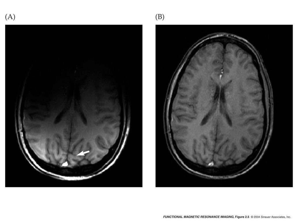
10 MRI can yield many types of Contrast : Proton Density; T1-Weighted; T2-Weighted; MRA PD contrast enhanced; 95% T1 a c b d T2 MRA Pittsburgh, June 13-17, 2001 Functional MRI in Clinical Research Robert Savoy
11 What is NMR? NUCLEAR MAGNETIC RESONANCE A magnet, a glass of water, and a radio wave source and detector. Almost every idea in MRI is easy... but the combined collection is complicated.


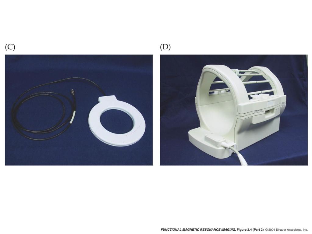
12 A Modern MRI; circa 2003 Figure from Huettel, et. alia (2004) Figure from Huettel, et. alia (2004) 12
13 B protons Earthʼs Field N W E compass S
14 Compass needles υ N Earthʼs Field z North Main Field Bo W E y S Freq = γ B MHz/T x
15 Gyroscopic motion Main Field Bo z North M y Proton has magnetic moment Proton has spin (angular momentum) >>gyroscopic precession Larmor precession freq. = MHz/T x υ = γ B o
16 EXCITATION : Displacing the spins from Equilibrium (North) Problem: It must be moving for us to detect it. Solution: knock out of equilibrium so it oscillates How? 1) Tilt the magnet or compass suddenly 2) Drive the magnetization (compass needle) with a periodic magnetic field
17 Excitation: Resonance Why does only one frequency efficiently tip protons? Resonant driving force. It s like pushing a child on a swing in time with the natural oscillating frequency.
18 z is "longitudinal" direction x-y is "transverse" plane z Static Field B0 y Applied RF Field M0 x The RF pulse rotates M0 the about applied field
19 The NMR Signal RF time Voltage (Signal) time υ υ o B o Mo z y z 90 y z y x υ o x V(t) x
20 Physical Foundations of MRI NMR: 60 year old phenomena that generates the signal from water that we detect. MRI: using NMR signal to generate an image Six magnetic fields (5 are generated by coils): 1) magnetic field of a nucleus, e.g., H1 on water 2) static magnetic field Bo 3) RF field that excites the spins B1 4-6) gradient fields that encode spatial info Gx, Gy, Gz
21 Three Steps in MR: 0) Equilibrium (magnetization points along B0) 1) RF Excitation (tip magn. away from equil.) 2) Precession induces signal, dephasing (timescale = T2, T2*). 3) Return to equilibrium (timescale = T1).
22 Magnetization vector during the NMR experiment RF Voltage (Signal) encode time Mz Mxy
23 A bit more to temporal scale... FID: Free Induction Decay RF Voltage (Signal) TE encode T2* time Mz Mxy T2* Wald, fmri MR Physics
24 Three places in the process to make a measurement (image) 0) Equilibrium (magnetization points along Bo) 1) RF Excitation (tip magnetization away from equilibrium) proton density weighting 2) Precession induces signal, allow to dephase for time TE. 3) Return to equilibrium (timescale =T1). T2 or T2* weighting T1 Weighting
25 Contrast in MRI: proton density Form image immediately after excitation (creation of signal). Tissue with more protons per cc give more signal and is thus brighter on the image. No chance to dephase, thus no differences due to different tissue T2 or T2* values. Magnetization starts fully relaxed (full Mz), thus no T1 weighting.
26 Contrast in MRI: proton density Image immediately after excitation No time for relaxation! Signal proportional to # of spins in voxel All MRI images have proton density weighting underlying them CSF > gray > white Wald, fmri MR Physics
27 RF T1 T2 white matter voxel grey matter voxel TR Voltage (Signal) From Mxy Mz encode encode encode T1 T2 grey matter (long T1) (long T2*) white matter (short T1) (short T2*) time
28 RF PD Proton Density Weighting (not used in fmri) TR Voltage (Signal) From Mxy Mz encode encode encode T1 T2 grey matter (long T1) (long T2*) white matter (short T1) (short T2*) time
29 T2*-Dephasing Wait time TE after excitation before measuring M. Shorter T2* spins have dephased z z z y y y initially x at t= TE x vector sum x Is smaller
30 T2* Dephasing Just showing the tips of the vectors in the laboratory frame and in the rotating frame
31 1.0 T2* decay graphs Transverse Magnetization T2* = 200 Tissue #1 T2* = 60 Tissue # Time (milliseconds)
32 T2* Weighting... usually used for BOLD contrast RF TR: MUCH LONGER Voltage (Signal) From Mxy Mz encode encode T1 T2 grey matter (long T1) (long T2*) white matter (short T1) (short T2*) time
33 T2* Weighting Phantoms with four different T2* decay rates... There is no contrast difference immediately after excitation, must wait (but not too long!). Choose TE for maximum intensity difference.
34 Spin Echo (T2 contrast) Some dephasing can be refocused because its due to static fields. z 90 y z y 180 z y Echo! z y t = 0 x t = T x t = T (+) x t = 2T x Blue arrows precesses faster due to local field inhomogeneity than red arrow

35 Spin Echo 180 pulse only helps cancel static inhomogeneity The runners can have static speed distribution. If a runner trips, he will not make it back in phase with the others. TE TE/2 TE/2 Shown in Laboratory Frame Shown in Rotating Frame
 Wald, fmri MR")
36 T2 weighed spin echo image NMR Signal white gray Time to Echo, TE (ms) Wald, fmri MR Physics
37 T2 Weighting 90 RF 180 RF...usually used clinical anatomy, but may be used for BOLD, especially at higher fields. TR: MUCH LONGER Voltage (Signal) From Mxy Mz encode enco T1 T2 grey matter (long T1) (long T2*) white matter (short T1) (short T2*) time
38 T1-Weighting Signal white matter T1 = 600 grey matter T1 = 1000 CSF T1 = TR (milliseconds) 3000
39 T1 weighting in MRI......How is it possible?
 (long T2*) white matter (short T1) (short T2*)")
40 RF PD Proton Density Weighting (not used in fmri) TR Voltage (Signal) From Mxy Mz encode encode encode T1 T2 grey matter (long T1) (long T2*) white matter (short T1) (short T2*) time
41 T2* Weighting... usually used for BOLD contrast RF TR: MUCH LONGER Voltage (Signal) From Mxy Mz encode encode T1 T2 grey matter (long T1) (long T2*) white matter (short T1) (short T2*) time
42 RF T1 T2 white matter voxel grey matter voxel T1 Weighting it possible? TR... How is Voltage (Signal) From Mxy Mz encode encode encode T1 T2 grey matter (long T1) (long T2*) white matter (short T1) (short T2*) time


43 Image contrast summary: TR, TE TR Long Short Proton Density T1 T2, T2* poor! Short TE Long
44 Basis of fmri: BOLD contrast Qualitative Changes During Activation Observation of Hemodynamic Changes!! Direct Flow effects!!! Blood oxygenation effects
45 Blood cell magnetization and Oxygen State B o M = 0 M = χb Oxygenated Red Cell de-oxygenated Red Cell
46 Addition of paramagnetic compound to blood: T2* effect B o Local field is heterogeneous Water is dephased T2* shortens, Signal goes down on EPI H 2 O

47 Deoxygenated blood attenuates signal in T 2* -weighted MR images O 2 MRI volume element Pittsburgh, June 13-17, 2001 Functional MRI in Clinical Research Figure from Rick Hoge Robert Savoy

48 Decrease of Venous Deoxyhemoglobin Concentration During Increased Perfusion O 2 Figure from Rick Hoge Pittsburgh, June 13-17, 2001 Functional MRI in Clinical Research Robert Savoy
49 Close up on a few hydrogen nuclei, the Larmor frequencies of each, and how they add up + = O 2 + = MRI volume element Pittsburgh, June 13-17, 2001 Functional MRI in Clinical Research Robert Savoy

50 Addition of paramagnetic compound to blood B o Signal from water is dephased T2* shortens, Signal goes down on T2* weighted image
51 Neuronal Activation... Produces local hemodynamic changes (Roy and Sherrington, 1890) Increases local blood flow Increases local blood volume BUT, relatively little change in oxygen consumption
52 Deoxyhemoglobin concentration goes down when flow goes up 1 sec 1 sec flow (4 balls/ sec.) 1 sec consumption = 3 balls/sec. Venous out 1 sec flow (6 balls/ sec.) consumption = 3 balls/sec. Venous out
53 Paramagnetic compound in blood: T2 also changes. B o H 2 O Water diffusion path Diffusion through local fields gives dynamic phase changes not refocused by spin echo T2 shortens, S goes down on spin echo EPI T2 effect increases with B o 2 10um
54 Why does T2 in extravascular water not change for large vessels? Field outside large magnetized venule is approx. constant on length scale of water mean path (~25um) B o θ Field is constant over water path, but magnitude depends on vessle orientation. Thus T2* effect only. 50um Water diffusion path
55 Extravascular T2 does not change for large vessels Field outside large magnetized venule is approx. constant on length scale of water mean path (~25um) θ venule capillary B o Water diffusion path B o 50um Wald, fmri MR Physics Water samples different fields while diffusing
56 MR pulse sequences to see BOLD Considerations: Signal increase = 0 to 5% (small) Motion artifact on conventional image is 0.5% - 3% => need to freeze motion Need to see changes on timescale of hemodynamic changes (seconds) Requirement:! Fast, single shot imaging, image in 80ms, set of slices every 1-3 seconds.
57 Part II MR imaging methods for fmri
58 Magnetic field gradient: the key to image encoding Bo Gx x Bo + Gx x Uniform magnet z x Field from gradient coils Total field


59 Figures from Huettel, et. alia (2004) 59
60 A gradient causes a spread of frequencies y B o MR frequency of the protons in a given location is proportional to the local applied field. z v = γb TOT = γ(b o + G z z) B Field Bo B o + G z z z υ o υ # of spins υ
61 Step one: excite a slice y B o z While the grad. is on, excite only band of frequencies. B Field (w/ z gradient) B o B o + G z z z RF t Signal inten. G z t Δv v Why?
62 Step two: encode spatial info. in-plane B o along z y Frequency encoding x B B TOT = B o + G z x x Signal with gradient υ υ o υ without gradient
63 ʻPulse sequenceʼ so far RF slice select G z t t freq. encode (read-out) G x S(t) t Sample points t
64 Step 3: Phase encoding slice select phase encode RF G z G y t t t freq. encode (read-out) G x S(t) t t
65 Spin-warp encoding slice select RF G z t t k y phase enc G y t freq. enc (read-out) G x S(t) a 1 a 2 t t k x one excitation, one line of kspace...
66 Spin-warp encoding mathematics Keep track of the phase... Phase due to readout: θ(t) = ωo t + γ G x x t RF t Phase due to P.E. θ(t) = ωo t + γ G y y τ Δθ(t) = ωo t + γ G x x t + γ G y y τ G z G y G x S(t) a 1 a 2 t t t t
67 What s the difference? conventional MRI fast imaging slice select RF G z t RF G z t G y G y freq. enc (read-out) G x S(t) G x S(t) T2* k y etc... t k y k x k x
68 Susceptibility in MR Gives us BOLD (i.e., The Good) Gives us dropouts (i.e., The Bad) Gives us distortion (i.e., The Ugly)
69 What do we mean by susceptibility? In physics, it refers to a material s tendency to magnetize when placed in an external field. In MR, it refers to the effects of magnetized material on the image through its local distortion of the static magnetic field B o.
70 What is the source of susceptibility? 1) deoxyheme is paramagnetic 2) Water is diamagnetic (χ = ) 3) Air is paramagnetic (χ = 4x10⁻⁶ ) B o The magnet has a spatially uniform field but your head is magnetic Pattern of B field outside magnetic object in a uniform field
 B o Field map (coronal image) 1.")
71 Ping-pong ball in water Susceptibility effects occur near magnetically dis-similar materials Field disturbance around air surrounded by water (e.g. sinuses) B o Field map (coronal image) 1.5T

72 B o map in head: itʼs the air tissue interface Sagittal Bo field maps at 3T


73 Susceptibility field (in Gauss) increases w/ B o Ping-pong ball in H 2 0: Field maps (ΔTE = 5ms), black lines spaced by 0.024G (0.8ppm at 3T) 1.5T 3T 7T
74 What is the effect of having a non-uniform field on the MR image? Sagittal B o field map at 3T Local field changes with position. To the extent the change is linear, = local suscept. field gradient. We expect uniform field and controllable external gradients
75 Local susceptibility gradients: two effects 1) Local dephasing of the signal (signal loss) within a voxel, mainly from thru-plane gradients 2) Local geometric distortions, (voxel location improperly reconstructed) mainly from local in-plane gradients.
76 1) Non-uniform Local Field Causes Local Dephasing Sagittal B o field map at 3T 5 water protons in different parts of the voxel z 90 z y slowest x T = 0 fastest T = TE

77 Thru-plane dephasing gets worse at longer TE 3T, TE = 21, 30, 40, 50, 60ms
78 Mitigation: thru-plane dephasing; easy to implement 1) Good shimming. (1st and 2nd order) 2) Use thinner slices, preferably with isotropic voxels. Drawback: takes more slices to cover the brain. 3) Use shorter TE. Drawback: BOLD contrast is optimized for TE = T2* local. Thus BOLD is only optimized for the poor susceptibility regions.
79 Problem #2 Susceptibility Causes Image Distortion in EPI Field near sinus y y To encode the image, we control phase evolution as a function of position with applied gradients. Local suscept. Gradient causes unwanted phase evolution. The phase encode error builds up with time. Δθ = γ B local Δt

80 Susceptibility Causes Image Distortion Field near sinus y y Conventional grad. echo, Δθ α encode time α 1/BW
81 Bandwidth is asymmetric in EPI Adjacent points in k x have short Δt = 5 us (high bandwidth) k y Adjacent points along ky are taken with long Δt (= 500us). (low bandwidth) The phase error (and thus distortions) are in the phase encode direction. k x
82 Susceptibility Causes Image Distortion Echoplanar Image, Δθ α encode time α 1/BW z Field near sinus 3T head gradients Encode time = 34, 26, 22, 17ms

83 Susceptibility in EPI can give either a compression or expansion Altering the direction kspace is transversed causes either local compression or expansion. choose your poison 3T whole body gradients
84 EPI needs second order shims EPI distortion is partially alleviated with second order shims Movie of EPI at 1.5T with and without second order shims
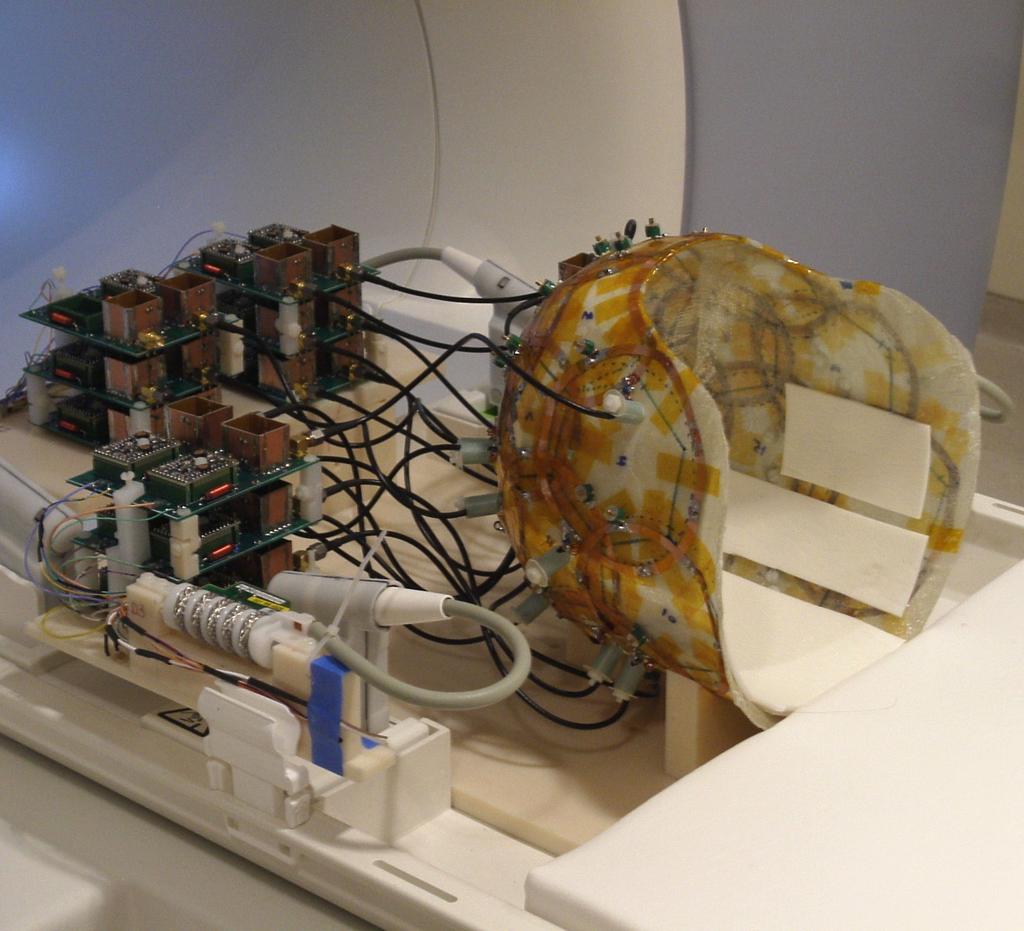
85 Coils 23-Channel Experimental 85
86 With fast gradients, add parallel imaging { Acquisition: SMASH Reduced k-space sampling SENSE Folded images in each receiver channel Reconstruction: Folded datasets + Coil sensitivity maps
 = 0, 2x, 3x Fast gradients are the")
87 GRAPPA for EPI susceptibility 3T Trio, MRI Devices Inc. 8 channel array b=1000 DWI images ipat (GRAPPA) = 0, 2x, 3x Fast gradients are the foundation, but EPI still suffers distortion


88 fmri w/ 8 channel phased array 1.5mm isotropic TE=30ms EPI 3T, 8ch array, GRAPPA =2
89
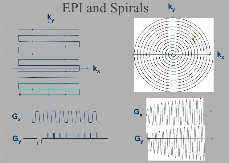
90 EPI Spirals Susceptibility: distortion, blurring, dephasing dephasing Eddy currents: ghosts blurring k = 0 is sampled: 1/2 through 1st Corners of kspace: yes no Gradient demands: very high pretty high

91 EPI and Spirals EPI at 3T Spirals at 3T (courtesy Stanford group)
HST.583 Functional Magnetic Resonance Imaging: Data Acquisition and Analysis Fall 2008
 MIT OpenCourseWare http://ocw.mit.edu HST.583 Functional Magnetic Resonance Imaging: Data Acquisition and Analsis Fall 2008 For information about citing these materials or our Terms of Use, visit: http://ocw.mit.edu/terms.
Contrast Mechanisms in MRI. Michael Jay Schillaci
 Contrast Mechanisms in MRI Michael Jay Schillaci Overview Image Acquisition Basic Pulse Sequences Unwrapping K-Space Image Optimization Contrast Mechanisms Static and Motion Contrasts T1 & T2 Weighting,
Introduction to MRI Acquisition
 Introduction to MRI Acquisition James Meakin FMRIB Physics Group FSL Course, Bristol, September 2012 1 What are we trying to achieve? 2 What are we trying to achieve? Informed decision making: Protocols
EL-GY 6813/BE-GY 6203 Medical Imaging, Fall 2016 Final Exam
 EL-GY 6813/BE-GY 6203 Medical Imaging, Fall 2016 Final Exam (closed book, 1 sheets of notes double sided allowed, no calculator or other electronic devices allowed) 1. Ultrasound Physics (15 pt) A) (9
The NMR Inverse Imaging Problem
 The NMR Inverse Imaging Problem Nuclear Magnetic Resonance Protons and Neutrons have intrinsic angular momentum Atoms with an odd number of proton and/or odd number of neutrons have a net magnetic moment=>
Field trip: Tuesday, Feb 5th
 Pulse Sequences Field trip: Tuesday, Feb 5th Hardware tour of VUIIIS Philips 3T Meet here at regular class time (11.15) Complete MRI screening form! Chuck Nockowski Philips Service Engineer Reminder: Project/Presentation
Introduction to MRI. Spin & Magnetic Moments. Relaxation (T1, T2) Spin Echoes. 2DFT Imaging. K-space & Spatial Resolution.
 Spin Echoes. 2DFT Imaging. K-space & Spatial Resolution.") Introduction to MRI Spin & Magnetic Moments Relaxation (T1, T2) Spin Echoes 2DFT Imaging Selective excitation, phase & frequency encoding K-space & Spatial Resolution Contrast (T1, T2) Acknowledgement:
Introduction to MRI Spin & Magnetic Moments Relaxation (T1, T2) Spin Echoes 2DFT Imaging Selective excitation, phase & frequency encoding K-space & Spatial Resolution Contrast (T1, T2) Acknowledgement:
MRI in Review: Simple Steps to Cutting Edge Part I
 MRI in Review: Simple Steps to Cutting Edge Part I DWI is now 2 years old... Mike Moseley Radiology Stanford DWI, b = 1413 T2wt, 28/16 ASN 21 San Francisco + Disclosures: Funding NINDS, NCRR, NCI 45 minutes
MRI Physics II: Gradients, Imaging. Douglas C. Noll, Ph.D. Dept. of Biomedical Engineering University of Michigan, Ann Arbor
 MRI Physics II: Gradients, Imaging Douglas C., Ph.D. Dept. of Biomedical Engineering University of Michigan, Ann Arbor Magnetic Fields in MRI B 0 The main magnetic field. Always on (0.5-7 T) Magnetizes
Basic MRI physics and Functional MRI
 Basic MRI physics and Functional MRI Gregory R. Lee, Ph.D Assistant Professor, Department of Radiology June 24, 2013 Pediatric Neuroimaging Research Consortium Objectives Neuroimaging Overview MR Physics
Physics of MR Image Acquisition
 Physics of MR Image Acquisition HST-583, Fall 2002 Review: -MRI: Overview - MRI: Spatial Encoding MRI Contrast: Basic sequences - Gradient Echo - Spin Echo - Inversion Recovery : Functional Magnetic Resonance
Introduction to Biomedical Imaging
 Alejandro Frangi, PhD Computational Imaging Lab Department of Information & Communication Technology Pompeu Fabra University www.cilab.upf.edu MRI advantages Superior soft-tissue contrast Depends on among
Advanced Topics and Diffusion MRI
 Advanced Topics and Diffusion MRI Slides originally by Karla Miller, FMRIB Centre Modified by Mark Chiew (mark.chiew@ndcn.ox.ac.uk) Slides available at: http://users.fmrib.ox.ac.uk/~mchiew/teaching/ MRI
MRI Physics I: Spins, Excitation, Relaxation
 MRI Physics I: Spins, Excitation, Relaxation Douglas C. Noll Biomedical Engineering University of Michigan Michigan Functional MRI Laboratory Outline Introduction to Nuclear Magnetic Resonance Imaging
FREQUENCY SELECTIVE EXCITATION
 PULSE SEQUENCES FREQUENCY SELECTIVE EXCITATION RF Grad 0 Sir Peter Mansfield A 1D IMAGE Field Strength / Frequency Position FOURIER PROJECTIONS MR Image Raw Data FFT of Raw Data BACK PROJECTION Image Domain
Part III: Sequences and Contrast
 Part III: Sequences and Contrast Contents T1 and T2/T2* Relaxation Contrast of Imaging Sequences T1 weighting T2/T2* weighting Contrast Agents Saturation Inversion Recovery JUST WATER? (i.e., proton density
Magnetic Resonance Imaging. Pål Erik Goa Associate Professor in Medical Imaging Dept. of Physics
 Magnetic Resonance Imaging Pål Erik Goa Associate Professor in Medical Imaging Dept. of Physics pal.e.goa@ntnu.no 1 Why MRI? X-ray/CT: Great for bone structures and high spatial resolution Not so great
Introduction to Magnetic Resonance Imaging (MRI) Pietro Gori
 Pietro Gori") Introduction to Magnetic Resonance Imaging (MRI) Pietro Gori Enseignant-chercheur Equipe IMAGES - Télécom ParisTech pietro.gori@telecom-paristech.fr September 20, 2017 P. Gori BIOMED 20/09/2017 1 / 76
Introduction to Magnetic Resonance Imaging (MRI) Pietro Gori Enseignant-chercheur Equipe IMAGES - Télécom ParisTech pietro.gori@telecom-paristech.fr September 20, 2017 P. Gori BIOMED 20/09/2017 1 / 76
Nuclear Magnetic Resonance Imaging
 Nuclear Magnetic Resonance Imaging Jeffrey A. Fessler EECS Department The University of Michigan NSS-MIC: Fundamentals of Medical Imaging Oct. 20, 2003 NMR-0 Background Basic physics 4 magnetic fields
Tissue Characteristics Module Three
 Tissue Characteristics Module Three 1 Equilibrium State Equilibrium State At equilibrium, the hydrogen vector is oriented in a direction parallel to the main magnetic field. Hydrogen atoms within the vector
K-space. Spin-Warp Pulse Sequence. At each point in time, the received signal is the Fourier transform of the object s(t) = M( k x
 = M( k x") Bioengineering 280A Principles of Biomedical Imaging Fall Quarter 2015 MRI Lecture 4 k (t) = γ 2π k y (t) = γ 2π K-space At each point in time, the received signal is the Fourier transform of the object
Bioengineering 280A Principles of Biomedical Imaging Fall Quarter 2015 MRI Lecture 4 k (t) = γ 2π k y (t) = γ 2π K-space At each point in time, the received signal is the Fourier transform of the object
Apodization. Gibbs Artifact. Bioengineering 280A Principles of Biomedical Imaging. Fall Quarter 2013 MRI Lecture 5. rect(k x )
") Bioengineering 280A Principles of Biomedical Imaging Fall Quarter 2013 MRI Lecture 5 GE Medical Systems 2003 Gibbs Artifact Apodization rect(k ) Hanning Window h(k )=1/2(1+cos(2πk ) 256256 image 256128
Bioengineering 280A Principles of Biomedical Imaging Fall Quarter 2013 MRI Lecture 5 GE Medical Systems 2003 Gibbs Artifact Apodization rect(k ) Hanning Window h(k )=1/2(1+cos(2πk ) 256256 image 256128
Sketch of the MRI Device
 Outline for Today 1. 2. 3. Introduction to MRI Quantum NMR and MRI in 0D Magnetization, m(x,t), in a Voxel Proton T1 Spin Relaxation in a Voxel Proton Density MRI in 1D MRI Case Study, and Caveat Sketch
Nuclei, Excitation, Relaxation
 Outline 4.1 Principles of MRI uclei, Excitation, Relaxation Carolyn Kaut Roth, RT (R)(MR)(CT)(M)(CV) FSMRT CEO Imaging Education Associates www.imaginged.com candi@imaginged.com What nuclei are MR active?
Introductory MRI Physics
 C HAPR 18 Introductory MRI Physics Aaron Sodickson EXRNAL MAGNETIC FIELD, PROTONS AND EQUILIBRIUM MAGNETIZATION Much of the bulk of the magnetic resonance imaging (MRI) scanner apparatus is dedicated to
M R I Physics Course. Jerry Allison Ph.D., Chris Wright B.S., Tom Lavin B.S., Nathan Yanasak Ph.D. Department of Radiology Medical College of Georgia
 M R I Physics Course Jerry Allison Ph.D., Chris Wright B.S., Tom Lavin B.S., Nathan Yanasak Ph.D. Department of Radiology Medical College of Georgia M R I Physics Course Spin Echo Imaging Hahn Spin Echo
G Medical Imaging. Outline 4/13/2012. Physics of Magnetic Resonance Imaging
 G16.4426 Medical Imaging Physics of Magnetic Resonance Imaging Riccardo Lattanzi, Ph.D. Assistant Professor Department of Radiology, NYU School of Medicine Department of Electrical and Computer Engineering,
BASIC MRI PHYSICS SPIN GYMNASTICS Don Plewes PhD, Walter Kucharczyk MD
 BASIC MRI PHYSICS SPIN GYMNASTICS Don Plewes PhD, Walter Kucharczyk MD Introduction To understand MRI, it is first necessary to understand the physics of proton Nuclear Magnetic Resonance (NMR). The most
Basis of MRI Contrast
 Basis of MRI Contrast MARK A. HORSFIELD Department of Cardiovascular Sciences University of Leicester Leicester LE1 5WW UK Tel: +44-116-2585080 Fax: +44-870-7053111 e-mail: mah5@le.ac.uk 1 1.1 The Magnetic
Chapter 14:Physics of Magnetic Resonance
 Chapter 14:Physics of Magnetic Resonance Slide set of 141 slides based on the chapter authored by Hee Kwon Song of the publication (ISBN 978-92-0-131010-1): Diagnostic Radiology Physics: A Handbook for
NMR/MRI examination (8N080 / 3F240)
") NMR/MRI examination (8N080 / 3F240) Remarks: 1. This test consists of 3 problems with at total of 26 sub-questions. 2. Questions are in English. You are allowed to answer them in English or Dutch. 3. Please
NMR/MRI examination (8N080 / 3F240) Remarks: 1. This test consists of 3 problems with at total of 26 sub-questions. 2. Questions are in English. You are allowed to answer them in English or Dutch. 3. Please
Principles of Magnetic Resonance Imaging
 Principles of Magnetic Resonance Imaging Hi Klaus Scheffler, PhD Radiological Physics University of 1 Biomedical Magnetic Resonance: 1 Introduction Magnetic Resonance Imaging Contents: Hi 1 Introduction
The Basics of Magnetic Resonance Imaging
 The Basics of Magnetic Resonance Imaging Nathalie JUST, PhD nathalie.just@epfl.ch CIBM-AIT, EPFL Course 2013-2014-Chemistry 1 Course 2013-2014-Chemistry 2 MRI: Many different contrasts Proton density T1
Basic Pulse Sequences II - Spin Echoes. TE=12ms TE=47ms TE=106ms TE=153ms UCLA. Radiology
 TE TR 90 180 90 Basic Pulse Sequences II - Spin Echoes TE=12ms TE=47ms TE=106ms TE=153ms TE=235ms Lecture #6 Summary B1(t) RF TR RF t ~M (1) (0 )= ~ M 0 = 2 4 0 0 M 0 3 5 Initial Condition ~M (1) (0 +
Nuclear Magnetic Resonance Imaging
 Nuclear Magnetic Resonance Imaging Simon Lacoste-Julien Electromagnetic Theory Project 198-562B Department of Physics McGill University April 21 2003 Abstract This paper gives an elementary introduction
MRI in Practice. Catherine Westbrook MSc, DCRR, CTC Senior Lecturer Anglia Polytechnic University Cambridge UK. John Talbot MSc, DCRR
 MRI in Practice Third edition Catherine Westbrook MSc, DCRR, CTC Senior Lecturer Anglia Polytechnic University Cambridge UK and Carolyn Kaut RothRT(R) (MR) (CT) (M) (CV) Fellow SMRT (Section for Magnetic
Spin Echo Imaging Sequence
 1 MRI In Stereotactic Procedures Edward F. Jackson, Ph.D. The University of Texas M.D. Anderson Cancer Center Houston, Texas 2 RF G slice G phase G freq Signal k-space Spin Echo Imaging Sequence TE 1st
NMR and MRI : an introduction
 Intensive Programme 2011 Design, Synthesis and Validation of Imaging Probes NMR and MRI : an introduction Walter Dastrù Università di Torino walter.dastru@unito.it \ Introduction Magnetic Resonance Imaging
BMB 601 MRI. Ari Borthakur, PhD. Assistant Professor, Department of Radiology Associate Director, Center for Magnetic Resonance & Optical Imaging
 BMB 601 MRI Ari Borthakur, PhD Assistant Professor, Department of Radiology Associate Director, Center for Magnetic Resonance & Optical Imaging University of Pennsylvania School of Medicine A brief history
Applications of Spin Echo and Gradient Echo: Diffusion and Susceptibility Contrast
 Applications of Spin Echo and Gradient Echo: Diffusion and Susceptibility Contrast Chunlei Liu, PhD Department of Electrical Engineering & Computer Sciences and Helen Wills Neuroscience Institute University
Fundamental MRI Principles Module 2 N. Nuclear Magnetic Resonance. X-ray. MRI Hydrogen Protons. Page 1. Electrons
 Fundamental MRI Principles Module 2 N S 1 Nuclear Magnetic Resonance There are three main subatomic particles: protons positively charged neutrons no significant charge electrons negatively charged Protons
Index. p, lip, 78 8 function, 107 v, 7-8 w, 7-8 i,7-8 sine, 43 Bo,94-96
 p, lip, 78 8 function, 107 v, 7-8 w, 7-8 i,7-8 sine, 43 Bo,94-96 B 1,94-96 M,94-96 B oro!' 94-96 BIro!' 94-96 I/r, 79 2D linear system, 56 2D FFT, 119 2D Fourier transform, 1, 12, 18,91 2D sinc, 107, 112
Physical fundamentals of magnetic resonance imaging
 Physical fundamentals of magnetic resonance imaging Stepan Sereda University of Bonn 1 / 26 Why? Figure 1 : Full body MRI scan (Source: [4]) 2 / 26 Overview Spin angular momentum Rotating frame and interaction
Chemistry 431. Lecture 23
 Chemistry 431 Lecture 23 Introduction The Larmor Frequency The Bloch Equations Measuring T 1 : Inversion Recovery Measuring T 2 : the Spin Echo NC State University NMR spectroscopy The Nuclear Magnetic
} B 1 } Coil } Gradients } FFT
 Introduction to MRI Daniel B. Ennis, Ph.D. Requirements for MRI UCLA DCVI Requirements for MRI Dipoles to Images MR Active uclei e.g. 1 H in H20 Cryogen Liquid He and 2 Magnetic Field (B0) Polarizer ystem
Outlines: (June 11, 1996) Instructor:
 Instructor:") Magnetic Resonance Imaging (June 11, 1996) Instructor: Tai-huang Huang Institute of Biomedical Sciences Academia Sinica Tel. (02) 2652-3036; Fax. (02) 2788-7641 E. mail: bmthh@ibms.sinica.edu.tw Reference:
Magnetic Resonance Imaging (June 11, 1996) Instructor: Tai-huang Huang Institute of Biomedical Sciences Academia Sinica Tel. (02) 2652-3036; Fax. (02) 2788-7641 E. mail: bmthh@ibms.sinica.edu.tw Reference:
Medical Imaging Physics Spring Quarter Week 9-1
 Medical Imaging Physics Spring Quarter Week 9-1 NMR and MRI Davor Balzar balzar@du.edu www.du.edu/~balzar Intro MRI Outline NMR & MRI Guest lecturer fmri Thursday, May 22 Visit to CUHSC It s not mandatory
Sequence Overview. Gradient Echo Spin Echo Magnetization Preparation Sampling and Trajectories Parallel Imaging. B.Hargreaves - RAD 229
 Sequence Overview Gradient Echo Spin Echo Magnetization Preparation Sampling and Trajectories Parallel Imaging 75 Pulse Sequences and k-space RF k y G z k x G x 3D k-space G y k y k z Acq. k x 76 Gradient
HST.583 Functional Magnetic Resonance Imaging: Data Acquisition and Analysis Fall 2008
 MIT OpenCourseWare http://ocw.mit.edu HST.583 Functional Magnetic Resonance Imaging: Data Acquisition and Analysis Fall 2008 For information about citing these materials or our Terms of Use, visit: http://ocw.mit.edu/terms.
Fundamental MRI Principles Module Two
 Fundamental MRI Principles Module Two 1 Nuclear Magnetic Resonance There are three main subatomic particles: protons neutrons electrons positively charged no significant charge negatively charged Protons
Introduction to functional MRI in humans. Michael Hallquist University of Pittsburgh
 Introduction to functional MRI in humans Michael Hallquist University of Pittsburgh Goals of human neuroimaging Localization of brain function (mapping) Understanding large-scale functional integration
On Signal to Noise Ratio Tradeoffs in fmri
 On Signal to Noise Ratio Tradeoffs in fmri G. H. Glover April 11, 1999 This monograph addresses the question of signal to noise ratio (SNR) in fmri scanning, when parameters are changed under conditions
Basic p rinciples COPYRIGHTED MATERIAL. Introduction. Atomic s tructure
 1 Basic p rinciples Introduction 1 Atomic structure 1 Motion in the atom 2 MR active nuclei 2 The hydrogen nucleus 4 Alignment 4 Precession 8 The Larmor equation 9 Introduction The basic principles of
Topics. The concept of spin Precession of magnetic spin Relaxation Bloch Equation. Bioengineering 280A Principles of Biomedical Imaging
 Bioengineering 280A Principles of Biomedical Imaging Fall Quarter 2006 MRI Lecture 1 Topics The concept of spin Precession of magnetic spin Relaxation Bloch Equation 1 Spin Intrinsic angular momentum of
Tissue Parametric Mapping:
 Tissue Parametric Mapping: Contrast Mechanisms Using SSFP Sequences Jongho Lee Department of Radiology University of Pennsylvania Tissue Parametric Mapping: Contrast Mechanisms Using bssfp Sequences Jongho
Magnetic Resonance Imaging (MRI)
") Magnetic Resonance Imaging Introduction The Components The Technology (MRI) Physics behind MR Most slides taken from http:// www.slideworld.org/ viewslides.aspx/magnetic- Resonance-Imaging- %28MRI%29-MR-Imaging-
Magnetic Resonance Imaging Introduction The Components The Technology (MRI) Physics behind MR Most slides taken from http:// www.slideworld.org/ viewslides.aspx/magnetic- Resonance-Imaging- %28MRI%29-MR-Imaging-
Background II. Signal-to-Noise Ratio (SNR) Pulse Sequences Sampling and Trajectories Parallel Imaging. B.Hargreaves - RAD 229.
 Pulse Sequences Sampling and Trajectories Parallel Imaging. B.Hargreaves - RAD 229.") Background II Signal-to-Noise Ratio (SNR) Pulse Sequences Sampling and Trajectories Parallel Imaging 1 SNR: Signal-to-Noise Ratio Signal: Desired voltage in coil Noise: Thermal, electronic Noise Thermal
Background II Signal-to-Noise Ratio (SNR) Pulse Sequences Sampling and Trajectories Parallel Imaging 1 SNR: Signal-to-Noise Ratio Signal: Desired voltage in coil Noise: Thermal, electronic Noise Thermal
Magnetization Gradients, k-space and Molecular Diffusion. Magnetic field gradients, magnetization gratings and k-space
 2256 Magnetization Gradients k-space and Molecular Diffusion Magnetic field gradients magnetization gratings and k-space In order to record an image of a sample (or obtain other spatial information) there
Fundamentals of MR Imaging
 Fundamentals of MR Imaging Shantanu Sinha. Department of Radiology UCSD School of Medicine, San Diego, CA-92103. E-mail: shsinha@ucsd.edu Background References: R.B.Lufkin, The MRI Manual (2nd Edition).
Outline. Superconducting magnet. Magnetic properties of blood. Physiology BOLD-MRI signal. Magnetic properties of blood
 Magnetic properties of blood Physiology BOLD-MRI signal Aart Nederveen Department of Radiology AMC a.j.nederveen@amc.nl Outline Magnetic properties of blood Moses Blood oxygenation BOLD fmri Superconducting
The Theory of Nuclear Magnetic Resonance Behind Magnetic Resonance Imaging. Catherine Wasko Physics 304 Physics of the Human Body May 3, 2005
 The Theory of Nuclear Magnetic Resonance Behind Magnetic Resonance Imaging Catherine Wasko Physics 304 Physics of the Human Body May 3, 2005 Magnetic resonance imaging (MRI) is a tool utilized in the medical
CHEM / BCMB 4190/6190/8189. Introductory NMR. Lecture 10
 CHEM / BCMB 490/690/889 Introductory NMR Lecture 0 - - CHEM 490/690 Spin-Echo The spin-echo pulse sequence: 90 - τ - 80 - τ(echo) Spins echoes are widely used as part of larger pulse sequence to refocus
A Hands on Introduction to NMR Lecture #1 Nuclear Spin and Magnetic Resonance
 A Hands on Introduction to NMR 22.920 Lecture #1 Nuclear Spin and Magnetic Resonance Introduction - The aim of this short course is to present a physical picture of the basic principles of Nuclear Magnetic
Magnetic Resonance Imaging in a Nutshell
 Magnetic Resonance Imaging in a Nutshell Oliver Bieri, PhD Department of Radiology, Division of Radiological Physics, University Hospital Basel Department of Biomedical Engineering, University of Basel,
The Animated Physics of MRI:
 The Animated Physics of MRI: The Notes Introduction and using the CD. DB Plewes, PhD Department of Medical Biophysics and Medical Imaging University of Toronto The enclosed is an identical copy of the
Relaxation times in nuclear magnetic resonance
 Relaxation times in TEP Related topics Nuclear spins, atomic nuclei with a magnetic moment, precession movement of the nuclear spins, Landau-Lifshitz equation, Bloch equation, magnetisation, resonance
Principles of MRI EE225E / BIO265. Name That Artifact. RF Interference During Readout. RF Interference During Readout. Lecture 19
 Name That Artifact Principles of MRI EE225E / BIO265 Lecture 19 Instructor: Miki Lustig UC Berkeley, EECS 1 http://mri-info.net 2 RF Interference During Readout RF Interference During Readout 1D FFT 1D
Pulse Sequences: RARE and Simulations
 Pulse Sequences: RARE and Simulations M229 Advanced Topics in MRI Holden H. Wu, Ph.D. 2018.04.19 Department of Radiological Sciences David Geffen School of Medicine at UCLA Class Business Final project
Magnetic Resonance Imaging. Qun Zhao Bioimaging Research Center University of Georgia
 Magnetic Resonance Imaging Qun Zhao Bioimaging Research Center University of Georgia The Nobel Prize in Physiology or Medicine 2003 "for their discoveries concerning magnetic resonance imaging" Paul C.
Functional Magnetic Resonance Imaging (FMRI) is an imaging technique for
 is an imaging technique for") Chapter 2 Principles of FMRI Functional Magnetic Resonance Imaging (FMRI) is an imaging technique for examining brain function. Since its first appearance in 1991 (Belliveau et al.[8]) the use of FMRI
Chapter 2 Principles of FMRI Functional Magnetic Resonance Imaging (FMRI) is an imaging technique for examining brain function. Since its first appearance in 1991 (Belliveau et al.[8]) the use of FMRI
How is it different from conventional MRI? What is MR Spectroscopy? How is it different from conventional MRI? MR Active Nuclei
 What is MR Spectroscopy? MR-Spectroscopy (MRS) is a technique to measure the (relative) concentration of certain chemical or biochemical molecules in a target volume. MR-Spectroscopy is an in vivo (in
2.1.1 A Brief History of NMR The conception of NMR sprouted after the Pauli s prediction of nuclear spin in
 CHAPTER--2 BASICS OF NMR IMAGING AND SPECTROSCOPY 2.1 Introduction 2.1.1 A Brief History of NMR The conception of NMR sprouted after the Pauli s prediction of nuclear spin in 1924. Later Gorter (1936)
Correction Gradients. Nov7, Reference: Handbook of pulse sequence
 Correction Gradients Nov7, 2005 Reference: Handbook of pulse sequence Correction Gradients 1. Concomitant-Field Correction Gradients 2. Crusher Gradients 3. Eddy-Current Compensation 4. Spoiler Gradients
Exam 8N080 - Introduction to MRI
 Exam 8N080 - Introduction to MRI Friday April 10 2015, 18.00-21.00 h For this exam you may use an ordinary calculator (not a graphical one). In total there are 5 assignments and a total of 50 points can
The physics of medical imaging US, CT, MRI. Prof. Peter Bogner
 The physics of medical imaging US, CT, MRI Prof. Peter Bogner Clinical radiology curriculum blocks of lectures and clinical practice (7x2) Physics of medical imaging Neuroradiology Head and neck I. Head
SENSE & SUSCEPTIBILITY: RESPIRATION-RELATED SUSCEPTIBILITY EFFECTS AND THEIR INTERACTIONS WITH PARALLEL IMAGING. John Sexton.
 SENSE & SUSCEPTIBILITY: RESPIRATION-RELATED SUSCEPTIBILITY EFFECTS AND THEIR INTERACTIONS WITH PARALLEL IMAGING By John Sexton Thesis Submitted to the Faculty of the Graduate School of Vanderbilt University
The physics US and MRI. Prof. Peter Bogner
 The physics US and MRI Prof. Peter Bogner Sound waves mechanical disturbance, a pressure wave moves along longitudinal wave compression rarefaction zones c = nl, (c: velocity, n: frequency, l: wavelength
HY Ιατρική Απεικόνιση. Διδάσκων: Kώστας Μαριάς
 HY 571 - Ιατρική Απεικόνιση Διδάσκων: Kώστας Μαριάς 11. MRI Τ1,Τ2, PD and physiological parameter imaging Summary and Clarifications Resonance is referred to as the property of an atom to absorb energy
Lab 2: Magnetic Resonance Imaging
 EE225E/BIOE265 Spring 2013 Principles of MRI Miki Lustig Developed by: Galen Reed and Miki Lustig Lab 2: Magnetic Resonance Imaging Introduction In this lab, we will get some hands-on experience with an
MRI Physics (Phys 352A)
") MRI Physics (Phys 352A) Manus J. Donahue: mj.donahue@vanderbilt.edu Department of Radiology, Neurology, Physics, and Psychiatry Office: Vanderbilt University Institute of Imaging Science (VUIIS) AAA-3115
MRI Physics (Phys 352A) Manus J. Donahue: mj.donahue@vanderbilt.edu Department of Radiology, Neurology, Physics, and Psychiatry Office: Vanderbilt University Institute of Imaging Science (VUIIS) AAA-3115
Principles of MRI. Vinyl Record. Last time: Today: Homework Due tonight! EE225E / BIO265. Transforms a temporal signal to a spatial signal
 What is this? ` Principles of MRI Lecture 05 EE225E / BIO265 Instructor: Miki Lustig UC Berkeley, EECS The first NMR spectrum of ethanol 1951. 1 2 Today Last time: Linear systems, Fourier Transforms, Sampling
Spin Echo Review. Static Dephasing: 1/T2 * = 1/T2 + 1/T2 Spin echo rephases magnetization Spin echoes can be repeated. B.Hargreaves - RAD 229
 Spin-Echo Sequences Spin Echo Review Echo Trains Applications: RARE, Single-shot, 3D Signal and SAR considerations Hyperechoes 1 Spin Echo Review Static Dephasing: 1/T2 * = 1/T2 + 1/T2 Spin echo rephases
Topics. The History of Spin. Spin. The concept of spin Precession of magnetic spin Relaxation
 Topics Bioengineering 280A Principles of Biomedical Imaging Fall Quarter 2008 MRI Lecture 1 The concept of spin Precession of magnetic spin Relaation Spin The History of Spin Intrinsic angular momentum
10.4 Continuous Wave NMR Instrumentation
 10.4 Continuous Wave NMR Instrumentation coherent detection bulk magnetization the rotating frame, and effective magnetic field generating a rotating frame, and precession in the laboratory frame spin-lattice
With that first concept in mind, it is seen that a spinning nucleus creates a magnetic field, like a bar magnet
 NMR SPECTROSCOPY This section will discuss the basics of NMR (nuclear magnetic resonance) spectroscopy. Most of the section will discuss mainly 1H or proton spectroscopy but the most popular nuclei in
NMR Spectroscopy: A Quantum Phenomena
 NMR Spectroscopy: A Quantum Phenomena Pascale Legault Département de Biochimie Université de Montréal Outline 1) Energy Diagrams and Vector Diagrams 2) Simple 1D Spectra 3) Beyond Simple 1D Spectra 4)
Physics and Brain Imaging
 Physics and Brain Imaging Nuclear Magnetic Resonance (NMR) Magnetic Resonance Imaging (MRI) Functional MRI (fmri) Talk at Quarknet FSU Summer Workshop, July 24, 2017 Per Arne Rikvold Leonardo da Vinci
Introduction to Magnetic Resonance Imaging
 Introduction to Magnetic Resonance Imaging MRI of the brain, ca. 1978. ca. 1993 ca. 2006 2014 Modality Characteristics and Comparison Radiography CT scanning Nuclear medicine MRI transmission modalities
Topics. Spin. The concept of spin Precession of magnetic spin Relaxation Bloch Equation
 Bioengineering 280A Principles of Biomedical Imaging Fall Quarter 2005 MRI Lecture 1 Topics The concept of spin Precession of magnetic spin Relaation Bloch Equation Spin Intrinsic angular momentum of elementary
BME I5000: Biomedical Imaging
 BME I5000: Biomedical Imaging Lecture 9 Magnetic Resonance Imaging (imaging) Lucas C. Parra, parra@ccny.cuny.edu Blackboard: http://cityonline.ccny.cuny.edu/ 1 Schedule 1. Introduction, Spatial Resolution,
Technical University of Denmark
 Technical University of Denmark Page 1 of 10 pages Written test, 12 December 2012 Course name: Introduction to medical imaging Course no. 31540 Aids allowed: None. Pocket calculator not allowed "Weighting":
Introduction to Relaxation Theory James Keeler
 EUROMAR Zürich, 24 Introduction to Relaxation Theory James Keeler University of Cambridge Department of Chemistry What is relaxation? Why might it be interesting? relaxation is the process which drives
Cambridge University Press MRI from A to Z: A Definitive Guide for Medical Professionals Gary Liney Excerpt More information
 Main glossary Aa AB systems Referring to molecules exhibiting multiply split MRS peaks due to spin-spin interactions. In an AB system, the chemical shift between the spins is of similar magnitude to the
Physical Background Of Nuclear Magnetic Resonance Spectroscopy
 Physical Background Of Nuclear Magnetic Resonance Spectroscopy Michael McClellan Spring 2009 Department of Physics and Physical Oceanography University of North Carolina Wilmington What is Spectroscopy?
MR Fundamentals. 26 October Mitglied der Helmholtz-Gemeinschaft
 MR Fundamentals 26 October 2010 Mitglied der Helmholtz-Gemeinschaft Mitglied der Helmholtz-Gemeinschaft Nuclear Spin Nuclear Spin Nuclear magnetic resonance is observed in atoms with odd number of protons
Chapter 26 Sequence Design, Artifacts and Nomenclature. Yongquan Ye, Ph.D. Assist. Prof. Radiology, SOM Wayne State University
 Chapter 26 Sequence Design, Artifacts and Nomenclature Yongquan Ye, Ph.D. Assist. Prof. Radiology, SOM Wayne State University Previous classes: RF pulse, Gradient, Signal Readout Gradient echo, spin echo,
Biomedical Imaging Magnetic Resonance Imaging
 Biomedical Imaging Magnetic Resonance Imaging Charles A. DiMarzio & Eric Kercher EECE 4649 Northeastern University May 2018 Background and History Measurement of Nuclear Spins Widely used in physics/chemistry
NMR, the vector model and the relaxation
 NMR, the vector model and the relaxation Reading/Books: One and two dimensional NMR spectroscopy, VCH, Friebolin Spin Dynamics, Basics of NMR, Wiley, Levitt Molecular Quantum Mechanics, Oxford Univ. Press,
RADIOLOGIV TECHNOLOGY 4912 COMPREHENSEIVE REVIEW/MRI WORSHEET #1- PATIENT CARE AND SAFETY/PHYSICAL PRINCIPLES
 RADIOLOGIV TECHNOLOGY 4912 COMPREHENSEIVE REVIEW/MRI WORSHEET #1- PATIENT CARE AND SAFETY/PHYSICAL PRINCIPLES 1. What are potential consequences to patients and personnel should there be a release of gaseous
Magnetic resonance imaging MRI
 Magnetic resonance imaging MRI Introduction What is MRI MRI is an imaging technique used primarily in medical settings that uses a strong magnetic field and radio waves to produce very clear and detailed