Diffusion Weighted MRI. Zanqi Liang & Hendrik Poernama
|
|
|
- Noah Norman
- 6 years ago
- Views:
Transcription

1 Diffusion Weighted MRI Zanqi Liang & Hendrik Poernama 1
2 Outline MRI Quick Review What is Diffusion MRI? Detecting Diffusion Stroke and Tumor Detection Presenting Diffusion Anisotropy and Diffusion Tensor Cyst Detection Fiber Tractography Tumor Surgery and Schizophrenia Future Research Challenges 2
3 Outline MRI Quick Review What is Diffusion MRI? Detecting Diffusion Stroke and Tumor Detection Presenting Diffusion Anisotropy and Diffusion Tensor Cyst Detection Fiber Tractography Tumor Surgery and Schizophrenia Future Research Challenges 3
4 1. MRI Procedure Magnetic field Tissue protons align with magnetic field (equilibrium state) RF RF pulses 1.Magnetic Field Protons absorb Relaxation Spatial encoding RF RF energy 2.Radio-Frequency processes using magnetic (excited Pulse field state) gradients 3.Relaxation NMR signal detection Protons emit RF RF energy (return to to equilibrium state) Relaxation processes Repeat RAW DATA MATRIX Fourier transform IMAGE 4
5 Outline MRI Quick Review What is Diffusion MRI? Detecting Diffusion Stroke and Tumor Detection Presenting Diffusion Anisotropy and Diffusion Tensor Cyst Detection Fiber Tractography Tumor Surgery and Schizophrenia Future Research Challenges 5
, heat or momentum Rate of concentration change")
6 What is the D in DMRI? Diffusion The spontaneous spreading of matter (particle), heat or momentum Rate of concentration change proportional to diffusion coefficient Water makes up 60 80% of our body weight. 6
7 Diffusion and MRI When the patient enters the large tunnel of a static magnetic field, nuclear spins (small magnets inside each proton nucleus) are lined up along the direction of the big magnet Magnetic field gradients of certain duration will then add a smaller magnetic field to spins located in different regions within the tissue. (Magic ink) 7
8 Conventional MRI and DMRI Conventional MRI Measures the phase changed due to local magnetic properties of the surrounding tissue Can distinguish different types of tissue. Eg: liver, fat, muscle and water... Diffusion MRI Measures the phase changed due to the changed position of individual spins More sensitive to cellular changes than conventional MRI sequences 8
9 Outline MRI Quick Review What is Diffusion MRI? Detecting Diffusion Stroke and Tumor Detection Presenting Diffusion Anisotropy and Diffusion Tensor Cyst Detection Fiber Tractography Tumor Surgery and Schizophrenia Future Research Challenges 9
10 Detecting Diffusion Particles move around randomly (Brownian Motion) Average displacement determined by diffusion coefficient Given observation time and displacement, the diffusion coefficient could be calculated Movement of particles attenuates signal in addition to T2 and T1 relaxations in a magnetic field gradient Angular frequency depends on magnetic field Movement of particles decreases phase coherence much like T2 relaxation 10
11 11

12 Diffusion Represents Molecular Events 12
13 Magnetic Field Gradient Field inhomogeneities exist in traditional MRI, interfering with T2 signal Applying stronger field gradient amplifies diffusion effect Gradient timing Constant gradient Pulsed gradient S = S 0 e ( b ADC Diffusion weighted image (Also called trace image)measure s S ADC image weighted image measures ADC ) 13
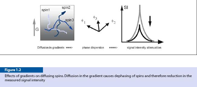
14 Figure 1.1 A typical pulse sequence for diffusion imaging.the shaded areas represent field gradient pulses.dw diffusion weighted,te time evolution 14
15 Apparent Diffusion Coefficient (ADC) Water self diffusion is constant Water movement is restricted by tissues Since brain cerebrospinal fluid (CSF) contains water that can move around freely, its ADC value is much higher than that of other brain tissues (either gray matter or white matter) S = S 0 e ( b ADC Low ADC gives strong echo, High ADC gives low echo ) 15
16 Outline MRI Quick Review What is Diffusion MRI? Detecting Diffusion Stroke and Tumor Detection Presenting Diffusion Anisotropy and Diffusion Tensor Cyst Detection Fiber Tractography Tumor Surgery and Schizophrenia Future Research Challenges 16
17 Acute Cerebral Ischemia Ischemia: Lack of blood supply Ischemic tissues have lower ADC Causes: Blood clot Shock (excessive bleeding, heart failure) Consequences: Stroke symptoms Permanent CNS damage (CNS does not regenerate) High risk of death Treatments must be rapid to prevent permanent damage 17
18 Importance of Diffusion weighted MRI Required to decide whether risky treatments are necessary Localization of Attack Measuring Severity Distinguish hemorrhage from Ischemia CT and T2 wighted MRI shows damage 5 6 hours after attack Diffusion weighted MRI detects within minutes 18

19 Acute Ischemia 19

20 Edema Stroke can also be due to edema. Edema means water molecule transfer from extracellula to intracellula. More water molecule is restricted leads to decreasing ADC 20

21 Edema Stroke due to Edema Lesioned neurons: Decreased ADC Increased Trace T2 and CT does not show the damage 21
22 Tumor 22
23 Outline MRI Quick Review What is Diffusion MRI? Detecting Diffusion Stroke and Tumor Detection Presenting Diffusion Anisotropy and Diffusion Tensor Cyst Detection Fiber Tractography Tumor Surgery and Schizophrenia Future Research Challenges 23
24 Diffusion Anisotropy Cell membranes decreases diffusion rate Diffusion rate at a point is direction dependent Axons act like pipes, myelin amplifies this effect Consequences: Diffusion must be scanned in all 3 axes Diffusion rate is represented as vector (modeled as ellipsoid) 24
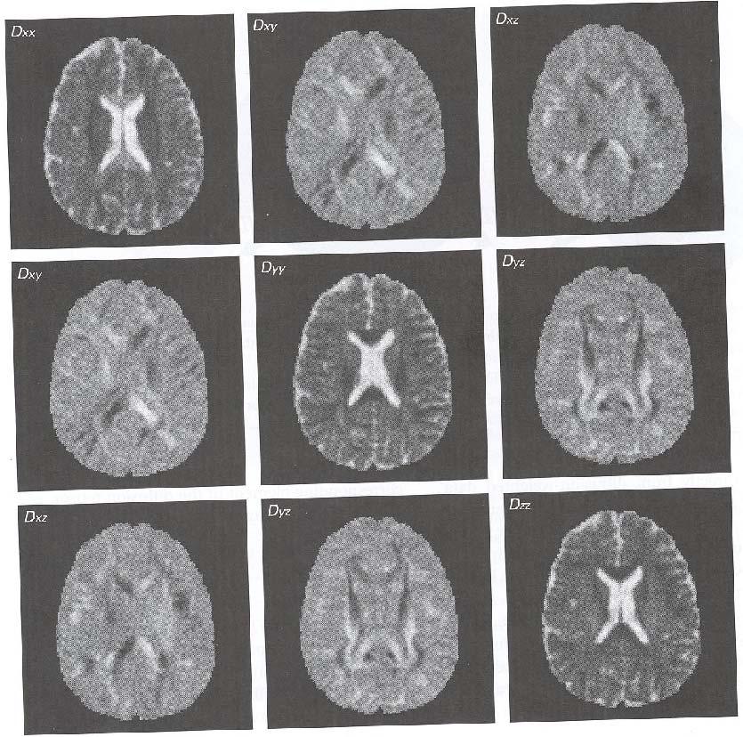
25 Diffusion Tensor Imaging Scanning in 3 axes is not sufficient Head alignment causes inconsistent data Solution: Use a tensor matrix and scan from 6 directions Calculate eigenvalues D = D D D xx yx zx D D D xy yy zy D D D xz yz zz 25
26 26
27 Tensor Calculations ( D λi) X = 0 D xx λ D xy D xz D yx D yy λ D yz = 0 D zx D zy D zz λ 27

 λ2 λ3 Isotropic diffusion occurs when there is no restriction to water movement")
28 Diffusion Tensors & Anisotropy DTI allows researchers to quantify the diffusion of water in brain tissue Diffusion for each image voxel is described by 3 perpendicular vectors λ1 λ1 λ2 λ3 Anisotropic diffusion occurs when water movement is restricted to one primary direction (e.g., myelinated fibers) λ2 λ3 Isotropic diffusion occurs when there is no restriction to water movement (e.g., ventricles, CSF) 28

29 Diffusion Ellipsoids Diffusion ellipsoids reconstructed from real DTI data. 29
 + (")

30 Mean Diffusivity & Fractional Anisotropy Mean Diffusivity (ADC) Fractional Anisotropy λ λ = 1 + λ3 λ2 + 3 FA = ( λ λ ) + ( λ λ ) + ( λ λ ) λ + λ + λ Addition of eigenvalues Difference in eigenvalues Overall diffusion Directional diffusion 30


31 Color Coded Direction Color map used to indicate dominant diffusion direction 31


32 Left: Conventional T2W image does not show white matter fiber tracts in the brain. Middle: Anisotropy map highlights the white matter bundles in the brain. Right: The z-map high intensity regions correspond to large out of plane diffusion. 32
33 Interpreting Diffusivity and FA Diffusivity and FA help determine the number, size and myelination of fibers, whereas only FA gives information about directionality. Number of fibers Myelination of fibers High Diffusivity Low FA High FA Low Diffusivity High Diffusivity Low FA High FA Low Diffusivity Size of fibers Directionality of Fibers High Diffusivity Low FA High FA Low Diffusivity Low FA Same Diffusivity High FA Same Diffusivity 33
34 Outline MRI Quick Review What is Diffusion MRI? Detecting Diffusion Stroke and Tumor Detection Presenting Diffusion Anisotropy and Diffusion Tensor Cyst Detection Fiber Tractography Tumor Surgery and Schizophrenia Future Research Challenges 34
35 Tumor 35

36 Cyst 36
37 Outline MRI Quick Review What is Diffusion MRI? Detecting Diffusion Stroke and Tumor Detection Presenting Diffusion Anisotropy and Diffusion Tensor Cyst Detection Fiber Tractography Tumor Surgery and Schizophrenia Future Research Challenges 37
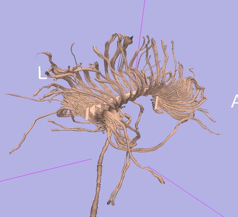
38 Fiber Tracking 38

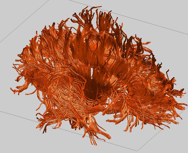
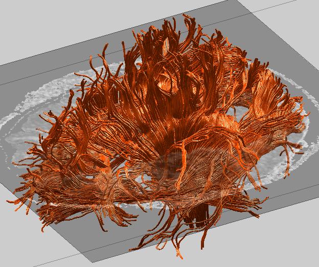
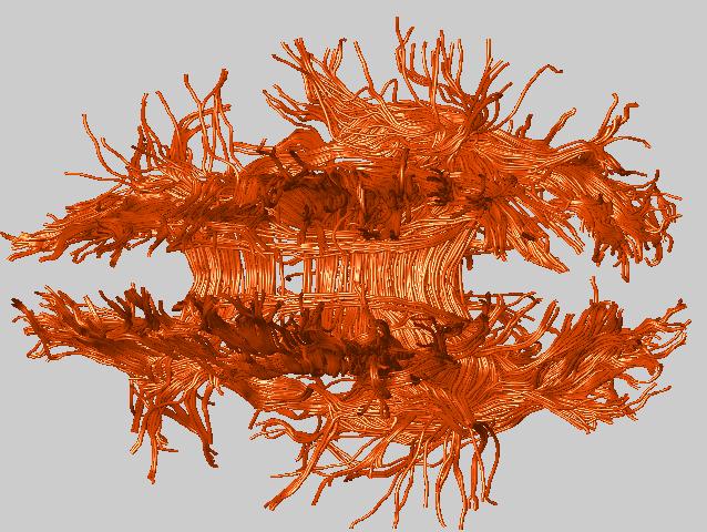
39 Whole Brain Tractography 39
40 40
41 Outline MRI Quick Review What is Diffusion MRI? Detecting Diffusion Stroke and Tumor Detection Presenting Diffusion Anisotropy and Diffusion Tensor Cyst Detection Fiber Tractography Tumor Surgery and Schizophrenia Future Research Challenges 41
42 Application of Fiber Tracking White matter research White matter disruption due to tumor Diseases related to fiber dysfunction 42
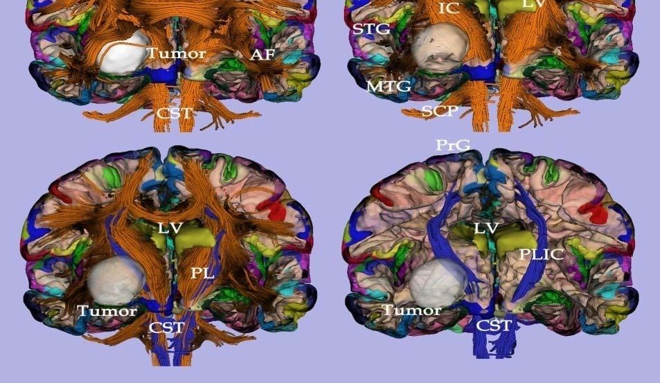
43 Other applications: Neurosurgery, Brain Tumors 43
44 White matter fiber tracts and Schizophrenia There are widespread gray matter deficits reported in MRI structural studies, but fewer reports of white matter abnormalities. Functional abnormalities are reported in different brain regions and different systems using fmri and PET. Several theories link schizophrenia with disconnection between different brain regions. 44

45 Cingulum Bundle The most prominent connection between limbic structures. Consolidates information by interconnecting thalamus, prefrontal, parietal, temporal lobes (including amygdala, hippocampus and parahippocampal gyrus) with cingulate gyrus. 45

46 Schizophrenia related symptoms most frequently linked with cingulate dysfunction Thought disorder Disorganized behavior Hallucinations Flattening of affect Delusions Lack of attention 46
(arrows), above the corpus")

47 Coronal sections of diffusion tensor maps show cingulate fasciculi (out of plane diffusion component- coded in orange)(arrows), above the corpus callosum (in plane component- coded in blue). Patient with schizophrenia on the left, comparison subject on the right. Note the difference in area of the bundle. 47
48 normal controls schizophrenics 450 RA right left Diffusion anisotropy within the left cingulum bundle in schizophrenia group was 7.4 % lower than in normal comparison subjects (mean of the percentage difference for all eight slices), while diffusion anisotropy on the right side within the CB in schizophrenics was only 2 % lower than in normal comparisons. 48
49 Outline MRI Quick Review What is Diffusion MRI? Detecting Diffusion Stroke and Tumor Detection Presenting Diffusion Anisotropy and Diffusion Tensor Cyst Detection Fiber Tractography Tumor Surgery and Schizophrenia Future Research Challenges 49
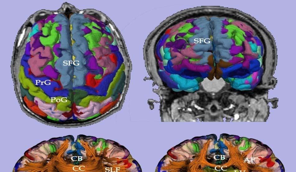
50 Research Challenges Image Resolution. (Limited by gradient strength) White Matter Segmentation. Statistical Analysis. Current Region Of Interests: Superior Temporal Gyrus. Uncinate Fasciculus. Cingulate Bundle. Arcuate Fasciculus. 50


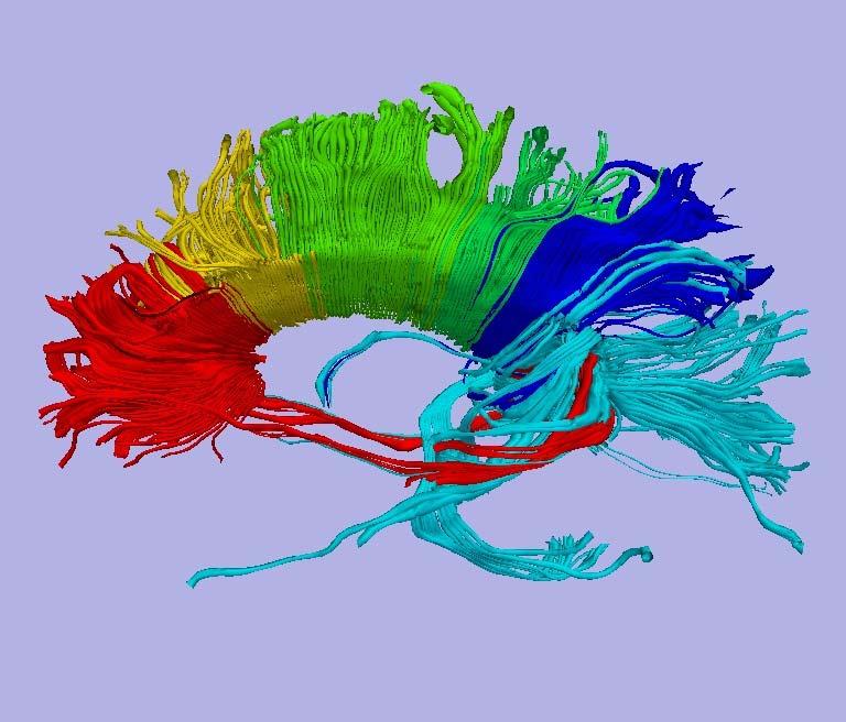
51 Fiber Clustering Automated tools separate fibers on the basis of their shape and projections 51
52 Conclusion The concept behind Diffusion MRI is relatively simple, yet there are many different applications utilizing this technology. 52
53 References Gillard, Jonathan, Adam Waldman, and Peter Barker. Clinical MR Neuroimaging. Cambridge: Cambridge University Press, Yoshiura T,Wu O, Sorensen AG (1999) Advanced MR techniques: Diffusion MR imaging,perfusion MR imaging, and Spectroscopy. Neuroimaging Clin N Am 9: Chun T, Filippi CG, Zimmerman RD, Ulug AM (2000) Diffusion changes in the Edemic human brain. Am J Neuroradi-ol 21: Engelter ST, Provenzale JM, Petrella JR, DeLong DM,Mac-Fall JR (2000) The effect of stroke on the apparent diffusion coefficient of normal-appearing white matter. Am JRoentgenol 175: Helenius J, Soinne L, Perkio J (2002) Diffusion-weighted MR imaging in normal human brains in various age groups. Am J Neuroradiol 23: Gideon P, Thomsen C, Henriksen O (1994) Increased self-diffusion of brain water. J Magn Reson Imaging 4:
Diffusion Tensor Imaging (DTI): An overview of key concepts
: An overview of key concepts") Diffusion Tensor Imaging (DTI): An overview of key concepts (Supplemental material for presentation) Prepared by: Nadia Barakat BMB 601 Chris Conklin Thursday, April 8 th 2010 Diffusion Concept [1,2]:
Diffusion Tensor Imaging (DTI): An overview of key concepts (Supplemental material for presentation) Prepared by: Nadia Barakat BMB 601 Chris Conklin Thursday, April 8 th 2010 Diffusion Concept [1,2]:
Diffusion Tensor Imaging I: The basics. Jennifer Campbell
 Diffusion Tensor Imaging I: The basics Jennifer Campbell Diffusion Tensor Imaging I: The basics Jennifer Campbell Diffusion Imaging MRI: many different sources of contrast T1W T2W PDW Perfusion BOLD DW
醫用磁振學 MRM 擴散張量影像 擴散張量影像原理. 本週課程內容 MR Diffusion 擴散張量造影原理 擴散張量造影應用 盧家鋒助理教授國立陽明大學生物醫學影像暨放射科學系
 本週課程內容 http://www.ym.edu.tw/~cflu 擴散張量造影原理 擴散張量造影應用 醫用磁振學 MRM 擴散張量影像 盧家鋒助理教授國立陽明大學生物醫學影像暨放射科學系 alvin4016@ym.edu.tw MRI The Basics (3rd edition) Chapter 22: Echo Planar Imaging MRI in Practice, (4th edition)
DIFFUSION MAGNETIC RESONANCE IMAGING
 DIFFUSION MAGNETIC RESONANCE IMAGING from spectroscopy to imaging apparent diffusion coefficient ADC-Map anisotropy diffusion tensor (imaging) DIFFUSION NMR - FROM SPECTROSCOPY TO IMAGING Combining Diffusion
Contrast Mechanisms in MRI. Michael Jay Schillaci
 Contrast Mechanisms in MRI Michael Jay Schillaci Overview Image Acquisition Basic Pulse Sequences Unwrapping K-Space Image Optimization Contrast Mechanisms Static and Motion Contrasts T1 & T2 Weighting,
Tensor Visualization. CSC 7443: Scientific Information Visualization
 Tensor Visualization Tensor data A tensor is a multivariate quantity Scalar is a tensor of rank zero s = s(x,y,z) Vector is a tensor of rank one v = (v x,v y,v z ) For a symmetric tensor of rank 2, its
A Neurosurgeon s Perspectives of Diffusion Tensor Imaging(DTI) Diffusion Tensor MRI (DTI) Background and Relevant Physics.
 Diffusion Tensor MRI (DTI) Background and Relevant Physics.") A Neurosurgeon s Perspectives of Diffusion Tensor Imaging(DTI) Kalai Arasu Muthusamy, D.Phil(Oxon) Senior Lecturer & Consultant Neurosurgeon. Division of Neurosurgery. University Malaya Medical Centre.
A Neurosurgeon s Perspectives of Diffusion Tensor Imaging(DTI) Kalai Arasu Muthusamy, D.Phil(Oxon) Senior Lecturer & Consultant Neurosurgeon. Division of Neurosurgery. University Malaya Medical Centre.
Diffusion imaging of the brain: technical considerations and practical applications
 Diffusion imaging of the brain: technical considerations and practical applications David G. Norris FC Donders Centre for Cognitive Neuroimaging Nijmegen Sustaining the physiologist in measuring the atomic
Advanced Topics and Diffusion MRI
 Advanced Topics and Diffusion MRI Slides originally by Karla Miller, FMRIB Centre Modified by Mark Chiew (mark.chiew@ndcn.ox.ac.uk) Slides available at: http://users.fmrib.ox.ac.uk/~mchiew/teaching/ MRI
Diffusion Imaging II. By: Osama Abdullah
 iffusion Imaging II By: Osama Abdullah Review Introduction. What is diffusion? iffusion and signal attenuation. iffusion imaging. How to capture diffusion? iffusion sensitizing gradients. Spin Echo. Gradient
Magnetic resonance imaging MRI
 Magnetic resonance imaging MRI Introduction What is MRI MRI is an imaging technique used primarily in medical settings that uses a strong magnetic field and radio waves to produce very clear and detailed
Quantitative Metrics for White Matter Integrity Based on Diffusion Tensor MRI Data. Stephanie Lee
 Quantitative Metrics for White Matter Integrity Based on Diffusion Tensor MRI Data Stephanie Lee May 5, 2005 Quantitative Metrics for White Matter Integrity Based on Diffusion Tensor MRI Data ABSTRACT
Applications of Spin Echo and Gradient Echo: Diffusion and Susceptibility Contrast
 Applications of Spin Echo and Gradient Echo: Diffusion and Susceptibility Contrast Chunlei Liu, PhD Department of Electrical Engineering & Computer Sciences and Helen Wills Neuroscience Institute University
Ordinary Least Squares and its applications
 Ordinary Least Squares and its applications Dr. Mauro Zucchelli University Of Verona December 5, 2016 Dr. Mauro Zucchelli Ordinary Least Squares and its applications December 5, 2016 1 / 48 Contents 1
Application of diffusion MRI to cancer, heart and brain connectome imaging
 Colloquium @ Department of Physics, NTU Application of diffusion MRI to cancer, heart and brain connectome imaging March 11, 2014 Wen-Yih Isaac Tseng MD, PhD Advanced Biomedical MRI Lab Center for Optoelectronic
Diffusion Tensor Imaging tutorial
 NA-MIC http://na-mic.org Diffusion Tensor Imaging tutorial Sonia Pujol, PhD Surgical Planning Laboratory Harvard University DTI tutorial This tutorial is an introduction to the advanced Diffusion MR capabilities
Basics of Diffusion Tensor Imaging and DtiStudio
 Basics of Diffusion Tensor Imaging and DtiStudio DTI Basics 1 DTI reveals White matter anatomy Gray matter White matter DTI uses water diffusion as a probe for white matter anatomy Isotropic diffusion
Diffusion Tensor Imaging in Humans: Practical Implications for Neuroanatomy
 Diffusion Tensor Imaging in Humans: Practical Implications for Neuroanatomy Collaborators Center for Morphometric Analysis: Nikos Makris Andy Worth Verne S. Caviness George Papadimitriou MGH-NMR Center
How does this work? How does this method differ from ordinary MRI?
 361-Lec41 Tue 18nov14 How does this work? How does this method differ from ordinary MRI? NEW kinds of MRI (magnetic resononance imaging (MRI) Diffusion Magnetic Resonance Imaging Tractographic reconstruction
Diffusion Tensor Imaging I. Jennifer Campbell
 Diffusion Tensor Imaging I Jennifer Campbell Diffusion Imaging Molecular diffusion The diffusion tensor Diffusion weighting in MRI Alternatives to the tensor Overview of applications Diffusion Imaging
Tissue Characteristics Module Three
 Tissue Characteristics Module Three 1 Equilibrium State Equilibrium State At equilibrium, the hydrogen vector is oriented in a direction parallel to the main magnetic field. Hydrogen atoms within the vector
MRI Physics I: Spins, Excitation, Relaxation
 MRI Physics I: Spins, Excitation, Relaxation Douglas C. Noll Biomedical Engineering University of Michigan Michigan Functional MRI Laboratory Outline Introduction to Nuclear Magnetic Resonance Imaging
EL-GY 6813/BE-GY 6203 Medical Imaging, Fall 2016 Final Exam
 EL-GY 6813/BE-GY 6203 Medical Imaging, Fall 2016 Final Exam (closed book, 1 sheets of notes double sided allowed, no calculator or other electronic devices allowed) 1. Ultrasound Physics (15 pt) A) (9
Field trip: Tuesday, Feb 5th
 Pulse Sequences Field trip: Tuesday, Feb 5th Hardware tour of VUIIIS Philips 3T Meet here at regular class time (11.15) Complete MRI screening form! Chuck Nockowski Philips Service Engineer Reminder: Project/Presentation
Basic MRI physics and Functional MRI
 Basic MRI physics and Functional MRI Gregory R. Lee, Ph.D Assistant Professor, Department of Radiology June 24, 2013 Pediatric Neuroimaging Research Consortium Objectives Neuroimaging Overview MR Physics
Physics of MR Image Acquisition
 Physics of MR Image Acquisition HST-583, Fall 2002 Review: -MRI: Overview - MRI: Spatial Encoding MRI Contrast: Basic sequences - Gradient Echo - Spin Echo - Inversion Recovery : Functional Magnetic Resonance
Higher Order Cartesian Tensor Representation of Orientation Distribution Functions (ODFs)
") Higher Order Cartesian Tensor Representation of Orientation Distribution Functions (ODFs) Yonas T. Weldeselassie (Ph.D. Candidate) Medical Image Computing and Analysis Lab, CS, SFU DT-MR Imaging Introduction
Higher Order Cartesian Tensor Representation of Orientation Distribution Functions (ODFs) Yonas T. Weldeselassie (Ph.D. Candidate) Medical Image Computing and Analysis Lab, CS, SFU DT-MR Imaging Introduction
Introduction to MRI. Spin & Magnetic Moments. Relaxation (T1, T2) Spin Echoes. 2DFT Imaging. K-space & Spatial Resolution.
 Spin Echoes. 2DFT Imaging. K-space & Spatial Resolution.") Introduction to MRI Spin & Magnetic Moments Relaxation (T1, T2) Spin Echoes 2DFT Imaging Selective excitation, phase & frequency encoding K-space & Spatial Resolution Contrast (T1, T2) Acknowledgement:
Introduction to MRI Spin & Magnetic Moments Relaxation (T1, T2) Spin Echoes 2DFT Imaging Selective excitation, phase & frequency encoding K-space & Spatial Resolution Contrast (T1, T2) Acknowledgement:
Outlines: (June 11, 1996) Instructor:
 Instructor:") Magnetic Resonance Imaging (June 11, 1996) Instructor: Tai-huang Huang Institute of Biomedical Sciences Academia Sinica Tel. (02) 2652-3036; Fax. (02) 2788-7641 E. mail: bmthh@ibms.sinica.edu.tw Reference:
Magnetic Resonance Imaging (June 11, 1996) Instructor: Tai-huang Huang Institute of Biomedical Sciences Academia Sinica Tel. (02) 2652-3036; Fax. (02) 2788-7641 E. mail: bmthh@ibms.sinica.edu.tw Reference:
Magnetic Resonance Imaging. Pål Erik Goa Associate Professor in Medical Imaging Dept. of Physics
 Magnetic Resonance Imaging Pål Erik Goa Associate Professor in Medical Imaging Dept. of Physics pal.e.goa@ntnu.no 1 Why MRI? X-ray/CT: Great for bone structures and high spatial resolution Not so great
The NMR Inverse Imaging Problem
 The NMR Inverse Imaging Problem Nuclear Magnetic Resonance Protons and Neutrons have intrinsic angular momentum Atoms with an odd number of proton and/or odd number of neutrons have a net magnetic moment=>
Introductory MRI Physics
 C HAPR 18 Introductory MRI Physics Aaron Sodickson EXRNAL MAGNETIC FIELD, PROTONS AND EQUILIBRIUM MAGNETIZATION Much of the bulk of the magnetic resonance imaging (MRI) scanner apparatus is dedicated to
Magnetic Resonance Imaging
 http://www.qldxray.com.au/filelibrary/mri_cardiovascular_system_ca_0005.jpg Magnetic Resonance Imaging 1 Overview 1. The magnetic properties of nuclei, and how they behave in strong magnetic fields. 2.
The physics US and MRI. Prof. Peter Bogner
 The physics US and MRI Prof. Peter Bogner Sound waves mechanical disturbance, a pressure wave moves along longitudinal wave compression rarefaction zones c = nl, (c: velocity, n: frequency, l: wavelength
Diffusion-Weighted MRI may be used to measure the apparent diffusion coefficient of water in tissue.
 Specialty Area: MR Physics for Physicists Speaker: Jennifer A. McNab, Ph.D. Assistant Professor, Radiology, Stanford University () Highlights The Bloch-Torrey equation is a generalization of the Bloch
Magnetic Resonance Spectroscopy: Basic Principles and Selected Applications
 Magnetic Resonance Spectroscopy: Basic Principles and Selected Applications Sridar Narayanan, PhD Magnetic Resonance Spectroscopy Unit McConnell Brain Imaging Centre Dept. of Neurology and Neurosurgery
The physics of medical imaging US, CT, MRI. Prof. Peter Bogner
 The physics of medical imaging US, CT, MRI Prof. Peter Bogner Clinical radiology curriculum blocks of lectures and clinical practice (7x2) Physics of medical imaging Neuroradiology Head and neck I. Head
New developments in Magnetic Resonance Spectrocopy and Diffusion MRI. Els Fieremans Steven Delputte Mahir Ozdemir
 New developments in Magnetic Resonance Spectrocopy and Diffusion MRI Els Fieremans Steven Delputte Mahir Ozdemir Overview Magnetic Resonance Spectroscopy (MRS) Basic physics of MRS Quantitative MRS Pitfalls
Basis of MRI Contrast
 Basis of MRI Contrast MARK A. HORSFIELD Department of Cardiovascular Sciences University of Leicester Leicester LE1 5WW UK Tel: +44-116-2585080 Fax: +44-870-7053111 e-mail: mah5@le.ac.uk 1 1.1 The Magnetic
Diffusion Tensor Imaging (DTI) e Neurite Orientation Dispersion and Density Imaging (NODDI)
 e Neurite Orientation Dispersion and Density Imaging (NODDI)") Diffusion Tensor Imaging (DTI) e Neurite Orientation Dispersion and Density Imaging (NODDI) Claudia AM Gandini Wheeler-Kingshott, PhD Prof. of MRI Physics Overview Diffusion and microstructure NODDI theoretical
Diffusion Tensor Imaging (DTI) e Neurite Orientation Dispersion and Density Imaging (NODDI) Claudia AM Gandini Wheeler-Kingshott, PhD Prof. of MRI Physics Overview Diffusion and microstructure NODDI theoretical
Quantitative Susceptibility Mapping and Susceptibility Tensor Imaging. Magnetization and Susceptibility
 Quantitative Susceptibility Mapping and Susceptibility Tensor Imaging 1, Chunlei Liu, Ph.D. 1 Brain Imaging and Analysis Center Department of Radiology Duke University, Durham, NC, USA 1 Magnetization
From Pixels to Brain Networks: Modeling Brain Connectivity and Its Changes in Disease. Polina Golland
 From Pixels to Brain Networks: Modeling Brain Connectivity and Its Changes in Disease Polina Golland MIT Computer Science and Artificial Intelligence Laboratory Joint work with Archana Venkataraman C.-F.
Part III: Sequences and Contrast
 Part III: Sequences and Contrast Contents T1 and T2/T2* Relaxation Contrast of Imaging Sequences T1 weighting T2/T2* weighting Contrast Agents Saturation Inversion Recovery JUST WATER? (i.e., proton density
Bayesian multi-tensor diffusion MRI and tractography
 Bayesian multi-tensor diffusion MRI and tractography Diwei Zhou 1, Ian L. Dryden 1, Alexey Koloydenko 1, & Li Bai 2 1 School of Mathematical Sciences, Univ. of Nottingham 2 School of Computer Science and
Medical Imaging Physics Spring Quarter Week 9-1
 Medical Imaging Physics Spring Quarter Week 9-1 NMR and MRI Davor Balzar balzar@du.edu www.du.edu/~balzar Intro MRI Outline NMR & MRI Guest lecturer fmri Thursday, May 22 Visit to CUHSC It s not mandatory
Introduction to Magnetic Resonance Imaging (MRI) Pietro Gori
 Pietro Gori") Introduction to Magnetic Resonance Imaging (MRI) Pietro Gori Enseignant-chercheur Equipe IMAGES - Télécom ParisTech pietro.gori@telecom-paristech.fr September 20, 2017 P. Gori BIOMED 20/09/2017 1 / 76
Introduction to Magnetic Resonance Imaging (MRI) Pietro Gori Enseignant-chercheur Equipe IMAGES - Télécom ParisTech pietro.gori@telecom-paristech.fr September 20, 2017 P. Gori BIOMED 20/09/2017 1 / 76
Cambridge University Press MRI from A to Z: A Definitive Guide for Medical Professionals Gary Liney Excerpt More information
 Main glossary Aa AB systems Referring to molecules exhibiting multiply split MRS peaks due to spin-spin interactions. In an AB system, the chemical shift between the spins is of similar magnitude to the
FREQUENCY SELECTIVE EXCITATION
 PULSE SEQUENCES FREQUENCY SELECTIVE EXCITATION RF Grad 0 Sir Peter Mansfield A 1D IMAGE Field Strength / Frequency Position FOURIER PROJECTIONS MR Image Raw Data FFT of Raw Data BACK PROJECTION Image Domain
G Medical Imaging. Outline 4/13/2012. Physics of Magnetic Resonance Imaging
 G16.4426 Medical Imaging Physics of Magnetic Resonance Imaging Riccardo Lattanzi, Ph.D. Assistant Professor Department of Radiology, NYU School of Medicine Department of Electrical and Computer Engineering,
Fundamental MRI Principles Module Two
 Fundamental MRI Principles Module Two 1 Nuclear Magnetic Resonance There are three main subatomic particles: protons neutrons electrons positively charged no significant charge negatively charged Protons
Chem8028(1314) - Spin Dynamics: Spin Interactions
 - Spin Dynamics: Spin Interactions") Chem8028(1314) - Spin Dynamics: Spin Interactions Malcolm Levitt see also IK m106 1 Nuclear spin interactions (diamagnetic materials) 2 Chemical Shift 3 Direct dipole-dipole coupling 4 J-coupling 5 Nuclear
Chem8028(1314) - Spin Dynamics: Spin Interactions Malcolm Levitt see also IK m106 1 Nuclear spin interactions (diamagnetic materials) 2 Chemical Shift 3 Direct dipole-dipole coupling 4 J-coupling 5 Nuclear
MRI in Review: Simple Steps to Cutting Edge Part I
 MRI in Review: Simple Steps to Cutting Edge Part I DWI is now 2 years old... Mike Moseley Radiology Stanford DWI, b = 1413 T2wt, 28/16 ASN 21 San Francisco + Disclosures: Funding NINDS, NCRR, NCI 45 minutes
PhD THESIS. prepared at INRIA Sophia Antipolis
 PhD THESIS prepared at INRIA Sophia Antipolis and presented at the University of Nice-Sophia Antipolis Graduate School of Information and Communication Sciences A dissertation submitted in partial satisfaction
Medical Visualization - Tensor Visualization. J.-Prof. Dr. Kai Lawonn
 Medical Visualization - Tensor Visualization J.-Prof. Dr. Kai Lawonn Lecture is partially based on the lecture by Prof. Thomas Schultz 2 What is a Tensor? A tensor is a multilinear transformation that
Anisotropy of HARDI Diffusion Profiles Based on the L 2 -Norm
 Anisotropy of HARDI Diffusion Profiles Based on the L 2 -Norm Philipp Landgraf 1, Dorit Merhof 1, Mirco Richter 1 1 Institute of Computer Science, Visual Computing Group, University of Konstanz philipp.landgraf@uni-konstanz.de
The Diffusion Tensor Imaging Toolbox
 7418 The Journal of Neuroscience, May 30, 2012 32(22):7418 7428 Toolbox Editor s Note: Toolboxes are intended to describe and evaluate methods that are becoming widely relevant to the neuroscience community
Introduction to Biomedical Imaging
 Alejandro Frangi, PhD Computational Imaging Lab Department of Information & Communication Technology Pompeu Fabra University www.cilab.upf.edu MRI advantages Superior soft-tissue contrast Depends on among
The effect of different number of diffusion gradients on SNR of diffusion tensor-derived measurement maps
 J. Biomedical Science and Engineering, 009,, 96-101 The effect of different number of diffusion gradients on SNR of diffusion tensor-derived measurement maps Na Zhang 1, Zhen-Sheng Deng 1*, Fang Wang 1,
Professor Stuart Bunt 217
 Professor Stuart Bunt 217 Traditional Anatomy Phrenology, the study of bumps on the skull. Measuring brain weights and size (still being done..see the fuss about Einstein s brain). Little link between
Diffusion tensor imaging (DTI):
:") Diffusion tensor imaging (DTI): A basic introduction to data acquisition and analysis Matthew Cykowski, MD Postdoctoral fellow Research Imaging Center UTHSCSA Room 2.320 cykowski@uthscsa.edu PART I: Acquiring
Diffusion tensor imaging (DTI): A basic introduction to data acquisition and analysis Matthew Cykowski, MD Postdoctoral fellow Research Imaging Center UTHSCSA Room 2.320 cykowski@uthscsa.edu PART I: Acquiring
Improved Correspondence for DTI Population Studies via Unbiased Atlas Building
 Improved Correspondence for DTI Population Studies via Unbiased Atlas Building Casey Goodlett 1, Brad Davis 1,2, Remi Jean 3, John Gilmore 3, and Guido Gerig 1,3 1 Department of Computer Science, University
Developing a Method for Distortion Correction in High b-value Diffusion-Weighted Magnetic Resonance Imaging HENRIK HANSSON
 Developing a Method for Distortion Correction in High b-value Diffusion-Weighted Magnetic Resonance Imaging Master s thesis in Complex Adaptive Systems HENRIK HANSSON Department of Applied Physics Division
Improving White Matter Tractography by Resolving the Challenges of Edema
 Improving White Matter Tractography by Resolving the Challenges of Edema Jérémy Lecoeur, Emmanuel Caruyer, Luke Macyszyn, Ragini Verma To cite this version: Jérémy Lecoeur, Emmanuel Caruyer, Luke Macyszyn,
Lecture #7 In Vivo Water
 Lecture #7 In Vivo Water Topics Hydration layers Tissue relaxation times Magic angle effects Magnetization Transfer Contrast (MTC) CEST Handouts and Reading assignments Mathur-De Vre, R., The NMR studies
HST.583 Functional Magnetic Resonance Imaging: Data Acquisition and Analysis Fall 2006
 MIT OpenCourseWare http://ocw.mit.edu HST.583 Functional Magnetic Resonance Imaging: Data Acquisition and Analysis Fall 2006 For information about citing these materials or our Terms of Use, visit: http://ocw.mit.edu/terms.
MRI in Practice. Catherine Westbrook MSc, DCRR, CTC Senior Lecturer Anglia Polytechnic University Cambridge UK. John Talbot MSc, DCRR
 MRI in Practice Third edition Catherine Westbrook MSc, DCRR, CTC Senior Lecturer Anglia Polytechnic University Cambridge UK and Carolyn Kaut RothRT(R) (MR) (CT) (M) (CV) Fellow SMRT (Section for Magnetic
Magnetic Resonance Imaging (MRI)
") Magnetic Resonance Imaging Introduction The Components The Technology (MRI) Physics behind MR Most slides taken from http:// www.slideworld.org/ viewslides.aspx/magnetic- Resonance-Imaging- %28MRI%29-MR-Imaging-
Magnetic Resonance Imaging Introduction The Components The Technology (MRI) Physics behind MR Most slides taken from http:// www.slideworld.org/ viewslides.aspx/magnetic- Resonance-Imaging- %28MRI%29-MR-Imaging-
Diffusion MRI. Outline. Biology: The Neuron. Brain connectivity. Biology: Brain Organization. Brain connections and fibers
 Outline Diffusion MRI Alfred Anwander Download of Slides: www.cbs.mpg.de/events/ teaching/brainsignals1112 password: mpi-brain CBSWIKI: Cornet/DiffusionMRI Neuroanatomy Diffusion MRI Diffusion Tensor Imaging
Nuclear Magnetic Resonance Imaging
 Nuclear Magnetic Resonance Imaging Simon Lacoste-Julien Electromagnetic Theory Project 198-562B Department of Physics McGill University April 21 2003 Abstract This paper gives an elementary introduction
Spatially Regularized Spherical Reconstruction: A Cross-Domain Filtering Approach for HARDI Signals
 Spatially Regularized Spherical Reconstruction: A Cross-Domain Filtering Approach for HARDI Signals by Iván Camilo Salgado Patarroyo A thesis presented to the University of Waterloo in fulfillment of the
RADIOLOGIV TECHNOLOGY 4912 COMPREHENSEIVE REVIEW/MRI WORSHEET #1- PATIENT CARE AND SAFETY/PHYSICAL PRINCIPLES
 RADIOLOGIV TECHNOLOGY 4912 COMPREHENSEIVE REVIEW/MRI WORSHEET #1- PATIENT CARE AND SAFETY/PHYSICAL PRINCIPLES 1. What are potential consequences to patients and personnel should there be a release of gaseous
An introduction to Solid State NMR and its Interactions
 An introduction to Solid State NMR and its Interactions From tensor to NMR spectra CECAM Tutorial September 9 Calculation of Solid-State NMR Parameters Using the GIPAW Method Thibault Charpentier - CEA
10.4 Continuous Wave NMR Instrumentation
 10.4 Continuous Wave NMR Instrumentation coherent detection bulk magnetization the rotating frame, and effective magnetic field generating a rotating frame, and precession in the laboratory frame spin-lattice
Predictive Diagnosis of Alzheimer s Disease using Diffusion MRI
 Predictive Diagnosis of Alzheimer s Disease using Diffusion MRI by Syeda Maryam A thesis presented to the University of Waterloo in fulfillment of the thesis requirement for the degree of Master of Applied
Fundamental MRI Principles Module 2 N. Nuclear Magnetic Resonance. X-ray. MRI Hydrogen Protons. Page 1. Electrons
 Fundamental MRI Principles Module 2 N S 1 Nuclear Magnetic Resonance There are three main subatomic particles: protons positively charged neutrons no significant charge electrons negatively charged Protons
Sketch of the MRI Device
 Outline for Today 1. 2. 3. Introduction to MRI Quantum NMR and MRI in 0D Magnetization, m(x,t), in a Voxel Proton T1 Spin Relaxation in a Voxel Proton Density MRI in 1D MRI Case Study, and Caveat Sketch
Diffusion tensor imaging: brain pathway reconstruction
 Neda Sepasian, Jan ten Thije Boonkkamp, Anna Vilanova Diffusion tensor imaging: brain pathway reconstruction NAW 5/6 nr. 4 december 205 259 Neda Sepasian Department of Biomedical Engineering Eindhoven
MRI Homework. i. (0.5 pt each) Consider the following arrangements of bar magnets in a strong magnetic field.
 Consider the following arrangements of bar magnets in a strong magnetic field.") MRI Homework 1. While x-rays are used to image bones, magnetic resonance imaging (MRI) is used to examine tissues within the body by detecting where hydrogen atoms (H atoms) are and their environment (e.g.
MRI Homework 1. While x-rays are used to image bones, magnetic resonance imaging (MRI) is used to examine tissues within the body by detecting where hydrogen atoms (H atoms) are and their environment (e.g.
Spatial normalization of diffusion models and tensor analysis
 University of Iowa Iowa Research Online Theses and Dissertations Summer 2009 Spatial normalization of diffusion models and tensor analysis Madhura Aditya Ingalhalikar University of Iowa Copyright 2009
Tract-Specific Analysis for DTI of Brain White Matter
 Tract-Specific Analysis for DTI of Brain White Matter Paul Yushkevich, Hui Zhang, James Gee Penn Image Computing & Science Lab Department of Radiology University of Pennsylvania IPAM Summer School July
Lecture 8 Analyzing the diffusion weighted signal. Room CSB 272 this week! Please install AFNI
 Lecture 8 Analyzing the diffusion weighted signal Room CSB 272 this week! Please install AFNI http://afni.nimh.nih.gov/afni/ Next lecture, DTI For this lecture, think in terms of a single voxel We re still
One patient and one control were unable to complete the TOMM. An additional HC had abnormally low scores
 Ling et al. 2013-1 Supplemental Methods Participants One patient and one control were unable to complete the TOMM. An additional HC had abnormally low scores on the TOMM only (T-score < 0), and was not
Introduction to the Course and the Techniques. Jeffry R. Alger, PhD Ahmanson-Lovelace Brain Mapping Center Department of Neurology
 Introduction to the Course and the Techniques Jeffry R. Alger, PhD Ahmanson-Lovelace Brain Mapping Center Department of Neurology (jralger@ucla.edu) CTSI Neuroimaging April 2013 Rationale for the Course
Research Article Thalamus Segmentation from Diffusion Tensor Magnetic Resonance Imaging
 Biomedical Imaging Volume 2007, Article ID 90216, 5 pages doi:10.1155/2007/90216 Research Article Thalamus Segmentation from Diffusion Tensor Magnetic Resonance Imaging Ye Duan, Xiaoling Li, and Yongjian
Diffusion Tensor Processing and Visualization
 NA-MIC National Alliance for Medical Image Computing http://na-mic.org Diffusion Tensor Processing and Visualization Guido Gerig University of Utah NAMIC: National Alliance for Medical Image Computing
The diffusion tensor is derived from diffusion-weighted
 Increased Anisotropy in Acute Stroke A Possible Explanation Hadrian A.L. Green, MB ChB; Alonso Peña, PhD; Christopher J. Price, BSc, MRCP; Elizabeth A. Warburton, MRCP, DM; John D. Pickard, MS, FRCS, FMedSci;
Magnetic Resonance Spectroscopy. Saurabh Bhaskar Shaw Dwip Shah
 Magnetic Resonance Spectroscopy By Saurabh Bhaskar Shaw Dwip Shah What is Magnetic Resonance Spectroscopy? [1] Non invasive method to look at concentration of metabolites invivo. 2 Basics of MRS Physics
DEVIL PHYSICS THE BADDEST CLASS ON CAMPUS IB PHYSICS
 DEVIL PHYSICS THE BADDEST CLASS ON CAMPUS IB PHYSICS TSOKOS OPTION I-2 MEDICAL IMAGING Reading Activity Answers IB Assessment Statements Option I-2, Medical Imaging: X-Rays I.2.1. I.2.2. I.2.3. Define
An Anisotropic Material Model for Image Guided Neurosurgery
 An Anisotropic Material Model for Image Guided Neurosurgery Corey A. Kemper 1, Ion-Florin Talos 2, Alexandra Golby 2, Peter M. Black 2, Ron Kikinis 2, W. Eric L. Grimson 1, and Simon K. Warfield 2 1 Massachusetts
The Basics of Magnetic Resonance Imaging
 The Basics of Magnetic Resonance Imaging Nathalie JUST, PhD nathalie.just@epfl.ch CIBM-AIT, EPFL Course 2013-2014-Chemistry 1 Course 2013-2014-Chemistry 2 MRI: Many different contrasts Proton density T1
Chapter 14:Physics of Magnetic Resonance
 Chapter 14:Physics of Magnetic Resonance Slide set of 141 slides based on the chapter authored by Hee Kwon Song of the publication (ISBN 978-92-0-131010-1): Diagnostic Radiology Physics: A Handbook for
III, Diffusion, and Susceptibility. August 25, Departments of Mathematics and Applied Math and Computational Science University of Pennsylvania
 III,, and Departments of Mathematics and Applied Math and Computational Science University of Pennsylvania August 25, 2010 Copyright Page All material in this lecture, except as noted within the text,
Bloch Equations & Relaxation UCLA. Radiology
 Bloch Equations & Relaxation MRI Systems II B1 I 1 I ~B 1 (t) I 6 ~M I I 5 I 4 Lecture # Learning Objectives Distinguish spin, precession, and nutation. Appreciate that any B-field acts on the the spin
BME I5000: Biomedical Imaging
 BME I5000: Biomedical Imaging Lecture 9 Magnetic Resonance Imaging (imaging) Lucas C. Parra, parra@ccny.cuny.edu Blackboard: http://cityonline.ccny.cuny.edu/ 1 Schedule 1. Introduction, Spatial Resolution,
Shape Anisotropy: Tensor Distance to Anisotropy Measure
 Shape Anisotropy: Tensor Distance to Anisotropy Measure Yonas T. Weldeselassie, Saba El-Hilo and M. Stella Atkins Medical Image Analysis Lab, School of Computing Science, Simon Fraser University ABSTRACT
Magnetic Resonance Imaging. Qun Zhao Bioimaging Research Center University of Georgia
 Magnetic Resonance Imaging Qun Zhao Bioimaging Research Center University of Georgia The Nobel Prize in Physiology or Medicine 2003 "for their discoveries concerning magnetic resonance imaging" Paul C.
Improved Correspondence for DTI Population Studies Via Unbiased Atlas Building
 Improved Correspondence for DTI Population Studies Via Unbiased Atlas Building Casey Goodlett 1,BradDavis 1,2,RemiJean 3, John Gilmore 3, and Guido Gerig 1,3 1 Department of Computer Science, University
Noise considerations in the determination of diffusion tensor anisotropy
 Magnetic Resonance Imaging () 659 669 Noise considerations in the determination of diffusion tensor anisotropy Stefan Skare a,b, *, Tie-Qiang Li c, Bo Nordell a,b, Martin Ingvar a a MR Center, Karolinska
Advanced MRI: Diffusion MRI 1: DTI and k-space
 k y Advanced MRI: Diffusion MRI 1: DTI and k-space k X Eric Sigmund, PhD February 26th, 2013 LECTURE 1 Neuro Diffusion MRI 3-5 m White matter axons Body 15 m Renal medulla Musculoskeletal 50 m Skeletal
A Brief Introduction to Medical Imaging. Outline
 A Brief Introduction to Medical Imaging Outline General Goals Linear Imaging Systems An Example, The Pin Hole Camera Radiations and Their Interactions with Matter Coherent vs. Incoherent Imaging Length