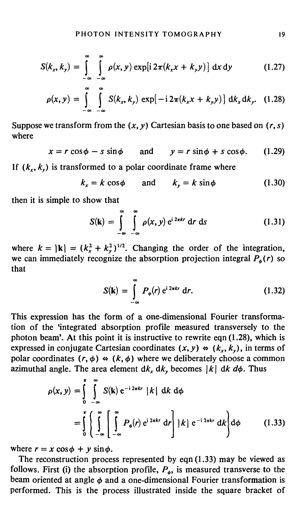
PhD THESIS. prepared at INRIA Sophia Antipolis
|
|
|
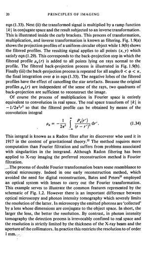
- Hugh Shaw
- 5 years ago
- Views:
Transcription
1 PhD THESIS prepared at INRIA Sophia Antipolis and presented at the University of Nice-Sophia Antipolis Graduate School of Information and Communication Sciences A dissertation submitted in partial satisfaction of the requirements for the degree of DOCTOR OF SCIENCE Specialized in Signal and Image Processing Automated In Vivo Dissection of White Matter Structures from Diffusion Magnetic Resonance Imaging Demian Wassermann Advisor Dr. Rachid Deriche INRIA Sophia-Antipolis, France Reviewers Dr. Cyril Poupon CEA NeuroSpin, France Dr. Carl-Fredrik Westin Harvard University, USA Examiners Dr. Peter Basser National Institute of Health, Bethesda, USA Dr. Habib Benali INSERM / Pitié-Salpêtrière, France Dr. Ragini Verma University of Pennsylvania, USA
2 56 CHAPTER 2. BRAIN ANATOMY AND DIFFUSION MRI by Basser et al. [1994b]. In this simplified model of water diffusion, Einstein s and Fick s laws of diffusion are generalized. The scalar diffusion coefficient D is replaced by a positive symmetric semi-definite matrix D representing diffusion, the diffusion tensor. Therefore, Einstein s relation (equation 2.1) is generalized, considering the covariance matrix of the net displacement vector R 0 1 D xx D xy D xz B C D D xy D yy D yz A = 1 6 hrrt i. (2.3) D xz D yz D zz We can use this generalization to characterize the diffusion propagator. First, we notate the probability that a particle moves along the vector R in a time as P (R, ). Then, we make a first order approximation to P (R, ) ignoring the high order terms and using equation 2.3. Finally, we obtain a partial differential equation which governs the diffusion propagator 1 (R, = Dr 2 P (R, ). (2.4) Under the assumption that the diffusion is Gaussian, the solution of this equation is the propagator model given by Basser et al. [1994b]: P (R, ) = 1 p (4 ) 3 D exp R T D 1 R The characteristics of this propagator model have proved to be an invaluable resource in characterizing diffusion in living tissue, particularly in the human brain [Johansen-Berg and Behrens: 2009]. In summary, diffusion is a fundamental physical process in nature and particularly physiology. The random motion of water molecules within a tissue are influenced by a variety of factors like cell membranes, the cytoskeleton, and macromolecules [Tanner and Stejskal: 1968]. It is due to this that being able to measure and characterize diffusion, and in fact the diffusion propagator, is a valuable tool to elucidate the microstructural and physiological features of tissues [Basser and Pierpaoli: 1996]. In the past twenty years, nuclear magnetic resonance has made a breakthrough in this area providing, through diffusion magnetic resonance imaging (dmri), a probe into the microstructure of living tissue [Alexander et al.: 2007]. NUCLEAR MAGNETIC RESONANCE In 1946, Bloch et al. [1946] and Purcell et al. [1946] simultaneously described Nuclear Magnetic Resonance (NMR) which yielded them a joint Nobel Prize in Physics in The basic principle behind NMR is that after aligning a magnetic nu- 1. The detailed intermediate mathematical derivations can be found in the work by Tuch [2002]

3 57 Figure 2.10: Water diffusion measured by the first NMR-obtained signal: The decay of transverse polarization associated with water at 25 C used by Carr and Purcell [1954] to measure the diffusion coefficient of water. In this work, using a small magnetic gradient and the Spin-Echo method proposed earlier by Hahn [1950] they observed that the decay is largely determined by the molecular diffusion through the magnetic gradient. Image adapted from Carr and Purcell [1954] cleus, for instance the proton 1 H, with a very strong external magnetic field, its response to a perturbation of the alignment by an electromagnetic field is characteristic. Shortly after this, Hahn [1950] published a paper on the NMR spin echo in which he noted that the random thermal motion of the spins would reduce the amplitude of the observed signal in the presence of an inhomogeneity of the magnetic field. His work was influential on NMR and fundamental in understanding magnetic resonance imaging (MRI). Inspired by this work, Carr and Purcell [1954] used the Spin-Echo NMR sequence proposed by Hahn [1950] and a sequence of their own to measure the diffusion coefficient of water at 25 C, see figure Carr and Purcell used a small magnetic gradient and the Spin-Echo sequence and showed that the decay on the transverse polarization was affected by water diffusion. Then they proposed a second NMR sequence which was not influenced by water diffusion and used both decays obtained by both sequences to measure the diffusion constant. This was the first NMR signal ever obtained. Almost 20 years later, the first acquisition of a bi-dimensional image using NMR was performed by Lauterbur [1973], see figure As the image was obtained using two coupled magnetic gradients, he called the process zeugmatography, from the Greek work zeugma, that which is used to join. Mansfield [1977] improved on the previous technique. By using mathematical properties of the MRI signal, he proposed a new ultrafast acquisition procedure: echo-planar imaging. The two previous techniques, zeugmatography and echo-planar imaging, became a fundamental parts of medical MRI yielded a joint Nobel Prize in Physiology or Medicine 2003 to their authors. There is a single piece missing to fully describe the techniques at the heart of diffusion MRI: the gradient spin echo sequence developed by Stejskal and Tanner [1965]. Due to its critical role, we dedicate the next section to the description of their technique.
4 58 CHAPTER 2. BRAIN ANATOMY AND DIFFUSION MRI Figure 2.11: First bi-dimensional image obtained from NMR. Lauterbur [1973] was the first to propose the use of two coupled magnetic gradients in order to obtain a bi-dimensional slice. He called this process zeugmatography. On the left we can see a drawing of the imaged object, with two capillaries filled with water, and on the right the resulting image obtained by NMR. Both images adapted from Lauterbur [1973] Pulse Gradient Spin Echo (PGSE) The Stejskal and Tanner [1965] imaging sequence is used to measure the diffusion of water molecules in a given direction g. This sequence uses two gradient pulses, g(t), of duration time, in the direction g to control the diffusion-weighting. The gradient pulses are placed before and after a 180 refocusing pulse (RF). More specifically, a first 90 RF is applied to flip the magnetization in the transverse plane. first gradient pulse induces a phase shift of the spins whose position are now a function of time. The position of the spins is assumed to stay constant during time. Finally, after a time, the 180 combined with the second gradient pulse causes a second phase shift. For static spins only, this pulse cancels the phase shift. Alternatively, spins under Brownian motion during the time period The, undergo different phase shifts by the two gradient pulses, resulting in a T2 signal attenuation [Cercignani and Horsfield: 2001]. This pulse sequence is illustrated in figure 2.12 Figure 2.13 shows examples of diffusion weighted images of the brain acquired at different directions. In this figure it can be observed that direction specific attenuation is related to the orientation of white matter fibres. By assuming that the pulses are infinitely narrow (narrow pulse approximation), meaning the gradient pulse duration is short enough to neglect the diffusion of the water molecule at that time, Stejskal and Tanner [1965] showed that the attenuation of the signal S(q, ) is expressed as the 3-dimensional Fourier transform F of the ensemble average propagator P, Z S(q, ) = P (r r 0, )exp( 2 iq T R)dr = F[P (r r 0, )]. (2.5) S 0 R 3 In this equation, q = G/2, with the nuclear gyromagnetic ratio for water protons, G the applied diffusion gradient vector, S 0 is the baseline image acquired without any diffusion gradient (also called the B 0 image) and P (r r 0, ) is the diffusion p.d.f. or diffusion propagator introduced in chapter 2. This P ( ) is ultimately the
5 PhD THESIS prepared at INRIA Sophia Antipolis and presented at the University of Nice-Sophia Antipolis Graduate School of Information and Communication Sciences A dissertation submitted in partial fulfillment of the requirements for the degree of DOCTOR OF SCIENCE Specialized in Control, Signal and Image Processing Geometric and Variational Methods for Diffusion Tensor MRI Processing Christophe Lenglet Advisors Pr. Rachid Deriche INRIA Sophia Antipolis, France Pr. Olivier Faugeras INRIA Sophia Antipolis, France Reviewers Pr. Peter Basser NICHD, USA Pr. Guillermo Sapiro University of Minnesota, USA Pr. Carl-Fredrik Westin Harvard University, USA Examiners Pr. Nicholas Ayache INRIA Sophia Antipolis, France Dr. Maher Moakher ENIT Tunis, Tunisia Invited members Pr. Stéphane Lehéricy Hospital La Pitié-Salpêtrière, France Dr. Jean-Philippe Thirion QuantifiCare S.A. Sophia Antipolis, France
6 In the next section, we will introduce the foundation of MRI and diffusion MRI. We will show that diffusion MRI constitutes an amazing and non-invasive means to investigate the three-dimensional architecture of the human brain white matter that has just been described. 3.3 MAGNETIC RESONANCE IMAGING Magnetic Resonance Imaging (MRI) is by far the most popular application of Nuclear Magnetic Resonance (NMR), for medical diagnosis. However, NMR is also widely used in chemistry to perform NMR spectroscopy, ie. to study the atomic composition of a given sample. NMR was simultaneously described by Felix Bloch [40] at Stanford University and by Edward Mills Purcell [253] at Harvard University in In 1952, they both received the Nobel Prize in Physics for their discovery. The basic principle behind NMR is that, after aligning a magnetic nucleus like Hydrogen-1 with a very strong external magnetic field, its response to a perturbation of the alignment by an electromagnetic field is characteristic. Four year after this discovery, in 1950, Herman Carr, proposed to create the first one-dimensional MR images by introducing a gradient in the magnetic field. In 1971, it was shown by Raymond Damadian that T1 and T2 relaxation times of tumoral tissues are significantly longer than for the corresponding normal tissues, hence opening great hopes for cancer diagnosis. Shortly after Bloch and Purcell discovery, Hahn published his seminal paper [138] on the NMR spin echo in which he noted that the random thermal motion of the spins would reduce the amplitude of the observed signal in the presence of a magnetic field inhomogeneity. This is a fundamental notion to understand diffusion MRI. As soon as 1973, Paul Lauterbur proposed a method [172], based on gradients of magnetic fields, to reconstruct two dimensional MR images. Peter Mansfield [204] further developed the use of magnetic fields gradients and, by studying the mathematical properties of the MRI signal, proposed a new ultrafast acquisition technique known as the echo-planar technique. In 2003, they jointly received the Nobel prize in Physiology and Medicine for their discoveries on MRI. MRI allows one to acquire non-invasively 3D images at high spatial resolution. Various modalities can be obtained with the same device such as detailed anatomy (structural MRI), functional activity (functional MRI), water-molecules diffusion (diffusion weighted MRI), blood flow measurements (perfusion MRI), distribution of various metabolites (MR Spectroscopy) and also blood vessels (MR Angiography). The first part of this section briefly exposes the basic principles of MR imaging. We then give the outlines of diffusion MRI. 46
7 B 0 a) Random spins directions b) Spins aligned with B 0 Figure 3.8: Random directions of spins in the absence of an external magnetic field (a) and aligned spins in the presence of an external magnetic field B 0 (b). Note that the actual spin rotation around B 0 occurs within a cone around B MRI Principles Physical model Atoms are made of electrons, which hold a negative charge and rotate around a nucleus. The nucleus can be divided in neutrons (not charged) and protons (charged positively). It rotates around itself. MRI is based on this rotation motion. Some nuclei have the property to align with a magnetic field if their mass number is odd, i.e. if the sum of numbers of protons and neutrons is odd. This is named angular moment or spin. Among others, 1 H atoms, which represent 99.89% of naturally found hydrogens atoms and are widely represented in biological systems, have a spin. MRI is thus particularly relevant to study the structure of biological tissues such as the human brain. Spin nuclei being positively charged, their motion induces a magnetic field. Conversely, the resulting magnetic moment can be oriented by the application of a magnetic field. This reciprocity is largely used in MRI. From a macroscopic point of view, no resulting field can be observed directly since each spin has its own, independent, random orientation (figure 3.8 (a). However, when placed in a powerful external magnetic field B 0, the spin directions align parallel to this field (figure 3.8 (b). More precisely, each spin rotates within a cone around B 0. This is called spin precession. The frequency of rotation, called the Larmor frequency, is related to the magnetic field B 0 through the gyromagnetic ratio γ by the following equation: ω 0 = γ B 0 (3.1) γ depends on the nucleus. Hydrogen, for instance, has a gyromagnetic ratio γ = 42.57MHz/T. This corresponds to a rotation frequency of f1 H = 63.86MHz in a 1.5 Tesla magnetic field. Because of this rotation motion, a spin can be modeled by a small magnetic dipole with moment m, such that d m dt = γ m B 0 47
8 It is actually cumbersome to understand MRI at a microscopic scale. It is convenient, at a macroscopic level, to replace the individual spin by a single magnetization vector representing the spin of all the particles in a voxel (about protons/mm 3 ). The net resulting magnetization M is the sum of all the elementary moments and, by making the assumption of a uniform distribution of the dipoles orientations in a given voxel, we simply end up with a M = 0. However, under the action of a static magnetic field B 0 (from 1 and up to more than 9.4 Tesla), particles get aligned in the direction of that field and induce a magnetization parallel to B 0 at equilibrium. In that state, the amplitude of M represents only a small fraction of what it would have been if all the particles were aligned in the same direction. Actually, by the laws of thermodynamics, the number of spins following the orientation imposed by B 0 (low energy state, called spin-up) slightly outnumbers the amount of spins anti-parallel to the outer field (high energy state, called spin-down). The difference is small and given by the ratio: ( N N + =exp E B 0 kt where N and N + are respectively the number of spins in the upper and lower states, k is the Boltzmann constant and T the temperature in Kelvin. Applying the Boltzmann relation, one can estimate that, at the ambient temperature and within a 1.5 T field, there is a difference of 10 in favor of low energy protons among a total of 1 million protons. ) The net magnetization M can be decomposed into two components (figure 3.9): A longitudinal component M z, i.e. parallel to B 0 A transverse component M xy, orthogonal to B 0 At equilibrium, after a sufficient exposure time to B 0, the transverse component M xy vanishes. All the individual spins are indeed precessing, but they are all out of phase with each other. Excitation phase By applying an oscillating electromagnetic (radio-frequency) pulse toward the area of the body to be examined, it is possible to perturb the difference in the number of atoms between the two energy states. The idea is to use a much weaker field than B 0 at the Larmor frequency of the targeted nuclei and to apply it through a rotating reference frame orthogonal to B 0. It causes the particles in that area to absorb the energy required to make them spin in a different direction and move from the lower energy state towards the higher. The exposure to the radio-frequency pulse causes the net magnetization to spiral 48
9 Longitudinal Component Z Precession Trajectory B 0 M z M Net Magnetization Vector M xy Y X Transversal Component Figure 3.9: The net magnetization vector M, decomposed into a longitudinal component M z and a transverse component M xy. away from B 0. M rotates away from the longitudinal position in an amount proportional to the duration of the pulse. It is even possible to flip the original direction of M. A pulse of 90 degrees would zero out the longitudinal component of M (figure 3.10) while a 180 degrees pulse, or inversion pulse, completely inverts the longitudinal component through an excess of antiparallel spins. The net magnetization also starts to dephase since different particles experience a slightly different magnetic field. This is usually referred to as phase coherence. All the magnetic moments are in phase in their precession motion. TheMRIsignalisacquired by measuring a current induced in the plane where the radio-frequency pulse was applied. The frequency of this current is the Larmor frequency of the nucleus and its amplitude is directly linked to the amount of magnetization in that plane. Relaxation phase By removing the radio-frequency pulse, particles begin to return to their initial energy state, aligned with the external field, from the higher to the lower. This is associated with a loss of stored excess energy to surrounding particles which can be detected by the coil of the MRI scanner. We can then observe two different types of relaxation processes: T1 weighted images follow the evolution of the increasing longitudinal component of M T2 weighted images follow the evolution of the decreasing transversal component of M 49
10 Z RF M α Y X Figure 3.10: Excitation phase: the energy given by the RF pulse flips the net magnetization vector M of an angle α (here α = 90 ). In clinical MRI, the radio-frequency pulse is typically chosen to coincide with the Larmor frequency of the hydrogen nucleus. The energy release during relaxation is thus an estimate of the number of protons or, in other words, the amount of water. Spin lattice relaxation (T1): exchange between protons and surrounding molecules. The spin lattice relaxation is based on the energy This energy dissipation is characterized by the restoration of the longitudinal component to its equilibrium value. This recovery process is modeled by an exponential function characterized by a time constant T1, the period for the longitudinal magnetization to recover 63% of its equilibrium value (figure 3.11). For a 90-degree excitation pulse, we have: ( M z = M 0 (1 exp t ) ) T 1 The recovery process is considered as finished after 5 T1 periods. Spin-spin relaxation (T2): Spin-spin relaxation refers to the loss of net magnetization in the transverse plane related to protons dephasing. Spins do not only give up their energy to surrounding lattice molecules but also to other neighboring nonexcited spins. This process is also modeled by an exponential function characterized by another time constant T2, which corresponds to the period for the transversal component to loose 63% of its value just after the RF pulse: ( M xy = M 0 exp t ) T 2 50
11 100% 63% T1 Time Figure 3.11: Spin lattice relaxation describes the longitudinal component recovery as a function of time and is characterized by the T1 constant. This dephasing is actually further increased by local magnetic field inhomogeneities, since the Larmor frequency will also be nonuniform throughout the region. A time constant slightly different to T2, T2, is therefore used. The transverse component induces a current in a coil, known as Free Induction Decay (FID). The T2 constant can be evaluated through the convex envelop of the FID curve (figure 3.12). Signal 37% T2 Time Figure 3.12: Spin-spin relaxation describes the exponential decrease of the transversal component as a function of time and is characterized by the T2 constant. The different biological tissues are characterized by respective T1 and T2 values, as shown in table 3.1. The intensities of MR images comes from these values. Image construction through pulse sequence A pulse sequence is a series of RF pulses and/or magnetic field gradients applied to a sample to produce a specific form of MR signal. It is indeed possible to encode and thus recover the MR signal from specific regions in the volume of interest by means of RF and linear gradients applied along the 3 spatial directions. 51
12 Tissue T1 (ms) T2 (ms) CSF Grey matter White matter Fat Blood (deoxygenated) Blood (oxygenated) Muscles Table 3.1: Approximate T1 and T2 values (ms) in various tissues at 1.5T. Figure 3.13 illustrates a basic pulse sequence. A first gradient G z in the B 0 magnetic field direction results in a linear intensity variation of the magnetic field that can be used to select a slice. In this case, a slice is a plane orthogonal to B 0 with a typical thickness of 1-10mm. Based on relationship (3.1), the spins of a given slice are hence characterized by a specific Larmor frequency. After the RF pulse at the frequency related to the target slice, two transient gradients are applied to encode the x and y dimensions in the slice plane. A first gradient G y in the y direction induces a phase shift related to the position along the y axis: this is the phase encoding. A second gradient G x in the remaining x direction is applied, leading to a precession frequency variation along the x axis: this is the frequency encoding. This process actually performs an acquisition of the plane data in the frequency space (or k-space). For each selected slice, an inverse Fourier transform finally maps these data back into the 2D spatial domain. A pulse sequence is first characterized by the delay between two similar RF pulses, called the Repetition Time (TR). The other parameters of interest depend on the actual sequence. Indeed, different pulse sequences were developed to measure the relaxation times. For instance, Gradient Echo simply repeats the Free Induction Decay described above and allows to sample T2. Most sequences often comprise additional RF pulses following the slice selection one, to partially refocus the transverse magnetization and produce an echo, leading to a more reliable measure. Spin-Echo is the application of a 90 degree pulse followed by a 180 degree pulse after a time TE/2. This second pulse, which refocuses the transverse magnetization and results in an echo at time TE (Echo Time), removes local field inhomogeneities dephasing, hence allowing to directly measure the T2 decay. On the other hand, Inversion Recovery, which relies on a 180 degree pulse followed after a time TI (Inversion Time) by a 90 degree pulse, enhances the T1 weighting. The choice of the specific pulse sequence parameters (TR, TE, TI,...) finally determines the image contrast. Two distinct tissues may for instance have similar T1 values but distinct 52
13 Repetition Time (TR) RF RF pulse G z Slice selection G y Line selection G x Line readout Signal Free Induction Decay (FID) Signal Readout Figure 3.13: A simplified MRI pulse sequence timing diagram. T2 values, so the choice of the sequence depends on the information of interest. The straightforward application of a given pulse sequence allows to get a static image contrasting different tissues. However, based on the same principles, it is possible to indirectly image dynamic processes such as oxygen flow or the motion of water molecules. In the next section, we introduce the basic principles of diffusion MRI Diffusion MRI Diffusion MRI is the unique non-invasive technique that allows to probe and quantify the diffusion of water molecules in the body. By modeling the local anisotropy of this diffusion process, it becomes possible to indirectly infer the architecture and properties of tissues such as the brain white matter. Physical principles of Diffusion Tensor Imaging (DTI) Above the absolute zero temperature, water molecules freely move and collide with each other in an isotropic medium according to Brownian motion (figure 3.14) [43]. At a macroscopic scale, this phenomenon yields a diffusion process. In an isotropic medium, the diffusion coefficient D was related by Einstein [106] to the root mean square of the diffusion distance: D = 1 6τ RT R (3.2) 53
14 Figure 3.14: An image of Brownian motion, done with three different step sizes. The hierarchical structure is clearly visible. More saturated colors represent smaller step sizes. Image under the Gnu Free Documentation License 1.2 In this expression, τ is the diffusion time and R is the net displacement vector R = r r 0, with r 0 the original position of a particle and r, its position after the time τ. denotes an ensemble average. The scalar constant D, known as the diffusion coefficient, measures the molecules mobility in the isotropic case and depends on the molecule-type and the medium properties but not on the direction. For example, at normal brain temperature, 68% of the water molecules diffuse in 50ms in a sphere of 17 µm diameter. In anisotropic biological tissues, water molecules mobility is constrained by obstacles formed by surrounding structures, such as the axonal membranes in the brain. Moreover, it is known that the myelin sheath can modulate the anisotropy of the diffusion while the microtubules and neurofilaments do not modify it [30]. In this case, the scalar diffusion coefficient D must be replaced by a bilinear operator D. Einstein relation 3.2 can be generalized be considering the covariance matrix of the net displacement vector R D = D xx D xy D xz D xy D yy D yz D xz D yz D zz = 1 6τ RRT (3.3) It was proposed in 1994 by Basser et al. [25] to use this second order symmetric and positive-definite tensor to model the intrinsic diffusion properties of biological tissues. The diffusion coefficient d related to any direction u R 3 is given by: d = u T D u 54
15 Repetition Time (TR) RF RF pulse G z Slice selection G y Line selection g δ δ Diffusion Gradient G x Line readout Signal Spin Echo Signal Readout Figure 3.15: Stejskal-Tanner imaging sequence. It possible to introduce a Gaussian model for water molecules free diffusion. The probability to find a molecule, initially at position r 0, at r after a delay τ is given by: p(r r 0, τ) = ( 1 (4πτ) 3 D exp (r r 0) T D 1 ) (r r 0 ) 4τ (3.4) One of the first problems encountered in Diffusion Tensor Imaging (DTI) is to estimate the 6 independent parameters of D. This can be achieved with a minimum of 6 diffusion weighted images (DWI), each measuring a T 2 signal attenuation related to the diffusion coefficient in a specific direction g i = g i,i=1,...,n, plus one reference g i image acquired without any diffusion weighting. The diffusion weighted images can be obtained with an appropriate imaging sequence using diffusion gradients g i. Imaging sequence To measure water molecules diffusion in a given direction g i, i =1,...,N (for the sake of clarity, we note g i = g i in the remainder), the Stejskal-Tanner imaging sequence [276] is used (figure 3.15). This sequence uses two gradient pulses g(t) in the direction g, of duration time δ, to control the diffusion weighting. They are placed 55
16 before and after a 180 degrees refocusing pulse. More specifically, a first 90 degrees RF is applied to flip the magnetization in the transverse plane. The first gradient pulse then causes a phase shift φ 1 of the spins whose position is now a function of time r(t): δ φ 1 (t) =γ g(t) T r(t)dt (3.5) 0 Spin position is in fact assumed to stay constant during time δ. Finally, the 180 degrees pulse combined with the second gradient pulse induces another phase shift +δ φ 2 (t) = γ g(t) T r(t)dt (3.6) It is applied after a time separating the two gradient pulses. This pulse cancels the phase shift φ 1 only for static spins. On the other hand, spins under Brownian motion during the time period separating the two pulses undergo different phase shifts by the two gradient pulses, resulting in a T 2 signal attenuation [59]. Figure 3.16 shows examples of diffusion weighted images acquired with two different directions g(t). It illustrates the direction specific attenuation related to white matter fibers orientation. By assuming the pulses to be infinitely narrow (see [289] for instance), equations 3.5 and 3.6 can be rewritten to yield a net phase shift φ = φ 1 + φ 2 = γδg T (r(0) r( )) = γδg T R where R denotes the spin displacement between the two pulses. For the remaining of this section, it is convenient to introduce the displacement reciprocal vector q = γδg [289]. The signal attenuation can be modeled by the following equation [142] S(q, τ) =S 0 exp (iφ) (3.7) where S 0 is the reference signal without diffusion gradient. This expression can be rewritten as follows: S(q, τ) =S 0 R 3 p(r r 0, τ)exp ( iq T R ) dr (3.8) where p(r r 0, τ) is the so-called ensemble-average diffusion propagator (EAP) [162, 289]. It is easy to see in equation 3.8 that the ratio S(q,τ) S 0 is nothing but the Fourier transform of the EAP. This is a key observation that is at the core of q-space or diffusion displacement imaging [47] since it potentially gives access to the complex diffusion profile of water molecules at each voxel. However, the actual computation of the inverse Fourier transform of S(q, τ) is difficult in practice and has given rise to many acquisition and computational techniques to approximate the EAP. Diffusion Spectrum Imaging (DSI) was proposed by Tuch et al. [290, 193] and 56
.")
17 Figure 3.16: Axial slice of diffusion-weighted images (DWI) with two different diffusion gradient directions (red arrows). MR signal attenuation is found in regions having fibers mostly aligned with diffusion gradient direction (yellow arrows). is based on the sampling of a three-dimensional Cartesian grid (of typical size ) at each voxel. The subsequent 3D inverse Fourier transform of the modulus of the diffusion signal yields the EAP. The major drawback of this technique is its extremely high acquisition time. In order to alleviate this constraint, Tuch proposed to sample the q-space only on a shell since we are, in fact, only interested in the angular information of the EAP to differentiate multiple fiber orientations within a given voxel. He showed [288] that it was indeed possible to reconstruct the Orientation Distribution Function (ODF), i.e. the radial projection of the EAP, ψ(u) = 0 p(ρu r 0, τ)dρ by working directly on the sphere and thus bypassing the 3D grid sampling necessary for DSI. Many techniques have been proposed to compute ODFs from High Angular Resolution Imaging (HARDI) [119, 291, 226, 51, 91, 92]. HARDI typically requires the acquisition of 30 to several hundreds diffusion weighted images with different non-collinear diffusion gradients, g i, to be able to clearly discriminate multiple diffusion directions. If we make the assumption of free diffusion, the probability density function p(r r 0, τ) can be written as p(r r 0, τ) = 1 r r0 2 exp (4πτD) 3 4τD 57
18 for isotropic media and it becomes as in equation 3.4 for anisotropic media. Using these expressions of the EAP yields simple expressions for the signal S(q, τ), ie. respectively [276] S(q i, τ) =S 0 exp ( bd) (it is independent on the direction g i ) and S(q i, τ) =S 0 exp ( bgi T ) Dg i where b is the diffusion weighting factor depending on scanner parameters and proposed by Le Bihan et al. [37]: ( b = γ 2 δ 2 g 2 δ ) 3 We recall that g is the magnitude of the diffusion gradient pulse, δ its duration and the time separating two pulses (see figure 3.15). Hence, signal attenuation, i.e. the signal sensitivity to water molecules diffusion, is stronger if the diffusion coefficient g T i Dg i is important. Note also the importance of the b factor that has to be appropriately tuned with respect to g T i Dg i to avoid either a very low signal attenuation if b is too small or a poor SNR if b is too high. A typical value is b = 1000s.mm 2. For the purpose of DTI, images are collected with one or more b factor(s) and at least 6 independent gradient directions g i and one reference image S 0. The diffusion tensor D can then be estimated at each voxel using the S(q i, τ) and S 0. The classical method to derive the tensors uses least squares technique, but various alternative methods have been proposed. We will come back to this particular point in chapter 6. We finally end-up with a diffusion tensor image, i.e. a 3D image with 6 parameters describing the local tensor D at each voxel. From the eigenvalue decomposition of D, one can visualize the diffusion in each voxel by a diffusion ellipsoid: the directions of the main axes are given by the eigenvectors of D and their lengths are proportional to the square root of their respective eigenvalues. If all the eigenvalues are of the same magnitude, the ellipsoid will be spherical, while if one of the eigenvalues is much greater than the others, it will be more elongated. More details can be found for instance in [28, 313]. Figure 3.17 illustrates the corresponding ellipsoids field in an axial slice. The blue (respectively red) color refers to elongated anisotropic (resp. spherical isotropic) ellipsoids. 3.4 CONCLUSION We have presented, in the first section of this chapter, an overview of the 58
19
20
21
22
23

24
25
26
27
28
29
30
31
32
33
34
35
36

37
38
39
40

41
42
43
44

45
46
47
48
49
50
51
52
53
54
55
56
57
58
59
60
61
62
63
64

65
66
67
68
69
70
71
72
73
74

75
76
77
78
79
80
81
82
83
84
85
86
87
88
89
90
91
92
93
94
95
96
97
98
99
100
101
102
103
104
105
106
107
108
109
110
111
MRI Physics I: Spins, Excitation, Relaxation
 MRI Physics I: Spins, Excitation, Relaxation Douglas C. Noll Biomedical Engineering University of Michigan Michigan Functional MRI Laboratory Outline Introduction to Nuclear Magnetic Resonance Imaging
Diffusion Tensor Imaging (DTI): An overview of key concepts
: An overview of key concepts") Diffusion Tensor Imaging (DTI): An overview of key concepts (Supplemental material for presentation) Prepared by: Nadia Barakat BMB 601 Chris Conklin Thursday, April 8 th 2010 Diffusion Concept [1,2]:
Diffusion Tensor Imaging (DTI): An overview of key concepts (Supplemental material for presentation) Prepared by: Nadia Barakat BMB 601 Chris Conklin Thursday, April 8 th 2010 Diffusion Concept [1,2]:
Magnetic Resonance Imaging. Pål Erik Goa Associate Professor in Medical Imaging Dept. of Physics
 Magnetic Resonance Imaging Pål Erik Goa Associate Professor in Medical Imaging Dept. of Physics pal.e.goa@ntnu.no 1 Why MRI? X-ray/CT: Great for bone structures and high spatial resolution Not so great
Nuclear Magnetic Resonance Imaging
 Nuclear Magnetic Resonance Imaging Jeffrey A. Fessler EECS Department The University of Michigan NSS-MIC: Fundamentals of Medical Imaging Oct. 20, 2003 NMR-0 Background Basic physics 4 magnetic fields
Introduction to MRI. Spin & Magnetic Moments. Relaxation (T1, T2) Spin Echoes. 2DFT Imaging. K-space & Spatial Resolution.
 Spin Echoes. 2DFT Imaging. K-space & Spatial Resolution.") Introduction to MRI Spin & Magnetic Moments Relaxation (T1, T2) Spin Echoes 2DFT Imaging Selective excitation, phase & frequency encoding K-space & Spatial Resolution Contrast (T1, T2) Acknowledgement:
Introduction to MRI Spin & Magnetic Moments Relaxation (T1, T2) Spin Echoes 2DFT Imaging Selective excitation, phase & frequency encoding K-space & Spatial Resolution Contrast (T1, T2) Acknowledgement:
Introduction to Biomedical Imaging
 Alejandro Frangi, PhD Computational Imaging Lab Department of Information & Communication Technology Pompeu Fabra University www.cilab.upf.edu MRI advantages Superior soft-tissue contrast Depends on among
Lecture 12 February 11, 2016
 MATH 262/CME 372: Applied Fourier Analysis and Winter 2016 Elements of Modern Signal Processing Lecture 12 February 11, 2016 Prof. Emmanuel Candes Scribe: Carlos A. Sing-Long, Edited by E. Bates 1 Outline
The NMR Inverse Imaging Problem
 The NMR Inverse Imaging Problem Nuclear Magnetic Resonance Protons and Neutrons have intrinsic angular momentum Atoms with an odd number of proton and/or odd number of neutrons have a net magnetic moment=>
Ordinary Least Squares and its applications
 Ordinary Least Squares and its applications Dr. Mauro Zucchelli University Of Verona December 5, 2016 Dr. Mauro Zucchelli Ordinary Least Squares and its applications December 5, 2016 1 / 48 Contents 1
BMB 601 MRI. Ari Borthakur, PhD. Assistant Professor, Department of Radiology Associate Director, Center for Magnetic Resonance & Optical Imaging
 BMB 601 MRI Ari Borthakur, PhD Assistant Professor, Department of Radiology Associate Director, Center for Magnetic Resonance & Optical Imaging University of Pennsylvania School of Medicine A brief history
Introduction to Magnetic Resonance Imaging (MRI) Pietro Gori
 Pietro Gori") Introduction to Magnetic Resonance Imaging (MRI) Pietro Gori Enseignant-chercheur Equipe IMAGES - Télécom ParisTech pietro.gori@telecom-paristech.fr September 20, 2017 P. Gori BIOMED 20/09/2017 1 / 76
Introduction to Magnetic Resonance Imaging (MRI) Pietro Gori Enseignant-chercheur Equipe IMAGES - Télécom ParisTech pietro.gori@telecom-paristech.fr September 20, 2017 P. Gori BIOMED 20/09/2017 1 / 76
Topics. The concept of spin Precession of magnetic spin Relaxation Bloch Equation. Bioengineering 280A Principles of Biomedical Imaging
 Bioengineering 280A Principles of Biomedical Imaging Fall Quarter 2006 MRI Lecture 1 Topics The concept of spin Precession of magnetic spin Relaxation Bloch Equation 1 Spin Intrinsic angular momentum of
Nuclear Magnetic Resonance Imaging
 Nuclear Magnetic Resonance Imaging Simon Lacoste-Julien Electromagnetic Theory Project 198-562B Department of Physics McGill University April 21 2003 Abstract This paper gives an elementary introduction
Advanced Topics and Diffusion MRI
 Advanced Topics and Diffusion MRI Slides originally by Karla Miller, FMRIB Centre Modified by Mark Chiew (mark.chiew@ndcn.ox.ac.uk) Slides available at: http://users.fmrib.ox.ac.uk/~mchiew/teaching/ MRI
Basic MRI physics and Functional MRI
 Basic MRI physics and Functional MRI Gregory R. Lee, Ph.D Assistant Professor, Department of Radiology June 24, 2013 Pediatric Neuroimaging Research Consortium Objectives Neuroimaging Overview MR Physics
The Theory of Nuclear Magnetic Resonance Behind Magnetic Resonance Imaging. Catherine Wasko Physics 304 Physics of the Human Body May 3, 2005
 The Theory of Nuclear Magnetic Resonance Behind Magnetic Resonance Imaging Catherine Wasko Physics 304 Physics of the Human Body May 3, 2005 Magnetic resonance imaging (MRI) is a tool utilized in the medical
The Basics of Magnetic Resonance Imaging
 The Basics of Magnetic Resonance Imaging Nathalie JUST, PhD nathalie.just@epfl.ch CIBM-AIT, EPFL Course 2013-2014-Chemistry 1 Course 2013-2014-Chemistry 2 MRI: Many different contrasts Proton density T1
Physical fundamentals of magnetic resonance imaging
 Physical fundamentals of magnetic resonance imaging Stepan Sereda University of Bonn 1 / 26 Why? Figure 1 : Full body MRI scan (Source: [4]) 2 / 26 Overview Spin angular momentum Rotating frame and interaction
Basis of MRI Contrast
 Basis of MRI Contrast MARK A. HORSFIELD Department of Cardiovascular Sciences University of Leicester Leicester LE1 5WW UK Tel: +44-116-2585080 Fax: +44-870-7053111 e-mail: mah5@le.ac.uk 1 1.1 The Magnetic
MR Fundamentals. 26 October Mitglied der Helmholtz-Gemeinschaft
 MR Fundamentals 26 October 2010 Mitglied der Helmholtz-Gemeinschaft Mitglied der Helmholtz-Gemeinschaft Nuclear Spin Nuclear Spin Nuclear magnetic resonance is observed in atoms with odd number of protons
Fundamental MRI Principles Module 2 N. Nuclear Magnetic Resonance. X-ray. MRI Hydrogen Protons. Page 1. Electrons
 Fundamental MRI Principles Module 2 N S 1 Nuclear Magnetic Resonance There are three main subatomic particles: protons positively charged neutrons no significant charge electrons negatively charged Protons
Fundamental MRI Principles Module Two
 Fundamental MRI Principles Module Two 1 Nuclear Magnetic Resonance There are three main subatomic particles: protons neutrons electrons positively charged no significant charge negatively charged Protons
Chapter 14:Physics of Magnetic Resonance
 Chapter 14:Physics of Magnetic Resonance Slide set of 141 slides based on the chapter authored by Hee Kwon Song of the publication (ISBN 978-92-0-131010-1): Diagnostic Radiology Physics: A Handbook for
NMR and MRI : an introduction
 Intensive Programme 2011 Design, Synthesis and Validation of Imaging Probes NMR and MRI : an introduction Walter Dastrù Università di Torino walter.dastru@unito.it \ Introduction Magnetic Resonance Imaging
Principles of Nuclear Magnetic Resonance Microscopy
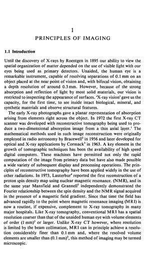
 Principles of Nuclear Magnetic Resonance Microscopy Paul T. Callaghan Department of Physics and Biophysics Massey University New Zealand CLARENDON PRESS OXFORD CONTENTS 1 PRINCIPLES OF IMAGING 1 1.1 Introduction
Field trip: Tuesday, Feb 5th
 Pulse Sequences Field trip: Tuesday, Feb 5th Hardware tour of VUIIIS Philips 3T Meet here at regular class time (11.15) Complete MRI screening form! Chuck Nockowski Philips Service Engineer Reminder: Project/Presentation
FREQUENCY SELECTIVE EXCITATION
 PULSE SEQUENCES FREQUENCY SELECTIVE EXCITATION RF Grad 0 Sir Peter Mansfield A 1D IMAGE Field Strength / Frequency Position FOURIER PROJECTIONS MR Image Raw Data FFT of Raw Data BACK PROJECTION Image Domain
Contrast Mechanisms in MRI. Michael Jay Schillaci
 Contrast Mechanisms in MRI Michael Jay Schillaci Overview Image Acquisition Basic Pulse Sequences Unwrapping K-Space Image Optimization Contrast Mechanisms Static and Motion Contrasts T1 & T2 Weighting,
DIFFUSION MAGNETIC RESONANCE IMAGING
 DIFFUSION MAGNETIC RESONANCE IMAGING from spectroscopy to imaging apparent diffusion coefficient ADC-Map anisotropy diffusion tensor (imaging) DIFFUSION NMR - FROM SPECTROSCOPY TO IMAGING Combining Diffusion
EL-GY 6813/BE-GY 6203 Medical Imaging, Fall 2016 Final Exam
 EL-GY 6813/BE-GY 6203 Medical Imaging, Fall 2016 Final Exam (closed book, 1 sheets of notes double sided allowed, no calculator or other electronic devices allowed) 1. Ultrasound Physics (15 pt) A) (9
Tissue Characteristics Module Three
 Tissue Characteristics Module Three 1 Equilibrium State Equilibrium State At equilibrium, the hydrogen vector is oriented in a direction parallel to the main magnetic field. Hydrogen atoms within the vector
Basic p rinciples COPYRIGHTED MATERIAL. Introduction. Atomic s tructure
 1 Basic p rinciples Introduction 1 Atomic structure 1 Motion in the atom 2 MR active nuclei 2 The hydrogen nucleus 4 Alignment 4 Precession 8 The Larmor equation 9 Introduction The basic principles of
Applications of Spin Echo and Gradient Echo: Diffusion and Susceptibility Contrast
 Applications of Spin Echo and Gradient Echo: Diffusion and Susceptibility Contrast Chunlei Liu, PhD Department of Electrical Engineering & Computer Sciences and Helen Wills Neuroscience Institute University
COPYRIGHTED MATERIAL. Production of Net Magnetization. Chapter 1
 Chapter 1 Production of Net Magnetization Magnetic resonance (MR) is a measurement technique used to examine atoms and molecules. It is based on the interaction between an applied magnetic field and a
Principles of Magnetic Resonance Imaging
 Principles of Magnetic Resonance Imaging Hi Klaus Scheffler, PhD Radiological Physics University of 1 Biomedical Magnetic Resonance: 1 Introduction Magnetic Resonance Imaging Contents: Hi 1 Introduction
Physics of MR Image Acquisition
 Physics of MR Image Acquisition HST-583, Fall 2002 Review: -MRI: Overview - MRI: Spatial Encoding MRI Contrast: Basic sequences - Gradient Echo - Spin Echo - Inversion Recovery : Functional Magnetic Resonance
Magnetic Resonance Imaging (MRI)
") Magnetic Resonance Imaging Introduction The Components The Technology (MRI) Physics behind MR Most slides taken from http:// www.slideworld.org/ viewslides.aspx/magnetic- Resonance-Imaging- %28MRI%29-MR-Imaging-
Magnetic Resonance Imaging Introduction The Components The Technology (MRI) Physics behind MR Most slides taken from http:// www.slideworld.org/ viewslides.aspx/magnetic- Resonance-Imaging- %28MRI%29-MR-Imaging-
K-space. Spin-Warp Pulse Sequence. At each point in time, the received signal is the Fourier transform of the object s(t) = M( k x
 = M( k x") Bioengineering 280A Principles of Biomedical Imaging Fall Quarter 2015 MRI Lecture 4 k (t) = γ 2π k y (t) = γ 2π K-space At each point in time, the received signal is the Fourier transform of the object
Bioengineering 280A Principles of Biomedical Imaging Fall Quarter 2015 MRI Lecture 4 k (t) = γ 2π k y (t) = γ 2π K-space At each point in time, the received signal is the Fourier transform of the object
The physics US and MRI. Prof. Peter Bogner
 The physics US and MRI Prof. Peter Bogner Sound waves mechanical disturbance, a pressure wave moves along longitudinal wave compression rarefaction zones c = nl, (c: velocity, n: frequency, l: wavelength
Magnetic Resonance Imaging in a Nutshell
 Magnetic Resonance Imaging in a Nutshell Oliver Bieri, PhD Department of Radiology, Division of Radiological Physics, University Hospital Basel Department of Biomedical Engineering, University of Basel,
Bloch Equations & Relaxation UCLA. Radiology
 Bloch Equations & Relaxation MRI Systems II B1 I 1 I ~B 1 (t) I 6 ~M I I 5 I 4 Lecture # Learning Objectives Distinguish spin, precession, and nutation. Appreciate that any B-field acts on the the spin
Magnetic Resonance Imaging
 Magnetic Resonance Imaging History Nuclear magnetic resonance was first described by Isidor Rabi in 1938 - Columbia University, New York City, (Nobel Prize Nobel Prize in Physics 1944) 1946 - Edward Mills
Tensor Visualization. CSC 7443: Scientific Information Visualization
 Tensor Visualization Tensor data A tensor is a multivariate quantity Scalar is a tensor of rank zero s = s(x,y,z) Vector is a tensor of rank one v = (v x,v y,v z ) For a symmetric tensor of rank 2, its
Physics and Brain Imaging
 Physics and Brain Imaging Nuclear Magnetic Resonance (NMR) Magnetic Resonance Imaging (MRI) Functional MRI (fmri) Talk at Quarknet FSU Summer Workshop, July 24, 2017 Per Arne Rikvold Leonardo da Vinci
Apodization. Gibbs Artifact. Bioengineering 280A Principles of Biomedical Imaging. Fall Quarter 2013 MRI Lecture 5. rect(k x )
") Bioengineering 280A Principles of Biomedical Imaging Fall Quarter 2013 MRI Lecture 5 GE Medical Systems 2003 Gibbs Artifact Apodization rect(k ) Hanning Window h(k )=1/2(1+cos(2πk ) 256256 image 256128
Bioengineering 280A Principles of Biomedical Imaging Fall Quarter 2013 MRI Lecture 5 GE Medical Systems 2003 Gibbs Artifact Apodization rect(k ) Hanning Window h(k )=1/2(1+cos(2πk ) 256256 image 256128
Chemistry 431. Lecture 23
 Chemistry 431 Lecture 23 Introduction The Larmor Frequency The Bloch Equations Measuring T 1 : Inversion Recovery Measuring T 2 : the Spin Echo NC State University NMR spectroscopy The Nuclear Magnetic
MRI in Review: Simple Steps to Cutting Edge Part I
 MRI in Review: Simple Steps to Cutting Edge Part I DWI is now 2 years old... Mike Moseley Radiology Stanford DWI, b = 1413 T2wt, 28/16 ASN 21 San Francisco + Disclosures: Funding NINDS, NCRR, NCI 45 minutes
Introductory MRI Physics
 C HAPR 18 Introductory MRI Physics Aaron Sodickson EXRNAL MAGNETIC FIELD, PROTONS AND EQUILIBRIUM MAGNETIZATION Much of the bulk of the magnetic resonance imaging (MRI) scanner apparatus is dedicated to
G Medical Imaging. Outline 4/13/2012. Physics of Magnetic Resonance Imaging
 G16.4426 Medical Imaging Physics of Magnetic Resonance Imaging Riccardo Lattanzi, Ph.D. Assistant Professor Department of Radiology, NYU School of Medicine Department of Electrical and Computer Engineering,
NMR/MRI examination (8N080 / 3F240)
") NMR/MRI examination (8N080 / 3F240) Remarks: 1. This test consists of 3 problems with at total of 26 sub-questions. 2. Questions are in English. You are allowed to answer them in English or Dutch. 3. Please
NMR/MRI examination (8N080 / 3F240) Remarks: 1. This test consists of 3 problems with at total of 26 sub-questions. 2. Questions are in English. You are allowed to answer them in English or Dutch. 3. Please
M R I Physics Course. Jerry Allison Ph.D., Chris Wright B.S., Tom Lavin B.S., Nathan Yanasak Ph.D. Department of Radiology Medical College of Georgia
 M R I Physics Course Jerry Allison Ph.D., Chris Wright B.S., Tom Lavin B.S., Nathan Yanasak Ph.D. Department of Radiology Medical College of Georgia M R I Physics Course Spin Echo Imaging Hahn Spin Echo
Part III: Sequences and Contrast
 Part III: Sequences and Contrast Contents T1 and T2/T2* Relaxation Contrast of Imaging Sequences T1 weighting T2/T2* weighting Contrast Agents Saturation Inversion Recovery JUST WATER? (i.e., proton density
Doppler echocardiography & Magnetic Resonance Imaging. Doppler echocardiography. History: - Langevin developed sonar.
 1 Doppler echocardiography & Magnetic Resonance Imaging History: - Langevin developed sonar. - 1940s development of pulse-echo. - 1950s development of mode A and B. - 1957 development of continuous wave
Spatial encoding in Magnetic Resonance Imaging. Jean-Marie BONNY
 Spatial encoding in Magnetic Resonance Imaging Jean-Marie BONNY What s Qu est an image ce qu une? image? «a reproduction of a material object by a camera or a related technique» Multi-dimensional signal
Spin Dynamics Basics of Nuclear Magnetic Resonance. Malcolm H. Levitt
 Spin Dynamics Basics of Nuclear Magnetic Resonance Second edition Malcolm H. Levitt The University of Southampton, UK John Wiley &. Sons, Ltd Preface xxi Preface to the First Edition xxiii Introduction
Magnetic Resonance Imaging
 http://www.qldxray.com.au/filelibrary/mri_cardiovascular_system_ca_0005.jpg Magnetic Resonance Imaging 1 Overview 1. The magnetic properties of nuclei, and how they behave in strong magnetic fields. 2.
Biomedical Imaging Magnetic Resonance Imaging
 Biomedical Imaging Magnetic Resonance Imaging Charles A. DiMarzio & Eric Kercher EECE 4649 Northeastern University May 2018 Background and History Measurement of Nuclear Spins Widely used in physics/chemistry
Classical Description of NMR Parameters: The Bloch Equations
 Classical Description of NMR Parameters: The Bloch Equations Pascale Legault Département de Biochimie Université de Montréal 1 Outline 1) Classical Behavior of Magnetic Nuclei: The Bloch Equation 2) Precession
10.4 Continuous Wave NMR Instrumentation
 10.4 Continuous Wave NMR Instrumentation coherent detection bulk magnetization the rotating frame, and effective magnetic field generating a rotating frame, and precession in the laboratory frame spin-lattice
Classical Description of NMR Parameters: The Bloch Equations
 Classical Description of NMR Parameters: The Bloch Equations Pascale Legault Département de Biochimie Université de Montréal 1 Outline 1) Classical Behavior of Magnetic Nuclei: The Bloch Equation 2) Precession
Technical University of Denmark
 Technical University of Denmark Page 1 of 10 pages Written test, 12 December 2012 Course name: Introduction to medical imaging Course no. 31540 Aids allowed: None. Pocket calculator not allowed "Weighting":
Relaxation times in nuclear magnetic resonance
 Relaxation times in TEP Related topics Nuclear spins, atomic nuclei with a magnetic moment, precession movement of the nuclear spins, Landau-Lifshitz equation, Bloch equation, magnetisation, resonance
Topics. Spin. The concept of spin Precession of magnetic spin Relaxation Bloch Equation
 Bioengineering 280A Principles of Biomedical Imaging Fall Quarter 2005 MRI Lecture 1 Topics The concept of spin Precession of magnetic spin Relaation Bloch Equation Spin Intrinsic angular momentum of elementary
How does this work? How does this method differ from ordinary MRI?
 361-Lec41 Tue 18nov14 How does this work? How does this method differ from ordinary MRI? NEW kinds of MRI (magnetic resononance imaging (MRI) Diffusion Magnetic Resonance Imaging Tractographic reconstruction
Diffusion-Weighted MRI may be used to measure the apparent diffusion coefficient of water in tissue.
 Specialty Area: MR Physics for Physicists Speaker: Jennifer A. McNab, Ph.D. Assistant Professor, Radiology, Stanford University () Highlights The Bloch-Torrey equation is a generalization of the Bloch
Chapter 7. Nuclear Magnetic Resonance Spectroscopy
 Chapter 7 Nuclear Magnetic Resonance Spectroscopy I. Introduction 1924, W. Pauli proposed that certain atomic nuclei have spin and magnetic moment and exposure to magnetic field would lead to energy level
The physics of medical imaging US, CT, MRI. Prof. Peter Bogner
 The physics of medical imaging US, CT, MRI Prof. Peter Bogner Clinical radiology curriculum blocks of lectures and clinical practice (7x2) Physics of medical imaging Neuroradiology Head and neck I. Head
CONTENTS. 2 CLASSICAL DESCRIPTION 2.1 The resonance phenomenon 2.2 The vector picture for pulse EPR experiments 2.3 Relaxation and the Bloch equations
 CONTENTS Preface Acknowledgements Symbols Abbreviations 1 INTRODUCTION 1.1 Scope of pulse EPR 1.2 A short history of pulse EPR 1.3 Examples of Applications 2 CLASSICAL DESCRIPTION 2.1 The resonance phenomenon
Magnetic Resonance Imaging. Qun Zhao Bioimaging Research Center University of Georgia
 Magnetic Resonance Imaging Qun Zhao Bioimaging Research Center University of Georgia The Nobel Prize in Physiology or Medicine 2003 "for their discoveries concerning magnetic resonance imaging" Paul C.
NMR, the vector model and the relaxation
 NMR, the vector model and the relaxation Reading/Books: One and two dimensional NMR spectroscopy, VCH, Friebolin Spin Dynamics, Basics of NMR, Wiley, Levitt Molecular Quantum Mechanics, Oxford Univ. Press,
Spatial encoding in Magnetic Resonance Imaging. Jean-Marie BONNY
 Spatial encoding in Magnetic Resonance Imaging Jean-Marie BONNY What s Qu est an image ce qu une? image? «a reproduction of a material object by a camera or a related technique» Multi-dimensional signal
Exam 8N080 - Introduction to MRI
 Exam 8N080 - Introduction to MRI Friday April 10 2015, 18.00-21.00 h For this exam you may use an ordinary calculator (not a graphical one). In total there are 5 assignments and a total of 50 points can
Biophysical Chemistry: NMR Spectroscopy
 Spin Dynamics & Vrije Universiteit Brussel 25th November 2011 Outline 1 Pulse/Fourier Transform NMR Thermal Equilibrium Effect of RF Pulses The Fourier Transform 2 Symmetric Exchange Between Two Sites
Index. p, lip, 78 8 function, 107 v, 7-8 w, 7-8 i,7-8 sine, 43 Bo,94-96
 p, lip, 78 8 function, 107 v, 7-8 w, 7-8 i,7-8 sine, 43 Bo,94-96 B 1,94-96 M,94-96 B oro!' 94-96 BIro!' 94-96 I/r, 79 2D linear system, 56 2D FFT, 119 2D Fourier transform, 1, 12, 18,91 2D sinc, 107, 112
Sketch of the MRI Device
 Outline for Today 1. 2. 3. Introduction to MRI Quantum NMR and MRI in 0D Magnetization, m(x,t), in a Voxel Proton T1 Spin Relaxation in a Voxel Proton Density MRI in 1D MRI Case Study, and Caveat Sketch
NMR Spectroscopy of Polymers
 UNESCO/IUPAC Course 2005/2006 Jiri Brus NMR Spectroscopy of Polymers Brus J 1. part At the very beginning the phenomenon of nuclear spin resonance was studied predominantly by physicists and the application
The Physical Basis of Nuclear Magnetic Resonance Part I ESMRMB. Jürgen R. Reichenbach
 The Physical Basis of Nuclear agnetic Resonance Part I Jürgen R. Reichenbach odule 1 October 17, 216 Outline of odule Introduction Spin and magnetic moment Spin precession, Larmor frequency agnetic properties
BASIC MRI PHYSICS SPIN GYMNASTICS Don Plewes PhD, Walter Kucharczyk MD
 BASIC MRI PHYSICS SPIN GYMNASTICS Don Plewes PhD, Walter Kucharczyk MD Introduction To understand MRI, it is first necessary to understand the physics of proton Nuclear Magnetic Resonance (NMR). The most
Magnetic Resonance Imaging in Medicine
 Institute for Biomedical Engineering University and ETH Zurich Gloriastrasse 35 CH- 8092 Zurich Switzerland Magnetic Resonance Imaging in Medicine D. Meier, P. Boesiger, S. Kozerke 2012 All rights reserved.
Predictive Diagnosis of Alzheimer s Disease using Diffusion MRI
 Predictive Diagnosis of Alzheimer s Disease using Diffusion MRI by Syeda Maryam A thesis presented to the University of Waterloo in fulfillment of the thesis requirement for the degree of Master of Applied
Anisotropy of HARDI Diffusion Profiles Based on the L 2 -Norm
 Anisotropy of HARDI Diffusion Profiles Based on the L 2 -Norm Philipp Landgraf 1, Dorit Merhof 1, Mirco Richter 1 1 Institute of Computer Science, Visual Computing Group, University of Konstanz philipp.landgraf@uni-konstanz.de
Magnetic resonance imaging MRI
 Magnetic resonance imaging MRI Introduction What is MRI MRI is an imaging technique used primarily in medical settings that uses a strong magnetic field and radio waves to produce very clear and detailed
Spectral Broadening Mechanisms
 Spectral Broadening Mechanisms Lorentzian broadening (Homogeneous) Gaussian broadening (Inhomogeneous, Inertial) Doppler broadening (special case for gas phase) The Fourier Transform NC State University
Technical University of Denmark
 Technical University of Denmark Page 1 of 11 pages Written test, 9 December 2010 Course name: Introduction to medical imaging Course no. 31540 Aids allowed: none. "Weighting": All problems weight equally.
Diffusion Imaging II. By: Osama Abdullah
 iffusion Imaging II By: Osama Abdullah Review Introduction. What is diffusion? iffusion and signal attenuation. iffusion imaging. How to capture diffusion? iffusion sensitizing gradients. Spin Echo. Gradient
NMR Spectroscopy: A Quantum Phenomena
 NMR Spectroscopy: A Quantum Phenomena Pascale Legault Département de Biochimie Université de Montréal Outline 1) Energy Diagrams and Vector Diagrams 2) Simple 1D Spectra 3) Beyond Simple 1D Spectra 4)
Topics. The History of Spin. Spin. The concept of spin Precession of magnetic spin Relaxation
 Topics Bioengineering 280A Principles of Biomedical Imaging Fall Quarter 2008 MRI Lecture 1 The concept of spin Precession of magnetic spin Relaation Spin The History of Spin Intrinsic angular momentum
Developing a Method for Distortion Correction in High b-value Diffusion-Weighted Magnetic Resonance Imaging HENRIK HANSSON
 Developing a Method for Distortion Correction in High b-value Diffusion-Weighted Magnetic Resonance Imaging Master s thesis in Complex Adaptive Systems HENRIK HANSSON Department of Applied Physics Division
Generalizing Diffusion Tensor Model Using Probabilistic Inference in Markov Random Fields
 Generalizing Diffusion Tensor Model Using Probabilistic Inference in Markov Random Fields Çağatay Demiralp and David H. Laidlaw Brown University Providence, RI, USA Abstract. We give a proof of concept
Part II: Magnetic Resonance Imaging (MRI)
") Part II: Magnetic Resonance Imaging (MRI) Contents Magnetic Field Gradients Selective Excitation Spatially Resolved Reception k-space Gradient Echo Sequence Spin Echo Sequence Magnetic Resonance Imaging
Part II: Magnetic Resonance Imaging (MRI) Contents Magnetic Field Gradients Selective Excitation Spatially Resolved Reception k-space Gradient Echo Sequence Spin Echo Sequence Magnetic Resonance Imaging
Nuclear magnetic resonance in condensed matter
 University of Ljubljana Faculty of mathematics and physics Physics department SEMINAR Nuclear magnetic resonance in condensed matter Author: Miha Bratkovič Mentor: prof. dr. Janez Dolinšek Ljubljana, October
Lecture 02 Nuclear Magnetic Resonance Spectroscopy Principle and Application in Structure Elucidation
 Application of Spectroscopic Methods in Molecular Structure Determination Prof. S. Sankararaman Department of Chemistry Indian Institution of Technology Madras Lecture 02 Nuclear Magnetic Resonance Spectroscopy
Diffusion Tensor Imaging I: The basics. Jennifer Campbell
 Diffusion Tensor Imaging I: The basics Jennifer Campbell Diffusion Tensor Imaging I: The basics Jennifer Campbell Diffusion Imaging MRI: many different sources of contrast T1W T2W PDW Perfusion BOLD DW
2.1.1 A Brief History of NMR The conception of NMR sprouted after the Pauli s prediction of nuclear spin in
 CHAPTER--2 BASICS OF NMR IMAGING AND SPECTROSCOPY 2.1 Introduction 2.1.1 A Brief History of NMR The conception of NMR sprouted after the Pauli s prediction of nuclear spin in 1924. Later Gorter (1936)
Chem 325 NMR Intro. The Electromagnetic Spectrum. Physical properties, chemical properties, formulas Shedding real light on molecular structure:
 Physical properties, chemical properties, formulas Shedding real light on molecular structure: Wavelength Frequency ν Wavelength λ Frequency ν Velocity c = 2.998 10 8 m s -1 The Electromagnetic Spectrum
III, Diffusion, and Susceptibility. August 25, Departments of Mathematics and Applied Math and Computational Science University of Pennsylvania
 III,, and Departments of Mathematics and Applied Math and Computational Science University of Pennsylvania August 25, 2010 Copyright Page All material in this lecture, except as noted within the text,
Medical Imaging Physics Spring Quarter Week 9-1
 Medical Imaging Physics Spring Quarter Week 9-1 NMR and MRI Davor Balzar balzar@du.edu www.du.edu/~balzar Intro MRI Outline NMR & MRI Guest lecturer fmri Thursday, May 22 Visit to CUHSC It s not mandatory
Introduction to the Physics of NMR, MRI, BOLD fmri
 Pittsburgh, June 13-17, 2011 Introduction to the Physics of NMR, MRI, BOLD fmri (with an orientation toward the practical aspects of data acquisition) Pittsburgh, June 13-17, 2001 Functional MRI in Clinical
NMR Imaging in porous media
 NMR Imaging in porous media What does NMR give us. Chemical structure. Molecular structure. Interactions between atoms and molecules. Incoherent dynamics (fluctuation, rotation, diffusion). Coherent flow
Chapter 1 Pulsed Field Gradient NMR Sequences
 Chapter 1 Pulsed Field Gradient NMR Sequences Abstract The mechanism via which an NMR signal is generated and how diffusion can be measured from solving the equation of motion of the nuclear spins is described.