Diffusion Imaging II. By: Osama Abdullah
|
|
|
- Letitia Weaver
- 5 years ago
- Views:
Transcription
1 iffusion Imaging II By: Osama Abdullah
2 Review Introduction. What is diffusion? iffusion and signal attenuation. iffusion imaging. How to capture diffusion? iffusion sensitizing gradients. Spin Echo. Gradient Echo. Quantitative description. What is the b-value? High b-value problems. iffusion imaging pulse sequence. Pulsed Gradient Spin Echo. Single shot EPI. RARE RARE with crushers Split Acquisition of fast spin echo (SPLICE) diffusion preparation. Split echoes of FSE or STEAM. iffusion basics. Einstein equation. Factors that affect diffusion. iffusion tensor. Anisotropic vs. isotropic diffusion. iffusion Imaging techniques: Introduction Family of techniques iffusion weighted imaging (WI). Quantitative apparent diffusion coefficient (AC). Ref: Handbook of MRI pulse sequences (P and ) 2
3 Review iffusion weighted (W) image An MRI image acquired in the presence of diffusion weighting gradient (at single b-value). S=S exp(-b) Its contrast depends on the direction of the applied gradient. To remove orientation dependence, three W images with gradients applied along three orthogonal directions can be geometrically averaged to give an Isotropic (or Trace weighted) diffusion weighted image S xyz = 3 S x S y S z = S e b(xx + yy + zz) / 3 = S e b trace / 3 3
4 Apparent diffusion coefficient (AC) Constructed from more than one W image (at least two W images), with different b-values. S 1 b1 b2 = Se,S2 = Se...S n = S e b n The first image should be calculated without diffusion weighting (S ) and the rest with different b-values (i.e., different gradients amplitudes in the same direction). Then the AC map can be constructed by means of Linear fitting (approximation) or a Nonlinear fitting. The resulted AC map contrast is inverted compared to W image 4

5 W image AC map 5
6 Biexponential ecay In some tissues (e.g. brain), diffusion signal follows a biexponential signal decay according to: f S = S ( ζe + ( 1 b b ζ)e s ) Where: ξ and (1-ξ) are the compartmental fractions. f is the fast diffusing component. s is the slow diffusing component. 6
7 Lecture 2 outline Family of diffusion techniques, cont d iffusion tensor imaging Quantitative description. Finding the diffusion tensor. Scalar and vector parameters extracted from the diffusion tensor (RA, FA, M, PE, and Tractography) q-space imaging Quatitative description. efinition of q. How to conduct a q-space experiment? Quick paper discussion: Assaf et al 22: High b-value q-space analyzed diffusion-weighted MRI: Application to multiple sclerosis. Magn Reson Med 47: , 22 Wiley-Liss, Inc. 7
8 III. iffusion Tensor Imaging (TI) Many tissue structures imposes a nonspherical geometry on water diffusion boundaries, i.e., diffusion anisotropy. To fully describe diffusion anisotropy, diffusion tensor notation is used. = xx xy xz It maps diffusion anisotropy at each spatial location, xy yy yz xz yz zz 8
9 9 iffusion tensor = zz yz xz yz yy xy xz xy xx = Eigenvalues


10 1 iffusion models Isotropic No orientation dependence. Anisotropic iffusion depends on spatial orientation. = = 3 2 1
11 Stejskal and Tanner (1965) solved Bloch equation with diffusion weighting in a pulsed gradient spin echo (PGSE) experiment, which made this sequence amenable for describing free diffusion in anisotropic medium. 11
12 They related the applied gradient, G(t) G j (t) = G j [u j v j w j ] and γ k(t) = 2π to the echo intensity, S, in a spin echo experiment according to: t o ' G(t )dt ' S S TE ' ' TE TE T ' ' = exp( [k(t ) 2U(t )k( )] [k(t ) 2U(t TE TE ' )k( )]dt ) 2 U is the step function (unit Heaviside function) [uj vj wj] are unit vectors in the direction of the applied gradient field. 12
13 Basser, Mattiello and Le Bilhan (1994) defined the effective diffusivity tensor, eff, to be the mean value of the exponent in Stejskal s equation. Re-writing it in term of the new parameter: S S = exp( 4π 2 TE k j. eff T. k j dt) ' γ k = π j(t ) G j(t 2 t o ' ) dt ' 13
14 Neglecting the contribution from imaging gradients, we obtain the previous equation in matrix form: S S j IM j = exp( b.[u = ln(s j / S j ) v = j w j]. bq j xx xy xz xy yy yz xz yz zz uj. vj ) wj Q j is the quadratic form of the diffusion tensor matrix. G j = G [u j v j w j ] and b = c 1.G c 1 = γ 2 [ δ 2 ( Δ δ ) ζ 3 δξ 6 2 ] 14
15 Calculating the diffusion tensor Requires at least two b-values, one of them without diffusion weighting (b~). Acquire six W images with gradients applied along at least six noncolinear, noncoplanar directions at any nonzero b-values. In total, at least seven images are needed: one with b= which gives S, and six images with nonzero b-values, giving S 1, S 2,, S 6 15
16 We have a system of six linear equations with six unknowns, (the six independent elements of ): IM j = b.[u j v j w j]. xx xy xz xy yy yz xz yz zz uj. vj wj Solving this system of linear equations will yield the diffusion tensor at each voxel. 16
17 iffusion tensor anisotropy-maps Scalar maps: Can be exploited in Visualizing location, size and integrity of fibrous structures (white matter). Examples: Mean diffusivity, relative anisotropy (RA), fractional anisotropy (FA) and Volume ratio (VR) Vector maps Provide information on diffusion spatial orientation that can infer fiber tracts connectivity. Examples: Eigenvectors and Tractography. 17
18 Scalar diffusion anisotropy calculations In order to remove patient orientation dependence, the diffusion tensor matrix will not be used directly, instead, its eigenvalues will be used. =
19 Maps: Apparent diffusion coefficient AC 1 = 1, AC 2 = 2, AC 3 = 3 AC 1 /AC 2 or max[ac 1,AC 2, AC 3 ]/min[ac 1, AC 2, AC 3 ] Trace weighted image Trace() = Mean diffusivity = Trace() / 3 19
 due to anisotropic diffusion effects, the use of images weighted by the trace of")
20 TI in acute stroke (7 hours). Acute ischemic regions appear bright in diffusion-weighted images. As normal white matter regions may also appear bright in some orientations (indicated by the bold arrows) due to anisotropic diffusion effects, the use of images weighted by the trace of the diffusion tensor is more adequate (the ischemic lesion is indicated by the thin arrow). 2
21 Relative Anisotropy (RA) Normalized standard deviation: Represents the ratio of the anisotropic part of to its isotropic part. Ranges from (isotropic diffusion) to 2 1/2 (infinite anisotropic) Fractional Anisotropy (FA) Normalized standard deviation: FA measures the fraction of the magnitude of that can be ascribed to anisotropic diffusion Ranges from (isotropic diffusion) to 1 (infinite anisotropic) 21
22 Volume Ratio (VR) Represents the ratio of the ellipsoid volume to the volume of a sphere of radius avg. Ranges from 1 (isotropic diffusion) to (infinite anisotropic), hence, (1-VR) may be used. 22
. c: Volume ratio index (VR).")
23 Comparison of various anisotropy indices. a: Simple anisotropy index given by: (/ avg -1)/2. b: Relative anisotropy index (RA). c: Volume ratio index (VR). d: Fractional anisotropy index (FA). 23

24 Principal eigenvector Two-dimensional display of the diffusion tensor. A: Projection of the main eigenvector on a high-resolution T2-weighted image. B: Representation of the main eigenvector direction using a color scale (red = x axis, green = y axis, blue = z axis). 24
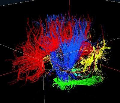
25 Tractography Maps the tissue fiber orientation and connectivity in three directions, in vivo. The assumption is that the direction of the fibers is colinear with the direction of the eigen-vector, λ 1,associated with the largest eigen diffusivity. 25
26 Quantitative escription It was proposed that a white matter fiber tract trajectory could be represented as a 3 space curve, i.e., a vector, h(r), parameterized by the arc length, r, of the trajectory. The Frenet equation describing the evolution of h(r) is: dh (r) dr = t(r) Where t(r) is a unit tangent vector to h(r) at r. 26
27 λ 1 Let be the eigenvector associated with the largest eigenvalue at location r. λ 1 Since is assumed to be parallel to the fiber tracts, then it is equal to the tangent of the 3 space curve h(r): λ 1 = t(r) = dh(r) dr Solving this equation can be done using iterative methods based on Euler s Method or more robustly Runge-Kutta Method, to interpolate the h(r) curve. 27
28 Usually, color mapping is used to represent the tract directions: Red: left-right Green: anterior-posterior Blue: Superior-inferior 28

29 Upper left panel displays fiber tractography combined with cortical thickness map obtained with Free Surfer. Upper right panel demonstrates a sagittal cross-sectional view of brain parenchyma segmented into white and gray matter combined with fiber tractography map. Lower left figure shows a coronal cross-sectional view of automatic parcellation of white matter overplayed with the gray matter surface. Lower right figure shows the parcellation of gray matter surface and corresponding white matter fibers. 29

30 3
31 IV. q-space imaging Proposed by. Cory and N. Garroway (199) and P. Callaghan (1991). It can provide structural information on samples with a higher spatial resolution compared to an MR image. What is q-space? 31
32 What is k-space? Fourier transformation of MRI image (spin density) in the spatial domain with respect to k-vector (1/cm), defined as : γ k(t) = π G d(t 2 t o Yielding the Echo Intensity S(k) ' ) dt ' What is q-space? Fourier transformation of the the iplacement Probability Profile, P s (R,Δ), with respect to reciprocal spatial vector, q (1/cm), defined as: γ q = δg d 2 π Yielding the Echo Intensity S(q) 32
33 The Stejskal s equation, with a rectangular pulse, δ S / S = exp( b) = exp[ γ g δ ( Δ / 3)] Using the definition of q, We get: γ q = δg d 2 π 2 2 δ S(q) / S = exp[ 4π q ( Δ / 3)] 33
34 Taking the FFT of the previous equation, we will get the isplacement Probability profile: F(r) = FT{S(q)} = α.exp(-ß.r 2 ) r is the Fourier conjugate of q α and ß are diffusion dependent parameters The displacement probability profile is a Gaussian profile. 34
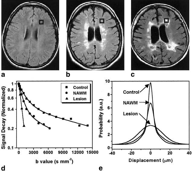
35 How to conduct a q-space experiment? Acquire a set of W images (16 for example), with increasing the gradient amplitude. The applied gradients must be in the same direction. For the diffusion weighting gradient: Short pulse gradient approximation (or Long time scale limit) should be met. δ<<δ 35
36 36
37 Paper iscussion High b-value q-space analyzed diffusion-weighted MRI: Application to multiple sclerosis By: Y. Assaf, et al 22. Magnetic Resonance in Medicine 47: , Wiley- Liss, Inc. 37
38 38
39 Multiple Sclerosis (MS) MS is an autoimmune-mediated disease of the Central Nervous System (CNS) characterized by: 1. emyelination of axons 2. Focal inflammatory reactions in the MS lesions 39
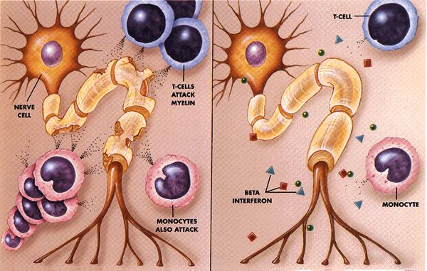
40 4
41 Consequences of destroying the Myelin Sheath: Progressive decline of motor and sensory functions. Permanent disability. 41
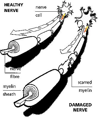
42 42
43 Imaging modalities that supports the clinical diagnosis of MS: MRI: T 1 -IR weighted MRI. T 2 Fluid-Attenuated Inversion Recovery (FLAIR). iffusion Tensor Imaging (TI). Problems: Normal Appearing White Matter (NAWM). New imaging technique is needed. 43
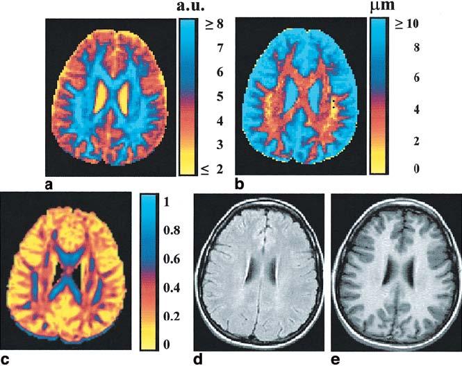
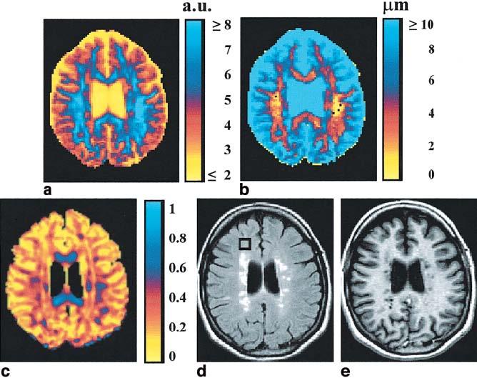
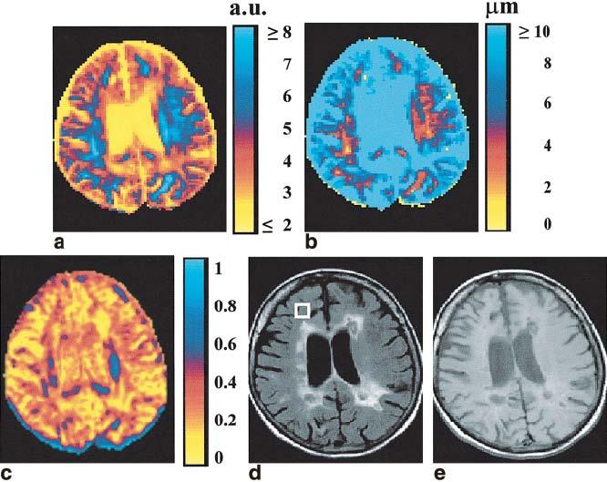
44 MS and q-space 44
45 45
46 46
47 47
48 48
49 Conclusion The q-space analysis provides images displaying widespread disease load which provide evidence of abnormalities in the NAWM of MS patients that are not detected by FLAIR, T1, or even conventional TI. 49
50 References [1] Stejskal EO. Use of spin echoes in a pulsed magnetic-field gradient to study restricted diffusion and flow. J Chem Phys 1965; 43; [2] E. O. Stejskal, J. E. Tanner. Spin echoes in the presence of a time-dependent field gradient. J Chem Phys 1965; 42: [3] enis Le Bilhan, et al. iffusion Tensor Imaging: Concepts and Applications. J Magn Reson Imaging,13: (21). [4] P. Basser, et al, Estimation of the effective self-diffusion tensor from an NMR spin echo. J of Magn Reson, Sereis B 13, (1994). [5] P. Basser, et al, A simplified method to measure the diffusion tensor from seven MR images. MRM 39: (1998) [6]. G. Cory, A. N. Garroway. Measurement of translational displacement probability by NMR: and indicator of compartmentation. Magn Reson Med 14: , 199. [7] P. Callaghan, NMR imaging, NMR diffraction and applications of pulsed gradient spin echoes in porous media. Magn Reson Imaging,Vol 14 Nos. 7/8, pp , [8] Assaf et al 22: High b-value q-space analyzed diffusion-weighted MRI: Application to multiple sclerosis. Magn Reson Med 47: , 22 Wiley-Liss, Inc. [9] Y. Assaf, A. Mayk. isplacement Imaging of Spinal Cord Using q-space iffusion- Weighted MRI.. Magn Reson Med 44: , 2. 5
Diffusion Tensor Imaging (DTI): An overview of key concepts
: An overview of key concepts") Diffusion Tensor Imaging (DTI): An overview of key concepts (Supplemental material for presentation) Prepared by: Nadia Barakat BMB 601 Chris Conklin Thursday, April 8 th 2010 Diffusion Concept [1,2]:
Diffusion Tensor Imaging (DTI): An overview of key concepts (Supplemental material for presentation) Prepared by: Nadia Barakat BMB 601 Chris Conklin Thursday, April 8 th 2010 Diffusion Concept [1,2]:
DIFFUSION MAGNETIC RESONANCE IMAGING
 DIFFUSION MAGNETIC RESONANCE IMAGING from spectroscopy to imaging apparent diffusion coefficient ADC-Map anisotropy diffusion tensor (imaging) DIFFUSION NMR - FROM SPECTROSCOPY TO IMAGING Combining Diffusion
Tensor Visualization. CSC 7443: Scientific Information Visualization
 Tensor Visualization Tensor data A tensor is a multivariate quantity Scalar is a tensor of rank zero s = s(x,y,z) Vector is a tensor of rank one v = (v x,v y,v z ) For a symmetric tensor of rank 2, its
Applications of Spin Echo and Gradient Echo: Diffusion and Susceptibility Contrast
 Applications of Spin Echo and Gradient Echo: Diffusion and Susceptibility Contrast Chunlei Liu, PhD Department of Electrical Engineering & Computer Sciences and Helen Wills Neuroscience Institute University
HST.583 Functional Magnetic Resonance Imaging: Data Acquisition and Analysis Fall 2006
 MIT OpenCourseWare http://ocw.mit.edu HST.583 Functional Magnetic Resonance Imaging: Data Acquisition and Analysis Fall 2006 For information about citing these materials or our Terms of Use, visit: http://ocw.mit.edu/terms.
Diffusion Weighted MRI. Zanqi Liang & Hendrik Poernama
 Diffusion Weighted MRI Zanqi Liang & Hendrik Poernama 1 Outline MRI Quick Review What is Diffusion MRI? Detecting Diffusion Stroke and Tumor Detection Presenting Diffusion Anisotropy and Diffusion Tensor
Ordinary Least Squares and its applications
 Ordinary Least Squares and its applications Dr. Mauro Zucchelli University Of Verona December 5, 2016 Dr. Mauro Zucchelli Ordinary Least Squares and its applications December 5, 2016 1 / 48 Contents 1
Diffusion Tensor Imaging I. Jennifer Campbell
 Diffusion Tensor Imaging I Jennifer Campbell Diffusion Imaging Molecular diffusion The diffusion tensor Diffusion weighting in MRI Alternatives to the tensor Overview of applications Diffusion Imaging
醫用磁振學 MRM 擴散張量影像 擴散張量影像原理. 本週課程內容 MR Diffusion 擴散張量造影原理 擴散張量造影應用 盧家鋒助理教授國立陽明大學生物醫學影像暨放射科學系
 本週課程內容 http://www.ym.edu.tw/~cflu 擴散張量造影原理 擴散張量造影應用 醫用磁振學 MRM 擴散張量影像 盧家鋒助理教授國立陽明大學生物醫學影像暨放射科學系 alvin4016@ym.edu.tw MRI The Basics (3rd edition) Chapter 22: Echo Planar Imaging MRI in Practice, (4th edition)
Medical Visualization - Tensor Visualization. J.-Prof. Dr. Kai Lawonn
 Medical Visualization - Tensor Visualization J.-Prof. Dr. Kai Lawonn Lecture is partially based on the lecture by Prof. Thomas Schultz 2 What is a Tensor? A tensor is a multilinear transformation that
Quantitative Metrics for White Matter Integrity Based on Diffusion Tensor MRI Data. Stephanie Lee
 Quantitative Metrics for White Matter Integrity Based on Diffusion Tensor MRI Data Stephanie Lee May 5, 2005 Quantitative Metrics for White Matter Integrity Based on Diffusion Tensor MRI Data ABSTRACT
A Neurosurgeon s Perspectives of Diffusion Tensor Imaging(DTI) Diffusion Tensor MRI (DTI) Background and Relevant Physics.
 Diffusion Tensor MRI (DTI) Background and Relevant Physics.") A Neurosurgeon s Perspectives of Diffusion Tensor Imaging(DTI) Kalai Arasu Muthusamy, D.Phil(Oxon) Senior Lecturer & Consultant Neurosurgeon. Division of Neurosurgery. University Malaya Medical Centre.
A Neurosurgeon s Perspectives of Diffusion Tensor Imaging(DTI) Kalai Arasu Muthusamy, D.Phil(Oxon) Senior Lecturer & Consultant Neurosurgeon. Division of Neurosurgery. University Malaya Medical Centre.
Bayesian multi-tensor diffusion MRI and tractography
 Bayesian multi-tensor diffusion MRI and tractography Diwei Zhou 1, Ian L. Dryden 1, Alexey Koloydenko 1, & Li Bai 2 1 School of Mathematical Sciences, Univ. of Nottingham 2 School of Computer Science and
Advanced Topics and Diffusion MRI
 Advanced Topics and Diffusion MRI Slides originally by Karla Miller, FMRIB Centre Modified by Mark Chiew (mark.chiew@ndcn.ox.ac.uk) Slides available at: http://users.fmrib.ox.ac.uk/~mchiew/teaching/ MRI
Diffusion Tensor Imaging I: The basics. Jennifer Campbell
 Diffusion Tensor Imaging I: The basics Jennifer Campbell Diffusion Tensor Imaging I: The basics Jennifer Campbell Diffusion Imaging MRI: many different sources of contrast T1W T2W PDW Perfusion BOLD DW
Diffusion-Weighted MRI may be used to measure the apparent diffusion coefficient of water in tissue.
 Specialty Area: MR Physics for Physicists Speaker: Jennifer A. McNab, Ph.D. Assistant Professor, Radiology, Stanford University () Highlights The Bloch-Torrey equation is a generalization of the Bloch
Diffusion Magnetic Resonance Imaging Part 1: Theory & Methods
 Diffusion Magnetic Resonance Imaging Part 1: Theory & Methods Benjamin M. Ellingson, Ph.D. Assistant Professor of Radiology, Biomedical Physics and Bioengineering Dept. of Radiological Sciences UCLA Neuro-Oncology
The effect of different number of diffusion gradients on SNR of diffusion tensor-derived measurement maps
 J. Biomedical Science and Engineering, 009,, 96-101 The effect of different number of diffusion gradients on SNR of diffusion tensor-derived measurement maps Na Zhang 1, Zhen-Sheng Deng 1*, Fang Wang 1,
Diffusion Tensor Imaging tutorial
 NA-MIC http://na-mic.org Diffusion Tensor Imaging tutorial Sonia Pujol, PhD Surgical Planning Laboratory Harvard University DTI tutorial This tutorial is an introduction to the advanced Diffusion MR capabilities
Contrast Mechanisms in MRI. Michael Jay Schillaci
 Contrast Mechanisms in MRI Michael Jay Schillaci Overview Image Acquisition Basic Pulse Sequences Unwrapping K-Space Image Optimization Contrast Mechanisms Static and Motion Contrasts T1 & T2 Weighting,
Diffusion Tensor Imaging (DTI) e Neurite Orientation Dispersion and Density Imaging (NODDI)
 e Neurite Orientation Dispersion and Density Imaging (NODDI)") Diffusion Tensor Imaging (DTI) e Neurite Orientation Dispersion and Density Imaging (NODDI) Claudia AM Gandini Wheeler-Kingshott, PhD Prof. of MRI Physics Overview Diffusion and microstructure NODDI theoretical
Diffusion Tensor Imaging (DTI) e Neurite Orientation Dispersion and Density Imaging (NODDI) Claudia AM Gandini Wheeler-Kingshott, PhD Prof. of MRI Physics Overview Diffusion and microstructure NODDI theoretical
Diffusion imaging of the brain: technical considerations and practical applications
 Diffusion imaging of the brain: technical considerations and practical applications David G. Norris FC Donders Centre for Cognitive Neuroimaging Nijmegen Sustaining the physiologist in measuring the atomic
Diffusion Tensor Imaging in Humans: Practical Implications for Neuroanatomy
 Diffusion Tensor Imaging in Humans: Practical Implications for Neuroanatomy Collaborators Center for Morphometric Analysis: Nikos Makris Andy Worth Verne S. Caviness George Papadimitriou MGH-NMR Center
Diffusion tensor imaging: brain pathway reconstruction
 Neda Sepasian, Jan ten Thije Boonkkamp, Anna Vilanova Diffusion tensor imaging: brain pathway reconstruction NAW 5/6 nr. 4 december 205 259 Neda Sepasian Department of Biomedical Engineering Eindhoven
CIND Pre-Processing Pipeline For Diffusion Tensor Imaging. Overview
 CIND Pre-Processing Pipeline For Diffusion Tensor Imaging Overview The preprocessing pipeline of the Center for Imaging of Neurodegenerative Diseases (CIND) prepares diffusion weighted images (DWI) and
Anisotropy of HARDI Diffusion Profiles Based on the L 2 -Norm
 Anisotropy of HARDI Diffusion Profiles Based on the L 2 -Norm Philipp Landgraf 1, Dorit Merhof 1, Mirco Richter 1 1 Institute of Computer Science, Visual Computing Group, University of Konstanz philipp.landgraf@uni-konstanz.de
Physics of MR Image Acquisition
 Physics of MR Image Acquisition HST-583, Fall 2002 Review: -MRI: Overview - MRI: Spatial Encoding MRI Contrast: Basic sequences - Gradient Echo - Spin Echo - Inversion Recovery : Functional Magnetic Resonance
Characterizing Non-Gaussian Diffusion by Using Generalized Diffusion Tensors
 Magnetic Resonance in Medicine 51:924 937 (2004) Characterizing Non-Gaussian Diffusion by Using Generalized Diffusion Tensors Chunlei Liu, 1,2 Roland Bammer, 1 Burak Acar, 3 and Michael E. Moseley 1 *
Generalizing Diffusion Tensor Model Using Probabilistic Inference in Markov Random Fields
 Generalizing Diffusion Tensor Model Using Probabilistic Inference in Markov Random Fields Çağatay Demiralp and David H. Laidlaw Brown University Providence, RI, USA Abstract. We give a proof of concept
III, Diffusion, and Susceptibility. August 25, Departments of Mathematics and Applied Math and Computational Science University of Pennsylvania
 III,, and Departments of Mathematics and Applied Math and Computational Science University of Pennsylvania August 25, 2010 Copyright Page All material in this lecture, except as noted within the text,
NMR Advanced methodologies to investigate water diffusion in materials and biological systems
 NMR Advanced methodologies to investigate water diffusion in materials and biological systems PhD Candidate _Silvia De Santis PhD Supervisors _dott. Silvia Capuani _prof. Bruno Maraviglia Outlook Introduction:
Application of diffusion MRI to cancer, heart and brain connectome imaging
 Colloquium @ Department of Physics, NTU Application of diffusion MRI to cancer, heart and brain connectome imaging March 11, 2014 Wen-Yih Isaac Tseng MD, PhD Advanced Biomedical MRI Lab Center for Optoelectronic
Basic Pulse Sequences I Saturation & Inversion Recovery UCLA. Radiology
 Basic Pulse Sequences I Saturation & Inversion Recovery Lecture #5 Learning Objectives Explain what the most important equations of motion are for describing spin systems for MRI. Understand the assumptions
Higher Order Cartesian Tensor Representation of Orientation Distribution Functions (ODFs)
") Higher Order Cartesian Tensor Representation of Orientation Distribution Functions (ODFs) Yonas T. Weldeselassie (Ph.D. Candidate) Medical Image Computing and Analysis Lab, CS, SFU DT-MR Imaging Introduction
Higher Order Cartesian Tensor Representation of Orientation Distribution Functions (ODFs) Yonas T. Weldeselassie (Ph.D. Candidate) Medical Image Computing and Analysis Lab, CS, SFU DT-MR Imaging Introduction
PhD THESIS. prepared at INRIA Sophia Antipolis
 PhD THESIS prepared at INRIA Sophia Antipolis and presented at the University of Nice-Sophia Antipolis Graduate School of Information and Communication Sciences A dissertation submitted in partial satisfaction
Advanced MRI: Diffusion MRI 1: DTI and k-space
 k y Advanced MRI: Diffusion MRI 1: DTI and k-space k X Eric Sigmund, PhD February 26th, 2013 LECTURE 1 Neuro Diffusion MRI 3-5 m White matter axons Body 15 m Renal medulla Musculoskeletal 50 m Skeletal
Research Article Thalamus Segmentation from Diffusion Tensor Magnetic Resonance Imaging
 Biomedical Imaging Volume 2007, Article ID 90216, 5 pages doi:10.1155/2007/90216 Research Article Thalamus Segmentation from Diffusion Tensor Magnetic Resonance Imaging Ye Duan, Xiaoling Li, and Yongjian
FREQUENCY SELECTIVE EXCITATION
 PULSE SEQUENCES FREQUENCY SELECTIVE EXCITATION RF Grad 0 Sir Peter Mansfield A 1D IMAGE Field Strength / Frequency Position FOURIER PROJECTIONS MR Image Raw Data FFT of Raw Data BACK PROJECTION Image Domain
Master of Science Thesis. Using q-space Diffusion MRI for Structural Studies of a Biological Phantom at a 3T Clinical Scanner
 Master of Science Thesis Using q-space Diffusion MRI for Structural Studies of a Biological Phantom at a 3T Clinical Scanner Anna Rydhög Supervisor: Sara Brockstedt, Jimmy Lätt Medical Radiation Physics
Observation of Microscopic Diffusion Anisotropy in the Spinal Cord Using Double-Pulsed Gradient Spin Echo MRI
 Magnetic Resonance in Medicine 59:803 809 (2008) Observation of Microscopic Diffusion Anisotropy in the Spinal Cord Using Double-Pulsed Gradient Spin Echo MRI M.E. Komlosh, 1 * M.J. Lizak, 2 F. Horkay,
Quantitative Susceptibility Mapping and Susceptibility Tensor Imaging. Magnetization and Susceptibility
 Quantitative Susceptibility Mapping and Susceptibility Tensor Imaging 1, Chunlei Liu, Ph.D. 1 Brain Imaging and Analysis Center Department of Radiology Duke University, Durham, NC, USA 1 Magnetization
Diffusion MRI. Outline. Biology: The Neuron. Brain connectivity. Biology: Brain Organization. Brain connections and fibers
 Outline Diffusion MRI Alfred Anwander Download of Slides: www.cbs.mpg.de/events/ teaching/brainsignals1112 password: mpi-brain CBSWIKI: Cornet/DiffusionMRI Neuroanatomy Diffusion MRI Diffusion Tensor Imaging
Cambridge University Press MRI from A to Z: A Definitive Guide for Medical Professionals Gary Liney Excerpt More information
 Main glossary Aa AB systems Referring to molecules exhibiting multiply split MRS peaks due to spin-spin interactions. In an AB system, the chemical shift between the spins is of similar magnitude to the
Effect of Bulk Tissue Motion on Quantitative Perfusion and Diffusion Magnetic Resonance Imaging *
 MAGNETIC RESONANCE IN MEDICINE 19,261-265 (1991) Effect of Bulk Tissue Motion on Quantitative Perfusion and Diffusion Magnetic Resonance Imaging * THOMAS L. CHENEVERT AND JAMES G. PIPE University of Michigan
A Riemannian Framework for Denoising Diffusion Tensor Images
 A Riemannian Framework for Denoising Diffusion Tensor Images Manasi Datar No Institute Given Abstract. Diffusion Tensor Imaging (DTI) is a relatively new imaging modality that has been extensively used
Shape Anisotropy: Tensor Distance to Anisotropy Measure
 Shape Anisotropy: Tensor Distance to Anisotropy Measure Yonas T. Weldeselassie, Saba El-Hilo and M. Stella Atkins Medical Image Analysis Lab, School of Computing Science, Simon Fraser University ABSTRACT
Two-step Anomalous Diffusion Tensor Imaging
 Two-step Anomalous Diffusion Tensor Imain Thomas R. Barrick 1, Matthew G. Hall 2 1 Centre for Stroke and Dementia, Division of Cardiac and Vascular Sciences, St. Geore s University of London, 2 Department
Robust estimator framework in diffusion tensor imaging
 The Open-Access Journal for the Basic Principles of Diffusion Theory, Experiment and Application Robust estimator framework in diffusion tensor imaging Ivan I. Maximov 1,*, Farida Grinberg 1, and N. Jon
Basics of Diffusion Tensor Imaging and DtiStudio
 Basics of Diffusion Tensor Imaging and DtiStudio DTI Basics 1 DTI reveals White matter anatomy Gray matter White matter DTI uses water diffusion as a probe for white matter anatomy Isotropic diffusion
Noise considerations in the determination of diffusion tensor anisotropy
 Magnetic Resonance Imaging () 659 669 Noise considerations in the determination of diffusion tensor anisotropy Stefan Skare a,b, *, Tie-Qiang Li c, Bo Nordell a,b, Martin Ingvar a a MR Center, Karolinska
Spatial encoding in Magnetic Resonance Imaging. Jean-Marie BONNY
 Spatial encoding in Magnetic Resonance Imaging Jean-Marie BONNY What s Qu est an image ce qu une? image? «a reproduction of a material object by a camera or a related technique» Multi-dimensional signal
Basis of MRI Contrast
 Basis of MRI Contrast MARK A. HORSFIELD Department of Cardiovascular Sciences University of Leicester Leicester LE1 5WW UK Tel: +44-116-2585080 Fax: +44-870-7053111 e-mail: mah5@le.ac.uk 1 1.1 The Magnetic
MRI beyond Fourier Encoding: From array detection to higher-order field dynamics
 MRI beyond Fourier Encoding: From array detection to higher-order field dynamics K. Pruessmann Institute for Biomedical Engineering ETH Zurich and University of Zurich Parallel MRI Signal sample: m γκ,
Suppression of Static Magnetic Field in Diffusion Measurements of Heterogeneous Materials
 PIERS ONLINE, VOL. 5, NO. 1, 2009 81 Suppression of Static Magnetic Field in Diffusion Measurements of Heterogeneous Materials Eva Gescheidtova 1 and Karel Bartusek 2 1 Faculty of Electrical Engineering
Improved Correspondence for DTI Population Studies via Unbiased Atlas Building
 Improved Correspondence for DTI Population Studies via Unbiased Atlas Building Casey Goodlett 1, Brad Davis 1,2, Remi Jean 3, John Gilmore 3, and Guido Gerig 1,3 1 Department of Computer Science, University
An Analytical Model of Water Diffusion and Exchange in White Matter from Diffusion MRI and Its Application in Measuring Axon Radii
 An Analytical Model of Water Diffusion and Exchange in White Matter from Diffusion MRI and Its Application in Measuring Axon Radii Wenjin Zhou, Student Member, IEEE, and David H. Laidlaw, Senior Member,
Rician Noise Removal in Diffusion Tensor MRI
 Rician Noise Removal in Diffusion Tensor MRI Saurav Basu, Thomas Fletcher, and Ross Whitaker University of Utah, School of Computing, Salt Lake City, UT 84112, USA Abstract. Rician noise introduces a bias
Pulse Sequences: RARE and Simulations
 Pulse Sequences: RARE and Simulations M229 Advanced Topics in MRI Holden H. Wu, Ph.D. 2018.04.19 Department of Radiological Sciences David Geffen School of Medicine at UCLA Class Business Final project
Principles of Nuclear Magnetic Resonance Microscopy
 Principles of Nuclear Magnetic Resonance Microscopy Paul T. Callaghan Department of Physics and Biophysics Massey University New Zealand CLARENDON PRESS OXFORD CONTENTS 1 PRINCIPLES OF IMAGING 1 1.1 Introduction
Spatial encoding in Magnetic Resonance Imaging. Jean-Marie BONNY
 Spatial encoding in Magnetic Resonance Imaging Jean-Marie BONNY What s Qu est an image ce qu une? image? «a reproduction of a material object by a camera or a related technique» Multi-dimensional signal
Lecture 8 Analyzing the diffusion weighted signal. Room CSB 272 this week! Please install AFNI
 Lecture 8 Analyzing the diffusion weighted signal Room CSB 272 this week! Please install AFNI http://afni.nimh.nih.gov/afni/ Next lecture, DTI For this lecture, think in terms of a single voxel We re still
Extracting Quantitative Measures from EAP: A Small Clinical Study using BFOR
 Extracting Quantitative Measures from EAP: A Small Clinical Study using BFOR A. Pasha Hosseinbor, Moo K. Chung, Yu-Chien Wu, John O. Fleming, Aaron S. Field, and Andrew L. Alexander University of Wisconsin-Madison,
An introduction to Solid State NMR and its Interactions
 An introduction to Solid State NMR and its Interactions From tensor to NMR spectra CECAM Tutorial September 9 Calculation of Solid-State NMR Parameters Using the GIPAW Method Thibault Charpentier - CEA
Field trip: Tuesday, Feb 5th
 Pulse Sequences Field trip: Tuesday, Feb 5th Hardware tour of VUIIIS Philips 3T Meet here at regular class time (11.15) Complete MRI screening form! Chuck Nockowski Philips Service Engineer Reminder: Project/Presentation
Basic Pulse Sequences II - Spin Echoes. TE=12ms TE=47ms TE=106ms TE=153ms UCLA. Radiology
 TE TR 90 180 90 Basic Pulse Sequences II - Spin Echoes TE=12ms TE=47ms TE=106ms TE=153ms TE=235ms Lecture #6 Summary B1(t) RF TR RF t ~M (1) (0 )= ~ M 0 = 2 4 0 0 M 0 3 5 Initial Condition ~M (1) (0 +
The Diffusion Tensor Imaging Toolbox
 7418 The Journal of Neuroscience, May 30, 2012 32(22):7418 7428 Toolbox Editor s Note: Toolboxes are intended to describe and evaluate methods that are becoming widely relevant to the neuroscience community
Introduction to MRI. Spin & Magnetic Moments. Relaxation (T1, T2) Spin Echoes. 2DFT Imaging. K-space & Spatial Resolution.
 Spin Echoes. 2DFT Imaging. K-space & Spatial Resolution.") Introduction to MRI Spin & Magnetic Moments Relaxation (T1, T2) Spin Echoes 2DFT Imaging Selective excitation, phase & frequency encoding K-space & Spatial Resolution Contrast (T1, T2) Acknowledgement:
Introduction to MRI Spin & Magnetic Moments Relaxation (T1, T2) Spin Echoes 2DFT Imaging Selective excitation, phase & frequency encoding K-space & Spatial Resolution Contrast (T1, T2) Acknowledgement:
EL-GY 6813/BE-GY 6203 Medical Imaging, Fall 2016 Final Exam
 EL-GY 6813/BE-GY 6203 Medical Imaging, Fall 2016 Final Exam (closed book, 1 sheets of notes double sided allowed, no calculator or other electronic devices allowed) 1. Ultrasound Physics (15 pt) A) (9
D-eigenvalues of diffusion kurtosis tensors
 Journal of Computational and Applied Mathematics 221 (2008) 150 157 www.elsevier.com/locate/cam D-eigenvalues of diffusion kurtosis tensors Liqun Qi a,, Yiju Wang b, Ed X. Wu c a Department of Applied
Multiple Integrals and Vector Calculus (Oxford Physics) Synopsis and Problem Sets; Hilary 2015
 Synopsis and Problem Sets; Hilary 2015") Multiple Integrals and Vector Calculus (Oxford Physics) Ramin Golestanian Synopsis and Problem Sets; Hilary 215 The outline of the material, which will be covered in 14 lectures, is as follows: 1. Introduction
Multiple Integrals and Vector Calculus (Oxford Physics) Ramin Golestanian Synopsis and Problem Sets; Hilary 215 The outline of the material, which will be covered in 14 lectures, is as follows: 1. Introduction
Regularization of Diffusion Tensor Field Using Coupled Robust Anisotropic Diffusion Filters
 Regularization of Diffusion Tensor Field Using Coupled Robust Anisotropic Diffusion Filters Songyuan Tang a, Yong Fan a, Hongtu Zhu b, Pew-Thian Yap a Wei Gao a, Weili Lin a, and Dinggang Shen a a Department
1 Diffusion Tensor. x 1, , x n
 Tensor Field Visualization Tensor is the extension of concept of scalar and vector, it is the language of mechanics. Therefore, tensor field visualization is a challenging issue for scientific visualization.
Artefact Correction in DTI
 Artefact Correction in DTI (ACID) Wellcome Trust Centre for Neuroimaging, UCL Institute of Neurology, University College London Siawoosh Mohammadi Motivation High-end DTI: tractography Potential problems
Problem Set #6 BioE 326B/Rad 226B
 . Chemical shift anisotropy Problem Set #6 BioE 26B/Rad 226B 2. Scalar relaxation of the 2 nd kind. 0 imaging 4. NMRD curves Chemical Shift Anisotropy The Hamiltonian a single-spin system in a magnetic
New developments in Magnetic Resonance Spectrocopy and Diffusion MRI. Els Fieremans Steven Delputte Mahir Ozdemir
 New developments in Magnetic Resonance Spectrocopy and Diffusion MRI Els Fieremans Steven Delputte Mahir Ozdemir Overview Magnetic Resonance Spectroscopy (MRS) Basic physics of MRS Quantitative MRS Pitfalls
A generalized CSA-ODF model for Fiber Orientation Mapping
 A generalized CSA-ODF model for Fiber Orientation Mapping A THESIS SUBMITTED TO THE FACULTY OF THE GRADUATE SCHOOL OF THE UNIVERSITY OF MINNESOTA BY Amith J. Kamath IN PARTIAL FULFILLMENT OF THE REQUIREMENTS
Recent Advances in Diffusion MRI Modeling: Angular and Radial Reconstruction
 Recent Advances in Diffusion MRI Modeling: Angular and Radial Reconstruction Haz-Edine Assemlal a, David Tschumperlé b, Luc Brun b, Kaleem Siddiqi a a School of Computer Science, McGill University, 3480
Part III: Sequences and Contrast
 Part III: Sequences and Contrast Contents T1 and T2/T2* Relaxation Contrast of Imaging Sequences T1 weighting T2/T2* weighting Contrast Agents Saturation Inversion Recovery JUST WATER? (i.e., proton density
Diffusion tensor imaging (DTI):
:") Diffusion tensor imaging (DTI): A basic introduction to data acquisition and analysis Matthew Cykowski, MD Postdoctoral fellow Research Imaging Center UTHSCSA Room 2.320 cykowski@uthscsa.edu PART I: Acquiring
Diffusion tensor imaging (DTI): A basic introduction to data acquisition and analysis Matthew Cykowski, MD Postdoctoral fellow Research Imaging Center UTHSCSA Room 2.320 cykowski@uthscsa.edu PART I: Acquiring
Magnetic Resonance Spectroscopy: Basic Principles and Selected Applications
 Magnetic Resonance Spectroscopy: Basic Principles and Selected Applications Sridar Narayanan, PhD Magnetic Resonance Spectroscopy Unit McConnell Brain Imaging Centre Dept. of Neurology and Neurosurgery
Anisotropic Acquisition and Analysis for Diffusion Tensor Magnetic Resonance Imaging
 Anisotropic Acquisition and Analysis for iffusion Tensor Magnetic Resonance Imaging by Jee Eun Lee A dissertation submitted in partial fulfillment of the requirements for the degree of octor of Philosophy
Image enhancement. Why image enhancement? Why image enhancement? Why image enhancement? Example of artifacts caused by image encoding
 13 Why image enhancement? Image enhancement Example of artifacts caused by image encoding Computer Vision, Lecture 14 Michael Felsberg Computer Vision Laboratory Department of Electrical Engineering 12
Outlines: (June 11, 1996) Instructor:
 Instructor:") Magnetic Resonance Imaging (June 11, 1996) Instructor: Tai-huang Huang Institute of Biomedical Sciences Academia Sinica Tel. (02) 2652-3036; Fax. (02) 2788-7641 E. mail: bmthh@ibms.sinica.edu.tw Reference:
Magnetic Resonance Imaging (June 11, 1996) Instructor: Tai-huang Huang Institute of Biomedical Sciences Academia Sinica Tel. (02) 2652-3036; Fax. (02) 2788-7641 E. mail: bmthh@ibms.sinica.edu.tw Reference:
Anisotropic Interpolation of DT-MRI
 Anisotropic Interpolation of DT-MRI Carlos A. Castaño-Moraga 1, Miguel A. Rodriguez-Florido 1, Luis Alvarez 2, Carl-Fredrik Westin 3, and Juan Ruiz-Alzola 1,3 1 Medical Technology Center, Signals & Communications
MRI in Review: Simple Steps to Cutting Edge Part I
 MRI in Review: Simple Steps to Cutting Edge Part I DWI is now 2 years old... Mike Moseley Radiology Stanford DWI, b = 1413 T2wt, 28/16 ASN 21 San Francisco + Disclosures: Funding NINDS, NCRR, NCI 45 minutes
Part II: Magnetic Resonance Imaging (MRI)
") Part II: Magnetic Resonance Imaging (MRI) Contents Magnetic Field Gradients Selective Excitation Spatially Resolved Reception k-space Gradient Echo Sequence Spin Echo Sequence Magnetic Resonance Imaging
Part II: Magnetic Resonance Imaging (MRI) Contents Magnetic Field Gradients Selective Excitation Spatially Resolved Reception k-space Gradient Echo Sequence Spin Echo Sequence Magnetic Resonance Imaging
Mathematical Segmentation of Grey Matter, White Matter
 Tina Memo No. 2000-006 Short Version published in: British Journal of Radiology, 74, 234-242, 2001. Mathematical Segmentation of Grey Matter, White Matter and Cerebral Spinal Fluid from MR image Pairs.
NEURONAL FIBER TRACKING IN DT-MRI
 NEURONAL FIBER TRACKING IN DT-MRI By TIM E. MCGRAW A THESIS PRESENTED TO THE GRADUATE SCHOOL OF THE UNIVERSITY OF FLORIDA IN PARTIAL FULFILLMENT OF THE REQUIREMENTS FOR THE DEGREE OF MASTER OF SCIENCE
NIH Public Access Author Manuscript Med Image Comput Comput Assist Interv. Author manuscript; available in PMC 2014 May 19.
 NIH Public Access Author Manuscript Published in final edited form as: Med Image Comput Comput Assist Interv. 2009 ; 12(0 1): 919 926. Bias of Least Squares Approaches for Diffusion Tensor Estimation from
Problem Set 2 Due Tuesday, September 27, ; p : 0. (b) Construct a representation using five d orbitals that sit on the origin as a basis: 1
 Construct a representation using five d orbitals that sit on the origin as a basis: 1") Problem Set 2 Due Tuesday, September 27, 211 Problems from Carter: Chapter 2: 2a-d,g,h,j 2.6, 2.9; Chapter 3: 1a-d,f,g 3.3, 3.6, 3.7 Additional problems: (1) Consider the D 4 point group and use a coordinate
Problem Set 2 Due Tuesday, September 27, 211 Problems from Carter: Chapter 2: 2a-d,g,h,j 2.6, 2.9; Chapter 3: 1a-d,f,g 3.3, 3.6, 3.7 Additional problems: (1) Consider the D 4 point group and use a coordinate
EE225E/BIOE265 Spring 2013 Principles of MRI. Assignment 9 Solutions. Due April 29th, 2013
 EE5E/BIOE65 Spring 013 Principles of MRI Miki Lustig This is the last homework in class. Enjoy it. Assignment 9 Solutions Due April 9th, 013 1) In class when we presented the spin-echo saturation recovery
Longitudinal growth analysis of early childhood brain using deformation based morphometry
 Longitudinal growth analysis of early childhood brain using deformation based morphometry Junki Lee 1, Yasser Ad-Dab'bagh 2, Vladimir Fonov 1, Alan C. Evans 1 and the Brain Development Cooperative Group
Extended Phase Graphs (EPG)
") Extended Phase Graphs (EPG) Purpose / Definition Propagation Gradients, Relaxation, RF Diffusion Examples 1 EPG Motivating Example: RF with Crushers RF G z Crushers are used to suppress spins that do not
Extended Phase Graphs (EPG) Purpose / Definition Propagation Gradients, Relaxation, RF Diffusion Examples 1 EPG Motivating Example: RF with Crushers RF G z Crushers are used to suppress spins that do not
Supporting Information Elucidating Lithium-Ion and Proton Dynamics in Anti- Perovskite Solid Electrolytes
 Electronic Supplementary Material (ESI) for Energy & Environmental Science. This journal is The Royal Society of Chemistry 2018 Supporting Information Elucidating Lithium-Ion and Proton Dynamics in Anti-
How Many Gradients are Sufficient in High-Angular Resolution Diffusion Imaging (HARDI)?
?") How Many Gradients are Sufficient in High-Angular Resolution Diffusion Imaging (HARDI)? Liang Zhan 1, Ming-Chang Chiang 1, Alex D. Leow 1, Siwei Zhu 2, Marina Barysheva 1, Arthur W. Toga 1, Katie L. McMahon
How Many Gradients are Sufficient in High-Angular Resolution Diffusion Imaging (HARDI)? Liang Zhan 1, Ming-Chang Chiang 1, Alex D. Leow 1, Siwei Zhu 2, Marina Barysheva 1, Arthur W. Toga 1, Katie L. McMahon
Diffusion Tensor Processing and Visualization
 NA-MIC National Alliance for Medical Image Computing http://na-mic.org Diffusion Tensor Processing and Visualization Guido Gerig University of Utah NAMIC: National Alliance for Medical Image Computing
Lecture #7 In Vivo Water
 Lecture #7 In Vivo Water Topics Hydration layers Tissue relaxation times Magic angle effects Magnetization Transfer Contrast (MTC) CEST Handouts and Reading assignments Mathur-De Vre, R., The NMR studies
The diffusion tensor is derived from diffusion-weighted
 Increased Anisotropy in Acute Stroke A Possible Explanation Hadrian A.L. Green, MB ChB; Alonso Peña, PhD; Christopher J. Price, BSc, MRCP; Elizabeth A. Warburton, MRCP, DM; John D. Pickard, MS, FRCS, FMedSci;
Tissue Characteristics Module Three
 Tissue Characteristics Module Three 1 Equilibrium State Equilibrium State At equilibrium, the hydrogen vector is oriented in a direction parallel to the main magnetic field. Hydrogen atoms within the vector
Diffusion MRI for Brain Connectivity Mapping and Analysis
 Diffusion MRI for Brain Connectivity Mapping and Analysis Brian G. Booth and Ghassan Hamarneh Contents 1 Diffusion Weighted Image Acquision 2 1.1 Biological Basis for Diffusion MRI..........................