New developments in Magnetic Resonance Spectrocopy and Diffusion MRI. Els Fieremans Steven Delputte Mahir Ozdemir
|
|
|
- Edward Hall
- 5 years ago
- Views:
Transcription


1 New developments in Magnetic Resonance Spectrocopy and Diffusion MRI Els Fieremans Steven Delputte Mahir Ozdemir
2 Overview Magnetic Resonance Spectroscopy (MRS) Basic physics of MRS Quantitative MRS Pitfalls MRS of the prostate Diffusion MRI Basic physics of diffusion MRI Sequence development Validation (hardware phantom) Diffusion Tensor Tractography Validation (software phantom) More MR research at UGent
3 General Introduction: Nuclear spin A nucleus with an odd atomic number or an odd mass number has a nuclear spin. The spinning charged nucleus generates a magnetic field.

4 General Introduction: Net magnetization M The magnetic fields of the spinning nuclei will align either parallel with the external field, or antiparallel to the field. M
5 General Introduction: Larmor frequency ν = γ B 0 0 ν 0 is the Larmor precession frequency γ is the gyromagnetic ratio (42.58 MHz/T for hydrogen) B 0 is the main magnetic field (typically 1T to 3T) ν 0


6 General Introduction: RF excitation & FID M is tilted from its original longitudinal z-axis orientation by B 1 matching the larmor frequency of M. The oscillation of M xy produces a fluctuating magnetic field that generates a current in the receiver coil: FID. I Spectrum: peak area is proportional to proton concentration
7 Introduction Absolute quantification Pitfalls Application Introduction MRS: real spectrum If all the proton nuclei in a mixture of molecules had the same Larmor frequency, spectra would be limited to a single peak! Mahir
8 Introduction Absolute quantification Pitfalls Application Introduction MRS: Magnetic shielding B local B 0 B i a bare nucleus (H + ) feels the full effect of the external field (B 0 ) electrons generate an induced field (B i ) which opposes B 0 electron density partially shields the nucleus from B 0 so it feels B local ν = γ local B local Mahir
9 Introduction Absolute quantification Pitfalls Application Introduction MRS: chemical shift The difference between the resonance frequency and a standard reference frequency in Hz (chemical shift) is characteristic for each metabolite and is dependent on the magnetic field strength. This difference, divided by that standard frequency is independent of the field strength: δ (ppm)= shift (Hz) / frequency of excitation pulse (MHz) Mahir


10 Introduction Absolute quantification Pitfalls Application Introduction MRS: CSI versus SVS Chemical Shift Imaging Single Voxel Spectroscopy Courtesy: Siemens Mahir

 Ambiguity: is numerator or denominator")
11 Introduction Absolute quantification Pitfalls Application Introduction MRS: Ratio based results Can generate maps of certain metabolites Maps of metabolite ratios such as NAA/Cre, or Cho/Cre Courtesy: GE The ratio based results can be used for the classification of tissues (eg. for discrimination between malignant versus benign tissues) Ambiguity: is numerator or denominator changing? Mahir
12 Introduction Absolute quantification Pitfalls Application Absolute quantification Resolves ambiguities caused by ratio based results. S [ M ] = [ reference] x S Correction for metabolite dependent values of T1, T2 and # of protons per molecule needed!! M ref Choice of reference substance is of key importance: internal (creatine, water, ) versus external reference Since last couple of years: internal water signal most popular as reference.» Pathology related changes are relatively small compared to Cre» Concentration is very well known Mahir
13 Introduction Absolute quantification Pitfalls Application Absolute quantification Concentration of water: 1000g 1liter 1mole 18g = 55.5M 2moles moleh H O M = [ protons] Concentration of metabolite is typically only in the order of 10mM! Severe dynamic range problem (factor of 10000)! But it is too time consuming to record both water unsuppressed and water suppressed data sets. Mahir
14 Introduction Absolute quantification Pitfalls Application Absolute quantification: Singular Value Decomposition Unsuppressed water spectrum SVD Metabolite spectrum Mahir
 5 11.7 20 3.9 50 1.3 100 0.")
15 Introduction Absolute quantification Pitfalls Application Pitfall 1: Signal loss due to SVD P_true Forward Problem SVD P_true>P_res 18% signal loss P_res P_res n (number of steps) Inverse problem Iterative Algorithm Remaining signal loss (%) Mahir P_true Iterative Algorithm
16 Introduction Absolute quantification Pitfalls Application Pitfall 2: Sideband artifacts Sitebands = gradient induced frequency modulations of the unsuppressed water signal Residual water signal NAA Mahir
17 Introduction Absolute quantification Pitfalls Application Pitfall 2: Corrected sideband artifacts Mahir

18 Introduction Absolute quantification Pitfalls Application Application: MRS of the prostate Second cause of cancer related death in men (*) Prostrate: 2x4x3 cm, 20 g Normal tissue Reduced signal ratio between citrate & choline *Imperial cancer research Fund, American Cancer Society Tumor Mahir


19 Application: MRS of the prostate, validation with a pelvis phantom The phantom Introduction Absolute quantification Pitfalls Application 90 mm Citrate Solution ER coil m = mm, std = 17.3 mm Mahir

20
21 Overview Magnetic Resonance Spectroscopy (MRS) Basic physics of MRS Quantitative MRS Pitfalls MRS of the prostate Diffusion MRI Basic physics of diffusion MRI Sequence development Validation (hardware phantom) Diffusion Tensor Tractography Validation (software phantom) More MR research at UGent


22 Introduction General introduction to Diffusion MRI DTI can disclose the 3D organization of fibrous tissue DTT enables us to reconstruct non-invasively the white matter axonal pathways


23 Introduction Basics of diffusion MRI The random movement of protons. Mean step = 2Dt Einstein equation D = diffusion coefficient in free medium t = observation time» Typically: 8μm in 35ms (D=1.0x10⁻³ mm 2 s -1 )
24 Introduction Origin of diffusion signal in brain white matter Dendrites Cell body Nucleus Axon Myelin Sheath Axon terminals 10 μm
25 Introduction Diffusion signal: Extra cellular FAST diffusion Intra cellular SLOW diffusion Exchange IC / EC
26 Introduction B x x x Δ B x x t


27 Introduction Basics of diffusion MRI By applying diffusion gradients, the random movement of protons in the extra cellular space along a chosen direction is measured (DWI). Molecular mobility is not the same in all directions due to barriers (myelin and axon membranes) anisotropy DTI: probing the three-dimensional architecture of brain white matter Diffusion Tensor Tractography (DTT): non-invasive tool for reconstructing the white matter axonal pathways of the human brain in vivo.
.")

28 Spiral acquisition Validation: head phantom Diffusion Tensor Imaging Sequences DTI in brain white matter:» Intra-voxel heterogeneity a voxel may contain multiple fiber directions (eg. crossing fibers).» Low SNR Solution: Increase the number of acquisitions and angular resolution by applying diffusion gradients in many directions: for DTI and for High Angular Resolution Diffusion Imaging (HARDI, does not suppose any model for the diffusion). Speed of the sequence becomes crucial! Els

 Image space Frequence space Fourier space")
29 Spiral acquisition Validation: head phantom MRI Imaging Basic principle of magnetic resonance imaging: k-space formalism I r (r ) = I = S (k r ) Image space Frequence space Fourier space k-space Els

 Spiral imaging Spiral sequences show")

 blurring.")
30 Spiral acquisition Validation: head phantom K-space sampling strategies Need for fast MR imaging sequences for fmri, DTI, HARDI, 2 strategies for sampling the k-space rapidly: Echo planar imaging (EPI) Spiral imaging Spiral sequences show some advantages/differences in comparison with cartesian EPI:» Smoother trajectory less demands on hardware performance» Less sensitive to motion artifacts.» Spirals (radial symmetric PSF) blurring.» Cartesian EPI (anisotropic PSF) distortion artifacts Els


31 Spiral acquisition Validation: head phantom K-space sampling strategies Comparison between Cartesian EPI and Spiral Imaging Els


32 Spiral acquisition Validation: head phantom DTI optimization strategies Elimination of the artifacts: Spiral image Spiral image SHIMMING Spiral image Correction for eddy currents imperfection of the magnetic gradients Els
.")
33 Spiral acquisition Validation: head phantom Validation of DTI sequences In vivo single shot spiral scan images with diffusion encoding along the x-, y-, z-direction and corresponding isotropic diffusion-weighted imaging (from left to right). Bammer R, Basic principles of diffusionweighted imaging, European journal of radiology, 45: , 2003 Hardware diffusion phantom Els
34 Spiral acquisition Validation: head phantom Validation: head diffusion phantom Synthetic fibers to imitate the neural fascicle bundles. Anthropomorphic phantom of the major neural fiber tracts. MRI-compatibility: T1 and T2-relaxation times similar with brain white matter. DTI-compatibility: similar diffusion behavior as brain white matter (Monte Carlo diffusion simulations and quantitative measurements of D App (t)-curves for different fiber materials). Els

 (Dyneema ) FA = 0.")
35 Spiral acquisition Validation: head phantom Validation: a phantom bundle 400 parallel wires tightly held together by a shrinking tube Wire = woven strand of Ultrahigh-Molecular Weight Polyethylene fibers (UHMWPE) (Dyneema ) FA = 0.45 (± σ = 0.15) Els


36 Spiral acquisition Validation: head phantom Validation: head diffusion phantom Els


37 Spiral acquisition Validation: head phantom Validation: head diffusion phantom FA FA Fractional Anisotropy 3T, TE = 60ms, TR= 3s spin echo sequence with TRSE-diffusion preparation 12 directions, b-factors of 0 and 700 s/mm² Tracking result of the corticospinal tract. FA = (± σ = 0.15) Els
38 Overview Magnetic Resonance Spectroscopy (MRS) Basic physics of MRS Quantitative MRS Pitfalls MRS of the prostate Diffusion MRI Basic physics of diffusion MRI Sequence development Validation (hardware phantom) Diffusion Tensor Tractography Validation (software phantom) More MR research at UGent
39 DTT: introduction DRFT Validation: software phantom DTT: reconstruction of axonal connections line propagation Solve: d r ds ( s) = For each step, 2 decisions to make: New direction?» Principal diffusion direction» Tensor deflection» Tensor deflection with subpixel adaptive step size, Integration method? e» Euler (first order integration: i+1 i )» Runge-Kutta (fourth order integration)» FACT (1999, Mori et al.), r = r + c e Steven


 Likelihood")

40 DTT: introduction DRFT Validation: software phantom DTT algorithms & visualization Point to point rigid connections Diagnostically valuable Fast Cumulative error propagation (spurious tracts) Likelihood of connectivity maps eg. Fast marching More information slower Seedvoxel Steven
41 DTT: introduction DRFT Validation: software phantom Density Regularized Fiber Tracking (DRFT) Point to point connections + pointwise estimate of probability + environmental architectural information Based on the fact that the architectural environment plays a dominant role in the reproducibility of each tracking result Steven
42 DTT: introduction DRFT Validation: software phantom Density Regularized Fiber Tracking (DRFT) d i > d +1.7σ d Temporary track CM tract Stop temporary track Steven
 Transparency encodes estimate of probability Width encodes σ d")
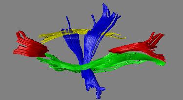
43 DTT: introduction DRFT Validation: software phantom DRFT results: visualization Body of the corpus callosum Color encodes directional information (A/P: green, I/S: blue, L/R: red) Transparency encodes estimate of probability Width encodes σ d (dispersion) Steven

 DRFT ground truth fibers Build the")
 C Add noise & try to")

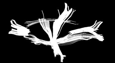
44 DTT: introduction DRFT Validation: software phantom DTT validation: framework A B In vivo DWI acquisition Anisotropic smoothing of DWIs RESTORE (robust tensor estimation) DRFT ground truth fibers Build the anatomically realistic phantom 1 (using environmental architectural information) C Add noise & try to reconstruct the ground truth fibers Compute similarity measures 1 extension of work by A. Leemans (MRM 2005, 53: )

 A good")


45 (a) In vivo (a,c) (c) DTT: introduction DRFT Validation: software phantom (b) A good correspondence is found between the colour coded synthetic FA images and the original in vivo ones (d) Phantom (b,d) Steven
46 DTT: introduction DRFT Validation: software phantom Validation results: similarity measurements Steven
47 DTT: introduction DRFT Validation: software phantom DRFT and DTT validation DRFT results in diagnostically valuable 3D pathways AND at the same time gives an estimate of probability. By using in vivo DRFT results, we were able to build a noise-free and anatomically realistic dataset.» Noise and MRI acquisition artifacts can be incorporated in the synthetic phantom as well. With an anatomically realistic synthetic DT dataset we can:» quantitatively predict how a (new) DTT algorithm will perform on real in vivo data (and not just for ad hoc cases such as helices etc.).» optimize internal and operator dependant tractography parameters. Steven
48 More More MR research at Ugent (1.5T and 3T) GifMi:» fmri studies of language and memory of epileptic patients, fmri of stuttering» MRI techniques for measuring the biological malfunctioning of neurovascular units in migraine patients» Neuropsychology: Emotional disorders, mental rotation, cognitive dysfunctions structural brain damage in MS patients Radiotherapy:» Quantitative T2-mapping for 3D geldosimetry & tissue classification» Molecular imaging
49 New developments in Magnetic Resonance Spectrocopy and Diffusion MRI Els Fieremans Steven Delputte Mahir Ozdemir
Advanced Topics and Diffusion MRI
 Advanced Topics and Diffusion MRI Slides originally by Karla Miller, FMRIB Centre Modified by Mark Chiew (mark.chiew@ndcn.ox.ac.uk) Slides available at: http://users.fmrib.ox.ac.uk/~mchiew/teaching/ MRI
DIFFUSION MAGNETIC RESONANCE IMAGING
 DIFFUSION MAGNETIC RESONANCE IMAGING from spectroscopy to imaging apparent diffusion coefficient ADC-Map anisotropy diffusion tensor (imaging) DIFFUSION NMR - FROM SPECTROSCOPY TO IMAGING Combining Diffusion
Field trip: Tuesday, Feb 5th
 Pulse Sequences Field trip: Tuesday, Feb 5th Hardware tour of VUIIIS Philips 3T Meet here at regular class time (11.15) Complete MRI screening form! Chuck Nockowski Philips Service Engineer Reminder: Project/Presentation
醫用磁振學 MRM 擴散張量影像 擴散張量影像原理. 本週課程內容 MR Diffusion 擴散張量造影原理 擴散張量造影應用 盧家鋒助理教授國立陽明大學生物醫學影像暨放射科學系
 本週課程內容 http://www.ym.edu.tw/~cflu 擴散張量造影原理 擴散張量造影應用 醫用磁振學 MRM 擴散張量影像 盧家鋒助理教授國立陽明大學生物醫學影像暨放射科學系 alvin4016@ym.edu.tw MRI The Basics (3rd edition) Chapter 22: Echo Planar Imaging MRI in Practice, (4th edition)
A Neurosurgeon s Perspectives of Diffusion Tensor Imaging(DTI) Diffusion Tensor MRI (DTI) Background and Relevant Physics.
 Diffusion Tensor MRI (DTI) Background and Relevant Physics.") A Neurosurgeon s Perspectives of Diffusion Tensor Imaging(DTI) Kalai Arasu Muthusamy, D.Phil(Oxon) Senior Lecturer & Consultant Neurosurgeon. Division of Neurosurgery. University Malaya Medical Centre.
A Neurosurgeon s Perspectives of Diffusion Tensor Imaging(DTI) Kalai Arasu Muthusamy, D.Phil(Oxon) Senior Lecturer & Consultant Neurosurgeon. Division of Neurosurgery. University Malaya Medical Centre.
QUALITY ASSURANCE OF MAGNETIC RESONANCE IMAGING FOR ADAPTIVE RADIOTHERAPY: PRELIMINARY INVESTIGATIONS TREVOR THANG 1 Supervisors: Dr.
 QUALITY ASSURANCE OF MAGNETIC RESONANCE IMAGING FOR ADAPTIVE RADIOTHERAPY: PRELIMINARY INVESTIGATIONS TREVOR THANG 1 Supervisors: Dr. Eugene Wong 2, Dr. Rob Bartha 1 Department of Medical Biophysics 1,
Magnetic Resonance Imaging. Pål Erik Goa Associate Professor in Medical Imaging Dept. of Physics
 Magnetic Resonance Imaging Pål Erik Goa Associate Professor in Medical Imaging Dept. of Physics pal.e.goa@ntnu.no 1 Why MRI? X-ray/CT: Great for bone structures and high spatial resolution Not so great
Cambridge University Press MRI from A to Z: A Definitive Guide for Medical Professionals Gary Liney Excerpt More information
 Main glossary Aa AB systems Referring to molecules exhibiting multiply split MRS peaks due to spin-spin interactions. In an AB system, the chemical shift between the spins is of similar magnitude to the
Diffusion Tensor Imaging I: The basics. Jennifer Campbell
 Diffusion Tensor Imaging I: The basics Jennifer Campbell Diffusion Tensor Imaging I: The basics Jennifer Campbell Diffusion Imaging MRI: many different sources of contrast T1W T2W PDW Perfusion BOLD DW
Contrast Mechanisms in MRI. Michael Jay Schillaci
 Contrast Mechanisms in MRI Michael Jay Schillaci Overview Image Acquisition Basic Pulse Sequences Unwrapping K-Space Image Optimization Contrast Mechanisms Static and Motion Contrasts T1 & T2 Weighting,
Introduction to MRI Acquisition
 Introduction to MRI Acquisition James Meakin FMRIB Physics Group FSL Course, Bristol, September 2012 1 What are we trying to achieve? 2 What are we trying to achieve? Informed decision making: Protocols
Introduction to MRI. Spin & Magnetic Moments. Relaxation (T1, T2) Spin Echoes. 2DFT Imaging. K-space & Spatial Resolution.
 Spin Echoes. 2DFT Imaging. K-space & Spatial Resolution.") Introduction to MRI Spin & Magnetic Moments Relaxation (T1, T2) Spin Echoes 2DFT Imaging Selective excitation, phase & frequency encoding K-space & Spatial Resolution Contrast (T1, T2) Acknowledgement:
Introduction to MRI Spin & Magnetic Moments Relaxation (T1, T2) Spin Echoes 2DFT Imaging Selective excitation, phase & frequency encoding K-space & Spatial Resolution Contrast (T1, T2) Acknowledgement:
Diffusion Tensor Imaging (DTI): An overview of key concepts
: An overview of key concepts") Diffusion Tensor Imaging (DTI): An overview of key concepts (Supplemental material for presentation) Prepared by: Nadia Barakat BMB 601 Chris Conklin Thursday, April 8 th 2010 Diffusion Concept [1,2]:
Diffusion Tensor Imaging (DTI): An overview of key concepts (Supplemental material for presentation) Prepared by: Nadia Barakat BMB 601 Chris Conklin Thursday, April 8 th 2010 Diffusion Concept [1,2]:
MRI Physics I: Spins, Excitation, Relaxation
 MRI Physics I: Spins, Excitation, Relaxation Douglas C. Noll Biomedical Engineering University of Michigan Michigan Functional MRI Laboratory Outline Introduction to Nuclear Magnetic Resonance Imaging
Principles of Magnetic Resonance Imaging
 Principles of Magnetic Resonance Imaging Hi Klaus Scheffler, PhD Radiological Physics University of 1 Biomedical Magnetic Resonance: 1 Introduction Magnetic Resonance Imaging Contents: Hi 1 Introduction
Magnetic Resonance Spectroscopy: Basic Principles and Selected Applications
 Magnetic Resonance Spectroscopy: Basic Principles and Selected Applications Sridar Narayanan, PhD Magnetic Resonance Spectroscopy Unit McConnell Brain Imaging Centre Dept. of Neurology and Neurosurgery
MR Spectroscopy: The Physical Basis and Acquisition Strategies
 AAPM 2010 SAM Imaging Session MR Spectroscopy: The Physical Basis and Acquisition Strategies Edward F. Jackson, PhD Department of Imaging Physics Objectives Understand the physical basis of in vivo MRS
Diffusion Tensor Imaging I. Jennifer Campbell
 Diffusion Tensor Imaging I Jennifer Campbell Diffusion Imaging Molecular diffusion The diffusion tensor Diffusion weighting in MRI Alternatives to the tensor Overview of applications Diffusion Imaging
Basic MRI physics and Functional MRI
 Basic MRI physics and Functional MRI Gregory R. Lee, Ph.D Assistant Professor, Department of Radiology June 24, 2013 Pediatric Neuroimaging Research Consortium Objectives Neuroimaging Overview MR Physics
EL-GY 6813/BE-GY 6203 Medical Imaging, Fall 2016 Final Exam
 EL-GY 6813/BE-GY 6203 Medical Imaging, Fall 2016 Final Exam (closed book, 1 sheets of notes double sided allowed, no calculator or other electronic devices allowed) 1. Ultrasound Physics (15 pt) A) (9
MRS: IN VIVO SPECTROSCOPIC IMAGING MAIN POINTS
 MRS: IN VIVO SPECTROSCOPIC IMAGING MAIN POINTS 1. A MR spectrum can identify many metabolites other than water by: Locating the peak(s) determined by a characteristic chemical shift (ppm) resulting from
PROTEIN NMR SPECTROSCOPY
 List of Figures List of Tables xvii xxvi 1. NMR SPECTROSCOPY 1 1.1 Introduction to NMR Spectroscopy 2 1.2 One Dimensional NMR Spectroscopy 3 1.2.1 Classical Description of NMR Spectroscopy 3 1.2.2 Nuclear
Basics of Diffusion Tensor Imaging and DtiStudio
 Basics of Diffusion Tensor Imaging and DtiStudio DTI Basics 1 DTI reveals White matter anatomy Gray matter White matter DTI uses water diffusion as a probe for white matter anatomy Isotropic diffusion
Introduction to Magnetic Resonance Imaging (MRI) Pietro Gori
 Pietro Gori") Introduction to Magnetic Resonance Imaging (MRI) Pietro Gori Enseignant-chercheur Equipe IMAGES - Télécom ParisTech pietro.gori@telecom-paristech.fr September 20, 2017 P. Gori BIOMED 20/09/2017 1 / 76
Introduction to Magnetic Resonance Imaging (MRI) Pietro Gori Enseignant-chercheur Equipe IMAGES - Télécom ParisTech pietro.gori@telecom-paristech.fr September 20, 2017 P. Gori BIOMED 20/09/2017 1 / 76
Introduction to Biomedical Imaging
 Alejandro Frangi, PhD Computational Imaging Lab Department of Information & Communication Technology Pompeu Fabra University www.cilab.upf.edu MRI advantages Superior soft-tissue contrast Depends on among
How is it different from conventional MRI? What is MR Spectroscopy? How is it different from conventional MRI? MR Active Nuclei
 What is MR Spectroscopy? MR-Spectroscopy (MRS) is a technique to measure the (relative) concentration of certain chemical or biochemical molecules in a target volume. MR-Spectroscopy is an in vivo (in
Diffusion imaging of the brain: technical considerations and practical applications
 Diffusion imaging of the brain: technical considerations and practical applications David G. Norris FC Donders Centre for Cognitive Neuroimaging Nijmegen Sustaining the physiologist in measuring the atomic
Lecture #7 In Vivo Water
 Lecture #7 In Vivo Water Topics Hydration layers Tissue relaxation times Magic angle effects Magnetization Transfer Contrast (MTC) CEST Handouts and Reading assignments Mathur-De Vre, R., The NMR studies
MRI Physics II: Gradients, Imaging. Douglas C. Noll, Ph.D. Dept. of Biomedical Engineering University of Michigan, Ann Arbor
 MRI Physics II: Gradients, Imaging Douglas C., Ph.D. Dept. of Biomedical Engineering University of Michigan, Ann Arbor Magnetic Fields in MRI B 0 The main magnetic field. Always on (0.5-7 T) Magnetizes
The physics of medical imaging US, CT, MRI. Prof. Peter Bogner
 The physics of medical imaging US, CT, MRI Prof. Peter Bogner Clinical radiology curriculum blocks of lectures and clinical practice (7x2) Physics of medical imaging Neuroradiology Head and neck I. Head
The NMR Inverse Imaging Problem
 The NMR Inverse Imaging Problem Nuclear Magnetic Resonance Protons and Neutrons have intrinsic angular momentum Atoms with an odd number of proton and/or odd number of neutrons have a net magnetic moment=>
Applications of Spin Echo and Gradient Echo: Diffusion and Susceptibility Contrast
 Applications of Spin Echo and Gradient Echo: Diffusion and Susceptibility Contrast Chunlei Liu, PhD Department of Electrical Engineering & Computer Sciences and Helen Wills Neuroscience Institute University
Diffusion Tensor Imaging (DTI) e Neurite Orientation Dispersion and Density Imaging (NODDI)
 e Neurite Orientation Dispersion and Density Imaging (NODDI)") Diffusion Tensor Imaging (DTI) e Neurite Orientation Dispersion and Density Imaging (NODDI) Claudia AM Gandini Wheeler-Kingshott, PhD Prof. of MRI Physics Overview Diffusion and microstructure NODDI theoretical
Diffusion Tensor Imaging (DTI) e Neurite Orientation Dispersion and Density Imaging (NODDI) Claudia AM Gandini Wheeler-Kingshott, PhD Prof. of MRI Physics Overview Diffusion and microstructure NODDI theoretical
Tensor Visualization. CSC 7443: Scientific Information Visualization
 Tensor Visualization Tensor data A tensor is a multivariate quantity Scalar is a tensor of rank zero s = s(x,y,z) Vector is a tensor of rank one v = (v x,v y,v z ) For a symmetric tensor of rank 2, its
How does this work? How does this method differ from ordinary MRI?
 361-Lec41 Tue 18nov14 How does this work? How does this method differ from ordinary MRI? NEW kinds of MRI (magnetic resononance imaging (MRI) Diffusion Magnetic Resonance Imaging Tractographic reconstruction
G Medical Imaging. Outline 4/13/2012. Physics of Magnetic Resonance Imaging
 G16.4426 Medical Imaging Physics of Magnetic Resonance Imaging Riccardo Lattanzi, Ph.D. Assistant Professor Department of Radiology, NYU School of Medicine Department of Electrical and Computer Engineering,
The physics US and MRI. Prof. Peter Bogner
 The physics US and MRI Prof. Peter Bogner Sound waves mechanical disturbance, a pressure wave moves along longitudinal wave compression rarefaction zones c = nl, (c: velocity, n: frequency, l: wavelength
Nuclear Magnetic Resonance Imaging
 Nuclear Magnetic Resonance Imaging Jeffrey A. Fessler EECS Department The University of Michigan NSS-MIC: Fundamentals of Medical Imaging Oct. 20, 2003 NMR-0 Background Basic physics 4 magnetic fields
Anisotropy of HARDI Diffusion Profiles Based on the L 2 -Norm
 Anisotropy of HARDI Diffusion Profiles Based on the L 2 -Norm Philipp Landgraf 1, Dorit Merhof 1, Mirco Richter 1 1 Institute of Computer Science, Visual Computing Group, University of Konstanz philipp.landgraf@uni-konstanz.de
Magnetic Resonance Imaging (MRI)
") Magnetic Resonance Imaging Introduction The Components The Technology (MRI) Physics behind MR Most slides taken from http:// www.slideworld.org/ viewslides.aspx/magnetic- Resonance-Imaging- %28MRI%29-MR-Imaging-
Magnetic Resonance Imaging Introduction The Components The Technology (MRI) Physics behind MR Most slides taken from http:// www.slideworld.org/ viewslides.aspx/magnetic- Resonance-Imaging- %28MRI%29-MR-Imaging-
NMR and MRI : an introduction
 Intensive Programme 2011 Design, Synthesis and Validation of Imaging Probes NMR and MRI : an introduction Walter Dastrù Università di Torino walter.dastru@unito.it \ Introduction Magnetic Resonance Imaging
MRI beyond Fourier Encoding: From array detection to higher-order field dynamics
 MRI beyond Fourier Encoding: From array detection to higher-order field dynamics K. Pruessmann Institute for Biomedical Engineering ETH Zurich and University of Zurich Parallel MRI Signal sample: m γκ,
Diffusion Weighted MRI. Zanqi Liang & Hendrik Poernama
 Diffusion Weighted MRI Zanqi Liang & Hendrik Poernama 1 Outline MRI Quick Review What is Diffusion MRI? Detecting Diffusion Stroke and Tumor Detection Presenting Diffusion Anisotropy and Diffusion Tensor
Nuclei, Excitation, Relaxation
 Outline 4.1 Principles of MRI uclei, Excitation, Relaxation Carolyn Kaut Roth, RT (R)(MR)(CT)(M)(CV) FSMRT CEO Imaging Education Associates www.imaginged.com candi@imaginged.com What nuclei are MR active?
MRI in Practice. Catherine Westbrook MSc, DCRR, CTC Senior Lecturer Anglia Polytechnic University Cambridge UK. John Talbot MSc, DCRR
 MRI in Practice Third edition Catherine Westbrook MSc, DCRR, CTC Senior Lecturer Anglia Polytechnic University Cambridge UK and Carolyn Kaut RothRT(R) (MR) (CT) (M) (CV) Fellow SMRT (Section for Magnetic
Chapter 7. Nuclear Magnetic Resonance Spectroscopy
 Chapter 7 Nuclear Magnetic Resonance Spectroscopy I. Introduction 1924, W. Pauli proposed that certain atomic nuclei have spin and magnetic moment and exposure to magnetic field would lead to energy level
The Basics of Magnetic Resonance Imaging
 The Basics of Magnetic Resonance Imaging Nathalie JUST, PhD nathalie.just@epfl.ch CIBM-AIT, EPFL Course 2013-2014-Chemistry 1 Course 2013-2014-Chemistry 2 MRI: Many different contrasts Proton density T1
Tissue Parametric Mapping:
 Tissue Parametric Mapping: Contrast Mechanisms Using SSFP Sequences Jongho Lee Department of Radiology University of Pennsylvania Tissue Parametric Mapping: Contrast Mechanisms Using bssfp Sequences Jongho
NMR/MRI examination (8N080 / 3F240)
") NMR/MRI examination (8N080 / 3F240) Remarks: 1. This test consists of 3 problems with at total of 26 sub-questions. 2. Questions are in English. You are allowed to answer them in English or Dutch. 3. Please
NMR/MRI examination (8N080 / 3F240) Remarks: 1. This test consists of 3 problems with at total of 26 sub-questions. 2. Questions are in English. You are allowed to answer them in English or Dutch. 3. Please
Magnetic resonance imaging MRI
 Magnetic resonance imaging MRI Introduction What is MRI MRI is an imaging technique used primarily in medical settings that uses a strong magnetic field and radio waves to produce very clear and detailed
Basic Concepts of MR Imaging, Diffusion MR Imaging, and Diffusion Tensor Imaging
 Basic Concepts of MR Imaging, Diffusion MR Imaging, and Diffusion Tensor Imaging Eduardo H.M.S.G. de Figueiredo, BSc a, *, Arthur F.N.G. Borgonovi, BSc b,c, Thomas M. Doring, MSc d,e KEYWORDS Magnetic
COPYRIGHTED MATERIAL. Production of Net Magnetization. Chapter 1
 Chapter 1 Production of Net Magnetization Magnetic resonance (MR) is a measurement technique used to examine atoms and molecules. It is based on the interaction between an applied magnetic field and a
Fundamental MRI Principles Module Two
 Fundamental MRI Principles Module Two 1 Nuclear Magnetic Resonance There are three main subatomic particles: protons neutrons electrons positively charged no significant charge negatively charged Protons
Basis of MRI Contrast
 Basis of MRI Contrast MARK A. HORSFIELD Department of Cardiovascular Sciences University of Leicester Leicester LE1 5WW UK Tel: +44-116-2585080 Fax: +44-870-7053111 e-mail: mah5@le.ac.uk 1 1.1 The Magnetic
Magnetic Resonance Imaging. Qun Zhao Bioimaging Research Center University of Georgia
 Magnetic Resonance Imaging Qun Zhao Bioimaging Research Center University of Georgia The Nobel Prize in Physiology or Medicine 2003 "for their discoveries concerning magnetic resonance imaging" Paul C.
MRI in Review: Simple Steps to Cutting Edge Part I
 MRI in Review: Simple Steps to Cutting Edge Part I DWI is now 2 years old... Mike Moseley Radiology Stanford DWI, b = 1413 T2wt, 28/16 ASN 21 San Francisco + Disclosures: Funding NINDS, NCRR, NCI 45 minutes
Part II: Magnetic Resonance Imaging (MRI)
") Part II: Magnetic Resonance Imaging (MRI) Contents Magnetic Field Gradients Selective Excitation Spatially Resolved Reception k-space Gradient Echo Sequence Spin Echo Sequence Magnetic Resonance Imaging
Part II: Magnetic Resonance Imaging (MRI) Contents Magnetic Field Gradients Selective Excitation Spatially Resolved Reception k-space Gradient Echo Sequence Spin Echo Sequence Magnetic Resonance Imaging
Diffusion MRI. Outline. Biology: The Neuron. Brain connectivity. Biology: Brain Organization. Brain connections and fibers
 Outline Diffusion MRI Alfred Anwander Download of Slides: www.cbs.mpg.de/events/ teaching/brainsignals1112 password: mpi-brain CBSWIKI: Cornet/DiffusionMRI Neuroanatomy Diffusion MRI Diffusion Tensor Imaging
7.3.A. The expression for signal recovery is similar to that derived under exercise 7.2 and is given by:
 7..A. Chemical shift difference 3..0. ppm, which equals 54.5 Hz at 3.0 T. Spatial displacement 54.5/00 0.87, which equals.03 cm along the 8 cm side and 0.77 cm along the 6 cm. The cm slice does not have
Magnetic Resonance Spectroscopy. Saurabh Bhaskar Shaw Dwip Shah
 Magnetic Resonance Spectroscopy By Saurabh Bhaskar Shaw Dwip Shah What is Magnetic Resonance Spectroscopy? [1] Non invasive method to look at concentration of metabolites invivo. 2 Basics of MRS Physics
Introductory MRI Physics
 C HAPR 18 Introductory MRI Physics Aaron Sodickson EXRNAL MAGNETIC FIELD, PROTONS AND EQUILIBRIUM MAGNETIZATION Much of the bulk of the magnetic resonance imaging (MRI) scanner apparatus is dedicated to
Fundamental MRI Principles Module 2 N. Nuclear Magnetic Resonance. X-ray. MRI Hydrogen Protons. Page 1. Electrons
 Fundamental MRI Principles Module 2 N S 1 Nuclear Magnetic Resonance There are three main subatomic particles: protons positively charged neutrons no significant charge electrons negatively charged Protons
Application of diffusion MRI to cancer, heart and brain connectome imaging
 Colloquium @ Department of Physics, NTU Application of diffusion MRI to cancer, heart and brain connectome imaging March 11, 2014 Wen-Yih Isaac Tseng MD, PhD Advanced Biomedical MRI Lab Center for Optoelectronic
Medical Visualization - Tensor Visualization. J.-Prof. Dr. Kai Lawonn
 Medical Visualization - Tensor Visualization J.-Prof. Dr. Kai Lawonn Lecture is partially based on the lecture by Prof. Thomas Schultz 2 What is a Tensor? A tensor is a multilinear transformation that
DWI acquisition schemes and Diffusion Tensor estimation
 DWI acquisition schemes and Diffusion Tensor estimation A simulation based study Santiago Aja-Fernández, Antonio Tristán-Vega, Pablo Casaseca-de-la-Higuera Laboratory of Image Processing L A B O R A T
Quantitative Susceptibility Mapping and Susceptibility Tensor Imaging. Magnetization and Susceptibility
 Quantitative Susceptibility Mapping and Susceptibility Tensor Imaging 1, Chunlei Liu, Ph.D. 1 Brain Imaging and Analysis Center Department of Radiology Duke University, Durham, NC, USA 1 Magnetization
Spin Relaxation and NOEs BCMB/CHEM 8190
 Spin Relaxation and NOEs BCMB/CHEM 8190 T 1, T 2 (reminder), NOE T 1 is the time constant for longitudinal relaxation - the process of re-establishing the Boltzmann distribution of the energy level populations
Introduction to the Physics of NMR, MRI, BOLD fmri
 Pittsburgh, June 13-17, 2011 Introduction to the Physics of NMR, MRI, BOLD fmri (with an orientation toward the practical aspects of data acquisition) Pittsburgh, June 13-17, 2001 Functional MRI in Clinical
Nuclear Magnetic Resonance Imaging
 Nuclear Magnetic Resonance Imaging Simon Lacoste-Julien Electromagnetic Theory Project 198-562B Department of Physics McGill University April 21 2003 Abstract This paper gives an elementary introduction
Background II. Signal-to-Noise Ratio (SNR) Pulse Sequences Sampling and Trajectories Parallel Imaging. B.Hargreaves - RAD 229.
 Pulse Sequences Sampling and Trajectories Parallel Imaging. B.Hargreaves - RAD 229.") Background II Signal-to-Noise Ratio (SNR) Pulse Sequences Sampling and Trajectories Parallel Imaging 1 SNR: Signal-to-Noise Ratio Signal: Desired voltage in coil Noise: Thermal, electronic Noise Thermal
Background II Signal-to-Noise Ratio (SNR) Pulse Sequences Sampling and Trajectories Parallel Imaging 1 SNR: Signal-to-Noise Ratio Signal: Desired voltage in coil Noise: Thermal, electronic Noise Thermal
Sketch of the MRI Device
 Outline for Today 1. 2. 3. Introduction to MRI Quantum NMR and MRI in 0D Magnetization, m(x,t), in a Voxel Proton T1 Spin Relaxation in a Voxel Proton Density MRI in 1D MRI Case Study, and Caveat Sketch
Bayesian multi-tensor diffusion MRI and tractography
 Bayesian multi-tensor diffusion MRI and tractography Diwei Zhou 1, Ian L. Dryden 1, Alexey Koloydenko 1, & Li Bai 2 1 School of Mathematical Sciences, Univ. of Nottingham 2 School of Computer Science and
Diffusion-Weighted MRI may be used to measure the apparent diffusion coefficient of water in tissue.
 Specialty Area: MR Physics for Physicists Speaker: Jennifer A. McNab, Ph.D. Assistant Professor, Radiology, Stanford University () Highlights The Bloch-Torrey equation is a generalization of the Bloch
Quantitative Metrics for White Matter Integrity Based on Diffusion Tensor MRI Data. Stephanie Lee
 Quantitative Metrics for White Matter Integrity Based on Diffusion Tensor MRI Data Stephanie Lee May 5, 2005 Quantitative Metrics for White Matter Integrity Based on Diffusion Tensor MRI Data ABSTRACT
Chapter 14:Physics of Magnetic Resonance
 Chapter 14:Physics of Magnetic Resonance Slide set of 141 slides based on the chapter authored by Hee Kwon Song of the publication (ISBN 978-92-0-131010-1): Diagnostic Radiology Physics: A Handbook for
10.4 Continuous Wave NMR Instrumentation
 10.4 Continuous Wave NMR Instrumentation coherent detection bulk magnetization the rotating frame, and effective magnetic field generating a rotating frame, and precession in the laboratory frame spin-lattice
Recent advances in quantitative MR spectroscopy
 Recent advances in quantitative MR spectroscopy Anke Henning, PhD Institute for Biomedical Engineering, University and ETH Zurich, Switzerland July 2009 MOTIVATION: non-invasive metabolite quantification
Advanced MRI: Diffusion MRI 1: DTI and k-space
 k y Advanced MRI: Diffusion MRI 1: DTI and k-space k X Eric Sigmund, PhD February 26th, 2013 LECTURE 1 Neuro Diffusion MRI 3-5 m White matter axons Body 15 m Renal medulla Musculoskeletal 50 m Skeletal
HST.583 Functional Magnetic Resonance Imaging: Data Acquisition and Analysis Fall 2006
 MIT OpenCourseWare http://ocw.mit.edu HST.583 Functional Magnetic Resonance Imaging: Data Acquisition and Analysis Fall 2006 For information about citing these materials or our Terms of Use, visit: http://ocw.mit.edu/terms.
Diffusion tensor imaging: brain pathway reconstruction
 Neda Sepasian, Jan ten Thije Boonkkamp, Anna Vilanova Diffusion tensor imaging: brain pathway reconstruction NAW 5/6 nr. 4 december 205 259 Neda Sepasian Department of Biomedical Engineering Eindhoven
Correction Gradients. Nov7, Reference: Handbook of pulse sequence
 Correction Gradients Nov7, 2005 Reference: Handbook of pulse sequence Correction Gradients 1. Concomitant-Field Correction Gradients 2. Crusher Gradients 3. Eddy-Current Compensation 4. Spoiler Gradients
Velocity Images. Phase Contrast Technique. G. Reiter 1,2, U. Reiter 1, R. Rienmüller 1
 Velocity Images - the MR Phase Contrast Technique G. Reiter 1,2, U. Reiter 1, R. Rienmüller 1 SSIP 2004 12 th Summer School in Image Processing, Graz, Austria 1 Interdisciplinary Cardiac Imaging Center,
M R I Physics Course. Jerry Allison Ph.D., Chris Wright B.S., Tom Lavin B.S., Nathan Yanasak Ph.D. Department of Radiology Medical College of Georgia
 M R I Physics Course Jerry Allison Ph.D., Chris Wright B.S., Tom Lavin B.S., Nathan Yanasak Ph.D. Department of Radiology Medical College of Georgia M R I Physics Course Spin Echo Imaging Hahn Spin Echo
Magnetic Resonance Imaging
 http://www.qldxray.com.au/filelibrary/mri_cardiovascular_system_ca_0005.jpg Magnetic Resonance Imaging 1 Overview 1. The magnetic properties of nuclei, and how they behave in strong magnetic fields. 2.
Topics. The concept of spin Precession of magnetic spin Relaxation Bloch Equation. Bioengineering 280A Principles of Biomedical Imaging
 Bioengineering 280A Principles of Biomedical Imaging Fall Quarter 2006 MRI Lecture 1 Topics The concept of spin Precession of magnetic spin Relaxation Bloch Equation 1 Spin Intrinsic angular momentum of
Physics of MR Image Acquisition
 Physics of MR Image Acquisition HST-583, Fall 2002 Review: -MRI: Overview - MRI: Spatial Encoding MRI Contrast: Basic sequences - Gradient Echo - Spin Echo - Inversion Recovery : Functional Magnetic Resonance
Chem 325 NMR Intro. The Electromagnetic Spectrum. Physical properties, chemical properties, formulas Shedding real light on molecular structure:
 Physical properties, chemical properties, formulas Shedding real light on molecular structure: Wavelength Frequency ν Wavelength λ Frequency ν Velocity c = 2.998 10 8 m s -1 The Electromagnetic Spectrum
Tissue Characteristics Module Three
 Tissue Characteristics Module Three 1 Equilibrium State Equilibrium State At equilibrium, the hydrogen vector is oriented in a direction parallel to the main magnetic field. Hydrogen atoms within the vector
Principles of Nuclear Magnetic Resonance Microscopy
 Principles of Nuclear Magnetic Resonance Microscopy Paul T. Callaghan Department of Physics and Biophysics Massey University New Zealand CLARENDON PRESS OXFORD CONTENTS 1 PRINCIPLES OF IMAGING 1 1.1 Introduction
The effect of different number of diffusion gradients on SNR of diffusion tensor-derived measurement maps
 J. Biomedical Science and Engineering, 009,, 96-101 The effect of different number of diffusion gradients on SNR of diffusion tensor-derived measurement maps Na Zhang 1, Zhen-Sheng Deng 1*, Fang Wang 1,
Apodization. Gibbs Artifact. Bioengineering 280A Principles of Biomedical Imaging. Fall Quarter 2013 MRI Lecture 5. rect(k x )
") Bioengineering 280A Principles of Biomedical Imaging Fall Quarter 2013 MRI Lecture 5 GE Medical Systems 2003 Gibbs Artifact Apodization rect(k ) Hanning Window h(k )=1/2(1+cos(2πk ) 256256 image 256128
Bioengineering 280A Principles of Biomedical Imaging Fall Quarter 2013 MRI Lecture 5 GE Medical Systems 2003 Gibbs Artifact Apodization rect(k ) Hanning Window h(k )=1/2(1+cos(2πk ) 256256 image 256128
Spin Echo Imaging Sequence
 1 MRI In Stereotactic Procedures Edward F. Jackson, Ph.D. The University of Texas M.D. Anderson Cancer Center Houston, Texas 2 RF G slice G phase G freq Signal k-space Spin Echo Imaging Sequence TE 1st
Diffusion Imaging II. By: Osama Abdullah
 iffusion Imaging II By: Osama Abdullah Review Introduction. What is diffusion? iffusion and signal attenuation. iffusion imaging. How to capture diffusion? iffusion sensitizing gradients. Spin Echo. Gradient
Diffusion MRI for Brain Connectivity Mapping and Analysis
 Diffusion MRI for Brain Connectivity Mapping and Analysis Brian G. Booth and Ghassan Hamarneh Contents 1 Diffusion Weighted Image Acquision 2 1.1 Biological Basis for Diffusion MRI..........................
A Riemannian Framework for Denoising Diffusion Tensor Images
 A Riemannian Framework for Denoising Diffusion Tensor Images Manasi Datar No Institute Given Abstract. Diffusion Tensor Imaging (DTI) is a relatively new imaging modality that has been extensively used
FREQUENCY SELECTIVE EXCITATION
 PULSE SEQUENCES FREQUENCY SELECTIVE EXCITATION RF Grad 0 Sir Peter Mansfield A 1D IMAGE Field Strength / Frequency Position FOURIER PROJECTIONS MR Image Raw Data FFT of Raw Data BACK PROJECTION Image Domain
Medical Imaging Physics Spring Quarter Week 9-1
 Medical Imaging Physics Spring Quarter Week 9-1 NMR and MRI Davor Balzar balzar@du.edu www.du.edu/~balzar Intro MRI Outline NMR & MRI Guest lecturer fmri Thursday, May 22 Visit to CUHSC It s not mandatory
NMR BMB 173 Lecture 16, February
 NMR The Structural Biology Continuum Today s lecture: NMR Lots of slides adapted from Levitt, Spin Dynamics; Creighton, Proteins; And Andy Rawlinson There are three types of particles in the universe Quarks
Diffusion Tensor Imaging tutorial
 NA-MIC http://na-mic.org Diffusion Tensor Imaging tutorial Sonia Pujol, PhD Surgical Planning Laboratory Harvard University DTI tutorial This tutorial is an introduction to the advanced Diffusion MR capabilities
Rochester Institute of Technology Rochester, New York. COLLEGE of Science Department of Chemistry. NEW (or REVISED) COURSE:
 COURSE:") Rochester Institute of Technology Rochester, New York COLLEGE of Science Department of Chemistry NEW (or REVISED) COURSE: 1014-730 1.0 Title: Magnetic Resonance Imaging (MRI) Date: July 2006 Credit Hours:
Rochester Institute of Technology Rochester, New York COLLEGE of Science Department of Chemistry NEW (or REVISED) COURSE: 1014-730 1.0 Title: Magnetic Resonance Imaging (MRI) Date: July 2006 Credit Hours: