Introduction to the Course and the Techniques. Jeffry R. Alger, PhD Ahmanson-Lovelace Brain Mapping Center Department of Neurology
|
|
|
- Patricia McDaniel
- 6 years ago
- Views:
Transcription
1 Introduction to the Course and the Techniques Jeffry R. Alger, PhD Ahmanson-Lovelace Brain Mapping Center Department of Neurology CTSI Neuroimaging April 2013
2 Rationale for the Course
3 Translational Clinical Science Cycle
 Use of imaging to make clinical trials of drugs or devices more scientifically valid (imaging biomarkers) or less")
4 Translational Clinical Science Cycle Discovery of new approaches to image human anatomy and physiology (basic science bench to bedside) Using imaging to discover new information about human disease Routine clinical use of imaging (diagnosis and treatment response assessment) Use of imaging to make clinical trials of drugs or devices more scientifically valid (imaging biomarkers) or less expensive (surrogate endpoints) Clinical trials that test the diagnostic power of imaging
5 Course Components Discovery of new approaches to image human anatomy and physiology Nader Pouratian, MD Jack van Horn, PhD Danny JJ Wang, PhD Using imaging to discover new information about human disease Katherine Narr, PhD Paul M. Vespa, MD Use of multimodal imaging in clinical trials David S. Liebeskind, MD Benjamin M. Ellingson, PhD
6 MRI 101 Laboratory Friday April 12 at 11 AM Meet at the entrance to the Ahmanson-Lovelace Brain Mapping Center (660 CE Young Drive South) Content MRI Safety The look and feel of MRI For patients and investigators Anatomic Brain Imaging Functional Brain Imaging Physiological Brain Imaging Metabolic Brain Imaging
7 Medical Imaging Medical Imaging is based on the interaction between electromagnetic radiation and living tissue The brain is surrounded by bone Ultrasonography does not play a major role in brain imaging although several niche approaches are developing Neuroimaging is equally based on interaction between Electromagnetic radiation and bone Electromagnetic radiation and brain
8 Neuroimaging Requirements Brain imaging requires electromagnetic radiation that is safe penetrates bone interacts with brain to produce contrast to anatomy or function or both

9 Bone Minerals
10 Water Salts Metabolites Neurotransmitters Protein Lipids Membranes Cells Blood vessels Hemoglobin Brain Components

11 Electromagnetic radiation is composed on oscillating electric and magnetic fields (wave frequency is inversely proportional to wavelength)
12 Electric Field
13 Electrocortigraphy (EcoG) Uses electrodes placed on the brain cortex to detect time varying electric fields produced by neural activity A functional brain imaging technique Detection of E-fields is much enhanced if electrodes are placed directly on the brain surface Bone is an electric insulator Electric field strength decays rapidly with distance Safety Requires neurosurgery Applicable only to specific patient populations
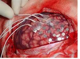
14 Electrocortigraphy
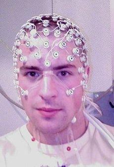
15 Electroencephalagraphic Imaging

16 Magnetic Fields
17 Magnetic Fields Movement of electric charge (electric current) produces a dipole magnetic field Monopole magnetic fields do not exist Movement of a magnetic field produces electric current Magnetic fields always have two or more poles All magnetism is fundamentally due to electric current
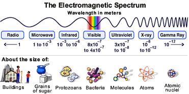
18 The Electromagnetic Spectrum
19 Bone penetration Bone excludes Infrared Light Visible Light Ultraviolet Light Bone is penetrated by High intensity Xrays Computed Tomography Xray Angiography Radio frequency waves Magnetic Resonance Imaging Microwaves Gamma rays Positron Emission Tomography

20 Microwave Imaging?
21 Imaging with Microwaves? Microwave-based imaging is impractical due to Tissue heating Poor penetration Small charged particles (Na +, K +, Cl - ) in tissue can move in resonance with the applied microwave field Friction between the moving ions and medium produces heat. Beginning to be a factor for MRI because newest MRI frequencies approach the microwave region of the electromagnetic spectrum
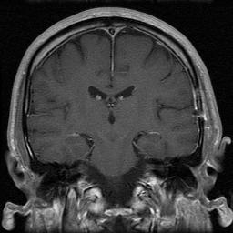
22 Magnetic Resonance Imaging
23 Magnetic Resonance Imaging MRI utilizes the magnetic resonance signal produced by the protons ( 1 H) of tissue water. MRI has become the method of choice for nondestructive visualization of brain anatomy Anatomic contrast depends on microscopic biophysical properties of tissue that (mostly) influence how fast water molecules can move Functional MRI techniques trick the proton MR signal to depend on brain function Usually indirect detection of blood flow or oxygenation changes that accompany neural activity

24 B0 B0 H O Magnetic Resonance Image H O H H H B0 B0 One volume element from the imaged tissue section H Proton spin magnets oriented parallel or antiparallel to the applied magnetic field and precessing about it
25 The Static Magnetic Field (B 0 ) B 0 penetrate bony structures allowing the brain to be seen through bone B 0 is typically 1.5 Tesla or greater 1.5 T is 30,000 times the strength of the earth s magnetic field Nuclear magnets rotate around B 0 at a characteristic frequency, known as the Larmor frequency Larmor frequency is in the radio frequency region of the electromagnetic spectrum 127 Mhz at 3.0 T 63 Mhz at 1.5 T
26 Large ensembles of nuclei The typical MRI volume element has about protons Some nuclei oriented parallel to B 0 Some nuclei oriented antiparallel to B 0 Nature favors the parallel configuration Lower energy
27 B0 B0 N S Individual spin magnets within the voxel Bulk magnetization generated by all the nuclei in the voxel
28 Large ensembles of nuclei The two configurations do not differ to a very great extent in their energy stability Almost equal numbers in the two configurations Most of the protons cancel each other The uncancelled nuclei act like a macroscopic bar magnet Referred to as nuclear magnetization Not quantized The MRI signal results from a small fraction of the uncancelled nuclei MRI signal is relatively weak
29 Oscillatory magnetic field (B 1 ) The stable configuration of the magnetization can be disturbed by the addition of a second magnetic field (B 1 ) that oscillates in time in coherence with the nuclei B 1 penetrates bone The oscillatory frequency of B 1 must be very near to the Larmor frequency The ensemble of nuclear magnets resonates with B 1 B 1 is created by a transmitter which is turned on and off under computer control as needed
30 B0 B0 N transmitter excitation pulse S Signal Phase Voltage Free Induction Decay (FID) Signal frequency Signal Amplitude time (This is the basic MRI signal detected by acoil of wire placed somewhere near the brain)
31 Magnetic Field Gradient(s) and Imaging A magnetic field gradient is a smooth variation in B 0 from one position to another position Anterior-to-posterior variation Superior-to-inferior variation Left-to-right variation Gradients are purposefully applied as part of the imaging process Takes advantage of the linear Larmor relationship between magnetic field and frequency


32 High High Frequency bandwidth of selective excitation Low B0 in presence of S/I magnetic field gradient Low MR signal frequency MRI of the selected slice


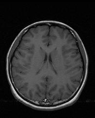
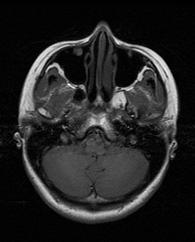
33 sagittal axial coronal
34
35 The Image Signal discussed thus far is depicted as gray level of each element in the image White = greater signal Gray = less signal Black = no signal MR Images usually have no absolute calibration Contrast usually depends on signal relaxation characteristics
36 Relaxation Recovery to original state after some perturbation T1 relaxation time Characteristic time for recovery of magnetization along B 0 axis T2 relaxation time Characteristic time for decay of magnetization in the plane perpendicular to B 0
37 B0 B0 B0 time T2 relaxation process Magnitude of the magnetization component in the plane perpendicular to B 0 decays to zero
38 B0 B0 B0 N S N S N S time T1 relaxation process Magnitude of the magnetization component parallel to B 0 grows and ultimately reaches an equilibrium value
39 T1 and T2 are tissue dependent T1 and T2 are (mostly) related to how quickly the water molecules can tumble Water tumbling speed depends on Viscosity of the biological fluid Solutes -- especially macromolecules Amount of surface (biological membranes) that water can stick to Different biological tissues have differences in macromolecules and membranes
40 MRI Contrast Signal creation in MRI uses a complex set of pulses Sensitivity to relaxation and therefore contrast is adjusted by selection of pulse sequence pulse sequence timing parameters


41 Spin Echo Imaging Signal Intensity PDw Regime T2w regime CSF WM GM TE (msec)

42 Saturation recovery T1 weighted imaging T1-weighted regime Signal Intensity WM Contrast-enhanced GM CSF TR (msec)
43 MRI Contrast Agents Typical contrast reagents are chelates of metals ions (e.g. gadolinium) having unpaired electron spins The electron spin decreases the T1 of water r Water molecules must touch (r << 20 A) the agent to enhance T1 relaxation

44 MRI Contrast Agents and T1 Typical contrast reagents are designed to not pass the blood brain barrier T1 relaxation is enhanced only where blood brain barrier is damaged
45 Positron Emission Tomography
46 Positron Emission Tomography Based on the existence of an unusual subatomic particle called a positron (a positive electron) Certain unstable nuclear isotopes emit positrons Requires an onsite cyclotron Emitted positrons move a small distance before encountering a negative electron causing decay to two gamma rays

47 PET Technique

48 PET Imaging
49 PET Tracers 18 Fluorodeoxyglucose (FDG) is a tracer of glucose metabolism Injected into vascular system Transported into cells by glucose transporters FDG metabolism reaches a dead end after phosphorylation tracer of glucose metabolism H 2 15 O Injected into the vascular system Penetrates all membranes Washout rate depends on cerebral blood flow 15 O 2 Delivered by inhalation Tracer converted to CO 2 by respiration and washed out by blood flow Analysis of time course can be used to determine the rate of oxygen metabolism if a CBF measurement is also done Many other PET tracers are in development The basis of molecular imaging

50 PET Tracers
51 PET Safety Issues Gamma ray exposure Chemical safety Positron emitters tend to have short half lives Tracer molecules must be synthesized immediately before use Pharmacological effects of the tracer Many PET tracers are analogs of drugs or neurotransmitters
52 Computed Tomography

53 Computed Tomography Technique

54 CT Contrast
55 CT contrast Intrinsic CT contrast is within brain is limited Gray matter, white matter and CSF can be distinguished, but not as effectively as with MRI CT neuroimaging often uses intravenous contrast agent (iodinated materials) to create vascular contrast CT angiography (CTA) Visualization of larger blood vessels (usually arteries) CT perfusion (CTP) Dynamic measurement of contrast passage to measure/image microvascular perfusion

56 CT Contrast
57 CT Safety Xrays strip electrons from molecules Ionizing radiation Damages molecules In neuroimaging large xray intensity is used to overcome absorption by calcium in bone Xray exposure is sometimes a limiting factor Dynamic studies such as CTP Multiple studies on the same patient Allergic reaction to contrast agent Renal insufficiency that results in poor clearance of contrast agent
58 Introduction to Image Interpretation and Processing
59 What property is shown by the image? Gray scale and color are used to convey information about tissue properties The image looks cool but what is being shown? Is the image quantitative or only approximate MRI and CT data are often approximate PET data is often quantitative
60 Learning Points Neuroimaging techniques can be described at a basic level without using complex physics All techniques use some form of electromagnetic radiation that can pass through bone Contrast depends on interaction between radiation in brain
61 Learning Points EcoG Functional contrast Requires neurosurgery MRI Uses radio waves and magnetic fields Anatomic, vascular and functional contrast Extensive use of contrast agents PET Primarily uses tracers that emit positrons and gamma rays Ionizing radiation Requires cyclotron and chemistry CT Uses xrays Ionizing radiation Anatomic and vascular contrast
EL-GY 6813/BE-GY 6203 Medical Imaging, Fall 2016 Final Exam
 EL-GY 6813/BE-GY 6203 Medical Imaging, Fall 2016 Final Exam (closed book, 1 sheets of notes double sided allowed, no calculator or other electronic devices allowed) 1. Ultrasound Physics (15 pt) A) (9
Basic MRI physics and Functional MRI
 Basic MRI physics and Functional MRI Gregory R. Lee, Ph.D Assistant Professor, Department of Radiology June 24, 2013 Pediatric Neuroimaging Research Consortium Objectives Neuroimaging Overview MR Physics
MRI Physics I: Spins, Excitation, Relaxation
 MRI Physics I: Spins, Excitation, Relaxation Douglas C. Noll Biomedical Engineering University of Michigan Michigan Functional MRI Laboratory Outline Introduction to Nuclear Magnetic Resonance Imaging
Nuclear Medicine RADIOPHARMACEUTICAL CHEMISTRY
 Nuclear Medicine RADIOPHARMACEUTICAL CHEMISTRY An alpha particle consists of two protons and two neutrons Common alpha-particle emitters Radon-222 gas in the environment Uranium-234 and -238) in the environment
Basis of MRI Contrast
 Basis of MRI Contrast MARK A. HORSFIELD Department of Cardiovascular Sciences University of Leicester Leicester LE1 5WW UK Tel: +44-116-2585080 Fax: +44-870-7053111 e-mail: mah5@le.ac.uk 1 1.1 The Magnetic
2015 U N I V E R S I T I T E K N O L O G I P E T R O N A S
 Multi-Modality based Diagnosis: A way forward by Hafeez Ullah Amin Centre for Intelligent Signal and Imaging Research (CISIR) Department of Electrical & Electronic Engineering 2015 U N I V E R S I T I
The Basics of Magnetic Resonance Imaging
 The Basics of Magnetic Resonance Imaging Nathalie JUST, PhD nathalie.just@epfl.ch CIBM-AIT, EPFL Course 2013-2014-Chemistry 1 Course 2013-2014-Chemistry 2 MRI: Many different contrasts Proton density T1
DEVIL PHYSICS THE BADDEST CLASS ON CAMPUS IB PHYSICS
 DEVIL PHYSICS THE BADDEST CLASS ON CAMPUS IB PHYSICS TSOKOS OPTION I-2 MEDICAL IMAGING Reading Activity Answers IB Assessment Statements Option I-2, Medical Imaging: X-Rays I.2.1. I.2.2. I.2.3. Define
Technical University of Denmark
 Technical University of Denmark Page 1 of 11 pages Written test, 9 December 2010 Course name: Introduction to medical imaging Course no. 31540 Aids allowed: none. "Weighting": All problems weight equally.
Magnetic resonance imaging MRI
 Magnetic resonance imaging MRI Introduction What is MRI MRI is an imaging technique used primarily in medical settings that uses a strong magnetic field and radio waves to produce very clear and detailed
Technical University of Denmark
 Technical University of Denmark Page 1 of 10 pages Written test, 12 December 2012 Course name: Introduction to medical imaging Course no. 31540 Aids allowed: None. Pocket calculator not allowed "Weighting":
ELG7173 Topics in signal Processing II Computational Techniques in Medical Imaging
 ELG7173 Topics in signal Processing II Computational Techniques in Medical Imaging Topic #1: Intro to medical imaging Medical Imaging Classifications n Measurement physics Send Energy into body Send stuff
Magnetic Resonance Imaging
 http://www.qldxray.com.au/filelibrary/mri_cardiovascular_system_ca_0005.jpg Magnetic Resonance Imaging 1 Overview 1. The magnetic properties of nuclei, and how they behave in strong magnetic fields. 2.
The physics of medical imaging US, CT, MRI. Prof. Peter Bogner
 The physics of medical imaging US, CT, MRI Prof. Peter Bogner Clinical radiology curriculum blocks of lectures and clinical practice (7x2) Physics of medical imaging Neuroradiology Head and neck I. Head
Radioisotopes and PET
 Radioisotopes and PET 1 Radioisotopes Elements are defined by their number of protons, but there is some variation in the number of neutrons. Atoms resulting from this variation are called isotopes. Consider
The physics US and MRI. Prof. Peter Bogner
 The physics US and MRI Prof. Peter Bogner Sound waves mechanical disturbance, a pressure wave moves along longitudinal wave compression rarefaction zones c = nl, (c: velocity, n: frequency, l: wavelength
Part III: Sequences and Contrast
 Part III: Sequences and Contrast Contents T1 and T2/T2* Relaxation Contrast of Imaging Sequences T1 weighting T2/T2* weighting Contrast Agents Saturation Inversion Recovery JUST WATER? (i.e., proton density
Magnetic Resonance Imaging. Pål Erik Goa Associate Professor in Medical Imaging Dept. of Physics
 Magnetic Resonance Imaging Pål Erik Goa Associate Professor in Medical Imaging Dept. of Physics pal.e.goa@ntnu.no 1 Why MRI? X-ray/CT: Great for bone structures and high spatial resolution Not so great
Nuclear Magnetic Resonance Imaging
 Nuclear Magnetic Resonance Imaging Simon Lacoste-Julien Electromagnetic Theory Project 198-562B Department of Physics McGill University April 21 2003 Abstract This paper gives an elementary introduction
Introduction to MRI. Spin & Magnetic Moments. Relaxation (T1, T2) Spin Echoes. 2DFT Imaging. K-space & Spatial Resolution.
 Spin Echoes. 2DFT Imaging. K-space & Spatial Resolution.") Introduction to MRI Spin & Magnetic Moments Relaxation (T1, T2) Spin Echoes 2DFT Imaging Selective excitation, phase & frequency encoding K-space & Spatial Resolution Contrast (T1, T2) Acknowledgement:
Introduction to MRI Spin & Magnetic Moments Relaxation (T1, T2) Spin Echoes 2DFT Imaging Selective excitation, phase & frequency encoding K-space & Spatial Resolution Contrast (T1, T2) Acknowledgement:
MEDICAL IMAGING. METHODS OF MODERN IMAGING, BASED ON ELECTRO-MAGNETIC RADIATION (radiowaves, infrared radiation, X-rays, γ-rays ) AND ULTRASOUND
 AND ULTRASOUND") MEDICAL IMAGING MEDICAL IMAGING METHODS OF MODERN IMAGING, BASED ON ELECTRO-MAGNETIC RADIATION (radiowaves, infrared radiation, X-rays, γ-rays ) AND ULTRASOUND MEDICAL IMAGING RADIOLOGY NUCLEAR MEDICINE
MEDICAL IMAGING MEDICAL IMAGING METHODS OF MODERN IMAGING, BASED ON ELECTRO-MAGNETIC RADIATION (radiowaves, infrared radiation, X-rays, γ-rays ) AND ULTRASOUND MEDICAL IMAGING RADIOLOGY NUCLEAR MEDICINE
Tissue Characteristics Module Three
 Tissue Characteristics Module Three 1 Equilibrium State Equilibrium State At equilibrium, the hydrogen vector is oriented in a direction parallel to the main magnetic field. Hydrogen atoms within the vector
Professor Stuart Bunt 217
 Professor Stuart Bunt 217 Traditional Anatomy Phrenology, the study of bumps on the skull. Measuring brain weights and size (still being done..see the fuss about Einstein s brain). Little link between
Fundamental MRI Principles Module 2 N. Nuclear Magnetic Resonance. X-ray. MRI Hydrogen Protons. Page 1. Electrons
 Fundamental MRI Principles Module 2 N S 1 Nuclear Magnetic Resonance There are three main subatomic particles: protons positively charged neutrons no significant charge electrons negatively charged Protons
Magnetic Resonance Imaging (MRI)
") Magnetic Resonance Imaging Introduction The Components The Technology (MRI) Physics behind MR Most slides taken from http:// www.slideworld.org/ viewslides.aspx/magnetic- Resonance-Imaging- %28MRI%29-MR-Imaging-
Magnetic Resonance Imaging Introduction The Components The Technology (MRI) Physics behind MR Most slides taken from http:// www.slideworld.org/ viewslides.aspx/magnetic- Resonance-Imaging- %28MRI%29-MR-Imaging-
ENG4BF3 Medical Image Processing
 ENG4BF3 Medical Image Processing Medical Imaging Modalities Imaging in Medical Sciences Imaging is an essential aspect of medical sciences for visualization of anatomical structures and functional or metabolic
MRI in Review: Simple Steps to Cutting Edge Part I
 MRI in Review: Simple Steps to Cutting Edge Part I DWI is now 2 years old... Mike Moseley Radiology Stanford DWI, b = 1413 T2wt, 28/16 ASN 21 San Francisco + Disclosures: Funding NINDS, NCRR, NCI 45 minutes
Introduction to Biomedical Imaging
 Alejandro Frangi, PhD Computational Imaging Lab Department of Information & Communication Technology Pompeu Fabra University www.cilab.upf.edu MRI advantages Superior soft-tissue contrast Depends on among
Fundamental MRI Principles Module Two
 Fundamental MRI Principles Module Two 1 Nuclear Magnetic Resonance There are three main subatomic particles: protons neutrons electrons positively charged no significant charge negatively charged Protons
11/10/2014. Chapter 1: Introduction to Medical Imaging. Projection (Transmission) vs. Emission Imaging. Emission Imaging
 vs. Emission Imaging. Emission Imaging") Chapter 1: Introduction to Medical Imaging Overview of Modalities Properties of an Image: Limitations on Information Content Contrast (both object & image): Brightness difference Sharpness (blur): Smallest
Chapter 1: Introduction to Medical Imaging Overview of Modalities Properties of an Image: Limitations on Information Content Contrast (both object & image): Brightness difference Sharpness (blur): Smallest
Contrast Mechanisms in MRI. Michael Jay Schillaci
 Contrast Mechanisms in MRI Michael Jay Schillaci Overview Image Acquisition Basic Pulse Sequences Unwrapping K-Space Image Optimization Contrast Mechanisms Static and Motion Contrasts T1 & T2 Weighting,
COPYRIGHTED MATERIAL. Production of Net Magnetization. Chapter 1
 Chapter 1 Production of Net Magnetization Magnetic resonance (MR) is a measurement technique used to examine atoms and molecules. It is based on the interaction between an applied magnetic field and a
Medical Imaging Physics Spring Quarter Week 9-1
 Medical Imaging Physics Spring Quarter Week 9-1 NMR and MRI Davor Balzar balzar@du.edu www.du.edu/~balzar Intro MRI Outline NMR & MRI Guest lecturer fmri Thursday, May 22 Visit to CUHSC It s not mandatory
Medical Biophysics II. Final exam theoretical questions 2013.
 Medical Biophysics II. Final exam theoretical questions 2013. 1. Early atomic models. Rutherford-experiment. Franck-Hertz experiment. Bohr model of atom. 2. Quantum mechanical atomic model. Quantum numbers.
The NMR Inverse Imaging Problem
 The NMR Inverse Imaging Problem Nuclear Magnetic Resonance Protons and Neutrons have intrinsic angular momentum Atoms with an odd number of proton and/or odd number of neutrons have a net magnetic moment=>
Outlines: (June 11, 1996) Instructor:
 Instructor:") Magnetic Resonance Imaging (June 11, 1996) Instructor: Tai-huang Huang Institute of Biomedical Sciences Academia Sinica Tel. (02) 2652-3036; Fax. (02) 2788-7641 E. mail: bmthh@ibms.sinica.edu.tw Reference:
Magnetic Resonance Imaging (June 11, 1996) Instructor: Tai-huang Huang Institute of Biomedical Sciences Academia Sinica Tel. (02) 2652-3036; Fax. (02) 2788-7641 E. mail: bmthh@ibms.sinica.edu.tw Reference:
MRI in Practice. Catherine Westbrook MSc, DCRR, CTC Senior Lecturer Anglia Polytechnic University Cambridge UK. John Talbot MSc, DCRR
 MRI in Practice Third edition Catherine Westbrook MSc, DCRR, CTC Senior Lecturer Anglia Polytechnic University Cambridge UK and Carolyn Kaut RothRT(R) (MR) (CT) (M) (CV) Fellow SMRT (Section for Magnetic
Nuclear Chemistry. Background Radiation. Three-fourths of all exposure to radiation comes from background radiation.
 Chapter 11 Nuclear Chemistry Background Radiation Three-fourths of all exposure to radiation comes from background radiation. Most of the remaining one-fourth comes from medical irradiation such as X-rays.
ELECTRON SPIN RESONANCE & MAGNETIC RESONANCE TOMOGRAPHY
 ELECTRON SPIN RESONANCE & MAGNETIC RESONANCE TOMOGRAPHY 1. AIM OF THE EXPERIMENT This is a model experiment for electron spin resonance, for clear demonstration of interaction between the magnetic moment
Year 12 Notes Radioactivity 1/5
 Year Notes Radioactivity /5 Radioactivity Stable and Unstable Nuclei Radioactivity is the spontaneous disintegration of certain nuclei, a random process in which particles and/or high-energy photons are
G Medical Imaging. Outline 4/13/2012. Physics of Magnetic Resonance Imaging
 G16.4426 Medical Imaging Physics of Magnetic Resonance Imaging Riccardo Lattanzi, Ph.D. Assistant Professor Department of Radiology, NYU School of Medicine Department of Electrical and Computer Engineering,
12/1/17 OUTLINE KEY POINTS ELEMENTS WITH UNSTABLE NUCLEI Radioisotopes and Nuclear Reactions 16.2 Biological Effects of Nuclear Radiation
 OUTLINE 16.1 Radioisotopes and Nuclear Reactions 16.2 Biological Effects of Nuclear Radiation PET scan X-ray technology CT scan 2009 W.H. Freeman KEY POINTS Radioactivity is the consequence of an unstable
 www.aask24.com www.aask24.com www.aask24.com P=Positron E= Emission T=Tomography Positron emission or beta plus decay (+ ) is a particular type of radioactive decay, in which a proton inside a radionuclide
Radioisotopes in action. Diagnostic application of radioisotopes. Steps of diagnostic procedure. Information from various medical imaging techniques
 Radioisotopes in action Diagnostic application of radioisotopes Steps of diagnostic procedure - Radioactive material introduced into the patient - Distribution and alteration of activity is detected -
Introductory MRI Physics
 C HAPR 18 Introductory MRI Physics Aaron Sodickson EXRNAL MAGNETIC FIELD, PROTONS AND EQUILIBRIUM MAGNETIZATION Much of the bulk of the magnetic resonance imaging (MRI) scanner apparatus is dedicated to
Chapter. Nuclear Chemistry
 Chapter Nuclear Chemistry Nuclear Reactions 01 Chapter 22 Slide 2 Chapter 22 Slide 3 Alpha Decay: Loss of an α-particle (a helium nucleus) 4 2 He 238 92 U 234 4 U He 90 + 2 Chapter 22 Slide 4 Beta Decay:
How does this work? How does this method differ from ordinary MRI?
 361-Lec41 Tue 18nov14 How does this work? How does this method differ from ordinary MRI? NEW kinds of MRI (magnetic resononance imaging (MRI) Diffusion Magnetic Resonance Imaging Tractographic reconstruction
Differentiating Chemical Reactions from Nuclear Reactions
 Differentiating Chemical Reactions from Nuclear Reactions 1 CHEMICAL Occurs when bonds are broken or formed. Atoms remained unchanged, though may be rearranged. Involves valence electrons Small energy
Modern physics ideas are strange! L 36 Modern Physics [2] The Photon Concept. How are x-rays produced? The uncertainty principle
![Modern physics ideas are strange! L 36 Modern Physics [2] The Photon Concept. How are x-rays produced? The uncertainty principle](/thumbs/88/117098787.jpg "Modern physics ideas are strange! L 36 Modern Physics [2] The Photon Concept. How are x-rays produced? The uncertainty principle") L 36 Modern Physics [2] X-rays & gamma rays How lasers work Medical applications of lasers Applications of high power lasers Medical imaging techniques CAT scans MRI s Modern physics ideas are strange!
L 36 Modern Physics [2] X-rays & gamma rays How lasers work Medical applications of lasers Applications of high power lasers Medical imaging techniques CAT scans MRI s Modern physics ideas are strange!
Lecture 1 Bioradiation
 1 1 Radiation definition: Radiation, when broadly defined, includes the entire spectrum of electromagnetic waves : radiowaves, microwaves, infrared, visible light, ultraviolet, and x-rays and particles.
Magnetic Resonance Imaging
 Magnetic Resonance Imaging History Nuclear magnetic resonance was first described by Isidor Rabi in 1938 - Columbia University, New York City, (Nobel Prize Nobel Prize in Physics 1944) 1946 - Edward Mills
Basic p rinciples COPYRIGHTED MATERIAL. Introduction. Atomic s tructure
 1 Basic p rinciples Introduction 1 Atomic structure 1 Motion in the atom 2 MR active nuclei 2 The hydrogen nucleus 4 Alignment 4 Precession 8 The Larmor equation 9 Introduction The basic principles of
Doppler echocardiography & Magnetic Resonance Imaging. Doppler echocardiography. History: - Langevin developed sonar.
 1 Doppler echocardiography & Magnetic Resonance Imaging History: - Langevin developed sonar. - 1940s development of pulse-echo. - 1950s development of mode A and B. - 1957 development of continuous wave
This Week. 3/23/2017 Physics 214 Summer
 This Week Atoms and nuclei What are we made of? The periodic table Why does it stop? How were the elements made? Radioactive decay Useful but can be toxic Discovery of X Rays: Cathode Rays and TV sets
Nuclear Radiation. Natural Radioactivity. A person working with radioisotopes wears protective clothing and gloves and stands behind a shield.
 Nuclear Radiation Natural Radioactivity A person working with radioisotopes wears protective clothing and gloves and stands behind a shield. 1 Radioactive Isotopes A radioactive isotope has an unstable
III. Proton-therapytherapy. Rome SB - 2/5 1
 Outline Introduction: an historical review I Applications in medical diagnostics Particle accelerators for medicine Applications in conventional radiation therapy II III IV Hadrontherapy, the frontier
Sodium isotopes in biology
 Stable Relative Mole isotope atomic mass fraction 23 Na 22.989 769 28 1 Sodium isotopes in biology Both 22 Na and 24 Na can be used as radioactive tracers to study electrolytes in the human body [102-104].
Bioimage Informatics. Lecture 23, Spring Emerging Applications: Molecular Imaging
 Bioimage Informatics Lecture 23, Spring 2012 Emerging Applications: Molecular Imaging Lecture 23 April 25, 2012 1 Outline Overview of molecular imaging Molecular imaging modalities Molecular imaging applications
Physics and Brain Imaging
 Physics and Brain Imaging Nuclear Magnetic Resonance (NMR) Magnetic Resonance Imaging (MRI) Functional MRI (fmri) Talk at Quarknet FSU Summer Workshop, July 24, 2017 Per Arne Rikvold Leonardo da Vinci
Atomic & Nuclear Physics
 Atomic & Nuclear Physics Life and Atoms Every time you breathe you are taking in atoms. Oxygen atoms to be exact. These atoms react with the blood and are carried to every cell in your body for various
Imaging Brain Structure and Function
 Imaging Brain Structure and Function Thomas J. Grabowski, Jr., MD Professor, Radiology and Neurology (joint) Director, UW Integrated Brain Imaging Center Director, UW Alzheimer s Disease Research Center
The Physics in Psychology. Jonathan Flynn
 The Physics in Psychology Jonathan Flynn Wilhelm Wundt August 16, 1832 - August 31, 1920 Freud & Jung 6 May 1856 23 September 26 July 1875 6 June Behaviorism September 14, 1849 February 27, 1936 August
NMR/MRI examination (8N080 / 3F240)
") NMR/MRI examination (8N080 / 3F240) Remarks: 1. This test consists of 3 problems with at total of 26 sub-questions. 2. Questions are in English. You are allowed to answer them in English or Dutch. 3. Please
NMR/MRI examination (8N080 / 3F240) Remarks: 1. This test consists of 3 problems with at total of 26 sub-questions. 2. Questions are in English. You are allowed to answer them in English or Dutch. 3. Please
A Hands on Introduction to NMR Lecture #1 Nuclear Spin and Magnetic Resonance
 A Hands on Introduction to NMR 22.920 Lecture #1 Nuclear Spin and Magnetic Resonance Introduction - The aim of this short course is to present a physical picture of the basic principles of Nuclear Magnetic
Relaxation times in nuclear magnetic resonance
 Relaxation times in TEP Related topics Nuclear spins, atomic nuclei with a magnetic moment, precession movement of the nuclear spins, Landau-Lifshitz equation, Bloch equation, magnetisation, resonance
Lecture Presentation. Chapter 21. Nuclear Chemistry. James F. Kirby Quinnipiac University Hamden, CT Pearson Education, Inc.
 Lecture Presentation Chapter 21, Inc. James F. Kirby Quinnipiac University Hamden, CT Energy: Chemical vs. Chemical energy is associated with making and breaking chemical bonds. energy is enormous in comparison.
Magnetic Resonance Imaging. Qun Zhao Bioimaging Research Center University of Georgia
 Magnetic Resonance Imaging Qun Zhao Bioimaging Research Center University of Georgia The Nobel Prize in Physiology or Medicine 2003 "for their discoveries concerning magnetic resonance imaging" Paul C.
(INCLUDING THIS FRONT PAGE)
") I'IFIITIIBIFI UNIVERSITY OF SCIEI'ICE RITD TECHNOLOGY FACULTY OF HEALTH AND APPLIED SCIENCES DEPARTMENT OF NATURAL AND APPLIED SCIENCES QUALIFICATION: BACHELOR OF SCIENCE (MAJOR AND MINOR) QUALIFICATION
I'IFIITIIBIFI UNIVERSITY OF SCIEI'ICE RITD TECHNOLOGY FACULTY OF HEALTH AND APPLIED SCIENCES DEPARTMENT OF NATURAL AND APPLIED SCIENCES QUALIFICATION: BACHELOR OF SCIENCE (MAJOR AND MINOR) QUALIFICATION
Physics of MR Image Acquisition
 Physics of MR Image Acquisition HST-583, Fall 2002 Review: -MRI: Overview - MRI: Spatial Encoding MRI Contrast: Basic sequences - Gradient Echo - Spin Echo - Inversion Recovery : Functional Magnetic Resonance
Nuclear Physics and Astrophysics
 Nuclear Physics and Astrophysics PHY-302 Dr. E. Rizvi Lecture 24 Medical Imaging Effects of Radiation We now know what radiation is But what does it mean for our bodies? Radioactivity is quantified in
Tomography is imaging by sections. 1
 Tomography is imaging by sections. 1 It is a technique used in clinical medicine and biomedical research to create images that show how certain tissues are performing their physiological functions. 1 Conversely,
Name: COMBINED SCIENCE Topics 4, 5 & 6 LEARNING OUTCOMES. Maintain a record of your progress Use the booklet to guide revision
 Name: COMBINED SCIENCE Topics 4, 5 & 6 LEARNING OUTCOMES Maintain a record of your progress Use the booklet to guide revision Close the Gap Contemporary record of the Topics / Learning outcomes that I
Magnetic Resonance Imaging in a Nutshell
 Magnetic Resonance Imaging in a Nutshell Oliver Bieri, PhD Department of Radiology, Division of Radiological Physics, University Hospital Basel Department of Biomedical Engineering, University of Basel,
Electron Spin Resonance, Basic principle of NMR, Application of NMR in the study of Biomolecules, NMR imaging and in vivo NMR spectromicroscopy
 Electron Spin Resonance, Basic principle of NMR, Application of NMR in the study of Biomolecules, NMR imaging and in vivo NMR spectromicroscopy Mitesh Shrestha Electron Spin Resonance Electron paramagnetic
Structure of Biological Materials
 ELEC ENG 3BA3: Structure of Biological Materials Notes for Lecture #19 Monday, November 22, 2010 6.5 Nuclear medicine imaging Nuclear imaging produces images of the distribution of radiopharmaceuticals
AQA Physics /7408
 AQA Physics - 7407/7408 Module 10: Medical physics You should be able to demonstrate and show your understanding of: 10.1 Physics of the eye 10.1.1 Physics of vision The eye as an optical refracting system,
Nuclear Medicine Intro & Physics from Medical Imaging Signals and Systems, Chapter 7, by Prince and Links
 Nuclear Medicine Intro & Physics from Medical Imaging Signals and Systems, Chapter 7, by Prince and Links NM - introduction Relies on EMISSION of photons from body (versus transmission of photons through
INAYA MEDICAL COLLEGE (IMC) RAD LECTURE 1 RADIATION PHYSICS DR. MOHAMMED MOSTAFA EMAM
 RAD LECTURE 1 RADIATION PHYSICS DR. MOHAMMED MOSTAFA EMAM") INAYA MEDICAL COLLEGE (IMC) RAD 232 - LECTURE 1 RADIATION PHYSICS DR. MOHAMMED MOSTAFA EMAM Radiation: It is defined as the process by which energy is emitted from a source and propagated through the surrounding
INAYA MEDICAL COLLEGE (IMC) RAD 232 - LECTURE 1 RADIATION PHYSICS DR. MOHAMMED MOSTAFA EMAM Radiation: It is defined as the process by which energy is emitted from a source and propagated through the surrounding
MRI Homework. i. (0.5 pt each) Consider the following arrangements of bar magnets in a strong magnetic field.
 Consider the following arrangements of bar magnets in a strong magnetic field.") MRI Homework 1. While x-rays are used to image bones, magnetic resonance imaging (MRI) is used to examine tissues within the body by detecting where hydrogen atoms (H atoms) are and their environment (e.g.
MRI Homework 1. While x-rays are used to image bones, magnetic resonance imaging (MRI) is used to examine tissues within the body by detecting where hydrogen atoms (H atoms) are and their environment (e.g.
The Photon Concept. Modern Physics [2] How are x-rays produced? Gamma rays. X-ray and gamma ray photons. X-rays & gamma rays How lasers work
![The Photon Concept. Modern Physics [2] How are x-rays produced? Gamma rays. X-ray and gamma ray photons. X-rays & gamma rays How lasers work](/thumbs/75/72921848.jpg "The Photon Concept. Modern Physics [2] How are x-rays produced? Gamma rays. X-ray and gamma ray photons. X-rays & gamma rays How lasers work") Modern Physics [2] X-rays & gamma rays How lasers work Medical applications of lasers Applications of high power lasers Medical imaging techniques CAT scans MRI s The Photon Concept a beam of light waves
Modern Physics [2] X-rays & gamma rays How lasers work Medical applications of lasers Applications of high power lasers Medical imaging techniques CAT scans MRI s The Photon Concept a beam of light waves
Lecture PowerPoints. Chapter 31 Physics: Principles with Applications, 7th edition Giancoli
 Lecture PowerPoints Chapter 31 Physics: Principles with Applications, 7th edition Giancoli This work is protected by United States copyright laws and is provided solely for the use of instructors in teaching
Science 30 Unit C Review Outline GCCHS. Negatively charged Positively charged Coulomb Conductor Electric potential difference
 Science 30 Unit C Review Outline GCCHS Negatively charged Positively charged Coulomb Conductor Electric potential difference volt voltage Insulator Test body Gravitational field Field lines Solar wind
INAYA MEDICAL COLLEGE (IMC) RAD LECTURE 1 RADIATION PHYSICS DR. MOHAMMED MOSTAFA EMAM
 RAD LECTURE 1 RADIATION PHYSICS DR. MOHAMMED MOSTAFA EMAM") INAYA MEDICAL COLLEGE (IMC) RAD 232 - LECTURE 1 RADIATION PHYSICS DR. MOHAMMED MOSTAFA EMAM LECTURES & CLASS ACTIVITIES https://inayacollegedrmohammedemam.wordpress.com/ Password: drmohammedemam 16-02-2015
INAYA MEDICAL COLLEGE (IMC) RAD 232 - LECTURE 1 RADIATION PHYSICS DR. MOHAMMED MOSTAFA EMAM LECTURES & CLASS ACTIVITIES https://inayacollegedrmohammedemam.wordpress.com/ Password: drmohammedemam 16-02-2015
Lecture PowerPoint. Chapter 31 Physics: Principles with Applications, 6 th edition Giancoli
 Lecture PowerPoint Chapter 31 Physics: Principles with Applications, 6 th edition Giancoli 2005 Pearson Prentice Hall This work is protected by United States copyright laws and is provided solely for the
Lecture #7 In Vivo Water
 Lecture #7 In Vivo Water Topics Hydration layers Tissue relaxation times Magic angle effects Magnetization Transfer Contrast (MTC) CEST Handouts and Reading assignments Mathur-De Vre, R., The NMR studies
Procesamiento de Imágenes y Bioseñales
 Procesamiento de Imágenes y Bioseñales Dr. Víctor Castañeda Agenda Physical basis of X-ray- CT, NMR, Ultrasound, Nuclear Medicine Sensors (cameras, gamma probes, microphone) Computational Tomography (CT)
Hole s Human Anatomy and Physiology Eleventh Edition. Chapter 2
 Hole s Human Anatomy and Physiology Eleventh Edition Shier Butler Lewis Chapter 2 1 Copyright The McGraw-Hill Companies, Inc. Permission required for reproduction or display. CHAPTER 2 CHEMICAL BASIS OF
MEDICAL EQUIPMENT: NUCLEAR MEDICINE. Prof. Yasser Mostafa Kadah
 MEDICAL EQUIPMENT: NUCLEAR MEDICINE Prof. Yasser Mostafa Kadah www.k-space.org Recommended Textbook Introduction to Medical Imaging: Physics, Engineering and Clinical Applications, by Nadine Barrie Smith
Radiopharmaceuticals and Contrast Media. Lec: 7
 Radiopharmaceuticals and Contrast Media Lec: 7 Radiopharmaceuticals Radioisotopes every atom of an element is composed of a nucleus, containing protons and neutrons, surrounded by electrons. In the electrically
Introduction to SPECT & PET TBMI02 - Medical Image Analysis 2017
 Introduction to SPECT & PET TBMI02 - Medical Image Analysis 2017 Marcus Ressner, PhD, Medical Radiation Physicist, Linköping University Hospital Content What is Nuclear medicine? Basic principles of Functional
RADIOLOGIV TECHNOLOGY 4912 COMPREHENSEIVE REVIEW/MRI WORSHEET #1- PATIENT CARE AND SAFETY/PHYSICAL PRINCIPLES
 RADIOLOGIV TECHNOLOGY 4912 COMPREHENSEIVE REVIEW/MRI WORSHEET #1- PATIENT CARE AND SAFETY/PHYSICAL PRINCIPLES 1. What are potential consequences to patients and personnel should there be a release of gaseous
Basic Principles of MRI
 MRI for Technologists Basic Principles of MRI PROGRAM INFORMATION MRI for Technologists is a training program designed to meet the needs of radiologic technologists entering or working in the field of
A Brief Introduction to Medical Imaging. Outline
 A Brief Introduction to Medical Imaging Outline General Goals Linear Imaging Systems An Example, The Pin Hole Camera Radiations and Their Interactions with Matter Coherent vs. Incoherent Imaging Length
NMR and MRI : an introduction
 Intensive Programme 2011 Design, Synthesis and Validation of Imaging Probes NMR and MRI : an introduction Walter Dastrù Università di Torino walter.dastru@unito.it \ Introduction Magnetic Resonance Imaging
Nuclei, Excitation, Relaxation
 Outline 4.1 Principles of MRI uclei, Excitation, Relaxation Carolyn Kaut Roth, RT (R)(MR)(CT)(M)(CV) FSMRT CEO Imaging Education Associates www.imaginged.com candi@imaginged.com What nuclei are MR active?
Introduction to Medical Imaging. Medical Imaging
 Introduction to Medical Imaging BME/EECS 516 Douglas C. Noll Medical Imaging Non-invasive visualization of internal organs, tissue, etc. I typically don t include endoscopy as an imaging modality Image
Chapter 1 Introduction
 Chapter 1 Introduction A journey of a thousand miles must begin with a single step. LaoZi Tomography is an important area in the ever-growing field of imaging science. The term tomos (rofio
The Theory of Nuclear Magnetic Resonance Behind Magnetic Resonance Imaging. Catherine Wasko Physics 304 Physics of the Human Body May 3, 2005
 The Theory of Nuclear Magnetic Resonance Behind Magnetic Resonance Imaging Catherine Wasko Physics 304 Physics of the Human Body May 3, 2005 Magnetic resonance imaging (MRI) is a tool utilized in the medical
Rad Tech 4912 MRI Registry Review. Outline of the Registry Exam: Certification Fees
 Rad Tech 4912 MRI Registry Review Outline of the Registry Exam: Category: # of questions: A. Patient Care 30 B. Imaging Procedures 62 C. Data Acquisition and Processing 65 D. Physical Principles of Image
Welcome to MR! CT PET (with FDG) MRI (T2 w.) MR Image types: T2 weighted T1 weighted Sequence parameters FLAIR Diffusion
 MRI (T2 w.) MR Image types: T2 weighted T1 weighted Sequence parameters FLAIR Diffusion") Phsics Images Welcome to R! Introduction to agnetic Resonance Imaging Adam Espe Hansen, PET/R-phsicist Department of Clinical Phsiolog, Nuclear medicine & PET Rigshospitalet Basic Kinetic odeling in olecular
Phsics Images Welcome to R! Introduction to agnetic Resonance Imaging Adam Espe Hansen, PET/R-phsicist Department of Clinical Phsiolog, Nuclear medicine & PET Rigshospitalet Basic Kinetic odeling in olecular