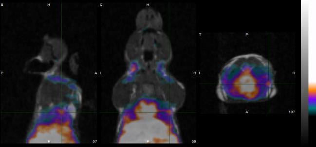
1st Faculty of Medicine, Charles University in Prague Center for Advanced Preclinical Imaging (CAPI)
|
|
|
- Kristina Nelson
- 5 years ago
- Views:
Transcription


1
2 Radioation
3 Resolution and Sensitivity
4 Nuclear Imaging PET + SPECT Radioactive Decay (EC,Ɣ), (β -,Ɣ), (I.T.,Ɣ) β + Projection imaging collimator needed one angular view Projection imaging coincidence imaging, no colimator needed complete set of angular views Single Photon Emission Computed Tomography (SPECT) Positron Emission Tomography (PET)


5 Functional Imaging Principles in Nuclear Imaging

6 Positron Emission Tomography PET
7 Positron Emission Tomography in vivo PET imaging Tomographic imaging modality Functional information Non-invasive High sensitivity pmol Short lived radioisotopes Large variety of labeled compounds Energy metabolism (FDG) Amino acid metabolism ( 18 F and 11 C labeled AA) Protein biosynthesis (DOTA conjugated puromycin analogues) Neurotransmitter Receptor imaging (neuro, onco, ) Hemodynamic parameters Gene expression Cell tracking (stem cells) 1-2 mm spacial resolution 6-10 % sensitivity temporal resolution < 0.5 sec QUANTIFIABLE


8 Positron Emission Tomography Positron Electron Annihilation Positron-emitting Rradionuclide e F 511 kev Photon Positron path-length depends on tissue density and positron energy 18 8O Positron-Electron Annihilation e + e kev Photon

9 Positron Emission Tomography Influence of Positron Energy on Resolution
10 Positron Emission Tomography Positron Emitting Radionuclides Isotope Halflife + fraction Max. Energy range(mm) production C mins MeV 0.4 mm cyclotron N mins MeV 0.7 mm cyclotron O secs MeV 1.1 mm cyclotron F mins MeV 0.3 mm cyclotron Cu mins MeV 2.7 mm generator Cu hours MeV 0.3 mm cyclotron Ga mins MeV 1.2 mm generator Br hours MeV 1.2 mm cyclotron Rb secs MeV 2.8 mm generator I days MeV 0.9 mm cyclotron
11 Positron Emission Tomography ALBIRA ɣ-ray Detector Principle PSPMT position sensitive photomultiplier tube Single continuous crystal Advanced Detector Electronics

12 Positron Emission Tomography Positron Electron Annihilation

13 Positron Emission Tomography Positron Electron Annihilation Current technology utilized packed crystals with dead zones Tighter packing yields more dead zones Susceptible to the parallax error (ignoring depth and order of interaction)

14 Positron Emission Tomography Operation of a PET-Scanner Scintilator Crystals Coincidence Unit Coincidences
15 Positron Emission Tomography ɣ-ray Detection in a PET system True Coincidences both ɣ-rays escape without scatter and interact in detctors Scatter coincidences one, or both ɣ-rays scatter in tissue Random coincidences two ɣ-rays from different origins strike the detectors at the same time (a.k.a. accidental coincidences)

16 Positron Emission Tomography Scatter Effects Buchholz et al., Eur J Nucl Med Mol Imaging (2003) 30:

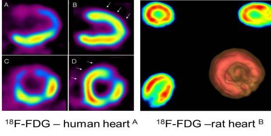
17 Positron Emission Tomography Temporal resolution Consecutive 0.3-s frames show passage of tracer bolus through RV cavity, lungs, and LV chamber of mouse on coronal and transverse slices. Times are those after start of image acquisition / injection. For better anatomic orientation, PET scan is overlaid with coregistered CT scan. Michael C. Kreissl et al. J Nucl Med 2006;47:
18 Positron Emission Tomography PET Hardware Scintilators Light-Detectors Detectortype High stopping power High light output Fast scintillator Small crystal size High spatial resolution LSO, LYSO, YAP, etc. Photomultiplier Tubes (PMT) Single Channel Multi Channel Solid State Detectors Avalanche Photo Diodes (APD) Geiger-Mode APDs Silicon-PMTs Single Crystal Coupling Block Detector Detectors with DOI capabilities (Phoswitch) A full PET system comprises several detector rings summing up to several 1000 to individual crystals The performance of a PET system as well as physical limitations will be determined by the choice of hardware
19 Positron Emission Tomography Important Scanner Parameters Energy Resolution detection limit for measured energy of detected ɣ-rays Timing Resolution time variation (inaccuracy) of the system for detection of two single events originating from the same annihilation Spatial Resolution Sensitivity smallest object that can be visualized (partial volume effect) detection limit for radiotracer (isotope) or contrast media Temporal Resolution < 0.5 sec. per frame allows for fast kinetic acquisition (e.g. first pass of tracer through heart)

20 Single Photon Emission Computed Tomography SPECT
21 Single Photon Emission computed Tomography SPECT Tomographic imaging modality Functional information Non-invasive High sensitivity nmol (not as good as PET) Longer lived radioisotopes than PET Large variety of labeled compounds mm spatial resolution temporal resolution much slower than PET Quantification nearly impossible temporal resolution > 10 sec.

22 SPECT Gamma-Radiation 99m Tc 99 Tc + Ɣ Nucleus in an excited state decays to ground state 111 In 111 Cd + Ɣ Electron capture: Nucleus possesses too many protons but is unable to emit a positron and instead captures an electron 67 Cu 67 Zn + e - + Ɣ

23 SPECT Important SPECT Radionuclides


24 SPECT Anger Camera Anger camera (NaI-scintillator and photo multipliers) Hal Oscar Anger ( )
25 SPECT Scintillation Material

26 SPECT Parallel Hole Collimator

27 SPECT Parallel Hole Collimator


28 SPECT Pinhole Collimator Camera Obscura Magnification of the projected object


29 SPECT Pinhole vs. Parallel Hole Collimator Pinhole: Parallel hole: 177 LuCl 3 bone scan in a normal mouse


30 SPECT Multiple Pinhole Technology Higher sensitivity and better resolution
")

31 SPECT Multiple Pinhole Technology - Performance Choroid Plexus (folate receptor positive organ) 99mTc-Folate (tumor and kidney FR-positiv) female nude mice with human KB-cell tumors, 24 h p.i.

32 SPECT Principle of SPECT Flat panel head used for detection Acquisition time depending on: detector, collimator size of the imaging region amount of activity available. Multiple angle detection (minimum 2 detectors (180 ))
 123 I (green-yellow) thyroid imaging 123 I (159.")
33 SPECT Dual Isotope Imaging 99m Tc-MDP (red-blue) bone scintigraphy 99m Tc (140.5 kev) 123 I (green-yellow) thyroid imaging 123 I (159.0 kev)

34 Functional Imaging Tracer Principle A radioactive tracer is a chemical compound in which one or more atoms have been replaced by a radioisotope. It is applied in minimal amounts, therefore, it has no pharmacologic effect in vivo. It can also be used to explore the mechanism of bio- /chemical reactions by tracing the path that the radioisotope follows from reactant to product George de Hevesy ( ); Nobel Prize for Chemistry in 1943 E.g. 370 MBq of 11 C-tracer necessary for a brain scan with 11 C-Raclopride (D2-receptor ligand) corresponds to 100 picogram total mass injected.


 I-131")

35 Radiopharmacy Radionuclide Production Cyclotron C-11 N-13 F-18 Cu-64 Cu-67 In Reactor (neutron bombardment) I-131 Sm-153 Ho-166 Lu-177 W-188 Radionuclide generators Sc-44 Ga-68 Tc-99m Re-188

36 Radiopharmacy Do we need so many radionuclides?


37 Radiopharmacy Radionuclides for Diagnosis Direct labeling with non-metal radionuclides Indirect labeling strategies via bifunctional chelators 60 % of suitable radionuclides are metals!


38 Radiopharmacy Radiolabeling Critical Issues of Functionalization organic inorganic Labeling yields Synthetic steps Avoid cross reactivity with other functional groups Avoid mixtures of products and formation of isomers Optimal pharmacokinetic Retention of biological activity and integrity
39 Radiopharmacy Radiotracer Production

40 THANK YOU FOR YOUR ATTENTION
Radioisotopes in action. Diagnostic application of radioisotopes. Steps of diagnostic procedure. Information from various medical imaging techniques
 Radioisotopes in action Diagnostic application of radioisotopes Steps of diagnostic procedure - Radioactive material introduced into the patient - Distribution and alteration of activity is detected -
 www.aask24.com www.aask24.com www.aask24.com P=Positron E= Emission T=Tomography Positron emission or beta plus decay (+ ) is a particular type of radioactive decay, in which a proton inside a radionuclide
Introduction to SPECT & PET TBMI02 - Medical Image Analysis 2017
 Introduction to SPECT & PET TBMI02 - Medical Image Analysis 2017 Marcus Ressner, PhD, Medical Radiation Physicist, Linköping University Hospital Content What is Nuclear medicine? Basic principles of Functional
Radionuclide Imaging MII Positron Emission Tomography (PET)
") Radionuclide Imaging MII 3073 Positron Emission Tomography (PET) Positron (β + ) emission Positron is an electron with positive charge. Positron-emitting radionuclides are most commonly produced in cyclotron
Radionuclide Imaging MII 3073 Positron Emission Tomography (PET) Positron (β + ) emission Positron is an electron with positive charge. Positron-emitting radionuclides are most commonly produced in cyclotron
Radioisotopes in action. Diagnostic application of radioisotopes. Steps of diagnostic procedure. Information from various medical imaging techniques
 Radioisotopes in action Diagnostic application of radioisotopes Steps of diagnostic procedure - Radioactive material introduced into the patient - Distribution and alteration of activity is detected -Monitoring
Radioisotopes and PET
 Radioisotopes and PET 1 Radioisotopes Elements are defined by their number of protons, but there is some variation in the number of neutrons. Atoms resulting from this variation are called isotopes. Consider
Nuclear Medicine Intro & Physics from Medical Imaging Signals and Systems, Chapter 7, by Prince and Links
 Nuclear Medicine Intro & Physics from Medical Imaging Signals and Systems, Chapter 7, by Prince and Links NM - introduction Relies on EMISSION of photons from body (versus transmission of photons through
Medical Physics. Nuclear Medicine Principles and Applications
 Medical Physics Nuclear Medicine Principles and Applications Dr Roger Fulton Department of PET & Nuclear Medicine Royal Prince Alfred Hospital Sydney Email: rfulton@mail.usyd.edu.au Lectures: http://www-personal.usyd.edu.au/~rfulton/medical_physics
Radiochemistry and Radiopharmacy III
 Radiochemistry and Radiopharmacy III Compact course held at UFSCAR, September 20123 Ulrich Abram Freie Universität Berlin Institute of Chemistry and Biochemistry Radiochemistry and Radiopharmacy 1. Fundamentals
MEDICAL EQUIPMENT: NUCLEAR MEDICINE. Prof. Yasser Mostafa Kadah
 MEDICAL EQUIPMENT: NUCLEAR MEDICINE Prof. Yasser Mostafa Kadah www.k-space.org Recommended Textbook Introduction to Medical Imaging: Physics, Engineering and Clinical Applications, by Nadine Barrie Smith
A. I, II, and III B. I C. I and II D. II and III E. I and III
 BioE 1330 - Review Chapters 7, 8, and 9 (Nuclear Medicine) 9/27/2018 Instructions: On the Answer Sheet, enter your 2-digit ID number (with a leading 0 if needed) in the boxes of the ID section. Fill in
Bases of radioisotope diagnostic methods
 Medical, pharmaceutical applications of radioisotopes Bases of radioisotope diagnostic methods Dr. István Voszka Basis of application: radioisotopes have identical behavior in the organism to corresponding
Mayneord-Phillips Summer School St Edmund Hall, University of Oxford July Proton decays to n, e +, ν
 Positron Emission Tomography Physics & Instrumentation Dimitra G. Darambara, Ph.D Multimodality Molecular Imaging Joint Department of Physics RMH/ICR Outline Introduction PET Physics overview Types of
Detector technology. Aim of this talk. Principle of a radiation detector. Interactions of gamma photons (gas) Gas-filled detectors: examples
 Gas-filled detectors: examples") Aim of this tal Detector technology WMIC Educational Program Nuclear Imaging World Molecular Imaging Congress, Dublin, Ireland, Sep 5-8, 202 You can now the name of a bird in all the languages of the world,
Aim of this tal Detector technology WMIC Educational Program Nuclear Imaging World Molecular Imaging Congress, Dublin, Ireland, Sep 5-8, 202 You can now the name of a bird in all the languages of the world,
Outline Chapter 14 Nuclear Medicine
 Outline Chapter 14 uclear Medicine Radiation Dosimetry I Text: H.E Johns and J.R. Cunningham, The physics of radiology, 4 th ed. http://www.utoledo.edu/med/depts/radther Introduction Detectors for nuclear
What is scintigraphy? The process of obtaining an image or series of sequential images of the distribution of a radionuclide in tissues, organs, or
 Let's remind... What is nuclear medicine? Nuclear medicine can be broadly divided into two branches "in vitro" and "in vivo" procedures. There are numerous radioisotopic "in vitro" procedures for genotyping
6: Positron Emission Tomography
 6: Positron Emission Tomography. What is the principle of PET imaging? Positron annihilation Electronic collimation coincidence detection. What is really measured by the PET camera? True, scatter and random
Radiation Detectors. How do we detect ionizing radiation? What are these effects? Types of Ionizing Radiation Detectors
 Radiation Detectors 1 How do we detect ionizing radiation? Indirectly, by its effects as it traverses matter? What are these effects? Ionization and excitation of the atoms and molecules Heat 2 Types of
69 Ga Ga
 Stable isotope Relative atomic mass Mole fraction 69 Ga 68.925 574 0.601 08 71 Ga 70.924 703 0.398 92 Gallium isotopes in medicine 68 Ga is a radioactive isotope that emits positrons, which are used to
Physics in Nuclear Medicine
 SIMON R. CHERRY, PH.D. Professor Department of Biomedical Engineering University of California-Davis Davis, California JAMES A. SORENSON, PH.D. Emeritus Professor of Medical Physics University of Wisconsin-Madison
Dana-Farber Cancer Institute, 44 Binney Street, Boston, MA 02115, USA ramsey
 SPECIAL FEATURE: MEDICAL PHYSICS www.iop.org/journals/physed Nuclear medicine Ramsey D Badawi Dana-Farber Cancer Institute, 44 Binney Street, Boston, MA 02115, USA E-mail: ramsey badawi@dfci.harvard.edu
3. Which of the following statements is (are) TRUE about detector crystals in Anger cameras?
 TRUE about detector crystals in Anger cameras?") BioE 1330 - Exam 2 11/13/2018 Answer Sheet - Correct answer is A for all questions 1. Unlike CT, in nuclear medicine A. Bremsstrahlung is not used to produce high-energy photons. B. signal can be increased
BioE 1330 - Exam 2 11/13/2018 Answer Sheet - Correct answer is A for all questions 1. Unlike CT, in nuclear medicine A. Bremsstrahlung is not used to produce high-energy photons. B. signal can be increased
Year 12 Notes Radioactivity 1/5
 Year Notes Radioactivity /5 Radioactivity Stable and Unstable Nuclei Radioactivity is the spontaneous disintegration of certain nuclei, a random process in which particles and/or high-energy photons are
EEE4106Z Radiation Interactions & Detection
 EEE4106Z Radiation Interactions & Detection 2. Radiation Detection Dr. Steve Peterson 5.14 RW James Department of Physics University of Cape Town steve.peterson@uct.ac.za May 06, 2015 EEE4106Z :: Radiation
Tomography is imaging by sections. 1
 Tomography is imaging by sections. 1 It is a technique used in clinical medicine and biomedical research to create images that show how certain tissues are performing their physiological functions. 1 Conversely,
PET scan simulation. Meysam Dadgar. UMSU, Iran. IFMP, Elbasan, Fig 1: PET camera simulation in gate by cylindrical phantom
 PET scan simulation Meysam Dadgar UMSU, Iran IFMP, Elbasan, 2016 Meysamdadgar10@gmail.com 1 Fig 1: PET camera simulation in gate by cylindrical phantom 2 What is PET? Positron emission tomography (PET),
Nuclear Reactions A Z. Radioactivity, Spontaneous Decay: Nuclear Reaction, Induced Process: x + X Y + y + Q Q > 0. Exothermic Endothermic
 Radioactivity, Spontaneous Decay: Nuclear Reactions A Z 4 P D+ He + Q A 4 Z 2 Q > 0 Nuclear Reaction, Induced Process: x + X Y + y + Q Q = ( m + m m m ) c 2 x X Y y Q > 0 Q < 0 Exothermic Endothermic 2
Nuclear Radiation. Natural Radioactivity. A person working with radioisotopes wears protective clothing and gloves and stands behind a shield.
 Nuclear Radiation Natural Radioactivity A person working with radioisotopes wears protective clothing and gloves and stands behind a shield. 1 Radioactive Isotopes A radioactive isotope has an unstable
Application of Nuclear Physics
 Application of Nuclear Physics Frontier of gamma-ray spectroscopy 0.1 IR visible light UV soft X-ray X-ray hard X-ray gamma-ray 1 10 100 1e3 1e4 1e5 1e6 energy [ev] Photoelectric effect e - Compton scattering
What is Albira and How Does the System Work and Why is it Differentiated Technology? Page 1
 What is Albira and How Does the System Work and Why is it Differentiated Technology? Page 1 Why Albira? Tri-modality: PET, SPECT, CT Modular and state-of-the-art electronics 6 configurations Novel, Proprietary
Chapter 2 PET Imaging Basics
 Chapter 2 PET Imaging Basics Timothy G. Turkington PET Radiotracers Positron emission tomography (PET) imaging is the injection (or inhalation) of a substance containing a positron emitter, the subsequent
Positron Emission Tomography
 Positron Emission Tomography Presenter: Difei Wang June,2018 Universität Bonn Contents 2 / 24 1 2 3 4 Positron emission Detected events Detectors and configuration Data acquisition Positron emission Positron
Nuclear Physics and Astrophysics
 Nuclear Physics and Astrophysics PHY-302 Dr. E. Rizvi Lecture 24 Medical Imaging Effects of Radiation We now know what radiation is But what does it mean for our bodies? Radioactivity is quantified in
Lecture Presentation. Chapter 21. Nuclear Chemistry. James F. Kirby Quinnipiac University Hamden, CT Pearson Education, Inc.
 Lecture Presentation Chapter 21, Inc. James F. Kirby Quinnipiac University Hamden, CT Energy: Chemical vs. Chemical energy is associated with making and breaking chemical bonds. energy is enormous in comparison.
Nuclear Medicine Treatments and Clinical Applications
 INAYA MEDICAL COLLEGE (IMC) RAD 243- LECTURE 2 Nuclear Medicine Treatments and Clinical Applications DR. MOHAMMED MOSTAFA EMAM Next Lectures Outlines Introduction to Nuclear Physics Physics of Radioactivity
Dosimetry of patients injected with tracers Ga-68, Zr-89 and Lu-177. Bruno Vanderlinden
 Dosimetry of patients injected with tracers Ga-68, Zr-89 and Lu-177 Bruno Vanderlinden What is NM speciality? Imaging radiology Physics Diagnostic Treatment assessment Clinical pathology Biological marker
Differentiating Chemical Reactions from Nuclear Reactions
 Differentiating Chemical Reactions from Nuclear Reactions 1 CHEMICAL Occurs when bonds are broken or formed. Atoms remained unchanged, though may be rearranged. Involves valence electrons Small energy
There are three mechanisms by which gamma rays interact with absorber atoms from which two are important for nuclear medicine.
 Measurement of radioactivity. Radioactive decay is a random process and therefore fluctuations are expected in the radioactivity measurement. That is why measurement of radioactivity must be treated by
Nuclear Medicine: Physics and Imaging Methods (SPECT and PET)
") EL-GY 6813 / BE-GY 6203 / G16.4426 Medical Imaging Nuclear Medicine: Physics and Imaging Methods (SPECT and PET) Jonathan Mamou and Yao Wang Polytechnic School of Engineering New York University, Brooklyn,
EL-GY 6813 / BE-GY 6203 / G16.4426 Medical Imaging Nuclear Medicine: Physics and Imaging Methods (SPECT and PET) Jonathan Mamou and Yao Wang Polytechnic School of Engineering New York University, Brooklyn,
Radiochemistry in nuclear medicine
 Lecce, Jan 14 2011 Radiochemistry in nuclear medicine Giancarlo Pascali, PhD radiochemist IFC-CNR, Pisa pascali@ifc.cnr.it Do not say contrast media (but Contrast media Mainly anatomical High injected
PET/MRI Principle, History, and Perspective. Main Imaging Techniques. X-ray Tube. History of X-ray & CT. How to Look inside the Human Body
 PET/MRI Principle, History, and Perspective Jae Sung Lee, PhD Dept. of Nuclear Medicine and Biomedical Sciences WCU Dept. of Brain and Cognitive Sciences Seoul National University Basic Imaging Principles
Nuclear Medicine RADIOPHARMACEUTICAL CHEMISTRY
 Nuclear Medicine RADIOPHARMACEUTICAL CHEMISTRY An alpha particle consists of two protons and two neutrons Common alpha-particle emitters Radon-222 gas in the environment Uranium-234 and -238) in the environment
Radioactivity. The Nobel Prize in Physics 1903 for their work on radioactivity. Henri Becquerel Pierre Curie Marie Curie
 Radioactivity Toward the end of the 19 th century, minerals were found that would darken a photographic plate even in the absence of light. This phenomenon is now called radioactivity. Marie and Pierre
Nuclear Medicine: Physics and Imaging Methods (SPECT and PET)
") EL-GY 6813 / BE-GY 6203 / G16.4426 Medical Imaging Nuclear Medicine: Physics and Imaging Methods (SPECT and PET) Yao Wang Polytechnic School of Engineering New York University, Brooklyn, NY 11201 Based
EL-GY 6813 / BE-GY 6203 / G16.4426 Medical Imaging Nuclear Medicine: Physics and Imaging Methods (SPECT and PET) Yao Wang Polytechnic School of Engineering New York University, Brooklyn, NY 11201 Based
Gamma ray coincidence and angular correlation
 University of Cape Town Department of Physics Course III laboratory Gamma ray coincidence and angular correlation Introduction Medical imaging based on positron emission tomography (PET) continues to have
General Physics (PHY 2140)
") General Physics (PHY 2140) Lecture 19 Modern Physics Nuclear Physics Nuclear Reactions Medical Applications Radiation Detectors Chapter 29 http://www.physics.wayne.edu/~alan/2140website/main.htm 1 Lightning
General Physics (PHY 2140) Lecture 19 Modern Physics Nuclear Physics Nuclear Reactions Medical Applications Radiation Detectors Chapter 29 http://www.physics.wayne.edu/~alan/2140website/main.htm 1 Lightning
General Physics (PHY 2140)
") General Physics (PHY 2140) Lightning Review Lecture 19 Modern Physics Nuclear Physics Nuclear Reactions Medical Applications Radiation Detectors Chapter 29 http://www.physics.wayne.edu/~alan/2140website/main.htm
General Physics (PHY 2140) Lightning Review Lecture 19 Modern Physics Nuclear Physics Nuclear Reactions Medical Applications Radiation Detectors Chapter 29 http://www.physics.wayne.edu/~alan/2140website/main.htm
β and γ decays, Radiation Therapies and Diagnostic, Fusion and Fission Final Exam Surveys New material Example of β-decay Beta decay Y + e # Y'+e +
 β and γ decays, Radiation Therapies and Diagnostic, Fusion and Fission Last Lecture: Radioactivity, Nuclear decay Radiation damage This lecture: nuclear physics in medicine and fusion and fission Final
Chapter. Nuclear Chemistry
 Chapter Nuclear Chemistry Nuclear Reactions 01 Chapter 22 Slide 2 Chapter 22 Slide 3 Alpha Decay: Loss of an α-particle (a helium nucleus) 4 2 He 238 92 U 234 4 U He 90 + 2 Chapter 22 Slide 4 Beta Decay:
Chapter 11 Nuclear Chemistry
 Chapter 11 Nuclear Chemistry 11.1 Nuclear Reactions Nuclear reactions involve the particles located in the nucleus of the atom: The nucleus contains: An atom is characterized by: X A Z - Z the gives the
Bioimage Informatics. Lecture 23, Spring Emerging Applications: Molecular Imaging
 Bioimage Informatics Lecture 23, Spring 2012 Emerging Applications: Molecular Imaging Lecture 23 April 25, 2012 1 Outline Overview of molecular imaging Molecular imaging modalities Molecular imaging applications
Compton Camera. Compton Camera
 Diagnostic Imaging II Student Project Compton Camera Ting-Tung Chang Introduction The Compton camera operates by exploiting the Compton Effect. It uses the kinematics of Compton scattering to contract
Positron Annihilation in Material Research
 Positron Annihilation in Material Research Introduction Positron sources, positron beams Interaction of positrons with matter Annihilation channels: Emission of 1, 2 or 3 γ-quanta Annihilation spectroscopies:
Technical University of Denmark
 Technical University of Denmark Page 1 of 11 pages Written test, 9 December 2010 Course name: Introduction to medical imaging Course no. 31540 Aids allowed: none. "Weighting": All problems weight equally.
PRODUCTION OF RADIOISOTOPES FOR IMAGING AND THERAPY AT LOW ENERGY
 PRODUCTION OF RADIOISOTOPES FOR IMAGING AND THERAPY AT LOW ENERGY THOMAS J. RUTH TRIUMF Vancouver, BC, Canada truth@triumf.ca 1 Introduction The production of radioisotopes for use in biomedical procedures
Study of the feasibility of a compact gamma camera for real-time cancer assessment
 Study of the feasibility of a compact gamma camera for real-time cancer assessment L. Caballero Instituto de Física Corpuscular - CSIC - University of Valencia; C/Catedrático José Beltrán, 2; E-46980;
Overview of Nuclear Medical Imaging Instrumentation and Techniques*
 Overview of Nuclear Medical Imaging Instrumentation and Techniques* William W. Moses Lawrence Berkeley National Laboratory, University of California, Berkeley, CA 94720 USA Abstract. Nuclear medical imaging
Name Date Class NUCLEAR RADIATION. alpha particle beta particle gamma ray
 25.1 NUCLEAR RADIATION Section Review Objectives Explain how an unstable nucleus releases energy Describe the three main types of nuclear radiation Vocabulary radioisotopes radioactivity radiation alpha
Atomic Notation (or Nuclear Symbol): Shorthand for keeping track of protons and neutrons in the nucleus
: Shorthand for keeping track of protons and neutrons in the nucleus") Name Section CHM52LL: Nuclear Chemistry: Radioactivity, Decay, Dating, and Other Hazards There is no prelab assignment this week I. Radioactive Isotopes and Nuclear Equations Atoms are composed of three
Name Section CHM52LL: Nuclear Chemistry: Radioactivity, Decay, Dating, and Other Hazards There is no prelab assignment this week I. Radioactive Isotopes and Nuclear Equations Atoms are composed of three
Chapter 20 Nuclear Chemistry. 1. Nuclear Reactions and Their Characteristics
 Chapter 2 Nuclear Chemistry 1. Nuclear Reactions and Their Characteristics Nuclear reactions involve the particles located in the nucleus of the atom: nucleons:. An atom is characterized by its atomic
The Physics of PET/CT scanners
 The Physics of PET/CT scanners Ruth E. Schmitz, Adam M. Alessio, and Paul E. Kinahan Imaging Research Laboratory Department of Radiology University of Washington What Makes PET Useful? Positron emission
PET. Technical aspects
 PET Technical aspects 15 N 15 O Detector 1 β+ Detector 2 e- Evolution of PET Detectors CTI/Siemens 15 N 15 O Detector block 1 β+ Detector block 2 x e- x y y location line of response Constant fraction
MEDICAL IMAGING. METHODS OF MODERN IMAGING, BASED ON ELECTRO-MAGNETIC RADIATION (radiowaves, infrared radiation, X-rays, γ-rays ) AND ULTRASOUND
 AND ULTRASOUND") MEDICAL IMAGING MEDICAL IMAGING METHODS OF MODERN IMAGING, BASED ON ELECTRO-MAGNETIC RADIATION (radiowaves, infrared radiation, X-rays, γ-rays ) AND ULTRASOUND MEDICAL IMAGING RADIOLOGY NUCLEAR MEDICINE
MEDICAL IMAGING MEDICAL IMAGING METHODS OF MODERN IMAGING, BASED ON ELECTRO-MAGNETIC RADIATION (radiowaves, infrared radiation, X-rays, γ-rays ) AND ULTRASOUND MEDICAL IMAGING RADIOLOGY NUCLEAR MEDICINE
CLINICALLY USEFUL RADIONUCLIDES:
 INTRODUCTION It is important that Nuclear Medicine Technologists be familiar with the imaging properties of all commonly used radionuclides to insure correct choice of isotope for a particular study as
RADIOCHEMICAL METHODS OF ANALYSIS
 RADIOCHEMICAL METHODS OF ANALYSIS 1 Early Pioneers in Radioactivity Rutherfo rd: Discoverer Alpha and Beta rays 1897 Roentge n: Discoverer of X- rays 1895 The Curies: Discoverers of Radium and Polonium
III. Proton-therapytherapy. Rome SB - 2/5 1
 Outline Introduction: an historical review I Applications in medical diagnostics Particle accelerators for medicine Applications in conventional radiation therapy II III IV Hadrontherapy, the frontier
Isotope Production for Nuclear Medicine
 Isotope Production for Nuclear Medicine Eva Birnbaum Isotope Program Manager February 26 th, 2016 LA-UR-16-21119 Isotopes for Nuclear Medicine More than 20 million nuclear medicine procedures are performed
Applied Nuclear Physics (Fall 2006) Lecture 21 (11/29/06) Detection of Nuclear Radiation: Pulse Height Spectra
 Lecture 21 (11/29/06) Detection of Nuclear Radiation: Pulse Height Spectra") 22.101 Applied Nuclear Physics (Fall 2006) Lecture 21 (11/29/06) Detection of Nuclear Radiation: Pulse Height Spectra References: W. E. Meyerhof, Elements of Nuclear Physics (McGraw-Hill, New York, 1967),
22.101 Applied Nuclear Physics (Fall 2006) Lecture 21 (11/29/06) Detection of Nuclear Radiation: Pulse Height Spectra References: W. E. Meyerhof, Elements of Nuclear Physics (McGraw-Hill, New York, 1967),
MINERVA MEDICA COPYRIGHT
 Q J NUCL MED MOL IMAGING 2008;52:151-8 Image quality with non-standard nuclides in PET Non-standard positron emission tomography (PET) nuclides bring with them the prospect of new chemistry leading the
GLOSSARY OF BASIC RADIATION PROTECTION TERMINOLOGY
 GLOSSARY OF BASIC RADIATION PROTECTION TERMINOLOGY ABSORBED DOSE: The amount of energy absorbed, as a result of radiation passing through a material, per unit mass of material. Measured in rads (1 rad
Mitigation of External Radiation Exposures
 Mitigation of External Radiation Exposures The three (3) major principles to assist with maintaining doses ALARA are :- 1) Time Minimizing the time of exposure directly reduces radiation dose. 2) Distance
Nuclear Physics and Astrophysics
 Nuclear Physics and Astrophysics PHY-30 Dr. E. Rizvi Lecture 4 - Detectors Binding Energy Nuclear mass MN less than sum of nucleon masses Shows nucleus is a bound (lower energy) state for this configuration
DETECTORS. I. Charged Particle Detectors
 DETECTORS I. Charged Particle Detectors A. Scintillators B. Gas Detectors 1. Ionization Chambers 2. Proportional Counters 3. Avalanche detectors 4. Geiger-Muller counters 5. Spark detectors C. Solid State
Chapter 21 Nuclear Chemistry
 Chapter 21 Nuclear Chemistry The Nucleus Remember that the nucleus is comprised of the two nucleons, protons and neutrons. The number of protons is the atomic number. The number of protons and neutrons
ELG7173 Topics in signal Processing II Computational Techniques in Medical Imaging
 ELG7173 Topics in signal Processing II Computational Techniques in Medical Imaging Topic #1: Intro to medical imaging Medical Imaging Classifications n Measurement physics Send Energy into body Send stuff
Properties of the nucleus. 9.1 Nuclear Physics. Isotopes. Stable Nuclei. Size of the nucleus. Size of the nucleus
 Properties of the nucleus 9. Nuclear Physics Properties of nuclei Binding Energy Radioactive decay Natural radioactivity Consists of protons and neutrons Z = no. of protons (tomic number) N = no. of neutrons
Structure of the course
 Structure of the course 1) Introduc1on 2) Interac1on of par1cles with ma9er } principles / tools 3) Therapy with proton and ion beams 4) Sources for nuclear medicine 5) X- ray sources sources 6) Image
Fundamentals of Radionuclide Metrology
 Fundamentals of Radionuclide Metrology Brian E. Zimmerman, PhD Physical Measurement Laboratory National Institute of Standards and Technology Gaithersburg, MD USA SIM Metrology Workshop Buenos Aires, Argentina
Essentials of nuclear medicine
 Essentials of nuclear medicine Medical imaging CT Rtg X- rays usg Ultrasound MR Nuclear Magnetic Resonance Nuclear Medicine SPECT PET A conventional radiological, ultrasound and magnetic resonance diagnostics
A Brief Introduction to Medical Imaging. Outline
 A Brief Introduction to Medical Imaging Outline General Goals Linear Imaging Systems An Example, The Pin Hole Camera Radiations and Their Interactions with Matter Coherent vs. Incoherent Imaging Length
A dual scintillator - dual silicon photodiode detector module for intraoperative gamma\beta probe and portable anti-compton spectrometer
 University of Wollongong Research Online Faculty of Engineering - Papers (Archive) Faculty of Engineering and Information Sciences 2008 A dual scintillator - dual silicon photodiode detector module for
Some nuclei are unstable Become stable by ejecting excess energy and often a particle in the process Types of radiation particle - particle
 Radioactivity George Starkschall, Ph.D. Lecture Objectives Identify methods for making radioactive isotopes Recognize the various types of radioactive decay Interpret an energy level diagram for radioactive
Neutron Detection. n interactions with matter n detection High/low energy n detectors
 Neutron Detection Example of n detection: Well logging Reservoir/Formation Evaluation Brief introduction to neutron generation Continuous sources Large accelerators Pulsed neutron generators n interactions
QUIZ: Physics of Nuclear Medicine Atomic Structure, Radioactive Decay, Interaction of Ionizing Radiation with Matter
 QUIZ: Physics of Nuclear Medicine Atomic Structure, Radioactive Decay, Interaction of Ionizing Radiation with Matter 1. An atomic nucleus contains 39 protons and 50 neutrons. Its mass number (A) is a)
Prompt gamma measurements for the verification of dose deposition in proton therapy. Contents. Two Proton Beam Facilities for Therapy and Research
 Prompt gamma measurements for the verification of dose deposition in proton therapy Two Proton Beam Facilities for Therapy and Research Ion Beam Facilities in Korea 1. Proton therapy facility at National
Positron Emission Tomography
 Positron Emission Tomography CERN Accelerator School Small Accelerators Zeegse, the Netherlands A.M.J. Paans Nuclear Medicine & Molecular Imaging UMC Groningen Elements of Life PET-nuclide Hydrogen Carbon
Functional Neuroimaging with PET
 Functional Neuroimaging with PET Terry Oakes troakes@wisc.edu W.M.Keck Lab for Functional Brain Imaging and Behavior Seeing the Brain Just look at it! Anatomic Images (MRI) Functional Images PET fmri (Just
Chapter 21
 Chapter 21 http://youtu.be/kwasz59f8ga Nuclear reactions involve the nucleus The nucleus opens, and protons and neutrons are rearranged. The opening of the nucleus releases a tremendous amount of energy
Current Challenges and Opportunities in Positron Emission Tomography. Paul Marsden King s College London
 Current Challenges and Opportunities in Positron Emission Tomography Paul Marsden King s College London STFC Particle Physics Department Rutherford Appleton Laboratory - 3 Sept 2014 Current Challenges
Dosimetry. Sanja Dolanski Babić May, 2018.
 Dosimetry Sanja Dolanski Babić May, 2018. What s the difference between radiation and radioactivity? Radiation - the process of emitting energy as waves or particles, and the radiated energy Radioactivity
APPLIED RADIATION PHYSICS
 A PRIMER IN APPLIED RADIATION PHYSICS F A SMITH Queen Mary & Westfield College, London fe World Scientific m Singapore * New Jersey London Hong Kong CONTENTS CHAPTER 1 : SOURCES of RADIATION 1.1 Introduction
Procesamiento de Imágenes y Bioseñales
 Procesamiento de Imágenes y Bioseñales Dr. Víctor Castañeda Agenda Physical basis of X-ray- CT, NMR, Ultrasound, Nuclear Medicine Sensors (cameras, gamma probes, microphone) Computational Tomography (CT)
Structure of Biological Materials
 ELEC ENG 3BA3: Structure of Biological Materials Notes for Lecture #19 Monday, November 22, 2010 6.5 Nuclear medicine imaging Nuclear imaging produces images of the distribution of radiopharmaceuticals
CHIPP Plenary Meeting University of Geneva, June 12, 2008 W. Lustermann on behalf of the AX PET Collaboration
 CHIPP Plenary Meeting University of Geneva, June 12, 2008 W. Lustermann on behalf of the AX PET Collaboration INFN Bari, Ohio State University, CERN, University of Michigan, University of Oslo, INFN Roma,
12/1/17 OUTLINE KEY POINTS ELEMENTS WITH UNSTABLE NUCLEI Radioisotopes and Nuclear Reactions 16.2 Biological Effects of Nuclear Radiation
 OUTLINE 16.1 Radioisotopes and Nuclear Reactions 16.2 Biological Effects of Nuclear Radiation PET scan X-ray technology CT scan 2009 W.H. Freeman KEY POINTS Radioactivity is the consequence of an unstable
DEVIL PHYSICS THE BADDEST CLASS ON CAMPUS IB PHYSICS
 DEVIL PHYSICS THE BADDEST CLASS ON CAMPUS IB PHYSICS TSOKOS OPTION I-2 MEDICAL IMAGING Reading Activity Answers IB Assessment Statements Option I-2, Medical Imaging: X-Rays I.2.1. I.2.2. I.2.3. Define
Name Date Class. alpha particle radioactivity gamma ray radioisotope beta particles radiation X-ray radioactive decay
 Name Date _ Class _ Nuclear Chemistry Section.1 Nuclear Radiation In your textbook, read about the terms used to describe nuclear changes. Use each of the terms below just once to complete the passage.
Nuclear Chemistry. Chapter 23
 Nuclear Chemistry Chapter 23 n/p too large beta decay X Y n/p too small positron decay or electron capture Nuclear Stability Certain numbers of neutrons and protons are extra stable n or p = 2, 8, 20,
22.56J Noninvasive Imaging in Biology and Medicine Instructor: Prof. Alan Jasanoff Fall 2005, TTh 1-2:30
 22.56J Noninvasive Imaging in Biology and Medicine Instructor: Prof. Alan Jasanoff Fall 2005, TTh 1-2:30 Sample problems HW1 1. Look up (e.g. in the CRC Manual of Chemistry and Physics www.hbcpnetbase.com)
Properties of the nucleus. 8.2 Nuclear Physics. Isotopes. Stable Nuclei. Size of the nucleus. Size of the nucleus
 Properties of the nucleus 8. Nuclear Physics Properties of nuclei Binding Energy Radioactive decay Natural radioactivity Consists of protons and neutrons Z = no. of protons (Atomic number) N = no. of neutrons