Application of Nuclear Physics
|
|
|
- Justin Parks
- 5 years ago
- Views:
Transcription


![1e6 energy [ev] Photoelectric effect e -](/docs-images/96/127011453/images/1-4.jpg "Compton scattering e - Germanium")
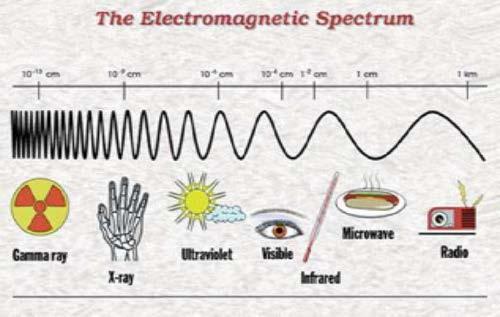
1 Application of Nuclear Physics Frontier of gamma-ray spectroscopy 0.1 IR visible light UV soft X-ray X-ray hard X-ray gamma-ray e3 1e4 1e5 1e6 energy [ev] Photoelectric effect e - Compton scattering e - Germanium semi-conductor detectors


2 Why imaging gamma-rays? High energy astrophysics Correlate the detected photon to source object as known from more precise observations in other wavelength Biomedical research Precise localization of radioactive tracers in the body Cancer diagnosis Molecular targeted radiation therapy Monitor changes in the tracer distribution dynamic studies National security Nuclear non-proliferation / nuclear counter terrorism Contraband detection Stockpile stewardship Nuclear waste monitoring and management Industrial non-destructive assessments Determination of the material density distribution between the source and detector
 Standard X-ray imaging Absorption of X-rays by bones and")

3 Application of Nuclear Physics First X-ray image by Wilhelm Conrad Roentgen (1895) Standard X-ray imaging Absorption of X-rays by bones and transmission through soft tissue produces image What if we want a 3D image? What if we want detailed information on organs, bones and muscles etc? Nobel Prize 1901

4 Tomographic Imaging PET SPEC CT OPTICAL MRI


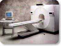
5 Computed Tomography CT scanning (originally known as CAT) X-rays taken at a range of angles around the patient Generation of 3D images Radiation detector X-ray source Standard CT-system

6 Positron Emission Tomography

7 Positron emission tomography

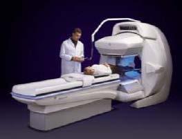
8 Single Photon Emission Computed Tomography Most commonly used tracer in SPECT is 99 Tc m, 140 kev a pure single photon emitter T 1/2 = 6.02 h. Utilizes a gamma-ray camera rotated in small ~3 0 steps around the patient.

9 The motivation behind the project Existing technology relies on BGO scintillator technology - Limited position resolution - High patient dose requirement. - Poor energy resolution only accept photopeak events. - Will not function in large magnetic field SPECT applications utilizing Compton Camera techniques.


10 Which γ-ray camera to be used? Requirements: Excellent resolution Δx = 2 mm Large field of view (FOV) = 8x9 cm 2 Large FOV of ~20 cm diam. Low spatial resolution cm Small FOV of 3-4 cm diam. High spatial resolution 2-3 mm Gem-imaging.com


11 Gamma Camera: Individual multi-anode readout 16 wires in X axis and 16 wires in Y axis LYSO scintillator d = 76 mm t = 3 mm ρ = 7.4 g/cm 3 Hamamatsu R2486 PSPMT C.Domingo Pardo, N. Goel, et.al., IEEE, Vol.28, Dec Photocathode = mm
12 Which γ-ray camera to be used? Requirements: Excellent resolution Δx = 2 mm Large field of view (FOV) = 8x9 cm 2 Small FOV of 3-4 cm diam. High spatial resolution 2-3 mm Scintillator Position Sensitive PMT Gem-imaging.com
13 Which γ-ray camera to be used? Requirements: Excellent resolution Δx = 2 mm Large field of view (FOV) = 8x9 cm 2 γ-ray Small FOV of 3-4 cm diam. High spatial resolution 2-3 mm Scintillator Position Sensitive PMT Gem-imaging.com
14 Which γ-ray camera to be used? Requirements: Excellent resolution Δx = 2 mm Large field of view (FOV) = 8x9 cm 2 γ-ray Small FOV of 3-4 cm diam. High spatial resolution 2-3 mm Scintillator Position Sensitive PMT Gem-imaging.com
15 Which γ-ray camera to be used? Requirements: Excellent resolution Δx = 2 mm Large field of view (FOV) = 8x9 cm 2 γ-ray Small FOV of 3-4 cm diam. High spatial resolution 2-3 mm Scintillator Position Sensitive PMT Gem-imaging.com
16 Which γ-ray camera to be used? Requirements: Excellent resolution Δx = 2 mm Large field of view (FOV) = 8x9 cm 2 γ-ray 2 γ-ray Small FOV of 3-4 cm diam. High spatial resolution 2-3 mm Scintillator Position Sensitive PMT Gem-imaging.com
17 HPGe detector working principle
18 HPGe detector working principle E = -V V + HV = 4000 Volts
19 HPGe detector working principle E = -V V γ-ray + HV = 4000 Volts electrons holes
20 HPGe detector working principle E = -V V γ-ray + HV = 4000 Volts electrons holes Electric signal
21 HPGe detector working principle E = -V V γ-ray + HV = 4000 Volts electrons holes Electric signal
22 HPGe detector working principle E = -V V γ-ray + HV = 4000 Volts electrons holes Electric signal
23 HPGe detector working principle E = -V V γ-ray γ-ray 2 electrons holes + HV = 4000 Volts Electric signal
24 HPGe detector working principle E = -V V γ-ray + HV = 4000 Volts electrons holes Electric signal


25 Characterization of planar HPGe detector Front view Side view Planar Ge d = 4 cm 22 Na Position sensitive detector t = 2 cm


 t(ns)")
26 Planar HPGe detector scan Intensity distribution for photopeak events (a) t(ns) amplitude, Φ (b) t(ns)


27 Outlook The GSI system uses conventional NIM and VME electronics, which makes it not easily portable, not easily scalable and rather expensive if one wants to build many of these devices. However, this drawback could be overcome thanks to the increasing technology of electronics, e.g. a new acquisition system based on ASIC, FPGA, etc. technologies. This would also make the system more suitable for medical applications. APV25 chip (from CERN CMS experiment)? 128-channel analogue pipeline chip M.J. French et al., NIMA 466 (2001) 359
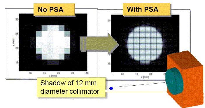
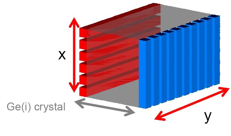
28 Position Extraction in Planar Detectors

29 Position Extraction in Planar Detectors

30 Gamma-Ray Imaging Gamma-ray imagers are detectors that separate radioactive objects from local background. Conventional detectors accept gamma-rays from all directions and can be overwhelmed by local backgrounds.
MEDICAL EQUIPMENT: NUCLEAR MEDICINE. Prof. Yasser Mostafa Kadah
 MEDICAL EQUIPMENT: NUCLEAR MEDICINE Prof. Yasser Mostafa Kadah www.k-space.org Recommended Textbook Introduction to Medical Imaging: Physics, Engineering and Clinical Applications, by Nadine Barrie Smith
Dual Isotope Imaging with LaBr3:Ce Crystal and H8500 PSPMT
 Dual Isotope Imaging with LaBr3:Ce Crystal and H8500 PSPMT Dr. Andrea Fabbri, University of Rome Roma Tre I.N.F.N. (National Institue of Nuclear Physics) γ-ray imaging with scintillator and PSPMT γ-ray
A Brief Introduction to Medical Imaging. Outline
 A Brief Introduction to Medical Imaging Outline General Goals Linear Imaging Systems An Example, The Pin Hole Camera Radiations and Their Interactions with Matter Coherent vs. Incoherent Imaging Length
Radioisotopes in action. Diagnostic application of radioisotopes. Steps of diagnostic procedure. Information from various medical imaging techniques
 Radioisotopes in action Diagnostic application of radioisotopes Steps of diagnostic procedure - Radioactive material introduced into the patient - Distribution and alteration of activity is detected -
Radionuclide Imaging MII Positron Emission Tomography (PET)
") Radionuclide Imaging MII 3073 Positron Emission Tomography (PET) Positron (β + ) emission Positron is an electron with positive charge. Positron-emitting radionuclides are most commonly produced in cyclotron
Radionuclide Imaging MII 3073 Positron Emission Tomography (PET) Positron (β + ) emission Positron is an electron with positive charge. Positron-emitting radionuclides are most commonly produced in cyclotron
11/10/2014. Chapter 1: Introduction to Medical Imaging. Projection (Transmission) vs. Emission Imaging. Emission Imaging
 vs. Emission Imaging. Emission Imaging") Chapter 1: Introduction to Medical Imaging Overview of Modalities Properties of an Image: Limitations on Information Content Contrast (both object & image): Brightness difference Sharpness (blur): Smallest
Chapter 1: Introduction to Medical Imaging Overview of Modalities Properties of an Image: Limitations on Information Content Contrast (both object & image): Brightness difference Sharpness (blur): Smallest
ELG7173 Topics in signal Processing II Computational Techniques in Medical Imaging
 ELG7173 Topics in signal Processing II Computational Techniques in Medical Imaging Topic #1: Intro to medical imaging Medical Imaging Classifications n Measurement physics Send Energy into body Send stuff
Lecture 5: Tomographic nuclear systems: SPECT
 Lecture 5: Tomographic nuclear systems: SPECT Field trip this saturday at 11 AM at UWMC meet in main hospital lobby at 11 AM if you miss the 'boat', page me at 540-4950 should take ~1 to 1.5 hours, depending
 www.aask24.com www.aask24.com www.aask24.com P=Positron E= Emission T=Tomography Positron emission or beta plus decay (+ ) is a particular type of radioactive decay, in which a proton inside a radionuclide
Detector technology. Aim of this talk. Principle of a radiation detector. Interactions of gamma photons (gas) Gas-filled detectors: examples
 Gas-filled detectors: examples") Aim of this tal Detector technology WMIC Educational Program Nuclear Imaging World Molecular Imaging Congress, Dublin, Ireland, Sep 5-8, 202 You can now the name of a bird in all the languages of the world,
Aim of this tal Detector technology WMIC Educational Program Nuclear Imaging World Molecular Imaging Congress, Dublin, Ireland, Sep 5-8, 202 You can now the name of a bird in all the languages of the world,
Nuclear Medicine Intro & Physics from Medical Imaging Signals and Systems, Chapter 7, by Prince and Links
 Nuclear Medicine Intro & Physics from Medical Imaging Signals and Systems, Chapter 7, by Prince and Links NM - introduction Relies on EMISSION of photons from body (versus transmission of photons through
A. I, II, and III B. I C. I and II D. II and III E. I and III
 BioE 1330 - Review Chapters 7, 8, and 9 (Nuclear Medicine) 9/27/2018 Instructions: On the Answer Sheet, enter your 2-digit ID number (with a leading 0 if needed) in the boxes of the ID section. Fill in
Development of a High Precision Axial 3-D PET for Brain Imaging
 Development of a High Precision Axial 3-D PET for Brain Imaging On behalf of the AX-PET Collaboration SIENA - IPRD08 October 1st 4th, 2008 1 Outline Basics of Positron Emission Tomography (PET); Principle
Rad T 290 Worksheet 2
 Class: Date: Rad T 290 Worksheet 2 1. Projectile electrons travel from a. anode to cathode. c. target to patient. b. cathode to anode. d. inner shell to outer shell. 2. At the target, the projectile electrons
hν' Φ e - Gamma spectroscopy - Prelab questions 1. What characteristics distinguish x-rays from gamma rays? Is either more intrinsically dangerous?
 Gamma spectroscopy - Prelab questions 1. What characteristics distinguish x-rays from gamma rays? Is either more intrinsically dangerous? 2. Briefly discuss dead time in a detector. What factors are important
Bases of radioisotope diagnostic methods
 Medical, pharmaceutical applications of radioisotopes Bases of radioisotope diagnostic methods Dr. István Voszka Basis of application: radioisotopes have identical behavior in the organism to corresponding
DEVIL PHYSICS THE BADDEST CLASS ON CAMPUS IB PHYSICS
 DEVIL PHYSICS THE BADDEST CLASS ON CAMPUS IB PHYSICS TSOKOS OPTION I-2 MEDICAL IMAGING Reading Activity Answers IB Assessment Statements Option I-2, Medical Imaging: X-Rays I.2.1. I.2.2. I.2.3. Define
Radioisotopes in action. Diagnostic application of radioisotopes. Steps of diagnostic procedure. Information from various medical imaging techniques
 Radioisotopes in action Diagnostic application of radioisotopes Steps of diagnostic procedure - Radioactive material introduced into the patient - Distribution and alteration of activity is detected -Monitoring
CHIPP Plenary Meeting University of Geneva, June 12, 2008 W. Lustermann on behalf of the AX PET Collaboration
 CHIPP Plenary Meeting University of Geneva, June 12, 2008 W. Lustermann on behalf of the AX PET Collaboration INFN Bari, Ohio State University, CERN, University of Michigan, University of Oslo, INFN Roma,
Sample Spectroscopy System Hardware
 Semiconductor Detectors vs. Scintillator+PMT Detectors Semiconductors are emerging technology - Scint.PMT systems relatively unchanged in 50 years. NaI(Tl) excellent for single-photon, new scintillation
Radiation Detection and Measurement
 Radiation Detection and Measurement June 2008 Tom Lewellen Tkldog@u.washington.edu Types of radiation relevant to Nuclear Medicine Particle Symbol Mass (MeV/c 2 ) Charge Electron e-,! - 0.511-1 Positron
A dual scintillator - dual silicon photodiode detector module for intraoperative gamma\beta probe and portable anti-compton spectrometer
 University of Wollongong Research Online Faculty of Engineering - Papers (Archive) Faculty of Engineering and Information Sciences 2008 A dual scintillator - dual silicon photodiode detector module for
IPNDV Working Group 3: Technical Challenges and Solutions Nuclear Material (3) Technology Data Sheet
 Technology Data Sheet") Nuclear Material (NM) Technology Name: Gamma-Ray Imaging Physical Principle/Methodology of Technology: Gamma-ray imaging provides the location and shape information of gamma-ray emitting radionuclides.
Nuclear Material (NM) Technology Name: Gamma-Ray Imaging Physical Principle/Methodology of Technology: Gamma-ray imaging provides the location and shape information of gamma-ray emitting radionuclides.
Radioisotopes and PET
 Radioisotopes and PET 1 Radioisotopes Elements are defined by their number of protons, but there is some variation in the number of neutrons. Atoms resulting from this variation are called isotopes. Consider
1st Faculty of Medicine, Charles University in Prague Center for Advanced Preclinical Imaging (CAPI)
") Radioation Resolution and Sensitivity Nuclear Imaging PET + SPECT Radioactive Decay (EC,Ɣ), (β -,Ɣ), (I.T.,Ɣ) β + Projection imaging collimator needed one angular view Projection imaging coincidence imaging,
Radioation Resolution and Sensitivity Nuclear Imaging PET + SPECT Radioactive Decay (EC,Ɣ), (β -,Ɣ), (I.T.,Ɣ) β + Projection imaging collimator needed one angular view Projection imaging coincidence imaging,
6: Positron Emission Tomography
 6: Positron Emission Tomography. What is the principle of PET imaging? Positron annihilation Electronic collimation coincidence detection. What is really measured by the PET camera? True, scatter and random
Charge collection in PET detectors
 University of Wollongong Research Online University of Wollongong Thesis Collection 1954-2016 University of Wollongong Thesis Collections 2007 Charge collection in PET detectors Tony Young University of
NEW DETECTION TECHNIQUES FOR GAMMA RADIATIONS AND POSSIBLE APPLICATIONS
 National Scientific Session of the Academy of Romanian Scientists ISSN 2067-2160 Spring 2009 147 NEW DETECTION TECHNIQUES FOR GAMMA RADIATIONS AND POSSIBLE APPLICATIONS Dorel BUCURESCU 1 Abstract. One
Introduction to Medical Imaging. Medical Imaging
 Introduction to Medical Imaging BME/EECS 516 Douglas C. Noll Medical Imaging Non-invasive visualization of internal organs, tissue, etc. I typically don t include endoscopy as an imaging modality Image
Time-of-Flight PET using Cherenkov Photons Produced in PbF 2
 Photons Produced in PbF 2 R. Dolenec a, S. Korpar b,a, P. Križan c,a, R. Pestotnik a, A. Stanovnik d,a a, Ljubljana, Slovenia b Faculty of Chemistry and Chemical Engineering, University of Maribor, Slovenia
Nuclear Physics and Astrophysics
 Nuclear Physics and Astrophysics PHY-30 Dr. E. Rizvi Lecture 4 - Detectors Binding Energy Nuclear mass MN less than sum of nucleon masses Shows nucleus is a bound (lower energy) state for this configuration
Positron Emission Tomography (PET)
") Positron Emission Tomography (PET) A radiological technique for functional imaging Please note that this exercise takes place at the Stockholm Centre for Physics, Astronomy and Biotechniques (Alba Nova).
Positron Emission Tomography (PET) A radiological technique for functional imaging Please note that this exercise takes place at the Stockholm Centre for Physics, Astronomy and Biotechniques (Alba Nova).
Chemical Engineering 412
 Chemical Engineering 412 Introductory Nuclear Engineering Lecture 26 Radiation Detection & Measurement II Spiritual Thought 2 I would not hold the position in the Church I hold today had I not followed
Structure of Biological Materials
 ELEC ENG 3BA3: Structure of Biological Materials Notes for Lecture #19 Monday, November 22, 2010 6.5 Nuclear medicine imaging Nuclear imaging produces images of the distribution of radiopharmaceuticals
(INCLUDING THIS FRONT PAGE)
") I'IFIITIIBIFI UNIVERSITY OF SCIEI'ICE RITD TECHNOLOGY FACULTY OF HEALTH AND APPLIED SCIENCES DEPARTMENT OF NATURAL AND APPLIED SCIENCES QUALIFICATION: BACHELOR OF SCIENCE (MAJOR AND MINOR) QUALIFICATION
I'IFIITIIBIFI UNIVERSITY OF SCIEI'ICE RITD TECHNOLOGY FACULTY OF HEALTH AND APPLIED SCIENCES DEPARTMENT OF NATURAL AND APPLIED SCIENCES QUALIFICATION: BACHELOR OF SCIENCE (MAJOR AND MINOR) QUALIFICATION
Nuclear Medicine RADIOPHARMACEUTICAL CHEMISTRY
 Nuclear Medicine RADIOPHARMACEUTICAL CHEMISTRY An alpha particle consists of two protons and two neutrons Common alpha-particle emitters Radon-222 gas in the environment Uranium-234 and -238) in the environment
Applied Nuclear Physics (Fall 2006) Lecture 21 (11/29/06) Detection of Nuclear Radiation: Pulse Height Spectra
 Lecture 21 (11/29/06) Detection of Nuclear Radiation: Pulse Height Spectra") 22.101 Applied Nuclear Physics (Fall 2006) Lecture 21 (11/29/06) Detection of Nuclear Radiation: Pulse Height Spectra References: W. E. Meyerhof, Elements of Nuclear Physics (McGraw-Hill, New York, 1967),
22.101 Applied Nuclear Physics (Fall 2006) Lecture 21 (11/29/06) Detection of Nuclear Radiation: Pulse Height Spectra References: W. E. Meyerhof, Elements of Nuclear Physics (McGraw-Hill, New York, 1967),
Queen s University PHYS 352
 Page 1 of 5 Queen s University Faculty of Applied Science; Faculty of Arts and Science Department of Physics, Engineering Physics and Astronomy PHYS 352 Measurement, Instrumentation and Experiment Design
Compton Camera with PositionSensitive Silicon Detectors
 University of Ljubljana Faculty of mathematics and physics Andrej Studen Compton Camera with PositionSensitive Silicon Detectors Doctoral thesis Supervisor: Professor Marko Mikuž Outline: Motivation Basic
Dana-Farber Cancer Institute, 44 Binney Street, Boston, MA 02115, USA ramsey
 SPECIAL FEATURE: MEDICAL PHYSICS www.iop.org/journals/physed Nuclear medicine Ramsey D Badawi Dana-Farber Cancer Institute, 44 Binney Street, Boston, MA 02115, USA E-mail: ramsey badawi@dfci.harvard.edu
Polaris 3-D CdZnTe (CZT) Gamma-Ray Imaging Spectrometers
 Gamma-Ray Imaging Spectrometers") Polaris 3-D CdZnTe (CZT) Gamma-Ray Imaging Spectrometers Zhong He ISOE-ALARA Symposium, January 12, 2015 Acknowledgements: DOD, DOE and DHS John M. Palms Outstanding Innovation Began in early 1960s Thank
Polaris 3-D CdZnTe (CZT) Gamma-Ray Imaging Spectrometers Zhong He ISOE-ALARA Symposium, January 12, 2015 Acknowledgements: DOD, DOE and DHS John M. Palms Outstanding Innovation Began in early 1960s Thank
FXA UNIT G485 Module X-Rays. Candidates should be able to : I = I 0 e -μx
 1 Candidates should be able to : HISTORY Describe the nature of X-rays. Describe in simple terms how X-rays are produced. X-rays were discovered by Wilhelm Röntgen in 1865, when he found that a fluorescent
MEDICAL IMAGING. METHODS OF MODERN IMAGING, BASED ON ELECTRO-MAGNETIC RADIATION (radiowaves, infrared radiation, X-rays, γ-rays ) AND ULTRASOUND
 AND ULTRASOUND") MEDICAL IMAGING MEDICAL IMAGING METHODS OF MODERN IMAGING, BASED ON ELECTRO-MAGNETIC RADIATION (radiowaves, infrared radiation, X-rays, γ-rays ) AND ULTRASOUND MEDICAL IMAGING RADIOLOGY NUCLEAR MEDICINE
MEDICAL IMAGING MEDICAL IMAGING METHODS OF MODERN IMAGING, BASED ON ELECTRO-MAGNETIC RADIATION (radiowaves, infrared radiation, X-rays, γ-rays ) AND ULTRASOUND MEDICAL IMAGING RADIOLOGY NUCLEAR MEDICINE
Study of the feasibility of a compact gamma camera for real-time cancer assessment
 Study of the feasibility of a compact gamma camera for real-time cancer assessment L. Caballero Instituto de Física Corpuscular - CSIC - University of Valencia; C/Catedrático José Beltrán, 2; E-46980;
Recent advances and future perspectives of gamma imagers for scintimammography
 3rd International Conference on Imaging Technologies in Biomedical Sciences: ITBS2005 Innovation in Nuclear and Radiological Imaging: From Basic Research to Clinical Application Milos Conference Center,
Compton Camera. Compton Camera
 Diagnostic Imaging II Student Project Compton Camera Ting-Tung Chang Introduction The Compton camera operates by exploiting the Compton Effect. It uses the kinematics of Compton scattering to contract
Nuclear Physics and Astrophysics
 Nuclear Physics and Astrophysics PHY-302 Dr. E. Rizvi Lecture 24 Medical Imaging Effects of Radiation We now know what radiation is But what does it mean for our bodies? Radioactivity is quantified in
Radiation Detectors. How do we detect ionizing radiation? What are these effects? Types of Ionizing Radiation Detectors
 Radiation Detectors 1 How do we detect ionizing radiation? Indirectly, by its effects as it traverses matter? What are these effects? Ionization and excitation of the atoms and molecules Heat 2 Types of
Detector R&D at KIPAC. Hiro Tajima Kavli InStitute of Particle Astrophysics and Cosmology
 Detector R&D at KIPAC Hiro Tajima Kavli InStitute of Particle Astrophysics and Cosmology Detector R&D Overview Si detector ASIC Integration GLAST GeV Gamma-ray Observatory ASIC DAQ Next generation X-ray
List of Nuclear Medicine Radionuclides. Nuclear Medicine Imaging Systems: The Scintillation Camera. Crystal and light guide
 Nuclear Medicine Imaging Systems: The Scintillation Camera List of Nuclear Medicine Radionuclides Tc99m 140.5 kev 6.03 hours I-131 364, 637 kev 8.06 days I-123 159 kev 13.0 hours I-125 35 kev 60.2 days
Technical University of Denmark
 Technical University of Denmark Page 1 of 11 pages Written test, 9 December 2010 Course name: Introduction to medical imaging Course no. 31540 Aids allowed: none. "Weighting": All problems weight equally.
Gamma Spectroscopy. References: Objectives:
 Gamma Spectroscopy References: G.F. Knoll, Radiation Detection and Measurement (John Wiley & Sons, New York, 2000) W. R. Leo, Techniques for Nuclear and Particle Physics Experiments: A How-to Approach,
Radioactivity. Lecture 6 Detectors and Instrumentation
 Radioactivity Lecture 6 Detectors and Instrumentation The human organs Neither humans nor animals have an organ for detecting radiation from radioactive decay! We can not hear it, smell it, feel it or
Slides by: Prof. Abeer Alharbi
 Slides by: Prof. Abeer Alharbi electromagnetic radiation of high energy. They are produced by sub-atomic particle interactions, such as electron-positron annihilation, neutral pion decay, radioactive decay,
Nuclear Instruments and Methods in Physics Research A
 Nuclear Instruments and Methods in Physics Research A () Contents lists available at ScienceDirect Nuclear Instruments and Methods in Physics Research A journal homepage: www.elsevier.com/locate/nima The
Gamma-ray spectroscopy with the scintillator/photomultiplierand with the high purity Ge detector: Compton scattering, photoeffect, and pair production
 Experiment N2: Gamma-ray spectroscopy with the scintillator/photomultiplierand with the high purity Ge detector: Compton scattering, photoeffect, and pair production References: 1. Experiments in Nuclear
3. Which of the following statements is (are) TRUE about detector crystals in Anger cameras?
 TRUE about detector crystals in Anger cameras?") BioE 1330 - Exam 2 11/13/2018 Answer Sheet - Correct answer is A for all questions 1. Unlike CT, in nuclear medicine A. Bremsstrahlung is not used to produce high-energy photons. B. signal can be increased
BioE 1330 - Exam 2 11/13/2018 Answer Sheet - Correct answer is A for all questions 1. Unlike CT, in nuclear medicine A. Bremsstrahlung is not used to produce high-energy photons. B. signal can be increased
University of Ljubljana Faculty of mathematics and physics Department of physics. Tomography. Mitja Eržen. August 6, Menthor: Dr.
 University of Ljubljana Faculty of mathematics and physics Department of physics Tomography Mitja Eržen August 6, 2009 Menthor: Dr. Matjaž Vencelj Abstract We ll describe some methods for medical imaging.i
DETECTORS. I. Charged Particle Detectors
 DETECTORS I. Charged Particle Detectors A. Scintillators B. Gas Detectors 1. Ionization Chambers 2. Proportional Counters 3. Avalanche detectors 4. Geiger-Muller counters 5. Spark detectors C. Solid State
High-Sensitivity Gamma-Ray Imaging With Double Sided Strip Detectors
 High-Sensitivity Gamma-Ray Imaging With Double Sided Strip Detectors Thomas Niedermayr, Morgan Burks, Lucian Mihailescu, Karl Nelson, David Lange, John Valentine, Kai Vetter Lawrence Livermore National
Particle Energy Loss in Matter
 Particle Energy Loss in Matter Charged particles loose energy when passing through material via atomic excitation and ionization These are protons, pions, muons, The energy loss can be described for moderately
Radiation Dose, Biology & Risk
 ENGG 167 MEDICAL IMAGING Lecture 2: Sept. 27 Radiation Dosimetry & Risk References: The Essential Physics of Medical Imaging, Bushberg et al, 2 nd ed. Radiation Detection and Measurement, Knoll, 2 nd Ed.
Particle Energy Loss in Matter
 Particle Energy Loss in Matter Charged particles, except electrons, loose energy when passing through material via atomic excitation and ionization These are protons, pions, muons, The energy loss can
Modern physics ideas are strange! L 36 Modern Physics [2] The Photon Concept. How are x-rays produced? The uncertainty principle
![Modern physics ideas are strange! L 36 Modern Physics [2] The Photon Concept. How are x-rays produced? The uncertainty principle](/thumbs/88/117098787.jpg "Modern physics ideas are strange! L 36 Modern Physics [2] The Photon Concept. How are x-rays produced? The uncertainty principle") L 36 Modern Physics [2] X-rays & gamma rays How lasers work Medical applications of lasers Applications of high power lasers Medical imaging techniques CAT scans MRI s Modern physics ideas are strange!
L 36 Modern Physics [2] X-rays & gamma rays How lasers work Medical applications of lasers Applications of high power lasers Medical imaging techniques CAT scans MRI s Modern physics ideas are strange!
Gamma ray coincidence and angular correlation
 University of Cape Town Department of Physics Course III laboratory Gamma ray coincidence and angular correlation Introduction Medical imaging based on positron emission tomography (PET) continues to have
Energy resolution and absolute detection efficiency for LSO crystals: a comparison between Monte Carlo simulation and experimental data
 Energy resolution and absolute detection efficiency for LSO crystals: a comparison between Monte Carlo simulation and experimental data Harold Rothfuss a,b, Larry Byars c, Michael E. Casey a, Maurizio
New perspectives in X-ray detection of concealed illicit materials brought by CdTe/CdZnTe spectrometric detectors
 New perspectives in X-ray detection of concealed illicit materials brought by CdTe/CdZnTe spectrometric detectors Jean-Marc Dinten, Jean-Louis Amans, Loïck Verger, Olivier Peyret CEA-LETI, MINATEC, Recherche
CT-PET calibration : physical principles and operating procedures F.Bonutti. Faustino Bonutti Ph.D. Medical Physics, Udine University Hospital.
 CT-PET calibration : physical principles and operating procedures Faustino Bonutti Ph.D. Medical Physics, Udine University Hospital Topics Introduction to PET physics F-18 production β + decay and annichilation
1. Motivation & Detector concept 2. Performance 3. Applications 4. Summary
 A. Takada, T. Tanimori, H. Kubo, K. Miuchi, J. D. Parker, T. Mizumoto, Y. Mizumura, T. Sawano, Y. Matsuoka, S. Komura, S. Nakamura, M. Oda, S. Iwaki, K. Nakamura, S. Sonoda, D. Tomono (Kyoto Univ.) 1.
First Results and Realization Status of a Proton Computed Radiography Device
 First Results and Realization Status of a Proton Computed Radiography Device V. Sipala for the PRIMA collaboration V.Sipalaa,b, D.LoPrestia,b, N.Randazzob, M.Bruzzid,e, D.Menichellie,d, C.Civininid, M.Bucciolinic,d,
ISPA-Tubes with YAP:Ce Active Windows for X and Gamma Ray Imaging.
 PIXEL 2000 International Workshop on Semiconductor Pixel Detectors for Particles and X-Rays Genova - Porto Antico - Magazzini del Cotone (Sala Libeccio) June 5-8, 2000 ISPA-Tubes with YAP:Ce Active Windows
Isotope Production for Nuclear Medicine
 Isotope Production for Nuclear Medicine Eva Birnbaum Isotope Program Manager February 26 th, 2016 LA-UR-16-21119 Isotopes for Nuclear Medicine More than 20 million nuclear medicine procedures are performed
Ultra High Quantum Efficiency PMT for energy resolution measurements of LaBr 3 (Ce) scintillation crystals
 scintillation crystals") Ultra High Quantum Efficiency PMT for energy resolution measurements of LaBr 3 (Ce) scintillation crystals Roberto Pani INFN and Sapienza - University of Rome, Italy On behalf of ECORAD Collaboration Cinti
Ultra High Quantum Efficiency PMT for energy resolution measurements of LaBr 3 (Ce) scintillation crystals Roberto Pani INFN and Sapienza - University of Rome, Italy On behalf of ECORAD Collaboration Cinti
Gamma-ray spectroscopy with the scintillator/photomultiplierand with the high purity Ge detector: Compton scattering, photoeffect, and pair production
 Experiment N2: Gamma-ray spectroscopy with the scintillator/photomultiplierand with the high purity Ge detector: Compton scattering, photoeffect, and pair production References: 1. Experiments in Nuclear
New trends in CdTe detectors for X and γ-ray applications
 New trends in CdTe detectors for X and γ-ray applications Olivier Limousin CEA Saclay / DSM / DAPNIA Service d Astrophysique France New developments in photodetection, Beaune 2002 / Solid state detectors
Chapter 4 Scintillation Detectors
 Med Phys 4RA3, 4RB3/6R03 Radioisotopes and Radiation Methodology 4-1 4.1. Basic principle of the scintillator Chapter 4 Scintillation Detectors Scintillator Light sensor Ionizing radiation Light (visible,
Detection and measurement of gamma-radiation by gammaspectroscopy
 Detection and measurement of gamma-radiation by gammaspectroscopy Gamma-radiation is electromagnetic radiation having speed equal to the light in vacuum. As reaching a matter it interact with the different
Physics of Radiography
 EL-GY 6813 / BE-GY 6203 / G16.4426 Medical Imaging Physics of Radiography Jonathan Mamou and Yao Wang Polytechnic School of Engineering New York University, Brooklyn, NY 11201 Based on Prince and Links,
General Physics (PHY 2140)
") General Physics (PHY 2140) Lecture 19 Modern Physics Nuclear Physics Nuclear Reactions Medical Applications Radiation Detectors Chapter 29 http://www.physics.wayne.edu/~alan/2140website/main.htm 1 Lightning
General Physics (PHY 2140) Lecture 19 Modern Physics Nuclear Physics Nuclear Reactions Medical Applications Radiation Detectors Chapter 29 http://www.physics.wayne.edu/~alan/2140website/main.htm 1 Lightning
General Physics (PHY 2140)
") General Physics (PHY 2140) Lightning Review Lecture 19 Modern Physics Nuclear Physics Nuclear Reactions Medical Applications Radiation Detectors Chapter 29 http://www.physics.wayne.edu/~alan/2140website/main.htm
General Physics (PHY 2140) Lightning Review Lecture 19 Modern Physics Nuclear Physics Nuclear Reactions Medical Applications Radiation Detectors Chapter 29 http://www.physics.wayne.edu/~alan/2140website/main.htm
Nuclear Reactions A Z. Radioactivity, Spontaneous Decay: Nuclear Reaction, Induced Process: x + X Y + y + Q Q > 0. Exothermic Endothermic
 Radioactivity, Spontaneous Decay: Nuclear Reactions A Z 4 P D+ He + Q A 4 Z 2 Q > 0 Nuclear Reaction, Induced Process: x + X Y + y + Q Q = ( m + m m m ) c 2 x X Y y Q > 0 Q < 0 Exothermic Endothermic 2
GLOSSARY OF BASIC RADIATION PROTECTION TERMINOLOGY
 GLOSSARY OF BASIC RADIATION PROTECTION TERMINOLOGY ABSORBED DOSE: The amount of energy absorbed, as a result of radiation passing through a material, per unit mass of material. Measured in rads (1 rad
CHAPTER 3 Prelude to Quantum Theory. Observation of X Rays. Thomson s Cathode-Ray Experiment. Röntgen s X-Ray Tube
 CHAPTER Prelude to Quantum Theory.1 Discovery of the X Ray and the Electron. Determination of Electron Charge. Line Spectra.4 Quantization.5 Blackbody Radiation.6 Photoelectric Effect.7 X-Ray Production.8
Recent Advances of Planar Silicon APD Technology
 Recent Advances of Planar Silicon APD Technology M. McClish 1, R. Farrell 1, R. Myers 1, F. Olschner 2, G. Entine 1, K.S. Shah 1 1 Radiation Monitoring Devices Inc., Watertown, MA 2 Cremat Inc., Newton,
LUND UNIVERSITY MASTER THESIS. Dissertation of Edita MJEKIQI
 LUND UNIVERSITY MASTER THESIS Estimation of the absorbed dose to patients treated with 177 Lu- Dotatate with regards to the long-term retention and radionuclide impurity in the form of 177m Lu Dissertation
Radiation (Particle) Detection and Measurement
 Detection and Measurement") Radiation (Particle) Detection and Measurement Radiation detection implies that the radiation interacts (e.g. leaves at least part of its energy) in the material. A specific material is chosen, because
Radiation (Particle) Detection and Measurement Radiation detection implies that the radiation interacts (e.g. leaves at least part of its energy) in the material. A specific material is chosen, because
The Photon Concept. Modern Physics [2] How are x-rays produced? Gamma rays. X-ray and gamma ray photons. X-rays & gamma rays How lasers work
![The Photon Concept. Modern Physics [2] How are x-rays produced? Gamma rays. X-ray and gamma ray photons. X-rays & gamma rays How lasers work](/thumbs/75/72921848.jpg "The Photon Concept. Modern Physics [2] How are x-rays produced? Gamma rays. X-ray and gamma ray photons. X-rays & gamma rays How lasers work") Modern Physics [2] X-rays & gamma rays How lasers work Medical applications of lasers Applications of high power lasers Medical imaging techniques CAT scans MRI s The Photon Concept a beam of light waves
Modern Physics [2] X-rays & gamma rays How lasers work Medical applications of lasers Applications of high power lasers Medical imaging techniques CAT scans MRI s The Photon Concept a beam of light waves
Compton suppression spectrometry
 Compton suppression spectrometry In gamma ray spectrometry performed with High-purity Germanium detectors (HpGe), the detection of low intensity gamma ray lines is complicated by the presence of Compton
Problem Solving. radians. 180 radians Stars & Elementary Astrophysics: Introduction Press F1 for Help 41. f s. picture. equation.
 Problem Solving picture θ f = 10 m s =1 cm equation rearrange numbers with units θ factors to change units s θ = = f sinθ fθ = s / cm 10 m f 1 m 100 cm check dimensions 1 3 π 180 radians = 10 60 arcmin
12/1/17 OUTLINE KEY POINTS ELEMENTS WITH UNSTABLE NUCLEI Radioisotopes and Nuclear Reactions 16.2 Biological Effects of Nuclear Radiation
 OUTLINE 16.1 Radioisotopes and Nuclear Reactions 16.2 Biological Effects of Nuclear Radiation PET scan X-ray technology CT scan 2009 W.H. Freeman KEY POINTS Radioactivity is the consequence of an unstable
AQA Physics /7408
 AQA Physics - 7407/7408 Module 10: Medical physics You should be able to demonstrate and show your understanding of: 10.1 Physics of the eye 10.1.1 Physics of vision The eye as an optical refracting system,
The Physics in Psychology. Jonathan Flynn
 The Physics in Psychology Jonathan Flynn Wilhelm Wundt August 16, 1832 - August 31, 1920 Freud & Jung 6 May 1856 23 September 26 July 1875 6 June Behaviorism September 14, 1849 February 27, 1936 August
III. Proton-therapytherapy. Rome SB - 2/5 1
 Outline Introduction: an historical review I Applications in medical diagnostics Particle accelerators for medicine Applications in conventional radiation therapy II III IV Hadrontherapy, the frontier
Photon Instrumentation. First Mexican Particle Accelerator School Guanajuato Oct 6, 2011
 Photon Instrumentation First Mexican Particle Accelerator School Guanajuato Oct 6, 2011 Outline The Electromagnetic Spectrum Photon Detection Interaction of Photons with Matter Photoelectric Effect Compton
PET scan simulation. Meysam Dadgar. UMSU, Iran. IFMP, Elbasan, Fig 1: PET camera simulation in gate by cylindrical phantom
 PET scan simulation Meysam Dadgar UMSU, Iran IFMP, Elbasan, 2016 Meysamdadgar10@gmail.com 1 Fig 1: PET camera simulation in gate by cylindrical phantom 2 What is PET? Positron emission tomography (PET),
Procesamiento de Imágenes y Bioseñales
 Procesamiento de Imágenes y Bioseñales Dr. Víctor Castañeda Agenda Physical basis of X-ray- CT, NMR, Ultrasound, Nuclear Medicine Sensors (cameras, gamma probes, microphone) Computational Tomography (CT)
High performance 3D CadmiumZinc-Telluride spectro-imager for. X and gamma-ray applications 3CaTS
 Call 18203/2016 per il finanziamento di 6 progetti per giovani ricercatori High performance 3D CadmiumZinc-Telluride spectro-imager for X and gamma-ray applications 3CaTS Nicoletta Protti, National Institute
Ba (Z = 56) W (Z = 74) preferred target Mo (Z = 42) Pb (Z = 82) Pd (Z = 64)
 W (Z = 74) preferred target Mo (Z = 42) Pb (Z = 82) Pd (Z = 64)") Produced by accelerating electrons with high voltage and allowing them to collide with metal target (anode), e.g, Tungsten. Three Events (Two types of x-ray) a) Heat X-Ray Tube b) bremsstrahlung (braking
Produced by accelerating electrons with high voltage and allowing them to collide with metal target (anode), e.g, Tungsten. Three Events (Two types of x-ray) a) Heat X-Ray Tube b) bremsstrahlung (braking
BSO Crystals for the HHCAL Detector Concept
 BSO Crystals for the HHCAL Detector Concept Fan Yang 1, Hui Yuan 2, Liyuan Zhang 1, Ren-Yuan Zhu 1 1 California Institute of Technology 2 Shanghai Institute of Ceramics 1 Homogeneous Hadronic Calorimeter
Units and Definition
 RADIATION SOURCES Units and Definition Activity (Radioactivity) Definition Activity: Rate of decay (transformation or disintegration) is described by its activity Activity = number of atoms that decay