Essentials of nuclear medicine
|
|
|
- Moses Byrd
- 6 years ago
- Views:
Transcription
1 Essentials of nuclear medicine


2 Medical imaging CT Rtg X- rays usg Ultrasound MR Nuclear Magnetic Resonance Nuclear Medicine SPECT PET
3 A conventional radiological, ultrasound and magnetic resonance diagnostics is of morphological character, i.e. the imaging using these modalities gives insight into the structure of human tissues and organs. As such these modalities enable detection of foci or regions of abnormal composition: the radiological image visualises differences in intensity of X-ray absorption ultrasound image reflects different echogenicity by registration of acoustic wave reflection by structures of varying acoustic resistance (impedance) imaging by magnetic resonance modality records local changes in intensity of magnetic field which occur while absorbing electromagnetic waves of radiofrequency by atomic nuclei contained in a uniform magnetic field of high intensity. The resulting image reflects spatial distribution of such atomic nuclei (mostly commonly protons nuclei of hydrogen atoms) and their chemical bounds.
4 Nuclear medicine This modality uses radionuclides (radioactive atoms) introduced into human body in form of unsealed sources of radiation (in form of radiopharmaceuticals and sometimes also in atomic form, e.g. noble gases) for purposes of diagnostics and therapy.

5 Atomic structure A Z X protons electrons neutrons A mass number a sum of protons and neutrons in an atomic nucleus Z-atomic number a number of protons in the atom of a given element
 in the nucleus Hydrogen")
6 Isotopes Variants of the same element differing in numbers of neutrons (A) in the nucleus Hydrogen isotopes

 charged particles (β electrons, positrons, α")
7 Radionuclides Atoms of elements with unstable nuclei, undergoing spontaneous decay with emission of energy (photons, particles) Atomic nucleus electromagnetic quanta (photons γ) charged particles (β electrons, positrons, α particles)
 Penetrating the tissue and leaving")

.")
8 Radiations emitted as a result of radioactive decay Photons (gamma) Penetrating the tissue and leaving the body Particles (beta rays) - electrons with negative charge - positrons with positive charge. Maximum range in tissues a few mm. alfa particles (helium nuclei). Maximum range in tissues a few tens of μm
 = 1 decay/sec 1 MBq = 10 6 Becquerel - Energy of emitted photons and")
9 Quantities characterizing the radioactive decay - Half-life time needed for decay of 50% of the initially present atoms of radionuclide. - Activity number of radioactive decays in a given sample per unit of time Unit: Becquerel (1 Bq) = 1 decay/sec 1 MBq = 10 6 Becquerel - Energy of emitted photons and particles Unit: ev electronvolt
10 Radionuclides used in nuclear medicine Diagnostics: emitters of gamma rays with half-lives from a few hours to several tens of hours, for instance: technetium - 99m Tc, iodine 131 I, 123 I, indium 111 In, thallium 201 Tl, gallium 67 Ga; emitters of positrons b + particles (PET): 18 F, 11 C, 15 O, 68 Ga, etc. Therapy: emitters of b - particles with range of a few mm, for instance: iodine I, strontium 89 Sr, samarium 153 Sm, renium 186 Re, ytrium - 90 Y, lutetium Lu (emitters of a particles under consideration experimental and clinical tests: astatine At, bismuth - 212/213 Bi)
 Energy of emitted gamma rays (140 kev)")

11 99m Tc Medycyna Nuclear nuklearna medicine Radioactive technetium 99m Tc, (a metastable radionuclide), is the most frequently used radioisotope in nuclear medicine. This is due to: low dose of radiation absorbed in a patient body (short half life of 6h, very low contribution of electrons to the energy emitted) Energy of emitted gamma rays (140 kev) well suited to detection by scintillation camera High chemical reactivity of the element, which is prone to form complexes with a wide range of chemical compounds (ligands) Due to the fact that 99m Tc atoms originate from decay of 99 Mo it can be obtained from Mo/Tc generators in every nuclear medicine lab, day by day for ab 10 days (restoration of the radioactive equilibrium between 99 Mo and 99m Tc)
12 Radiopharmaceutical This is a substance containing a radioactive atom in the molecule, which emits gamma rays that can be utilized for diagnostics or charged particles (alfa, beta minus) of very short range in tissues that may kill cells in the vicinity of deposited molecules, and therefore utilized for treatment of pathological tissue at the site of deposition or in vicinity. RF 131 I 131 I Hippuran
13 Routes of administration of radiopharmaceuticals By inhalation Per os Inj. into vertebral channel Inj. intravenously Introduction into urinary bladder Inj. intraarticularly (joints) Inj. subcutaneously
 Static scintigraphy organs whole body } planar emission tomography SPECT emission")
14 Functions of nuclear medicine Diagnostics Therapy Imaging (scintigraphy) Non-imaging (eg. clearances, metabolism) Static scintigraphy organs whole body } planar emission tomography SPECT emission tomography PET Dynamic scintigraphy
15 Nuclear medicine Diagnostics Imaging (scintigraphy) - image generated by emitted photons - image represents specific function of an organ (normal, pathological) - Scintigraphy: 1) static (planar or tomography) provides distribution of activity (2 or 3D), represents functional state of specific tissues, as well as localization, shape and dimensions of the organ of interest 2) dynamic by providing qualitative or quantitative data on passage of the radiopharmaceutical through a given organ. This may provide information on organ s function, e.g. flow of urine via urinary tract, kinetic behavior of the left heart ventricle, etc, etc. Resolution of scintigraphic images is inferior to that of US, X-rays or MRI.
16 A scintigraphic image (scintigram) presents distribution (2D or 3D) of the activity in a studied organ (or system); such an image reflects always a given function of the organ (normal physiological or pathological)

 In this case after administration of 99m Tc labelled colloid in the liver (distribution reflects regional phagocytic")
17 Static scintigraphic image (example) External detection of radiation by means of scintillation camera provides information on distribution of administered activity in a given organ Example: static scintigraphy of the liver Liver normal image Liver cancer Developing tumour destroys the tissue (including the cells of R.E. system) In this case after administration of 99m Tc labelled colloid in the liver (distribution reflects regional phagocytic function of the reticulo-endothelial system in liver)
18 Nonimaging studies utilizing radiopharmaceuticals Investigation of distribution of radiopharmaceuticals in various compartments of the system as well as rates of the transfer between compartments, and elimination from the body (e.g. clearance rates). C p Example 99m Tc DTPA conc. in plasma diethylenetriamine pentaacetic acid time This curve can be used for calculation of glomerular filtration rate (GFR)
 emitted by radionuclide taken up by the cells or deposited in their vicinity (within the range of particles in")
19 Nuclear medicine - therapy Therapy of pathologically altered cells and tissues using radiation (beta minus and alfa particles) emitted by radionuclide taken up by the cells or deposited in their vicinity (within the range of particles in question)
20 Nuclear medicine - therapy The therapy is used for treatment of: diseases of the thyroid (benign and malignant) pains due to malignant metastases into the skeleton (paliative treatment) exsudates and proliferation of the synovia in chronic arthritis of selected malignancies (receptor affinity, antibodies)
21
.")
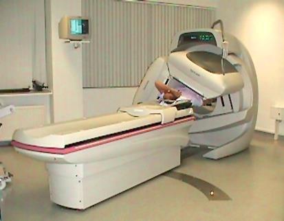
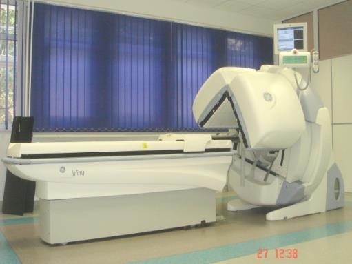
22 Scintigraphy Basic tool: SCINTILLATION CAMERA preamplifiers collimator electrical signals to console Gamma ray hits a scintillation crystal, causing emission of light photons by a crystal (scintillations). Those photons are absorbed in photocathodes of photomultiplers releasing electrons, thus forming an electrical current, which is then amplified in photomultipliers and registered in form of electrical signals at the detector output photomultipliers photocatode scintillator (NaI(Tl) crystal) collimator (passes only gamma rays moving in a direction parallel to axes of collimator holes)

23 Scintigraphy Basic tool: SCINTILLATION CAMERA collimator Light photons (scintillations) scintillator Distribution of a radiopharmaceutical in a given organ is reflected in a scintillation crystal in the form of light photons. Coordinates of every flash event are noted by a special electronic system, thus enabling a 2D presentation of a distribution of a radiopharmaceutical in a patient organ. Localization of every flash event in x,y coordinates Origination of a planar scintigram


24 Digital image n 51 cm n 51 cm Komórki matrycy cyfrowej Matrix with pixels Digital images are in fact matrices of pixels, presented in colors.
25 Static scintigraphy Conditions to be met: 1. Distribution of activity in an organ must not change over the interval of image acquisition. 2. The acquisition should only start when stable activity level in an organ has been reached. Information gathered: 1. Presence or absence of regions in an organ of abnormal (reduced or enhanced) uptake of the radiopharmaceutical administered. 2. In addition, data on position, shape and magnitude of the organ are obtained

 are trapped in those arteries thus enabling imaging of lung")
26 Normal lung perfusion scintigrams anterior mediastinum Anterior view posterior mediastinum Posterior view R silhouette L L R of heart Technetium labeled microspheres (small albumin spheres of diameters comparable to diameters of capillary arteries in lungs - in micrometers) are trapped in those arteries thus enabling imaging of lung perfusion.

 Bone")

27 Whole body static scintigraphy (skeletal scanning) Skeleton of an adult - normal image Skeleton of a child - normal image Abnormal image: numerous metastases to the skeleton (cancer) Bone scintigraphy an image presenting bone metabolism. Abnormal image lesions of enhanced bone metabolism at sites of cancer metastases

28 Single Photon Emission Computed Tomography (SPECT) Dynamic model of 3D-activity distribution reconstruction A method for presentation of a distribution of a radiopharmaceutical in 3 dimensions. Study acquisition is performed with a camera detector turning around a patient body. Then, using a special method for tomographic reconstruction, organ cuts along respective planes can be obtained.
 Various planes of cutting the 3D brain")
29 Single Photon Emission Computed Tomography (SPECT) Various planes of cutting the 3D brain matrix
 From left to right and from top to the bottom: from base to the top of the")
30 Brain SPECT perfusion study after Tc-HMPAO administration Transverse planes (cuts) From left to right and from top to the bottom: from base to the top of the brain
31 Dynamic scintigraphy The acquisition of scintigraphic information is started (usually) at the moment of i.v. injection of the RPharm. The study consist of a series of scintigraphic image acquisitions. The frequency of image acquisition depends upon the rate of the studied process.

32 Dynamic scintigraphy of the urinary system (renoscintigraphy) Serial acquisition of images takes place continuously over 20 min. (images provide information recorded over 20 minutes at 1 min intervals)

33 Renographic curves counts Left kidney Right kidney Background Ur. bladder time Counts are recorded at acquisition intervals over several regions of interest: kidneys, urinary bladder and over region representing the blood in organs background. Curves of activity in theses regions are automatically converted into renographic curves.

34 Principles of Positron Emission Tomography (PET) Another kind of tomography used in nuclear medicine. This modality makes advantage of radionuclides emitting positrons. As a result of a phenomenon of annihilation of positron with an electron in a patient body 2 photons appear escaping in opposite directions and they are detected in PET detector ring with a coincidence system. Positron + electron anihilation 2 photons of 512 kev each Detector Coincidence system Detector Radionuclide 18 F 15 O 11 C 13 N 82 Rb T 1/2 [min] ,1-20, ,2 Preconditions: on site production of positron emitting nuclides in a cyclotron; automatic on site synthesis of radiopharmaceuticals. Main benefits: a possibility to use for PET imaging the natural compounds (or their closely related analogues) of well understood physiological and biochemical behavior. Disadvantage: high cost
35 CLEARANCES Definition: The volume of plasma completely cleared of a specific compound (substance S) per minute and measured as a test of kidneys or liver function.
36 There is an obvious relationship between a clearance and rate of the decline of the substance (S) concentration in the plasma Sp(t) (conc. in plasma) lack of kidneys reduced function The faster decline of the curve, the higher the clearance of the substance. A clearance is calculated as a ratio of an activity administered to a patient and a definite integral of a function Sp over the interval from zero ( moment of injection) to infinity CL = I p 0 4 A 0 S (t)dt normal function (Activity of subst. S administered to a patient ) (Area under the curve presenting declining concentration of substance S in plasma) t
37 Determination of kidney and liver clearances - the selected RPhs GFR - 99m Tc DTPA ERPF I orthohipuric acid - 99m Tc MAG3-99m Tc Ethylene dicystein (EC) Cl liver - 99m Tc Hepida (derivative of an imin-bi-acetic acid) Advantages of radionuclide methods: high accuracy of determination of plasma concentrations possibility to measure CL after a single intravenous injection determination of CL is possible without collection of urine or gall low labour input required (particularly for simplified procedure, based on one sample of blood).
38 Patients care in nuclear diagnostics or therapy General information 1. Diagnostics or therapy by means of radiopharmaceuticals are in general non-invasive (there are very few exceptions e.g. intraarticular administration). 2. Majority of diagnostic procedures of nuclear medicine do not require pretreatment of the patient (there are a few exceptions). 3. Administration of a radiopharmaceutical is not followed by complications resulting from hypersensitivity (this applies also to patients hypersensitive to iodine contained in contrast media). 4. Doses to patients of ionizing radiation, resulting from diagnostic procedures are small, comparable to those accompanying most common radiological examinations. 5. Doses to the personnel injecting the radiopharmaceuticals to patients are very small if appropriate syringe shields are used and the patient has been prepared by introduction a venflon needle into his/her vein prior to injection.
Radioisotopes and PET
 Radioisotopes and PET 1 Radioisotopes Elements are defined by their number of protons, but there is some variation in the number of neutrons. Atoms resulting from this variation are called isotopes. Consider
Radioisotopes in action. Diagnostic application of radioisotopes. Steps of diagnostic procedure. Information from various medical imaging techniques
 Radioisotopes in action Diagnostic application of radioisotopes Steps of diagnostic procedure - Radioactive material introduced into the patient - Distribution and alteration of activity is detected -
Radioisotopes in action. Diagnostic application of radioisotopes. Steps of diagnostic procedure. Information from various medical imaging techniques
 Radioisotopes in action Diagnostic application of radioisotopes Steps of diagnostic procedure - Radioactive material introduced into the patient - Distribution and alteration of activity is detected -Monitoring
Nuclear Medicine RADIOPHARMACEUTICAL CHEMISTRY
 Nuclear Medicine RADIOPHARMACEUTICAL CHEMISTRY An alpha particle consists of two protons and two neutrons Common alpha-particle emitters Radon-222 gas in the environment Uranium-234 and -238) in the environment
Nuclear Medicine Intro & Physics from Medical Imaging Signals and Systems, Chapter 7, by Prince and Links
 Nuclear Medicine Intro & Physics from Medical Imaging Signals and Systems, Chapter 7, by Prince and Links NM - introduction Relies on EMISSION of photons from body (versus transmission of photons through
Bases of radioisotope diagnostic methods
 Medical, pharmaceutical applications of radioisotopes Bases of radioisotope diagnostic methods Dr. István Voszka Basis of application: radioisotopes have identical behavior in the organism to corresponding
Tomography is imaging by sections. 1
 Tomography is imaging by sections. 1 It is a technique used in clinical medicine and biomedical research to create images that show how certain tissues are performing their physiological functions. 1 Conversely,
Basic physics of nuclear medicine
 Basic physics of nuclear medicine Nuclear structure Atomic number (Z): the number of protons in a nucleus; defines the position of an element in the periodic table. Mass number (A) is the number of nucleons
DEVIL PHYSICS THE BADDEST CLASS ON CAMPUS IB PHYSICS
 DEVIL PHYSICS THE BADDEST CLASS ON CAMPUS IB PHYSICS TSOKOS OPTION I-2 MEDICAL IMAGING Reading Activity Answers IB Assessment Statements Option I-2, Medical Imaging: X-Rays I.2.1. I.2.2. I.2.3. Define
 www.aask24.com www.aask24.com www.aask24.com P=Positron E= Emission T=Tomography Positron emission or beta plus decay (+ ) is a particular type of radioactive decay, in which a proton inside a radionuclide
Outline Chapter 14 Nuclear Medicine
 Outline Chapter 14 uclear Medicine Radiation Dosimetry I Text: H.E Johns and J.R. Cunningham, The physics of radiology, 4 th ed. http://www.utoledo.edu/med/depts/radther Introduction Detectors for nuclear
MEDICAL EQUIPMENT: NUCLEAR MEDICINE. Prof. Yasser Mostafa Kadah
 MEDICAL EQUIPMENT: NUCLEAR MEDICINE Prof. Yasser Mostafa Kadah www.k-space.org Recommended Textbook Introduction to Medical Imaging: Physics, Engineering and Clinical Applications, by Nadine Barrie Smith
A. I, II, and III B. I C. I and II D. II and III E. I and III
 BioE 1330 - Review Chapters 7, 8, and 9 (Nuclear Medicine) 9/27/2018 Instructions: On the Answer Sheet, enter your 2-digit ID number (with a leading 0 if needed) in the boxes of the ID section. Fill in
CLINICALLY USEFUL RADIONUCLIDES:
 INTRODUCTION It is important that Nuclear Medicine Technologists be familiar with the imaging properties of all commonly used radionuclides to insure correct choice of isotope for a particular study as
Nuclear Radiation. Natural Radioactivity. A person working with radioisotopes wears protective clothing and gloves and stands behind a shield.
 Nuclear Radiation Natural Radioactivity A person working with radioisotopes wears protective clothing and gloves and stands behind a shield. 1 Radioactive Isotopes A radioactive isotope has an unstable
Year 12 Notes Radioactivity 1/5
 Year Notes Radioactivity /5 Radioactivity Stable and Unstable Nuclei Radioactivity is the spontaneous disintegration of certain nuclei, a random process in which particles and/or high-energy photons are
Radionuclide Imaging MII Positron Emission Tomography (PET)
") Radionuclide Imaging MII 3073 Positron Emission Tomography (PET) Positron (β + ) emission Positron is an electron with positive charge. Positron-emitting radionuclides are most commonly produced in cyclotron
Radionuclide Imaging MII 3073 Positron Emission Tomography (PET) Positron (β + ) emission Positron is an electron with positive charge. Positron-emitting radionuclides are most commonly produced in cyclotron
This Week. 3/23/2017 Physics 214 Summer
 This Week Atoms and nuclei What are we made of? The periodic table Why does it stop? How were the elements made? Radioactive decay Useful but can be toxic Discovery of X Rays: Cathode Rays and TV sets
Introduction to SPECT & PET TBMI02 - Medical Image Analysis 2017
 Introduction to SPECT & PET TBMI02 - Medical Image Analysis 2017 Marcus Ressner, PhD, Medical Radiation Physicist, Linköping University Hospital Content What is Nuclear medicine? Basic principles of Functional
1st Faculty of Medicine, Charles University in Prague Center for Advanced Preclinical Imaging (CAPI)
") Radioation Resolution and Sensitivity Nuclear Imaging PET + SPECT Radioactive Decay (EC,Ɣ), (β -,Ɣ), (I.T.,Ɣ) β + Projection imaging collimator needed one angular view Projection imaging coincidence imaging,
Radioation Resolution and Sensitivity Nuclear Imaging PET + SPECT Radioactive Decay (EC,Ɣ), (β -,Ɣ), (I.T.,Ɣ) β + Projection imaging collimator needed one angular view Projection imaging coincidence imaging,
Nuclear Physics and Astrophysics
 Nuclear Physics and Astrophysics PHY-302 Dr. E. Rizvi Lecture 24 Medical Imaging Effects of Radiation We now know what radiation is But what does it mean for our bodies? Radioactivity is quantified in
Stable isotope. Relative atomic mass. Mole fraction 203 Tl Tl Thallium isotopes in Earth/planetary science
 Stable isotope Relative atomic mass Mole fraction 203 Tl 202.972 345 0.2952 205 Tl 204.974 428 0.7048 Thallium isotopes in Earth/planetary science Because molecules, atoms, and ions of the stable isotopes
Dana-Farber Cancer Institute, 44 Binney Street, Boston, MA 02115, USA ramsey
 SPECIAL FEATURE: MEDICAL PHYSICS www.iop.org/journals/physed Nuclear medicine Ramsey D Badawi Dana-Farber Cancer Institute, 44 Binney Street, Boston, MA 02115, USA E-mail: ramsey badawi@dfci.harvard.edu
Nuclear Chemistry. Background Radiation. Three-fourths of all exposure to radiation comes from background radiation.
 Chapter 11 Nuclear Chemistry Background Radiation Three-fourths of all exposure to radiation comes from background radiation. Most of the remaining one-fourth comes from medical irradiation such as X-rays.
MEDICAL IMAGING. METHODS OF MODERN IMAGING, BASED ON ELECTRO-MAGNETIC RADIATION (radiowaves, infrared radiation, X-rays, γ-rays ) AND ULTRASOUND
 AND ULTRASOUND") MEDICAL IMAGING MEDICAL IMAGING METHODS OF MODERN IMAGING, BASED ON ELECTRO-MAGNETIC RADIATION (radiowaves, infrared radiation, X-rays, γ-rays ) AND ULTRASOUND MEDICAL IMAGING RADIOLOGY NUCLEAR MEDICINE
MEDICAL IMAGING MEDICAL IMAGING METHODS OF MODERN IMAGING, BASED ON ELECTRO-MAGNETIC RADIATION (radiowaves, infrared radiation, X-rays, γ-rays ) AND ULTRASOUND MEDICAL IMAGING RADIOLOGY NUCLEAR MEDICINE
Differentiating Chemical Reactions from Nuclear Reactions
 Differentiating Chemical Reactions from Nuclear Reactions 1 CHEMICAL Occurs when bonds are broken or formed. Atoms remained unchanged, though may be rearranged. Involves valence electrons Small energy
Number of protons. 2. What is the nuclear symbol for a radioactive isotope of copper with a mass number of 60? A) Cu
 Cu") Chapter 5 Nuclear Chemistry Practice Problems 1. Fill in the missing information in the chart: Medical Use Atomic Mass symbol number Heart imaging 201 Tl 81 Number of protons Number of neutrons Abdominal
Chapter 5 Nuclear Chemistry Practice Problems 1. Fill in the missing information in the chart: Medical Use Atomic Mass symbol number Heart imaging 201 Tl 81 Number of protons Number of neutrons Abdominal
69 Ga Ga
 Stable isotope Relative atomic mass Mole fraction 69 Ga 68.925 574 0.601 08 71 Ga 70.924 703 0.398 92 Gallium isotopes in medicine 68 Ga is a radioactive isotope that emits positrons, which are used to
RADIOCHEMICAL METHODS OF ANALYSIS
 RADIOCHEMICAL METHODS OF ANALYSIS 1 Early Pioneers in Radioactivity Rutherfo rd: Discoverer Alpha and Beta rays 1897 Roentge n: Discoverer of X- rays 1895 The Curies: Discoverers of Radium and Polonium
Atomic Notation (or Nuclear Symbol): Shorthand for keeping track of protons and neutrons in the nucleus
: Shorthand for keeping track of protons and neutrons in the nucleus") Name Section CHM52LL: Nuclear Chemistry: Radioactivity, Decay, Dating, and Other Hazards There is no prelab assignment this week I. Radioactive Isotopes and Nuclear Equations Atoms are composed of three
Name Section CHM52LL: Nuclear Chemistry: Radioactivity, Decay, Dating, and Other Hazards There is no prelab assignment this week I. Radioactive Isotopes and Nuclear Equations Atoms are composed of three
Lecture Presentation. Chapter 21. Nuclear Chemistry. James F. Kirby Quinnipiac University Hamden, CT Pearson Education, Inc.
 Lecture Presentation Chapter 21, Inc. James F. Kirby Quinnipiac University Hamden, CT Energy: Chemical vs. Chemical energy is associated with making and breaking chemical bonds. energy is enormous in comparison.
What is scintigraphy? The process of obtaining an image or series of sequential images of the distribution of a radionuclide in tissues, organs, or
 Let's remind... What is nuclear medicine? Nuclear medicine can be broadly divided into two branches "in vitro" and "in vivo" procedures. There are numerous radioisotopic "in vitro" procedures for genotyping
Introduction to Medical Imaging. Medical Imaging
 Introduction to Medical Imaging BME/EECS 516 Douglas C. Noll Medical Imaging Non-invasive visualization of internal organs, tissue, etc. I typically don t include endoscopy as an imaging modality Image
Physics in Nuclear Medicine
 SIMON R. CHERRY, PH.D. Professor Department of Biomedical Engineering University of California-Davis Davis, California JAMES A. SORENSON, PH.D. Emeritus Professor of Medical Physics University of Wisconsin-Madison
This Week. 7/20/2016 Physics 214 Spring
 This Week Atoms and nuclei What are we made of? The periodic table Why does it stop? How were the elements made? Radioactive decay Useful but can be toxic Discovery of X Rays: Cathode Rays and TV sets
Chapter 2. Atomic Structure and Nuclear Chemistry. Atomic Structure & Nuclear Chemistry page 1
 Chapter 2 Atomic Structure and Nuclear Chemistry Atomic Structure & Nuclear Chemistry page 1 Atoms & Elements Part 0: Atomic Structure An Introduction Electrostatics an underlying force throughout chemistry
Dosimetry of patients injected with tracers Ga-68, Zr-89 and Lu-177. Bruno Vanderlinden
 Dosimetry of patients injected with tracers Ga-68, Zr-89 and Lu-177 Bruno Vanderlinden What is NM speciality? Imaging radiology Physics Diagnostic Treatment assessment Clinical pathology Biological marker
There are three mechanisms by which gamma rays interact with absorber atoms from which two are important for nuclear medicine.
 Measurement of radioactivity. Radioactive decay is a random process and therefore fluctuations are expected in the radioactivity measurement. That is why measurement of radioactivity must be treated by
Technical University of Denmark
 Technical University of Denmark Page 1 of 11 pages Written test, 9 December 2010 Course name: Introduction to medical imaging Course no. 31540 Aids allowed: none. "Weighting": All problems weight equally.
Properties of the nucleus. 8.2 Nuclear Physics. Isotopes. Stable Nuclei. Size of the nucleus. Size of the nucleus
 Properties of the nucleus 8. Nuclear Physics Properties of nuclei Binding Energy Radioactive decay Natural radioactivity Consists of protons and neutrons Z = no. of protons (Atomic number) N = no. of neutrons
12/1/17 OUTLINE KEY POINTS ELEMENTS WITH UNSTABLE NUCLEI Radioisotopes and Nuclear Reactions 16.2 Biological Effects of Nuclear Radiation
 OUTLINE 16.1 Radioisotopes and Nuclear Reactions 16.2 Biological Effects of Nuclear Radiation PET scan X-ray technology CT scan 2009 W.H. Freeman KEY POINTS Radioactivity is the consequence of an unstable
Chapter 2 PET Imaging Basics
 Chapter 2 PET Imaging Basics Timothy G. Turkington PET Radiotracers Positron emission tomography (PET) imaging is the injection (or inhalation) of a substance containing a positron emitter, the subsequent
Laboratory 3: Kit Preparation and Chromatography. Design Considerations for a Radiopharmaceutical
 Laboratory 3: Kit Preparation and Chromatography PART 1: KIT PREPARATION Introduction In nuclear medicine, radionuclides are rarely used in their simplest chemical form. Instead they are incorporated into
Fusion Gamma radiation Half-life Radioactive tracer
 Unit Vocabulary: Alpha particle Artificial transmutation Beta particle Fission Fusion Gamma radiation Half-life Radioactive tracer Radioisotope Transmutation Unit Objectives: Upon completion of this unit
It s better to have a half-life than no life! Radioactive Decay Alpha, Beta, and Gamma Decay
 It s better to have a half-life than no life! Radioactive Decay Alpha, Beta, and Gamma Decay What does it mean to be radioactive? Some atoms have nuclei that are unstable. These atoms spontaneously decompose
ELG7173 Topics in signal Processing II Computational Techniques in Medical Imaging
 ELG7173 Topics in signal Processing II Computational Techniques in Medical Imaging Topic #1: Intro to medical imaging Medical Imaging Classifications n Measurement physics Send Energy into body Send stuff
β and γ decays, Radiation Therapies and Diagnostic, Fusion and Fission Final Exam Surveys New material Example of β-decay Beta decay Y + e # Y'+e +
 β and γ decays, Radiation Therapies and Diagnostic, Fusion and Fission Last Lecture: Radioactivity, Nuclear decay Radiation damage This lecture: nuclear physics in medicine and fusion and fission Final
Medical Physics. Nuclear Medicine Principles and Applications
 Medical Physics Nuclear Medicine Principles and Applications Dr Roger Fulton Department of PET & Nuclear Medicine Royal Prince Alfred Hospital Sydney Email: rfulton@mail.usyd.edu.au Lectures: http://www-personal.usyd.edu.au/~rfulton/medical_physics
General, Organic, and Biological Chemistry, 3e (Frost) Chapter 2 Atoms and Radioactivity. 2.1 Multiple-Choice
 Chapter 2 Atoms and Radioactivity. 2.1 Multiple-Choice") General, Organic, and Biological Chemistry, 3e (Frost) Chapter 2 Atoms and Radioactivity 2.1 Multiple-Choice 1) The smallest particle of an element that can be identified as that element is: A) a proton
General, Organic, and Biological Chemistry, 3e (Frost) Chapter 2 Atoms and Radioactivity 2.1 Multiple-Choice 1) The smallest particle of an element that can be identified as that element is: A) a proton
Chapter. Nuclear Chemistry
 Chapter Nuclear Chemistry Nuclear Reactions 01 Chapter 22 Slide 2 Chapter 22 Slide 3 Alpha Decay: Loss of an α-particle (a helium nucleus) 4 2 He 238 92 U 234 4 U He 90 + 2 Chapter 22 Slide 4 Beta Decay:
3. Which of the following statements is (are) TRUE about detector crystals in Anger cameras?
 TRUE about detector crystals in Anger cameras?") BioE 1330 - Exam 2 11/13/2018 Answer Sheet - Correct answer is A for all questions 1. Unlike CT, in nuclear medicine A. Bremsstrahlung is not used to produce high-energy photons. B. signal can be increased
BioE 1330 - Exam 2 11/13/2018 Answer Sheet - Correct answer is A for all questions 1. Unlike CT, in nuclear medicine A. Bremsstrahlung is not used to produce high-energy photons. B. signal can be increased
Structure of Biological Materials
 ELEC ENG 3BA3: Structure of Biological Materials Notes for Lecture #19 Monday, November 22, 2010 6.5 Nuclear medicine imaging Nuclear imaging produces images of the distribution of radiopharmaceuticals
Chapter 18: Radioactivity And Nuclear Transformation. Presented by Mingxiong Huang, Ph.D.,
 Chapter 18: Radioactivity And Nuclear Transformation Presented by Mingxiong Huang, Ph.D., mxhuang@ucsd.edu 18.1 Radionuclide Decay Terms and Relationships Activity Decay Constant Physical Half-Life Fundamental
General, Organic, and Biochemistry, 2e (Frost) Chapter 2 Atoms and Radioactivity. 2.1 Multiple-Choice
 Chapter 2 Atoms and Radioactivity. 2.1 Multiple-Choice") General, Organic, and Biochemistry, 2e (Frost) Chapter 2 Atoms and Radioactivity 2.1 Multiple-Choice 1) Two atoms must represent the same element if they both have the same: A) number of electron shells
General, Organic, and Biochemistry, 2e (Frost) Chapter 2 Atoms and Radioactivity 2.1 Multiple-Choice 1) Two atoms must represent the same element if they both have the same: A) number of electron shells
Some nuclei are unstable Become stable by ejecting excess energy and often a particle in the process Types of radiation particle - particle
 Radioactivity George Starkschall, Ph.D. Lecture Objectives Identify methods for making radioactive isotopes Recognize the various types of radioactive decay Interpret an energy level diagram for radioactive
PANEL DISCUSSION. Radionuclides and Health A promising future! OCTOBER 14-16, 2014 PARIS LE BOURGET FRANCE
 PANEL DISCUSSION Radionuclides and Health A promising future! Hosted by Richard Zimmermann, Chrysalium Consulting Discussion coordinated by François Sarkozy, President of FSNB Health & Care Speakers: Remigiusz
Unit 12: Nuclear Chemistry
 Unit 12: Nuclear Chemistry 1. Stability of isotopes is based on the ratio of neutrons and protons in its nucleus. Although most nuclei are stable, some are unstable and spontaneously decay, emitting radiation.
Radiopharmaceuticals and Contrast Media. Lec: 7
 Radiopharmaceuticals and Contrast Media Lec: 7 Radiopharmaceuticals Radioisotopes every atom of an element is composed of a nucleus, containing protons and neutrons, surrounded by electrons. In the electrically
Lecture PowerPoints. Chapter 31 Physics: Principles with Applications, 7th edition Giancoli
 Lecture PowerPoints Chapter 31 Physics: Principles with Applications, 7th edition Giancoli This work is protected by United States copyright laws and is provided solely for the use of instructors in teaching
Chapter 11 Nuclear Chemistry
 Chapter 11 Nuclear Chemistry 11.1 Nuclear Reactions Nuclear reactions involve the particles located in the nucleus of the atom: The nucleus contains: An atom is characterized by: X A Z - Z the gives the
GLOSSARY OF BASIC RADIATION PROTECTION TERMINOLOGY
 GLOSSARY OF BASIC RADIATION PROTECTION TERMINOLOGY ABSORBED DOSE: The amount of energy absorbed, as a result of radiation passing through a material, per unit mass of material. Measured in rads (1 rad
(INCLUDING THIS FRONT PAGE)
") I'IFIITIIBIFI UNIVERSITY OF SCIEI'ICE RITD TECHNOLOGY FACULTY OF HEALTH AND APPLIED SCIENCES DEPARTMENT OF NATURAL AND APPLIED SCIENCES QUALIFICATION: BACHELOR OF SCIENCE (MAJOR AND MINOR) QUALIFICATION
I'IFIITIIBIFI UNIVERSITY OF SCIEI'ICE RITD TECHNOLOGY FACULTY OF HEALTH AND APPLIED SCIENCES DEPARTMENT OF NATURAL AND APPLIED SCIENCES QUALIFICATION: BACHELOR OF SCIENCE (MAJOR AND MINOR) QUALIFICATION
Properties of the nucleus. 9.1 Nuclear Physics. Isotopes. Stable Nuclei. Size of the nucleus. Size of the nucleus
 Properties of the nucleus 9. Nuclear Physics Properties of nuclei Binding Energy Radioactive decay Natural radioactivity Consists of protons and neutrons Z = no. of protons (tomic number) N = no. of neutrons
Nuclear Medicine: Physics and Imaging Methods (SPECT and PET)
") EL-GY 6813 / BE-GY 6203 / G16.4426 Medical Imaging Nuclear Medicine: Physics and Imaging Methods (SPECT and PET) Jonathan Mamou and Yao Wang Polytechnic School of Engineering New York University, Brooklyn,
EL-GY 6813 / BE-GY 6203 / G16.4426 Medical Imaging Nuclear Medicine: Physics and Imaging Methods (SPECT and PET) Jonathan Mamou and Yao Wang Polytechnic School of Engineering New York University, Brooklyn,
PHY138Y Nuclear and Radiation Section
 PHY138Y Supplementary Notes V: Radioisotopes in Medicine. A.W. Key Page 1 of 10 PHY138Y Nuclear and Radiation Section Supplementary Notes V Radioisotopes in Medicine Contents. 5.1 Introduction 5.2 Radioisotopes
Radioactivity Outcomes. Radioactivity Outcomes. Radiation
 1 Radioactivity Outcomes Describe the experimental evidence for there being three types of radiation. Discuss the nature and properties of each type. Solve problems about mass and atomic numbers in radioactive
Radiation Detectors. How do we detect ionizing radiation? What are these effects? Types of Ionizing Radiation Detectors
 Radiation Detectors 1 How do we detect ionizing radiation? Indirectly, by its effects as it traverses matter? What are these effects? Ionization and excitation of the atoms and molecules Heat 2 Types of
Chapter 21
 Chapter 21 http://youtu.be/kwasz59f8ga Nuclear reactions involve the nucleus The nucleus opens, and protons and neutrons are rearranged. The opening of the nucleus releases a tremendous amount of energy
Lecture PowerPoint. Chapter 31 Physics: Principles with Applications, 6 th edition Giancoli
 Lecture PowerPoint Chapter 31 Physics: Principles with Applications, 6 th edition Giancoli 2005 Pearson Prentice Hall This work is protected by United States copyright laws and is provided solely for the
Radiochemistry and Radiopharmacy III
 Radiochemistry and Radiopharmacy III Compact course held at UFSCAR, September 20123 Ulrich Abram Freie Universität Berlin Institute of Chemistry and Biochemistry Radiochemistry and Radiopharmacy 1. Fundamentals
fission and fusion and classify a nuclear reaction as either a fission or fusion reaction.
 Chemistry HP Unit 11 Nuclear Chemistry Learning Targets (Your exam at the end of Unit 11 will assess the following:) 11. Nuclear Chemistry 11-1. Write the nuclide symbol for a given isotope. 11-2. Describe
Chapter 20 Nuclear Chemistry. 1. Nuclear Reactions and Their Characteristics
 Chapter 2 Nuclear Chemistry 1. Nuclear Reactions and Their Characteristics Nuclear reactions involve the particles located in the nucleus of the atom: nucleons:. An atom is characterized by its atomic
6: Positron Emission Tomography
 6: Positron Emission Tomography. What is the principle of PET imaging? Positron annihilation Electronic collimation coincidence detection. What is really measured by the PET camera? True, scatter and random
Notes: Unit 13 Nuclear Chemistry
 Name: Regents Chemistry: Notes: Unit 13 Nuclear Chemistry Name: KEY IDEAS: Stability of isotopes is based in the ratio of neutrons and protons in its nucleus. Although most nuclei are stable, some are
AQA Physics /7408
 AQA Physics - 7407/7408 Module 10: Medical physics You should be able to demonstrate and show your understanding of: 10.1 Physics of the eye 10.1.1 Physics of vision The eye as an optical refracting system,
Radioactivity. The Nobel Prize in Physics 1903 for their work on radioactivity. Henri Becquerel Pierre Curie Marie Curie
 Radioactivity Toward the end of the 19 th century, minerals were found that would darken a photographic plate even in the absence of light. This phenomenon is now called radioactivity. Marie and Pierre
Key Question: What role did the study of radioactivity play in learning more about atoms?
 Name Chemistry Essential question: How were the parts of the atom determined? Key Question: What role did the study of radioactivity play in learning more about atoms? Vocabulary: alpha particle fusion
NOTES: 25.2 Nuclear Stability and Radioactive Decay
 NOTES: 25.2 Nuclear Stability and Radioactive Decay Why does the nucleus stay together? STRONG NUCLEAR FORCE Short range, attractive force that acts among nuclear particles Nuclear particles attract one
RADIOACTIVITY & HALF-LIFE Part 2
 RADIOACTIVITY & HALF-LIFE Part 2 Radioactivity Radioactivity: Results from radioactive decay, which is the process whereby unstable atomic nuclei transform and emit radiation. Has existed longer than the
11/10/2014. Chapter 1: Introduction to Medical Imaging. Projection (Transmission) vs. Emission Imaging. Emission Imaging
 vs. Emission Imaging. Emission Imaging") Chapter 1: Introduction to Medical Imaging Overview of Modalities Properties of an Image: Limitations on Information Content Contrast (both object & image): Brightness difference Sharpness (blur): Smallest
Chapter 1: Introduction to Medical Imaging Overview of Modalities Properties of an Image: Limitations on Information Content Contrast (both object & image): Brightness difference Sharpness (blur): Smallest
Notes: Unit 14 Nuclear Chemistry
 Name: Regents Chemistry: Mr. Palermo Notes: Unit 14 Nuclear Chemistry www.mrpalermo.com Name: KEY IDEAS: Stability of isotopes is based in the ratio of neutrons and protons in its nucleus. Although most
National 5- Nuclear Chemistry past paper revision
 National 5- Nuclear Chemistry past paper revision 1. The diagram shows the paths of alpha, beta and gamma radiations as they pass through an electric field. Which line in the table correctly identifies
QUIZ: Physics of Nuclear Medicine Atomic Structure, Radioactive Decay, Interaction of Ionizing Radiation with Matter
 QUIZ: Physics of Nuclear Medicine Atomic Structure, Radioactive Decay, Interaction of Ionizing Radiation with Matter 1. An atomic nucleus contains 39 protons and 50 neutrons. Its mass number (A) is a)
INAYA MEDICAL COLLEGE (IMC) RAD LECTURE 1 RADIATION PHYSICS DR. MOHAMMED MOSTAFA EMAM
 RAD LECTURE 1 RADIATION PHYSICS DR. MOHAMMED MOSTAFA EMAM") INAYA MEDICAL COLLEGE (IMC) RAD 232 - LECTURE 1 RADIATION PHYSICS DR. MOHAMMED MOSTAFA EMAM Radiation: It is defined as the process by which energy is emitted from a source and propagated through the surrounding
INAYA MEDICAL COLLEGE (IMC) RAD 232 - LECTURE 1 RADIATION PHYSICS DR. MOHAMMED MOSTAFA EMAM Radiation: It is defined as the process by which energy is emitted from a source and propagated through the surrounding
The Case of Melting Ice
 Nuclear Chemistry A is for Atom - 1953 (15 minutes) http://www.youtube.com/watch?v=fn1oslamdgw part 1 (7:15) http://www.youtube.com/watch?v=cggskffgg7g part 2 (7:29) The Case of Melting Ice Frosty the
Name Date Class NUCLEAR RADIATION. alpha particle beta particle gamma ray
 25.1 NUCLEAR RADIATION Section Review Objectives Explain how an unstable nucleus releases energy Describe the three main types of nuclear radiation Vocabulary radioisotopes radioactivity radiation alpha
Units and Definition
 RADIATION SOURCES Units and Definition Activity (Radioactivity) Definition Activity: Rate of decay (transformation or disintegration) is described by its activity Activity = number of atoms that decay
INAYA MEDICAL COLLEGE (IMC) RAD LECTURE 1 RADIATION PHYSICS DR. MOHAMMED MOSTAFA EMAM
 RAD LECTURE 1 RADIATION PHYSICS DR. MOHAMMED MOSTAFA EMAM") INAYA MEDICAL COLLEGE (IMC) RAD 232 - LECTURE 1 RADIATION PHYSICS DR. MOHAMMED MOSTAFA EMAM LECTURES & CLASS ACTIVITIES https://inayacollegedrmohammedemam.wordpress.com/ Password: drmohammedemam 16-02-2015
INAYA MEDICAL COLLEGE (IMC) RAD 232 - LECTURE 1 RADIATION PHYSICS DR. MOHAMMED MOSTAFA EMAM LECTURES & CLASS ACTIVITIES https://inayacollegedrmohammedemam.wordpress.com/ Password: drmohammedemam 16-02-2015
and have low penetrating power) Alpha particles are released through alpha decay. Beta Particles: An electron that comes from a nucleus through
 Alpha particles are released through alpha decay. Beta Particles: An electron that comes from a nucleus through") TOPIC 13: Nuclear Chemistry 1. When the atomic nucleus of one element is changed into the nucleus of a different element, the reaction is called transmutation. Stability of a Nucleus: Any element containing
TOPIC 13: Nuclear Chemistry 1. When the atomic nucleus of one element is changed into the nucleus of a different element, the reaction is called transmutation. Stability of a Nucleus: Any element containing
Nuclear Medicine Treatments and Clinical Applications
 INAYA MEDICAL COLLEGE (IMC) RAD 243- LECTURE 2 Nuclear Medicine Treatments and Clinical Applications DR. MOHAMMED MOSTAFA EMAM Next Lectures Outlines Introduction to Nuclear Physics Physics of Radioactivity
AEPHY: Nuclear Physics Practise Test
 AEPHY: Nuclear Physics Practise Test Name: OVERALL: Additional 1 mark for units and significant figures. 1. Complete the table below: (2 marks) (63 marks + overall = 64 marks) Element Nuclide Atomic Number
P7 Radioactivity. Student Book answers. P7.1 Atoms and radiation. Question Answer Marks Guidance
 P7. Atoms and radiation a radiation from U consists = particles, radiation from lamp = electromagnetic waves, radiation from U is ionising, radiation from lamp is non-ionising b radioactive atoms have
Radionuclide Imaging MII Detection of Nuclear Emission
 Radionuclide Imaging MII 3073 Detection of Nuclear Emission Nuclear radiation detectors Detectors that are commonly used in nuclear medicine: 1. Gas-filled detectors 2. Scintillation detectors 3. Semiconductor
Particles involved proton neutron electron positron gamma ray 1
 TOPIC : Nuclear and radiation chemistry Nuclide - an atom with a particular mass number and atomic number Isotopes - nuclides with the same atomic number (Z) but different mass numbers (A) Notation A Element
Supervised assessment: Ionising radiation
 Physics 27 Sample assessment instrument and indicative Supervised assessment: Ionising radiation This sample is intended to inform the design of assessment instruments in the senior phase of learning.
Procesamiento de Imágenes y Bioseñales
 Procesamiento de Imágenes y Bioseñales Dr. Víctor Castañeda Agenda Physical basis of X-ray- CT, NMR, Ultrasound, Nuclear Medicine Sensors (cameras, gamma probes, microphone) Computational Tomography (CT)
Professor Stuart Bunt 217
 Professor Stuart Bunt 217 Traditional Anatomy Phrenology, the study of bumps on the skull. Measuring brain weights and size (still being done..see the fuss about Einstein s brain). Little link between
Unit 3: Chemistry in Society Nuclear Chemistry Summary Notes
 St Ninian s High School Chemistry Department National 5 Chemistry Unit 3: Chemistry in Society Nuclear Chemistry Summary Notes Name Learning Outcomes After completing this topic you should be able to :
Chemical Engineering 412
 Chemical Engineering 412 Introductory Nuclear Engineering Lecture 30 Medical Applications Beneficial Uses of Radiation Imaging X-Ray Projection Imaging Fluoroscopy Mammography Bone Densitometry X-Ray Computed
Chemistry 52 Chapter 11 ATOMIC STRUCTURE. The general designation for an atom is shown below:
 ATOMIC STRUCTURE An atom is composed of a positive nucleus surrounded by negatively charged electrons. The nucleus is composed of protons and neutrons. The protons and neutrons in a nucleus are referred