craniofacial development and WNT signaling. Shachi Bhatt
|
|
|
- Owen Green
- 5 years ago
- Views:
Transcription
1 MED23: a Mediator subunit's role in global gene transcription, regulation of craniofacial development and WNT signaling. BY 2013 Shachi Bhatt Submitted to the graduate degree program in Anatomy and Cell Biology and to the graduate faculty of University of Kansas in partial fulfillment of the requirements for the degree of Doctor of Philosophy Co-chair Paul Trainor, Ph.D. Co-chair Douglas Wright, Ph.D. Joan Conaway, Ph.D. Timothy Fields, M.D., Ph.D. Robb Krumlauf, Ph.D. Date Defended: August 23 rd, 2013
2 The Defense committee for Shachi Bhatt certifies that this is the approved version of the following dissertation: MED23: a Mediator subunit s role in global gene transcription, regulation of craniofacial development and WNT signaling Co-chair Paul Trainor, Ph.D. Co-chair Douglas Wright, Ph.D. Date approved: September 9 th, 2013 ii
3 Abstract Expression of genes at the right time and place is crucial during adult homeostasis as well as embryonic development. Multicellular organisms regulate this spatiotemporal expression of genes by employing tissue specific transcription factors which bind to enhancer and repressor elements distant from the gene transcription start site. In embryonic development, most of the research understanding cell differentiation has focused on identifying such tissue specific transcription factors. What we do not understand clearly, is how these transcription factors that bind so far away from the gene promoter site influence the decision of RNA Pol II to transcribe or not transcribe. A recent discovery of a mega-dalton protein complex, called Mediator, has begun to answer this question. Mediator complex physically interacts with RNA Pol II and general transcription factors on one side and with transcription factors bound to enhancer/repressor sites on the other side. Mediator has thus been shown to act as a bridge that relays information between the transcription factors and RNA Pol II machinery and thus regulate gene expression. The various subunits of Mediator complex have been shown to interact with distinct transcription factors to regulate expression of specific genes. In this work, I describe the role of one such Mediator subunit, MED23. The role of MED23 during mammalian embryonic development was identified through a forward genetics screen done in the lab. Loss of med23 leads to mid-gestational lethality in iii
4 mice embryos along with defects in craniofacial, neural and vascular development. We currently do not know the exact cause for embryonic lethality in med23 mutant embryos, but my results indicate that MED23 is crucial for endothelial cell-cell junction formation at E9.5, defects in which have previously been shown to affect embryonic survival. Specifically the formation of adherens and tight junctions between endothelial cells is affected in med23 mutant embryos. Analysis of neuronal defects in med23 mutant embryos suggests that MED23 is required during various steps of cranial placode development and this is regulated by MED23-mediated regulation of canonical WNT signaling. How and why loss of MED23 leads to defects in these specific tissues is currently unknown, but work with conditional mutant analysis as well as transcription factor-binding screens are underway to figure this out. My work thus highlights a unique link between general transcription co-factor, Mediator, and its subunit MED23 with development of neural, vascular and craniofacial tissues and a crucial signaling pathway, WNT signaling. iv
5 Acknowledgements First and foremost, I would like to dedicate this dissertation to the four most important people in my life, my parents, my sister and Rushi. This accomplishment would have been incomplete without your presence in my life. Mummy, Papa - In spite of all our disagreements ;), you have been the two strong pillars of my life and the strength that I gained from you has held me upright before and during my PhD; and Didu if there is one person I can count on who will always be on my side no matter what, I would say it s you. Thank you for all your love and support that have sustained me through the thick and thin of my past seven years in US. We might have been physically apart but you were always there. Rushi words are not enough for the amount of gratitude I owe to you for being by my side during smallest victories and the biggest scares of grad school. There isn t anybody else who could understand and relate to the frustrations and the excitement that grad school offers and I feel blessed to have had you to share it with. You are the fairest individual I have known in my life and daily inspire me to challenge myself and make things better in everyday life and in science. It is said that graduate school is one of the most stressful times in the career of a scientist, but your presence made it fun. Of course, my dissertation would be incomplete without thanking you, Paul. I have yet to come across a more well-rounded personality than yours in my life. It seems to me that you v
6 have figured it out; you are fabulous scientist who never forgets the bigger picture in life, a great mentor and just an amazing individual. Thank you for everything that you have done for me and for everything that I learnt from you in the past for years. I hope that my growth as a scientific researcher is something that makes you proud. Lisa, Amanda, Daisuke and Kim, you guys taught me the basics in the lab and I will never forget your contribution to my achievement today and hereafter. Every member of Trainor lab has been a great friend and a great colleague. From our scientific discussions to our fun hangouts at Westport, you all have provided me a family away from home. I would also like to thank my committee members, Robb, Joan, Doug, Tim and Jay, whose inputs have been valuable to shaping this body of work and in helping me grow as a researcher. I am also very grateful to the department of Anatomy and Cell Biology at KUMC and all the core facilities at Stowers Institute for Medical Research who have provided me support and facilitated my work. vi
7 Table of Contents Abstract iii Acknowledgements v Table of Contents vii List of Figures and Tables ix Introduction 1 Part 1: Craniofacial Development 2 Part 2: Mediator and Transcriptional control of gene expression 32 Chapter 1: MED23 is required for mammalian embryonic development 50 Summary 51 Introduction 52 Results 55 Discussion 81 Chapter 2: MED23 is required for mammalian vascular development 86 Summary 87 Introduction 88 Results 98 Discussion 106 vii
8 Chapter 3: MED23 is required for peripheral nervous system formation 111 Summary 112 Introduction 113 Results 128 Discussion 138 Chapter 4: MED23 is required for anterior development in mammalian embryos 146 Summary 147 Introduction 148 Results 156 Discussion 167 Chapter 5: MED23 regulates WNT signaling during cranial ganglia development 171 Summary 172 Introduction 173 Results 180 Discussion 196 Discussion 200 Materials and Methods 217 Bibliography 223 viii
9 List of Figures and Tables Introduction Figure 1: Emergence of asymmetry within a developing mouse embryo Figure 2: Mammalian neural crest cells Figure 3: Ectodermal placodes of a developing mouse embryo Figure 4: Composition of the Mediator Complex Table 1: Interactions between Mediator subunits and Transcription co-activators Table 2: Mediator subunits and vertebrate embryonic development Chapter 1 Figure 5: snouty mutation display a smaller size and craniofacial development at E9.5 Figure 6: snouty embryos have defects in peripheral nervous system formation Figure 7: snouty embryos have defects in vascular remodeling Figure 8: snouty is a mutant in med23 Figure 9: Generation of med23 bgeo allele and its embryonic expression pattern Figure 10: med23 bgeo/bgeo embryos halt their development at E8.75 ix
10 Figure 11: Schematic design for complementation test Figure 12: snouty is a functionally null allele Figure 13: Mating scheme to generate conditional mice from med23 bgeo animals Figure 14: Mosaic activity of Flpper mice Figure 15: Tamoxifen inducible ERT2-Cre is able to excise loxp floxed alleles at 5mg/kg dosage Chapter 2 Figure 16: Endothelial cell-cell junctions Figure 17: flk1 expression in wildtype and med23 sn/sn embryos Figure 18: Adherens junction marker, ve-cadherin expression is reduced in med23 sn/sn embryos at E9.5 but not at E9.0 Figure 19: Expression of tight junction marker, claudin5 is reduced in med23 sn/sn embryos at E9.5 Figure 20: Early formation of vascular smooth muscle cells is not affected in control and med23 sn/sn littermate embryos at E9.0 Chapter 3 Table 3: Cranial nerve innervation x
11 Figure 21: Contribution of neural crest and cranial placodes to the cranial sensory ganglia in vertebrates Figure 22: Hierarchy of genes expressed in placodal development from PPR to neuronal differentiation Figure 23:sox10+ cells are reduced in med23 sn/sn embryos Figure 24: neurod1+ cells are drastically reduced in the developing cranial ganglia of med23 sn/sn embryos at E9.5 Figure 25: neurogenin expression is affected in the cranial ganglia of med23 sn/sn embryos Figure 26: Coalescence but not specification of cranial placodes is affected in med23 sn/sn embryos Figure 27: med23 sn/sn embryos do not maintain the expression of PPR markers in cranial sensory ganglia Chapter 4 Figure 28: Reduced contribution of crabp1+ neural crest cells to the craniofacial regions in med23 sn/sn embryos. Figure 29: Cell proliferation and cell death are not altered in the frontonasal process of med23 sn/sn embryos at E9.5 xi
12 Figure 30: SHH signaling is unaffected in med23 sn/sn embryos Figure 31: six3 expression in forebrain is not affected in med23 sn/sn embryos Figure 32: WNT signaling is upregulated and ectopically expressed in the lateral nasal processes of med23 sn/sn embryos at E9.5. Figure 33: Craniofacial defects inmed23 sn/sn embryos are evident at E9.0. Chapter 5 Figure 34: WNT signaling pathways Figure 35 Expression of Dkk-1 (WNT inhibitor) is reduced in med23 sn/sn embryos Figure 36: WNT signaling is up-regulated throughout the embryo specifically at E9.5 Figure 37: Formation of nervous system is not affected by losing a copy of lrp6 or a copy of wise Figure 38: Modulation of canonical WNT signaling partially restores epibranchial ganglia development in med23 sn/sn embryos. Figure 39: Wise and Lrp6 activate WNT signaling during cranial ganglia formation. Figure 40: Epibranchial neurons restored by modulating of WNT signaling express neurod1 Figure 41: Defects in maintenance of early PPR gene expression of med23 sn/sn embryos are not restored by modulating the WNT signaling levels. xii
13 Figure 42: Loss of med23 does not affect wise expression Figure 43: Frontonasal process defects of med23 sn/sn embryos is not rescued by loss of lrp6 or wise Discussion Figure 44: egr2 is expressed in hindbrain of med23 sn/sn embryos xiii
14 Introduction 1
15 Part 1: Craniofacial Development One third of all congenital deformities have a craniofacial structure anomaly. In their severe forms, craniofacial defects affect the basic functioning of the head, ranging from jaw closure defects that affect the ability of patients to talk and eat, to debilitating defects in sensory organs affecting cognition. Even in their mildest forms where the functioning is not highly affected, defects in facial appearance affect well-being of patients. Prevention of occurrence of these craniofacial occurrences warrants a detailed understanding of how a normal head structure forms and an understanding of crucial genetic and environmental factors responsible for its normal development. While some genes and environmental factors required for proper craniofacial development have been identified, we clearly do not have a comprehensive understanding of head development and defects. The high occurrence of craniofacial anomalies is not a surprise given the highly complex nature of craniofacial development. Head induction begins as soon as the anterioposterior axis of the embryo is defined at gastrulation. All three germ layers, ectoderm, mesoderm and endoderm contribute to the developing craniofacial complex and their induction, differentiation, proliferation and patterning have to be highly orchestrated for proper head development. In head, ectoderm gives rise to the nervous system (central and peripheral) and majority of the craniofacial skeleton; mesoderm forms the cephalic 2
16 musculature and blood vessels as well as some of the skull bones while the endoderm forms the foregut lining, pharynx and trachea along with other organs. This section gives a general overview of head induction including formation of anterio-posterior axis and gastrulation followed by brief description (when and how) of differentiation of each germ layer into the various structures within craniofacial complex. I) Gastrulation and Formation of Anterio-posterior axis Mouse embryos begin as a single cell zygote which undergoes a series of cell divisions and a process of compaction eventually forming a fluid filled epithelial vesicle known as blastocyst. At late blastocyst stage, the outer epithelium cells are committed to the trophectoderm lineage which subsequently forms the placenta while the cells inside the vesicle, known as inner cell mass (ICM), is composed of two cell types, epiblast which gives rise to all cell of the embryo proper and some extra-embryonic membranes and primitive endoderm which primarily forms the endoderm layer of the extraembryonic yolk sacs 1. These early events in a mouse embryo describing formation of blastocyst and first asymmetry with ICM have been extensively described elsewhere and summarized in Figure 1 2. One of the crucial aspects of embryonic development is formation of the three axes, Organisms like Drosophila and Zebrafish, rely on asymmetric distribution of maternal 3
17 transcripts and proteins to generate polarity in the embryo. In mammals, however, transition of transcription under exclusive zygotic control (maternal-to-zygotic transition) occurs earlier than specification of axes. Following implantation the mouse blastocyst forms an egg cylinder with a proximal (the pole with the trophectoderm derived ectoplacental cone) distal (P/D) axis 1. Recent lineage tracing experiments have identified a specific group of lefty-1 expressing cells within the ICM prior to implantation 3. These lefty-1+ cells form the distal visceral endoderm (DVE) of the egg cylinder, which populates the distal pole of the P/D axis, suggesting a possibility that P/D axis is formed prior to implantation. DVE cells were originally believed to give rise to anterior visceral endoderm (AVE), an extra-embryonic signaling center crucial for imparting anterior character to the embryonic region adjacent to it 2,4,5, 6. Recent works show that AVE cells are specified de novo and DVE is not required for its induction 3,7-9. DVE however has been shown to be required initiating AVE migration toward the prospective anterior pole 3. Epiblast cells receiving signals from AVE acquire anterior fate and contribute to head and brain structures while the cells away from AVE acquire a posterior fate 1. The process of gastrulation forms the mesoderm and endoderm lineages from epiblast cells at the posterior end of the mouse embryo. In mice embryos, gastrulation begins with the formation of primitive streak. Primitive streak is formed by the epiblast cells undergoing polonaise movement at the future posterior end of the embryo. The polonaise motion results in 4
. ICM cells already display asymmetric gene expression, and the lefty1+ cells (dark blue and green) will eventually contribute to DVE (dark blue).")
18 Figure 1: Emergence of asymmetry within a developing mouse embryo A) At blastocyst stage, the embryo is already separated into three distinct cell types, ICM (light blue), Primitive endoderm (pink) and trophectoderm (grey). ICM cells already display asymmetric gene expression, and the lefty1+ cells (dark blue and green) will eventually contribute to DVE (dark blue). AVE cells (green), also lefty1+, are specified later than DVE. B) Implantation induced proximal (ectoplacental cone)-distal axis. Primitive endoderm eventually forms the visceral endoderm that surrounds the epiblast and extraembryonic tissues. DVE and AVE cells migrate to the distal pole of the embryo (boxed area with red dotted lines). WNT signaling is activated (orange) at the future posterior end of the embryo. C) Future posterior end expresses WNT and Nodal signaling ligands. Gastrulation begins. DVE and AVE cells migrate to the future anterior side of the embryo (boxed area with green dotted lines) and express WNT and Nodal signaling inhibitors. 5
19 epiblast cells moving in circles around a morphologically visible midline, which is the primitive streak. As time progresses the epiblast cells undergo epithelial-to-mesenchymal transition (EMT) and ingress through the streak into the embryo. These newly ingressed mesenchymal cells are a mixture of mesoderm and endoderm progenitors and signals emanating from the site of ingression help specify their eventual fate within the embryo. As gastrulation proceeds the embryo gets divided into three distinct germ layers: ectoderm which is the inside-most layer, endoderm which is the outside-most layer; and mesoderm, in between the two. The inside-outside positioning of the ectoderm and endoderm gets reversed when the mouse embryo undergoes turning at around E9.0. Ia) Molecular determinants and signaling pathways in gastrulation and A/P axis determination The formation of the anterio-posterior (A/P) axis within the embryo requires two separate yet related processes; specification of the anterior end (involvement of AVE) and specification of the posterior end (marked by presence of primitive streak, site of gastrulation). Pre-gastrula embryos have been shown to display asymmetry in gene expression. For example, components of canonical WNT signaling, wnt3, wnt2b and activated β-catenin have a restricted pattern of expression at the future posterior end of the embryo Mouse knock- 6
20 out studies have also shown that embryos lacking functioning WNT signaling (Wnt3 -/- 13 or WNT co-receptors Lrp5 -/- or Lrp6 -/- 14 ) fail to establish a primitive streak and thus lack mesoderm and definitive endoderm lineages. All of this information together makes a strong case for the involvement of WNT signaling in establishing gastrulation site and hence in establishing posterior end of the embryo. At the same time it is crucial to block WNT signaling for the specification of the anterior pole. This blockage of WNT signaling is achieved by expression of WNT signaling inhibitors specifically at the anterior end. As mentioned earlier, cells within AVE secrete signals that specify the anterior end of the embryo. One of these secreted signals is a molecule called Dickkopf-1 (DKK-1), a well-known inhibitor of WNT signaling 15. The importance of blocking WNT signaling for anterior specification comes from the fact that the complete knock-out of Dkk-1 leads to expansion of WNT signaling and results in complete absence of anterior head structures 16. The seemingly straightforward role for WNT signaling does not hold true for the other signaling pathways also involved A/P axis formation. Nodal, a member of TGFβ family of secreted ligands has a more complex role in specifying anterior and posterior poles of the embryo. Although it is well established that inhibition of Nodal signaling, achieved by secretion of Nodal antagonist, LEFTY, from AVE cells is crucial for anterior specification 17, the specification of DVE/AVE itself requires active Nodal signaling from the epiblast cells. DVE cells 7
21 are first specified within the ICM of a pre-implantation embryo at the future distal end. The specification of proximal-distal axis at this stage (E3.5 E4.0) requires active Nodal signaling. Once the DVE cells are specified, active Nodal signaling is observed specifically at the posterior proximal end of the embryo and the newly specified DVE/AVE cells secrete Nodal antagonists to restrict Nodal signaling to the posterior end of the embryo. II) Ectoderm Following A/P axis specification and gastrulation, medial part of the ectoderm undergoes neural induction forming a neural plate, which will eventually give rise to the entire central nervous system (brain at the anterior end and spinal cord at the posterior end). Neural induction is the first step of ectoderm differentiation and segregates the homogenous ectoderm into neural ectoderm (forming the neural plate) and non-neural ectoderm or surface ectoderm (everything else). The boundary between the two is called the neural plate boundary (NPB) region. In the most anterior regions, NPB gives rise to two separate ectodermal derivatives, neural crest and ectodermal placodes. The majority of the craniofacial skeleton is derived from cranial neural crest cells, while the cranial peripheral nervous system has contributions from both cranial neural crest and ectodermal placodes. 8
22 IIa) Neural Crest Cells First observed by William His in 1868 as a string of cells delaminating from in between the neural tube and the future epidermis of the spinal cord in a chicken embryo, neural crest cells were originally given the name ganglionic crest 18. These cells are induced bilaterally at the border of neural and non-neural ectoderm throughout the A/P axis, undergo epithelial-tomesenchymal transition and emigrate out in stereotypical trajectories to contribute to a variety of tissues within the body (Figure 2). Specifically, in the head region, crest cells follow a dorsolateral trajectory to form the craniofacial mesenchyme. The timing when neural crest emigration begins differs among various vertebrate species and in mice emigration begins before the neural plate forms the neural tube. IIa.1) Induction of Neural Crest cells Induction of neural crest cells is a multi-step process that varies between the various vertebrate species (reviewed in details here 19 ). Work in Xenopus embryos has shown that high levels of BMP signaling are required for epidermal differentiation while complete inhibition of BMP signaling is required for neural induction 20. It has been proposed that chick neural crest cells, which arise from the territory in between the future epidermis and neural plate, require intermediate levels of BMP signaling for their induction, which are generated as a consequence 9
23 of the BMP gradient generated between BMP ligand expressing epidermal cells 21 and BMP antagonists (Cerberus, Noggin, Chordin, Follistatin etc) found in the underlying paraxial mesoderm 19,22,23. In mice embryos, conclusive evidence depicting a crucial role for BMP gradient in neural crest induction is not established. However, other early aspects of neural crest development seem to require BMP signaling, for example, BMP5 and BMP7 have redundant roles in survival of post-migratory neural crest cells 24 and BMP2 has been shown to be important for formation of migratory neural crest cells 25. It should be kept in mind that BMP signaling is in no way sufficient to induce neural crest cell marker expression even in Xenopus or chick embryos 23,26 suggesting that neural crest cell induction requires help from additional signals as well. A role for WNT signaling in neural crest induction has been shown in all major vertebrate model systems 20, WNT signaling ligands are expressed and secreted from the paraxial mesoderm and overlying surface ectoderm, both tissues that have been shown to be important for neural crest induction 33. Activation of WNT signaling expands neural crest cell domain, while inhibition of WNT signaling perturbs neural crest induction in Xenopus, chick and fish embryos 20,26,27,29,34. Interestingly, defects in neural crest induction caused by ablation of paraxial mesoderm in Xenopus embryos can be reversed by overexpression of WNT ligand, wnt3a 35. In Xenopus, a two-step mechanism for induction of neural crest involving both WNT 10
24 and BMP signaling has been proposed 36. In this model WNT and BMP signaling work antagonistically (WNT signaling activates and BMP signaling inhibits) in the first step for induction of crest cells, but once induced, neural crest cell maintenance requires tandem activation of both WNT and BMP signaling. This two-step model is supported by chick explant studies 37 ; however, there is a lack of in vivo evidence for it. Wnt1/Wnt3a double knock-out mice show reduced numbers of neural crest cells and minor defects in craniofacial skeleton formation 30. Elimination of β-catenin, the intracellular effector of canonical WNT signaling, exclusively within neural crest cells affects formation of craniofacial skeleton and cranial ganglia due to increased apoptosis of neural crest cells, suggesting a role for WNT signaling in neural crest survival but not in the neural crest induction 28. At later stages, WNT signaling has also been shown to regulate differentiation of neural crest cells into sensory and melanocyte lineages in mice embryos FGF ligands represent another set of signaling cues that are capable of inducing neural crest. The role for FGF signaling in neural crest induction is mostly depicted in nonmammalian species. Overexpression of FGF8 transiently induces neural crest cells in Xenopus embryos 41 albeit with the help of other signals as exogenous FGF8 alone is not sufficient to induce neural crest marker expression 42. FGF2 mediated up-regulation of neural crest cell and epithelial-to-mesenchymal transition marker, snail2 in Xenopus embryos, requires 11
25 concomitant attenuation of BMP signaling 43, 44. Recent evidence in Xenopus embryos suggests that FGF signaling induces neural crest cells indirectly through activation of wnt8 in lateral mesoderm 45. In chick embryos, however, FGF/MAPK signaling has been implicated in specification of the neural plate border during gastrulation 46. Similar to what is observed with WNT signaling, mouse embryos lacking FGF ligand, FGF8, FGF10, FGF18 or FGF receptors, FGFR1 or FGFR2 display defects in craniofacial skeleton; however, neural crest cell induction is not abolished in these mutants (reviewed in details here 47 ). Recently, in a new theory, Garcia-Castro et al. propose that neural crest induction begins earlier than previously assumed, at gastrulation stage, and is independent of neural or mesodermal tissues 48. Using in situ hybridization and immuno-staining, the researchers depict pax7+ cells in two bilaterally symmetric oblique bands lateral to the Hensen s node in gastrulating chick embryos, which can in vitro form melanocytes and neurons. In gastrulating embryos, these pax7+ cells do not overlap with neural plate, epidermis, ectodermal placode or neural plate border markers. Lack of marker expression however does not rule out the possibility that the pax7+ cells might belong of any of those lineages. Interestingly, lineage tracing of this domain by DiI labeling showed that these (pax7+) cells eventually incorporate into the dorsal neural folds, arguing against their neural crest specific lineage and suggesting that the pax7+ domain at gastrulation might represent a unique domain contributing the 12
 shows the neural crest cells arising at the dorsal end of the neural tube and migrating out.")
26 Figure 2: Mammalian neural crest cells A) Sox10 labeling shows dorsoventrally migrating neural crest cells throughout the anterio-posterior axis B) A transverse section through a Wnt1-Cre; Rosa eyfp embryo immunostained for YFP (green) shows the neural crest cells arising at the dorsal end of the neural tube and migrating out. Figure A shows the approximate axial location of where the section would be. 13
27 neuroectoderm. 48 The involvement of pax genes in neural crest formation has been implicated in both Xenopus and mouse as well; however, this early role of pax genes in neural crest induction is not supported by current work done in either of these species. In Xenopus, loss of pax3 or pax7 leads to neural crest induction defects indirectly through defective development of ectoderm and mesoderm respectively 49. Both pax3 and pax7 mouse mutants individually show defects in neural crest derivatives. Loss of pax7 causes postnatal lethality with malformations in facial structures involving maxilla and nose, while loss of pax3 leads to defects in the spinal cord, sensory ganglia and cardiac development 50, 51. Normal neural crest induction and formation in pax3 and pax7 mutant mice has been attributed to functional redundancy between the two, a notion supported by the fact that knock-in of pax7 into pax3 locus can rescue pax3 mutant phenotype 52. Interestingly, induction of avian pax7+ presumptive neural crest precursor population at gastrulation is affected by perturbation of WNT and BMP signaling 48. IIa.2) Delamination, Migration and Differentiation of Neural Crest cells Neural crest cells are indistinguishable from the progenitor neuroepithelial pool before the actual event of delamination. Delamination of neural crest cells from neuroepithelium requires two events, epithelial-to-mesenchymal transition and a G1/S transition of progenitors, 14
28 as neural crest cells always delaminate in S-phase. Work in chicken embryos has shown that delamination of neural crest requires a transition of expression pattern from N-cadherin+ (non-neural crest) to Cadherin 6B+ (pre-migratory neural crest) to Cadherin7/11+ (migratory neural crest (reviewed in 53 ). Down-regulation of Cadherin 6B has been shown to be directly downstream of a transcriptional repressor Snail2 54, whose expression in trunk (but not cephalic) neural crest cells have been shown to be under the control of WNT/BMP signaling A lot of our understanding of molecular control of neural crest delamination comes from work done on avian species and specifically in the trunk neural crest cells. Our understanding of delamination of cephalic neural crest cells, specifically, that of mouse cephalic neural crest cells is very murky. Even though the role for Snail proteins in inducing neural crest delamination is well understood in Xenopus and chick 54,59, mouse knock-out of both Snail homologues, Snail1 and Snail2, individually or in combination does not result in neural crest defects 60,61. There has been only one gene reported to have defects in mouse neural crest delamination, Zfhx1b (Zeb2 or Sip1: smad interacting protein 1). Zfhx1b has also been shown to be important for G1/S cell cycle transition in EMT 62. Mutations in Zfhx1b in mice result in complete lack of post-otic vagal neural crest cells and affect delamination of anterior cephalic neural crest cells 63 ; however, analysis of neural crest-specific deletion of Zfhx1b in mice embryos suggests that this defect observed in complete null embryos is not neural crest cell- 15
29 autonomous 64. No analysis of the role for Zfhx1b in neural crest delamination is done in other species. Cranial neural crest cells delaminate from the neuroepithelium as a continuous wave of cells that then swiftly segregates into distinct stereotypical streams along the anterio/posterior axis. A sub-population of these cells halt at a relatively dorsal location and contribute to the peripheral nervous system of the head (Cranial sensory nervous system), while the rest migrate to ventral locations to make the craniofacial skeleton. The mechanisms responsible for segregation of this continuous wave along the A/P axis are similar among cephalic neural crest from different species. Cranial neural crest migration is regulated by both positive and negative interactions that occur between migrating crest cells and mesoderm surrounding them. Repulsive signaling, non-cell autonomously mediated by receptor tyrosine kinase Erbb4 65 and cell-autonomously mediated by Neuropilin/Semaphorin signaling 66,67 (also reviewed in 68 ) have been shown to be required to maintain the neural crest streams entering branchial arches 1 and 2 separate. Similarly, repulsive interactions between Eph receptors and Ephrin ligands is required for segregation of neural crest streams entering pharyngeal arches 2, 3 and 4 69,70. On the other hand, FGF signaling has been suggested to be an attractive signal that creates a permissive environment for neural crest cells to enter pharyngeal arch
30 Cranial neural crest cells differentiate into a broad array of cell types within the head. There are two possible explanations as to how neural crest cells differentiate into these different cell types. Crest cells could be thought of as a multi-potent population of cells that subsequently differentiate into different cell types. Signals from surrounding tissues would be the major source of instructive cues to the multi-potent neural crest cells, suggesting extrinsic regulation of neural crest differentiation. Conversely, neural crest cells could comprise a heterogeneous mixture of progenitor cells, each of which can give rise to a separate cell type within the body. This would require pre-specification of neural crest cells as they migrate out of the neural tube, implying an intrinsic regulation of neural crest differentiation. This question of extrinsic versus intrinsic specification of neural crest has plagued researchers for more than a century, and our current understanding is that both these modes are utilized for neural crest differentiation and are in certain cases interdependent on each other. For instance, changes in a cell intrinsic factor like Sox10 influences how the crest cell interprets its external environment 72. The detailed mechanisms of differentiation of cranial neural crest into specific neuroglial or skeletal cell types will be discussed later in specific chapters pertaining to those topics (Chapter 3 and 5 respectively). IIb) Ectodermal placodes 17
31 Ectodermal or cranial placodes are a unique derivative of cranial ectoderm originally defined by the thickened morphology within the cranial epithelium. Cranial placodes contribute to the developing paired sense organs and sensory ganglia and differentiate into various cell types based on the sense organ or sensory ganglia they are contributing to. In mice, the cranial placodes include adenohyphyseal, olfactory, lens, trigeminal, otic and epibranchial placodes amongst which the lens and adenohypophyseal placodes are non-neurogenic. The lens placode forms the lenses of the paired eyes is while the adenohypophyseal placode eventually forms the anterior and intermediate lobes of the pituitary gland. Fish and amphibia also possess lateral line placodes which give rise to the lateral line system which detects motion currents and aids in determining schooling behavior and prey detection. The lens, olfactory, otic and lateral line placodal cells form the paired sensory organs, while the trigeminal and epibranchial placodal cells differentiate into sensory neurons of the cranial sensory nervous system 73. IIb.1) Induction of Cranial placodes Formation of cranial placodes is thought to follow a hierarchal pattern of gene expression that correlates with the status of placodal differentiation. Generally, a pre-placodal region (PPR) is specified first from the cranial ectoderm, which is then segregated into multiple 18

32 Figure 3: Ectodermal placodes of a developing mouse embryo 19
33 cranial placodes based on extrinsic and intrinsic signals. Each of these placodes then differentiates into a number of cell types specific to the particular placode. Most of the work trying to understand the origin of cranial placodes comes from nonmammalian species. All cranial placodes have been suggested to come from a common ectodermal territory called the Pre-placodal region (PPR), which is specified at neurulation 73. Similar to neural crest induction, the importance of underlying head mesoderm and pharyngeal endoderm in formation of placodes has been shown in chick embryos 74. Activation of FGF signaling and inhibition of WNT & BMP signaling together have been shown to be necessary and sufficient for PPR induction 75, 76. Fate map analysis in Zebrafish, Xenopus, and chick and mouse embryos indicates that around the time of gastrulation the precursors for different placodes are widely intermingled with future neural, epidermal and neural crest cells 77. IIb.2) Molecular determinants of cranial placodes Differentiation of PPR into the various cranial placodes has been covered in depth in reviews by Gerard Schlosser, Andrea Streit and Sally Moody 73, 78, 79. Around the time of neurulation, six and eya family of transcription factors (six1/4, eya1/2) are expressed in a horseshoe shaped region surrounding the rostral neural plate (forebrain to hindbrain levels) 77 20
34 and eventually this six1 + /eya + domain forms the pre-placodal region (PPR). Studies in Xenopus have shown that six1 is required for cells to acquire a PPR character and mis-expression of six1 leads to up-regulation of genes specific to placodal precursors at the expense of neural crest and epidermis markers 80. Recent evidence in chick suggests that six1 and eya2 act synergistically to promote PPR gene expression, suppressing the neural and neural crest fates. These are, however, not sufficient to impart placodal competence to non-placodal cells, hence do not induce ectopic placodes 81, suggesting that additional signals must be required to specify PPR from ectoderm. Convincing visual evidence showing the presence of pre-placodal territory in mice is not available. Hence, analysis of whether six and eya genes are responsible for its formation is not possible, But whether a pre-placodal territory exists in mouse embryos or not, the importance for six and eya gene families in overall formation of cranial placodes is demonstrated through loss of function mutations in mice as well as humans. Mice heterozygous for eya1 display a phenotype similar to the Branchio-oto-renal (BOR) syndrome in humans 82, in which the development of neck tissues, ear and kidneys is affected. Homozygous eya1 mutant mice have severe defects in inner ear formation. In addition, trigeminal ganglia of these mice are reduced in size while the epibranchial placode derived ganglia are completely missing or severely affected 83. Human six1 mutations have also been associated with BOR syndrome. Null mutation in six1 in mice also causes similar phenotype 21
35 like eya1: smaller otic vescicles, lack of vestibulo-cochlear ganglion, loss or reduction in the number of trigeminal and epibranchial placode derived neurons 84. Information about eya2 loss of function is very sparse, while Six4 mutant mice have not been yet shown to have a placode phenotype 85. However, six1 and six4 double mutant mice show a more severe phenotype than six1 mutants alone 86 suggesting that Six1 might compensate for loss of six4. Sox2 and Sox3 of the SoxB1 subfamily of HMG box containing transcription factors, as well as, homeobox transcription factors irx1 and irx6 have also been shown to be important for cranial placode development. These genes show widespread expression pattern in the PPR but do not cover it entirely. In Zebrafish, irx genes are required for placodal neurogenesis in the profundal/trigeminal placode 87 while sox3 appears to be important for neurogenesis in the epibranchial placodes 88. Cranial placode specific expression of sox3 is not observed in mice embryos, highlighting the differences in the mechanism of placode specification between species. Both irx and soxb1 family genes are down-regulated as soon as the placodal derived neurons migrate away and express neuronal determination and differentiation genes 88,89, suggesting their role in maintenance of a neural progenitor state in the placodes, similar to their function in the neural plate. 22
36 Members of the pax2/5/8, pax 3/7 and pax6 subfamilies but not pax1/9 subfamily also play a role in specifying the different cranial placodes from the more general PPR. Pax genes interact with the regulatory network of eya-six-dach genes 90 and these genes are expressed in PPR. But the fact that pax genes are expressed in a more regionally restricted manner in the forming placodes has led to a hypothesis that pax genes are involved in conferring placode specific identity to the preplacodal cells Pax6 is a marker of the olfactory and lens placodes while pax3/7 markers of the ophthalmic trigeminal placode. Pax3 and Pax7 are expressed in the lateral neural plate (later dorsal neural tube) and neural crest as well as in the trigeminal placode. pax3 mutants display severe hypoplasty in various cranial ganglia including the profundal ganglia (ophthalmic division of the trigeminal ganglion in amniotes), however whether these defects are of placodal origin or neural crest is not known 94. Pax 2/5/8 mark the otic placode and pax2/8 expression is also observed in the developing epibranchial placodes 73, 92. Pax2 has been suggested to control epibranchial neural identity and Pax2 and pax8 have crucial but redundant functions in otic placode development 91. Pax2 mice mutants exhibit severe defects in sensory organ formation and neurogenesis from the inner ear 95. Each pax gene is still expressed in multiple placodes and hence specification of placodal identity should be a result of cooperation between pax family of genes and some other transcription 23
37 factors. Detailed description of neuronal differentiation of some of these cranial placodes expressing pax gene subfamily members will be provided in later chapters (Chapter 3). IIc) Interactions between Neural crest and cranial placodes In vertebrate head, neurogenic cranial placodes and neural crest cells work together to form the cranial sensory nervous system. Although most neurons of the cranial sensory nervous system are derived from the neurogenic placodes, proper migration and functioning of neural crest cells is required for the differentiation of placodal cells into neurons. Neural crest cells have been shown to be important for proper guidance of placode derived epibranchial neurons as they extend their axons towards hindbrain 96. Improper migration of cranial neural crest cells, following loss of Semaphorin/Neuropilin signaling has also been shown to affect axon guidance of placode derived sensory neurons 97. Neural crest cells have also been shown to influence the formation of cell-cell junctions between placodal cells thus helping in organization of trigeminal and epibranchial placodes which then affects their neuronal differentiation 98. On the other hand, disruption of sensory neurons derived from epibranchial placodes has been shown to affect directional outgrowth of hindbrain motor neurons and the formation of neural crest derived proximal epibranchial ganglia 99. Proper formation of cranial 24
38 PNS is thus highly dependent on proper formation, migration and differentiation of both neural crest and cranial placodes; processes that have been shown to be interdependent. III) Mesoderm Mesoderm, the middle germ layer formed during gastrulation, is an exclusive feature of triploblastic organisms. Head mesoderm precursors undergo gastrulation through the primitive streak prior to precursors of trunk mesoderm 100. Within the head, mesoderm gives rise to most of the musculature of the head and neck, some bones of the neurocranium as well as the endothelial and supporting cells forming the cranial vasculature 101. Differentiation and patterning of head mesoderm into cranial musculature and blood vessels is highly interdependent and proper patterning of these mesoderm derived tissues require normal formation of craniofacial skeleton (neural crest derived) and pharyngeal endoderm. IIIa) Head Musculature Head musculature includes about 60 distinct skeletal muscles that can be divided into 2 groups, 1) Six extra-ocular muscles (EOM) that move and rotate the eye and 2) pharyngeal muscles (aka branchial arch muscles) which control jaw mastication, facial expression, breathing as well pharyngeal and laryngeal function. Both EOM and pharyngeal muscles are 25
39 derived from head mesoderm. It is well established that pharyngeal muscles are derived from the subset of head mesoderm known as pharyngeal mesoderm (PM) (or paraxial mesoderm) which is located within the core of branchial arches and surrounding the developing pharynx 102. EOM are largely derived from another subset of head mesoderm called prechordal mesoderm; however, whether there is any contribution from pharyngeal mesoderm is not clear 103. Some of the muscles of the neck are derived from the mesoderm cells that originate in the anterior-most somites and migrate into the branchial arches 103. IIIa.1) Regulation of Head musculature development Unlike trunk mesoderm, which is segmented into metameric units called somites, head mesoderm is unsegmented. Head mesoderm patterning is heavily dependent on signals from pharyngeal endoderm and cranial neural crest cells migrating into the pharyngeal arches and is regulated by gene regulatory networks distinct from that governing trunk mesoderm development. For example, paired homeodomain transcription factor, PAX3, which is considered to be the master regulator of trunk myogenesis, is not expressed in head mesoderm and mutations in pax3 do not affect head musculature formation cell-autonomously 104. MYF5 and MYOD are key regulators of myogenesis in general. Combined loss of Myod and Myf5 affects craniofacial muscle development much more drastically than trunk muscle 26
40 development, as presence of another transcription factor MRF4 is able to rescue trunk muscle differentiation 105. Four transcription factors have been shown to be crucial for pharyngeal mesoderm differentiation. CAPSULIN (TCF21) and MYOR are transcriptional repressors, required for MYF5 activation in first branchial arch 106. PITX2, a homeobox transcription factor is crucial for EOM formation from prechordal mesoderm as well as differentiation and survival of first arch derived muscles 107,108 while TBX1 is required for robust expression of MYF5 and MYOD throughout the pharyngeal mesoderm MYF5 activates SIX1, which has been shown to be important for maintenance of pharyngeal myogenesis 111,112 and together with a related family member SIX4, is required for formation of certain pharyngeal muscles 113. Interestingly, even though both pitx2 and tbx1 are eventually expressed throughout the head mesoderm, their expression begins in a relatively regionalized manner. Early on, pitx2 is expressed in the anterior head mesoderm (from di- to metencephalic levels) while tbx1 is expressed in more posterior head mesoderm (adjacent to hindbrain). Given that loss of PITX2 affects EOM development 107,108 but loss of TBX1 does not 114, and anterior and posterior head mesoderm has distinct cell fates (EOM vs. pharyngeal muscles respectively), it is possible that early pitx2 and tbx1 expression boundaries represent a regionalization within head mesoderm 115. These restricted patterns of pitx2 and tbx1 are achieved by a network of signaling pathways regulating each other. Recent work from Dietrich lab suggests that head 27
41 mesoderm patterning begins much earlier than thought. Regionalization within the head mesoderm is observed immediately following gastrulation (chicken embryo stage HH5-6). An initial inhibition of Retinoic acid signaling in the anterior head mesoderm combined with activation of FGF signaling in the posterior head mesoderm, at this time, is required for regionalizing pitx2 and tbx1 expression in anterior and posterior head mesoderm respectively. Anterior regionalization is refined by active BMP signaling, which activates expression of Myor and alx4 in the pitx2 expressing region, while posterior regionalization is refined by increasing levels of FGF signaling. Subsequently anteriorly spreading FGF signals spread tbx1 expression throughout the pharynx region eventually leading to a final pattern of expression where anterior pitx2 expression labels precursors of EOM while posterior tbx1 expression labels precursors of pharyngeal muscles 116. Analysis of mandibular muscle specification in Zebrafish shows that at much later stages (20 hours post fertilization ~ equivalent to a HH22 stage chicken embryo) BMP, Hedgehog and WNT signaling are dispensable for patterning of mandibular muscles, while active FGF signaling still seems to be important for its patterning 117. IIIb) Endothelial Cells Organogenesis is critically dependent on blood supply. Failure to form proper blood vessel network has been shown to lead to major defects during development and has been 28
42 shown to play a key role in many diseases (reviewed in 118 ). Blood vessels comprise of an endothelial tube, which is surrounded by a covering of support cells, comprising pericytes, vascular smooth muscle cells, and connective tissue. Almost all of the blood vessels in the entire embryo and extra-embryonic tissues are derived from mesoderm, except for the vascular smooth muscle cells and pericytes surrounding some of the cranial arteries (great arteries, branchial arch arteries) which are derived from cranial neural crest. Formation of blood vessel network in amniotes is usually believed to begin in extraembryonic tissues around the time of gastrulation. Mesoderm cells in yolk sac first differentiate into angioblasts, which are precursors to endothelial cells. The locally differentiated endothelial cells coalesce into the first tubular network, through a process called vasculogenesis. This initial vessel network, known as the primary capillary plexus, then expands and remodels by angiogenesis. Angiogenesis involves several morphogenetic events that can expand the primary plexus through endothelial cell proliferation as well as remodel the primary network through branching, directional migration and breaking and making of cell-cell junctions. The result of angiogenesis is formation of a mature blood vessel network. Although separated in time, both vasculogenesis and angiogenesis utilize similar mechanisms and signaling pathways for regulation of blood vessel formation. 29
43 One of the major signaling pathways crucial for differentiation of endothelial cells from mesoderm is Vascular Endothelial Growth Factor (VEGF) signaling. Expression of ligand Vegf is observed throughout the newly formed endoderm of a gastrulation stage embryo. At the same time, its receptors, VEGFR1 and VEGFR2 are expressed in the newly formed yolk sac mesoderm. VEGF signaling has a dose dependent requirement during blood vessel development. The importance of VEGF signaling is highlighted by the fact that loss of vegfr2 leads to a complete absence of endothelial and blood cell formation 119. Loss of one or both copies of Vegf ligand also leads to defects in blood vessel development resulting in embryonic lethality in mice 120. On the other hand too much VEGF signaling has also been shown to be detrimental for blood vessel formation. Loss of VEGFR1, which is a negative regulator of embryonic angiogenesis, or up-regulation of VEGF levels have both been shown to cause midgestational embryonic lethality in mice 121, 122. During angiogenic sprouting, VEGF ligand has been shown to act a chemoattractive cue 123. Interestingly, it was shown that not all endothelial cells react to VEGF chemoattractive cue equally. During angiogenic sprouting, endothelial cells could be of two types, tip cells or stalk cells, depending on their location within the forming sprout; tip cells are at the tip of the sprout, first ones to experience the external cues while stalk cells as the name suggests are 30
44 behind the tip cells. Recent study has found that the tip versus stalk cell fate of endothelial cells is highly dynamic and depends on cell-cell inhibition by active Notch signaling 124. IV) Endoderm The contribution of endoderm to the developing head is very poorly understood. Endoderm contributes to the pharynx and foregut lining in the head region and is required for segregating the developing pharyngeal arches and preventing mixing of the neural crest- and mesoderm- derived mesenchyme between the pharyngeal arches. The endoderm that is derived from gastrulation is known as Definitive Endoderm. Prior to gastrulation, the inner cell mass (ICM) of the embryo differentiates into two cell types: Epiblast, which contributes only to the embryo proper, and Primitive Endoderm, which was originally thought to contribute to the extra-embryonic tissues. Recently, live imaging has shown that some cells from the primitive endoderm get incorporated in the early gut tube 125. The eventual fate of these primitive endoderm derived cells within the gut of mammalian embryos is currently unknown. Similar to mesoderm, induction of endoderm during gastrulation requires active WNT and Nodal signaling. Post specification formation of the gut tube lining from endodermal cells requires a cohort of transcription factors, which include members of GATA, Forkhead factor, and Sox families (reviewed here 126 ). 31
45 Part 2: Mediator and Transcriptional Control of Gene Expression Expression of genes at the right time and place is crucial for proper functioning of all biological processes. This is especially true during embryonic development, where gene expression mistakes or delays can result in congenital deformities. This control of gene expression is achieved through many mechanisms, one of the most prominent being regulation of messenger RNA (mrna) synthesis by RNA polymerase II (Pol II). This regulation is achieved by use of transcription factors that bind to regulatory elements upstream of a gene and either activate or repress gene expression. The activation or repression of gene expression is achieved through recruitment of large co-activator or co-repressor complexes. About two decades ago, researchers identified a multi-subunit mega-dalton protein complex in yeast that acts as a transcriptional co-activator. This complex, called Mediator, which has now been shown to be conserved from yeast to humans, also plays an important role in gene expression regulation, through its various subunits and their interactions with other intracellular proteins and protein complexes. Mediator complex Mediator complex is required for transcription of most (if not all) protein-coding genes, housekeeping and activated. Binding studies have shown that Mediator complex, through its 32
46 various subunits, conveys information from the gene specific transcription factors bound upstream to the Pre-initiation complex present at the transcription initiation site When first identified in yeast, Mediator complex was shown to be composed of 25 subunits. Five more metazoan specific mediator subunits have been identified since then. Electron microscopy and biochemical studies have shown that the different subunits of Mediator complex reside within four modules, Head, Middle, Tail and Kinase (Figure 9), and this modular architecture is conserved from yeast to humans. Regulation of transcription by Mediator Mediator has been associated with both active and inactive genes 131, 132. Both yeast and human studies show Mediator binds to and recruits of RNA Pol II to the gene promoter site and thus enhances basal and activated transcription by facilitating pre-initiation complex (PIC) formation 137,138. Apart from being required to assemble the PIC, Mediator has also been shown to aid in post-recruitment processes. The interaction between Mediator and transcription factor, hepatocyte nuclear factor 4 (HNF4) is required for activation by HNF4 but not for recruitment of RNA Pol II 139. Similarly, interaction of Mediator tail subunit, MED23 with transcription factor ELK1 has been shown to affect downstream gene expression at a much higher fold level than its effect in RNA Pol II binding at the gene promoter site 140. CDK7, 33
 machinery (green) sitting at the gene promoters while the tail subunits interact with the transcription factors")
47 Figure 4: Composition of the Mediator Complex Mediator complex is divided into 4 modules, Head (H), middle (M), and tail (T) and kinase (CDK8). Subunits of the Head module interact with the pre-initiation complex (PIC) machinery (green) sitting at the gene promoters while the tail subunits interact with the transcription factors (orange) at the distal regulatory sites. 34
48 the kinase component of TFIIH, a general transcription co-factor subunit of PIC, is important for phosphorylating Serines 5 and 7 of RNA Pol II C-terminal domain (CTD) and allowing the transition of transcription initiation to transcription elongation 141. Notably, it has recently been shown that the phosphorylation of Serine 7 by CDK7 is regulated by Mediator, thus suggesting a role for Mediator beyond PIC assembly 142. Evidence showing a role for Mediator in transcription elongation comes from recent work showing interactions between Mediator subunit MED26 and elongation factors and a role for this interaction in releasing paused polymerase 143. The recruitment of RNA Pol II and other general transcription factors to pre-initiation complex is facilitated by the conformation changes caused in Mediator complex by its association with transcription factors. For example, binding of p53 Activator Domain to Mediator forms a large pocket domain in Mediator and transitions the pre-initiation complex from a stalled state to an elongation state 144, suggesting pre- and post- recruitment roles for Mediator. In a very elegant study, it was recently shown that Mediator facilitates gene expression by assembling enhancer/core promoter DNA loop complex 145. In this study, Chip- Sequence analysis showed Mediator occupancy at both enhancers and core promoters in actively transcribed genes in mouse ES cells along with Cohesin (protein required for chromatid attachments) and Cohesin loading factor Nipb1. Chromosome Conformation 35
49 Capture (3C) experiments had previously shown the proximity of enhancers and promoter regions during gene activation 146,147. All these observations combined suggest a formation of Mediator Cohesin Nipb1 complex, which allows for enhancer/core promoter DNA looping 145. Head Module: Interactions of Mediator with Pre-Initiation complex (PIC) The subunits of the Head module directly bind to components of the PIC (RNA Pol II + general transcription co-factors). Rbp3 subunit of RNA Pol II has been shown to interact with Med17, Med18 and Med22 subunits of the Head module 148, 149. Cryo-EM techniques have also shown that the interaction of Mediator head module with Rbp4-7 subunits of RNA Pol II triggers a conformational change in RNA Pol II structure so as to improve the accessibility of enzyme binding site to DNA sequence of the promoter 150. This data provides an explanation for findings that showed that Head module alone was able to influence basal transcription 151,152. Med1 and Med8 subunits of Head module interact with general transcription factors TFIIH and TBP 153, 154. Mediator interactions with the general transcription co-factors of pre-initiation complex help strengthen Mediator RNA Pol II interactions and help tether RNA Pol II at the site of gene promoter
50 Table 1: Mediator subunits and transcriptional activators. Table adapted from Interactions between subunits of the Mediator complex with gene-specific transcription factors by Borgreffe and Yue, Seminars in Cell and Developmental Biology, 2011 Mediator subunits Transcriptional activators MED1 MED12 MED14 MED15 MED16 MED17 MED19 MED21 MED23 MED25 MED26 MED28 MED29 CDK8 TRα/β, PPARα/gamma, RARα, RXRα; VDR; ERα/β; RORα; GR; FXR; AHR; HNF4; GATA-1; PGC-1α ; BRCA1; GABPα ; Pit-1; GATA2 ; C/EBPβ ; p53 SOX9; RTA; Gli3; β-catenin; G9a; AICD GR; HNF4; STAT2; SREBP-1_; PPARgamma Smad2/3/4; SREBP-1α DIF VP16; p53; HSF; DIF; RXR; STAT2; p65 REST TRα/β E1A-CR3; Elk1; DIF and HSF; ESX; C/EBPβ VP16; DIF and HSF; RARα; HNF4; SOX9 REST Merlin and Grb2 DSXF c-myc 37
51 Tail Module: Interactions of Mediator with Transcription regulators At the other end, Tail module subunits interact with gene specific transcription regulators. Interestingly, it has been shown that even though diverse in sequence composition, all the binding domains within transcription factors responsible for their interactions with Mediator Tail subunits, form similar 3-dimensional arrangements upon binding to Mediator tail subunits. These structural transitions in the binding domains of transcription factors are dynamic, thus generating a situation where the same binding domain is flexible to form varied conformations, allowing it to interact with different Mediator subunit interfaces 156. Transcription regulator Mediator Tail subunit interaction recruits Mediator complex to the site of gene expression. It also leads to a conformation change in the Mediator complex that allows formation of a binding pocket for interaction with Pol II and other transcriptional cofactors thus regulating gene expression 144, Mediator interactions with transcription factors: Involvement in Cell Differentiation Transcription factor specific binding of Mediator subunits has been reported in all organisms possessing Mediator complex (Table 1). In yeast, MED10 has been shown to specifically interact with GAL10, and mutations in both these proteins lead to severe defects in transcription of genes involved in carbon metabolism and amino acid synthesis, leaving the 38
52 transcription of all other genes intact 160. The C. elegans homologue of MED23 subunit of Mediator complex has been shown to directly interact with WNT and Ras signaling pathways and regulate vulval development 161, while MDT-15, C.elegans homologue for MED15 has been shown to be important for expression of fatty acid metabolism genes 162. RNAi screen analysis showed distinct requirements for Drosophila Mediator subunits, MED16 and MED23 in transcription of specific genes attacin A and hsp MED1, which is a subunit of the Middle module of Mediator complex and has been shown to be a part of only about 20% Mediator complexes 164, is one of the most studied subunits of Mediator in terms of transcription factor binding. Loss of MED1 does not affect the overall integrity of Mediator complex. However, its loss affects Mediator interactions with specific transcription factors like various nuclear hormone receptors (Vitamin D receptor, glucocorticoid receptor, estrogen receptor, retinoic acid receptors and others (Table 2)), GATA1 and GATA2, p53, C/EBPβ and many others MED1 is also important for adipogenesis through its interaction with PPARgamma 172. MED1 was the only Mediator subunit shown to interact with nuclear receptors until recently, when MED14 was shown to be important for adipogenesis as well through its interaction with PPARgamma 173. MED14 has also been shown to interact with glucocorticoid receptor and can act in an independent manner as well as in 39
53 co-operation with MED1 to enhance glucocorticoid receptor transactivation 174. A null mutation for MED14 has not been reported yet. Similar to MED1 and MED14, another subunit of Tail Mediator complex, MED23 has also been shown to be important for adipogenesis but through a different mechanism. Under the influence of active Insulin signaling, MED23 has been shown to interact with transcription factor ELK1, and this interaction is crucial for activation of egr2 (krox20), which is crucial for adipogenic differentiation 175. MED23 was originally identified as a co-regulator of adenovirus E1A and has been shown to be important for expression of egr1 (aka krox24) through its binding with ELK1 140,176. Both active and inactive forms of C/EBPβ interact with Mediator through MED MED15 is another Mediator Tail subunit, which has been shown to mediate gene expression downstream of TGFβ/Activin signaling through its interaction with intracellular TGFβ signaling effector complex Smad 2/3/ Like C.elegans, a lipid homeostasis role for MED15 in higher eukaryotes has not been reported. However, given the sequence similarity such a role would not be a surprise 162. MED25 has also been shown to regulate lipid metabolism through its interaction with transcription factor HNF4 179 and regulate mouse embryonic development through its interaction with an E3 ubiquitin ligase WWP MED19 and MED26, though not a part of the Mediator Tail complex, have been shown to interact with 40
54 RE-1 silencing transcription factor (REST) and regulating epigenetic silencing of neuronal gene expression 181. Similar to Mediator Tail subunits, subunits of CDK8 module have been shown to directly interact with specific transcription factors. Work done in HeLa cells has shown that the kinase module subunit, MED12 can bind to β-catenin and this interaction is crucial for transcription of WNT signaling responsive genes 182. CDK8 itself has been shown to bind to MYC, a protein highly up-regulated in cancer cells. Whether this binding has any role to play in cancer progression is unknown, but the recruitment of CDK8 by MYC has been shown to be sufficient for activation of a synthetic promoter construct 183. Mediator Kinase module (Activator or Repressor?) From the four modules making up Mediator complex, Kinase (CDK8) module is variably associated with the Mediator and its interaction with Mediator has been usually associated with rendering the complex transcriptionally inactive and unable to RNA Pol II C-terminal domain 158. The CDK8 module of Mediator consists of four subunits, MED12, MED13, cyclin C and CDK8. Reconstitution of human CDK8 module has shown that MED12, but not MED13, is required for its kinase activity 184 while MED13 is required for its interaction with the rest of the Mediator 185. In spite of being members of cyclin and cyclin dependent kinase families, 41
55 Cyclin C and CDK8 (respectively) have not been shown to have any role in cell cycle regulation 186. A paralog of CDK8, with high amino acid sequence conservation, has been observed in vertebrates, named CDK19 (earlier known as CDK11), which has also been shown to associate with Mediator complex 135. CDK8 knockout leads to embryonic lethality in mice and in spite of high sequence similarity, presence of CDK19 is unable to compensate for CDK8 loss, suggesting that the two kinases are not functionally redundant 187. In fact, work done on viral activator VP16-dependent transcription suggests that the two kinases, CDK8 and CDK19 have opposite roles 188, 189. Similar to CDK8, MED12 and MED13 also have vertebrate paralogs, MED12L and MED13L 135, and whether they have a preferential binding for CDK8 containing or CDK19 containing kinase modules is currently unknown 186. The first indication for the role of CDK8 kinase module in mediating transcription repression came from electron microscopy studies done in yeast, S. pombe. CDK8 module was shown to be bound to the same cleft in Mediator where RNA Pol II was observed to be bound earlier, suggesting a steric hindrance to Mediator RNA Pol II interaction 190. Repression of activated transcription by human CDK8 module was independent of its kinase activity but was dependent on availability of its subunits MED12 and MED Two in vivo studies have highlighted the role for CDK8 association with inactive Mediator. C/EBPβ is an intrinsic repressor whose conversion into activator form requires active RAS signaling. The Leutz group 42
56 has shown that the transition from repressor C/EBPβ to activated C/EBPβ under influence of active RAS signaling is associated with an exchange of Mediator containing CDK8 module with Mediator devoid of CDK8 module 177. In another study it was shown that active Retinoic Acid (RA) requires DNA binding protein PARP-1 for dissociation of CDK8 containing Mediator complex for target gene expression 191. Recently, an activator role for CDK8 module has come to light. These studies highlight a context dependent role for CDK8 module, which might act as an activator or a repressor in a gene-specific manner. CDK8 module and its kinase activity have been shown to be important for RNA Pol II recruitment to and activation of a gene called dio1 192 and for transcriptional activation of p53 target gene p CDK8 mediated gene activation has also been shown down-stream of signaling pathways like TGFβ and Notch signaling 194, 195. Colon cancer cell lines show a copy number gain in CDK8 and a requirement for CDK8 in cancer cell proliferation and β-catenin mediated gene transcription 196. The transactivator domain of β- catenin binds to MED12 subunit of CDK8 module and recruits Mediator to WNT responsive genes in vivo. Null mutations in MED12 or specific mutations in MED12 affecting β-catenin binding, block β-catenin mediated gene activation 182. CDK8 has also been shown to influence β-catenin mediated gene activation indirectly by phosphorylating and inhibiting transcription regulator, E2F1, known to cause protein degradation of β-catenin 197. RNA Pol II CTD 43
57 phosphorylation and assembly of elongation complexes requires active CDK8 module, suggesting that CDK8 module might be important for post-recruitment activation and transcription elongation 198. Mediator and Embryonic Development Above section showcases how Mediator proteins interact with various transcription factors to influence specific cellular differentiation cascades. Some of these known Mediator- Transcription factor interactions have been shown to be important in embryonic development and highlighted in the section above. Mutant analyses in drosophila, zebrafish, mice and human cell lines have shined light on Mediator subunits required for specific developmental processes, and for some of these no Mediator-Transcription factor interactions are currently known. Table 2 summarizes the mutant analyses for various Mediator subunits that appear to be involved in embryonic development. Although known as a general transcription co-factor, only one Mediator subunit has yet been shown to be involved in general transcription. ES cells homozygous for med21 deletion cannot be generated and med21-/- mouse embryos die at blastocyst stage, a time point correlated with onset of zygotic transcription 199. Similar to MED21, MED6 has been shown to regulate most, but not all, housekeeping and cell homeostasis genes in drosophila, and loss of 44
58 med6 affects drosophila larval development 200. C.elegans homologues of MED6, MED7 and MED10 (cemed6, cemed7 and cemed10) have been shown to be required for transcription of developmental genes but not housekeeping genes. RNAi mediated inhibition of cemed6, cemed7 and cemed10, individually showed defects C.elegans embryonic development post 300cell stage but not prior. Low doses of RNAi caused defects in germ cell development and generated sterile adults (Kwon PNAS 1999). As mentioned above, MED1 has been shown to interact with GATA transcription factors (amongst many others). This interaction between MED1 and GATA transcription factors seems to be most pertinent in terms of early embryonic development as med1-/- mouse mutants display embryonic lethality at 11.5dpc associated with impaired placental and cardiovascular development, processes for which its interaction with GATA proteins seems to be important Both MED12 and MED13 have been shown to be important for formation of eye and wing imaginal disc in drosophila Hedgehog and WNT signaling pathways have been 204,206, shown to be disrupted in eye and wing imaginal discs of med12 and med13 mutant flies 207. MED12 and MED13 have also been shown to be involved in mediating GATA/RUNX regulated embryonic crystal cell formation in Drosophila hematopoiesis 208, 209. The interaction of MED12 and WNT signaling in C.elegans is shown to be opposite of that observed in 45
59 Table 2 Mediator subunits and vertebrate embryonic development Mediator gene Organism Mutant phenotype Med1 Mouse Cardiac, vascular and growth defects Med12 Zebrafish Neural crest and cartilage development, hindbrain patterning and cardiovascular defects Med12 Mouse Neural tube, somitogenesis and cardiovascular defects Med14 Zebrafish Retina and rod cell development Med21 Mouse Arrest at blastocyst stage Med23 Mouse Systemic circulatory failure Med24 Zebrafish Enteric neuron differentiation defect Med24 Mouse Cardiovascular, neural tube and cell growth defects Med25 Zebrafish, Mouse Palatal malformations Med27 Zebrafish Retina development Med31 Mouse Cell proliferation defect and developmental delay 46
60 Drosophila. In Drosophila, MED12 is required for WNT target gene activation, while in C.elegans; MED12 is shown to be an inhibitor of WNT signaling during neurogenesis 210. MED12 has also been shown to be important for C.elegans vulval development; however, this role of MED12 is independent of WNT signaling, but dependent on active RAS signaling 211. Vertebrate requirement for MED12 is very widespread. In zebrafish, multiple med12 mutations have been discovered from mutagenesis screens, suggesting a role for MED12 in brain development, endoderm development, neural crest development and kidney development MED12 has been shown to be a co-activator for activation of SOX9 target genes involved in neural crest, cartilage and ear development in zebrafish; however, an involvement of WNT signaling has not been reported 215. In mice too, MED12 has been shown to interact with canonical and non-canonical WNT signaling. med12 mouse mutants display defects in neural tube development, axis elongation, somitogenesis and heart development through its interaction with canonical and non-canonical WNT signaling 216. MED12 and MED13 have also been shown to be important for pattern formation in Arabidopsis, highlighting the evolutionary conservation of the subunits and their importance in embryonic development 217. Loss of med23, a metazoan specific Mediator Tail subunit has been shown to cause embryonic lethality in mice at 10.5dpc possibly through a systemic circulation failure 218. Although a few transcription factor interactions have been reported for MED23, none can 47
61 provide an explanation for causing mid-gestational embryonic lethality. In absence of MED23, its two Tail module interacting partners MED16 and MED24 also do not incorporate into the Mediator complex 176. Loss of MED24 also leads to mid-gestational embryonic lethality along with defects in cardiovascular development, central nervous system development and placenta formation 219. MED23, MED16 and MED24 form a small subunit that is missing in both med23-/- and med24-/- mutants. Loss of MED24 has also been analyzed in zebrafish. Zebrafish med24 mutants display defects specifically in neural crest derived components like craniofacial structure and enteric nervous system 220. Differentiation of neural crest cells in mouse med24 mutants has not been undertaken owing to early embryonic lethality of mutant embryos. Hence, the defects observed in each of these mutants could be a due to a compound loss of both, a possibility that has not been explored in details yet. Loss of another metazoan specific subunit, MED25 also causes neural crest and craniofacial development defects in zebrafish through an indirect effect on SOX Most of the subunits show somewhat similar defects between species. Loss of MED31 however, seems to have different roles in drosophila and mouse development. In drosophila, MED31 seems to be important for anterior-posterior axis formation 221, while loss of MED31 in mouse embryos does not affect early patterning; however, general developmental delay and proliferation defects are observed postmidgestation
62 Summary The past two decades of work has shown us very clearly that Mediator and its subunits are crucial for general and activated gene transcription, a process especially crucial for embryonic development. While the interaction of head module subunits with RNA Pol II and general transcription co-factors anchors the pre-initiation complex at the polymerase start site, the selective interactions of the rest of the Mediator subunits with specific transcription factors is what gives Mediator a role in regulating gene transcription. The fact that loss of specific Mediator subunits themselves or specific Mediator subunit-transcription factor interactions affects transcription of only a subset of genes, calls for the presence of a Mediator Code, which regulates transcription of a specific group of genes. Evidence for presence of such multiple forms of Mediator complexes has been observed in vivo, but we are currently far away from understanding the finite details of how each of these different complexes function. This current work is compilation of studies done to understand the role of one of these Mediator subunits, MED23 in transcription of a specific set of genes involved in specific differentiation processes during mammalian embryonic development. 49
63 Chapter 1 MED23 is required for mammalian embryonic development 50
64 Summary Mammalian embryonic development is a complex process that requires orchestration of multiple differentiation events happening at the same time. Precise transcriptional control is one of the mechanisms by which the embryo ensures the expression of genes at the right time and place. Through an unbiased 3-generation forward genetics screen, our lab identified ten mutants that show defects in normal embryonic development, specifically craniofacial development 223. Here, I describe one of these mutants, snouty, which leads to embryonic lethality, craniofacial, neural and vascular development defects. Dr. Lisa Sandell, a previous post-doctoral fellow in our lab, identified snouty to be a point mutant in med23, which encodes a subunit of the Mediator complex. Our snouty phenotype phenocopies published med23 -/- phenotype, suggesting that the snouty allele is a null allele of med23. We think snouty is a great tool to understand the role of ubiquitously expressed transcription co-factors in mammalian embryonic development and have generated additional null and conditional alleles of med23 for a comprehensive study of its role in mammalian embryonic development. 51
65 Introduction Craniofacial anomalies are a part of one-third of all congenital anomalies. A lot of craniofacial anomalies have been found to be associated with defects in neural crest-derived craniofacial skeleton and could arise from defects in neural crest formation (Treacher Collins syndrome) 224,225, migration (Bardet-Biedl syndrome) 226 or differentiation (Craniosynostosis) 227. Bone and cartilage derivatives of neural crest cells serve as important scaffold for formation of mesoderm derived muscles, tendons and vasculature. At the same time, the migration of neural crest cells through the surrounding mesoderm requires proper formation and patterning of the mesoderm itself. Signals from ectoderm and endoderm are also required for proper neural crest survival, patterning and growth. Thus, overall, craniofacial disorders, though usually manifested as defects in bone and cartilage formation, may arise also due to causes extrinsic to neural crest cell development. Defects in proper development and patterning of any of the germ layers, ectoderm, mesoderm and endoderm can lead to defects in craniofacial development. Formation of the craniofacial complex uses common molecular pathways like WNT, FGF, SHH, BMP and RA (Retinoic acid). We have some understanding of how these signaling molecules and the factors involved in and affected by their intracellular cascades regulate the different aspects of craniofacial development, but not all the details are known. Most of our 52
66 understanding of these molecules in mammalian systems comes from gene knock-out strategies, which focus of loss-of-function phenotypes or very rarely occurring spontaneous mutations in the laboratory mouse. Completion of mouse genome sequence, however, now allows us to utilize the tool of forward genetics screen even with a small focus. Forward genetics screen, using alkylating agent N-ethyl-N-nitrosourea (commonly also known as ENU) has been successfully used in many model organisms, including laboratory mouse, to explore the genetic basis of many biological processes. ENU is injected into healthy adult males, where it potently induces point mutations in premeiotic male germ line. The point mutations are the most unique feature of ENU mediated mutagenesis as functionally they lead to all kinds of mutations, null, hypomorphic and hypermorphic. Also, point mutations can provide us an insight into the structural and functional details of the various domains within a protein and the single point mutations generated by ENU are unbiased. With all this in mind, our lab designed a simple three-generation forward genetics screen to identify novel genes that might be involved in craniofacial development 223. The overall scheme for the screen involved breeding ENU-injected C57Bl/6 male mice to generate founder males (F1) heterozygous for the ENU-induced mutation. The founder males were outcrossed to FVB females and the daughter offspring were then backcrossed to each founder male to generate lines that reproducibly produced autosomal recessive mutant phenotypes. The 53
67 time point of observation was E10.5 and the embryos were analyzed for morphological defects in craniofacial development. Using this screen, ten recessive mutant lines were identified, each of which was analyzed morphologically for defects in embryo size, frontonasal prominence and branchial arch patterning, development of sensory organs (eye, ear, olfactory), formation of neural tube, neural crest development and neurogenesis and vasculogenesis. The phenotypes of all the mutants identified from the screen ranges from exencephaly to frontonasal and branchial arch hypoplasty to defects in vascular development as well as remodeling. Almost all of the mutants identified from this screen are embryonically lethal at mid-gestation, suggesting the crucial role that the genes involved play during mammalian embryonic development. The embryos also typically display a smaller size and have mild-to-severe defects in neural crest cell formation, migration or differentiation. The goal for the screen was to identify novel genes involved in craniofacial development. Each mutant line was generated essentially from a cross between C57Bl/6; 128Sv mixed background mutagenized male and an FVB background female. Using a panel of microsatellite markers polymorphic between C57Bl/6; 128Sv genetic background from males and FVB genetic background from females, we have been able to identify the genomic interval associated with each phenotype. Once the genomic interval is identified various approaches, from RT-PCR to exon sequencing to whole chromosome sequencing have been used for the 54
68 different mutants to conclusively identify the one single gene whose mutation is responsible for the phenotype. We have been able to identify known and previously unknown roles of multiple genes during craniofacial development using this approach ( 205 & personal observation, data not published). Here, I characterize the phenotype of one such mutant, snouty, identified from the above-mentioned screen. snouty is an autosomal recessive allele, and its homozygosity causes defects in embryonic survival beyond E10.5. As described below in details, snouty mutant embryos display defects in frontonasal process and branchial arch-1 formation along with defects in neurogenesis and vascular remodeling. Dr. Sandell, a previous member of our lab identified snouty as a single point mutation in med23, a gene encoding for a universally expressed transcription co-factor complex subunit, MED23. Results I. snouty mutation leads to craniofacial development defects and embryonic lethality snouty mutant embryos have a variable phenotype with the most severe embryos being much smaller and are quite developmentally delayed (up to a day) than their wild-type littermates. Most of the severe-phenotype snouty embryos display an open neural tube and lack of axial turning. snouty embryos with mild phenotypes also display a reduced size in 55
69 comparison to the wild-type counterparts, however they are bigger in size than the most severe mutant embryos, and these embryos do complete most of the neural tube closure. Some mildphenotype snouty embryos do display lack of neural tube closure in the most anterior regions that include the forebrain and the midbrain. The mild snouty-phenotype is the predominant phenotype in these mutants; hence, most of my analysis from here on focuses on the mildphenotype embryos. The smaller size of the mild-phenotype mutant embryos, observed at E9.5, does not seem to be due to developmental delay, as these snouty embryos are only 4-6 hours behind in development as determined by their somite number. By E9.5, snouty embryos display a smaller and malformed frontonasal prominence (FNP) and first branchial arch (BA) (Figure 5A and B), the regions of the embryo which will give rise to the face (snout) of the adult mouse; a characteristic which lead to the mutant phenotype being christened snouty. snouty embryos also display defects in eye and ear development, as observed by the smaller optic and otic capsules in the mutants (arrows, figure 5A and B). The optic capsule of the snouty embryos seems to be ventrally shifted in comparison to their wild-type littermates; this could however be an artifact due to lack of FNP structures that are normally formed ventral to the optic capsule. The forming heart (h, Figure 5A and B) should have already undergone cardiac looping by E9.5, a process which is delayed in snouty embryos. The representative mild- 56

70 Figure 5: snouty embryos display a smaller size and craniofacial defects at E9.5 Wild-type (A) and snouty (B) E9.5 mouse embryo littermates were stained with DAPI and imaged with confocal microscope. Arrows- optic and otic capsules, h- heart, f - frontonasal process, b- branchial arch 1. 57
71 phenotype snouty embryo is about 2/3 rd size as that of its wild-type embryo (Figure 7A). By E10.5, snouty embryos, even though alive as assessed by live beating heart, have heavily stunted growth leading to a very sick looking mutant embryo. Considering this, we selected E9.5 E10.0 as our time-point for analysis to make sure that we are able to differentiate between the direct consequences of snouty mutation versus the indirect defects in the mutant embryos owing to their unhealthy state at E10.5. snouty mutation causes mid-gestation embryonic lethality and the mutant embryos die by E10.5. Majority of organ systems of the mouse embryo are established post-e10.5. Hence, the embryonic lethality phenotype of snouty, although proving a crucial role for the mutated gene in embryogenesis, prevents us from understanding its role in organ system formation. However, two systems, nervous system and cardio-vascular system, both extremely crucial for embryonic development, have already begun forming and differentiating by this time point. Defects in both nervous system development as well as embryonic and extra-embryonic vascular development have been associated with mid-gestational embryonic lethality. Considering this, we analyzed the formation of neurons and blood vessels in snouty embryos at E
72 II snouty mutant embryos have defects in neural and vascular development Neural Development Development of neurons was analyzed by immuno-staining with an antibody against neuronal cell specific cytoskeletal marker β-tubulin III. At E10.0, β-tubulin III labels both the cell bodies and axons of the developing neurons of the central and peripheral nervous systems (CNS and PNS). In Figure 6A, we can see that β-tubulin III (green fluorescence) labels the developing neuronal network of the midbrain (mb) which is a part of the CNS. Here, β-tubulin III is also labeling the cell bodies and axons of the developing neurons of the cranial sensory nervous system (CSNS), which includes trigeminal, facial/vestibuloacoustic, glossopharyngeal and vagal ganglia, as well as the developing trunk sensory neurons whose cell bodies coalesce to form dorsal root ganglia (DRG). snouty mutant embryos are able to form the CNS neurons as can be seen by the presence of β-tubulin III immuno-stained neuronal cells in the midbrain neuronal network (mb, Figure 6B), however, they lack the neuronal differentiation in the developing CSNS, with only few cells showing positive β-tubulin III immuno-staining in the forming trigeminal ganglion (TG) and no cells showing β-tubulin III immuno-staining in the epibranchial ganglia (EG). snouty embryos also show a reduced neuronal differentiation in the DRGs; however the extent of this defect is variable amongst the mutants. The neurons of the CSNS are derived from a mixture of two cell populations, neural crest and cranial placodes. 59
 but relatively normal")
73 Figure 6: snouty embryos have defects in peripheral nervous system formation. Wild-type and snouty E9.5 littermate embryos were immuno-stained for antibody against TuJ1 (neuron marker). snouty embryos display disrupted cranial sensory nervous system formation (very few TuJ1+ cells in TG trigeminal ganglion and no TuJ1+ cells in EG epibranchial ganglia) but relatively normal neurogenesis in midbrain (mb). 60
74 Subsequent chapters will analyze which of the two cell populations are affected in snouty embryos leading to the defect in CSNS differentiation. The question of whether this defect is a differentiation defect or a cell survival defect will also be addressed. Vascular defect Having established that snouty embryos have a defect in formation of PNS, we next analyzed the formation of the embryonic and extra-embryonic blood vasculature in these embryos. Platelet endothelial cell adhesion marker (Pecam-1) aka CD31 is a protein expressed at the junctions of embryonic and extra-embryonic endothelial cells as they are forming blood vessels. We analyzed snouty embryos for the blood vessel development by immuno-staining the embryos using an antibody against Pecam-1. We know that snouty embryos are smaller in size than their wild-type littermates at E9.5. This reduction in size could be a consequence of reduced nutrient and oxygen supply to the developing embryo owing to defects in blood vessel formation. Hence, we decided to first look at the development and organization of the blood vasculature in wild-type and snouty embryos and yolk sacs at E9.5 (Figure 7). By E9.5, wildtype embryos form a very nicely organized vascular tree in the head and epibranchial regions of the embryo (asterisk, Figure 7E). snouty embryos display a mis-organization of the vascular 61
 and in the head (*) and epibranchial regions (E, F).")
75 Figure 7: snouty embryos have defects in vascular remodeling Pecam-1 immuno-staining of Wildtype (A, C, E, F) and snouty (B, D, F, H) E9.5 embryos. Misregulation of vascular tree like network is visible in yolk sacs (C, D) and in the head (*) and epibranchial regions (E, F). Intersomitic vessel formation (arrow, G, H) is not affected. 62
76 tree in the head and epibranchial regions (asterisk, Figure 7F). The inter-somitic vessels (ISM, Figure 7G and H) develop normally in the snouty embryos. The vessels infiltrating the heart (Figure 7A, B) also seem to be dilated, but that could be a consequence of delayed and defective cardiac development in snouty embryos. The yolk sac vasculature (Figure 7C, D) of snouty embryos displays a similar phenotype to embryonic vasculature. The blood vessels of the yolk sacs in snouty embryos are also mis-organized, thicker and more dilated. The characteristic vascular tree formation in the embryos and yolk sac is achieved by remodeling of the newly formed blood vessels during angiogenesis. Given that the blood vessels are formed in snouty embryos but the refined vascular tree network is not formed, it seems that snouty embryos have a defect in the vascular remodeling. III snouty mutation was identified as a single nucleotide change in med23, a gene encoding for Mediator complex subunit, MED23 The mapping for snouty mutation was done in our lab by a previous post-doctoral candidate, Dr. Lisa Sandell. Using SNP and microsatellite markers exclusive to C57BL/6 or FVB strain the exact point of mutation in snouty was determined to be a single base pair change in exon 22 of a gene med23, located on chromosome 10 of mice (Figure 8). The single base pair change leads to a single amino acid change from Valine to Aspartic acid (V to D) in the 63
77 Figure 8: snouty is a mutant in med23 snouty mutation was identified to be a single base pair mutation on Exon 22 of med23 gene leading to a single amino acid change, valine to aspartic acid, in the protein. Western blot against N-terminal of MED23 shows drastic reduction of MED23 protein levels in snouty mutant embryos 64
78 transcribed protein. Putative structure analysis suggests that MED23 protein comprises of multiple alpha helices joined together by linker motifs V-to-D mutation in med23 sn/sn (sn=snouty) falls in one for the linkers connecting the alpha helices. We speculated that the mutation might lead to instability in the transcribed protein. Western blot against N-terminal domain of MED23 showed a that MED23 protein in med23 sn/sn embryo was reduced by ~85% in comparison to wild-type littermate embryos (Figure 8). IV Generation of knock-out med23 mouse strain To additionally confirm that snouty mutant carries a mutation in med23 and that med23 sn allele is a functionally null allele of med23, we procured ES cells carrying a gene-trap mutation in med23 from Knock-out mouse consortium project (KOMP). The schematic of the gene-trap mouse allele is shown in Figure 9A. The KOMP-med23 allele contains a (splice acceptor) SA-lacZ-neo cassette, known as β-geo from now on, introduced within the intron between exons 12 and 13. Because of the presence of SA site 5 of β-geo cassette and a polya tail 3 of β-geo cassette, this KOMP-med23 allele (known as med23 bgeo from hereon) transcribes into a truncated RNA product consisting of exons 1-12 and a fused β-geo (Figure 9B). The translated protein is hence truncated at exon 12 and fused with LACZ. Formation of such a protein product gives us two advantages. First, the truncated protein from med23 bgeo 65


79 FRT FRT 12 lacz neo SA pa loxp loxp (A) Med23bgeo 1 lacz neo (B) spliced mrna product of (A) C E7.5 E8.75 E9.5 Yolk sac 66
80 Figure 9: A) Schematic representation of med23 bgeo allele B) Formation of LacZ-fused truncated med23 RNA product from med23 bgeo allele C) Ubiquitous expression of LacZ (representative of med23 expression) throughout the embryonic and extraembryonic yolk sac from E7.5 to E
81 (MED23*) cannot perform same functions of MED23, and hence med23 bgeo allele serves as a functionally null mutation. Secondly, the MED23*-LACZ fusion protein can be utilized to analyze the expression pattern of med23 within the developing embryo. ES cells containing med23 bgeo allele were on C57BL/6 background. To generate med23 bgeo/+ mouse strain these ES cells were cultured and injected into the C57BL/6 blastocysts generating chimeras which were then injected into a pseudo-pregnant C57Bl/6 females. Germ-line transmission was confirmed through tail-genotyping of the first generation and second generation offspring. med23 bgeo/+ animals were intercrossed and embryos were obtained at E9.5. med23 is ubiquitously expressed in all cells of the developing embryo We made use of the MED23*-lacZ fusion protein formed in med23 bgeo/+ embryos to analyze the expression pattern of med23 during embryogenesis. med23 bgeo/+ males were mated with C57BL/6 females to harvest embryo litters from E7.5 to E9.5 (Figure 9C). At each stage the embryos were subjected to X-gal staining, in which regional distribution of LACZ enzyme is determined using its property to cleave X-gal molecule and generate a blue colored product. The presence of blue staining indicates the presence of LACZ, which in this case indicates the site of med23 expression. As can be seen from Figure 9C, med23 is expressed ubiquitously in all embryonic and extra-embryonic tissues. 68

82 Figure 10: med23 bgeo/bgeo embryos halt their development at E8.75 DAPI immuno-stained littermate control and med23 bgeo/bgeo embryos at E9.5 show the developmental delay in the latter, med23 bgeo/bgeo embryos fail to undergo axial turning, display edematous heart, and are developmentally halted at E
83 V med23 bgeo homozygosity is embryonically lethal and leads to phenotype more severe than med23 sn/sn embryos med23 bgeo strain is on C57BL/6 mouse background, which is different from the FVB background of med23 sn strain. Hence, we analyzed the morphology of med23 bgeo/bgeo embryos to compare to med23 sn/sn embryos and determine whether there is any strain dependence of med23 loss-of-function (LOF) phenotype. med23 bgeo/bgeo embryos do not survive beyond E10.5, a phenotype similar to med23 sn/sn embryos. The difference between med23 sn/sn and med23 bgeo/bgeo embryos is that med23 bgeo/bgeo embryos halt their development at E8.75. At E9.5, even though med23 bgeo/bgeo embryos are alive with a beating heart, the mutant hearts are edematous (Figure 10A and B), they have not undergone axial turning and their FNP and 1 st BA (future craniofacial structures) have not developed equivalent to the littermate E9.5 wild-type embryos.. VI Complementation test to confirm snouty mutation as a functionally null mutation in med23 The severity of med23 bgeo/bgeo phenotype as compared to med23 sn/sn embryos is most likely due to the strain differences between the backgrounds of the mouse lines. However, there remains a faint possibility that med23 sn/sn is a hypomorph and med23 bgeo/bgeo represents the 70
84 Figure 11: Schematic of Complementation test 71
85 complete null phenotype for med23 LOF. To test for this possibility we crossed med23 bgeo/+ and med23 sn/+ adult animals and harvested the double mutant embryos at E9.5. The scheme for mating and expected outcomes is described in Figure 11. The analyses are performed by examining the phenotype of med23 bgeo/sn (double mutant) embryos and compare it to med23 sn/sn and med23 bgeo/bgeo embryos. If med23 sn allele is indeed a hypomorphic allele, then combining one snouty - hypomorphic allele with a bgeo - null allele should still lead to formation of some functional MED23 protein (at levels lower than med23 sn/sn embryos), and the phenotype of the double mutant med23 bgeo/sn embryos should be more severe than med23 sn/sn embryos. In another case if med23 sn allele is a functionally null allele, then the phenotype of the combined knock-out med23 bgeo/sn embryos should be similar to that of med23 sn/sn embryos. There is also a third possibility where the phenotype of double mutant med23 bgeo/sn embryos is milder than med23 sn/sn embryos; this would suggest that the med23 bgeo allele is a hypomorph, something that has been observed in gene-trap alleles containing the β- geo cassette. Given the highly severe phenotype for med23 bgeo/bgeo embryos, the third possibility is highly unlikely in our case. We know that med23 sn/sn embryos display defects in anterior morphology, neural differentiation and blood vessel organization at E9.5. Hence, we analyzed the combined mutant med23 bgeo/sn embryos for the same three processes at E9.5. Embryonic morphology was 72
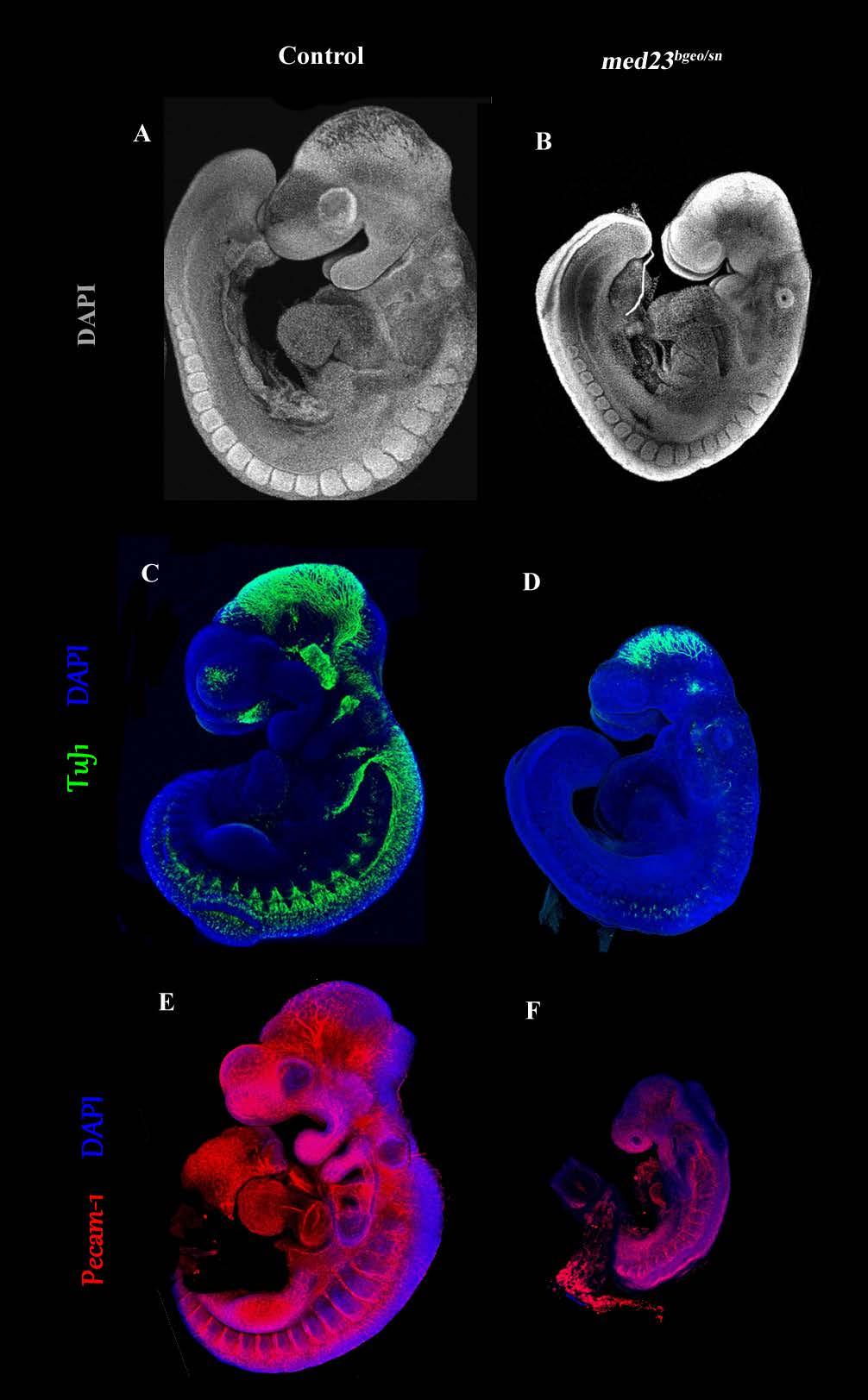
86 73
87 Figure 12: snouty is a functionally null allele Wildtype (A, C, E) and med23 bgeo/sn (B, D, F) littermate embryos were immunostained for DAPI (A, B), TuJ1 (C, D) and Pecam-1 (E, F). Morphological, neural and vascular defects in med23 bgeo/sn embryos are similar to med23 sn/sn embryos suggesting that med23 sn is a null allele. 74
88 determined by pseudo-sem DAPI staining (Figure 12A and B), neural differentiation was determined by β-tubulin III immuno-staining (Figure 12C and D) and blood vessel formation was determined by Pecam-1 immuno-staining of endothelial cells (Figure 12E and F). In all the three processes, the defects observed in med23 bgeo/sn embryos were similar (neither milder nor more severe) to med23 sn/sn embryos. At E9.5, med23 bgeo/sn embryos are smaller in size than their wild-type littermates, their FNP and 1 st BA (Figure 12B) are smaller in size and mis-formed and the heart is under-developed (Figure 12B). The forming nervous system of med23 bgeo/sn embryos displays similar defects as that of med23 sn/sn embryos. med23 bgeo/sn embryos also lack β-tubulin III immuno-stained differentiating neurons in the forming cranial sensory nervous system and have fewer and mis-organized β-tubulin III immuno-stained differentiating neurons in the forming trunk sensory nervous system (Figure 12D). Moreover, Pecam-1 immuno-staining shows that the defects in vascular organization of med23 bgeo/sn embryos (Figure 12F) are also similar to that of med23 sn/sn embryos. med23 bgeo/sn embryos do not survive beyond E10.5 similar to med23 sn/sn embryos. These results from the complementation test suggest that snouty is indeed a functionally null mutation of med23 and the phenotypic differences between med23 sn/sn and med23 bgeo/bgeo embryos are due to the background strain differences. 75
89 VII Generation of med23 conditional knock-out mice To overcome the early embryonic lethality of med23 bgeo/bgeo embryos and to analyze the role for MED23 during later gestation and postnatally, we decided to generate conditional mutant mouse lines for med23. med23 bgeo allele obtained from KOMP (Figure) has been designed such that the genetrap cassette within intron 12 is flanked by FRT sites. To make use of the Flp/FRT system, we crossed the med23 bgeo mice to a C57BL6 Flpper mouse strain. The flippase enzyme excises out the genetrap sequence flanked between FRT sites leaving two loxp sites flanking exons 13 and 14 of med23 (Figure). This mouse line, med23 floxed will be used to mate with various cre lines to generate tissue specific and time specific conditional mice. The scheme of matings is depicted in Figure 13. Unfortunately, much after the Flpper and med23 bgeo mating was underway we realized that the Flpper mouse is not very efficient. In the med23 bgeo allele, the FRT sites flank the complete bgeo cassette, which includes the genome sequence encoding for lacz as well. Using lacz staining in the developing gonads of embryos obtained from med23 bgeo x flpper mice, we observed that majority of the embryos that are carry both flp and med23 bgeo alleles are still unable to excise out bgeo cassette and express lacz and stain blue upon X-gal staining (Figure 14). The FLP/FRT mediated excision in this particular system is mosaic and as a result we ended up with animals of mixed genotypes containing med23 bgeo, med23 floxed and flp alleles all 76
90 Figure 13: Mating scheme to generate conditional mice from med23 bgeo animals 77

91 Figure 14: Mosaic activity of Flpper mice C57Bl/6 Flpper mice were crossed with med23 bgeo/+ animals and embryos were harvested at E14.5 to analyze their genital ridges for complete excision. LacZ staining from bgeo genetrap allele was used to determine if the excision occurred or not. Excision of bgeo genetrap by Flpper would result in absence of lacz staining, however 1 out of 9 (embryo # 6) embryos from C57B/6 Flpper x med23 bgeo cross still expressed lacz (in spite of having the Flp allele genotypically) showing that Flpper excision is not complete. 78
92 together. We did not want to generate our conditional mice with these mixed genotypes and getting around this ended up extending our timeline beyond previous expectations. In order to avoid leakiness of med23 bgeo allele into the conditional animals (med23 floxed ), we have screened all the offspring of Flpper x med23 bgeo mating by genotyping not just for the presence of the newly floxed allele but also for absence of lacz or bgeo allele and generated our med23 floxed founder males and females (med23 floxed F2 generation) only from these newly identified floxed+; lacz- animals (med23 floxed F1 generation). Since our genotyping is done with tail clips, we had to bear in mind that the mosaicism caused by inefficient Flppase enzyme activity could result in a situation where the all the tail cells end up with perfectly excised med23 floxed alleles (also lacz-) but the germ cells could still carry some residual med23 bgeo alleles. This indeed did happen as some of F2 animals were still genotyped as lacz+; our solution to this problem was to discard the lacz+ F2 animals and maintain only the floxed+; lacz- ones. This solution has been successful yet quite slow in generating the med23 floxed mouse line. In spite of these hurdles and time delays, we have now been able to cross the med23 floxed line to various cre lines (Wnt1 cre, Tek cre, ERT2 cre and ZP3 cre ). Wnt1 cre ; med23 floxed line will help us excise med23 specifically out of neural crest cells, while Tek cre (Tie2 cre ) will excise med23 out of all endothelial cells in yolk sac and embryo beginning E ERT cre is a mouse line which expresses cre in a tamoxifen-dependent manner, hence ERT2 cre ; med23 floxed mouse line 79

93 Figure 15: Tamoxifen inducible ERT2 Cre is able to excise loxp floxed alleles at 5mg/kg dosage ERT2 Cre mouse line was crossed with R26RlacZ mouse line, where stop codon upstream of lacz gene is flanked by loxp sites. Pregnant mothers were treated with oral gavage of 5mg/kg tamoxifen (+1mg/kg Progesterone) at E9.5 and embryos were analyzed for excision at E10.5. As shown above Embryo # 3 and Embryo # 5, carrying cre alleles show ubiquitous lacz staining suggesting ubiquitous excision by ERT2 cre allele at this dose. 80
94 will be used to surpass the early embryonic lethality of med23 sn/sn embryos and excise med23 out during later stages of embryonic development. Our preliminary studies have identified 5mg/kg dosage of Tamoxifen as optimum for complete excision of floxed allele by ERT2 cre (Figure 15). At this dose, Tamoxifen requires 24hours post-oral gavage to completely excise the floxed allele. The purpose of ZP3 Cre ; med23 floxed mouse line would be to excise med23 in the egg cells and generate another model for med23 null mutation to compare to med23 bgeo/bgeo and med23 sn/sn embryos. Discussion The mega-dalton protein complex, Mediator, is well conserved from yeast to humans. When first discovered, Mediator complex was assumed to serve as a tether between the regulating transcription factors and the PIC (Polymerase Initiation Complex) assembly. However, subsequent work done by many other labs has shown that Mediator is not a passive player in transcription. Mediator subunits themselves are arranged in three separate modules, Head, Middle and Tail. These subunits, through their interaction with transcription factors, intracellular signaling components, and polymerase machinery subunits create a system which provides an added layer of transcription initiation regulation. Mediator complex comes in two forms; the activator form of Mediator is associated with subunit MED26 while the repressor 81
95 form is associated with the CDK8 module. Both MED26 and CDK8 bind to the middle module of Mediator. Recent work has also shown that MED26 subunit containing Mediator complex plays a role in regulation of transcript elongation. MED23 subunit (mutated in snouty and med23 bgeo ) has been shown to be associated with both activator and repressor forms of Mediator 177. It is intriguing to see that loss of a single subunit of transcription co-factor complex, Mediator, results in such specific embryonic defects. Our data shows that med23 is ubiquitously expressed. Intuitively, we would expect a RNA Polymerase II co-factor to be crucial for all transcription within the embryo which would mean that losing a subunit of this co-factor complex would have severe defects within the homozygous embryo beginning as soon as the start of zygotic transcription, which happens really early in mammalian embryos. However, our data points to the contrary. How a mutation in a ubiquitously expressed subunit of a transcription co-factor complex can lead to defects specific to craniofacial, peripheral neurons and cardio-vascular tissue development, while sparing the other organ systems (central nervous system, muscle progenitor somatic tissue) is perplexing but not unprecedented. Some other Mediator subunits, like MED12, have shown roles in specific organ system development and specific signaling pathway regulation 216. One possible explanation for such tissue specific role for MED23 could be that loss of MED23 subunit does not affect the 82
96 assembly of the rest of the Mediator 176. We also know that MED23 is a subunit of the tail subcomplex of Mediator, whose function is to bind and interact with specific transcription factors. All of this suggests a system in which the different subunits of Tail sub-complex interact with specific transcription factors and these specific interactions have roles in regulation of specific processes only, thus making MED23 the tail subunit important for formation of frontonasal process, peripheral nervous system and cardiovascular system within the mammalian embryo. The med23 sn/sn mutant embryos die before we can analyze late gestational or post natal role for MED23. We have now generated conditional med23 mice that will help us overcome this midgestational lethality. snouty is a point mutant of med23; which means that snouty could be a null or a hypomorphic allele of med23. Our work, which also supports published reports 176,218 argues against snouty being a hypomorph. The phenotype of snouty embryos matches perfectly with the published med23 -/- embryo phenotype, in spite of differences in the mouse strains carrying the two mutations. We also see a loss of about 85% of MED23 protein product in snouty mutant embryos presumably due to instability of the mutant MED23 protein. We have generated a truncated allele of med23 (med23 bgeo ), which in homozygosity also leads to midgestational lethality, but slightly earlier than snouty. Our complementation test tells us that snouty and med23 bgeo allele are both functionally null, and the only reason for increased 83
97 severity of med23 bgeo/bgeo embryos is the background strain differences between snouty and med23 bgeo. med23 bgeo allele still forms a truncated RNA product consisting of first 12 exons. The expectation is that this truncated mrna would undergo Nonsense Mediated Decay (NMD) however, some transcript is still observed in the mutant embryos. No information is currently available from our or other labs to suggest that the truncated mrna product is still active, however, the possibility cannot be completely ruled out. The evidence that snouty and med23 bgeo are indeed functionally null alleles also comes from a comparison with a published phenotype of a med23 null allele. Berk lab has generated a mutant allele of med23 in which they have knocked out exons 2 and 3 of the gene (med23 -/- ) 176. Analysis of this med23 -/- phenotype revealed that the mutant embryos die between E9.5 to E10.5 as observed our snouty embryos 218. Berk lab med23 -/- embryos also display a growth defect at E9.5 along with defects in formation of the PNS and organization of blood vasculature. In spite of being on different mouse strain backgrounds and having different sites of mutation, snouty embryos (FVB strain; point mutation exon 22) and Berk lab med23 -/- embryos (F1 strain; deletion exon 2 and 3) display similar defects in embryogenesis. This suggests to us that even though med23 sn allele translates about 15% of the mutant MED23 protein; the translated product is functionally inactive. Our med23 bgeo/bgeo embryos phenotype is still more severe that the Berk lab med23 -/- and med23 sn/sn embryo phenotype, suggesting that these differences might be more due to the 84
98 underlying strain differences. Comparing the Berk lab med23-/- embryo phenotype with med23 sn/sn and med23 sn/bgo (complementation) embryo, it seems that med23 sn is a null allele. 85
99 Chapter 2 MED23 is required for mammalian vascular development 86
100 Summary Proper blood vasculature development ensures proper blood flow to all organs and cell types within the body. Availability of oxygenated blood and nutrition is just as important, if not more, during embryonic development as it is in maintaining body homeostasis. As described earlier, med23 sn/sn embryos seem to display a defect in formation for proper vascular network within the yolk sac and embryo. Our analysis shows that MED23 is dispensable for vasculogenesis (the process of formation of vasculature de novo) but seems to be important for remodeling of the blood vessels after the primary plexus has been established. It seems that loss of MED23 disrupts endothelial cell-cell junction formation. Later roles for med23 in differentiation of vascular smooth muscle cells has been shown in zebrafish embryos, however, med23 sn/sn mice embryos die much before those analyses could be performed. Early on, preliminary vascular smooth muscle development seems unaffected in these embryos. We have also observed an increase in Vegfa transcript levels following loss of med23, which has been reported to cause embryonic lethality in mice embryos due to cardiovascular development defects. Whether the combination of all these vascular defects in med23 sn/sn embryos is severe enough to cause the mid-gestational lethality is currently debatable. We hope that our analysis with endothelial cell-specific deletions of med23 will give us better understanding of how MED23 regulates blood vasculature development. 87
101 Introduction Development and functioning of all organs depends on a properly functioning vascular network. Blood vessels consist of an endothelial tube surrounded by mural or support cells (vascular smooth muscle cells for arteries and veins or pericytes for arterioles, venules and capillaries), which regulate vessel diameter and blood flow. Endothelial cells that line the entire blood vessel network are thought to derive de novo from mesoderm that first forms a primary vascular plexus within the extra-embryonic tissue and share their lineage with hematopoietic cells through a common progenitor, called hemangioblast, which has been described in drosophila, zebrafish, chick and mice embryos Blood vessel formation is typically divided into two stages, vasculogenesis (de novo formation of endothelial tubes from angioblasts) and angiogenesis (remodeling of existing blood vessels into new ones). Vasculogenesis Vasculogenesis begins in the yolk sac during gastrulation and involves differentiation of mesodermal cells into angioblasts. Angioblasts are endothelial cell precursors that come together and form the primary vascular plexus within the yolk sac. This differentiation of primary endothelial cells has been shown to require signals similar to the ones required for 88
102 mesoderm specification during gastrulation, BMPs and FGFs. Loss of BMP signaling effector proteins, SMAD4 or SMAD5, result in yolk sac vasculature defects leading to embryonic lethality In mouse embryonic stem cells, BMP signaling has been shown to initiate a FGF2-dependent signaling program that regulates specification of angioblasts 238, 239. The hierarchy of BMP and FGF signaling cascades is reversed in human embryonic stem cells where FGF signaling has been shown to be required for stem cell survival and proliferation, while BMP signaling is required to promote endothelial cell differentiation 240. BMP signaling in this case is under regulation of Hedgehog (IHH) pathway whose components are expressed in the yolk sac visceral endoderm and mesoderm at the time of vasculogenesis and their loss affects endothelial cell differentiation 241. Angiogenesis Angiogenesis or vascular remodeling involves proliferation, migration and sprouting of endothelial cells to form new vessels from pre-existing ones and also involves differentiation of endothelial cells into arterial, venous and blood-forming hemogenic phenotypes. One of the signaling pathways crucial for angiogenesis is Notch signaling 242, 243. Loss of ligands, notch1 and notch4 together, causes severe defects in embryonic and extra-embryonic vessel remodeling leading to embryonic lethality 244 while mice lacking a copy of Notch receptor, dll4, 89
103 exhibit smaller dorsal aorta and remodeling defects in yolk sac vessels 245. HEY1 and HEY2 are downstream effectors of Notch signaling and have been shown to be crucial for vascular development. Loss of both hey1 and hey2 together leads to embryonic lethality in mice at E9.5, specifically due to extra-embryonic (yolk sac and placenta) vessel remodeling defects 246, 247. The process of Angiogenic sprouting involves loss of cell-cell junctions between endothelial cells, degradation of basement membrane by proteolysis followed by migration of tip endothelial cells in response to angiogenic stimuli. The other endothelial cells that trail the earliest migrating tip endothelial cells are known as stalk cells and these stalk cells are responsible for proliferating and expanding the newly forming endothelial sprouts. The specification of tip versus stalk cells is regulated by VEGF and Notch signaling 248. Tip cells are characterized by their expression of Notch ligand, dll VEGF signaling regulates this expression of dll4 in tip cells 250, which then reduces the expression of flk1 in adjacent endothelial cells through activation of Notch signaling in those cells. Reduction in flk1 expression reduces the ability of the adjacent cells to respond to VEGF stimuli thus preventing them from acquiring tip cell fate and conferring them stalk cell identity Loss of dll4 and inhibition of Notch signaling results in increased angiogenic sprouting and hyper - proliferative endothelial cells 251, 254. To make matters more complex, the specification of tip- 90
104 versus-stalk identity within the sprouting endothelial cells itself has been shown to be a dynamic process. Through live imaging, it was shown that the sprouting endothelial cells are continuously in struggle to become the tip cells and at each point of time whichever cell expresses the dll4 ligand acquires the tip cell identity for that time period 255. Apart from angiogenic sprouting, VEGF and Notch signaling cross-talk is also crucial for arterial and venous differentiation of endothelial cells. Mice lacking a copy of Notch receptor, dll4, exhibit smaller dorsal aorta and remodeling defects in yolk sac vessels and have complete loss of arterial identity 256,257. On the other hand, overexpression of dll4 causes enhanced arterialization also leading to vascular defects 245. Dorsal aorta development was also affected in hey1; hey2 double mutant embryos Loss of Rbpj, encoding a transcription factor downstream of Notch signaling, also results in defects in arterial endothelial cell development 257, suggesting that Notch signaling pathway regulates arterial differentiation during endothelial cell development via RBPJ/HEY1-HEY2. VEGF signaling has also been suggested to have a role in arterio-venous differentiation of endothelial cells. In vitro VEGF induces expression of arterial markers in endothelial cells while morpholino knockdown of vegf in zebrafish embryos leads to sustenance of venous cell markers at the cost of arterial makers 258, 259. Specification of venous cell identity, on the other hand, is under the control of an orphan nuclear receptor, COUPTFII, which is expressed in endothelial cells specific to veins 91
105 and lymphatic vasculature beginning E Endothelial cell specific deletion of CouptfII (using Tie2 re ) leads to embryonic lethality and loss of venous identity. Overexpression of CouptfII in endothelial cells on the other hand also leads to aberrant vessel development due to fusion of artery-vein structures, a phenotype similar to notch- deficient embryos 261, 262. COUPTFII, thus seems to be required for suppressing Notch activity (which is regulates arterial endothelial cell differentiation) and thereby regulate venous cell identity. This hypothesis is supported by the fact that double knock-out for CouptfII and Rbpj (downstream target for Notch signaling) partially restores venous identity for endothelial cells 263. VEGF signaling in Vasculature development VEGF signaling has been shown to be important in regulating various aspects of endothelial cell function, including survival, proliferation and vessel permeability 264, 265. VEGFA is the most extensively studied VEGF family member and is expressed its expression in the extra-embryonic visceral endoderm at E7.5 in mice correlates perfectly with blood island formation in yolk sac 266. Loss of one or both copies of Vegfa leads to cardiovascular defects and embryonic lethality in mice 267, 268. Over-expression of Vegfa also impairs cardiac development and results in mid-gestational embryonic lethality suggesting a dose-dependent crucial role for VEGF in heart and vascular development 121. VEGFA signals mainly through two receptors, 92
106 VEGFR1 and VEGFR2 (or FLT1 and FLK1 respectively) but it has also been shown to interact with Neuropilin receptors 1 and 2 (NPN1 and NPN2). VEGFR1 (FLT1) has a higher affinity for VEGFA than VEGFR2 (FLK1), however VEGFR1 lacks the tyrosine kinase ability to signal downstream post- VEGFA binding event and hence has been considered to be a negative regulator (decoy receptor) of VEGFA 269. In support of this, loss of flt1 results in increased number of hemangioblasts leading to an overgrowth of endothelial cells and vascular channels and eventually causes mid-gestational embryonic lethality in mice 122. Role of VEGFR2 or FLK-1 in endothelial cell generation and blood vessel development is very well established. Loss of flk1 does not affect formation of angioblasts but still results in complete lack of yolk sac blood islands and embryonic and extra-embryonic blood vessels leading to embryonic lethality 119. Analysis of chimeric embryos generated from wild-type and flk1-null cells has shown that flk1 is cell autonomously required for endothelial cell differentiation and suggests that the main role for FLK1 is to ensure that the mesodermal precursors of blood islands end up at the correct locations within the yolk sac 270. This suggests that VEGFA signaling probably regulates survival and proliferation of endothelial cells and their precursors but not necessarily the differentiation of angioblasts from mesoderm. Angiopoeitin signaling in vascular development 93
107 Angiopoetin/Tie signaling pathway is unique such that it is almost exclusively endothelial cell specific and is crucial not for de novo endothelial cell differentiation (or vasculaogenesis) but for blood vessel remodeling and maturation (angiogenesis) during later stages of embryonic and postnatal development. Angiopoetins (ANG1, ANG2 and ANG4) are ligands that bind to tyrosine kinase receptor Tie2 (also known as Tek) and regulate endothelial cell survival and cell-cell junction stabilization. Deletion of tie2 in mice embryos leads to midgestational embryonic lethality due to impaired cardiac development and reduced numbers of endothelial cells 271, while loss of angiopoeitin-1 (ANG1) leads to vascular remodeling defects and a disruption of endothelial cell extracellular matrix interaction 272, 273. Tie1, a homolog of Tie2, on the other hand, does not directly bind to any Angiopoeitin ligands, but its loss still affects blood vasculature development, specifically angiogenic capillary growth and endothelial cell survival, leading to hemorrhage and death of mutant embryos 274,275. Formation and Regulation of Endothelial cell-cell junctions Endothelial cell-cell junctions are critical to regulate vascular permeability. During development, regulation of junction formation is crucial for forming endothelial cell tubes (blood vessels) and regulated disruption of the junctions is required during angiogenic sprouting. Homotypic junctions between endothelial cells come in multiple flavors, two of the 94
108 well-studied ones are tight and adherens junctions (Figure 16). Adhesion via adherens junctions is mediated via VE-Cadherin, an endothelial specific member of the Cadherin family. Inside the cells, VE-Cadherin is linked to a large number of intracellular partners, one of them being the actin cytoskeleton, which can control the dynamic opening and closing of the cell- cell junctions. Adhesion in tight junctions is mediated by the Claudin family of membrane proteins. Multiple claudin family members are expressed in tight junctions of all cell types, such as occluding, nectin and others; Claudin-5 amongst these is specific to endothelial cells 276. Disrupting VE-Cadherin in mouse embryonic stem cells does not affect differentiation of endothelial cells but affects their organization into a vessel like pattern 277. Loss of VE- Cadherin in embryos affects formation of capillary plexus within mouse allantois and leads to mid-gestational embryonic lethality following vascular defects 278. VE-Cadherin association with another junctional protein β-catenin seems to be crucial for its vascular remodeling function 279. Recently, it has been shown that VE-Cadherin is not required for formation of nascent blood vessels but is required for maintaining their vessel-like assembly by maintaining the endothelial cell-cell junctions 280. Another cadherin, N-cadherin, has also been shown to be expressed in endothelial cells and its ablation within the endothelial cell lineage leads to loss of ve-cadherin expression, vascular development defects and mid-gestational embryonic lethality 281. However, in vitro, embryonic stem cells deficient for N-cadherin were able to form 95

109 Figure 16: Endothelial cell-cell junctions 96
110 vessel like structures but showed defects in endothelial cell-support cell junctions (pericytes) suggesting a role for N-cadherin in maintaining heterotypic endothelial cell connections 282. Loss of Claudin-5, which is an endothelial cell specific claudin, surprisingly does not disrupt overall morphology of blood vasculature and tight junction structure; however, it does affect the formation of blood brain barrier which leads to perinatal embryonic lethality in mutants 283, 284. Apart from adherens and tight junctions, endothelial cells also form homo- and heterotypic junctions via proteins like PECAM-1 (Platelet and Endothelial Cell Adhesion Molecule-1) and VCAM-1 (Vascular Cell Adhesion Molecule-1), which are expressed preferentially in endothelial cells and hence are usually used as markers of vasculature but are not indispensable for blood vessel development 285, 286. Loss of MED23 results in mid-gestational embryonic lethality. Our initial analysis showed that med23 sn/sn embryos display disruption in embryonic and extra-embryonic blood vascular network at E9.5. Here we are trying to characterize this blood vessel formation defect in more details. We see defects in expression of endothelial cell-cell junction markers in embryonic vasculature at E9.5 suggesting a disruption in vascular remodeling due to loss of med23. Whether these defects are sufficient to cause embryonic lethality has currently not been tested, but is in process with conditional deletion of med23 from endothelial cells. 97
111 Results I. flk1 expression is similar in wild-type and med23 sn/sn littermate embryos PECAM-1 immuno-staining depicted the presence of disorganized blood vessel network in med23 sn/sn embryos and yolk sac at E9.5. A microarray done on E9.5 wild-type and med23 sn/sn embryos showed a 1.6fold increase in Vegfa transcript levels. Increasing Vegfa levels by 2-3 folds in mice embryos result in disruption of cardiovascular development and mid-gestational lethality 121. It has been hypothesized that in some mutants, lack of proper vasculature results in hypoxia-like conditions, which trigger an over-expression of Vegfa to compensate. Thus, the increase in Vegfa levels in med23 sn/sn embryos could be the cause for or result of abnormal vascular development. To determine if this mis-regulation of Vegfa transcription is real, we analyzed the expression pattern of VEGFA receptor, FLK1. flk1 is known to be expressed in all endothelial cells from the time that they first differentiate from mesoderm and by E9.5; it should be expressed throughout the vascular network. Wild-type embryos in med23 sn/sn embryos also, flk1+ cells are detected throughout the embryo (Figure 17). This is not surprising as we do see a vascular network being formed in med23 sn/sn embryos by PECAM-1 immunostaining and not a complete lack of vasculature, which would be expected had we lost flk1 expression. 98

112 Figure 17: flk1 expression in wildtype and med23 sn/sn embryos. flk1 expression in the med23 sn/sn embryo at late day E9.5 marks the blood vessel network. flk1+ cells are observed in the allantois, heart, intersomitic vessels and head and epibranchial regions of the mutant embryo similar to control littermate. 99
113 II. Formation of endothelial cell-cell junctions is affected in med23 sn/sn embryos In our effort to characterize the defects in blood vessel development in med23 sn/sn embryos we decided to analyze the expression of endothelial cell-cell junction markers in the mutant embryos. The two main Homotypic junctions formed between endothelial cells are adherens junctions (marked by VE-Cadherin) and tight junctions (marked by Claudin-5). Both VE-Cadherin and Claudin-5 are junction proteins exclusive to endothelial cells only. Adherens Junction When analyzed at E9.0, a time point when med23 sn/sn phenotype is first visible, expression of ve-cadherin seems to be normal in wild-type and mutant embryos alike (Figure 18A, B). However, at E9.5, two out of three embryos analyzed displayed a mild disruption in ve-cadherin expression pattern throughout the embryo (representative image, Figure 18 C, D). Instead of the beautiful network of blood vessels observed in control embryos, mutant embryos display dispersed ve-cadherin+ cells lacking a tree-like vascular network. One of the three mutant embryos analyzed had a severe defect, where the ve-cadherin+ endothelial cells were dispersed and fewer in number, especially in the head and epibranchial region (Figure 18 E, F). Specifically the network around the otic vescicle in the epibranchial region is severely disrupted in both mutant embryos (Figure 18D, F). Since, ve- cadherin+ cells are present 100
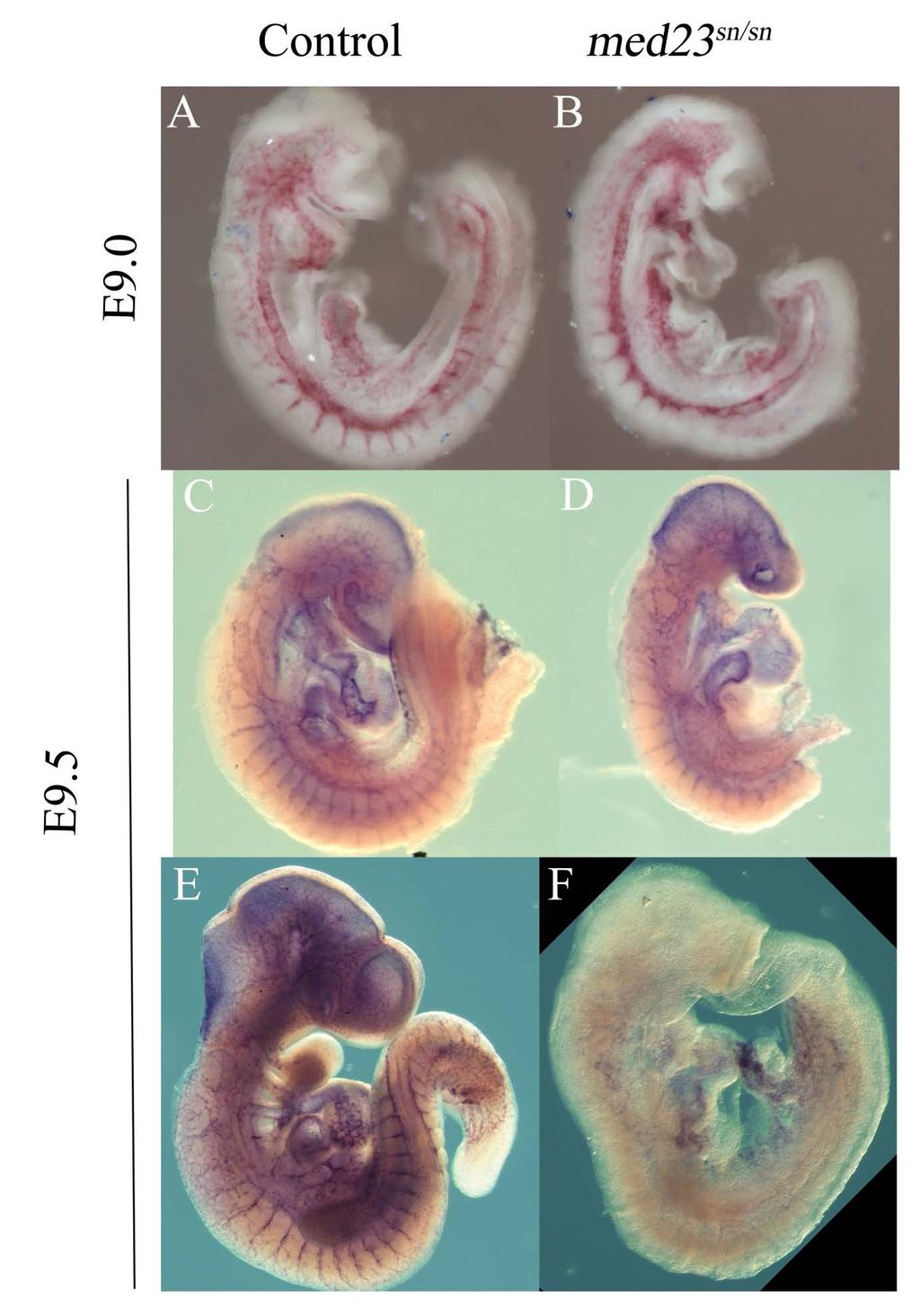
114 101
115 Figure 18: Adherens junction marker; ve-cadherin expression is reduced in med23 sn/sn embryos at E9.5 but not at E9.0 A, B ve-cadherin ISH at E9.0 shows similar expression between wild-type and mutant embryos. C, D At E9.5, mild phenotype ve-cadherin ISH shows abnormal vascular network in med23 sn/sn embryos reminding of Pecam-1 immunostaining phenotype. E, F In a severe phenotype, very few ve-cadherin+ cells are observed in the med23 sn/sn embryos and vascular pattern of expression is lacking. 102
116 in that region half a day earlier (Figure 17B), and PECAM-1 immuno-staining suggests the presence of a vascular network at E9.5, it seems that the expression of adherens junction marker is specifically affected at that stage and that MED23 might be required for its maintenance. Tight Junctions Since we saw a disruption of adherens junction marker expression in E9.5 med23 sn/sn embryos, we also decided to analyze the expression of tight junction marker in the mutant embryos. claudin-5 is a tight junction marker exclusive to endothelial cells. claudin-5 is expressed in all endothelial cells however its role is specific to blood brain barrier formation. RNA in situ hybridization of wild-type and med23 sn/sn embryos at E9.5, display a defect in claudin5 expression in mutant embryos (Figure 19 A, B). Similar to ve-cadherin, at E9.5, med23 sn/sn embryos display a disrupted claudin-5 expression pattern in the epibranchial region (Figure 19C, D). Given that PECAM-1 immuno-staining shows the presence of a vascular network in the epibranchial region, it seems that tight junction marker claudin-5 expression is specifically disrupted following loss of MED

117 104
118 Figure 19: Expression of tight junction marker, claudin5 is reduced in med23 sn/sn embryos at E9.5 Analysis of claudin5 expression in control and med23 sn/sn embryos at E9.5 shows reduced claudin5+ cells in the epibranchial and head region of mutant embryos (A D) and in the intersomitic vessels. 105
119 III. Early development of vascular smooth muscle cells is unaffected in med23 sn/sn embryos The vascular defect observed in med23 sn/sn embryos could be endothelial cellautonomous or non-autonomous. The loss of cell-cell junction markers in mutant embryos could also result from lack of support from mural cells. By E9.5, vascular smooth muscle cells have already begun to be recruited in the developing cardiac tissue and the developing aorta to surround and support the endothelial cell tubes. We utilized an antibody against SM22α (Smooth muscle 22 alpha), one of the earliest markers of vascular smooth muscle cells to analyze early stages of smooth muscle differentiation in mutant embryos. At E9.0, wild-type embryos express SM22α throughout out the developing heart as well as in yolk sac. SM22α expression in E9.0 med23 sn/sn embryos is also detected in the heart and yolk sac at levels similar to its wild-type littermate (Figure 20A, B) suggesting that early differentiation of vascular smooth muscle cells does not require MED23. Discussion Loss of MED23 results in disruption of vascular network and also causes midgestational embryonic lethality. We sought to figure out whether the embryonic and extraembryonic vascular defects of the mutant embryos are related to its lethality by analyzing the various aspects of vascular development in these embryos. Our analysis suggests that MED23 is 106

120 Figure 20: Early formation of vascular smooth muscle cells is not affected in control and med23 sn/sn littermate embryos at E9.0. Analysis of Control and med23 sn/sn embryos were immuno-stained for SM22α, a marker of vascular smooth muscles, showed similar staining in the developing heart of mutant and wildtype embryos. 107
121 required for vascular remodeling and maintenance of endothelial cell-cell junction markers, ve-cadherin and claudin-5. In spite of an increase in transcript levels of Vegfa in med23 sn/sn embryos, we do not see over-expression of VEGFA receptor flk1 in mutant endothelial cells, which matches with our PECAM-1 immuno-staining, where we do not see hyper-vascularization of the embryo. In vitro, the ability of VEGF to increase microvasculature permeability and cause vasodilation has been well known 287, 288, 289. VEGF signaling through its receptor VEGFR2 (or FLK1) induces phosphorylation of cell junction proteins (like ZO-1, Occludin and VE-Cadherin) leading to their destabilization resulting in increase in permeability 290,291,292,293,294. Ultrastructural analysis has shown that VEGF signaling activation leads to a very rapid disruption of endothelial cell-cell junctions, including loss of both tight and adherens junctions 295. What has not been investigated is if VEGF signaling has any role in regulating the formation of these endothelial cell-cell junctions via regulation of junctional protein transcription. Interestingly, in our system we see a reduction in expression of endothelial cell-cell adherens and tight junction markers (loss of ve-cadherin and claudin-5 transcripts) as well as an increase in Vegfa transcript levels. It would be interesting to look at the protein distribution of VE- Cadherin and Claudin-5 within the endothelial cells and see if their protein levels are reduced by loss of MED23. That would indicate that MED23 is required (directly or indirectly) to 108
122 suppress Vegfa transcript levels, mis-regulation of which affects endothelial cell junctional protein stability in med23 sn/sn embryos. On the other hand, MED23 might just be directly required for maintenance of ve-cadherin and/or claudin-5 expression. VE-Cadherin itself has been recently shown to regulate the formation of tight junctions 296 suggesting that loss of claudin-5 in E9.5 med23 sn/sn embryos might also be an indirect effect of loss of MED23- mediated VE-Cadherin expression. In my opinion, the loss of ve-cadherin and claudin-5 in med23 sn/sn embryos is less of a direct effect of MED23 loss on their expression and more of an effect in disruption of endothelial cell-cell junction maintenance. Specific loss of claudin-5 expression within the head region of med23 sn/sn embryos suggests that if allowed to develop till birth, loss of MED23 may affect the formation of blood brain barrier in perinatal and postnatal animals, similar to what is observed after loss of claudin Recently, a study in zebrafish suggested that MED23 is required for regulation of adipogenesis and smooth muscle cell formation. Morpholinos against med23 resulted in zebrafish embryos with increased vascular smooth muscle cells along with an increased vasculature at the cost of adipocytes; specifically the expressions of vascular smooth muscle markers, like SM22α and α-sma (smooth muscle actin) were both significantly increased in the med23 morpholino treated embryos in comparison to control embryos 297. Our analysis with one of the vascular smooth muscle markers, SM22α, shows a completely opposite result. 109
123 Loss of MED23 in mice embryos does not affect early smooth muscle cell differentiation. The major difference between the two studies is the time point of analysis. The increase in vascular smooth muscle cells, post-med23 morpholino treatment, was observed in fish at 48 hourspost-fertilization (hpf) is way beyond the time point E10.5 in mice embryos when med23 sn/sn embryos die. Thus, it might be that if somehow allowed to live longer, loss of MED23 might result in over-differentiation of vascular smooth muscle cells, a defect currently not observed in mutant embryos. Currently, we are focused on understanding whether the specification of arterial versus venous fate of endothelial cells is in any way affected by loss of MED23. Formation of endothelial cell-cell junctions is crucial to stabilize the proliferation of endothelial cells and thus stabilize the newly formed or sprouted endothelial tubes 298. It would be interesting to see if such endothelial cell specific hyper-proliferation would be observed in med23 sn/sn embryos due to loss of cell-cell junction markers. We are also working on understanding the endothelial cell specific role for MED23 in regulation of vascular development using Tie2 cre ; med23 floxed mice, which will excise out med23 specifically from endothelial cells beginning E9.5. Together all of this information should provide us a better understanding into how MED23 regulates vascular remodeling, endothelial cell-cell junction marker expression and thus endothelial cell-cell junction formation. 110
124 Chapter 3 MED23 is required for Peripheral Nervous system formation Parts of Introduction in this chapter (Trunk Sensory Nervous system and Autonomous Nervous system) have been adapted from a book chapter, Bhatt S., Diaz R., Trainor P. (2013) Signals and Switches in Mammalian Neural crest differentiation, published by Cold Spring Harbor Perspectives in Biology 111
125 Summary In our initial analysis, we observed a defect in peripheral nervous system formation in med23 sn/sn embryos, specifically the cranial sensory nervous system. In amniotes, the cranial sensory nervous system is comprised of the three paired sense organs, eyes, nose and ear and four neurogenic cranial ganglia, trigeminal, petrosal, nodose and glossopharyngeal as well as all the cranial nerves (sensory and/or motor) associated with each of the paired sensory organs and cranial ganglia. DAPI immuno-staining showed the defects in the morphology of the paired sensory organs while TuJ1 (neuron specific β-tubulin) marker immuno-staining showed defects in formation of the cranial nerves and associated cranial ganglia. RNA expression analysis determined that the major defect underlying the abnormal development of neurogenic cranial ganglia in med23 sn/sn embryos is in formation of cranial placodes. Cranial placodes form from the ectoderm at gastrulation as a single pre-placodal region (PPR) that then goes to segregate and differentiate into multiple placodes, which contribute to the various sensory organs and ganglia. Here, we show that in med23 sn/sn embryos, cranial placode formation is affected at multiple steps. Although very early specification of PPR is relatively unaffected, med23 seems to be required for proper organization and neurogenic differentiation of the neurogenic cranial placodes. 112
126 Introduction In multicellular organisms, the central nervous system (CNS) needs a way to sense and react to external stimuli. This function is carried out by the peripheral nervous system (PNS) of the body. PNS comprises of sensory and motor neurons that relay information back and forth from the viscera, smooth muscles, skin and the exocrine glands of the body to the brain and spinal cord of the central nervous system. In keeping with this function, PNS consists of nerves and ganglia located outside the brain and spinal cord which are grouped into two subsets, 1) Sensory nervous system: cranial and trunk sensory nervous system and 2) Autonomous nervous system (ANS): sympathetic, parasympathetic and enteric nervous system. The sensory nervous system, as the name suggests, is responsible for sensing external stimuli and consists of afferent fibers that transmit information from the somatic (skin, skeletal muscles, tendons and joints) and visceral (smooth and cardiac muscle and glands) structures to the CNS; while ANS consists of efferent motor neuron fibers which are responsible for transmitting information from CNS to the somatic and visceral structures. Sensory Nervous system Based on the location within the body, the sensory nervous system is divided into cranial and trunk sensory nervous systems. Cranial and trunk sensory neurons are pseudo- 113
127 unipolar; their cell bodies lie within the peripheral ganglia, cranial ganglia (for cranial sensory nervous system) and spinal or dorsal root ganglia (for trunk sensory nervous system) and their axons are split into two branches; one extending to the periphery while the other extending towards the CNS (brain for cranial and spinal cord for trunk sensory nervous system). Cranial Sensory Nervous System Cranial sensory neurons work in conjunction with cranial motor component to regulate the functions of paired sensory organs, eyes, ears and nose as well as movement of the craniofacial muscles including the muscles involved in facial expression and mastication (through the cranial ganglia) (Table 3). The sensory nerves are derived from two separate ectodermal-origin cell populations, neural crest and cranial placodes and associate with two anatomically distinct set of cranial sensory ganglia which have differential contributions from these two cell types (Figure 21). The proximal subset of cranial ganglia (dorso-medial region of trigeminal ganglion, a small portion of proximal geniculate ganglion, acoustic ganglion, superior ganglion and jugular ganglion associated with cranial nerves V (trigeminal), VII (facial), VIII (vestibulo-cochlear) and proximal portions of IX (glossopharyngeal) and X (vagal) respectively) is anatomically located closer to the central nervous system, and sensory neurons associated with these ganglia are derived from cranial neural crest, and trigeminal and otic 114
128 Table 3: Innervation of cranial nerves (human) Cranial Nerve Name Number Site of innervation Olfactory I Nasal cavity Optic II Retina Oculomotor III Eye muscles Trochlear IV Eye muscles Trigeminal V Skin of the face and muscles of mastication Abducens VI Muscle of the eye orbit Facial VII Muscles of facial expression, anterior 2/3 rd of the tongue Vestibulocochlear VIII Inner ear Glossopharyngeal IX Posterior 1/3 rd of the tongue Vagal X Laryngeal and pharyngeal muscles, soft palate Spinal accessory XI Sensory component joins Vagal nerve, motor component innervates muscles of the neck Hypoglossal XII Muscles of the tongue 115
129 Figure 21: Contribution of neural crest and cranial placodes to the cranial sensory ganglia in vertebrates 116
130 placodes. The distal subset of cranial ganglia (distal portions of geniculate (facial or cranial nerve VII), petrosal (glossopharyngeal or cranial nerve IX) and nodose (vagal or cranial nerve X) ganglia) is located further away from the central nervous system and neurons associated with these ganglia are derived from epibranchial placodes. Differentiation of cranial placodes into cranial sensory neurons The early development of cranial placodes and neural crest into cranial sensory neurons is independent of each other 299,300 however once specified the placodal derived sensory neurons require input from neural crest cells to form proper axonal projections 96, 300. Cranial placodes are first specified as a single pre-placodal region (PPR). This PPR region then segregates into separate cranial placodes (Introduction - pages 23-32). Four sets of genes are expressed in each placode during initial steps of differentiation in the following temporal manner, 1) six and eya, 2)sox, 3) pax and 4) bhlh (proneural determination and differentiation basic helix-loop-helix) genes (Figure 22). The temporal expression of genes in the developing cranial placodes has been reviewed in great details elsewhere 301. Knock-outs of some of these genes in mice have suggested an important role in mammalian placode development; however mechanisms by which they regulate mammalian placodal development are understudied. 117

131 Figure 22: Hierarchy of genes expressed in placodal development from PPR to neuronal differentiation. 118
132 six and eya family genes are expressed during initial placodal development and continue to be expressed throughout the various stages of cranial placode differentiation, albeit with a slight variation between various placodes (reviewed in 73 ). Their continued expression suggests a role in maintenance placodal identity and initiation of placodal differentiation. It has been suggested that six1 is involved in specification of PPR and maintenance of stem-like properties in placodal cells 78 as timing of placodal differentiation correlates with downregulation of six1 expression 302, 303 and its overexpression post-ppr establishment, diminishes expression of later placodal genes (reviewed by Sally Moody, Principles of Developmental Genetics, Book chapter, 2007). On the other hand, expression of other six family genes (six2, six4) and eya family genes (eya1 and eya2) persists into later stages of placodal development suggesting later roles in differentiation 304, 305. Work in non-mammalian model organisms has shown a cranial placode specific expression of sox family genes, sox2 and sox3. Both sox2 and sox3 are known to be expressed in the developing neural plate and seem to have a similar role in maintenance of neural progenitor pool in cranial placodes as well 306. However, the fact that sox2 and sox3 are also expressed in non-neurogenic lens placode and not expressed in neurogenic trigeminal placodes suggests that they may have non-neural roles as well 306, 307. In mice embryos, expression of sox2 is specific to the cranial placodes giving rise to the paired sensory organs (eye and ear) while sox3 expression is not placode specific. 119
133 Segregation of PPR into specific cranial placodes is visualized by expression of placodal specifier genes of pax family (reviewed in 73 ). pax3 is expressed in the ophthalmic branch of trigeminal placode, pax6 is expressed in olfactory and lens placodes and pax2/5/8 are expressed in the otic placode. Pre-placodal region (PPR) first segregates into anterior and posterior pre-placodal area (APA and PPA) and PPA then goes to form the otic and epibranchial placodes. pax2 has been shown to be expressed in the PPA and required for its maintenance 308. Work in chick embryos suggests a role for pax2 in regulation of epibranchial neuron identity 91, while in zebrafish pax2 acts synergistically and probably redundantly with pax8 in specifying the otic placode 309, 310. Mice knock-outs of pax2 and pax8 individually, both show defects in formation of the inner ear and associated ganglion 95, 311, 312. Loss of pax3 in mouse embryos leads to hypoplasty of all cranial ganglia including the trigeminal ganglion 313, 94. However, given the important role played by pax3 in neural crest development and the contribution of neural crest to sensory neurons and glia within the cranial ganglia, it is difficult to conclusively pinpoint these defects as cranial placode formation defects and claim a role for pax3 in cranial placode development. These restricted patterns of pax genes right before the cranial placodes begin to separate 92, 306 and their observed requirement in maintenance of the various cranial placodes in which they are expressed, suggest existence of a pax code that provides the identity to each cranial placode. 120
134 Once specified, each cranial placode (except for the lens placode) differentiates into cranial sensory neurons. Mouse knock-out studies have shown that neuronal differentiation of proximal subset of cranial ganglia, which derive from both neural crest and some cranial placodes require expression of a basic helix-loop-helix (bhlh) transcription factor, neurogenin1 314, while neuronal differentiation of distal subset of cranial ganglia that derive exclusively from cranial placodes, require expression of another bhlh transcription factor, neurogenin The importance of bhlh transcription factors in neurogenesis was first identified in Drosophila (reviewed in 316 ). In Xenopus, neurogenin (ngn1) expression is observed in prospective ganglion cells and is lost as the coalescing ganglion cells differentiate into neurons and glia. Both ngn1 and ngn2 directly regulate the expression of another bhlh transcription factor, neurod whose expression correlates well with the acquisition of neuronal and glial characteristics. Loss of neurod1 in mice affects formation of otic placode derived inner ear hair cells and in humans has been associated with Branchio-oto-renal disorder. Trunk Sensory Nervous system Trunk sensory nervous system comprises of a metameric string of Dorsal Root Ganglia (DRG) which consist of variety of sensory neuron cell bodies, axons of which innervate various parts of the body and enable us to sense touch, temperature, and pain and limb movements. 121
135 Each of these functions is attributed to a specific type of sensory neuron, all of which are derived from trunk NC cells. Each functional type of sensory neuron is characterized by its own unique set of ion channels and forms through interactions of unique sets of transcription factors. The different types of neurons within each DRG rely on separate neurotrophic factors for their survival and growth and hence express distinct neurotrophic receptors. The small diameter pain and thermal sensory neurons require NGF (Neurotrophic Growth factor) and express NGF receptor TrkA and Ret 317, 318, 319 while the subpopulation of touch neurons require BDNF (Brain derived neurotrophic factor) and express BDNF receptor TrkB 320, 321, 322. The larger diameter movement sensing neurons innervate skeletal muscles and depend on Neurotrophin-3 (NT-3) for survival and express NT-3 receptor TrkC 319, 323. Differentiation of neural crest into trunk sensory neurons Similar to cranial sensory neurons, trunk sensory neurogenesis also requires expression of bhlh transcription factors, neurogenin1 (ngn1) and neurogenin2 (ngn2). Trunk sensory neurogenesis occurs in two waves. The first wave of neurogenesis, controlled by ngn2, occurs in the migrating neural crest cells and forms the large diameter propioceptive and mechanoreceptive neurons while the second wave, controlled by ngn1, occurs after DRG coalescence and produces both small nociceptive and large mechanoreceptor and 122
136 proprioceptor neurons. Similar to cranial neurons, neurogenin expression in trunk controls expression of another family of bhlh transcription factors (neurod) which serve as neuronal differentiation factors. Combined loss of both ngn1 and ngn2 leads to complete loss of neurod expression and lack of DRGs 324, 325. Analyses of ngn1 and ngn2 null mutations suggests a predominant role for ngn1 in formation of small diameter pain TrkA+ neurons and a lesser role in the formation of large diameter TrkB+ and TrkC+ neurons, and a transient role for ngn2 in formation of large diameter TrkB+ and TrkC+ neurons. DRG formation in ngn2 mutant embryos is normal due to functional compensation by ngn1. ngn2+ cells also contribute to a small but significant fraction of TrkA+ nociceptive neurons 326. A number of transcription factors, like POU domain transcription factor, POU4F1, Runt related transcription factors, RUNX1 and RUNX3, and homeobox transcription factor, SHOX2, have all been shown to regulate the differentiation of DRG sensory neurons into various subtypes. Two major reports highlight the role of WNTs released from dorsal neural tube in trunk sensory neurogenesis 39, 38. Activation of canonical WNT signaling causes stabilization of cytoplasmic protein, β-catenin, which then translocates into the nucleus and activates WNT responsive genes with the help of Lef/TCF factors. Genetic ablation of β-catenin in neural crest leads to a complete absence of dorsal root ganglia and loss of both waves of sensory neurogenesis 39. Lack of active WNT signaling at the sites of post-migratory neural crest cells 123
137 and at DRG sites suggests that WNT signaling is not directly involved in the induction of the secondary wave of DRG neurogenesis 327. Constitutive activation of canonical WNT signaling in the neural crest progenitor cells in the dorsal neural tube promotes sensory neuron differentiation in vivo, at the expense of other neural crest progeny. In vitro clonal analysis proved that this effect of canonical WNT signaling in sensory cell differentiation is not caused by WNT mediated sensory progenitor cell proliferation and expansion but is a true cell fate specification 38. Autonomic Nervous system ANS is a neural crest derivative that provides a way for the central nervous system (CNS) to send commands to the rest of the body and regulate the involuntary functions of the visceral organs. Functionally, ANS is divided into three major components, sympathetic nervous system (SyNS) and parasympathetic nervous system (PSNS) which function together to maintain body homeostasis and the enteric nervous system (ENS) which controls the gut motility amongst other things. Axons from pre-ganglionic neurons of the CNS (brainstem and spinal cord in this case) project and synapse with the post-ganglionic cell bodies located paravertebrally (for SyNS) or inside the target organs (in case of PSNS); the post-ganglionic axons innervate the target site (organs and tissues throughout the body). In the gut, the nor- 124
138 adrenergic and cholinergic ganglia form the enteric nervous system (ENS) which exclusively innervates the gastro-intestinal (GI) tract. Differentiation of autonomic neurons Within the ANS, formation of sympathetic neurons has been studied in most details. Anatomically, sympathetic nervous system is hallmarked by a ventrally located string of sympathetic ganglia (SG) aligned parallel to the vertebrae on both sides of the embryo. Formation of SGs requires coalescence of sympathetic precursor neural crest cells which is achieved by combinatorial action of Eph/ Ephrin, Semaphorin/Neuropilin and N-cadherin signaling 328, 329, 330, 331. BMP signals emanating from the dorsal aorta have been shown to be the regulators of sympathetic neuron differentiation 332. Over-expression of Bmp4 and Bmp7 in migrating neural crest cells leads to an increase in the size of sympathetic ganglia as well as formation of ectopic ganglia in chick embryos while blocking BMP signaling in vivo leads to prevention of sympathetic neuron generation hallmarked by the absence of sympathetic neuron marker gene expression. Bmp4 and and Bmp2 and are expressed at dorsal aortae, the site of primary SG chain formation of chick and mice respectively while neural crest cells express BMP receptors Bmpr1a and Bmpr1b
139 BMP signaling activates expression of a multitude of transcription factors, MASH-1, Phox2a/2b, Hand2, and Gata2/3, which co-regulate each other and form a network key for sympathetic differentiation. Together, they regulate expression of Noradrenaline synthesis enzymes like TH (tyrosine hydroxylase) and DBH (Dopamine beta hydroxylase) in sympathetic neurons. Phox2 genes are highly conserved paired-type homeodomain transcription factor, expressed in sympathetic, parasympathetic as well as enteric ganglia of the developing peripheral autonomic nervous system and are necessary and sufficient for specification of sympathetic neurons from trunk neural crest cells 335, 336. PHOX2B is required to maintain the expression of MASH-1, another transcription factor involved in sympathetic differentiation which in turn regulates expression of another paired-type homeodomain transcription factor, phox2a 335,337,338, ,341. PHOX2A and PHOX2B can both, independently, induce transcription of adrenergic enzymes, TH and DBH ,344. The helix-loop-helix transcription factor HAND2 (also called dhand) works synergistically with PHOX2A to induce dbh transcription 345,346 and can also interact with MASH Over-expression of HAND2 in chick peripheral nerves leads to induction of pan-neuronal (SCG10 & NF160) and adrenergic marker (th and dbh) expression. Activation of PHOX2A/B, MASH-1 and HAND2 under active BMP signaling, is followed by activation of transcription of gata factors. GATA2 in chick and 126
140 GATA3 in mice embryos have been shown to be required for expression of th and dbh in sympathetic precursors 348, 349. Cholinergic vs. Nor-adrenergic neuronal differentiation Trunk NC cells generate both noradrenergic and cholinergic neurons (in limited numbers) while mesencephalic crest predominantly form cholinergic parasympathetic neurons (of the ciliary ganglia). BMPs synthesized in the local microenvironment are essential for the differentiation of both noradrenergic and cholinergic neurons (reviewed in 350, 351, 352. In the mesencephalic crest, phox2 but not hand2 genes are expressed under the influence of BMP signaling 353 suggesting that HAND2 may be required for th and dbh expression 353, 354. Recently, it has also been shown that trophic factors NGF and NT3 have differential requirements in cholinergic and adrenergic neuron differentiation respectively 355. As described in Chapter 1, loss of MED23 results in a defect in peripheral nervous system development. We have determined that MED23 is required for cranial placode development, loss of which leads to cranial sensory nervous system formation defects. Fewer neuro-gliogenic neural crest cells are observed in med23 sn/sn embryos but whether this leads to 127
141 a defect in formation of trunk sensory ganglia or the autonomic nervous system will be determined from conditional mutant analysis only. Results I. med23 sn/sn embryos do not have drastic defects in neural crest development Majority of the peripheral nervous system in vertebrates is derived from neural crest cells. Expression of sox10 is observed in all migrating neural crest cells at early stages but later on by E9.5 gets restricted to neuro-gliogenic fate neural crest only. We decided to make use of this specific expression pattern of sox10 at E9.5, to analyze the formation and migration of neuro-gliogenic neural crest. As can be seen in Figure 23A, in wild-type embryos, sox10 expression is observed in the neural crest cells populating the cranial ganglia. med23 sn/sn littermate embryos also display the presence of sox10 + neuro-gliogenic fate neural crest cells in the developing cranial ganglia; however the number of cells seems to be reduced (Figure 23B). Neural crest cells majorly contribute to the forming glia in the cranial ganglia. Given the presence of sox10 + cells in cranial, it seems neural crest cells are able to populate sites of cranial and trunk sensory ganglia in med23 sn/sn embryos, their numbers are however reduced. II med23 sn/sn embryos have drastic defects in cranial placode formation 128

142 Figure 23:sox10+ cells are reduced in med23 sn/sn embryos Sox10 ISH of control (A) and med23 sn/sn (B) embryos at E9.5 shows reduced sox10+ cells entering the cranial ganglia region near the 1 st branchial arch and around the otic vesicle. 129
143 As described earlier, the major defect in the peripheral nervous system formation in med23 sn/sn embryos seems to be in the formation of the cranial sensory nervous system. Cranial ganglia (ganglia of the cranial sensory nervous system) are derived from two separate populations of cells. All the neurons of epibranchial ganglia and a subset of the neurons of trigeminal ganglia come from cranial placodes. Since we did not see a drastic defect in formation and migration of cranial neural crest cells in med23 sn/sn embryos, we decided to analyze the formation of the cranial placodes in these embryos. IIA Neuronal differentiation of cranial placodes is affected in med23 sn/sn embryos Our first hint that neuronal differentiation could be affected in med23 sn/sn embryos came from analysis of microarray done on the wild-type and mutant littermate embryos at E9.5. neurod1, a gene involved in peripheral and central neuron differentiation was downregulated by 1.6fold on the microarray. In the peripheral neurons, neurod1 is expressed specifically during cranial sensory neuron differentiation. We confirmed the microarray data using RNA in situ hybridization. As expected, neurod1 transcripts are observed in the neurogenic trigeminal and epibranchial ganglia at E9.5 (Figure 24A). On the contrary, littermate med23 sn/sn embryos display an almost complete loss of neurod1 expression in the 130
 and med23 sn/sn embryos were analyzed for neurod1 expression at E9.5.")
144 Figure 24: neurod1+ cells are drastically reduced in the developing cranial ganglia of med23 sn/sn embryos at E9.5 Control (A) and med23 sn/sn embryos were analyzed for neurod1 expression at E9.5. Trigeminal and epibranchial ganglia in control embryos (arrows) display neurod1+ cells while med23 sn/sn embryos very few positive neurod1+ cells in the trigeminal ganglion and no neurod1+ cells in the epibranchial ganglia. 131
145 developing cranial ganglia (Figure 24B). Very few cells in the trigeminal ganglion still express neurod1. These few neurod1 + cells in the trigeminal ganglion co-relate with the few TuJ1 + cells observed in the trigeminal ganglion by immuno-staining, suggesting that lack of neurod1 expression might be the cause for lack of TuJ1 marker expression. As explained earlier in this chapter, cranial placode development follows a hierarchical pattern of gene expression. Thus, from beginning from specification of preplacodal territory to differentiation of cranial placodal cells, each stage of cranial placode development can be delineated using stage-specific gene expression as the marker. The lack of neurod1 + cells in the forming cranial ganglia region in med23 sn/sn embryos could be a result of loss of neurod1 or loss of cranial placodal cells. In order to figure that out, we analyzed cranial placode formation by analyzing the hierarchical gene expression. Neurogenins are transcription factors which during cranial ganglia development have been shown to be upstream of neurod1 expression. ngn1 (neurogenin1) in trigeminal ganglion and ngn2 (neurogenin2) in epibranchial ganglia have been shown to be directly upstream of neurod1 and are crucial to the neuronal differentiation of trigeminal ganglion and epibranchial ganglia respectively. We analyzed the expression of both ngn1 and ngn2 in wild-type and med23 sn/sn littermate embryos. At E9.5, we observe ngn1 expression mainly in the developing trigeminal ganglion and faintly in the developing epibranchial ganglia (Figure 25A). When analyzed in 132
 and epibranchial regions is lost in med23 sn/sn embryos. C, D ngn2+ cells are observed in the epibranchial ganglia of control embryos (arrows, C).")
146 Figure 25: neurogenin expression is affected in the cranial ganglia of med23 sn/sn embryos A, B ngn1 is expressed in the trigeminal (arrow) and few epibranchial cells in control embryos (A) ngn1 expression in the trigeminal (arrow) and epibranchial regions is lost in med23 sn/sn embryos. C, D ngn2+ cells are observed in the epibranchial ganglia of control embryos (arrows, C). ngn2+ cell number is drastically reduced in the epibranchial ganglia of med23 sn/sn embryos (arrows, D). 133
147 med23 sn/sn littermate embryos, very few cells in the developing trigeminal ganglion display presence of ngn1 transcript. No cells in the epibranchial ganglia/placode region are ngn1 + (Figure 25B). Similar results were observed with ngn2, ngn2 displays a temporally dynamic expression pattern. Although crucial for epibranchial ganglia development, ngn2 also has a brief time window during which it is expressed in the developing trigeminal ganglion. At E9.5, we observe ngn2 + cells present only in the epibranchial placodal cells in wild-type embryos (Figure 25C). The number of ngn2 + cells in the developing epibranchial placodes is drastically reduced in med23 sn/sn embryos (Figure 25D). Interestingly, these few ngn2 + epibranchial placodal cells fail to turn on the expression of neurod1 and differentiate into TuJ1 + epibranchial neurons. IIB Cranial placodes are properly specified but loosely organized in med23 sn/sn embryos In our quest to understand what steps in cranial placode development require MED23, we further analyzed if cranial placodes are appropriately specified inmed23 sn/sn embryos. Pax gene expression is an indication of newly specified cranial placodes. Specifically, pax2 and pax8 have been shown to be expressed in the newly specified otic and epibranchial placodes. 134
 in comparison to control embryos (arrows, C).")
148 Figure 26: Coalescence but not specification of cranial placodes is affected in med23 sn/sn embryos A, B pax2+ cells are present in the optic, otic and isthmus of both control and med23 sn/sn embryos C, D pax8+ cells are present but loosely organized in the epibranchial region, otic placode and isthmus of med23 sn/sn embryos (arrows, D) in comparison to control embryos (arrows, C). 135
149 At E9.5, pax2 expression is observed in the eye, nasal and otic placodes, in the epibranchial region and isthmus (Figure 26A). This expression pattern of pax2 persists in littermate med23 sn/sn embryos (Figure 26B), however, the delineation between the optic and nasal placode is difficult in mutant embryos due to abnormal frontonasal morphology. The expression of pax2 in the epibranchial region seems to be expanded in med23 sn/sn embryos, implying either an expansion in the pax2+ territory or a persistence of pax2 expression. pax8 expression is more specific to the epibranchial placodes and the gene is known to have roles in regulating the segregation of otic and epibranchial placodes from the posterior pre-placodal ectoderm (PPA). Wild-type embryos show expression of pax8 in the isthmus as well as the otic and epibranchial placodes (Figure 26C). Similar to pax2, this expression ofpax8 is also maintained in the littermate med23 sn/sn embryos, however the pax8+ cells in the developing epibranchial placodes do not seem to be as well coalesced in the mutants as in the wild-type embryos (Figure 26D). This lack of coalescence within the epibranchial placodes may be the cause for neuronal differentiation defect in the med23 mutant embryos. IIC med23 sn/sn embryos have defects in maintenance PPR markers Cranial placodes begin as a single ectodermal-cell population called Pre-placodal region (PPR), which then segregates into anterior and posterior pre-placodal regions that 136
 in mutant embryos E, F control embryos at E9.5 (E) express eya2 in trigeminal placode and few epibranchail placode cells (arrows, E).")
150 Figure 27: med23 sn/sn embryos do not maintain the expression of PPR markers in cranial sensory ganglia A D eya2 expression in control (A, C) and med23 sn/sn (B, D) embryos shows proper formation of anterior pre-placodal area (arrows, A D) in mutant embryos E, F control embryos at E9.5 (E) express eya2 in trigeminal placode and few epibranchail placode cells (arrows, E). Very few eya2+ cells are observed in the trigeminal and epibranchial regions of med23 sn/sn embryos (arrow, F). 137
151 separate into specific placodes. six and eya family members, specifically six1 and 4 and eya1 and 2, are expressed at the time of PPR specification and their expression is maintained as the PPR segregates into specific placodes and differentiates into various cell types. We analyzed the expression of one of these PPR genes, eya2, at two different stages of cranial placode segregation from PPR. At E8.5 (Figure 27A; lateral view & 27C; ventral view), eya2 expression is observed in the anterior pre-placodal region (that later separates into olfactory, optic and pituitary placodes) in both wild-type and mutant embryos. At E8.5 the expression of eya2 does not seem to be altered in the med23 sn/sn embryos (Figure 27B, D) suggesting that the formation of anterior pre-placodal region is not affected by loss of med23. On the other hand, the expression of eya2 is more pan-placodal at E9.5 (Figure 27E). Cells in trigeminal and epibranchial placodes are positive for eya2 in wild-type embryos at E9.5 but med23 sn/sn embryos display a reduction in the number of eya2+ cells in the developing trigeminal placode and no eya2 + cells are observed in the forming epibranchial placodes in the mutant embryos (Figure 27F). Thus, MED23 seems to be required for maintenance of placodal marker expression, suggesting that MED23 has a role in maintenance of placodal identity, loss of which may contribute to the neuronal differentiation phenotype observed in med23 mutants. Discussion 138
152 Our initial analysis showed to us that loss of MED23 leads to fewer TuJ1+ neurons in the cranial sensory nervous system. med23 sn/sn embryos display only a few differentiated TuJ1+ neurons in the trigeminal ganglion and no differentiated neurons in any of the epibranchial ganglia. We sought out to determine the cause for this neurogenesis defect in med23 sn/sn embryos. Our analysis has shown to us that there is a role for MED23 in development of neural crest and cranial placodes, the two ectoderm derived precursor of cranial sensory neurons. The extent of MED23 requirement, however, varies between the two. Despite almost complete loss of neurogenesis, med23 sn/sn embryos do display proper formation and migration of neural crest cells as observed by sox10 expression analysis, suggesting that MED23 is not required in the early steps of neural crest formation or in determining neural crest migratory pathways. By E9.5, expression of sox10 becomes restricted to neuro-gliogenic fated neural crest cells. In med23 sn/sn embryos, the total number of sox10+ neural crest cells is reduced, which could mean a role for MED23 in neural crest survival or proliferation. Reduction in the number of sox10+ neural crest cells was observed both in cranial and trunk regions. Trunk neural crest cells differentiate into sensory (DRG) and autonomic (sympathetic, parasympathetic and enteric) neurons. Whether this reduction in the trunk neural crest cell numbers leads to defects in sensory and autonomic neurogenesis within the trunk of med23 sn/sn embryos is not known. The timing of differentiation of trunk neural 139
153 crest cells into sensory and autonomic neurons is much later than that of cranial sensory neurons, and med23 sn/sn embryos die before a comprehensive analysis of trunk sensory and autonomic neuron differentiation can be done. Future understanding of the role for MED23 in differentiation of trunk neural crest cells requires analysis of conditional mutant med23 alleles that will surpass the early embryonic lethality of complete med23 knock-out. In our lab, we also study another mouse model, tcof1 +/-, which shows a reduction in neural crest numbers leading to defects in peripheral nervous system development as well. Similar to med23, tcof1 is also expressed in all cell types within the embryo. In tcof1 +/- mouse model, the cause for reduced number of neural crest cells is reduced survival and increased cell death of crest precursors, neural plate cells. Such a possibility cannot be ruled out right now in med23 sn/sn embryos. An analysis of the cell survival and proliferation within the distinct tissues of med23 sn/sn embryos should be able to answer this question. TuJ1 (β-tubulin III) is a neural specific cytoskeletal marker, whose expression coincides with acquisition of neural character. All differentiating neurons (central and peripheral) express TuJ1 as one of the final steps of neuronal differentiation. Work from various species supports a theory that cranial placodes originate as a single ectodermal cell population known as pre-placodal region (PPR) which segregates into different placodes that express neuronal determination genes and differentiate into neurons. Expression of TuJ1 140
154 happens during this final step of neuronal differentiation. In order to figure out which exact step(s) during cranial placodal differentiation require MED23, we analyzed the formation of cranial placodes all the way from pan-placodal region marker expression to neuronal differentiation. Our analysis of early pre-placodal region marker, eya2 suggests that formation of the anterior pre-placodal area (at E8.5) does not require MED23. Anterior pre-placodal area is fated to segregate and differentiate into optic, olfactory placodes and adenohypophysis of the pituitary gland. This fits in perfectly with our previous observation that a day later, med23 sn/sn embryos have smaller and more ventrally located but still morphologically normal eye development. On the other hand, it is known that even though a marker of pre-placodal region, eya2 is expressed throughout cranial placode differentiation. This persistent expression of eya2 is required for maintenance of placodal identity and maybe even for differentiation of cranial placodes. At later stages, med23 sn/sn embryos lose eya2 expression the developing otic and epibranchial ganglia as well as the number of eya2+ cells in the trigeminal ganglion of mutant embryos is reduced, thus suggesting in absence of MED23, the neurogenic cranial placodes are unable to maintain their placodal identity. MED23 however does not seem to be the ultimate authority for regulation of cranial placodal identity. PAX2 and PAX8 are both considered to be markers of otic and epibranchial placodal identity and their expression 141
155 usually follow that of eya genes. med23 sn/sn embryos have pax2+ and pax8+ cells in the developing otic and epibranchial ganglia, suggesting that cranial placodal identities are established even in absence of MED23. The pax2+ or pax8+ cells however, do not coalesce well within the epibranchial placodes. Lack of coalescence due to loss of Slit-Robo and N-cadherin signaling has been shown to be responsible for defects in trigeminal ganglion 98. It would not be surprising if similar compact three-dimensional arrangement is a requirement for differentiation of epibranchial placodes as well. It is possible that loss of MED23 affects intraplacodal cell-cell adherence, lack of which affects the maintenance of pan-placodal marker (eya2) which may cause the loss of neuronal differentiation in epibranchial ganglia of mutant embryos. It would be interesting to see if coalescence of newly segregated pax3+ trigeminal placodal cells is also affected by loss of MED23. The distal subset of epibranchial ganglia, geniculate, petrosal and nodose associate with the VIIth, IXth and Xth cranial nerves and are derived exclusively from epibranchial placodes. Neuronal differentiation of these distal epibranchial ganglia requires expression of neuronal determination gene, ngn2 which then turns on neurod1 and TuJ1 expression in these cells. Even though these placodal cells are specified for epibranchial fate in mutant embryos, loss of med23 leads to a complete loss of TuJ1+ and neurod1+ cells in the epibranchial region. Few cells expressing ngn2 are still observed in these distal epibranchial placode regions; this 142
156 reduction in ngn2+ cell number could be a result of a requirement for MED23 in maintenance of ngn2 expression, survival of ngn2+ cells or induction of ngn2 expression. Conversely, formation of a coalesced epibranchial placode could be required for neuronal differentiation of those cells and lack of such coalescence in med23 sn/sn embryos, as observed by pax2 and pax8 expression, could result in lack of ngn2 expression leading to defect in neuronal differentiation. Current data suggests that any or all of these possibilities could be true; however, one thing is for sure, that those few ngn2+ cells are unable to turn on the expression of neurod1 and TuJ1 in the developing distal epibranchial placodes of med23 sn/sn embryos, suggesting that MED23 is required (directly or indirectly) for acquisition of ultimate neuronal fate in epibranchial placode cells. Neurons in proximal set of epibranchial ganglia and trigeminal ganglion have mixed origins, from neural crest and placodes. Developing trigeminal and proximal epibranchial cells express another bhlh transcription factor, ngn1 and rely on it for their neuronal differentiation. At E9.5, apart from the site of developing trigeminal ganglion ngn1+ cells are also observed at the sites of vestibulocochlear (cranial nerve VIII) and proximal geniculate (cranial nerve VII) ganglia. Interestingly, amongst all the cranial sensory ganglia analyzed at E9.5 in med23 sn/sn embryos trigeminal ganglion is the only one that still shows some extent of neuronal differentiation, as observed by a few TuJ1+ cells. When we analyzed the expression of 143
157 neuronal determination genes in the cells (crest or placodal) destined for trigeminal fate, we observed that both ngn1 and its downstream gene neurod1 are expressed in the developing trigeminal neurons in med23 sn/sn embryos. The number of neurod1+ or ngn1+ cells in the putative trigeminal neurons in mutant embryos is fewer than that in wild-type littermates similar to the fewer number of TuJ1+ neurons observed in mutant embryos. Unlike trigeminal ganglion, no ngn1+ cells are observed at the sites of putative otic and proximal geniculate ganglia in med23 sn/sn embryos. Combining this with the results from distal epibranchial ganglia a theme emerges, where MED23 seems to be required for the neuronal determination of epibranchial ganglia in general (distal and proximal). The few TuJ1+, neurod1+ and ngn1+ cells in the trigeminal ganglion could be of neural crest origin or trigeminal placode origin. On the other hand there is a complete lack of TuJ1+, ngn1+ and neurd1+ cells in the crest- and/or placode- derived epibranchial ganglia. Given the normal formation and migration of neural crest cells we are tempted to hypothesize that in trigeminal ganglion the neuronal differentiation of cranial neural crest cells is unaffected but that of trigeminal placodal cells is affected in med23 sn/sn embryos. Conversely, MED23 might have a small role in neuronal differentiation of neural crest cells at proximal epibranchial ganglia. Neural crest specific med23 knock-out studies are currently underway to determine whether MED23 has a role for neuronal differentiation of cranial neural crest cells in trigeminal and proximal epibranchial 144
158 ganglia. These studies will also help us understand if MED23 is required for the sensory and autonomic differentiation of trunk neural crest cells. Thus, overall, this analysis unequivocally states a role for MED23 in various stages of cranial placode development, including placodal identity maintenance, organization of newly segregated cranial placodes and neuronal differentiation of neurogenic placodes, all together leading to a defect in formation and differentiation of cranial sensory nervous system. 145
159 Chapter 4 MED23 is important for the anterior development in mammalian embryos 146
160 Summary Our initial analysis showed a defect in frontonasal process and branchial arch-1 development in mice embryos following loss of MED23. Here, we are trying to understand what tissues and signaling pathways within the craniofacial apparatus are specifically affected by loss of MED23. Craniofacial defects can be identified in med23 sn/sn embryos as early as E9.0. Craniofacial skeleton develops majorly from neural crest cells, and its patterning depends on a complex interaction network between FGF, WNT, BMP and SHH signaling. Here we show that med23 sn/sn embryos display a reduction in the number of neural crest cells populating the frontonasal and branchial arch-1 mesenchyme; however this reduction is not due to reduced cell proliferation or increased cell apoptosis. Forebrain development (as assessed by expression of forebrain markers, otx2 and six3) and formation of zone of frontonasal ectoderm (FEZ) (as assessed by fgf8 expression and active SHH signaling) seems to be unaffected by loss of MED23. On the other hand, WNT signaling activity is up-regulated in the developing frontonasal process of med23 sn/sn embryos, and this could be the underlying cause for craniofacial defects in embryos lacking MED
161 Introduction All vertebrates share a common blueprint for facial development. A series of small buds of tissue called the facial prominences (or primordia) form around the primitive mouth (stomodeum) and are the basis for all structures of an adult face. These five prominences, single unpaired frontonasal prominence (FNP) and paired maxillary and mandibular prominences form the upper, lateral and lower borders of the stomodeum respectively. The paired maxillary and mandibular prominences are formed from the first pair of branchial arches on either side of the developing foregut and give rise to the cheek and sides of nose (maxillary) and lower lip and lower jaw structures (mandibular) of an adult face. All of these five prominences are filled with mesenchyme derived mostly from neural crest cells migrating from the fore-, mid- and hind- brain. As neural crest cells populate these prominences, they encounter signals from surrounding brain structures and covering surface ectoderm and differentiate into craniofacial skeleton. The mesenchymal core of each branchial arches also consists of some mesoderm derived cells which form the musculature of the head and is lined with ectoderm on the outside and endoderm on the inside. Formation of the final craniofacial structure requires fusion of these various prominences. Before any fusion event occurs, the frontonasal prominence develops two sets of bilateral swellings, optic and nasal (olfactory) placodes (that respectively contribute to the 148
162 paired sensory organs, eyes and nose see Chapter 3). Following this, the two (bilaterally located) mandibular prominences fuse at the midline forming a contiguous base underneath the mouth opening, stomodeum. The maxillary prominences on each side then fuse with the frontonasal prominence, and the two lines of fusion form the nasolacrimal groove. At the same time, nasal placodes invaginate to form nasal pits which are surrounded by horse shoe shaped eminences. The lateral portion of these eminences is called lateral nasal processes and it lies exactly next to the nasolacrimal groove (also known as the naso-optic furrow). Later on as development proceeds, the lateral nasal processes fuse with the maxillary processes on both sides obliterating the nasolacrimal grooves. The medial portion of these eminences is called medial nasal processes. Growth of the maxillary processes pushes the two medial nasal processes towards each other where they fuse with each other in the center and with the maxillary processes on the sides extending the mouth cavity. The fusion of the medial nasal processes at midline forms the intermaxillary process which contributes to the primary palate. In mice embryogenesis, this fusion occurs by 12.5dpc. Failure of this fusion leads to cleft lip, a congenital defect observed in 1 in 1000 live births. Along with this, the frontonasal prominence and medial nasal processes also contribute to forehead and nasal ridge in midface. The sides of the nose and cheek have contributions from both lateral nasal processes and maxillary prominences while the lower jaw comes from the fused mandibular prominences. 149
163 Later in development the outgrowths from maxillary prominences fuse to form the secondary palate, failure of which leads to a human congenital defect, cleft palate. Cranial Neural Crest contribution to the developing Face The face is largely the product of cranial neural crest cells. Dorsolateral migration of neural crest cells mainly from the forebrain and anterior midbrain contribute to the frontonasal ectomesenchyme and form the cartilage, bone and connective tissue of the face; while the ventromedially migrating neural crest cells from midbrain proper and anterior hindbrain axial levels populate the branchial arches and contribute to the cranial ganglia and branchial arch derived skeleton. Branchial arches made up of mesenchymal cores consisting of ectodermally derived cranial neural crest and some mesodermally derived cells, sandwiched between an outer ectodermal layer and the inner endodermal layer. Both the ectodermal and endodermal layers signal to the crest and mesoderm derived mesenchyme to regulate their differentiation into skeletal and muscular structures. Patterning the face Face is unique as in it does not possess a single facial organizer, but instead multiple organizing centers have been identified that regulate development of distinct facial 150
164 structures. Facial morphogenesis is achieved by controlled outgrowth of the above-mentioned facial prominences which is regulated by specialized signaling centers that express known important signaling molecules, like, SHH, BMPs, FGFs and WNTs. Release of these signals first regulates the specification and growth of the craniofacial tissues and later their differentiation and skeletogenesis. Inhibition of WNT signaling is crucial for anterior specification in vertebrates (Introduction); however at later stages, activation of WNT signaling is required for proper cell proliferation, differentiation, patterning and survival of the cells forming the various facial structures. The indication that WNT signaling is important for craniofacial development comes from the fact that various WNT ligands are expressed within the facial prominences 356, 357. Canonical WNT signaling stabilizes and enhances the function of β-catenin as a transcriptional co-activator and majorly all canonical WNT signaling target genes contain a β-catenin binding site in their promoter. Taking advantage of this, multiple WNT signaling reporter transgenes, TOPgal and BATgal have been constructed and their expression in facial prominences and their derivatives has been reported; further suggesting a role for WNT/β-catenin signaling in facial morphogenesis 358, 359, 360, 361. WNT signaling has been shown to coordinate multiple epithelial signals during the formation of upper and lower jaw elements. Increasing β-catenin levels specifically in the branchial arch-1 epithelium supports lower jaw development and induces 151
165 molecular reprogramming of mesenchymal cells from maxillary to mandibular-like fate while deletion of β-catenin specifically in the branchial arch-1 epithelium causes severe truncation or absence of mandibular skeletal elements 362. On the other hand, manipulation of canonical WNT signaling in frontonasal ectoderm 363 or both frontonasal and branchial arch-1 ectoderm 364 suggests a role for WNT signaling in formation of both maxillary and mandibular derived structures. Ablation of WNT signaling co-receptor, lrp6, causes hypoplasia of facial processes and reduced cell proliferation in the nasal processes leading to a Cleft lip like phenotype 365. Consistent with this, homozygous mutations in WNT ligands, wnt3 in both mice and humans and wnt9b in mice have been shown to cause cleft lip phenotype 366, 367. Changing levels of canonical WNT signaling, through its regulation of cell proliferation, 360, 368 has been implicated to be the basis of facial morphogenesis variety in nature For example, the increased cell proliferation in the maxillary processes while no cell proliferation in the frontonasal process in mice embryos correlates with presence of active WNT signaling in the maxillary processes whilst lack of active WNT signaling in the frontonasal process; resulting in a midline furrow that forms a snout or a muzzle in mice. On the other hand, in avian embryos which eventually form a beak, the frontonasal process undergoes higher cell proliferation than the maxillary process which is reflected by increasing WNT signaling responsiveness in the frontonasal process (vs. maxillary processes) in chick embryos
166 Hedgehog signaling from both foregut endoderm and overlaying ectoderm has been shown to regulate development of the upper face 369, 370, 371, 372, 373, 374. The most anterior pharyngeal endoderm is necessary for formation of premaxilla (front-most bone of the upper jaw) as well as mesethmoid (the ventral midline bar in nasal capsule) in chicken embryos as surgical removal of this SHH releasing endoderm zone prevents mesethmoid development while grafting of supernumerary endoderm zone leads to ectopic formation of the mesethmoid cartilage 371. The cause for disrupted mesethmoid cartilage development in ablation studies seems to be a massive apoptosis of the neural crest cells that populate the frontonasal mesenchyme and the 1 st branchial arch which could be somewhat restored by exogenous supplementation of SHH 369, suggesting a neural crest cell survival role for foregut endoderm and SHH signals released from it. Interfering with SHH signaling with a functionblocking antibody inhibits facial primordia fusion while SHH signaling removal causes failure of frontonasal growth 375. Hedgehog signaling is also required for proper patterning of stomodeum ectoderm which regulates neural crest condensation and formation of the anterior neurocranium (ANC) 374. The role for FGF signaling in the development of craniofacial mesenchyme has long been established. Among 22 FGF genes, at least seven (fgf3, fgf8, fgf9, fgf10, fgf15, fgf17 and fgf18) are expressed in the ectoderm of facial processes between 9.5dpc to 10.5dpc in mice 153
167 embryos 376. Mutations in FGF ligands, fgf3, fgf7, fgf10, fgf18 and FGF receptor, fgfr1 were recently shown to contribute to non-syndromic cleft lip and palate in humans 377. During craniofacial development, fgf8 expression is first observed in the head mesenchyme, as early as 8dpc. The frontonasal process and branchial arch-1 surface ectoderm specific expression is observed half a day later, 8.5dpc, which persists until later (10.5dpc) in the maxillary and mandibular ectoderm as well as ectoderm of nasopharynx and nasal pit ectoderm, in infundibulum, telencephalon, diencephalon and mesencephalon 378, 379. Branchial arch-1 ectoderm specific deletion of fgf8 reveals its dual role: promoting survival and differentiation of BA-1 mesenchyme during craniofacial morphogenesis 380. Even reduction in FGF8 levels within the branchial arch-1 ectoderm leads to neural crest cell apoptosis 381. On the other hand, too much FGF8 in cranial neural crest cell cultures increases cell proliferation and induces chondrogenesis 382. Another FGF ligand, FGF18 has been shown to be important survival and osteogenic and chondrogenic differentiation craniofacial mesenchyme, but its requirement seems to be later in development 383. Within the facial ectoderm, the expression domains of shh and fgf8 are adjacent to each other. This molecular boundary in the frontonasal process ectoderm, known as the Zone of Frontonasal Ectoderm (FEZ) has now been shown to be a site that regulates dorso-ventral and proximo-distal patterning of the frontonasal mass. Studies done by ectopically transplanting 154
168 the FEZ within avian species and between avian and mammalian species (mice-chick chimera) suggest that the regulatory role for FEZ in proliferation of neural crest mesenchyme into maxillary and mandibular structures is conserved through evolution 384, 385. Inhibition of FEZ formation leads to truncations in the developing upper jaw 386, 387. FEZ does not repattern the mesenchyme, as the tissues retain their identity (as frontonasal or maxillary); however it does determine the dorso-ventral patterning of the forming upper jaw 384. Formation of FEZ itself is not very clearly understood currently. The FEZ is established just prior to the outgrowth of the frontonasal prominence and regulates the three-dimensional expansion and orientation of the upper jaw. Expression of fgf8 has been shown to be required for initial FEZ activity, but its fgf8 down-regulation is required later for normal development of FEZ 388. Expression of shh, on the other hand, seems to be required for FNP outgrowth and ectopic expression of SHH leads to expansion of the FNP 386, 387, 375. Shh signaling from the forebrain is important for the proper positioning of the FEZ 385. Apart from fgf8 and shh, several genes encoding bone morphogenic proteins (bmp2, bmp4 and bmp7) are also expressed in the FEZ 389, 390, 384, 387 and regulation of BMP expression in FEZ seems to be important for development of neural crest mesenchyme 391. BMP signaling within the head is required for maxillary outgrowth 389 and disruption of BMP signaling has been shown to affect formation of FEZ itself 390. Fgf8 and shh signals themselves are not capable of replacing the FEZ in transplant experiments, suggesting that other factors 155
169 like the BMPs might be crucial for FEZ function. On the other hand, disruptions in WNT/βcatenin signaling leads to defects in FEZ formation suggesting an upstream role for canonical WNT signaling in FEZ formation and craniofacial development. Taking together all the work done on various vertebrate species, a picture emerges where cross-regulation between the crucial signaling pathways, like BMP, WNT, FGF and SHH and physical and molecular interactions between the various facial prominences and cell types is required for proper craniofacial development. As described in Chapter 1, loss of MED23 leads to morphological defects in frontonasal prominence and branchial arch-1 development. Early embryonic lethality of med23 sn/sn embryos prevents us from analyzing whether these defects translate into defects in craniofacial mesenchyme differentiation or not. In this chapter, I have tried to understand the underlying cause for the morphologically evident craniofacial defects in med23 sn/sn embryos and have probed whether the development of the FEZ, neural crest cells and forebrain is affected by loss of MED23. Results I. Cranial neural crest cells are marginally reduced in med23 sn/sn embryos The mesenchyme of the frontonasal prominence and branchial arch is majorly composed of cranial neural crest cells. Since we see a size difference in the frontonasal 156
.")
170 Figure 28: Reduced contribution of crabp1+ neural crest cells to the craniofacial regions in med23 sn/sn embryos. Control (A) and med23 sn/sn embryos were analyzed for crabp1 expression. Med23 sn/sn embryos display a reduced contribution of crabp1+ cells to the frontonasal process and 1 st branchial arch (arrows). Migration of crabp1+ neural crest cells in the second arch stream seems to have halted before it can enter the arch (B). 157
171 prominence of med23 sn/sn embryos at E9.5, we decided to analyze the contribution of neural crest cells to the developing FNP. crabp1 is an intracellular retinoic acid binding protein has been well utilized to characterize neural crest cell development 392. Using RNA in situ hybridization technique, we analyzed the expression of crabp1 in the neural crest cells of wildtype and med23 sn/sn littermate embryos at E9.5 (Figure 28). Wild-type E9.5 embryos show beautiful crabp1 + dorso-ventral neural crest streams entering into the frontonasal process, branchial arches (Figure 28A). Littermate med23 sn/sn embryos also display presence of crabp1 + neural crest cells following similar dorso-ventral trajectories and entering frontonasal process and branchial arches (Figure 28B). However, the number of crabp1+ crest cells is reduced in the mutant frontonasal process and 1 st branchial arch, specifically the distal part which gives rise to the mandibular process, suggesting that this reduction in the number of neural crest cells could be the cause for smaller FNP and first branchial arch in med23 sn/sn embryos. II. Neural crest cell reduction in frontonasal process of med23 sn/sn embryos is not due to lack of cell proliferation or increased cell apoptosis There could be multiple causes for the reduction in crabp1+ neural crest cells entering the frontonasal process and branchial arch-1in med23 sn/sn embryos. Since the crest cell streams were observed entering the frontonasal process and branchial arch-1 in med23 sn/sn embryos 158
 and TUNEL (green) labeling show no difference in number of mitotic and apoptotic cells (respectively) in cryosections through the frontonasal process of")
172 Figure 29: Cell proliferation and cell death are not altered in the frontonasal process of med23 sn/sn embryos at E9.5 Phosphohistone H3 immunostaining (red) and TUNEL (green) labeling show no difference in number of mitotic and apoptotic cells (respectively) in cryosections through the frontonasal process of wild-type (A), mild med23 sn/sn (B) and severe med23 sn/sn (C) phenotype embryos. 159
173 we wanted to know what happens to these cells once they populate the craniofacial region. We used phospho-histone H3 (phh3) and TUNEL to analyze for cell proliferation and cell death respectively in the frontonasal prominence (Figure 29). phh3 marks for cells in their mitotic phase of cell cycle. Increased phh3 immuno-staining would indicate either an increase in cell proliferation or a persistence of mitotic phase. TUNEL immuno-staining labels for nicks in DNA which occur following cell apoptosis or necrosis. Frontal cryosections through the frontonasal prominences of E9.5 wild-type and both severe and mild phenotype med23 sn/sn embryos were stained using antibodies against phh3 and TUNEL and analyzed by confocal microscopy. Interestingly, no changes in cell proliferation or cell death were observed in frontonasal ectoderm or frontonasal mesenchyme between med23 sn/sn embryos and its wild-type littermates at E9.5 (Figure 29A-C). III. Formation of FEZ seems to be unaffected in med23 sn/sn embryos In order to understand the cause behind frontonasal process defect in med23 sn/sn embryos, we also analyzed the formation of the signaling centers within the facial primordia. As explained above, the juxtaposition of shh and fgf8 expression and signaling within the facial ectoderm is crucial for proper dorsoventral expansion of frontonasal prominence, and this zone of juxtaposition is known as the zone of frontonasal ectoderm (FEZ). We looked at the 160
174 expression of fgf8 in wild-type and med23 sn/sn embryos, at E9.5. At E9.5, fgf8 expression within the facial ectoderm is specific to the anterior frontonasal ectoderm (also known as anterior neural ridge) and branchial arch-1 ectoderm (Figure 30A). fgf8 transcripts in the developing midbrain-hindbrain junction (or isthmus) are also required for survival of cells in the developing midbrain and hindbrain). In med23 sn/sn littermates at E9.5, fgf8 expression in the anterior neural ridge, branchial arch-1 ectoderm and midbrain-hindbrain junction is unaffected (Figure 30B). The role of FGF8 released from the facial ectoderm is to support cell survival and proliferation within the craniofacial mesenchyme. Similar levels of fgf8 transcripts in med23 sn/sn and wild-type littermate embryos, supports of our data where we do not see any changes in reduced cell proliferation or increased cell death within the med23 sn/sn craniofacial mesenchyme as compared to littermate wild type embryos. The other half of FEZ is composed of shh expression. SHH signaling functions by binding to its membrane receptor PATCHED-1, hence patched-1 expressing cells are considered SHH signaling recipient cells. SHH signaling itself is known to regulate the expression of patched-1. We utilized a reporter mouse, patched-1 lacz in which one of the alleles for patched-1 has been replaced with lacz sequence, thus the expression and activity of LacZ (as determined by the blue colored product formation post-x-gal staining) depicts the site of patched-1 expression and thus the site of active SHH signaling. We crossed med23 sn mouse 161
175 line to patched-1 lacz mouse line, and analyzed the LacZ pattern in wild-type; patched lacz and littermate med23 sn/sn ; patched lacz embryos obtained from their offsprings. In both wild-type and mutant embryos at E9.5, lacz expression and activity was observed to be the same in the endoderm and notochord region in the face and body respectively (Figure 30C, D), suggesting that SHH signaling activity is also unaffected in med23 sn/sn embryos. IV. Forebrain development seems to be unaffected in med23 sn/sn embryos Apart from FEZ, signals from the forebrain are also crucial for frontonasal process development. In our effort to understand the defects in anterior development in med23 sn/sn embryos we analyzed if the forebrain itself is developed well in absence of MED23. One of the genes crucial for forebrain and midbrain development in mouse embryos is a homeodomain transcription factor, otx2. Loss of otx2 leads to disruption and complete absence of forebrain and midbrain structures in mice embryos 393, 394. Another homeobox gene, whose expression in the craniofacial apparatus is restricted to forebrain, is six Six3 has been shown to be important to regulate expression of shh within the forebrain which in turn is important for shh expression in FEZ and loss of six3 causes holoprosencephaly in mice embryos, due to misregulation of SHH signaling 396, 397. We analyzed the expression of both otx2 and six3 in the forebrain of wild-type and littermate med23 sn/sn embryos at E9.5 and found no differences 162
176 between the two (Figure 31A-D), suggesting that patterning of forebrain is not affected in med23 sn/sn embryos. V. WNT signaling is disrupted in the developing frontonasal process of med23 sn/sn embryos One of the other crucial signaling pathways regulating cell growth, proliferation and patterning within the craniofacial mesenchyme is canonical WNT signaling. As explained above, WNT signaling has been proposed to correlate with zones of cell growth and proliferation in the developing embryos in avian and mammalian species. BATgal is a canonical WNT signaling transgenic reporter line that expresses LacZ under the regulation of β-catenin responsive sites. We crossed BATgal line with our med23 sn line to generate a med23 sn ; BATgal strain, animals from which were intercrossed to get wild-type and med23 sn/sn offspring that contained BATgal allele. In wild-type embryos, canonical WNT signaling has very regionally restrictive zones of activity. Within the craniofacial region of wild-type; BATgal embryos, at E9.5, strong WNT signaling activity was observed in the developing midbrain, hindbrain, and midline of frontonasal process, otic vesicle and 1 st branchial arch (Figure 32A). Interestingly, left and right lateral zones of the frontonasal process were devoid of WNT signaling activity, and this has been suggested to be crucial for the proper snout formation in mice. LacZ staining pattern in med23 sn/sn embryos at E9.5 had two key differences (Figure 32B). 163

177 Figure 32: WNT signaling is upregulated and ectopically expressed in the lateral nasal processes of med23 sn/sn embryos at E9.5. Frontal images of control; BATgal and med23 sn/sn ; BATGal embryos stained for lacz show an increase in WNT signaling in the frontonasal processes. Lateral nasal processes which are usually devoid of active WNT signaling (*, asterisk, control) show positive lacz staining (WNT activity) in med23 sn/sn embryos (*, asterisk, med23 sn/sn ). 164
178 Overall, the intensity of LacZ staining was highly up-regulated in the mutant embryos. And within the frontonasal process, we did not observe any zones (lateral) that were devoid of WNT signaling (Figure 32B). This result is supports an earlier observation made in the lab, where the levels of dkk-1 transcript, encoding for a WNT signaling inhibitor, were down-regulated in med23 sn/sn embryos (observed by microarray and qpcr). Increased WNT signaling can affect frontonasal process development, and complete loss of dkk-1 has been shown to lead to complete absence of anterior structures in mice. Thus, this increased WNT signaling might underlie the frontonasal process defects in med23 sn/sn embryos. (Disclaimer: The original med23 sn ; BATGal matings and analysis were done before I joined the lab, by Dr. Lisa Sandell). VI. Craniofacial defects in med23 sn/sn embryos are evident at E9.0 In whole mount, med23 sn/sn embryos are indistinguishable from the wild type littermates before E9.0. At E9.0, most med23 sn/sn embryos are of similar size in comparison to the wild-type littermates (Figure 33), however they have smaller first branchial arch (arrow, and their maxillary-mandibular furrow is reduced in depth. The frontonasal prominence of the control and med23 sn/sn embryos is also slightly deformed at this time point. 165

179 Figure 33: Craniofacial defects inmed23 sn/sn embryos are evident at E9.0. DAPI immunostaining of Control and med23 sn/sn embryos at E9.0. med23 sn/sn embryos are not smaller in size than their wild-type littermates at E9.0, but do display smaller and abnormal branchial arch-1 (arrow) at this stage. 166
180 Discussion Loss of MED23 leads to defects specific to the formation of frontonasal process and branchial arch 1 suggesting a specific role for MED23 in anterior development. Here, we tried to characterize the craniofacial defects in med23 sn/sn embryos. Homozygosity of the ENU allele for med23 leads to mid-gestational embryonic lethality which precludes us from understanding the role for MED23 in skeletogenesis and musculogenesis during craniofacial development, but by E9.5 the frontonasal process and the first branchial arch are already filled with cranial neural crest cells and the blueprint for dorsoventral and proximo-distal patterning is already in place. We first analyzed if neural crest cells populate the developing frontonasal process and 1 st branchial arch in med23 sn/sn embryos. Neural crest cells migrate into the frontonasal process and the 1 st branchial arch as early as E In our observation, when analyzed by neural crest marker expression, by E9.5, wild-type neural crest cells populate the frontonasal process, and 1 st and 2 nd branchial arches as well as the DRGs in trunk (data not shown). In absence of MED23, fewer neural crest cells are observed in the developing frontonasal prominence and branchial arch-1, but as we showed this reduction in neural crest numbers are not due to increased cell death or reduced cell proliferation. We currently do not know how early the neural crest cell defects in med23 sn/sn embryos begin. The defects could be due to 167
181 a reduction in the number of crest cells emigrating out of the dorsal neural tube or could be caused due to a lack of survival of crest cells within the frontonasal prominence and branchial arch-1 at earlier stages. The first morphological phenotype of med23 loss is a very apparent smaller and deformed branchial arch-1 and a very slightly deformed frontonasal process at E9.0. Given this, we suspect that if analyzed we would already be able to see a reduction in crest cell numbers in the frontonasal process and 1 st branchial arch of med23 sn/sn embryos at this early stage as well. One thing to bear in mind is that expression analysis can never substitute for lineage tracing. Even though less likely in this case, it is always possible that the crest cells populating the frontonasal process and 1 st branchial arch in med23 sn/sn embryos are present but have turned down crabp1 expression. Smaller size of the frontonasal process and 1 st arch in mutant embryos argues against this possibility. Also, using RNA in situ hybridization for another neural crest cell marker, we observed a reduced number of sox10+ neural crest cells migrating out of the neural tube (Chapter 3), suggesting that loss of MED23 might truly affect the formation or migration of neural crest cells into the craniofacial regions. Lineage tracing of neural crest cells (using Wnt1 cre or Pax3 cre mouse lines) in presence or absence of med23 would be able to confirm this. Genetic manipulation leading to loss of neural crest cell potential to respond to SHH stimuli leads to formation of smaller frontonasal process and branchial arch-1 in mutants 399 ; 168
182 the occurrence of this phenotype however is later in time in comparison to med23 sn/sn embryos. Similarly, loss of fgf8 from the facial ectoderm has been shown to lead to increased mesenchymal apoptosis in med23 sn/sn embryos, which we do not observe at E9.5 and later stages. fgf8 expression in the facial ectoderm begins as early as E8.5, and neural crest cells have already migrated into the frontonasal process and 1 st branchial arch by then, thus we need to analyze the expression of cell survival factor, fgf8 as well as cell proliferation and cell death markers, phh3 and TUNEL at earlier time points to see if an early hit on neural crest cell population within the craniofacial mesenchyme would lead to smaller frontonasal and arch size in med23 sn/sn embryos. Out of the four signaling pathways crucial for craniofacial development, we show that canonical WNT signaling pathway is mis-regulated in med23 mutant embryos. Canonical WNT signaling affects species specific facial morphogenesis by specifically activating cell proliferation in regionally restricted fashion. In med23 sn/sn embryos, this regional restriction of WNT signaling activity is lost, specifically at E9.5 but not really at half a day earlier, E9.0 when the defect is first observed (see data in Chapter 5). The activity of WNT signaling during craniofacial development is very dynamic 358, 400 WNT signaling upregulation following homozygous loss of dkk-1 is however also associated with some forebrain patterning defects (assessed by six3 expression) 400, a feature that we do not observe in med23 sn/sn embryos. However, it does seem that MED23 regulates WNT signaling in a unique 169
183 way that does not affect forebrain or facial ectoderm patterning, but still affects frontonasal prominence and branchial arch-1 development. Stabilizing β-catenin specifically in the facial ectoderm, leads to increased BATgal activity in the lateral nasal processes and maxillary and mandibular processes at E9.5, similar to our observation, however it also leads to defects in specification of FEZ and shows no overt morphological defects in frontonasal and 1 st arch formation at this early stage 364. This is contrary to what we observe; however, it has to be kept in mind that we currently have no evidence to conclude that WNT signaling is the only pathway affected during frontonasal and branchial arch-1 development in med23 sn/sn embryos, as we have not yet analyzed downstream targets of FGF8 and BMP pathways in med23 sn/sn mutants. Also, in our mouse line, med23 is lost in both facial ectoderm and mesenchyme, which would probably affect WNT signaling in both these populations, leading to a different phenotype than what is observed by stabilizing β-catenin in facial ectoderm only. 170
184 Chapter 5 MED23 regulates WNT signaling during cranial ganglia development 171
185 Summary To understand the underlying mechanisms by which MED23 regulates craniofacial, vascular and neural development during mouse embryogenesis, our lab had performed a microarray on wild-type and med23 sn/sn littermate embryos at E9.5. The microarray was already performed before I joined the lab and insights from med23 sn/sn transcriptome data were crucial to my understanding of MED23 role. One of the crucial signaling pathways that seemed to be mis-regulated in mutant embryos was WNT signaling. WNT signaling has been shown to be crucial for anterior-posterior axis specification and head development, induction of neural crest cells, formation and differentiation of cranial sensory ganglia and endothelial cell remodeling and differentiation, some of which are disturbed by loss of MED23. The upregulation of WNT signaling observed in med23 sn/sn embryos seems to be responsible for cranial sensory differentiation defects as manipulating the levels back to approximate normal partially restores neuronal differentiation in cranial ganglia. This, however, does not diminish craniofacial development or vascular remodeling defects in the mutants, suggesting that they might be WNT signaling-independent. 172
186 Introduction WNT proteins are a family of highly conserved secreted proteins that were first identified for their role in carcinogenesis and fly embryonic development 401, 402. Mice boast 19 secreted WNT ligands which bind to anyone of its 15 transmembrane protein receptors and co-receptors and activate three types of intra-cellular cascades, that have been historically classified as β-catenin dependent and β-catenin independent (Figure 34). Through these intracellular cascades, WNT signaling regulates a multitude of embryonic developmental processes, including specification of embryonic axis formation, body patterning as well as stem cell development 403, 404. The complexity of WNT signaling is amplified by the presence of various antagonists, like dickkopf related protein 1 (Dkk1), secreted frizzled related proteins (sfrps), WNT inhibitory factor (WIF), Sclerostin, other agonists like R-spondin family members and Norrin and the presence of WNT signaling modulators (context dependent activators or inhibitors) like Wise, all of which can bind to the various WNT signaling receptors and coreceptors and influence WNT signaling. The various WNT ligands activate β-catenin dependent or β-catenin independent pathways depending upon their cellular and molecular context. Much work has been done over the past two decades to define a WNT ligand receptor combinatorial code that can predict the intra-cellular cascade the ligand-receptor binding activates. Barring certain exceptional circumstances, it is generally believed that WNT ligands, 173
187 Figure 34: WNT signaling pathways 174
188 WNT1, WNT3A and WNT8 activate a β-catenin dependent signaling while ligands, WNT5A and WNT11 typically activate a β-catenin independent pathway. β-catenin dependent or Canonical WNT signaling The canonical WNT signaling or WNT/β-catenin pathway is one of the most studied pathways during mammalian embryonic development and is described in Figure 34A. β- catenin is a multifactorial protein that interacts with cadherins to maintain apical junctions between cells 405. Current understanding of subcellular localization of β-catenin suggests that cells have two pools of β-catenin molecules: one that is required for maintenance of cell-cell junctions while another that responds to canonical WNT signaling and translocates to the nucleus. Cross-talk between these two β-catenin pools has been observed but is not completely understood. In absence of WNT signaling ligands, the intracellular pool of β-catenin molecules gets targeted for proteasomal degradation by the destruction complex. The destruction complex comprises adenomatosis polyposis coli (APC), Axin2, casein kinase 1α and (β-catenin phosphorylating enzyme) GSK3β. Interaction between WNT ligands and its transmembrane receptor, Frizzled, along with WNT co-receptors, LRP5 and/or LRP6, leads to inhibition of GSK3β thereby preventing the degradation of β-catenin. Cytoplasmic β-catenin, hence, accumulates and can now translocate into the nucleus, where it serves as a transcriptional co- 175
189 activator for transcriptional factors TCF (T-cell factor) and LEF (lymphoid enhancer binding factor) to regulate transcription of WNT signaling target genes 406. β-catenin independent or Non-canonical WNT signaling Non-canonical WNT signaling encompasses all those pathways that do not use β- catenin as their transcriptional effector (Figure 34B). Based on the intracellular cascades involved, non-canonical WNT signaling pathways are classified as WNT/JNK pathway (closely related to drosophila planar cell polarity (PCP) signaling), WNT/Ca++ pathway, WNT/ROR2 pathway, WNT/PKA pathway, WNT/mTOR pathway, WNT/Rap1 pathway, WNT/GSK3MT pathway and WNT/αPKC pathway 407. Signaling through non-canonical WNT pathways may or may not affect the transcriptome of the receiving cells. WNT/PCP signaling is one of the best characterized non-canonical WNT signaling pathways and involves activation of small GTPases of Rho family (including RACK, CDC42, RHO, JNK and RHO Kinase). Activation of WNT/PCP signaling is required for establishing cellular polarity and asymmetric distribution of polarized proteins within the cell. During embryogenesis, WNT/PCP signaling components are important during all processes that require polarized cell movements, for example, gastrulation, neural crest migration and cardiac outflow tract development. WNT/Ca++ signaling, on the other hand, has also been shown to be important during development for 176
190 establishment of dorsoventral polarity, convergent extension during gastrulation and organogenesis (liver, heart and kidney formation) 408. Role of canonical WNT signaling during embryonic development The role of WNT/β-catenin signaling during anterior-posterior axis specification in mice embryos has been described in earlier sections (Introduction-1). Briefly, WNT/β-catenin signaling is crucial for specification of mesendoderm in a gastrulating mouse embryo and inhibition of canonical WNT signaling is absolutely essential for anterior specification. Later on, however, WNT signals coming from dorsal neural tube influence survival and delamination of the newly formed neural crest cells as they migrate into the embryo to differentiate into craniofacial skeleton, peripheral nervous system derivatives and pigment cells. During neural crest cell differentiation, canonical WNT signaling is absolutely essential at two points, one during differentiation of trunk neural crest cells into sensory neurons and later during differentiation of dorsolaterally migrating neural crest cells into melanocytes. As described earlier, cranial sensory ganglia develop from a mixed population of cranial placodes and cranial neural crest cells. The role for WNT signaling in regulation of cranial placode specification and development is multifaceted. Inhibition of WNT signaling early on is required for specification of the pre-placodal region (PPR) a precursor tissue to all cranial 177
191 placodes 79. Within the neural plate border (NPB) region, the level of WNT signaling determines if the NPB cell will adopt a neural crest fate or a PPR fate and sustained activation of canonical WNT signaling prevents expression of PPR specific gene markers 76. On the other hand, WNT/β-catenin signaling which has been shown to be active in the ophthalmic branch of trigeminal placode is required by those cells to maintain their placodal identity and differentiate into trigeminal neurons 409. Canonical WNT signaling has also shown to be required for determining otic placode versus epidermis fate as well as determine otic placode size and regulate otic placode cell survival 410, 411, 412. Inhibiting canonical WNT signaling in pax2+ otic placodal cells, reverts their fate back to epidermis while activation of canonical WNT signaling increases the size of the otic placode. On the contrary formation of epibranchial placode is inhibited by persistent WNT/β-catenin signaling, suggesting that the segregation of PPA (posterior pre-placodal area) into otic and epibranchial placodal fates is WNT signaling dependent 79. Canonical WNT signaling is also later required for both proliferation and differentiation of inner hair cells within the cochlea, which is a derivative of the otic placode 413. BATGal is a transgenic canonical WNT signaling reporter mouse line, that expresses lacz under the regulation of β-catenin responsive sites. LacZ staining in BATGal mice embryos has shown that canonical WNT signaling is active in endothelial cells 359. Targeted mutations of 178
192 many canonical WNT signaling ligands, wnt2, wnt4, wnt7b and a canonical WNT signaling receptor, fzd5 show variable degrees of defects in vascular development 414, 415, 416, 417, 418. Specifically loss of wnt2 and fzd5 leads to embryonic lethality due to yolk sac and placental angiogenesis defects 414, 418. Overexpression and inhibition of WNT signaling in chick paraxial mesoderm increases vascular density or inhibits angiogenesis respectively 419, 420. Inhibition of WNT signaling also leads to decreased flk1+ cells 421 and affects mesoderm specification while forced expression of stabilized β-catenin does not induce mesoderm marker expression, including expression of flk1 422, suggesting that canonical WNT signaling is necessary but not sufficient for endothelial cell differentiation. Inactivating β-catenin specifically in the endothelial cell lineage (using Tie2 cre ) causes defects in vascular remodeling and cardiovascular cushion and valve development leading to embryonic lethality while sustained expression of β-catenin in endothelial cells leads to defects in arterio-venous fate specification through regulation of Notch signaling 423, 424. WNT signaling is specifically important during CNS angiogenesis. Endothelial cells in the central nervous system form an elaborate network of tight junctions forming the blood-brain-barrier (BBB), which is essential for neural protection 425. Canonical WNT signaling has been shown to be necessary and sufficient for inducing BBB-characteristics in endothelial cells 426. Inactivation of β-catenin in flk1+ cells (flk1 cre ; β-catenin floxed ) induced defective angiogenesis in central nervous system with 179
193 phenotypically normal development in cardiac and hemovascular systems 427 suggesting a crucial role for WNT/β-catenin signaling in maintenance of BBB. Here, we describe up-regulation of WNT/β-catenin signaling in med23 sn/sn embryos. We questioned whether this up-regulation of canonical WNT signaling is the underlying cause for the various craniofacial, neural and vascular defects observed in med23 sn/sn embryos by genetically suppressing WNT signaling in the mutant embryos and observing the phenotype of combined mutants. Interestingly, reducing canonical WNT signaling by loss of a copy of WNTco-receptor lrp6 or WNT signaling modulator, wise, resulted in a partial restoration of epibranchial neuron differentiation, but not in craniofacial development or embryonic lethality. This suggests to us that MED23 is crucial for regulating canonical WNT signaling during cranial ganglia development, loss of which affects cranial ganglia differentiation in med23 sn/sn embryos. Results I. Canonical WNT signaling is up-regulated in med23 sn/sn embryos at E9.5 Microarray analysis of med23 sn/sn embryos suggested a reduction in dkk-1 (canonical WNT signaling inhibitor) transcript levels at E9.5. Dr. Sandell, a previous post-doctoral fellow in our lab, confirmed these results by quantitative PCR analysis (Figure 35). 180

194 Figure 35 Expression of Dkk-1 (WNT inhibitor) is reduced in med23 sn/sn embryos. qpcr analysis on cdna obtained from E9.5 wild-type (greens) and med23 sn/sn (oranges) littermate embryos shows a reduction in levels of dkk
195 To investigate the effect of loss of dkk-1 on WNT signaling in vivo in med23 sn/sn embryos, we took advantage of the BATGal reporter mouse line that expresses lacz under regulation of β-catenin responsive sites. med23 sn ; BATGal mouse line was generated and embryos from their intercrosses were analyzed for LacZ activity from E8.5 to E9.5 (Figure 36A- H). As depicted in Chapter 4, lateral frontonasal processes display up-regulated and ectopic canonical WNT signaling in med23 sn/sn embryos at E9.5. We also observe an overall upregulation of WNT signaling levels at E9.5 (Figure 36 G, H). At E8.5 however, the anterior regions of control and med23 sn/sn embryos display similar pattern and levels of LacZ staining, suggesting that a normal pattern for canonical WNT signaling in med23 sn/sn embryos at this stage. Lateral view also shows similar levels of WNT signaling overall in the embryo (Figure 36A, B). Anterio-posterior patterning of WNT signaling however is affected by loss of med23 at this stage (Figure 36C, D). In order to find out how early the frontonasal process of med23 sn/sn embryos shows aberrant WNT signaling, we analyzed med23 sn/sn ; BATGal embryos at E9.0. Interestingly, lacz staining levels in the frontonasal process and the rest of the med23 sn/sn embryo were comparable to their wild-type littermates at this stage (Figure 36 E, F). II. Genetic modulation of WNT signaling in med23 sn/sn embryos results in partial restoration of cranial sensory neuron differentiation 182
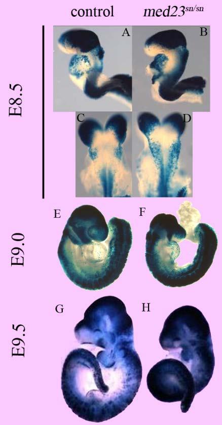
196 183
197 Figure 36: WNT signaling is up-regulated throughout the embryo specifically at E9.5. A D, X-gal staining of E8.5 wild-type; BATGal (A, C) and med23 sn/sn ; BATGal (B, D) shows ectopic WNT signaling in the hindbrain regions of mutant embryos (D) E, F X-Gal staining of E9.0 wild-type; BATGal (E) and med23 sn/sn ; BATGal (F) embryos shows similar levels of WNT signaling between the two embryos G, H WNT signaling is overall up-regulated in med23 sn/sn ; BATGal embryos (H) at E9.5 in comparison to wild-type; BATGal embryos (G) 184
198 Proper dosage of canonical WNT signaling is crucial for many aspects of mammalian embryonic development. We hypothesized that up-regulation of canonical WNT signaling underlies the defects observed in med23 sn/sn embryos. To test for this, we crossed med23 sn mouse line to mutants of WNT signaling pathway genes, WNT co-receptor, lrp6 and WNT signaling modulator, wise. med23 sn ; lrp6 +/- and med23 sn ; wise +/- mouse lines were generated and intercrossed to get med23 sn/sn embryos that had lost one or both copies of lrp6 and/or wise and the double or triple knock-out embryos were then analyzed for neuronal differentiation using antibodies against neuron specific β-tubulin III or TuJ1. We first analyzed the lrp6 +/- and wise +/- embryos themselves to confirm that loss of a copy of lrp6 or wise does not affect neuronal differentiation. As can be seen in Figure 37, embryos that have lost a copy of lrp6 or a copy of wise allele were able to generate and pattern TuJ1+ neurons similar to their wild-type littermates. We then analyzed the TuJ1 expression pattern in the double mutant embryos. Figure 38A D shows the epibranchial region in wildtype, med23 sn/sn, med23 sn/sn ; lrp6 +/- and med23 sn/sn ; wise +/- embryos at E9.5 respectively. As you can see in the boxed area, mutant embryos that have lost of a single copy of lrp6 or a single copy of wise show some TuJ1+ cells within the epibranchial region (while no 185

199 Figure 37: Formation of nervous system is not affected by losing a copy of lrp6 or a copy of wise TuJ1 immuno-staining of lrp6 +/- and wise +/- embryos at late day E9.5 shows beautiful formation of trigeminal, epibranchial and trunk peripheral nervous system as well as midbrain neuron differentiation. 186
200 TuJ1+ is observed in epibranchial region of exclusive med23 sn/sn embryos) (Figure 38B-D). Interestingly, the morphology of the epibranchial neuron ganglia is slightly different between med23 sn/sn ; lrp6 +/- and med23 sn/sn ; wise +/- embryos (Figure 38C, D). Mutant embryos that have lost a copy of lrp6 seem to coalesce the TuJ1+ cells in a more ganglion like form while the restored TuJ1+ neurons in mutants that have lost a copy of wise are much more dispersedly organized, suggesting that LRP6 and Wise regulate WNT signaling and cranial ganglia differentiation with slightly different mechanisms and might be important for separate aspects of cranial ganglia development. Since WISE has been shown to be a canonical WNT signaling modulator, such that it can act both as a WNT signaling inhibitor or an activator depending on the cellular and molecular context, similar phenotype of loss of lrp6 and loss of wise in med23 sn/sn embryos was intriguing and suggestive of a WNT signaling activator role in cranial neuron differentiation context. To confirm this, we analyzed the neuronal differentiation in triple mutants carrying med23 sn/sn ; lrp6 +/- ; wise +/- or med23 sn/sn ; lrp6 +/- ; wise -/- genotype (Figure 38A-D). Interestingly, the restoration of TuJ1 immuno-staining and cranial neuron differentiation in the triple mutants is similar to that of both double mutants indicating that in cranial neuron differentiation both WISE and LRP6 are required for positive canonical WNT signaling. 187
 med23 sn/sn embryo which has lost a copy of lrp6, and D) med23 sn/sn embryo that has lost a copy of wise, were all immuno-stained for TuJ1.")
201 Figure 38: Modulation of canonical WNT signaling partially restores epibranchial ganglia development in med23 sn/sn embryos. A)Wild-type E9.5 embryo, B) med23 sn/sn E9.5 embryo, C) med23 sn/sn embryo which has lost a copy of lrp6, and D) med23 sn/sn embryo that has lost a copy of wise, were all immuno-stained for TuJ1. Losing a copy of lrp6 or wise restores neuronal differentiation in epibranchial region of med23 sn/sn embryo (C, D). 188
 gives similar phenotype to loss of lrp6 only (Figure 37, C) suggesting that in cranial")
202 Figure 39: Wise and Lrp6 activate WNT signaling during cranial ganglia formation. Combined loss of lrp6 and wise (C, D) gives similar phenotype to loss of lrp6 only (Figure 37, C) suggesting that in cranial ganglia formation Wise activates WNT signaling. 189
203 III. Partially restored cranial neurons in med23 sn/sn ; wise +/- embryos express neurod1 Since loss of a copy of wise gave us much more widespread restoration of cranial neuron differentiation, we decided to do our further analysis with med23 sn/sn ; wise +/- double mutant embryos only. It was important for us to know whether manipulation of canonical WNT signaling by losing a copy of WISE (or LRP6) restored TuJ1 immuno-staining through restoration of neuronal differentiation of cranial placodal cells. To that effect we analyzed the expression of neurod1, neuronal differentiation marker of cranial placodal cells, in control andmed23 sn/sn ; wise +/- embryos at E9.5. As expected, similar to TuJ1 immuno-staining, double knock-out embryos, display neurod1+ cells in the epibranchial region at E9.5 (Figure 40), while no such cells were observed in this region in just med23 sn/sn embryos (Figure 24). Also similar to TuJ1immuno-staining, the pattern of neurod1+ cells in the epibranchial region of the double knock-out embryos is very dispersed and not in a coalesced ganglion form. IV. Early placodal marker expression is still not maintained in med23 sn/sn ; wise +/- embryos Partial restoration of neuronal differentiation and neurod1 expression in med23 sn/sn ; wise +/- embryos suggests that WNT signaling regulation is crucial for cranial neuron differentiation. Our expression marker analysis has shown to us that MED23 is required at multiple steps during cranial placode development. However, the time-point during cranial placode 190
")
204 Figure 40: Epibranchial neurons restored by modulating of WNT signaling express neurod1 Losing a copy of wise in med23 sn/sn embryos restores neurod1 expression in the epibranchial region (Compare with complete lack of neurod1+ cells med23 sn/sn embryos in Figure 24) 191
, med23 sn/sn (B) and med23 sn/sn ; wise +/- embryos were analyzed for expression of PPR marker eya2.")
205 Figure 41: Defects in maintenance of early PPR gene expression of med23 sn/sn embryos are not restored by modulating the WNT signaling levels. Wild-type (A), med23 sn/sn (B) and med23 sn/sn ; wise +/- embryos were analyzed for expression of PPR marker eya2. Fewer eya2+ cells are present in the trigeminal and epibranchial placodes of both med23 sn/sn and med23 sn/sn ; wise +/- embryos when compared to wild-type eya2 expression. 192
206 development, when loss of a wise copy restores neuronal differentiation, was still unknown. To that effect, we decided to analyze the earliest marker of cranial placode development. As showed earlier, eya2 expression in cranial placode development begins as soon as pre-placodal region (PPR) is specified and is maintained and required throughout cranial placode development. At E9.5, eya2 expression can still be observed in the cells in olfactory, trigeminal and some epibranchial placodal cells of wild-type embryos (Figure 41A) while in med23 sn/sn embryos only a few cells within the trigeminal placode are eya2+ (Figure 41B). Similar to med23 sn/sn embryos, even the med23 sn/sn ; wise +/- embryos display a reduced number of eya2+ in the trigeminal placode and absence of eya2+ cells in the epibranchial regions (Figure 41C), suggesting that even though the double mutants partially restore neurogenesis, loss of a single copy of wise does not completely restore cranial placode development in med23 sn/sn embryos. V. wise expression itself is unaffected in med23 sn/sn embryos Wise modulates WNT signaling by binding to WNT co-receptors, Lrp5 and Lrp6 428, 429. In vitro, Wise has been shown to bind to WNT antagonist Lrp4 430, transcript levels of which are reduced in med23 sn/sn embryos as observed by the microarray done on wild-type and med23 sn/sn E9.5 littermates. We wanted to analyze if similar to lrp4, Wise expression was 193

207 VI. Figure 42: Loss of med23 does not affect wise expression Using transgenic wise lacz mouse line, we determined that wild-type and med23 sn/sn embryos display similar wise expression pattern. 194
208 affected in med23 sn/sn embryos. Dr. Youngwook in Krumlauf lab generously shared the Wise lacz transgenic mouse line with us which we then used to mate with med23 sn to generate med23 sn ; wise lacz line. Animals from this line were intercrossed and embryos were analyzed for LacZ activity (indicating wise expression) in wild-type and med23 sn/sn embryos at E9.5. Earliest expression of Wise, as detected by lacz staining, was observed at E8.5 in the trunk region (data not shown). At E9.5, wild-type embryos display lacz activity specifically within the epibranchial region of the developing embryo. This epibranchial region specific expression of wise is maintained even in med23 sn/sn embryos (Figure 42 A, B) suggesting that loss of MED23 does not affect wise expression. VII. Manipulation of canonical WNT signaling does not restore Frontonasal process formation and embryonic lethality inmed23 sn/sn embryos Loss of dkk1 affects anterior development in mice embryos due to aberrant increases in canonical WNT signaling 16. Hence, our hypothesis was that the craniofacial defects observed in med23 sn/sn embryos were due to reduced dkk1 transcript and aberrant WNT signaling in the frontonasal prominence. Interestingly, manipulating the levels of WNT signaling in the mutant embryos, by loss of a copy of lrp6 or a copy of wise did not restore frontonasal prominence defects observed in med23 sn/sn embryos (Figure 43). Knock-out embryo phenotypes for lrp6 195
209 and wise are both milder than what is observed in med23 sn/sn embryos. Mouse embryos null for lrp6 have a smaller skull with a crooked nose while complete loss of wise affects tooth development and results in palatal defects 431, 432. Aberrant canonical WNT signaling might still be the cause for frontonasal prominence and first arch defects in med23 sn/sn embryos; however these results suggest that aberrant WNT signaling in frontonasal process of med23 sn/sn embryos is not mediated through LRP6 or Wise. Loss of a copy of lrp6 or a copy of wise is also unable to restore the embryonic lethal phenotype of caused by loss of MED23 as the double knock-out embryos die at around E10.5 as well. Discussion Mis-regulation of canonical WNT signaling seems to be at the heart of cranial ganglia defects in med23 sn/sn embryos. Even though both loss and gain of canonical WNT signaling have been shown to affect cranial ganglia development and differentiation, the detailed role for WNT signaling during each step of cranial placode development and cranial sensory neuron differentiation is not clearly understood in mice or other model organisms. Here we show that MED23 is important for regulation of WNT signaling levels during mouse embryogenesis, and this dosage dependent regulation of WNT signaling is crucial for appropriate cranial ganglia development. WNT signaling activation is temporally regulated during mouse development, 196
210 and loss of MED23 is specifically crucial at E9.5. Currently, we do not know if there is a tissue specific up-regulation of WNT signaling within the med23 sn/sn embryos in comparison to their wild-type littermates and whether MED23 is cell-autonomously required for regulation of WNT signaling. Analysis of conditional mutants should answer some of these questions. Regulation of WNT signaling by Mediator subunit is not unprecedented. Another Mediator subunit, MED12 has also been shown to regulate WNT signaling during mouse embryogenesis 216. MED12 has been shown to bind to β-catenin in vivo, but whether this binding event is crucial to its regulation of WNT signaling is not known 182. Such a binding has not been described for MED23, and right now we think that MED23-mediated regulation of WNT signaling might be through an indirect mechanism. Wise was identified as a conserved WNT modulator that interacts with WNT signaling co-receptors LRP5 and LRP6 433, 428, 429. Animal cap experiments in Xenopus have shown that Wise can also act as a WNT signaling activator, where ectopic expression of Wise lead to an altered A/P patterning phenotype that was similar to the one observed by over-expression of canonical WNT signaling ligand, wnt8a 433. On the other hand, the interaction between Wise and LRP5 and 6 has been shown to inhibit WNT signaling during tooth patterning 434 and bone development 428, 429. In our context, loss of lrp6 or wise or both together, in embryos already carrying a mutation for med23, lead to cranial neuron differentiation suggesting that they 197
211 might be acting in the same pathway. Loss of a wise allele seems to suppress WNT signaling levels similar to loss of alrp6 allele. In mice embryos, this is the first indication of Wise acting as a WNT signaling activator in vivo. The morphology of the cranial ganglia in med23 sn/sn ; lrp6 +/- and med23 sn/sn ; wise +/- is slightly different. WNT signaling modulation through loss of lrp6 seems to confer a more ganglion like morphology in med23 sn/sn embryos. Work in chick embryos has shown a role for wise in trigeminal ganglion coalescence 435 suggesting that this loss of ganglion like morphology in med23 sn/sn ; wise +/- embryos could be a compound effect of aberrant WNT signaling and loss of wise. Overall, this suggests a possibility that LRP6 and Wise-mediated canonical WNT signaling might be required for coalescence of epibranchial ganglia. There are multiple possibilities as to why double knock-out embryos display a restoration of TuJ1+ and neurod1+ cells but not that of eya2+ cells in the epibranchial region. One possibility is that MED23 regulates the various steps of cranial placode development and differentiation via separate mechanisms and mis-regulation of canonical WNT signaling specifically affects only the neuronal differentiation of the cranial ganglia. On the other hand, this disparity in restoration might be more due to the specific pattern and timing of expression of wise itself; wise expression in the epibranchial region of mouse embryos does not begin until E9.5, a time point by which the various cranial placodes have already been specified. Thus loss 198
212 of wise in med23 sn/sn embryos does not alter WNT signaling levels until after cranial placode specification and hence, the early defects in cranial placode development (like loss of eya2 expression) can no longer be restored. Unlike wise, lrp6 is expression in the developing mouse embryo has been observed as early as E It would be interesting to see the expression pattern of eya2 in med23 sn/sn ; lrp6 +/- embryos and compare it to that of med23 sn/sn ; wise +/- embryos. A lack of normal cranial placode eya2 expression pattern in med23 sn/sn ; lrp6 +/- embryos would suggest that LRP6 mediated canonical WNT signaling does not regulate early steps of cranial placode development. Lack of frontonasal process defect or embryonic lethality rescue by the double mutants suggests that disruption of LRP6 and Wise mediated WNT signaling is not the cause for these defects in med23 sn/sn embryos, but it does not rule out the involvement of other WNT signaling co-receptors and modulators. Overall, this work suggests that MED23 mediated regulation of canonical WNT signaling is required for multiple steps of cranial ganglia development. 199
213 Discussion 200
214 Mediator has been called an integrative hub of transcriptional regulation. The evidence for role of Mediator complex in embryonic development comes from mutation analysis. Specific Mediator subunits have been shown to regulate transcription of specific set of genes and control cell and tissue differentiation. This work describes the role for one such Mediator subunit, MED23, which seems to be required for regulation of craniofacial, vascular and neural development specifically. MED23 is a context dependent regulator of genes The human MED23 subunit was identified as a factor required for adenovirus E1A mediated transcription 436. Over the past decade a role for MED23 in regulation of Insulin- and Ras- signaling mediated gene transcription has emerged 175, 177, 297. Insulin signaling induces adipogenesis in wild-type mouse embryonic fibroblasts. This in vitro differentiation of adipocytes from MEFs was reduced in absence of med23 as MED23-deficient Mediator complex is unable to induce transcription of a crucial adipogenesis gene, egr2. Interestingly, egr2 gene activation requires binding of MED23 to phosphorylated form of ELK1, phosphorylation of which is regulated by Insulin signaling 175. Alternatively, MAPK signalingmediated ELK1 phosphorylation and MED23-pELK1 interaction induces expression egr1 140, 176. Epigenetic marks for active gene transcription as well as Pol II and general transcription factor 201
215 binding at egr1 locus in MAPK-induced med23 -/- cells was equivalent or only slightly reduced in comparison to the MAPK induced wild-type cells; however the transcription of egr1 gene itself was drastically reduced in cells devoid of MED Recently, it has been shown that MED23 regulated transcription of egr1 is also regulated at the transcript elongation step. med23 -/- cells are unable to recruit CDK-9 and through it, the elongation factor, P-TEFb, and thus egr1 expression is inhibited 437. Ras-signaling mediated MED23-pELK1 binding is crucial for proliferation and tumorigenicity of lung cancer cells 438. Another transcription factor, C/EBPβ has also been shown to interact with MED23 and this interaction is capable of transcription activation, however the role for this interaction in vivo has not been established 177. One important thing to bear in mind is that all the above studies have been performed in vitro and whether these interactions and gene transcription specific roles of MED23 hold true within a live animal or a developing embryo have not been analyzed. In our system, microarray analysis shows that med23 sn/sn embryos display a down-regulation of egr-1 transcript levels by 9-fold compared to wild-type. This provides us a good validation that our med23 sn mutation causes similar defects to the existent med23 - allele. Along with many other developmental defects, egr1 mutation in mice also causes a defect in growth hormone levels, which might explain the smaller size of med23 sn/sn embryos as compared to wild-type 202

216 Figure 44: egr2 (krox20) is expressed in the hindbrain of med23 sn/sn embryos Wild-type and med23 sn/sn embryos display hindbrain specific expression of egr2. Lack of egr2 expression in r5 is due to the developmental delay in med23 sn/sn embryos. Analysis of med23 sn/sn embryos at a later stage displays egr2 expression in both r3 and r5 (data not shown). 203
1. What are the three general areas of the developing vertebrate limb? 2. What embryonic regions contribute to the developing limb bud?
 Study Questions - Lecture 17 & 18 1. What are the three general areas of the developing vertebrate limb? The three general areas of the developing vertebrate limb are the proximal stylopod, zeugopod, and
Role of Organizer Chages in Late Frog Embryos
 Ectoderm Germ Layer Frog Fate Map Frog Fate Map Role of Organizer Chages in Late Frog Embryos Organizer forms three distinct regions Notochord formation in chick Beta-catenin localization How does beta-catenin
Cellular Neurobiology BIPN 140 Fall 2016 Problem Set #8
 Cellular Neurobiology BIPN 140 Fall 2016 Problem Set #8 1. Inductive signaling is a hallmark of vertebrate and mammalian development. In early neural development, there are multiple signaling pathways
Developmental Zoology. Ectodermal derivatives (ZOO ) Developmental Stages. Developmental Stages
 Developmental Stages. Developmental Stages") Developmental Zoology (ZOO 228.1.0) Ectodermal derivatives 1 Developmental Stages Ø Early Development Fertilization Cleavage Gastrulation Neurulation Ø Later Development Organogenesis Larval molts Metamorphosis
Developmental Zoology (ZOO 228.1.0) Ectodermal derivatives 1 Developmental Stages Ø Early Development Fertilization Cleavage Gastrulation Neurulation Ø Later Development Organogenesis Larval molts Metamorphosis
Conclusions. The experimental studies presented in this thesis provide the first molecular insights
 C h a p t e r 5 Conclusions 5.1 Summary The experimental studies presented in this thesis provide the first molecular insights into the cellular processes of assembly, and aggregation of neural crest and
Paraxial and Intermediate Mesoderm
 Biology 4361 Paraxial and Intermediate Mesoderm December 7, 2006 Major Mesoderm Lineages Mesodermal subdivisions are specified along a mediolateral axis by increasing amounts of BMPs more lateral mesoderm
Mesoderm Development
 Quiz rules: Spread out across available tables No phones, text books, or (lecture) notes on your desks No consultation with your colleagues No websites open other than the Quiz page No screen snap shots
Bio Section III Organogenesis. The Neural Crest and Axonal Specification. Student Learning Objectives. Student Learning Objectives
 Bio 127 - Section III Organogenesis The Neural Crest and Axonal Specification Gilbert 9e Chapter 10 Student Learning Objectives 1. You should understand that the neural crest is an evolutionary advancement
Paraxial and Intermediate Mesoderm
 Biology 4361 Paraxial and Intermediate Mesoderm December 6, 2007 Mesoderm Formation Chick Major Mesoderm Lineages Mesodermal subdivisions are specified along a mediolateral axis by increasing amounts of
Exam 3 (Final Exam) December 20, 2007
 December 20, 2007") Biology 4361 Exam 3 (Final Exam) December 20, 2007 Name: ID: Multiple choice (1 point each. Indicate the best answer.) 1. During Drosophila gastrulation, mesoderm moves in through the a. primitives streak.
Biology 4361 Exam 3 (Final Exam) December 20, 2007 Name: ID: Multiple choice (1 point each. Indicate the best answer.) 1. During Drosophila gastrulation, mesoderm moves in through the a. primitives streak.
Developmental processes Differential gene expression Introduction to determination The model organisms used to study developmental processes
 Date Title Topic(s) Learning Outcomes: Sept 28 Oct 3 1. What is developmental biology and why should we care? 2. What is so special about stem cells and gametes? Developmental processes Differential gene
9/4/2015 INDUCTION CHAPTER 1. Neurons are similar across phyla Thus, many different model systems are used in developmental neurobiology. Fig 1.
 INDUCTION CHAPTER 1 Neurons are similar across phyla Thus, many different model systems are used in developmental neurobiology Fig 1.1 1 EVOLUTION OF METAZOAN BRAINS GASTRULATION MAKING THE 3 RD GERM LAYER
MCDB 4777/5777 Molecular Neurobiology Lecture 29 Neural Development- In the beginning
 MCDB 4777/5777 Molecular Neurobiology Lecture 29 Neural Development- In the beginning Learning Goals for Lecture 29 4.1 Describe the contributions of early developmental events in the embryo to the formation
Neural development its all connected
 Neural development its all connected How do you build a complex nervous system? How do you build a complex nervous system? 1. Learn how tissue is instructed to become nervous system. Neural induction 2.
Life Sciences For NET & SLET Exams Of UGC-CSIR. Section B and C. Volume-08. Contents A. BASIC CONCEPT OF DEVELOPMENT 1
 Section B and C Volume-08 Contents 5. DEVELOPMENTAL BIOLOGY A. BASIC CONCEPT OF DEVELOPMENT 1 B. GAMETOGENESIS, FERTILIZATION AND EARLY DEVELOPMENT 23 C. MORPHOGENESIS AND ORGANOGENESIS IN ANIMALS 91 0
PRACTICE EXAM. 20 pts: 1. With the aid of a diagram, indicate how initial dorsal-ventral polarity is created in fruit fly and frog embryos.
 PRACTICE EXAM 20 pts: 1. With the aid of a diagram, indicate how initial dorsal-ventral polarity is created in fruit fly and frog embryos. No Low [] Fly Embryo Embryo Non-neural Genes Neuroectoderm Genes
Paraxial and Intermediate Mesoderm
 Biology 4361 Paraxial and Intermediate Mesoderm December 6, 2007 Mesoderm Formation Chick Major Mesoderm Lineages Mesodermal subdivisions are specified along a mediolateral axis by increasing amounts of
!!!!!!!! DB3230 Midterm 2 12/13/2013 Name:
 1. (10 pts) Draw or describe the fate map of a late blastula stage sea urchin embryo. Draw or describe the corresponding fate map of the pluteus stage larva. Describe the sequence of gastrulation events
Name. Biology Developmental Biology Winter Quarter 2013 KEY. Midterm 3
 Name 100 Total Points Open Book Biology 411 - Developmental Biology Winter Quarter 2013 KEY Midterm 3 Read the Following Instructions: * Answer 20 questions (5 points each) out of the available 25 questions
Cell-Cell Communication in Development
 Biology 4361 - Developmental Biology Cell-Cell Communication in Development October 2, 2007 Cell-Cell Communication - Topics Induction and competence Paracrine factors inducer molecules Signal transduction
Bio 127 Section I Introduction to Developmental Biology. Cell Cell Communication in Development. Developmental Activities Coordinated in this Way
 Bio 127 Section I Introduction to Developmental Biology Cell Cell Communication in Development Gilbert 9e Chapter 3 It has to be EXTREMELY well coordinated for the single celled fertilized ovum to develop
SUPPLEMENTARY INFORMATION
 doi:10.1038/nature11589 Supplementary Figure 1 Ciona intestinalis and Petromyzon marinus neural crest expression domain comparison. Cartoon shows dorsal views of Ciona mid gastrula (left) and Petromyzon
Mesoderm Induction CBT, 2018 Hand-out CBT March 2018
 Mesoderm Induction CBT, 2018 Hand-out CBT March 2018 Introduction 3. Books This module is based on the following books: - 'Principles of Developement', Lewis Wolpert, et al., fifth edition, 2015 - 'Developmental
Developmental Biology 3230 Midterm Exam 1 March 2006
 Name Developmental Biology 3230 Midterm Exam 1 March 2006 1. (20pts) Regeneration occurs to some degree to most metazoans. When you remove the head of a hydra a new one regenerates. Graph the inhibitor
presumptiv e germ layers during Gastrulatio n and neurulation Somites
 Vertebrate embryos are similar at the phylotypic stage Patterning the Vertebrate Body Plan II: Mesoderm & Early Nervous System Wolpert L, Beddington R, Jessell T, Lawrence P, Meyerowitz E, Smith J. (2001)
Biology 218, practise Exam 2, 2011
 Figure 3 The long-range effect of Sqt does not depend on the induction of the endogenous cyc or sqt genes. a, Design and predictions for the experiments shown in b-e. b-e, Single-cell injection of 4 pg
Developmental Biology Lecture Outlines
 Developmental Biology Lecture Outlines Lecture 01: Introduction Course content Developmental Biology Obsolete hypotheses Current theory Lecture 02: Gametogenesis Spermatozoa Spermatozoon function Spermatozoon
Sonic hedgehog (Shh) signalling in the rabbit embryo
 signalling in the rabbit embryo") Sonic hedgehog (Shh) signalling in the rabbit embryo In the first part of this thesis work the physical properties of cilia-driven leftward flow were characterised in the rabbit embryo. Since its discovery
Sonic hedgehog (Shh) signalling in the rabbit embryo In the first part of this thesis work the physical properties of cilia-driven leftward flow were characterised in the rabbit embryo. Since its discovery
Early Development in Invertebrates
 Developmental Biology Biology 4361 Early Development in Invertebrates October 25, 2006 Early Development Overview Cleavage rapid cell divisions divisions of fertilized egg into many cells Gastrulation
Making Headway: The Roles of Hox Genes and Neural Crest Cells in Craniofacial Development
 Mini-Review TheScientificWorldJOURNAL (2003) 3, 240 264 ISSN 1537-744X; DOI 10.1100/tsw.2003.11 Making Headway: The Roles of Hox Genes and Neural Crest Cells in Craniofacial Development Paul A Trainor
Name KEY. Biology Developmental Biology Winter Quarter Midterm 3 KEY
 Name KEY 100 Total Points Open Book Biology 411 - Developmental Biology Winter Quarter 2009 Midterm 3 KEY All of the 25 multi-choice questions are single-answer. Choose the best answer. (4 pts each) Place
4. Neural tube cells are specified by opposing dorsal-ventral gradients of a. Wnts and Nodal. b. FGF and Shh. c. BMPs and Wnts. d. BMPs and Shh.
 Biology 4361 Name: KEY Exam 4 ID#: August 1, 2008 Multiple choice (one point each; indicate the best answer) 1. Neural tube closure is accomplished by movement of the a. medial hinge point cells. b. medial
Question Set # 4 Answer Key 7.22 Nov. 2002
 Question Set # 4 Answer Key 7.22 Nov. 2002 1) A variety of reagents and approaches are frequently used by developmental biologists to understand the tissue interactions and molecular signaling pathways
Supplementary Figure 1: Mechanism of Lbx2 action on the Wnt/ -catenin signalling pathway. (a) The Wnt/ -catenin signalling pathway and its
 The Wnt/ -catenin signalling pathway and its") Supplementary Figure 1: Mechanism of Lbx2 action on the Wnt/ -catenin signalling pathway. (a) The Wnt/ -catenin signalling pathway and its transcriptional activity in wild-type embryo. A gradient of canonical
Supplementary Figure 1: Mechanism of Lbx2 action on the Wnt/ -catenin signalling pathway. (a) The Wnt/ -catenin signalling pathway and its transcriptional activity in wild-type embryo. A gradient of canonical
Axis Specification in Drosophila
 Developmental Biology Biology 4361 Axis Specification in Drosophila November 2, 2006 Axis Specification in Drosophila Fertilization Superficial cleavage Gastrulation Drosophila body plan Oocyte formation
Axis Specification in Drosophila
 Developmental Biology Biology 4361 Axis Specification in Drosophila November 6, 2007 Axis Specification in Drosophila Fertilization Superficial cleavage Gastrulation Drosophila body plan Oocyte formation
THE PROBLEMS OF DEVELOPMENT. Cell differentiation. Cell determination
 We emphasize these points from Kandel in Bi/CNS 150 Bi/CNS/NB 150: Neuroscience Read Lecture Lecture Friday, October 2, 2015 Development 1: pp 5-10 Introduction Brains evolved All higher animals have brains
BIOLOGY - CLUTCH CH.32 - OVERVIEW OF ANIMALS.
 !! www.clutchprep.com Animals are multicellular, heterotrophic eukaryotes that feed by ingesting their food Most animals are diploid, and produce gametes produced directly by meiosis Animals lack cell
10/2/2015. Chapter 4. Determination and Differentiation. Neuroanatomical Diversity
 Chapter 4 Determination and Differentiation Neuroanatomical Diversity 1 Neurochemical diversity: another important aspect of neuronal fate Neurotransmitters and their receptors Excitatory Glutamate Acetylcholine
Cell Cell Communication in Development
 Biology 4361 Developmental Biology Cell Cell Communication in Development June 25, 2008 Cell Cell Communication Concepts Cells in developing organisms develop in the context of their environment, including
Unicellular: Cells change function in response to a temporal plan, such as the cell cycle.
 Spatial organization is a key difference between unicellular organisms and metazoans Unicellular: Cells change function in response to a temporal plan, such as the cell cycle. Cells differentiate as a
Paraxial and Intermediate Mesoderm
 Biology 4361 Paraxial and Intermediate Mesoderm July 28, 2008 Paraxial and Intermediate Mesoderm Overview Development of major mesodermal lineages Somites: formation specification and differentiation Mesodermal
Head and Face Development
 Head and Face Development Resources: http://php.med.unsw.edu.au/embryology/ Larsen s Human Embryology The Developing Human: Clinically Oriented Embryology Dr Annemiek Beverdam School of Medical Sciences,
7.013 Problem Set
 7.013 Problem Set 5-2013 Question 1 During a summer hike you suddenly spot a huge grizzly bear. This emergency situation triggers a fight or flight response through a signaling pathway as shown below.
Unit 4 Evaluation Question 1:
 Name: Unit 4 Evaluation Question 1: /7 points A naturally occurring dominant mutant in mice is the Doublefoot (Dbf) mutant. Below is an image of the bones from a wildtype (wt) and Doublefoot mutant mouse.
Developmental Biology Biology Ectodermal Organs. November 22, 2005
 Developmental Biology Biology 4361 Ectodermal Organs November 22, 2005 Germinal neuroepithelium external limiting membrane neural tube neuroepithelium (stem cells) Figure 13.3 Figure 13.4 Neuroepithelial
MBios 401/501: Lecture 14.2 Cell Differentiation I. Slide #1. Cell Differentiation
 MBios 401/501: Lecture 14.2 Cell Differentiation I Slide #1 Cell Differentiation Cell Differentiation I -Basic principles of differentiation (p1305-1320) -C-elegans (p1321-1327) Cell Differentiation II
Axis Specification in Drosophila
 Developmental Biology Biology 4361 Axis Specification in Drosophila July 9, 2008 Drosophila Development Overview Fertilization Cleavage Gastrulation Drosophila body plan Oocyte formation Genetic control
Questions in developmental biology. Differentiation Morphogenesis Growth/apoptosis Reproduction Evolution Environmental integration
 Questions in developmental biology Differentiation Morphogenesis Growth/apoptosis Reproduction Evolution Environmental integration Representative cell types of a vertebrate zygote => embryo => adult differentiation
UNIVERSITY OF YORK BIOLOGY. Developmental Biology
 Examination Candidate Number: UNIVERSITY OF YORK BSc Stage 2 Degree Examinations 2017-18 Department: BIOLOGY Title of Exam: Developmental Biology Desk Number: Time allowed: 1 hour and 30 minutes Total
In ovo time-lapse analysis after dorsal neural tube ablation shows rerouting of chick hindbrain neural crest
 In ovo time-lapse analysis after dorsal neural tube ablation shows rerouting of chick hindbrain neural crest Paul Kulesa, Marianne Bronner-Fraser and Scott Fraser (2000) Presented by Diandra Lucia Background
The Radiata-Bilateria split. Second branching in the evolutionary tree
 The Radiata-Bilateria split Second branching in the evolutionary tree Two very important characteristics are used to distinguish between the second bifurcation of metazoans Body symmetry Germinal layers
Biology 224 Human Anatomy and Physiology - II Week 1; Lecture 1; Monday Dr. Stuart S. Sumida. Review of Early Development of Humans.
 Biology 224 Human Anatomy and Physiology - II Week 1; Lecture 1; Monday Dr. Stuart S. Sumida Review of Early Development of Humans Special Senses Review: Historical and Developmental Perspectives Ontogeny
Chapter 18 Lecture. Concepts of Genetics. Tenth Edition. Developmental Genetics
 Chapter 18 Lecture Concepts of Genetics Tenth Edition Developmental Genetics Chapter Contents 18.1 Differentiated States Develop from Coordinated Programs of Gene Expression 18.2 Evolutionary Conservation
Mesoderm Divided into three main types - Paraxial (somite) - Intermediate - Lateral (somatic and splanchnic)
 - Intermediate - Lateral (somatic and splanchnic)") Mesoderm Divided into three main types - Paraxial (somite) - Intermediate - Lateral (somatic and splanchnic) Fates of Mesoderm Paraxial - Dermis of skin - Axial Skeleton - Axial and limb muscles/tendons
Mesoderm Divided into three main types - Paraxial (somite) - Intermediate - Lateral (somatic and splanchnic) Fates of Mesoderm Paraxial - Dermis of skin - Axial Skeleton - Axial and limb muscles/tendons
2/23/09. Regional differentiation of mesoderm. Morphological changes at early postgastrulation. Segments organize the body plan during embryogenesis
 Regional differentiation of mesoderm Axial Paraxial Intermediate Somatic Splanchnic Chick embryo Morphological changes at early postgastrulation stages Segments organize the body plan during embryogenesis
Detailed Learning Outcomes
 Detailed Learning Outcomes Following lectures, workshops and directed study activities, students should: 1. Understand the principles behind the study of bioscience in relation to the profession of SLT
Chapter 10 Development and Differentiation
 Part III Organization of Cell Populations Chapter Since ancient times, people have wondered how organisms are formed during the developmental process, and many researchers have worked tirelessly in search
MOLECULAR CONTROL OF EMBRYONIC PATTERN FORMATION
 MOLECULAR CONTROL OF EMBRYONIC PATTERN FORMATION Drosophila is the best understood of all developmental systems, especially at the genetic level, and although it is an invertebrate it has had an enormous
THE SPECIFICATION OF DORSAL CELL FATES IN THE VERTEBRATE CENTRAL NERVOUS SYSTEM
 Annu. Rev. Neurosci. 1999. 22:261 94 Copyright c 1999 by Annual Reviews. All rights reserved THE SPECIFICATION OF DORSAL CELL FATES IN THE VERTEBRATE CENTRAL NERVOUS SYSTEM Kevin J. Lee and Thomas M. Jessell
Bi 117 Final (60 pts) DUE by 11:00 am on March 15, 2012 Box by Beckman Institute B9 or to a TA
 DUE by 11:00 am on March 15, 2012 Box by Beckman Institute B9 or to a TA") Bi 117 Final (60 pts) DUE by 11:00 am on March 15, 2012 Box by Beckman Institute B9 or to a TA Instructor: Marianne Bronner Exam Length: 6 hours plus one 30-minute break at your discretion. It should take
Bi 117 Final (60 pts) DUE by 11:00 am on March 15, 2012 Box by Beckman Institute B9 or to a TA Instructor: Marianne Bronner Exam Length: 6 hours plus one 30-minute break at your discretion. It should take
Folding of the embryo.. the embryo is becoming a tube like structure
 The embryo is a Folding of the embryo.. the embryo is becoming a tube like structure WEEK 4 EMBRYO General features Primordia of the brain Somites Primordia of the heart Branchial arches Primordia
Dynamic and Differential Regulation of Stem Cell Factor FoxD3 in the Neural Crest Is Encrypted in the Genome
 Dynamic and Differential Regulation of Stem Cell Factor FoxD3 in the Neural Crest Is Encrypted in the Genome Marcos S. Simões-Costa 1., Sonja J. McKeown 1., Joanne Tan-Cabugao 1, Tatjana Sauka-Spengler
Axon guidance I. Paul Garrity March 15, /9.013
 Axon guidance I Paul Garrity March 15, 2004 7.68/9.013 Neuronal Wiring: Functional Framework of the Nervous System Stretch reflex circuit Early theories of axonogenesis Schwann: many neurons link to form
DOWNLOAD PDF DETERMINATION OF PREPLACODAL ECTODERM AND SENSORY PLACODES SALLY A. MOODY
 Chapter 1 : Olfactory epithelium - Wikipedia The sensory organs of the vertebrate head derive from two embryological structures, the neural crest and the ectodermal placodes. Although quite a lot is known
Cell-Cell Communication in Development
 Biology 4361 - Developmental Biology Cell-Cell Communication in Development June 23, 2009 Concepts Cell-Cell Communication Cells develop in the context of their environment, including: - their immediate
Lecture 7. Development of the Fruit Fly Drosophila
 BIOLOGY 205/SECTION 7 DEVELOPMENT- LILJEGREN Lecture 7 Development of the Fruit Fly Drosophila 1. The fruit fly- a highly successful, specialized organism a. Quick life cycle includes three larval stages
Exam 2 ID#: November 9, 2006
 Biology 4361 Name: KEY Exam 2 ID#: November 9, 2006 Multiple choice (one point each) Circle the best answer. 1. Inducers of Xenopus lens and optic vesicle include a. pharyngeal endoderm and anterior neural
Exam 3 ID#: July 31, 2009
 Biology 4361 Name: KEY Exam 3 ID#: July 31, 2009 Multiple choice (one point each; indicate the best answer) 1. Neural tube closure is accomplished by movement of the a. medial hinge point cells. b. medial
purpose of this Chapter is to highlight some problems that will likely provide new
 119 Chapter 6 Future Directions Besides our contributions discussed in previous chapters to the problem of developmental pattern formation, this work has also brought new questions that remain unanswered.
Chapter 4 Evaluating a potential interaction between deltex and git in Drosophila: genetic interaction, gene overexpression and cell biology assays.
 Evaluating a potential interaction between deltex and git in Drosophila: genetic interaction, gene overexpression and cell biology assays. The data described in chapter 3 presented evidence that endogenous
Lecture 3 - Molecular Regulation of Development. Growth factor signaling, Hox genes and the body plan
 Lecture 3 - Molecular Regulation of Development. Growth factor signaling, Hox genes and the body plan Lecture Objectives Outline August 18, 2015, M.D., Ph.D. To understand how cell differentiation and
Mesoderm induction: from caps to chips
 Mesoderm induction: from caps to chips David Kimelman Abstract Vertebrate mesoderm induction is one of the classical problems in developmental biology. Various developmental biology approaches, particularly
10/15/09. Tetrapod Limb Development & Pattern Formation. Developing limb region is an example of a morphogenetic field
 Tetrapod Limb Development & Pattern Formation Figure 16.5(1) Limb Bud Formation derived from lateral plate (somatic) & paraxial (myotome) Fig. 16.2 Prospective Forelimb Field of Salamander Ambystoma maculatum
Introduction Chpt 1. Study Slides
 Introduction Chpt 1 Study Slides A group of molecules working together toward a common function is a: A. Cell B. Atom C. Organelle D. Tissue E. Organ ANSWER A group of molecules working together toward
The formation of the neural crest has been traditionally considered a classic example of
 CHAPTER 2 Neural Crest Inducing Signals Mardn L. Basch and Marianne Bronner-Fraser* Abstract The formation of the neural crest has been traditionally considered a classic example of secondary induction,
SIGNIFICANCE OF EMBRYOLOGY
 This lecture will discuss the following topics : Definition of Embryology Significance of Embryology Old and New Frontiers Introduction to Molecular Regulation and Signaling Descriptive terms in Embryology
Origin of mesenchymal stem cell
 Origin of mesenchymal stem cell Takumi Era Department of Cell Modulation, Institute of Molecular embryology and Genetics (IMEG) Kumamoto University Differentiation pathways of in vitro ES cell culture
Drosophila Life Cycle
 Drosophila Life Cycle 1 Early Drosophila Cleavage Nuclei migrate to periphery after 10 nuclear divisions. Cellularization occurs when plasma membrane folds in to divide nuclei into cells. Drosophila Superficial
Results and Problems in Cell Differentiation. Series Editor: Hennig
 Results and Problems in Cell Differentiation Series Editor: w. 38 Hennig Springer-Verlag Berlin Heidelberg GmbH Beate Brand-Saberi (Ed.) Vertebrate Myogenesis With 31 Figures Springer Professor Dr. Beate
Specification of neural crest cell formation and migration in mouse embryos
 Seminars in Cell & Developmental Biology 16 (2005) 683 693 Review Specification of neural crest cell formation and migration in mouse embryos Paul A. Trainor Stowers Institute for Medical Research, 1000
Coordination of Cell Differentiation and Migration in Mathematical Models of Caudal Embryonic Axis Extension
 Coordination of Cell Differentiation and Migration in Mathematical Models of Caudal Embryonic Axis Extension Nigel C. Harrison 1, Ruth Diez del Corral 2 *, Bakhtier Vasiev 1 * 1 Department of Mathematical
Introduction to Animals
 Introduction to Animals Characteristics of Animals multicellular Except for sponges, animal cells are arranged into tissues. Tissues are necessary to produce organs and organ systems. Tissues, organs,
3/8/ Complex adaptations. 2. often a novel trait
 Chapter 10 Adaptation: from genes to traits p. 302 10.1 Cascades of Genes (p. 304) 1. Complex adaptations A. Coexpressed traits selected for a common function, 2. often a novel trait A. not inherited from
Tsukushi Modulates Xnr2, FGF and BMP Signaling: Regulation of Xenopus Germ Layer Formation
 Tsukushi Modulates Xnr2, FGF and BMP Signaling: Regulation of Xenopus Germ Layer Formation Samantha A. Morris 1 *, Alexandra D. Almeida 1, Hideaki Tanaka 2, Kunimasa Ohta 2, Shin-ichi Ohnuma 1 * 1 Department
Axis determination in flies. Sem 9.3.B.5 Animal Science
 Axis determination in flies Sem 9.3.B.5 Animal Science All embryos are in lateral view (anterior to the left). Endoderm, midgut; mesoderm; central nervous system; foregut, hindgut and pole cells in yellow.
Axon Guidance. Multiple decision points along a growing axon s trajectory Different types of axon guidance cues:
 Axon Guidance Multiple decision points along a growing axon s trajectory Different types of axon guidance cues: Contact mediated - requires direct contact by growth cone Long range - growth cone responds
Maternal Control of GermLayer Formation in Xenopus
 Maternal Control of GermLayer Formation in Xenopus The zygotic genome is activated at the mid-blastula transition mid-blastula fertilized egg Xenopus gastrulae early-gastrula 7 hrs 10 hrs control not VP
Organization of Vertebrate Body. Organization of Vertebrate Body
 The Animal Body and Principles of Regulation Chapter 43 There are four levels of organization: 1. Cells 2. Tissues 3. Organs 4. Organ systems Bodies of vertebrates are composed of different cell types
Reading. Lecture VI. Making Connections 9/17/12. Bio 3411 Lecture VI. Making Connections. Bio 3411 Monday September 17, 2012
 Lecture VI. Making Connections Bio 3411 Monday September 17, 2012!! 1! Reading NEUROSCIENCE: 5 th ed, pp!507?536! 4 th ed, pp 577-609 Bentley, D., & Caudy, M. (1983). Nature, 304(5921), 62-65. Dickson,
BONE MORPHOGENETIC PROTEINS AS REGULATORS OF NEURAL TUBE DEVELOPMENT
 Papers on Anthropology XX, 2011, pp. 314 326 BONE MORPHOGENETIC PROTEINS AS REGULATORS OF NEURAL TUBE DEVELOPMENT Aimar Namm 1,2, Andres Arend 1, Marina Aunapuu 1,2 1 Department of Anatomy, University
Cells to Tissues. Peter Takizawa Department of Cell Biology
 Cells to Tissues Peter Takizawa Department of Cell Biology From one cell to ensembles of cells. Multicellular organisms require individual cells to work together in functional groups. This means cells
Segment boundary formation in Drosophila embryos
 Segment boundary formation in Drosophila embryos Development 130, August 2003 Camilla W. Larsen, Elizabeth Hirst, Cyrille Alexandre and Jean Paul Vincent 1. Introduction: - Segment boundary formation:
The Pax and large Maf families of genes in mammalian eye development
 The Pax and large Maf families of genes in mammalian eye development Vertebrate eye development is dependent on the coordinated action of thousands of genes. A specific group of over one hundred of regulatory
Comparison of the expression patterns of several sox genes between Oryzias latipes and Danio rerio
 Urun 1 Comparison of the expression patterns of several sox genes between Oryzias latipes and Danio rerio Fatma Rabia URUN ilkent University, nkara - TURKEY High mobility group domain containing transcription
Drosophila melanogaster- Morphogen Gradient
 NPTEL Biotechnology - Systems Biology Drosophila melanogaster- Morphogen Gradient Dr. M. Vijayalakshmi School of Chemical and Biotechnology SASTRA University Joint Initiative of IITs and IISc Funded by
Limb Development Involving the development of the appendicular skeleton and muscles
 Limb Development Involving the development of the appendicular skeleton and muscles 1 Objectives Timing and location of limb bud development The tissues from which limb buds are made Determining the position
PY4302 Developmental Neuroscience. Eye Development
 PY4302 Developmental Neuroscience Eye Development J. Martin Collinson School of Medical Sciences m.collinson@abdn.ac.uk F 55750 Vertebrate eye development Development of the retina 2D patterning. Specification
AP Biology Gene Regulation and Development Review
 AP Biology Gene Regulation and Development Review 1. What does the regulatory gene code for? 2. Is the repressor by default active/inactive? 3. What changes the repressor activity? 4. What does repressor
18.4 Embryonic development involves cell division, cell differentiation, and morphogenesis
 18.4 Embryonic development involves cell division, cell differentiation, and morphogenesis An organism arises from a fertilized egg cell as the result of three interrelated processes: cell division, cell