Investigating the Mechanism of Nur77-Induced Apoptosis in T Cells
|
|
|
- Pierce Cain
- 6 years ago
- Views:
Transcription

1 University of Massachusetts Amherst Amherst Masters Theses February Investigating the Mechanism of Nur77-Induced Apoptosis in T Cells Heather E. Fogarty University of Massachusetts Amherst Follow this and additional works at: Part of the Biology Commons, Cancer Biology Commons, Immunity Commons, and the Other Animal Sciences Commons Fogarty, Heather E., "Investigating the Mechanism of Nur77-Induced Apoptosis in T Cells" (2012). Masters Theses February Retrieved from This thesis is brought to you for free and open access by ScholarWorks@UMass Amherst. It has been accepted for inclusion in Masters Theses February 2014 by an authorized administrator of ScholarWorks@UMass Amherst. For more information, please contact scholarworks@library.umass.edu.
2 INVESTIGATING THE MECHANISM OF NUR77-INDUCED APOPTOSIS IN T CELLS A Thesis Presented by HEATHER E. FOGARTY Submitted to the Graduate School of the University of Massachusetts Amherst in partial fulfillment of the requirements for the degree of MASTER OF SCIENCE February 2012 Program in Veterinary and Animal Sciences
3 INVESTIGATING THE MECHANISM OF NUR77-INDUCED APOPTOSIS IN T CELLS A Thesis Presented by HEATHER E. FOGARTY Approved as to style and content by: Barbara A. Osborne, Chair Rafael A. Fissore, Member Lisa M. Minter, Member Rafael A. Fissore, Department Head Veterinary and Animal Sciences
4 DEDICATION Dedicated to my loving husband Jeffrey Arsenault.
5 ACKNOWLEDGMENTS I thank my advisor, Dr. Barbara Osborne, for being a wonderful mentor and guiding me through the course of my graduate study. Being able to study and learn in her laboratory was a valuable experience I am very grateful for. I appreciate her patience, encouragement and support even through the rough times of my project. I would also like to thank my committee members, Dr. Rafael Fissore and Dr. Lisa Minter, for their encouragement and helpful input in forming this thesis. I especially thank all of the former and current members of the Osborne lab: Dr. Jonathan Rud, Dr. Mulualem Tilahun, Rebecca Lawlor, Anushka Dongre, Srilalitha Surampudi, Reem Suleiman, Joseph Homsi, Elizabeth Schutten, Karthik Chandiran, Matthias Birtel, and Wipawee Wongchana. I thank all of these lab mates for being wonderful to work with, have interesting conversations with, or just having an answer to one of my silly questions. I would also like to specifically thank Anushka Dongre and Srilalitha Surampudi for their support throughout classes or presentations and friendship during my time in the Osborne Lab. I will also thank Nan Zhang for his technical support and guidance completing some experiments for this project. Finally, I thank my husband for his understanding, patience and support throughout the course of my studies. iv
6 ABSTRACT INVESTIGATING THE MECHANISMS OF NUR77-INDUCED APOPTOSIS IN T CELLS FEBRUARY 2011 HEATHER E. FOGARTY, B.S. UNIVERSITY OF MASSACHUSETTS, AMHERST M.S. UNIVERSITY OF MASSACHUSETTS, AMHERST Directed by: Professor Barbara A. Osborne Nur77 is a member of the orphan nuclear receptor family, where it is known to play an important role in apoptosis in both negative selection in T cells and in cancer cell lines. In the development of T cells, it is critical for the immune system to discriminate self from non-self by eliminating auto-reactive cells. It was originally thought that Nur77 initiated apoptosis by activating downstream gene targets. However, it is now clear that Nur77 has its own distinct role outside of the nucleus. Our laboratory has shown in T cells that Nur77 is phosphorylated by the MEK-ERK-RSK cascade, which facilitates its translocation from the nucleus to the cytosol where it activates the intrinsic apoptotic cascade. Others have suggested that, in cancer cell lines, Nur77 migrates to the mitochondria and binds to anti-apoptotic Bcl-2 thus converting it from a protector to a killer. However, the precise mechanisms by which Nur77 induces apoptosis in T cells still needs to be clarified. Calcium plays an important role as a second messenger in various cellular responses, one of which includes apoptosis. The IP3 receptor controls efflux of calcium from the ER and can be activated through TCR activation. This signal induces a rise in cytoplasmic calcium levels ultimately causing cell death through mechanisms that remain v
7 unclear. Preliminary studies in our laboratory have shown that Nur77 causes changes in two distinct isoforms of the IP3R, an increase in IP3R1 and a decrease in IP3R3. Here, we use a double positive DO11.10 T cell line with tetracycline responsive Nur77, which is constitutively exported from the nucleus to examine the effects of cytosolic Nur77. Through co-immunoprecipitation experiments we suggest, that the presence of Nur77 disrupts the IP3R/Bcl-2 interaction. Additionally, we asked whether the presence of Nur77 influences calcium release in activated T cells. In this study we show that Nur77 increases baseline calcium levels and causes emptying of ER calcium stores. We suggest a model where cytosolic Nur77 disrupts the IP3R/Bcl-2 interaction by binding Bcl-2 at the mitochondria or ER, causing calcium flux and apoptosis of the cell. Current experiments focus on confirming the findings presented here while also examining Nur77 s contribution to calcium levels in different subcellular compartments such as the ER and mitochondria. Other studies using the BiFC assay aim to confirm the Nur77/Bcl- 2 interaction and support earlier data that Nur77 disrupts the IP3R1/Bcl-2 interaction in T cells undergoing apoptosis. vi
8 TABLE OF CONTENTS Page ACKNOWLEDGMENTS... i ABSTRACT...v LIST OF TABLES... ix LIST OF FIGURES... ix CHAPTER I. GENERAL INTRODUCTION...1 Apoptosis: signaling pathways...1 The role of apoptosis in thymocyte development...2 Nur77 structure and function...5 Cellular localization of Nur77 and cell death...7 Bcl-2 family members...10 IP3 receptor, calcium and apoptosis...13 Changes in IP3R isoforms during thymocyte negative selection and in Nur77- dependent apoptosis...15 Aims of this thesis II. INVESTIGATING THE MECHANISMS OF NUR77-INDUCED APOPTOSIS...23 Introduction...23 Results...24 Nur77 may disrupt the interaction between Bcl-2 and IP3R1 in a D T cell hybridoma...24 An established calcium imaging protocol as a valuable tool for studying the effect of Nur77 on intracellular calcium levels...25 Exploring the effects of Nur77 on intracellular calcium...30 III. DISCUSSION AND FUTURE DIRECTIONS...46 vii
9 IV. MATERIALS AND METHODS...55 Antibodies...55 Calcium Imaging...55 Cell culture and stable cell lines...56 Flow Cytometry and ELISA...57 Immunoprecipitation...58 V. SUPPLEMENTARY DATA...59 BIBLIOGRAPHY...66 viii
10 LIST OF TABLES Table Page 1 Statistics summarizing Figure ix
11 LIST OF FIGURES Figure Page 1.1 The Extrinsic and Intrinsic apoptosis pathways T-cell development selection processes Schematic representations of Nur77 and the Bcl-2 family Calcium continuously cycles between the ER and mitochondria Possible mechanism of Nur77-induced apoptosis in T cells IP3R isoforms change during PMA/ionomycin and Nur77 induced apoptosis Nur77 may disrupt the interaction between IP3R1 and Bcl Nur77 may increase baseline calcium level, amplitude and number of calcium oscillations Optimization of antibody concentration in Jurkat cells for calcium imaging experiments Tet-Nur77 NES cells are poorly activated with 20µg/mL commercial anti- CD Tetracycline inducible Nur77 DO11.10 cell lines show decreased TCR/CD3 on cell surface when compared to parental DO11.10 cells Nur77 increases baseline calcium levels in the presence of extracellular Ca Nur77 may delay ER calcium release in response to thapsigarin Presence of Nur77 shows decreased CPA-induced calcium response at low concentrations Nur77 increases baseline calcium levels and decreases intracellular calcium stores Possible mechanism of Nur77-induced apoptosis in T cells x
12 5.1 Nur77 may disrupt the interaction between IP3R1 and Bcl Further optimization of IP protocol and co-ip antibodies are needed Anti-CD3/anti-CD28 treatment insufficiently activates Tet-Nur77 NES cells Commercial anti-cd3/28 is more efficient for cell activation Many concentrations of commercial anti-cd3 antibody tested are insufficient to activate Tet-Nur77 NES cells Variation in thapsigargin data Effect of Nur77 on baseline calcium levels (-) Ca 2+ in extracellular buffer xi
13 CHAPTER I GENERAL INTRODUCTION Apoptosis: signaling pathways Apoptosis, or programmed cell death, was originally described by Kerr et al in Apoptosis plays an important role in the successful embryonic development, as well as maintaining a functioning immune system. In the development of T cells, it is critical for the immune system to discriminate self from non-self by eliminating autoreactive cells. Malfunctions in this system may lead to autoimmunity, diabetes, and even certain types of cancer. During apoptosis, the cell undergoes morphological changes including nuclear fragmentation, fragmentation, and membrane blebbing. These physiological changes eventually result in phagocytosis and disposal of the cell. Apoptosis differs from another form of cell death called necrosis. In contrast to apoptosis, necrosis may be caused by physical damage or toxins, generally involves many localized cells, is characterized by nuclear chromatin lysis instead of condensation, and also provokes an inflammatory response (Saikumar 1999). The main orchestrators of cell death are cysteine proteases called caspases. Caspases cleave vital cellular substrates such as cytoskeletal or nuclear proteins which lead to cell disassembly and finally removal of cell debris by phagocytosis. The initiator caspases start the wheel turning in the caspase cascade (pro-caspase 8, 9, 10) to the activation of executioner caspases (caspase 3, 7). There are two apoptotic pathways that can trigger this caspase cascade called the extrinsic and the intrinsic apoptotic pathways (Figure 1.1). 1
14 The extrinsic pathway involves engagement of death receptor with ligand on the cell surface such as FasL, TNF or TRAIL interaction to transmit apoptotic signals. The extracellular ligand-binding domains of members in this death receptor family contain cysteine-rich subdomains (Naismith and Sprang, 1998). Following engagement of surface death receptors with ligand, the cytoplasmic death domain of the death receptor facilitates internal signaling. Adaptor molecules FADD or TRADD engage the death domain of the activated death receptor. This engagement causes the recruitment of a trimerized receptor-ligand complex (DISC) which rapidly activates the initiator caspase 8 and subsequent activation of executioner caspases (Figure 1.1). The intrinsic pathway of apoptosis is induced by proapoptotic signals that unite at the mitochondria. Therefore, this is also known as the mitochondrial apoptosis pathway. Apoptotic signals induce outer mitochondrial permeabilization (MOMP) release cytochrome c from the inner-mitochondrial membrane space. Both prosurvival and proapoptotic Bcl-2 family members control this permeabilization and release of cytochrome c (MacFarlane and Williams 2004). Once cytochrome c is released from the mitochondria, it binds to apoptotic protease-activating factor (Apaf1). The Apaf1-caspase 9 complex, also called the apoptosome, then initiates the effector caspases 3,6, and 7, which cleave cellular substrates and cause an apoptotic phenotype (Adams, 2003; Danial and Korsmeyer, 2004; Riedl and Salvesen, 2007). The role of apoptosis in thymocyte development Apoptosis plays an important role in the development of a host s effective immune system. There are two different forms of tolerance that control general lymphoid homeostasis; central and peripheral tolerance. Central tolerance occurs during the 2
15 development of lymphocytes where autoreactive cells are eliminated by programmed mechanisms as described below, whereas peripheral tolerance occurs after cells are released to the periphery to control lymphoid homeostasis (Hernandez 2010). During early T cell differentiation in the thymus, life and death decisions are made through processes called positive and negative selection (Figure 1.2). A functional T cell must be able to recognize self-major Histocompatibility Complex (MHC) (positive selection) while at the same time fail to react too strongly against self (negative selection). These selection processes are regulated by the induction of various Bcl-2 family members. Proapoptotic Bcl-2 member Bim and the orphan nuclear receptor Nur77 have been shown to play a role in negative selection (Tishner, 2010). TRIAL has also been suggested to play a role in negative selection, but has yet to be confirmed (Cretney, 2008). Eventually, these immature thymocytes will mature into single positive CD4 or CD8 T cells. In order for a thymocyte to mature into a T cell, it must go through several stages characterized by their cell surface markers. The first stage of thymocyte development is called the double negative stage (DN) because cells are CD4 - CD8 -. Based on their expression of CD117, CD25, and CD44, double negative cells can be further subdivided into four subsets (Godfrey, 1993). During this selection process, survival of cells from the DN1 through DN4 is facilitated by the IL-7 cytokine receptor. Once bound, IL-7R induces MCL-1, a prosurvival member of the Bcl-2 family. When DN1 cells enter the thymic microenvironment, they begin expressing CD25 and are called DN2 cells. TCR α and β chains undergo rearrangement resulting in vast and diverse repertoire of T cell receptor. When DN2 cells progress to the DN3 stage they become CD117 - CD44 - where they rearrange the variable to the diversity joining gene segments of the TCRβ chain. The 3
16 β chains plus pretcrα combine with CD3 to form the pre-tcr complex, which is important for further signaling, suppressing continued rearrangement of TCRβ chains, and the progression of cells to the CD4 + CD8 + double positive stage. Very few double positive cells will survive and differentiate into single positive cells. During the DP stage, cells undergo great proliferation. However, only when these cells stop proliferating and RAG-2 protein levels increase does the rearrangement of the TCR α-chain genes begin. DP thymocytes are also selected based on the strength of signal sent through the TCR by TCR-MHC interaction. Positive selection ensures that surviving cells can recognize self-peptide/mhc complexes. Thymocytes that lack a functional TCR die by neglect. Negative selection occurs when cells react too strongly with self-peptide/ MHC and these cells are eliminated by apoptosis (Hernandez, 2010; Kuby Immunology). The signaling mechanisms that determine pro or anti-apoptotic effectors still remain unclear. However, both processes of positive and negative selection have been shown to be partly regulated by various members of the Bcl-2 family. Bim and the nuclear receptor Nur77 family are two effectors among known pathways to apoptosis in thymocyte negative selection. In negative selection, Nur77 has been shown to be important in the apoptosis of autoreactive T cells (Tischner, 2010). However, Bim functions by activating as well as eliminating autoreactive T cells. Ludwinski et al. (2009) reported this conflicting role for Bim. Bim had already been shown to facilitate apoptosis of autoreactive thymocytes, however here they showed that Bim is also required for the activation of autoreactive T cells. They show Bim is important for the activation of autoreactive cells because when Bim is lost in both in vitro and in vivo 4
17 models, double positive thymocytes are rescued from TCR-induced apoptosis. This group showed that Bim-deficient T cells showed impaired signaling through their TCR. The loss of Bim functionality has been shown to assist auto-reactive T cells develop and escape into the periphery (Ludwinsky, 2009). Consistent with this notion that Bim may aid in cell activation, they used a model of experimental autoimmune encephalomyelitis, where Bim loss was crossed onto a C57BL/6 background. Bim -/- mice showed substantial reduction in disease severity compared to control mice. The authors suggest that Bim may alter ER calcium flux by binding Bcl-2. In the absence of Bim, more Bcl-2 may be available to bind IP3R, thus causing a smaller calcium release to reduce the activation of autoreactive T cells. (Ludwinsky, 2009; Tischner, 2010). However, negative selection may also happen even without Bim. Recently, it was shown in a recombination-dependent H-Y TCR transgenic mouse model that the absence of Bim increased the survival of auto-reactive thymocytes but failed to rescue differentiation of these cells into the double positive stage (Tischner, 2010). Further investigation with in vitro studies showed that these cells maintained high levels of the Nur77 protein. In the absence of Bim, Nur77 may regulate negative selection though a Bim-independent mechanism (Tischner, 2010). There is much known about the molecular mechanisms governing negative selection in thymocytes, however much still needs clarification. Nur77 structure and function Nur77 (TR3, NGFI-B, TIS1, or NAK-1) is a member of an orphan nuclear receptor family, which is part of the steroid/thyroid/retinoid nuclear receptor superfamily. The nuclear receptor superfamily members are characterized by their N-terminal 5
18 transactivation domain encoding activation function 1 (AF-1), a DNA-binding domain (DBD) and a conserved C-terminal ligand binding domain encoding AF-2 (LBD) (Figure 1.3A) (Mangelsdorf, 1995; Moll, 2006). Studies have shown that AF-2 is unnecessary, whereas AF-1 mediates Nur77 transactivation (Wansa, 2003). Within the nuclear receptor superfamily, closely related family members Nur77, Nor1, and Nurr1 form their own subgroup (Moll, 2006). These are known as orphan nuclear receptors because of the absence of a known activating ligand. Nur77 is unique because it is a gene induced by a wide variety of physiological signals including cell survival, differentiation, and apoptosis. These include stress, fatty acids, growth factors, calcium, peptide hormones, and phorbol esters (Maxwell and Muscat, 2006). More recently, the octaketide Cytosporone B has been shown to be a naturally occurring agonist for Nur77 (Zhan, 2008). Thus, Nur77 is characterized as an immediate-early or stress response gene involved in a diversity of cellular processes. Nur77 is known to play an important role in negative selection of thymocytes and apoptosis in T cell hybridomas (Cainan 1995; Liu 1994; Zhang 2002). Nur77 was originally identified in a nerve growth factor-responsive rat pheochromocytoma cell line, PC12 (Milbrant, 1988). It is also overexpressed in many types of cancer cells such as lung, breast, prostate and colon cancer. In prostate tissue, Nur77 is highly expressed in tumor cells when compared to benign tissues (Uemura and Chang, 1998; Moll, 2006). Nur77 was originally categorized as a growth factor-inducible gene, but later work brought to light its proapoptotic function. In 1994, two independent labs used subtractive hybridization screenings that identified the immediate early response gene, Nur77 (Liu 1994; Woronicz 1994). In thymocytes, expression of Nur77 or Nor1 causes 6
19 substantial cell death (Cheng, 1997). In contrast, single Nur77 or Nor1 single knockout mice display little defect in thymic and peripheral T cell development (Lee, 1995). During negative selection, a dominant-negative form of Nur77 blocks negative selection in thymocytes, whereas constitutive expression causes substantial apoptosis (Sohn, 2007). Kuang et al (1999) introduced a constitutively active or a C-terminally truncated less active Nur77 into mice and suggest that nuclear Nur77 functions as a transcription factor to cause cell death in vivo. Cellular localization of Nur77 and cell death Nur77 and its other family members have been shown to be important in the development of T cells through negative selection, although the precise mechanism by which they function still needs to be clarified. Nur77 is regulated by transcriptional and post-translational modifications. Nur77 is induced by a variety of factors and is modified during changes in sub-cellular localization. As mentioned previously, Nur77 is rapidly induced because of its role as an immediate early gene. It was originally identified as a transcription factor and was thought that its apoptotic function occurred through the activation of gene targets. However, little is known about the ability of Nur77 target genes to cause cell death. Nur77 functions in the nucleus as a transcription factor by binding as a monomer to the Nur77-binding response element (NBRE), as a homodimer to the Nur response element (NurRE: TGATATTTX 6 AAATGCCA) (Philips, 1997). Nur77 can crosstalk between other nuclear receptors by forming heterodimers with receptors such as retinoic X receptor, the orphan receptor Coup-TF, and glucocorticoid receptor (Perlmann and Jansson, 1995; Wu 1997; Philips, 1997b). Heterodimerization of Nur77 and other binding 7
20 partners has been shown to have varying effects on transcription of NBREs and changes intracellular localization. When the prostate cancer cell line,lncap, are treated with 12-Otetradecanoylphorbol-13-acetate (TPA), this causes Nur77-mediated apoptosis where Nur77 binds the apoptosis mediator E2F1 promoter to induce expression. In these cells, when E2F1 is blocked, Nur77-mediated apoptosis initiated by TPA was partially rescued. (Mu, 2003). T cells, the downstream Nur77 target genes during apoptosis still remain to be clarified. Rapjal et al (2003) identified targets of Nur77 in thymocytes TRAIL and Fas ligand which are already known pro-apoptotic genes. They also used microarray analysis from wild type and Nur77 knockout mice and two novel gene targets named NDG1 and NDG2 were identified. They point out that these may be direct or indirect targets of Nur77. They also suggest these novel genes which may lead to initiation of the death receptor (DR) mediated pathways. (Rajpal, 2003). However, continued studies have shown that there are no Nur77 recognition sequences in the promoters of NDG1 or NDG2 and Nur77 indeed influences these targets indirectly. Consistent with this idea, using the inhibitor FK506 during TCR activation in thymocytes inhibits Nur77-DNA binding, but interestingly cells treated with FK506 show normal levels of apoptosis (Stasik, 2007). Nur77 was originally characterized as a transcription factor. However, it has now become clear that Nur77 can function elsewhere than the nucleus. Surprisingly, when Li et al. (2000) stimulated LNCap cancer cells to undergo apoptosis, they observed that Nur77 translocates from the nucleus to mitochondria and induced cytochrome c release. 8
21 This showed that Nur77 acts independently of its transactivational activity and thus proposed a new mechanism as to how Nur77 induces apoptosis. Protein translocation between different subcellular compartments is critical for the effective regulation of apoptosis. An example of this is seen when pro-apoptotic Bax and Bid translocate from the cytosol to the mitochondria, where they then can promote MOMP. Additionally, nuclear proteins p53, Nur77, histone H1.2 and nucleophosmin have appeared in the cytosol to promote the intrinsic apoptotic pathway. Similar to p53, Nur77 also is a nuclear protein that migrates out of the nucleus for its effector functions. A classical mitochondrial targeting sequence is absent in Nur77 and p53 (Lindenboim, 2010). Nur77 is a highly phosphorylated protein, which may regulate its cellular localization. In lung and prostate cancer cell lines, induction of JNK kinase and inhibition of Akt determine Nur77 s mitochondrial targeting (Han, 2006). In DO11.10 T-cell hybridomas, Akt constitutively expressed inhibits the transcriptional activity of Nurr (Pekarsky, 2001; Masuyama, 2001). Recently, in thymocytes going through apoptosis, it was shown that protein kinase C (PKC) regulates mitochondrial targeting of Nur77 and Nor-1 (Thompson, 2010). Aside from Nur77 s role in the nucleus, in cancer cell lines, Nur77 migrates to the mitochondria where it binds anti-apoptotic B cell lymphoma-2 (Bcl-2) proteins activation of the intrinsic apoptotic cascade (Lin 2004). Nur77 binding to Bcl-2 causes a conformational change in Bcl-2 that exposes its BH3 domain, thus suggesting that Nur77 converts Bcl-2 from a protector to a killer (Lin, 2004; Thompson, 2008). Along with this notion, Kolluri et al. (2008) identified a short Nur77-derived peptide that binds to the 9
22 loop domain of Bcl-2, dislodging the Bcl-2-BH4 domain and exposing its proapoptotic BH3 domain. The involvement of Nur77 at the mitochondria is well established; however another lab has also shown the role of Nur77 with ER-targeted Bcl2. Liang et al (2007) showed in human neuroblastoma SK-N-SH cells, human esophageal squamous carcinoma cells EC109 and EC9706 that Nur77 translocates to the ER, where it interacts with Bcl-2 and causing calcium release from ER stores. This results in ER stress induced apoptosis through capsase-4 and subsequent activation of the executioner caspases (Liang 2007). During Nur77-induced apoptosis in T cells, our laboratory has shown that Nur77 is phosphorylated by the MEK-ERK-RSK cascade. RSK2 was shown to phosphorylate Nur77 at serine 354 and facilitate nuclear export and translocation to the cytosol, where Nur77 s pro-apoptotic functions are carried out (Figure 1.5). It was also shown in DO11.10 T cell hybridomas that cytosolic Nur77 expression is sufficient to cause pronounced cell death by day three after induction (Wang, 2009). Even though there is evidence that Nur77 causes apoptosis through the intrinsic apoptotic pathway, the precise molecular mechanisms by which Nur77 induces apoptosis still remain to be defined. Current studies focus on Nur77 s pro-apoptotic function with migration to the cytosol and possible functions at both ER and mitochondria. Bcl-2 family members A critical part of apoptosis is the activation of caspases, which is regulated by the Bcl-2 family. The Bcl-2 protein family acts on the mitochondria by regulating cytochrome c release and activation of the intrinsic apoptotic pathway. There are both proapoptotic and prosurvival members characterized by their specific Bcl-2 homology 10
23 domains BH1 through BH4 (Figure 1.3B) (Youle, 2008; Rodriquez, 2011). Antiapoptotic members Bcl-2 and Bcl-xL, and myeloid cell leukemia 1 (MCL-1) are usually located in the outer mitochondrial membrane (OMM) and contain BH1-4. These proteins function by inhibiting proapoptotic Bcl-2 family members and protect the OMM integrity (Chipuk, 2010). Bcl-2 BH3-only members such as BID, BIK, BIM, PUMA, and NOXA contain a single α- helical domain, which is important in initiating apoptosis. BH3-only proteins can be broken down into activators and sensitizers or inactivators. Activators tbid, BIM, and PUMA initiate a conformational change of Bcl-2-associated X protein (BAX) and Bcl-2 homologous antagonist killer (BAK) to release cytochrome c. Sensitizers or inactivators BAD and NOXA bind to anti-apoptotic Bcl-2 members and free activator BH3-only proteins (Rodriguez, 2011). BAX and BAK contain BH1 to BH3 domains and are at the center of triggering the intrinsic pathway. After activation, BAX and BAK promote mitochondrial outer membrane permeabilization (MOMP) by forming pores within the OMM and release of cytochrome c (Ow 2008). Cytochrome c engages the formation of a complex with apoptotic protease activating factor-1 (APAF1), also known as the apoptosome, and activation of a caspase cascade. As described above, different Bcl-2 family members play their own unique proor anti-apoptotic role at the mitochondria. The proapoptotic members Bax and Bak enhance calcium exchange between the ER and mitochondria (Nutt, 2002a; Nutt, 2002b). In PC-3 prostate cancer cells, when Bax and Bak are overexpressed, they localize to the ER membrane and induce calcium exchange from the ER to the mitochondria. This change in calcium dynamics triggers the release of cytochrome c and initiates the 11
24 intrinsic apoptotic cascade (Nutt,2002a; Nutt,2002b). BH-3 only proteins also play a vital role in interpreting calcium signals between the ER and mitochondria. ER-located Bik is expressed in response to genotoxic stress and responds by releasing calcium from the ER (Csordás, 2002). Bcl-2 family members described above promote apoptosis by enhancing calcium signaling. However, not all calcium signaling is apoptotic but rather some can be prosurvival. Certain Bcl-2 family members positively regulate cell proliferation and survival. In T cells that have been activated weakly through their TCR, Bcl-2 enhances prosurvival calcium oscillations. Pro-survival Bcl-XL aids in the number of T cells that respond to weak TCR activation (Rong and Distelhorst, 2008). Bcl-2 regulation of apoptosis at the mitochondria has been well established. However, more recently the role of Bcl-2 at the ER is also being investigated. Chen et al (2004) discovered in T cells that Bcl-2 interacts with IP3 receptors at the ER. After stimulation through the TCR, Bcl-2 inhibits IP3-mediated calcium release from the ER. During strong TCR signals, the Bcl-2/IP3R interaction reduces the ability of IP3R to release calcium into the cytoplasm (Zhong, 2006; Rong and Distelhorst, 2008). The IP3R/Bcl-2 interaction site was mapped to the regulatory and coupling domain of the IP3R. This interaction might regulate conformational changes of the IP3R and opening/closing of IP3R channels (Bezprozvanny, 2005). A direct interaction may regulate IP3R channel opening, but Bcl-2 may also play a role in phosphorylation of IP3R. Phosphorylation events promote IP3R channel opening whereas dephosphorylation antagonizes channel opening. When Rong et al (2009) used a synthetic peptide imitating 12
25 the Bcl-2 interaction site; this interrupted the Bcl-2/IP3R interaction and promoted IP3R calcium release and apoptosis. IP3 receptor, calcium and apoptosis Inositol 1,4,5-triphosphate receptors (IP3R) are ligand gated calcium channels located primarily in the endoplasmic reticulum (ER) (Figure 1.4). The ER is the main organelle that controls calcium signaling and maintains intracellular calcium homeostasis. During certain stimulation conditions such as during T cell or B cell activation, IP3 is generated and binds to IP3R and releases calcium from intracellular ER stores. These channels are regulated by factors including posttranslational modifications such as phosphorylation (Joseph and Hajnoczky, 2007; Rong 2008). Calcium has been shown to play an important role as a second messenger in various cellular responses including fertilization, muscle contraction, motility, exocytosis, gene expression, and in some cases apoptosis. An apoptotic stimulus can stimulate calcium release through the IP3Rs and release proapoptotic factors such as cytochrome c from the mitochondria and activate the intrinsic apoptotic pathway. Certain apoptotic stimuli can also cause ER calcium release. These include engagement of classical death ligands TNFα or Fas which, in some cells, can generate IP3 (Wozniak, 2006). Other stimuli that cause ER calcium release are growth factor withdrawal (Baffy, 1993), ceramide (Pinton, 2001), cytotoxic drugs (Nutt, 2002), or cisplatin (Nawrocki, 2005). Under normal cellular conditions, calcium is continuously cycled between the ER and mitochondria. Calcium is pumped into the ER by SERCA (sarcoplasmic/endoplasmic reticulum Ca 2 +-ATPase) and released from the ER into the cytoplasm by the opening IP3R or ryanodine receptor (RyR) channels. Calcium then enters mitochondria by a 13
26 calcium uniporter (mcu) and is released by a sodium/calcium exchanger (mnce) (Figure 1.4) (Berridge, 2000; Demaurex and Distelhorst, 2003). The ER luminal calcium concentration (0.5mM) is maintained at much higher levels than the cytoplasm (50nM to 1µM) (Robert, 2011). Released calcium in the cytoplasm is taken up into near-by mitochondria. This close physical distance between ER calcium channels and mitochondria allow the collaboration of calcium signals between the two organelles. Calcium signals generated by the ER and mitochondria can modify cellular metabolism and apoptosis (Demaurex and Distelhorst, 2003). Calcium accomplishes controlling various cellular processes partly by the pattern of calcium elevation. Cytoplasmic calcium can be either transient, or sustained and follow patterns such as spikes (oscillations), or wave patterns (Bootman, 2001). The frequency and amplitude of calcium spikes mediates physiological processes by transmitting information to downstream effectors such as calcium-sensitive kinases and phosphatases (Rong and Distelhorst, 2008). As mentioned previously, during T cell selection, thymocytes undergo positive or negative selection based on their TCR strength of signal. Each of these has their own calcium profiles: calcium oscillations (weak TCR activation) or sustained calcium release (strong TCR activation) (Rong and Distelhorst, 2008). Some studies have shown that reducing IP3R levels inhibits apoptosis in DT40 and Jurkat cells (Sugawara, 1997; Tantral, 2004). After TCR stimulation in IP3Rdeficient Jurkat cells, caspase-3 and -9 are not activated, which correlates with their resistance to apoptosis (Tantral, 2004). Calcium is regulated by recycling between the ER and the mitochondria, which remain in close proximity to each other to facilitate this process (Demaurex and Distelhorst, 2003). IP3R functions to enhance as well as initiate 14
27 apoptosis. It has been demonstrated that calcium specifically released from IP3Rs can directly cause mitochondrial permeabilization and release of cytochrome c (Szalai, 1999). Cytochrome c has also been shown to bind IP3Rs and increase calcium channel opening, thus creating a positive feedback loop that enhances mitochondrial dependent apoptosis. Other studies have shown that antiapoptotic Bcl-2 and Bcl-XL prevent apoptosis by blocking IP3R-induced calcium release, whereas proapoptotic Bax and Bak enhance ER calcium release and calcium uptake by mitochondria (Demaurex and Distelhorst, 2003). Bcl-2 family members have a well-established role at the mitochondria, but are also localized at the ER and might function here as a stabilizer (Cory, 2003). Changes in IP3R isoforms during thymocyte negative selection and in Nur77- dependent apoptosis There are three different IP3R isoforms and which one is significant in apoptosis remains controversial. In Jurkat T-lymphocytes with IP3R1 knocked down, there is a decreased response to apoptotic activation. One study suggested the importance of IP3R- 3, in that anti-sense RNA to IP3R-3 could abolish apoptosis. However, in certain animal models where IP3R-1 or IP3R2 and IP3R3 are knocked down, the results show little to no apoptotic phenotype. This suggests some redundancy between these three isoforms, and it still remains unclear which isoform is relevant in apoptosis (Hanson 2004; Joseph and Hajnoczky 2007). Preliminary data from our lab has shown in double positive thymocytes and in a tetracycline responsive Nur77 DO11.10 cell line activation with PMA and ionomycin causes changes in the IP3R isoforms. Over time, the levels of IP3R3 decrease and IP3R1 increase, possibly suggesting the importance of IP3R type 1 in thymocyte negative selection (Figure 1.6; Rud, 2010). The goal of our research is to 15
28 elucidate the involvement of Nur77 on the regulation of calcium release through the effects of IP3R and Bcl-2. Aims of this thesis The aim of my thesis research is to investigate possible mechanisms of Nur77- induced apoptosis in T cells. Negative selection involves the Nur77 orphan receptor family as well as the Bcl-2 family. Calcium release through IP3 receptors has also been shown to be critical in the live versus death decisions of the cell, also closely linked to Bcl-2 members. Bcl-2 has been shown to physically interact with IP3R at the ER and mitochondria. Nur77 has also been shown to bind Bcl-2. The goals of our research are to determine the roles of Nur77, Bcl-2, and IP3R in apoptosis or negative selection. Our hypothesis is that during apoptosis, Nur77 translocates from the nucleus to the cytoplasm, where it can then carry out its effector functions. Nur77 may then bind Bcl-2 at the ER, thus disrupting the already known interaction Bcl-2/IP3R interaction, causing calcium release through the IP3R and initiation apoptosis. 16
 receptor superfamily, which include CD95 and TRAIL.")
29 Figure 1.1 The extrinsic and intrinsic apoptosis pathways. The extrinsic pathway is activated by ligation of death receptors with ligand. These consist of tumor necrosis factor (TNF) receptor superfamily, which include CD95 and TRAIL. Ligation results in the recruitment of a trimerized receptor-ligand complex (DISC) and activation of initiator caspase 8. The intrinsic pathway is induced by stress signals that cause mitochondria to release cytochrome c, which is regulated by the Bcl-2 family.. Cytochrome c binds Apaf- 1, forms of the Apaf1-caspase 9 apoptosome and activation of initiator caspase 9. Active initiator caspases 8 and 9 in turn activate effector caspases 3,6 and 7 which cleave cellular substrates and cause apoptosis of the cell (MacFarlane and Williams, 2004). 17
30 Figure 1.2 T-cell development selection processes. Apoptosis contributes the development of a functional T cell repertoire at various stages. A functional T cell must recognize self MHC while at the same time fails to react too strongly against self. During T cell development in the thymus, cells undergo positive and negative selection. Cells progress from double negative (DN) 1-4, double positive (DP), and eventually single positive CD4 or CD8 cells. In the DN3 stage, the TCR β chain locus is rearranged. During positive selection, DP thymocytes that interact with self-mhc complexes survive. Negative selection occurs when DP thymocytes react too strongly against self they, die by apoptosis. Apoptosis during negative selection has been shown to be mediated by Bim and Nur77(Tischner, 2010). 18
 (Moll, 2006). B) The Bcl-2 family of proteins are characterized by the presence of four domains homologous to Bcl-2.")
31 Figure 1.3 Schematic representations of Nur77 and the Bcl-2 family. A) Nur77. AF- 1 is activation function (green). DBD is DNA-binding domain (red). LBD is ligandbinding domain (pink). AF-2 (pink) (Moll, 2006). B) The Bcl-2 family of proteins are characterized by the presence of four domains homologous to Bcl-2. These groups are divided into anti-apoptotic, pro-apoptotic multidomain, proapoptotic BH-3 only (Rodriguez, 2011). 19
 and is released through IP3R channels.")
32 Figure 1.4 Calcium continuously cycles between the ER and mitochondria. In a normal cell, Ca 2+ is pumped into the ER by Ca 2+ ATPases (SERCA) and is released through IP3R channels. A Ca 2+ uniporter (mcu) imports calcium into mitochondria and is released by a Na + / Ca 2+ exchanger (mnce) (Demaurex and Distelhorst, 2003). 20

33 Figure 1.5 Possible mechanism of Nur77-induced apoptosis in T cells. After stimulation through the TCR, ERK1/-2 phosphorylates RSK and causes its translocation to the cell membrane. Here, RSK is phosphorylated again and fully activated where it then translocates into the nucleus. In the nucleus, RSK phosphorylates Nur77. Nur77 is exported from the nucleus into the cytosol, where Nur77 associates with the mitochondria. This causes activation of the intrinsic apoptotic pathway (Wang, 2009) 21
34 Figure 1.6 IP3R isoforms change during PMA/ionomycin and Nur77 induced apoptosis. Densitometric analysis of IP3R1 and IP3R3 Western blots A) Purified double positive thymocytes were treated with PMA/ionomycin over indicated times. B) tet responsive Nur77 NES DO11.10 cells were induced to express Nur77 and cell lysates were collected at 24, 48 and 72 hours after induction for immunoblotting with IP3R1/3 antibodies (Rud, 2010). 22
35 CHAPTER II INVESTIGATING THE MECHANISMS OF NUR77-INDUCED APOPTOSIS Introduction The interaction between the Bcl-2 and all three subtypes of IP3R has been well established using multiple experimental methods and a variety of cells. These include coimmunoprecipitation, Blue Native Gel Electrophoresis, GST pull-down and Fluorescence Resonance Energy Transfer (Chen, 2004; Rong, 2008). The interaction between IP3R and Bcl-2 or Bcl-xL has been confirmed in different laboratories (Chen, 2004; Hanson, 2008; Rong, 2008; White, 2005; Xu, 2007). Nur77 has also been shown to functionally interact with Bcl-2, causing a conformational change in Bcl-2 and exposes is proapoptotic BH3 domain (Kolluri, 2008; Lin, 2004; Thompson, 2008;). Nur77 has already been shown to migrate from the nucleus to the mitochondria in a variety of cell types (Chang, 2011; Cheng, 2010; Li, 2000; Thompson, 2008) and has been shown to activate the intrinsic apoptotic pathway in T cells (Wang, 2009). If Nur77 is disrupting the interaction between IP3R and Bcl-2, we hypothesize that calcium may be released from the ER and subsequently activate the intrinsic apoptotic pathway. One group (Liang, 2007) suggested a possible role of Nur77 at the ER in human neuroblastoma SK-N-SH cells and human esophageal squamous carcinoma EC109 and EC9706 cells. Treatment of these cells with the synthetic retinoid CD437 induced Nur77 to translocate to the ER, where Nur77 interacted with Bcl-2 and prompted calcium release (Laing, 2007). To date, there has been no reported study investigating the effect of Nur77 on calcium responses in T cells and its role in negative selection. We hypothesize that the cytosolic role of Nur77 may be to bind Bcl-2, disrupting the 23
36 established interaction between Bcl-2/IP3R, causing calcium release and subsequent apoptosis of the cell. Results Nur77 may disrupt the interaction between Bcl-2 and IP3R1 in a D T cell hybridoma In order to study the direct effects of Nur77 on the IP3R/Bcl-2 interaction in T cells, we used the CD4 + CD8 + DO11.10 T cell hybridoma that has been previously characterized (referred to as Tet-Nur77 NES or TetNNES throughout) (Wang, 2009). First, we standardized our immunoprecipitation (IP) protocol in 293T cells. Bcl-2 and IP3R1 were coimmunoprecipitated (co-ip). However, the opposite IP3R1/Bcl-2 co-ip was not observed, under these experimental conditions (Figure 2.1A). Next, we observed this interaction in the parental DO11.10 cell line of the inducible Nur77 cells. IP3R1 was detected in complex with Bcl-2 in unstimulated cells or when stimulated with PMA and calcium ionophore (CaI) for 1 hour. The opposite Bcl-2/IP3R1 complex may also occur. However, there is also a band present in the beads plus IP3R1 antibody control lane, suggesting non-specific binding in this case (Figure 2.1B). To determine if Nur77 had an effect on the IP3R1/Bcl-2 interaction, doxycycline was removed from media to induce Nur77 expression for 48 hours. Interestingly, Nur77 greatly reduced the level of IP3R1/Bcl-2 complex (Figure 2.1C). However, further attempts at repeating this experiment proved unsuccessful (Figure 5.1 A&B). Figure 5.1A and 5.1B (top) shows ample amount of IP3R1 in both (-/+) Nur77 lysates. Co-IP of IP3R1 and Bcl-2 were absent in the control DO11.10 IP lane. In Figure 5.1B, Bcl-2 was visualized in lysates but absent in IP lanes. 24
37 After many unsuccessful attempts at repeating the IP3R1/Bcl-2 co-ip, the antibodies used were re-evaluated. A different antibody strategy was employed using rabbit polyclonal antibodies to immunoprecipitate IP3R1 or Bcl-2 and immunoblotting with mouse monoclonal antibodies. Previous experiments showed non-specific binding of antibodies used. To determine if these antibodies are specific to target protein, a positive control for Bcl-2 was used. A good IP3R1 positive control still remains to be tested. Bcl- 2 DO11.10 cells were treated with copper sulfate to initiate Bcl-2 expression. Bcl-2 was observed when treated with 50, 75, or 100µM copper sulfate. Control cells also displayed some Bcl-2 protein, suggesting the leakiness in this inducible system (Figure 5.2A). A different pull-down strategy was used with new antibodies. This was aimed to reduce non-specific binding of previous antibodies used. Polyclonal IP3R and Bcl-2 antibodies were used for IP and monoclonal antibodies were used for detection. However, IP3R1 was undetectable under all conditions. Bcl-2 was detected in BAL-17 lysate, a cell line that has a high endogenous level of Bcl-2. Neither protein was detectable in lysates or when immunoprecipitated with specific antibodies (Figure 5.2B and 5.2C). This shows there is further room for improvement in IP methods or antibodies do not recognize the specific exposed epitopes on these proteins. An established calcium imaging protocol as a valuable tool for studying the effect of Nur77 on intracellular calcium levels Calcium ions are second messengers that control a variety of signaling processes that regulate normal machinery, life, and death. Mitochondria decipher whether calcium signals will cause life or death of the cell. However, it is still unclear as to how calcium tips the balance or if these signals are necessary for triggering the cell death cascade 25
38 (Demaurex and Distelhorst, 2003). Our hypothesis is that Nur77 binds Bcl-2, disrupts the IP3R/Bcl-2 interaction, and causes calcium release through the IP3R. This led us to investigate the effect of Nur77 on intracellular calcium levels using a live cell imaging technique (Chen, 2004; Zhong, 2006). This allowed us to investigate intracellular calcium stores determined by calcium release in response to various stimuli. Additionally, we used a tetracycline inducible Nur77 DO11.10 cell line to selectively turn on the expression of Nur77 (Wang, 2009). This is a convenient model for these studies because we can investigate the direct effect of Nur77 on calcium homeostasis. The precise conditions for effective calcium imaging in the inducible DO11.10 Nur77 cell line (Tet Nur77 NES) were established through several experiments. In preliminary studies, we used anti-cd3/anti-cd28 to stimulate cells through their T cell receptor (TCR) and image calcium responses. Stimulation through the TCR with anti- CD3/CD28 or with PMA plus calcium ionophore can induce apoptosis in parental DO11.10 cells (Liu, 1994; Woronicz, 1994). The anti-cd3/cd28 antibodies were initially added before starting the imaging process due to technical restraints of the system at the time. In the presence of Nur77(+), about half of the cells showed calcium oscillations, whereas the other half failed to respond (Figure 2.2B,D). Nur77 appeared to increase baseline calcium levels, as well as increase the number of cells oscillating and the amplitude of oscillations (Figure 2.2, Table 1). However, stimulation of these cells Nur77(-) showed a weak calcium response and very few responding cells (Figure 2.2B). We had expected a better response and were concerned our activation conditions were insufficient to observe calcium signaling (Robert, 2011). 26
39 In order to better activate these cells and obtain a better calcium response in these cells, we attempted using a heated stage with or without the addition of pre-warmed media (Figure 5.3A-C). We also optimized the concentration of anti-cd3/cd28 antibodies used by activating the cells and determining percentage of cell death by staining with DAPI and determining death by counting cells with apoptotic nuclear morphology (Zhong, 2006). One or 5µg/mL anti-cd3 caused little cell death, but was more likely a weaker pro-survival signal. With similar results, protein A/G beads were used in an attempt to concentrate the dilute antibodies. A large amount of anti-cd3/cd28 with the addition of anti-hamster IgG to cross link the receptor was added at the same time or after a 1 hour incubation period. This condition caused substantial cell death (Figure 5.3D). As seen in Figure 5.4A, cells failed to respond when anti-cd3/cd28 antibody was added at 5 minutes. The addition of these antibodies also causes a great shift in the focus of the microscope, making it difficult for us to accurately monitor cells. This could be due to the antibodies being present in a large volume (1mL) for these experiments. To overcome this problem, used commercial anti-cd3 which could be used in a much smaller volume (<40µL) and the microscope focus could stay more consistent (Figure 5.4B). When Tet-Nur77 NES cells (-) Nur77 were activated with the commercial antibody, there was a delayed but substantial calcium release. These cells also were able to be activated by a strong signal using PMA+CaI. The continuous calcium release is due to the continual import of Ca 2+ into the cell from extracellular buffer (Figure 5.4C). Once the antibody application method was determined by using a smaller antibody volume, the concentration of commercial anti-cd3 needed to be optimized for 27
40 effective stimulation. For all subsequent experiments we used commercial anti-cd3. We tested low and high concentrations of anti-cd3 to activate Tet-Nur77 NES cells Nur77(-) expecting to see either transient or oscillatory calcium profiles (Figure 5.5) (Rong and Distelhorst, 2008; Zhong, 2006). Unexpectedly, all of the concentrations tested were unsuccessful in activating cells. Only at an extremely high concentration of antibody (100µg/mL) did any of these cells respond. We repeated this experiment again with high concentrations of the antibody, but realized that this concentration was much higher than normal and had to re-evaluate our protocol. When we were unable to observe transient or oscillatory calcium signals in the DO11.10 Tet-Nur77 NES cell line, we went back and optimized the protocol in Jurkat cells. Poly-D-lysine was the optimal glass slide-coated material because cells adhered more effectively for monitoring (see Materials and Methods for detailed protocol). Using Jurkat cells, the protocol was followed as described in Zhong et al. 2006, again using different concentrations of antibody. We expected to observe either a transient calcium release or calcium oscillations, depending on the strength of TCR activation (Zhong, 2006). We successfully imaged transient calcium release with 20 and 10µg/mL antibody treatment (Figure 2.3A,B). Oscillatory signals were also seen with 5 (data not shown) and 1µg/mL (Figure 2.3C). After we were confident in our staining and imaging protocol, we moved back to the inducible Nur77 cell line. As seen in the experiment shown in Figure 2.4, Jurkat cells could be stimulated effectively with anti-cd3. Tet-Nur77 NES cells Nur77(-) could be stimulated using PMA plus calcium ionophore (P+CaI), however failed to respond to 20µg/mL anti-cd3 treatment (Figure 2.4C). This indicated to us that our protocol and commercial anti-cd3 antibody were working because we saw an 28
41 expected transient calcium response in Jurkat cells. Tet-Nur77 NES cells also appeared to be healthy during this experiment, which would negatively impact signaling if the cells were stressed, because they responded to PMA+CaI treatment. P+CaI strongly activate cells through bypassing the TCR, which later causes dumping of calcium stores (Figure 2.4B). This experiment indicated to us that Tet-Nur77 NES cells could become activated downstream of the TCR, but at the same time were insensitive to signaling through the TCR itself. The observations that we could activate Tet-Nur77 with P+CaI, but failed to activate with anti-cd3 in these experiments made us suspicious of the amount of TCR on the surface of the cells available for signaling. Therefore, we looked at the level of CD3 on the cell surface and activation of parental DO11.10, Tet-Nur77/NES, and vector alone control cell lines (Figure 2.5). Cells were stained with an anti-cd3-pe antibody and analyzed by flow cytometry. Parental DO11.10 cells show normal amounts of CD3, whereas all other cell lines tested had about half as much (Figure 2.5A). The percentage of cells expressing CD3 remains about the same (Figure 2.5B). However, the density of CD3 on each cell is drastically reduced (Figure 2.5C). Notably, the cell line we were attempting to stimulate with anti-cd3 for previous experiments (DO11.10 Tet-Nur77 NES) showed the greatest reduction in CD3. This explained nicely why we obtained such poor calcium responses when trying to activate cells through their TCR. The ability of the cells to become activated was assessed by activating respective cell lines with PMA+CaI for 24 hours, then measuring the levels of IL-2 produced (Rochman, 2009). Activated DO11.10 cells responded by producing ample amount of IL-2, whereas vector alone cell 29
42 lines showed a dramatic decrease in IL-2. Surprisingly, both Tet-Nur77 and Tet-Nur77 NES did not produce detectable levels of IL-2 (Figure 2.5D). Exploring the effects of Nur77 on intracellular calcium After we optimized the calcium imaging protocol in Jurkat cells as described above and in Materials and Methods, we next investigated the effect of Nur77 on intracellular calcium. We asked the question: does Nur77 affect baseline calcium levels in DO11.10 cells? We expected to see higher baseline levels when Nur77 is induced because higher levels of cytosolic calcium are suggested to contribute to induction of apoptosis (Harr and Distelhorst, 2010). Here, we demonstrate that in the presence of calcium in extracellular media, the expression of Nur77 increases cytosolic baseline calcium levels (Figure 2.6). After examining the effect of Nur77 on baseline calcium levels, we next asked: does Nur77 affect ER calcium store content? To investigate this, we used SERCA inhibitors thapsigargin (TG) and cyclopiazonic acid (CPA), which both empty ER calcium stores and raise cytosolic calcium levels. TG and CPA were used in calcium-free extracellular buffer (ECB), thus only measuring the level of calcium already present in the ER. We measured the cytosolic calcium by staining cells with the fluorescent calcium indicator fura2-am and determined the fluorescence ratio (340/380). As seen in Figure 2.7C and 2.7D, when Nur77 was expressed and cells were treated with TG or CPA, the ER calcium content was decreased compared to Nur77 (-) cells. In these instances, the calcium response was also delayed. Even though we observed decreased ER calcium stores in Figures 2.7, we have also observed variation in one thapsigargin experiment (Figure 5.6). Further experiments are needed to clarify these data. 30
43 Similarly to thapsigargin, CPA is a SERCA inhibitor we also used to determine Nur77 s role on ER calcium levels. Tet-Nur77 NES cells (-/+) Nur77 were treated with 5µM, 10µM or 20µM CPA (Figure 2.8). When 5µM CPA was used on Nur77 (+) cells, there was a delayed calcium release and a lower level of calcium in the ER (Figure 2.8B & 2.8G). In the presence of Nur77, cells responded to 10µM or 20µM CPA treatment immediately after addition. Both of these groups also displayed the same amount of maximal calcium released (Figure 2.8G & 2.8H). Also in both cases, Nur77 caused a shorter duration of calcium release. In the absence of Nur77, 10µM CPA-induced calcium release begins to drop by about 13 minutes, whereas the presence of Nur77 causes calcium levels to drop almost immediately after it reaches its peak by around 8 minutes (Figure 2.8C & 2.8D). Similarly in the absence of Nur77, 20µM CPA-induced calcium release begins to drop by about 20 minutes, whereas the presence of Nur77 causes calcium decline around 17 minutes (Figure 2.8E & 2.8F). Our next studies focused on the effect of inducing Nur77 for two different time points (1hr and 4hr) and observe the calcium levels in response to cell activation by PMA+CaI. Consistent with previous data, Figure 12.9G, shows that 1hr Nur77 expression (+) Ca2 + in ECB increases baseline calcium levels. As expected, the calcium released >15 minutes in (-) Ca2 + groups is minimal because this shows the release of stored calcium inside the cell. There is no calcium in the extracellular buffer for the cell to pump inside and cause a sustained calcium release (Figure 2.9A, 2.9C, 2.9E). Interestingly, after 1 hour of Nur77 activation, activation of these cells shows a more robust influx and release of calcium (Figure 2.9D and 2.9I). By 4 hours of Nur77 activation, there seems to be a decrease in baseline calcium levels and lower intracellular 31
44 calcium stores (Figure 2.9F and 2.9G-I). These data suggest earlier activation of Nur77 (1hr) may cause a large amount of sustained calcium release, thus aiding in T cell apoptosis. 32
 Untreated lysates (Lys) from 293T cells were immunoprecipitated (IP) with IP3R1 (Abcam ab5804) or Bcl-2 antibodies (Abcam ab32124) and analyzed by immunoblotting (IP3R1 Abcam ab5804 or Bcl-2 Abcam")
45 Figure 2.1 Nur77 may disrupt the interaction between IP3R1 and Bcl-2. A) Untreated lysates (Lys) from 293T cells were immunoprecipitated (IP) with IP3R1 (Abcam ab5804) or Bcl-2 antibodies (Abcam ab32124) and analyzed by immunoblotting (IP3R1 Abcam ab5804 or Bcl-2 Abcam ab32124). The respective antibodies listed were used throughout this figure. Bcl-2 was detected to co-ip with IP3R1. B) DO11.10 cells were activated with PMA and CaI and harvested at indicated times. IP3R1 was detected to co-ip with Bcl-2 at 0 and 1 hr. after activation. Beads (Bd) plus antibody (Ab) were used as IP controls. C) In DO11.10 Tet-Nur77 NES (Tet-NNES) cells, Nur77 was induced for 48 hours. When Nur77 is present, the IP3R1/Bcl2 complex is greatly reduced. Parental DO11.10 cells were used as an IP control. Nur77-GFP can be detected in IP GFP lane and in induced cell lysate. 33
46 Figure 2.2 Nur77 may increase baseline calcium level, amplitude and number of calcium oscillations. Calcium traces from DO11.10 Tet-Nur77 NES cells (-/+) Nur77. Total calcium traces (left) and representative individual cell profiles (right) of unstimulated (A,C) or stimulated (B, D) cells. Cells were stimulated with anti-cd3 and anti-cd3 antibody by adding gently over coverslip before imaging Experiment. 34

47 Table 1 Statistics summarizing Figure 2.2. Nur77 alone increases baseline calcium level. When cells are stimulated using anti-cd3, Nur77 increases the number and amplitude of calcium oscillations. 35
48 Figure 2.3 Optimization of antibody concentration in Jurkat cells for calcium imaging experiments. Jurkat cells were activated with high (20 µg/ml) or low (1 µg/ml) concentrations of anti-cd3 in (+)Ca 2+ buffer. Both transient and oscillatory calcium responses can be generated. Left, calcium traces of total cells. Right, representative single cell calcium traces. (µg/ml) A) 20-transient. B)10-transient. C) 1- oscillatory. Arrow indicates time of activation Experiment. 36
49 Figure 2.4 Tet-Nur77 NES cells are poorly activated with 20µg/mL commercial anti- CD3. Cells were activated at 5 minutes A) Control Jurkat cells show a transient calcium release when activated with 20µg/mL anti-cd3 (+Ca 2+ buffer). B) DO11.10 Tet-Nur77 NES (-) Nur77 cells (Tet NNES) treated with PMA+ CaI show transient calcium spike (- Ca 2+ buffer). C) DO11.10 Tet-Nur77 NES (-) Nur77 cells treated with 20µg/mL commercial anti-cd3 (+Ca 2+ buffer). Arrow indicates time of activation Experiment. 37
50 Figure 2.5 Tetracycline inducible Nur77 DO11.10 cell lines show decreased TCR/CD3 on cell surface when compared to parental DO11.10 cells. A) Parental DO11.10, Tet-Nur77 NES, or vector alone control cell lines (tta & pretrox-tight) were stained with anti-cd3-pe and analyzed by flow cytometry. All Tet cell lines show decreased amount of CD3. B,C) Quantitative representation of data. D) IL-2 levels were measured by ELISA after activated with PMA+CaI for 24 hours. DMSO used as vehicle control. All Tet cell lines show dramatic decrease in IL-2 production. 38
51 Figure 2.6 Nur77 increases baseline calcium levels in the presence of extracellular Ca 2+. DO11.10 Tet-Nur77 NES (-/+) Nur77 cells were monitored for 30 minutes to establish baseline calcium levels (+) Ca 2+ in monitoring buffer. A) (-) Nur77 B) (+) Nur77. A+B) representative calcium traces from experiment. Pink (- Nur77) and green (+ Nur77) lines indicate a representative cell s calcium profile. C) Average baseline calcium levels from three independent experiments: , , & (-) Nur77 = 181 cells. (+) Nur77 = 184 cells. P value <
52 Figure 2.7 Nur77 may delay ER calcium release in response to thapsigarin. 100nM thapsigargin was added at 5 minutes for all groups (-) Ca 2+ monitoring buffer. A) Parental DO11.10 cells. B) DO11.10 Tet-Nur77 NES (-) Nur77 cells. C) DO11.10 Tet- Nur77 NES (+) Nur77 cells. D) Baseline Ca 2+ levels. Arrow indicates time of thapsigargin addition Experiment. 40
53 41
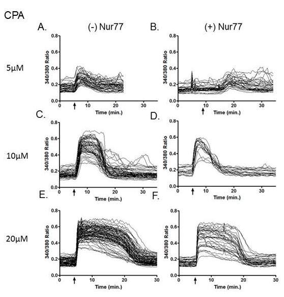
54 Figure 2.8 Presence of Nur77 shows decreased CPA-induced calcium response at low concentrations. DO11.10 Tet-Nur77 NES (-/+) Nur77 cells were treated with various concentrations of CPA in (-) Ca 2+ monitoring buffer. Arrow indicates time of CPA addition (A-F) Left, (-) Nur77. Right, (+) Nur77. A,B) 5µM C,D) 10µM. E,F) 20µM G,H) Baseline calcium levels. IJ) Calcium Amplitude. A-D = / E+F = Experiment. 42
55 43
56 Figure 2.9 Nur77 increases baseline calcium levels and decreases intracellular calcium stores. All conditions are DO11.10 Tet-Nur77 NES (-/+) Nur77 cells activated with PMA+CaI at 5 minutes.(a-f) Left, (-) Ca 2+ in monitoring buffer. Right, (+) Ca 2+ in monitoring buffer. A,B) (-) Nur77. C,D) (+) Nur77 1 hour. E,F) (+) Nur77 3 hours. G) Quantification of baseline calcium levels -/+ calcium in monitoring buffer. H, I) Quantification of calcium release amplitude during 5-10min. of trace or calcium released after 15 min. Arrow indicates time of activation Experiment. 44

57 Figure 2.10 Possible mechanism of Nur77-induced apoptosis in T cells. After stimulation through the TCR, ERK1/-2 phosphorylates RSK and causes its translocation to the cell membrane. Here, RSK is phosphorylated again and fully activated where it then translocates into the nucleus. In the nucleus, RSK phosphorylates Nur77. Nur77 is exported from the nucleus into the cytosol, where Nur77 associates with Bcl-2 at the mitochondria. This causes release of cytochrome c from the mitochondria and activation of the caspase cascade. However, phosphorylated Nur77 also can bind Bcl-2 at the ER. We hypothesize that Nur77 disrupts the already established Bcl-2/IP3R interaction, causing ER calcium release and eventual death of the cell (Pinton, 2006; Rud, 2010; Wang, 2009). 45
58 CHAPTER III DISCUSSION AND FUTURE DIRECTIONS Here, we present evidence that Nur77 may activate the intrinsic apoptotic pathway in T cells through a Bcl-2/IP3R-dependent mechanism. This is supported by the ability of Nur77 to modulate intracellular calcium levels and implied by coimmunoprecipitation studies. We propose that Nur77 may perhaps disrupt the Bcl- 2/IP3R1 interaction, releasing calcium from the ER and initiating the mitochondrial apoptotic pathway in a DO11.10 T cell hybridoma. However, further investigation is needed to confirm these studies. One potentially novel finding presented in this work is the role of Nur77 in disrupting the interaction between the known IP3R/Bcl-2 complexes (Chen, 2004). Chen et al., 2004 studied co-immunoprecipitated (co-ip) these complexes in Bcl-2 WEH17.2 T cells and endogenous levels in the T cell line S49.A2. Another group also reported IP3R1 (but not IP3R3)/Bcl-2 interaction at the ER in Bcl-2 MCF7 cell lines (Xu, 2007). Later, the BH4 domain of Bcl-2 was shown to be critical for the Bcl-2/IP3R interaction again in WEH17.2 T cells (Rong, 2009). Here, we use a tetracycline-inducible Nur77 in DO11.10 T cells where Nur77 is tagged with a NES sequence and is constitutively trafficked into the cytosol. In a few instances, we were able to co-immunoprecipitate the Bcl-2/IP3R1 complex in 293T, DO11.10, and Tet-Nur77 NES cells (Figure 2.1A-C). We were able to observe a reduction in the Bcl-2/IP3R1 complex in the presence of Nur77 in only one experiment (Figure 2.1C). In further trials we tried to repeat these data. However, we were unable to obtain reproducible results (Figure 5.1 and Figure 5.2). Possible explanations for these results are discussed below. 46
59 Because we were concerned with antibody specificity, a different antibody strategy was then used in an attempt to reduce non-specific bands. All of the subsequent antibodies described are reactive against mouse protein. Originally, these experiments were carried out by immunoprecipitating (IP) with rabbit antibodies and immunoblotting (IB) with the same rabbit antibodies. Bcl-2 was IP with a mouse monoclonal antibody and IB with an IP3R1 rabbit polyclonal antibody. We observed many nonspecific bands using both rabbit antibodies for IB, making it difficult to determine if the visualized bands were really IP3R1 or Bcl-2. A revised antibody strategy was employed trying to eliminate non-specific reactivity where proteins were first IP with rabbit polyclonal antibodies and detected with mouse monoclonal antibodies. However, we were unable to co-ip Bcl-2 and IP3R or detect either protein in the lysate under these experimental conditions (Figure 5.2B and 5.2C). As a positive control for Bcl-2, we used the B cell lymphoma line BAL-17, which has high endogenous levels of Bcl-2 (Figure 5.2A) (Merino, 1994). It is possible that the Bcl-2 antibody detected specific protein in BAL-17 cells, but is still unclear. Future obstacles to surpass include defining an effective IP protocol and optimizing detection with specific antibodies. In order to overcome these difficulties with our immunoprecipitation experiments, we sought to use the Bimolecular Fluorescence Complementation (BiFC) assay. This assay is used to detect protein-protein interactions in a living cell based on the association between two fluorescent fragments. For example, protein A is tagged with the N-terminus of the yellow fluorescent protein and protein B is tagged with the C-terminus. If these proteins interact and the two fluorescent fragments come together, fluorescence can be observed using a standard fluorescent microscope (Kerppola, 2008; Kerpolla, 2009). 47
60 The advantages of using this system are that one can observe protein-protein interactions in living cells, fluorescence is observed only if proteins interact, and protein levels in their intracellular compartment are comparable to biological levels. The limitations of the system, which should also be considered, are that the fluorescent complex formation is slow. This means the interactions observed are unable to be seen in real time. The fluorescent complex formed between the two fragments is also irreversible, thus not mimicking the endogenous sometimes fleeting protein interaction partners. (Kerppola, 2008; Kerpolla, 2009). The BiFC assay is currently being used and continuously standardized in our laboratory. One member has successfully observed interactions with specific proteins in different cell types. One such interaction was seen using BiFC for the previously reported interaction between Nur77NES/Notch1NES. Our laboratory had originally isolated Notch-1 in a yeast two-hybrid screen with Nur77 (Jehn, 1999). Therefore, it was expected to see this interaction using BiFC. We plan to use this technique in continuation of the current study described in this work. Our aim is to use BiFC to observe the reported interaction between Nur77 and Bcl-2 (Lin, 2004; Winoto, 2008, Kolluri, 2008). The use of a mitochondrial or ERtargeted Bcl-2 along with Nur77 NLS /NES can be used in various combinations to provide additional evidence of interactions. We obtained two plasmids containing mitochondria (Monoamine oxidase B) and ER (Cytochrome b5) targeted Bcl-2. Efforts were made to restriction digest and PCR these fragments from their pcmv-tag2b vector backbone. However, we were unable to cut at both restriction sties or obtain specific Bcl- 2 PCR products (data not shown). Further optimization of primers and PCR protocol is 48
61 needed before ligating either Bcl-2 products into a BiFC vector and transfecting cells to visualize protein-protein interactions. We would expect to see an interaction in the cytosol with Bcl-2 (ER or mitochondrial-targeted) with Nur77-NES. This technique would also be useful to confirm our hypothesis that Nur77 may disrupt the IP3R/Bcl-2 interaction. BiFC may provide us with a better understanding of these interactions because we could visualize the interactions in live cells instead of the possible, and sometimes unstable, protein interactions determined from cell lysates used for co-ip studies (Kerppola, 2008; Kerpolla, 2009). Bcl-2 has been shown to interact with the IP3R in the regulatory and coupling domain of the IP3R (Rong, 2008). This interacting portion of IP3R can be incorporated into a BiFC construct could be used along with Bcl-2-BiFC in Tet-Nur77 cell lines. These would be very interesting and informative experiments as to the nature of Nur77 s effects on IP3R and Bcl-2 proteins. As described above, we attempted to elucidate the direct effect of cytosolic Nur77 on Bcl-2 and IP3R proteins. The mechanism by which Nur77 activates the apoptotic machinery of the cell still remains to be clarified, although its cytosolic role has become increasingly appreciated (Chang, 2011; Liang, 2007; Thompson, 2010; Wang, 2009; Yang, 2011a; Yang, 2011b). Nur77 has been shown to specifically migrate to mitochondria and associate with Bcl-2 (Kolluri, 2008; Lin, 2004; Thompson, 2008). However, in DO11.10 T cells the physical interaction between endogenous Nur77 and Bcl-2 or Bcl-xL not detected (Wang, 2009). Nur77 has also been shown to interact with ER-targeted Bcl-2 (Laing, 2007). Laing et al. (2007) showed in cancer cell lines that ER-targeted Nur77 depletes ER calcium stores upstream of caspase activation. These data prompted us to explore the role 49
62 of Nur77 in T cells on intracellular calcium. We present data revealing some effects of Nur77 ER calcium stores T cells. We demonstrate that in the presence of calcium in extracellular media, the expression of Nur77 increases baseline calcium levels in the cytosol (Figure 2.11). Higher levels of cytosolic calcium are suggested to contribute to induction of apoptosis (Harr and Distelhorst, 2010). This could occur by increasing mitochondrial calcium load, and triggering apoptosis through mechanisms that still remain unclear (Demaurex and Distelhorst, 2003). Nonetheless, the amount and intracellular location of calcium remains an important factor when investigating apoptosis. Following TCR engagement, calcium enters the cell through Ca 2+ releaseactivated Ca 2+ (CRAC) channels and is a critical step for effective T cell activation (Feske, 2007; Quintana, 2011). Calcium-dependent signaling pathway calcineurin promotes nuclear factor of activated T cells (NFAT) which interacts with myocyte enhancer factor-2 (MEF2). MEF2 binds the Nur77 promoter and enhances its transcriptional activity (Youn, 1999; Youn, 2000). As mentioned above, when calcium is present in the imaging buffer, Nur77 increases baseline calcium levels (Figure 2.6). In contrast, when Nur77 is expressed and cells are placed without calcium in the imaging buffer, baseline calcium levels may be lowered (Figure 5.7C). This result was also not surprising because sustained Nur77-induced calcium release may require continuous calcium import into the cell. After examining the effect of Nur77 on baseline calcium levels, we next asked if Nur77 affects ER calcium store content. To investigate this, we used SERCA inhibitors thapsigargin (TG) and Cyclopiazonic acid (CPA), which both empty ER calcium stores 50
63 and raise cytosolic calcium levels (Dolmetsch and Lewis, 1994). It is important to note that all TG and CPA experiments were done in (-) Ca 2+ imaging buffer (ECB). We measured the cytosolic calcium by staining cells with the fluorescent calcium indicator fura2-am and determined the fluorescence ratio (340/380). As seen in Figure 2.7 and Figure 2.8, when Nur77 was expressed and cells were treated with TG or CPA, the ER calcium content was decreased in the presence of Nur77. In both of these instances, the calcium response was also delayed. This trend where Nur77 delays calcium response and shows lower levels of ER calcium leads us to suggest a model where Nur77 influences opening of the IP3R, releasing calcium from the ER into the cytosol. Calcium signaling is mediated by SERCA calcium pumps, but another pathway referred to as the leak mechanism balances the influx created by the pumps and are important in regulating calcium levels (Camello, 2002). Nur77 may increase the leakiness of calcium through IP3R, resulting in lower ER stores when emptied with either SERCA inhibitor. Even though we observed decreased ER calcium stores and delayed response to thapsigargin in Figure 2.7, we also observed variation in a different experiment (Figure 5.6). In response to strong activation signals such as during negative selection, Nur77 may promote a sustained calcium release (Reviewed in Rong and Diselhorst, 2008). Perhaps in this experiment (Figure 5.6) the ER stores were increased so Nur77 could cause this transient calcium release and subsequent apoptosis. In all experiments unless otherwise noted, Nur77 was induced for 1 hour. Perhaps in this experiment, Nur77 induction was not as efficient and we were actually looking at an earlier time point. This would explain why at the time of imaging, there is a greater amount of ER calcium. If 51
64 Nur77 has been induced for a longer period of time, Nur77 may have already caused this ER calcium release and could explain why we saw decreased ER calcium content in the experiments previously described (Figures 2.12E and 2.14I). Further experiments need to be performed to verify these results. As described earlier, we have seen that Nur77 increases baseline calcium levels when calcium is available in extracellular buffer. We have also seen that Nur77 expression might influence ER calcium stores. Our next study focused on the effect of Nur77 at two different time points (1hr and 4hr) on calcium levels in response to cell activation by PMA+CaI. Consistent with previous data, Figure 5.7B shows that 1hr Nur77 expression (+)Ca 2+ increases baseline calcium levels, whereas (-)Ca 2+ decreases baseline calcium levels. In Figure 2.15H, both Nur77 time points seem to release about the amount of intracellular calcium. As expected, the calcium released >15 minutes is minimal because there is no calcium in the extracellular buffer for the cell to pump inside and cause a sustained calcium release. In contrast, when extracellular calcium is present and Nur77 in induced for 1 hour, the calcium released at >15 min is increased (Figure 2.9I). This suggests to us that the cell is continually pumping calcium back into the cell, where Nur77 can then cause an even greater calcium release. However, at 4 hours the calcium released is lowered maybe because the cell has already continually dumped ER calcium stores. Much of the calcium imaging data presented here suggests Nur77 does affect calcium levels in cells undergoing apoptosis. We are confident in our data showing that when extracellular calcium is available, Nur77 expression results in an increase in baseline calcium levels. 52
65 Whether Nur77 decreases or increases the ER calcium stores will need to be verified in further experiments. Continued investigation of the effect of Nur77 on intracellular calcium could focus on determining the contribution of ER and mitochondrial calcium. Specific inhibitors to block calcium entry into the mitochondria would be useful to examine the effect of Nur77 on mitochondrial calcium stores. It has already been shown that Nur77 causes changes in mitochondrial membrane potential (Wang, 2009). Perhaps Nur77 causes increased uptake of calcium into the mitochondria, thus initiating apoptosis. Imaging cells in the absence of calcium (-/+ Nur77) in extracellular buffer then adding back calcium would be also an interesting experiment to show the engagement of the STIM/ORAI pathway in T cells (Robert, 2011). These calcium patterns are generated by interplay between various cellular compartments. During T cell activation, intracellular cytosolic calcium levels can rise from 50nM to around 1µM. Calcium is stored in organelles such as the ER, where calcium concentration can be about 400µM (Robert, 2011). In a normal cell, calcium is being continually cycled between the ER and mitochondria. However, calcium is also imported and exported from cell through channels at the cell membrane (Robert, 2011). In the experiments presented here, we measured the total amount of calcium released into the cytosol. The calcium concentration at various cellular compartments could be drastically different in the presence of Nur77, but may be undetectable by measuring total calcium. Determining exactly what compartments calcium is being stored and the concentration and the effect of Nur77 on these compartments would be very interesting for further studies. 53
66 Here, we hypothesize that Nur77 disrupts the known Bcl-2/IP3R interaction at the ER, causing calcium release through the IP3R and activation of apoptosis in T cells. This is supported through our co-immunoprecipitation studies. However, further experiments using BiFC will be used to confirm this. Studies of Nur77 on intracellular calcium concentration revealed that when extracellular calcium is present, Nur77 increases baseline calcium levels. Nur77 may also increase the leakiness of calcium through IP3R, resulting in lower ER stores when emptied with either TG or CPA. Additional data regarding subcellular calcium localization and concentration would be useful in determining the precise molecular mechanism as to how Nur77 induces apoptosis in T cells. Once more is known about Nur77 and its interacting partners, these could be a therapeutic target. Current research focuses on targeting Nur77 in cancer models. Recently, a group reported a novel compound PCH4 initiates apoptosis in DBTRG-05MG and glioblastoma multiform (GBM) 8401 cells by up regulating Nur77. They again observed the translocation of Nur77 protein from the nucleus to the cytoplasm by Western blot. Treatment of DBTRG xenografts in nude mice with PCH4 decreased tumor size over time compared to control groups (Chang, 2011). The Wan lab also published two related papers highlighting the role of Nur77 and its intracellular location as a target in cancer therapy (Yang, 2011a; Yang 2011b). Clearly, the advanced understanding of the molecular mechanisms underlying Nur77-induced apoptosis can provide new insights into cancer therapy, autoimmune diseases and the mechanisms underlying thymocyte negative selection. 54
67 CHAPTER IV MATERIALS AND METHODS Antibodies The following antibodies were used for immunoblotting: IP3R1 (ab5804), Bcl-2 (ab32124), Bcl-2 (ab-692) and GFP (ab5450) were from Abcam (Cambridge, MA). IP3R1 (sc ) was from Santa Cruz (Santa Cruz, CA). TetR antibody (631108) was from Clontech (Mountain View, CA). Actin (A3853) was from (Sigma-Aldrich). The following antibodies were used for IP: IP3R1 (ab5804), Bcl-2 (ab32124), and Bcl-2 (ab7973) were from Abcam (Cambridge, MA). Anti-CD3 used for activation (553058) was from BD Biosciences (San Diego, CA). Flow cytometry: PE-conjugated anti-mouse CD3e (553064) was from BD Biosciences (San Diego, CA). ELISA antibodies: rat anti-mouse IL-2 (554424), biotin rat anti-mouse IL-2 (554426), and avidin-hrp (554058) were from BD Biosciences (San Diego, CA). Calcium Imaging Methods of calcium imaging were described previously in Zhong et al., 2006; Chen et al., 2004, and used here with modifications x 10 6 cells were adhered directly to poly-d-lysine-coated coverslips (14-mm coverslip dishes; MatTek Corp, Ashland, MA) and incubated for 1 hour at 37 C in 7% CO 2. To induce Nur77, cells were washed three times with 1xPBS and resuspended in RDG medium before plating. After adhering cells to coverslip, cells were loaded with 1µM Fura-2-acetoxymethyl ester (Molecular Probes, Eugene, OR) for 45 min at 25 C in extracellular buffer (ECB; 130mM NaCI, 5mM KCI, 1.5mM CaCI 2, 1mM MgCI 2, 25mM Hepes ph 7.5, 1mg/ml 55
68 BSA, and 5mM glucose). The buffer was then replaced with fresh ECB and incubated at 25 C for an additional 45 minutes. Culture dishes were washed once with ECB and mounted on the nonheated stage of an inverted microscope (Nikon Diaphot). Intracellular calcium measurements up to 100 cells were monitored using SimplePCI (C-Imagaing System Cranberry Township, PA), which controls a filter wheel rotating between wavelengths of 340 and 380nm. Light was illuminated by a 75 W Xenon arc lamp and emitted light above 510nm was collected by a cooled Photometrics SenSys CCD camera (Roper Scientific, Tuscon, AZ). Light was collected every 20 seconds and used to calculate fluorescence ratios of 340/380 (Zhang et. al, 2011). ER calcium was assessed by determining the magnitude of calcium response induced by the addition of 100nM thapsigargin (TG) or 5,10,20 µm cyclopiazonic acid (CPA). For TG and CPA treatments, cells were maintained in Ca 2+- free conditions in ECB (-)Ca 2+ (-) BSA supplemented with 4mM EGTA. TG or CPA was added to this buffer during calcium monitoring at indicated times. Statistics were calculated using Graph Pad Prism (GraphPad Software, La Jolla, CA) by performing appropriate Student s t-tests. Cell culture and stable cell lines DO11.10, 293T, and Jurkat cells were cultured in RDG complete media composed of 45% RPMI 1640, 45% DMEM, 10% FBS (Lonza, Switzerland), 100U/mL Penicillin and Streptomycin (Lonza, Switzerland) at 37 C in 7% CO 2. DO11.10 and Jurkat cells were stimulated by using various concentrations of anti- CD3 antibody (BD Biosciences, San Diego, CA). Anti-CD3/CD28 antibodies used in 56
69 Figures 2.4, 2.5, 2.6 were made in our laboratory and were used at a 1:1 ratio. DO11.10 cells were stimulated with 80nM PMA and 500nM Calcium Ionophore. Flow cytometry and ELISA Flow Cytometry was performed using an LSRII (Beckon-Dickinson, Mountain View, CA) Cells were stained using PE-conjugated anti-mouse CD3e (BD Biosciences). ELISAs were performed using supernatants from activated cells using rat anti-mouse IL- 2 from BD Biosciences. Immunoblotting Whole cell lysates were prepared by lysing cells using RIPA buffer (50mM Tris, ph8.0, 150mM NaCl, 1.0% NP-40, 0.5% sodium deoxycholate, 0.1% SDS, protease inhibitors). Protein assays were performed using BCA Assay kit (Thermo Scientific, Rockford, IL). Cell lysates containing 30-50µg protin were subjected to SDS- PAGE and then transferred to nitrocellulose membranes. Blocking, primary, and secondary antibody incubations were implemented in BLOTTO (1x PBS containing 0.1% Tween-20 supplemented with 5% (wt/vol) dry fat-free milk powder (Carnation Nestle). Horseradish peroxidase-conjugated sheep anti-rabbit and anti-mouse antibodies (GE Health, Piscataway, NJ) were used at a dilution of 1:5000, and bands were detected with enhanced chemiluminescence (ECL, GE Health, Piscataway, NJ). Antibodies were used at 1:1000 or as directed by the manufacturer for immunoblotting. DO11.10 cells were stimulated to die with 80nM PMA and 500nM calcium ionophore. 57
70 Immunoprecipitation 293T cells or DO11.10 cells were lysed with IP-lysis buffer (50mM Tris, 100mM NaCl, 2mM EDTA, and 1% CHAPS) with protease inhibitors. Whole cell lysates were then incubated overnight with anti-ip3r1 or anti-bcl-2 antibody and cleared using Protein A/G Plus beads (Santa Cruz Biotechnology). Samples were then analyzed by SDS-PAGE and Western blot analysis. Tris-glycine SDS-Polyacrylamide Gels (IP3R1-6%, Bcl-2 12%) or 4-15% Ready Gel Tris-HCl ( , BIORAD) were used. 58
 Nur77 was induced for 48hours. The IP3R1/Bcl2 complex was greatly reduced. Parental DO11.10 cells were used as an IP control.")
71 CHAPTER V SUPPLEMENTARY DATA Figure 5.1 Nur77 may disrupt the interaction between IP3R1 and Bcl-2. A-C) Untreated Tet-Nur77 NES cells were IP with indicated antibodies and analyzed by immunoblotting. For IP and IB: IP3R1 Abcam ab5804, Bcl-2 Abcam ab A) Nur77 was induced for 48hours. The IP3R1/Bcl2 complex was greatly reduced. Parental DO11.10 cells were used as an IP control. Nur77 can be detected in IP GFP and in induced cells. B) IP3R1/Bcl-2 complex is absent in control cells. C) IP3R1/Bcl-2 or Bcl- 2/IP3R1 complex is absent in control cells. 59
 cell lysates were immunoprecipitated (IP) with either Bcl-2 (Abcam ab7973) or IP3R1 (Abcam ab5804) and proteins were detected by immunoblotting (Bcl-2: Ab692 or IP3R1:")
72 Figure 5.2 Further optimization of IP protocol and co-ip antibodies are needed. A- C) DO11.10 Tet-Nur77 NES (Tet-NNES) cell lysates were immunoprecipitated (IP) with either Bcl-2 (Abcam ab7973) or IP3R1 (Abcam ab5804) and proteins were detected by immunoblotting (Bcl-2: Ab692 or IP3R1: Santa Cruz sc ). A) Bcl-2 was induced in a DO11.10 cell line with varying concentrations of copper sulfate. BAL-17 cells also show high endogenous levels of Bcl-2. B+C) New antibodies listed above were unsuccessful in visualizing IP3R1 and Bcl-2 proteins through IP or in lysate. 60
73 Figure 5.3 Anti-CD3/anti-CD28 treatment insufficiently activates Tet-Nur77 NES cells. A-C) DO11.10 Tet-Nur77 NES (-/+) Nur77 cells were monitored on a heated microscope stage. A) Unstimulated. B.1) Cells placed directly on heated stage for imaging. B.2) Cells placed on heated stage for imaging and pre-warmed media was added over coverslip. C) Cells incubated on ice for 15 minutes before mounting on heated stage and 1mL additional warm media added to assist in activation. D) Percent death was determined in Tet-Nur77 NES cells by staining with DAPI and observing apoptotic nuclear morphology after 24 hours. PMA + CaI was used as a positive control. D-A) Cells were incubated with anti-cd3/anti-cd28 for 0 or 1 hour before adding hamster IgG to cross-link receptors. Antibodies were incubated with Protein A/G beads for 1 hour to concentrate. B) Cells stimulated with commercial anti-cd3 at 1 or 5 ug/ml. Nur77NES cells die better with co-stimulation by anti-cd3, anti-cd28, and anti-hamster IgG than with anti-cd3 alone. This activation condition was used for certain calcium imaging experiments. 61
74 Figure 5.4 Commercial anti-cd3/28 is more efficient for cell activation. DO11.10 Tet-Nur77 NES (-) Nur77 cells were imaged in (+)Ca 2+ buffer. A) Cells were activated with anti-cd3/28. B) Commercial anti-cd3/28. Activation through TCR with anti-cd3 shows delayed calcium release. C) PMA+CaI. Arrow indicates time of activation. 62
 Nur77 were monitored in (+)Ca 2+ buffer and were activated at 5 minutes with a variety of concentrations of commercial anti-cd3 antibody (µg/ml). A) 5. B) 20.")
75 Figure 5.5 Many concentrations of commercial anti-cd3 antibody tested are insufficient to activate Tet-Nur77 NES cells. Tet-Nur77 NES cells (-) Nur77 were monitored in (+)Ca 2+ buffer and were activated at 5 minutes with a variety of concentrations of commercial anti-cd3 antibody (µg/ml). A) 5. B) 20. C) 50. D) 75. E) 100. Arrow indicates time of activation. 63
Cell Death & Trophic Factors II. Steven McLoon Department of Neuroscience University of Minnesota
 Cell Death & Trophic Factors II Steven McLoon Department of Neuroscience University of Minnesota 1 Remember? Neurotrophins are cell survival factors that neurons get from their target cells! There is a
Apoptosis EXTRA REQUIREMETS
 Apoptosis Introduction (SLIDE 1) Organisms develop from a single cell, and in doing so an anatomy has to be created. This process not only involves the creation of new cells, but also the removal of cells
Apoptosis: Death Comes for the Cell
 Apoptosis: Death Comes for the Cell Joe W. Ramos joeramos@hawaii.edu From Ingmar Bergman s The Seventh Seal 1 2 Mutations in proteins that regulate cell proliferation, survival and death can contribute
 http://cwp.embo.org/w09-13/index.html Roads to Ruin is s o t p o ap healthy cell autophagic cell death ne cr os is Thanks to Seamus Martin! Evolution of apoptosis signalling cascades Inhibitor Adopted
Mechanisms of Cell Death
 Mechanisms of Cell Death CELL DEATH AND FORMATION OF THE SEMICIRCULAR CANALS Carol M. Troy, MD PhD August 24, 2009 FROM: Fekete et al., Development 124: 2451 (1997) PHENOMENOLOGY OF CELL DEATH I. DEVELOPMENT
Mechanisms. Cell Death. Carol M. Troy, MD PhD August 24, 2009 PHENOMENOLOGY OF CELL DEATH I. DEVELOPMENT 8/20/2009
 Mechanisms of Cell Death Carol M. Troy, MD PhD August 24, 2009 PHENOMENOLOGY OF CELL DEATH I. DEVELOPMENT A. MORPHOGENESIS: SCULPTING/SHAPING STRUCTURES CREATION OF CAVITIES AND TUBES FUSION OF TISSUE
Cell Survival Curves (Chap. 3)
") Cell Survival Curves (Chap. 3) Example of pedigress of EMT6 mouse cells Clone The in vitro cell survival curve Cell Survival Assay Dye exclusion assay: membrane integrity MTT assay: respiratory activity
Cell Survival Curves (Chap. 3) Example of pedigress of EMT6 mouse cells Clone The in vitro cell survival curve Cell Survival Assay Dye exclusion assay: membrane integrity MTT assay: respiratory activity
Regulation and signaling. Overview. Control of gene expression. Cells need to regulate the amounts of different proteins they express, depending on
 Regulation and signaling Overview Cells need to regulate the amounts of different proteins they express, depending on cell development (skin vs liver cell) cell stage environmental conditions (food, temperature,
Zool 3200: Cell Biology Exam 5 4/27/15
 Name: Trask Zool 3200: Cell Biology Exam 5 4/27/15 Answer each of the following short answer questions in the space provided, giving explanations when asked to do so. Circle the correct answer or answers
Programmed Cell Death
 Programmed Cell Death Dewajani Purnomosari Department of Histology and Cell Biology Faculty of Medicine Universitas Gadjah Mada d.purnomosari@ugm.ac.id What is apoptosis? a normal component of the development
Reception The target cell s detection of a signal coming from outside the cell May Occur by: Direct connect Through signal molecules
 Why Do Cells Communicate? Regulation Cells need to control cellular processes In multicellular organism, cells signaling pathways coordinate the activities within individual cells that support the function
Bio-Plex Pro RBM Apoptosis Assays
 Acute Phase Apoptosis Cancer Cardiovascular Disease Cytokines Chemokines, Growth Factors Diabetes Gene Expression Genotyping Immunoglobulin Isotyping MicroRNA Expression Bio-Plex Pro RBM Apoptosis Assays
A. Incorrect! The Cell Cycle contains 4 distinct phases: (1) G 1, (2) S Phase, (3) G 2 and (4) M Phase.
 G 1, (2) S Phase, (3) G 2 and (4) M Phase.") Molecular Cell Biology - Problem Drill 21: Cell Cycle and Cell Death Question No. 1 of 10 1. Which of the following statements about the cell cycle is correct? Question #1 (A) The Cell Cycle contains 3
Molecular Cell Biology - Problem Drill 21: Cell Cycle and Cell Death Question No. 1 of 10 1. Which of the following statements about the cell cycle is correct? Question #1 (A) The Cell Cycle contains 3
Signal Transduction. Dr. Chaidir, Apt
 Signal Transduction Dr. Chaidir, Apt Background Complex unicellular organisms existed on Earth for approximately 2.5 billion years before the first multicellular organisms appeared.this long period for
Apoptosis: Death comes for the Cell. Apoptosis: Programmed Cell Death. Apoptosis. Necrosis
 poptosis: eath comes for the Cell Mutations in proteins that regulate cell proliferation, survival and death can contribute to oncogenesis Joe W. Ramos jramos@crch.hawaii.edu From Ingmar Bergman s The
4.3 Intracellular calcium dynamics
 coupled equations: dv dv 2 dv 3 dv 4 = i + I e A + g,2 (V 2 V ) = i 2 + g 2,3 (V 3 V 2 )+g 2, (V V 2 )+g 2,4 (V 4 V 2 ) = i 3 + g 3,2 (V 2 V 3 ) = i 4 + g 4,2 (V 2 V 4 ) This can be written in matrix/vector
Embedded together: The life and death consequences of interaction of the Bcl-2 family with membranes
 DOI 10.1007/s10495-007-0746-4 Embedded together: The life and death consequences of interaction of the Bcl-2 family with membranes Brian Leber Jialing Lin David W. Andrews Published online: 15 February
Chapter 12: Intracellular sorting
 Chapter 12: Intracellular sorting Principles of intracellular sorting Principles of intracellular sorting Cells have many distinct compartments (What are they? What do they do?) Specific mechanisms are
The EGF Signaling Pathway! Introduction! Introduction! Chem Lecture 10 Signal Transduction & Sensory Systems Part 3. EGF promotes cell growth
 Chem 452 - Lecture 10 Signal Transduction & Sensory Systems Part 3 Question of the Day: Who is the son of Sevenless? Introduction! Signal transduction involves the changing of a cell s metabolism or gene
Chem Lecture 10 Signal Transduction
 Chem 452 - Lecture 10 Signal Transduction 111202 Here we look at the movement of a signal from the outside of a cell to its inside, where it elicits changes within the cell. These changes are usually mediated
Apoptosis and Carcinogenesis
 Death Becomes Us: Apoptosis and Carcinogenesis by Michele M. Cox Department of Biology, University of the Virgin Islands Part I What Is Cancer? Hi, Derek. Studying for your Cell and Molec final? asked
Types of biological networks. I. Intra-cellurar networks
 Types of biological networks I. Intra-cellurar networks 1 Some intra-cellular networks: 1. Metabolic networks 2. Transcriptional regulation networks 3. Cell signalling networks 4. Protein-protein interaction
Apoptosis in Mammalian Cells
 Apoptosis in Mammalian Cells 7.16 2-10-05 Apoptosis is an important factor in many human diseases Cancer malignant cells evade death by suppressing apoptosis (too little apoptosis) Stroke damaged neurons
Biochimica et Biophysica Acta
 Biochimica et Biophysica Acta 1843 (2014) 2100 2113 Contents lists available at ScienceDirect Biochimica et Biophysica Acta journal homepage: www.elsevier.com/locate/bbamcr Review Regulating cell death
IAPs. Structure of BIR2 Zn finger domain from XIAP
 IAPs The only known endogenous caspase inhibitors are members of a second family of proteins identified by apoptosis research, the IAP (inhibitor of apoptosis proteins) family. IAPs were originally described
The Caspase System: a potential role in muscle proteolysis and meat quality? Tim Parr
 The Caspase System: a potential role in muscle proteolysis and meat quality? Tim Parr Caroline Kemp, Ron Bardsley,, Peter Buttery Division of Nutritional Sciences, School of Biosciences, University of
Death signaling. Color PDF file of handouts can be found at Wu lab web-page:
 Death signaling Hao Wu References 1. Wang, X. The expanding role of mitochondria in apoptosis. Genes Dev 15, 2922-2933. (2001). 2. Fesik, S. W. Insights into programmed cell death through structural biology.
Bio 3411, Fall 2006, Lecture 19-Cell Death.
 Types of Cell Death Questions : Apoptosis (Programmed Cell Death) : Cell-Autonomous Stereotypic Rapid Clean (dead cells eaten) Necrosis : Not Self-Initiated Not Stereotypic Can Be Slow Messy (injury can
Monte Carlo study elucidates the type 1/type 2 choice in apoptotic death signaling in normal and cancer cells
 Cells 2013, 2, 1-x manuscripts; doi:10.3390/cells20x000x OPEN ACCESS cells ISSN 2073-4409 www.mdpi.com/journal/cells Article Monte Carlo study elucidates the type 1/type 2 choice in apoptotic death signaling
Introduction. Gene expression is the combined process of :
 1 To know and explain: Regulation of Bacterial Gene Expression Constitutive ( house keeping) vs. Controllable genes OPERON structure and its role in gene regulation Regulation of Eukaryotic Gene Expression
7.06 Cell Biology EXAM #3 April 21, 2005
 7.06 Cell Biology EXAM #3 April 21, 2005 This is an open book exam, and you are allowed access to books, a calculator, and notes but not computers or any other types of electronic devices. Please write
RANK. Alternative names. Discovery. Structure. William J. Boyle* SUMMARY BACKGROUND
 RANK William J. Boyle* Department of Cell Biology, Amgen, Inc., One Amgen Center Drive, Thousand Oaks, CA 91320-1799, USA * corresponding author tel: 805-447-4304, fax: 805-447-1982, e-mail: bboyle@amgen.com
ADAM FAMILY. ephrin A INTERAZIONE. Eph ADESIONE? PROTEOLISI ENDOCITOSI B A RISULTATO REPULSIONE. reverse. forward
 ADAM FAMILY - a family of membrane-anchored metalloproteases that are known as A Disintegrin And Metalloprotease proteins and are key components in protein ectodomain shedding Eph A INTERAZIONE B ephrin
Massive loss of neurons in embryos occurs during normal development (!)
") Types of Cell Death Apoptosis (Programmed Cell Death) : Cell-Autonomous Stereotypic Rapid Clean (dead cells eaten) Necrosis : Not Self-Initiated Not Stereotypic Can Be Slow Messy (injury can spread) Apoptosis
Types of Cell Death Apoptosis (Programmed Cell Death) : Cell-Autonomous Stereotypic Rapid Clean (dead cells eaten) Necrosis : Not Self-Initiated Not Stereotypic Can Be Slow Messy (injury can spread) Apoptosis
Big Idea 3: Living systems store, retrieve, transmit and respond to information essential to life processes. Tuesday, December 27, 16
 Big Idea 3: Living systems store, retrieve, transmit and respond to information essential to life processes. Enduring understanding 3.B: Expression of genetic information involves cellular and molecular
Proteome-wide High Throughput Cell Based Assay for Apoptotic Genes
 John Kenten, Doug Woods, Pankaj Oberoi, Laura Schaefer, Jonathan Reeves, Hans A. Biebuyck and Jacob N. Wohlstadter 9238 Gaither Road, Gaithersburg, MD 2877. Phone: 24.631.2522 Fax: 24.632.2219. Website:
COMPUTER SIMULATION OF DIFFERENTIAL KINETICS OF MAPK ACTIVATION UPON EGF RECEPTOR OVEREXPRESSION
 COMPUTER SIMULATION OF DIFFERENTIAL KINETICS OF MAPK ACTIVATION UPON EGF RECEPTOR OVEREXPRESSION I. Aksan 1, M. Sen 2, M. K. Araz 3, and M. L. Kurnaz 3 1 School of Biological Sciences, University of Manchester,
Richik N. Ghosh, Linnette Grove, and Oleg Lapets ASSAY and Drug Development Technologies 2004, 2:
 1 3/1/2005 A Quantitative Cell-Based High-Content Screening Assay for the Epidermal Growth Factor Receptor-Specific Activation of Mitogen-Activated Protein Kinase Richik N. Ghosh, Linnette Grove, and Oleg
CHAPTER 3. Cell Structure and Genetic Control. Chapter 3 Outline
 CHAPTER 3 Cell Structure and Genetic Control Chapter 3 Outline Plasma Membrane Cytoplasm and Its Organelles Cell Nucleus and Gene Expression Protein Synthesis and Secretion DNA Synthesis and Cell Division
Calcium dynamics, 7. Calcium pumps. Well mixed concentrations
 Calcium dynamics, 7 Calcium is an important ion in the biochemistry of cells Is used a signal carrier, i.e. causes contraction of muscle cells Is toxic at high levels The concentration is regulated through
Study Guide 11 & 12 MULTIPLE CHOICE. Choose the one alternative that best completes the statement or answers the question.
 Study Guide 11 & 12 MULTIPLE CHOICE. Choose the one alternative that best completes the statement or answers the question. 1) The receptors for a group of signaling molecules known as growth factors are
Apoptosis and Cancer. Carol M. Troy, MD, PhD October 26, 2016
 Apoptosis and Cancer Carol M. Troy, MD, PhD October 26, 2016 PHENOMENOLOGY OF CELL DEATH -1 I. DEVELOPMENT A. MORPHOGENESIS: SCULPTING/SHAPING STRUCTURES FUSION OF TISSUE MASSES (PALATE/NEURAL TUBE) CREATION
S1 Gene ontology (GO) analysis of the network alignment results
 analysis of the network alignment results") 1 Supplementary Material for Effective comparative analysis of protein-protein interaction networks by measuring the steady-state network flow using a Markov model Hyundoo Jeong 1, Xiaoning Qian 1 and
1 Supplementary Material for Effective comparative analysis of protein-protein interaction networks by measuring the steady-state network flow using a Markov model Hyundoo Jeong 1, Xiaoning Qian 1 and
APOPTOSIS REGULATOR BCL-2
 Papers on Anthropology Apoptosis regulator XXII, 2013, BCL-2 pp. 63 67 P. Hussar, M. Žuravskaja, M. Kärner APOPTOSIS REGULATOR BCL-2 Piret Hussar 1, Maria Žuravskaja 2, Martin Kärner 2 1 Institute of Anatomy,
RNA Synthesis and Processing
 RNA Synthesis and Processing Introduction Regulation of gene expression allows cells to adapt to environmental changes and is responsible for the distinct activities of the differentiated cell types that
cellular division cell division cell cycle cell cycle kinases chapter 18-19
 cellular division chapter 18-19 cell division when? growth replacement of older cells production of specialized cells asexual reproduction sexual reproduction production of gametes prokaryotic binary fission
TNFα 18hr. Control. CHX 18hr. TNFα+ CHX 18hr. TNFα: 18 18hr (KDa) PARP. Cleaved. Cleaved. Cleaved. Caspase3. Pellino3 shrna. Control shrna.
 PARP. Cleaved. Cleaved. Cleaved. Caspase3. Pellino3 shrna. Control shrna.") Survival ( %) a. TNFα 18hr b. Control sirna Pellino3 sirna TNFα: 18 18hr c. Control shrna Pellino3 shrna Caspase3 Actin Control d. Control shrna Pellino3 shrna *** 100 80 60 CHX 18hr 40 TNFα+ CHX 18hr
Survival ( %) a. TNFα 18hr b. Control sirna Pellino3 sirna TNFα: 18 18hr c. Control shrna Pellino3 shrna Caspase3 Actin Control d. Control shrna Pellino3 shrna *** 100 80 60 CHX 18hr 40 TNFα+ CHX 18hr
cellular division cell division cell cycle cell cycle kinases chapter 18-19
 cellular division chapter 18-19 cell division when? growth replacement of older cells production of specialized cells asexual reproduction sexual reproduction production of gametes prokaryotic binary fission
The majority of cells in the nervous system arise during the embryonic and early post
 Introduction Introduction The majority of cells in the nervous system arise during the embryonic and early post natal period. These cells are derived from population of neural stem cells first shown by
Host-Pathogen Interaction. PN Sharma Department of Plant Pathology CSK HPKV, Palampur
 Host-Pathogen Interaction PN Sharma Department of Plant Pathology CSK HPKV, Palampur-176062 PATHOGEN DEFENCE IN PLANTS A BIOLOGICAL AND MOLECULAR VIEW Two types of plant resistance response to potential
AP Biology Gene Regulation and Development Review
 AP Biology Gene Regulation and Development Review 1. What does the regulatory gene code for? 2. Is the repressor by default active/inactive? 3. What changes the repressor activity? 4. What does repressor
Cytochrome c, cytochrome b 5 and electron transfer
 Cytochrome c, cytochrome b 5 and electron transfer Covalent bonds Cytochrome c is a major player in membrane associated electron transport systems in bacteria and mitochondria. Cytochrome c has two axial
Chang 1. Characterization of the Drosophila Ortholog of the Mammalian Anti-apoptotic Protein Aven
 Chang 1 Characterization of the Drosophila Ortholog of the Mammalian Anti-apoptotic Protein Aven Joy Chang Georgetown University Senior Digital Thesis May 1, 2006 Chang 2 Table of Contents BACKGROUND 4
SUPPLEMENTARY INFORMATION
 DOI: 10.1038/ncb2362 Figure S1 CYLD and CASPASE 8 genes are co-regulated. Analysis of gene expression across 79 tissues was carried out as described previously [Ref: PMID: 18636086]. Briefly, microarray
6 Mechanotransduction
 6.1 Motivation The process of converting physical forces into biochemical signals and integrating these signals into the cellular response is referred to as mechnotransduction [11, 20]. To fully understand
Toxicological Targets. Russell L. Carr Department of Basic Sciences College of Veterinary Medicine
 Toxicological Targets Russell L. Carr Department of Basic Sciences College of Veterinary Medicine Toxicology Definitions = study of poisons Poison = any agent capable of producing a deleterious response
BIOH111. o Cell Biology Module o Tissue Module o Integumentary system o Skeletal system o Muscle system o Nervous system o Endocrine system
 BIOH111 o Cell Biology Module o Tissue Module o Integumentary system o Skeletal system o Muscle system o Nervous system o Endocrine system Endeavour College of Natural Health endeavour.edu.au 1 Textbook
Lipniacki 2004 Ground Truth
 Abstract Lipniacki 2004 Ground Truth The two-feedback-loop regulatory module of nuclear factor kb (NF-kB) signaling pathway is modeled by means of ordinary differential equations. signaling pathway: https://en.wikipedia.org/wiki/signaling_pathway
7.06 Problem Set
 7.06 Problem Set 5 -- 2006 1. In the first half of the course, we encountered many examples of proteins that entered the nucleus in response to the activation of a cell-signaling pathway. One example of
MAPK kinase kinase regulation of SAPK/JNK pathways
 MAPK kinase kinase regulation of SAPK/JNK pathways Lisa Stalheim and Gary L. Johnson Abstract SAPK/JNK members of the MAPK family are regulated by at least fourteen known MAPK kinase kinases (MKKKs). In
FSC-W FSC-H CD4 CD62-L
 Supplementary Fig. 1 a SSC-A FSC-A FSC-W FSC-H SSC-W SSC-H CD4 CD62-L b SSC-A FSC-A FSC-W FSC-A FSC-A 7-AAD FSC-A CD4 IL-9 CD4 c SSC-A FSC-A FSC-W FSC-H SSC-W SSC-H 7-AAD KI67 Annexin-V 7-AAD d I L -5
SUPPLEMENTARY INFORMATION
 GP2 Type I-piliated bacteria FAE M cell M cell pocket idc T cell mdc Generation of antigenspecific T cells Induction of antigen-specific mucosal immune response Supplementary Figure 1 Schematic diagram
Multiple Choice Review- Eukaryotic Gene Expression
 Multiple Choice Review- Eukaryotic Gene Expression 1. Which of the following is the Central Dogma of cell biology? a. DNA Nucleic Acid Protein Amino Acid b. Prokaryote Bacteria - Eukaryote c. Atom Molecule
Looking for LOV: Location of LOV1 function in Nicotiana benthamiana cells
 Looking for LOV: Location of LOV1 function in Nicotiana benthamiana cells By: Patrick Rutledge 1 Dr. Jennifer Lorang 2,3, Dr. Marc Curtis 2,3, Dr. Thomas Wolpert 2,3 BioResource Research 1, Botany and
Domain 6: Communication
 Domain 6: Communication 6.1: Cell communication processes share common features that reflect a shared evolutionary history. (EK3.D.1) 1. Introduction to Communication Communication requires the generation,
Death Ligand- Death Receptor. Annexin V. Fas PI MKK7 PIP3. pro-caspase-3. Granzyme B. Caspase Activation. Bid. Bcl-2 Caspase-3 Caspase-12
 International Corporation life. science. discovery. { Apoptosis Apoptosis Kits International Corporation Apoptosis Signal Cascade and Detection Kits Annexin V Kits MEBCYTO Apoptosis Kit Annexin V Apoptosis
Explain how cell size and shape affect the overall rate of nutrient intake and the rate of waste elimination. [LO 2.7, SP 6.2]
![Explain how cell size and shape affect the overall rate of nutrient intake and the rate of waste elimination. [LO 2.7, SP 6.2]](/thumbs/77/76271213.jpg "Explain how cell size and shape affect the overall rate of nutrient intake and the rate of waste elimination. [LO 2.7, SP 6.2]") Cells Learning Objectives Use calculated surface area-to-volume ratios to predict which cell(s) might eliminate wastes or procure nutrients faster by diffusion. [LO 2.6, SP 2.2] Explain how cell size and
Cells Learning Objectives Use calculated surface area-to-volume ratios to predict which cell(s) might eliminate wastes or procure nutrients faster by diffusion. [LO 2.6, SP 2.2] Explain how cell size and
Modeling a Snap-Action, Variable-Delay Switch Controlling Extrinsic Cell Death
 Modeling a Snap-Action, Variable-Delay Switch Controlling Extrinsic Cell Death John G. Albeck 1[, John M. Burke 1,2[, Sabrina L. Spencer 1,3, Douglas A. Lauffenburger 2,3, Peter K. Sorger 1,2* PLoS BIOLOGY
Sig2GRN: A Software Tool Linking Signaling Pathway with Gene Regulatory Network for Dynamic Simulation
 Sig2GRN: A Software Tool Linking Signaling Pathway with Gene Regulatory Network for Dynamic Simulation Authors: Fan Zhang, Runsheng Liu and Jie Zheng Presented by: Fan Wu School of Computer Science and
Mechanisms of Cell Death
 Mechanisms of Cell Death Etiology of cell death Major Factors Accidental Genetic Necrosis Apoptosis Necrosis: The sum of the morphologic changes that follow cell death in a living tissue or organ Apoptosis:
Muscle regulation and Actin Topics: Tropomyosin and Troponin, Actin Assembly, Actin-dependent Movement
 1 Muscle regulation and Actin Topics: Tropomyosin and Troponin, Actin Assembly, Actin-dependent Movement In the last lecture, we saw that a repeating alternation between chemical (ATP hydrolysis) and vectorial
Molecular Cell Biology 5068 In Class Exam 2 November 8, 2016
 Molecular Cell Biology 5068 In Class Exam 2 November 8, 2016 Exam Number: Please print your name: Instructions: Please write only on these pages, in the spaces allotted and not on the back. Write your
Cellular Neuroanatomy I The Prototypical Neuron: Soma. Reading: BCP Chapter 2
 Cellular Neuroanatomy I The Prototypical Neuron: Soma Reading: BCP Chapter 2 Functional Unit of the Nervous System The functional unit of the nervous system is the neuron. Neurons are cells specialized
Advanced Higher Biology. Unit 1- Cells and Proteins 2c) Membrane Proteins
 Membrane Proteins") Advanced Higher Biology Unit 1- Cells and Proteins 2c) Membrane Proteins Membrane Structure Phospholipid bilayer Transmembrane protein Integral protein Movement of Molecules Across Membranes Phospholipid
Advanced Higher Biology Unit 1- Cells and Proteins 2c) Membrane Proteins Membrane Structure Phospholipid bilayer Transmembrane protein Integral protein Movement of Molecules Across Membranes Phospholipid
Delivery. Delivery Processes. Delivery Processes: Distribution. Ultimate Toxicant
 Delivery Ultimate Toxicant The chemical species that reacts with the endogenous target. Toxicity depends on the concentration (dose) of the ultimate toxicant at the target site Delivery Processes Absorption
Enduring understanding 1.A: Change in the genetic makeup of a population over time is evolution.
 The AP Biology course is designed to enable you to develop advanced inquiry and reasoning skills, such as designing a plan for collecting data, analyzing data, applying mathematical routines, and connecting
AP Curriculum Framework with Learning Objectives
 Big Ideas Big Idea 1: The process of evolution drives the diversity and unity of life. AP Curriculum Framework with Learning Objectives Understanding 1.A: Change in the genetic makeup of a population over
Massachusetts Institute of Technology Harvard Medical School Brigham and Women s Hospital VA Boston Healthcare System 2.79J/3.96J/BE.
 Massachusetts Institute of Technology Harvard Medical School Brigham and Women s Hospital VA Boston Healthcare System 2.79J/3.96J/BE.441/HST522J INTEGRINS I.V. Yannas, Ph.D. and M. Spector, Ph.D. Regulator
7.06 Cell Biology EXAM #3 KEY
 7.06 Cell Biology EXAM #3 KEY May 2, 2006 This is an OPEN BOOK exam, and you are allowed access to books, a calculator, and notes BUT NOT computers or any other types of electronic devices. Please write
Biology Teach Yourself Series Topic 2: Cells
 Biology Teach Yourself Series Topic 2: Cells A: Level 14, 474 Flinders Street Melbourne VIC 3000 T: 1300 134 518 W: tssm.com.au E: info@tssm.com.au TSSM 2013 Page 1 of 14 Contents Cells... 3 Prokaryotic
Chemical aspects of the cell. Compounds that induce cell proliferation, differentiation and death
 Chemical aspects of the cell Compounds that induce cell proliferation, differentiation and death Cell proliferation The cell cycle Chapter 17 Molecular Biology of the Cell 2 Cell cycle Chapter 17 Molecular
NucView TM 488 Caspase-3 Assay Kit for Live Cells
 NucView TM 488 Caspase-3 Assay Kit for Live Cells Catalog Number: 30029 (100-500 assays) Contact Information Address: Biotium, Inc. 3423 Investment Blvd. Suite 8 Hayward, CA 94545 USA Telephone: (510)
Mathematical Modeling and Analysis of Crosstalk between MAPK Pathway and Smad-Dependent TGF-β Signal Transduction
 Processes 2014, 2, 570-595; doi:10.3390/pr2030570 Article OPEN ACCESS processes ISSN 2227-9717 www.mdpi.com/journal/processes Mathematical Modeling and Analysis of Crosstalk between MAPK Pathway and Smad-Dependent
Big Idea 1: The process of evolution drives the diversity and unity of life.
 Big Idea 1: The process of evolution drives the diversity and unity of life. understanding 1.A: Change in the genetic makeup of a population over time is evolution. 1.A.1: Natural selection is a major
SUPPLEMENTARY INFORMATION
 doi:10.1038/nature11419 Supplementary Figure 1 Schematic representation of innate immune signaling pathways induced by intracellular Salmonella in cultured macrophages. a, During the infection Salmonella
A MATHEMATICAL MODEL FOR DYNAMIC SIMULATION OF APOPTOSIS PATHWAYS
 A MATHEMATICAL MODEL FOR DYNAMIC SIMULATION OF APOPTOSIS PATHWAYS Paolo Massari Department of Information Engineering Bioengineering University of Padova April 2014 Advisor: Gianna Maria Toolo Coadvisor:
Validation of Petri Net Apoptosis Models Using P-Invariant Analysis
 2009 IEEE International Conference on Control and Automation Christchurch, New Zealand, December 9-11, 2009 WeMT5.1 Validation of Petri Net Apoptosis Models Using P-Invariant Analysis Ian Wee Jin Low,
Supplementary Figure 1. AnnexinV FITC and Sytox orange staining in wild type, Nlrp3 /, ASC / and casp1/11 / TEC treated with TNF /CHX.
 Supplementary Figure 1. AnnexinV FITC and Sytox orange staining in wild type, Nlrp3 /, ASC / and casp1/11 / TEC treated with TNF /CHX. Phase contrast and widefield fluorescence microscopy (20x magnification).
CENTRO ESCOLAR UNIVERSITY Biological Sciences Department Manila*Malolos*Makati SYLLABUS
 CENTRO ESCOLAR UNIVERSITY Biological Sciences Department Manila*Malolos*Makati SYLLABUS PRBS 131 BIOSCI131 CELL BIOLOGY 3 units 3 hours lec Course Number Course Title Descriptive Title Credit Unit(s) Hour(s)/Week
Unit 2: Cells Guided Reading Questions (60 pts total)
") Name: AP Biology Biology, Campbell and Reece, 7th Edition Adapted from chapter reading guides originally created by Lynn Miriello Chapter 6 A Tour of the Cell Unit 2: Cells Guided Reading Questions (60
Name: AP Biology Biology, Campbell and Reece, 7th Edition Adapted from chapter reading guides originally created by Lynn Miriello Chapter 6 A Tour of the Cell Unit 2: Cells Guided Reading Questions (60
A A A A B B1
 LEARNING OBJECTIVES FOR EACH BIG IDEA WITH ASSOCIATED SCIENCE PRACTICES AND ESSENTIAL KNOWLEDGE Learning Objectives will be the target for AP Biology exam questions Learning Objectives Sci Prac Es Knowl
Measuring TF-DNA interactions
 Measuring TF-DNA interactions How is Biological Complexity Achieved? Mediated by Transcription Factors (TFs) 2 Regulation of Gene Expression by Transcription Factors TF trans-acting factors TF TF TF TF
Cells to Tissues. Peter Takizawa Department of Cell Biology
 Cells to Tissues Peter Takizawa Department of Cell Biology From one cell to ensembles of cells. Multicellular organisms require individual cells to work together in functional groups. This means cells
16 The Cell Cycle. Chapter Outline The Eukaryotic Cell Cycle Regulators of Cell Cycle Progression The Events of M Phase Meiosis and Fertilization
 The Cell Cycle 16 The Cell Cycle Chapter Outline The Eukaryotic Cell Cycle Regulators of Cell Cycle Progression The Events of M Phase Meiosis and Fertilization Introduction Self-reproduction is perhaps
CELL SIGNALLING and MEMBRANE TRANSPORT. Mark Louie D. Lopez Department of Biology College of Science Polytechnic University of the Philippines
 CELL SIGNALLING and MEMBRANE TRANSPORT Mark Louie D. Lopez Department of Biology College of Science Polytechnic University of the Philippines GENERIC SIGNALLING PATHWAY CELL RESPONSE TO SIGNALS CELL RESPONSE
Gene Control Mechanisms at Transcription and Translation Levels
 Gene Control Mechanisms at Transcription and Translation Levels Dr. M. Vijayalakshmi School of Chemical and Biotechnology SASTRA University Joint Initiative of IITs and IISc Funded by MHRD Page 1 of 9
Drosophila Apoptosis and the Regulation of the Caspase Cascade
 Drosophila Apoptosis and the Regulation of the Caspase Cascade Kate Stafford March 18, 2005 Abstract The caspase cascade in Drosophila is controlled primarily by DIAP1 (Drosophila inhibitor of apoptosis),
Optimization of Immunoblot Protocol for Use with a Yeast Strain Containing the CDC7 Gene Tagged with myc
 OPTIMIZATION OF IMMUNOBLOT PROTOCOL 121 Optimization of Immunoblot Protocol for Use with a Yeast Strain Containing the CDC7 Gene Tagged with myc Jacqueline Bjornton and John Wheeler Faculty Sponsor: Anne
 1 of 13 8/11/2014 10:32 AM Units: Teacher: APBiology, CORE Course: APBiology Year: 2012-13 Chemistry of Life Chapters 1-4 Big Idea 1, 2 & 4 Change in the genetic population over time is feedback mechanisms
AND MOLECULAR CHARACT~RIZATION. By DEBORAH MARIE LONG,
 NITRATE REDUCTA3E IN MAIZE ROOTS: LOCALIZATION AND MOLECULAR CHARACT~RIZATION By DEBORAH MARIE LONG, B.Sc. A Thesis Submitted to the School of Graduate Studies in Partial Fulfilment of the Requirements