Nuclear resonant scattering of synchrotron radiation: a novel approach to the Mössbauer effect
|
|
|
- Rudolf Parks
- 5 years ago
- Views:
Transcription
1 Nuclear resonant scattering of synchrotron radiation: a novel approach to the Mössbauer effect Johan Meersschaut Instituut voor Kern- en Stralingsfysica, Katholieke Universiteit Leuven, Belgium Johan.Meersschaut@fys.kuleuven.be C. L abbé, ( ) Instituut voor Kern- en Stralingsfysica, K.U.Leuven, Belgium W. Sturhahn, T.S. Toellner, E.E. Alp, Advanced Photon Source, Argonne National Laboratory J.S. Jiang, S.D. Bader, Materials Science Division, Argonne National Laboratory Fund for Scientific Research Flanders (F.W.O.-Vlaanderen) and the Inter-University Attraction Pole IUAP P5/1 Work at Argonne and the use of the APS was supported by U.S. DOE, BES Office of Science, under Contract No. W ENG-38 European Commission (FP6) STREP NMP4-CT (DYNASYNC)
2 Introduction Mössbauer spectroscopy Nuclear Resonant Scattering of SR part1
3 Motivation Site-selective magnetization measurements : - XMCD - element-specific scattering - study different materials independently - Mössbauer spectroscopy - Nuclear resonant scattering of synchrotron radiation - isotope selective - study specific sites within the material separately motivation

4 Iron Isotopes Table of nuclides 57 Fe probe layer substrate 57Fe Other possible isotopes are 119 Sn, 181 Ta, 149 Sm, 153 Eu,
5 57 Fe isotope Nuclear properties: Excited level unstable (τ = ns) E = kev E = 4.66 nev I = 3/2, µ = µ n Q = 0.16 b Ground state (stable) I = 1/2, µ = µ n Q = 0 b Nat 57Fe µ N = Am b= 10 m h = ev s
6 57Co -> 57Fe Nuclear decay of 57 Co to 57 Fe

7 Hyperfine Interactions Electric monopole term: 57 Fe 57 Fe 57 Fe Isolated nucleus 57 Fe Electron density at the nucleus depends on the chemical properties Isomer shift Isomer shift I = 3/2 57 Fe I = 1/2 Monopole term
8 Hyperfine Interactions Electric quadrupole term: 57 Fe Electric field gradient due to non-cubic environment: * tetragonal or hexagonal lattice, * surface, * impurity in neigbouring shell Isolated nucleus Isomer shift 57 Fe I = 3/2 57 Fe I = 1/2 Quadrupo le term
9 Magnetic dipole interaction: Hyperfine Interactions 57 Fe B hf H B µ = I B hi I = 3/2 µ = µ n + 3/2 + 1/2-1/2-3/2 B hf = + 33 T E = 107 nev E M µ B = I m I = 1/2-1/2 + 1/2 HFI Zeeman µ n = J/T 1 J = ev µ n = ev/t
10 Summary 57 Fe Electric monopole term: Electric quadrupole interaction: Magnetic dipole interaction: I = 3/2 Isomer shift Quadrupole splitting + 3/2, -3/2 + 1/2, -1/2 magnetic splitting B hf = + 33 T + 3/2 + 1/2-1/2-3/2 E = 107 nev E = kev -1/2 + 1/2, -1/2 I = 1/2 + 1/2 HFI summary
11 Introduction Mössbauer spectroscopy Nuclear Resonant Scattering of SR part2
12 Mössbauer spectroscopy Nuclear emission : Nuclear absorption : 14.4 kev 57 Fe I = 3/ kev 57 Fe I = 3/2 0 I = 1/2 0 I = 1/2 drive source detector mm/s E = E0 1+ v c Mossbaue r
13 Mössbauer spectroscopy Nuclear emission : Nuclear absorption : 14.4 kev 57 Fe I = 3/ kev 57 Fe I = 3/2 0 I = 1/2 0 I = 1/2 drive source absorber detector Mossbaue r velocity
14 Mössbauer spectrum The Mössbauer spectrum depends on the strength of the magnetic field : magnetic splitting B = 33 T + 3/2 + 1/2-1/2 E M µ B = I m -3/2-1/2 B = 10 T + 1/2 MS
15 Mössbauer spectrum ( ) 2 1 Intensity : = I1 1 m1 m I2m2 Dm, σ θ, ϕρ, coupling of two nuclear radiation probability in a angular momentum direction with respect to states the quantization axis + 3/2 m = 1,0,-1 I = 3/2 + 1/2-1/2-3/2 m = -1 m = 0 m = +1 m = -1 m = 0 m = +1 Only six possible transitions -1/2 I = 1/2 + 1/2 Sel rules
16 Information from Mössbauer spectra e.g. hyperfine field along the photon direction ( ) 2 1 Intensity : = I1 1 m1 m I2m2 Dm, σ θ, ϕρ, I = 3/2 + 3/2 + 1/2-1/2-3/2 m = -1 m = +1 m = -1 m = +1 m = -1 m = 0 m = +1 m = -1 m = 0 m = +1-1/2 I = 1/2 B = + 33 T + 1/2 orientation
17 Mössbauer spectra Mössbauer spectra on polycrystalline Fe powder : Random orientation of M, B = 33 T External magnetic field along photon µ 0 H = 1 T k B M Thin film magnetized perpendicular to photon MS
18 Information from Mössbauer spectra Mössbauer spectroscopy is sensitive to the direction of the hyperfine field the magnitude of the hyperfine field Can we determine the sign? M -k M k k M B or k B M absorber absorber Info from
19 Determine the sign of B hf? B = 33 T, i.e. M -k + 3/2 + 1/2-1/2-3/2 m = -1 m = 0 m = +1 m = -1 m = 0 m = +1-1/2 + 1/2 m = -1 m = +1 m = -1 m = +1 I = 3/2 I = 1/2 µ B EM = m I B = - 33 T, i.e. M k -3/2-1/2 + 1/2 + 3/2 m = +1 m = 0 m = -1 m = +1 m = 0 m = /2-1/2 m = +1 m = -1 m = +1 m = -1
20 Frauenfelder Frauenfelder Spectra are NOT sensitive to the sign of the magnetization vector Explanation : because the incident radiation is unpolarized the scattering process is not sensitive to the sign of B Solution : Use circularly polarized radiation
21 I = 3/2 I = 1/2 Use left circularly polarized source! B = 33 T + 3/2 + 1/2-1/2-3/2 B = - 33 T m = -1 m = 0 m = +1 m = 0 m = +1-1/2 + 1/2-3/2-1/2 + 1/2 + 3/2 + 1/2-1/2 m = -1 m = +1 m = 0 m = -1 m = +1 m = 0 m = -1 m = +1 m = +1 m = +1 m = +1
22 Practical implementation How to create circularly polarized radiation? with a monochromatic source (Mössbauer source) : - use a magnetized absorber whose 3 rd line coincides with the source line intensity intensity m = ±1 +1 source magnetized absorber (B k : M -k) B = 33 T energy (Γ) photons with helicity +1 are absorbed transmitted radiation is highly polarized with helicity -1
23 MS with circularly polarized radiation Instrum. Meth. B 119 (1996) 438 MS Szymanski
24 MS with left circularly polarized radiation Magnetized iron foil B = - 33 T k B k B m = m = Szymanski MS K. Szymanski, NATO ARW 02 proceedings
25 Information in time spectra The quantum beat pattern is the signature of the hyperfine interaction : - isomer shift ~ chemical environment of probe nuclei - electric field gradient ~ lattice symmetry around the probe nuclei - magnetic hyperfine field ~ magnetization properties The magnetic hyperfine field is related to the magnetization vector in Fe, e.g., the magnetization vector is opposite to the hyperfine field B M The quantum beat pattern is the signature of the magnetization vector!
26 Information from Mössbauer spectra The Mössbauer spectrum is the signature of the hyperfine interaction : sensitive to the direction of the hyperfine field sensitive to the magnitude of the hyperfine field The hyperfine field is a measure for the magnetization vector : in Fe the magnetization vector is opposite to the hyperfine field Very simple! drive source detector Widely used to study magnetic properties of bulk materials. Unsufficient sensitivity (30 nm) to study nanostructures
27 Conversion Electron Mössbauer Spectroscopy Nuclear emission Nuclear absorption Internal conversion 14.4 kev 14.4 kev 14.4 kev 57 Fe 57 Fe + 57 Fe e Cems
28 Conversion Electron Mössbauer Spectroscopy Cems Conversion electron Mössbauer spectroscopy is sensitive enough to probe one monolayer
29 CEMS Example 1 Page ML Fe W(110) a) 2 nd monolayer from interface with Ag b) Interface monolayer with Ag c) Clean surface monolayer Magnetic hyperfine interaction B hf Isomer shift S Electric Quadrupole interaction ε Fe/W(110 )
30 Multilayer system: Fe/ 57 FeSi/Fe epitaxial CsCl-FeSi on Fe MBE growth 150 C Au-capping Nat Fe (40 Å) 57 Fe Si (x Å) Nat Fe (80 Å) Co-evaporated at a low rate (0.068 Å/s) MgO(001) FeSi structure

 en strained B2-FeSi (78%)")
31 Conversion electron Mössbauer spectroscopy Quadrupole splitting + 3/2, -3/2 + 1/2, -1/2 + 1/2, -1/2 α-fe (22%) en strained B2-FeSi (78%) FeSi Cems
32 Strain relaxation in CsCl-FeSi B. Croonenborghs et al., Appl. Phys. Lett. 85 (2004) 200 X-ray diffraction strain
33 Conversion electron Mössbauer spectroscopy Epitaxially grown FePt L1 0 k B -3/2 Unpolarized source : -1/2 + 1/2 m = m = +1 m = 0 m = -1 m = +1 m = 0 m = /2 + 1/2-1/2 B = -28 T L10 FePt
34 Conversion electron Mössbauer spectroscopy Epitaxially grown FePt L1 0 k B Unpolarized source : Polarized source : m = m = MS
35 Perspectives Perspectives Mössbauer spectroscopy can be used to probe the local properties of materials (structural & magnetic) Conversion electron Mössbauer spectroscopy (CEMS) allows to study magnetic properties of monolayer thick nanostructures The radioactive source illuminates the whole sample: no spatial in-plane resolution Mössbauer spectroscopy or CEMS are difficult to perform under extreme conditions: low/high temperatures applied magnetic field high pressure, possibly in cryomagnets
36 Summary Hyperfine interactions: interaction between the nucleus and its environment (isomer shift, el. Quadr., magn dipole) Mössbauer spectroscopy can probe the hyperfine fields, yielding structural & magnetic information Mössbauer spectroscopy using a circularly polarized source Conversion electron Mössbauer spectroscopy (CEMS) allows to study the structural and magnetic properties of monolayer thin nanostructures Ag/Fe/W(110) Fe/FeSi/Fe L1 0 FePt Summary
37 Introduction Mössbauer spectroscopy Nuclear Resonant Scattering of SR part3
38 Perspectives NRS Mössbauer spectroscopy can be used to probe the local properties of materials (structural & magnetic) Conversion electron Mössbauer spectroscopy (CEMS) allows to study magnetic properties of monolayer thick nanostructures The radioactive source illuminates the whole sample: no spatial in-plane resolution Mössbauer spectroscopy or CEMS are difficult to perform under extreme conditions: low/high temperatures applied magnetic field high pressure, possibly in cryomagnets
39 motivation Nuclear resonant scattering Motivation : study material properties via the hyperfine interactions Mössbauer spectroscopy : as sample dimensions decrease source sample detector need for more brilliant sources Nuclear resonant scattering with synchrotron radiation : synchrotron orbit σ k sample detector - high brilliance + small beamsize (~ 10 µm) - linear polarization - pulsed time structure - broad energy bandwidth 50 ps ns
40 57 Fe isotope Nuclear properties: Excited level unstable (τ = ns) E = kev E = 4.66 nev I = 3/2, µ = µ n Q = 0.16 b Ground state (stable) I = 1/2, µ = µ n Q = 0 b Nat 57Fe µ N = Am b= 10 m h = ev s
41 Isolated nucleus : 57 Fe 14.4 kev I = 3/2 0 I = 1/2 Energy domain : Time domain : intensity nev intensity τ = 141 ns energy 14.4 kev (Γ) time (ns) exponential decay due to lifetime of excited state
42 Nucleus embedded in lattice: I = 3/2 M1 I = 1/2 Energy domain : Time domain : intensity µev intensity energy 14.4 kev (Γ) time (ns) quantum beats due to hyperfine splitting of nuclear states
43 Information in time spectra The quantum beat pattern is the signature of the hyperfine interaction : sensitive to the direction of the hyperfine field in-plane synchrotron orbit σ k
44 Information in time spectra The quantum beat pattern is the signature of the hyperfine interaction : sensitive to the direction of the hyperfine field in-plane out-of-plane
45 Information in time spectra The quantum beat pattern is the signature of the hyperfine interaction : sensitive to the direction of the hyperfine field B k x σ B σ B k intensity energy (Γ) intensity energy (Γ) intensity energy (Γ) intensity intensity intensity time (ns) time (ns) time (ns) sensitive to orientation of B in-plane and out-of-plane
46 Information in time spectra The quantum beat pattern is the signature of the hyperfine interaction : sensitive to the direction of the hyperfine field sensitive to the magnitude of the hyperfine field B = 33 T B = 11 T intensity 100 E 100 E energy (Γ) intensity energy (Γ) intensity intensity quantum beat ~ cos( E t / ħ) time (ns) time (ns) beat frequency ~ magnitude of B
47 applications Applications for magnetism Thus, nuclear resonant scattering can be used to probe the magnetic properties of materials Examples : - measurement of spin rotation in exchange-coupled bilayers - measurement of spin orientation in nanoscale islands

48 Application: exchange springs depth-selective measurement of spinrotation in exchange-coupled bilayers : soft magnet hard magnet with uniaxial anisotropy Fe FePt H exchange spring insert an 57 Fe probe layer : M scattering plane 11 nm 20 mm 0.7 nm 57 Fe R. Rohlsb R. Röhlsberger et al., Phys. Rev. Lett. 89 (2002)
 0 30 60 Ag H = 160 mt Fe FePt H = 240 mt H = 500 mt 90 0 2 4 6 8 10 12 depth")
49 Applications scattering plane 11 nm 20 mm 0.7 nm 57 Fe rotation angle ( ) Ag H = 160 mt Fe FePt H = 240 mt H = 500 mt depth (nm) R. Rohlsb R. Röhlsberger et al., Phys. Rev. Lett. 89 (2002)

50 Application: nanoscale islands measurement of nanoscale islands Fe/W(110): 2 nm 57 Fe 1 atomic step W(110) coverage of 0.57 monolayer perpendicular spin orientation in Fe islands below 100 K Fe/W(110 R. Röhlsberger et al., Phys. Rev. Lett. 86 (2001) 5597
51 Polarized radiation Nuclear resonant scattering permits to retrieve detailed magnetic information There is one restriction : two opposite directions of M give exactly the same time spectrum!
52 Spectra are NOT sensitive to the sign of the magnetization vector Explanation : because the incident radiation is linearly polarized the scattering process is not sensitive to the sign of B intensity m = energy (Γ) B k B -k With linearly polarized radiation the same nuclear transitions are excited for opposite directions of the hyperfine field B
53 Sign of the hyperfine field How can one measure the sign of the hyperfine field (or magnetization)? Use circularly polarized radiation : 100 B k 100 B -k intensity intensity m = energy (Γ) 20 m = energy (Γ) depending on the sign of B, different transitions are excited Circ polarization
54 Even with circularly polarized radiation 100 B k 100 B -k intensity intensity m = energy (Γ) 20 0 m = energy (Γ) if transitions for two opposite field directions are symmetric around E 0 the quantum beat is the same for both field directions 100 B k B -k 100 intensity intensity time (ns) time (ns)
55 Trick Break the symmetry by adding an extra single-line transition at E E 0 intensity SL B k intensity SL B -k 20 m = energy (Γ) 20 0 m = energy (Γ) clear difference between two time spectra B k B -k intensity intensity time (ns) time (ns)
56 Practical implementation Extra single-line transition can be achieved by adding a single-line reference sample to the beam B k single-line reference magnetic sample the resonances in reference and sample are excited coherently intensity SL B k Extra single line 20 m = energy (Γ)
57 Practical implementation How to create circularly polarized radiation? with a monochromatic source (Mössbauer source) : - use a magnetized absorber whose 3 rd line coincides with the source line with a broadband source (synchrotron radiation) : - use an X-ray phase retarder linearly polarized circularly polarized 45 Phase retarder single crystal with the diffraction planes inclined at 45 Bragg reflection involves both a σ and π component offset the crystal from exact Bragg condition a phase retardation between the σ and π components is induced tune offset angle for maximal degree of circular polarization
58 To measure the sign of the hyperfine field (magnetization vector) one has to use circularly polarized radiation and an additional single-line reference sample Now the full magnetization information can be retrieved : the magnitude of the magnetization vector the direction of the magnetization vector the sign of the magnetization vector One can perform nuclear resonant magnetometry : measure magnetization curves as a function of the external field
59 Fe/Cr Interlayer coupling in Fe/Cr multilayers Fe/Cr multilayers : Fe Cr Depending on the Cr layer thickness, the Fe magnetization vectors will align : under 0 or 180 : bilinear coupling under 90 : biquadratic coupling
60 5-layers Study influence of growth mechanism on interlayer coupling : quintalayer samples grown on MgO(100) Fe 4 nm 4 nm 4 nm 1.1 nm 1.1 nm Cr strong AF coupling expected standard magnetization measurement : molecular beam epitaxy magnetron sputtering M/Ms µ 0 H (T) M/ Ms µ 0 H (T)
61 Iso enrichment In order to study the interlayer coupling in detail : measure the magnetization of 1 Fe layer selectively use the isotope-selectivity of nuclear resonant scattering 57 Fe 56 Fe 56 Fe buried 57 Fe layer grown on thick substrate isotopic enrichment does not change the magnetic properties of the sample Measurement yields the magnetization vector of the central Fe layer


62 Set-up 3ID Experiment at APS beamline 3-ID kev C (111) SS foil H undulator premono high-resolution monochromator phase retarder reference multilayer sample detector
63 Time spectra Sample grown with molecular beam epitaxy on MgO(100) : Nuclear resonant magnetometry M/Ms µ 0 H (T)
64 NRM sputtered Sample grown with magnetron sputtering on MgO(100) : M / Ms µ 0 H (T) at zero field, the central magnetization vector is NOT antiparallel!!
65 Retrieve quantitative values for coupling angle : ϕ coupl angle θ ϕ ϕ : angle of outer magnetization vectors θ : angle of central magnetization vector nuclear resonant magnetometry : standard magnetometry : M/Ms µ 0 H (T) central Fe layer M/M S = cos θ M/Ms µ 0 H (T) all Fe layers M/M S = (2cos ϕ + cos θ)/3
66 Retrieve quantitative values for coupling angle : ϕ coupl angle θ ϕ ϕ : angle of outer magnetization vectors θ : angle of central magnetization vector From the combination of nuclear resonant and standard magnetometry : 180 θ ϕ ( o ) µ 0 H (T) at zero field : θ ϕ = 162 ± 4 non-collinear coupling!!
67 Nuclear Resonant scattering of SR Nuclear resonant scattering with circularly polarized radiation and an additional single-line reference sample permits to retrieve the full magnetic information This allows to perform nuclear resonant magnetometry We measured a layer-selective magnetization curve in [Fe(5.0nm)/Cr(1.1nm)] 3 and found - bilinear coupling for MBE-grown samples - non-collinear coupling for sputtered samples we attribute the existence of non-collinear coupling to extrinsic properties of the multilayer which are determined by the preparation conditions C. L abbé et al., Phys. Rev. Lett. 93 (2004)
68 Summary Origin of quantum beats in time-domain Sensitivity to the direction of B Examples: exchange system FePt/Fe using isotopic marker layer low-temperature spin state in sub-monolayer Fe/W(110) How and why to introduce circularly polarized radiation into nuclear resonant scattering of synchrotron radiation additional single-line reference sample Example: interlayer coupled Fe/Cr/Fe/Cr/Fe quintalayer with isotope selective hysteresis curve comparison MBE vs sputtered samples Summary
69 Conclusion Applications for magnetism Mössbauer spectroscopy can be used to probe the magnetic properties of materials (including homogeneous ultrathin films) Nuclear resonant scattering of synchrotron radiation allows to measure magnetization curves of specific parts as a function of the external magnetic field or under extreme conditions
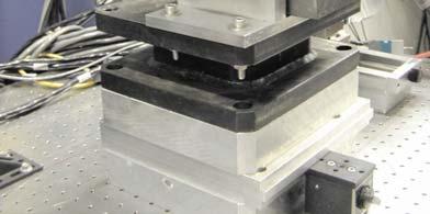
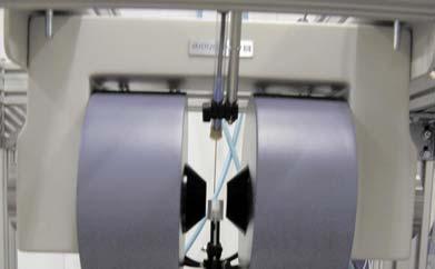
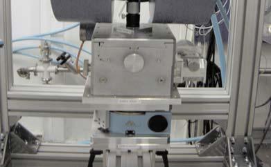
Drickamer type. Disk containing the specimen. Pressure cell. Press
 ε-fe Drickamer type Press Pressure cell Disk containing the specimen Low Temperature Cryostat Diamond Anvil Cell (DAC) Ruby manometry Re gasket for collimation Small size of specimen space High-density
Mossbauer Effect and Spectroscopy. Kishan Sinha Xu Group Department of Physics and Astronomy University of Nebraska-Lincoln
 Mossbauer Effect and Spectroscopy Kishan Sinha Xu Group Department of Physics and Astronomy University of Nebraska-Lincoln Emission E R γ-photon E transition hν = E transition - E R Photon does not carry
Gamma-ray decay. Introduction to Nuclear Science. Simon Fraser University Spring NUCS 342 March 7, 2011
 Gamma-ray decay Introduction to Nuclear Science Simon Fraser University Spring 2011 NUCS 342 March 7, 2011 NUCS 342 (Lecture 18) March 7, 2011 1 / 31 Outline 1 Mössbauer spectroscopy NUCS 342 (Lecture
Chapter 8 Magnetic Resonance
 Chapter 8 Magnetic Resonance 9.1 Electron paramagnetic resonance 9.2 Ferromagnetic resonance 9.3 Nuclear magnetic resonance 9.4 Other resonance methods TCD March 2007 1 A resonance experiment involves
RFSS: Lecture 6 Gamma Decay
 RFSS: Lecture 6 Gamma Decay Readings: Modern Nuclear Chemistry, Chap. 9; Nuclear and Radiochemistry, Chapter 3 Energetics Decay Types Transition Probabilities Internal Conversion Angular Correlations Moessbauer
3. Perturbed Angular Correlation Spectroscopy
 3. Perturbed Angular Correlation Spectroscopy Dileep Mampallil Augustine K.U.Leuven, Belgium Perturbed Angular Correlation Spectroscopy (PAC) is a gamma ray spectroscopy and can be used to investigate
Making the Invisible Visible: Probing Antiferromagnetic Order in Novel Materials
 Making the Invisible Visible: Probing Antiferromagnetic Order in Novel Materials Elke Arenholz Lawrence Berkeley National Laboratory Antiferromagnetic contrast in X-ray absorption Ni in NiO Neel Temperature
Determination of the hyperfine parameters of iron in aluminous (Mg,Fe)SiO 3 perovskite
SiO 3 perovskite") Determination of the hyperfine parameters of iron in aluminous (Mg,Fe)SiO 3 perovskite Jennifer M. Jackson Seismological Laboratory, Geological & Planetary Sciences California Institute of Technology VLab
Determination of the hyperfine parameters of iron in aluminous (Mg,Fe)SiO 3 perovskite Jennifer M. Jackson Seismological Laboratory, Geological & Planetary Sciences California Institute of Technology VLab
Disordered Materials: Glass physics
 Disordered Materials: Glass physics > 2.7. Introduction, liquids, glasses > 4.7. Scattering off disordered matter: static, elastic and dynamics structure factors > 9.7. Static structures: X-ray scattering,
Energy Spectroscopy. Ex.: Fe/MgO
 Energy Spectroscopy Spectroscopy gives access to the electronic properties (and thus chemistry, magnetism,..) of the investigated system with thickness dependence Ex.: Fe/MgO Fe O Mg Control of the oxidation
Nuclear Physics. (PHY-231) Dr C. M. Cormack. Nuclear Physics This Lecture
 Dr C. M. Cormack. Nuclear Physics This Lecture") Nuclear Physics (PHY-31) Dr C. M. Cormack 11 Nuclear Physics This Lecture This Lecture We will discuss an important effect in nuclear spectroscopy The Mössbauer Effect and its applications in technology
Nuclear Physics (PHY-31) Dr C. M. Cormack 11 Nuclear Physics This Lecture This Lecture We will discuss an important effect in nuclear spectroscopy The Mössbauer Effect and its applications in technology
X-Ray Magnetic Circular Dichroism: basic concepts and applications for 3d transition metals. Stefania PIZZINI Laboratoire Louis Néel CNRS- Grenoble
 X-Ray Magnetic Circular Dichroism: basic concepts and applications for 3d transition metals Stefania PIZZINI Laboratoire Louis Néel CNRS- Grenoble I) - Basic concepts of XAS and XMCD - XMCD at L 2,3 edges
Studying Metal to Insulator Transitions in Solids using Synchrotron Radiation-based Spectroscopies.
 PY482 Lecture. February 28 th, 2013 Studying Metal to Insulator Transitions in Solids using Synchrotron Radiation-based Spectroscopies. Kevin E. Smith Department of Physics Department of Chemistry Division
Soft X-ray Physics DELNOR-WIGGINS PASS STATE PARK
 Soft X-ray Physics Overview of research in Prof. Tonner s group Introduction to synchrotron radiation physics Photoemission spectroscopy: band-mapping and photoelectron diffraction Magnetic spectroscopy
PEEM and XPEEM: methodology and applications for dynamic processes
 PEEM and XPEEM: methodology and applications for dynamic processes PEEM methods and General considerations Chemical imaging Magnetic imaging XMCD/XMLD Examples Dynamic studies PEEM and XPEEM methods 1
APEX CARE INSTITUTE FOR PG - TRB, SLET AND NET IN PHYSICS
 Page 1 1. Within the nucleus, the charge distribution A) Is constant, but falls to zero sharply at the nuclear radius B) Increases linearly from the centre, but falls off exponentially at the surface C)
Probing Matter: Diffraction, Spectroscopy and Photoemission
 Probing Matter: Diffraction, Spectroscopy and Photoemission Anders Nilsson Stanford Synchrotron Radiation Laboratory Why X-rays? VUV? What can we hope to learn? 1 Photon Interaction Incident photon interacts
Hidden Interfaces and High-Temperature Magnetism in Intrinsic Topological Insulator - Ferromagnetic Insulator Heterostructures
 Hidden Interfaces and High-Temperature Magnetism in Intrinsic Topological Insulator - Ferromagnetic Insulator Heterostructures Valeria Lauter Quantum Condensed Matter Division, Oak Ridge National Laboratory,
Neutron Instruments I & II. Ken Andersen ESS Instruments Division
 Neutron Instruments I & II ESS Instruments Division Neutron Instruments I & II Overview of source characteristics Bragg s Law Elastic scattering: diffractometers Continuous sources Pulsed sources Inelastic
Supplementary Figure 1: Spin noise spectra of 55 Mn in bulk sample at BL =10.5 mt, before subtraction of the zero-frequency line. a, Contour plot of
 1 Supplementary Figure 1: Spin noise spectra of 55 Mn in bulk sample at BL =10.5 mt, before subtraction of the zero-frequency line. a, Contour plot of the spin noise spectra calculated with Eq. (2) for
Magnetic measurements (Pt. IV) advanced probes
 advanced probes") Magnetic measurements (Pt. IV) advanced probes Ruslan Prozorov October 2018 Physics 590B types of local probes microscopic (site-specific) NMR neutrons Mossbauer stationary Bitter decoration magneto-optics
Magnetic measurements (Pt. IV) advanced probes Ruslan Prozorov October 2018 Physics 590B types of local probes microscopic (site-specific) NMR neutrons Mossbauer stationary Bitter decoration magneto-optics
SUPPLEMENTARY INFORMATION
 SUPPLEMENTARY INFORMATION Conductance Measurements The conductance measurements were performed at the University of Aarhus. The Ag/Si surface was prepared using well-established procedures [1, 2]. After
X-Ray Scattering and Absorption by Magnetic Materials
 X-Ray Scattering and Absorption by Magnetic Materials S. W. Lovesey ISIS Facility, Rutherford Appleton Laboratory S. P. Collins Synchrotron Radiation Department, Daresbury Laboratory CLARENDON PRESS OXFORD
X-ray Magnetic Circular and Linear Dichroism (XMCD, XMLD) and X-ray Magnetic Imaging (PEEM,...)
 and X-ray Magnetic Imaging (PEEM,...)") X-ray Magnetic Circular and Linear Dichroism (XMCD, XMLD) and X-ray Magnetic Imaging (PEEM,...) Jan Vogel Institut Néel (CNRS, UJF), Nanoscience Department Grenoble, France - X-ray (Magnetic) Circular
X-ray Magnetic Circular and Linear Dichroism (XMCD, XMLD) and X-ray Magnetic Imaging (PEEM,...) Jan Vogel Institut Néel (CNRS, UJF), Nanoscience Department Grenoble, France - X-ray (Magnetic) Circular
arxiv:cond-mat/ v1 [cond-mat.str-el] 27 Oct 2003
![arxiv:cond-mat/ v1 [cond-mat.str-el] 27 Oct 2003](/thumbs/71/65811887.jpg "arxiv:cond-mat/ v1 [cond-mat.str-el] 27 Oct 2003") Magnetic versus crystal field linear dichroism in NiO thin films arxiv:cond-mat/0310634v1 [cond-mat.str-el] 27 Oct 2003 M. W. Haverkort, 1 S. I. Csiszar, 2 Z. Hu, 1 S. Altieri, 3 A. Tanaka, 4 H. H. Hsieh,
Magnetic versus crystal field linear dichroism in NiO thin films arxiv:cond-mat/0310634v1 [cond-mat.str-el] 27 Oct 2003 M. W. Haverkort, 1 S. I. Csiszar, 2 Z. Hu, 1 S. Altieri, 3 A. Tanaka, 4 H. H. Hsieh,
Neutron and x-ray spectroscopy
 Neutron and x-ray spectroscopy B. Keimer Max-Planck-Institute for Solid State Research outline 1. self-contained introduction neutron scattering and spectroscopy x-ray scattering and spectroscopy 2. application
Conclusion. 109m Ag isomer showed that there is no such broadening. Because one can hardly
 Conclusion This small book presents a description of the results of studies performed over many years by our research group, which, in the best period, included 15 physicists and laboratory assistants
Magnetic measurements (Pt. IV) advanced probes
 advanced probes") Magnetic measurements (Pt. IV) advanced probes Ruslan Prozorov 26 February 2014 Physics 590B types of local probes microscopic (site-specific) NMR neutrons Mossbauer stationary Bitter decoration magneto-optics
Magnetic measurements (Pt. IV) advanced probes Ruslan Prozorov 26 February 2014 Physics 590B types of local probes microscopic (site-specific) NMR neutrons Mossbauer stationary Bitter decoration magneto-optics
X-Ray Magnetic Dichroism. S. Turchini ISM-CNR
 X-Ray Magnetic Dichroism S. Turchini SM-CNR stefano.turchini@ism.cnr.it stefano.turchini@elettra.trieste.it Magnetism spin magnetic moment direct exchange: ferro antiferro superexchange 3d Ligand 2p 3d
Spins and spin-orbit coupling in semiconductors, metals, and nanostructures
 B. Halperin Spin lecture 1 Spins and spin-orbit coupling in semiconductors, metals, and nanostructures Behavior of non-equilibrium spin populations. Spin relaxation and spin transport. How does one produce
A facility for Femtosecond Soft X-Ray Imaging on the Nanoscale
 A facility for Femtosecond Soft X-Ray Imaging on the Nanoscale Jan Lüning Outline Scientific motivation: Random magnetization processes Technique: Lensless imaging by Fourier Transform holography Feasibility:
Spin Dynamics in Single GaAs Nanowires
 1 Dr. Max Mustermann Referat Kommunikation & Marketing Verwaltung Spin Dynamics in Single GaAs Nanowires F. Dirnberger, S. Furthmeier, M. Forsch, A. Bayer, J. Hubmann, B. Bauer, J. Zweck, E. Reiger, C.
Magnetic recording technology
 Magnetic recording technology The grain (particle) can be described as a single macrospin μ = Σ i μ i 1 0 1 0 1 W~500nm 1 bit = 300 grains All spins in the grain are ferromagnetically aligned B~50nm Exchange
Spectroscopy. Practical Handbook of. J. W. Robinson, Ph.D., D.Sc, F.R.C.S. Department of Chemistry Louisiana State University Baton Rouge, Louisiana
 Practical Handbook of Spectroscopy Edited by J. W. Robinson, Ph.D., D.Sc, F.R.C.S. Department of Chemistry Louisiana State University Baton Rouge, Louisiana CRC Press Boca Raton Ann Arbor Boston TABLE
An Introduction to Diffraction and Scattering. School of Chemistry The University of Sydney
 An Introduction to Diffraction and Scattering Brendan J. Kennedy School of Chemistry The University of Sydney 1) Strong forces 2) Weak forces Types of Forces 3) Electromagnetic forces 4) Gravity Types
Probing Magnetic Order with Neutron Scattering
 Probing Magnetic Order with Neutron Scattering G.J. Mankey, V.V. Krishnamurthy, F.D. Mackey and I. Zoto University of Alabama in collaboration with J.L. Robertson and M.L. Crow Oak Ridge National Laboratory
Photon Interaction. Spectroscopy
 Photon Interaction Incident photon interacts with electrons Core and Valence Cross Sections Photon is Adsorbed Elastic Scattered Inelastic Scattered Electron is Emitted Excitated Dexcitated Stöhr, NEXAPS
Reflection = EM strikes a boundary between two media differing in η and bounces back
 Reflection = EM strikes a boundary between two media differing in η and bounces back Incident ray θ 1 θ 2 Reflected ray Medium 1 (air) η = 1.00 Medium 2 (glass) η = 1.50 Specular reflection = situation
NMR: Formalism & Techniques
 NMR: Formalism & Techniques Vesna Mitrović, Brown University Boulder Summer School, 2008 Why NMR? - Local microscopic & bulk probe - Can be performed on relatively small samples (~1 mg +) & no contacts
Photoelectron spectroscopy Instrumentation. Nanomaterials characterization 2
 Photoelectron spectroscopy Instrumentation Nanomaterials characterization 2 RNDr. Věra V Vodičkov ková,, PhD. Photoelectron Spectroscopy general scheme Impact of X-ray emitted from source to the sample
The Island of of Inversion from a nuclear moments perspective
 The Island of of Inversion from a nuclear moments perspective Gerda Neyens Instituut voor Kern- en Stralingsfysica, K.U. Leuven, Belgium the LISE-NMR collaboration @ GANIL: E437 (,32,33 g-factors) E437a
Angular Correlation Experiments
 Angular Correlation Experiments John M. LoSecco April 2, 2007 Angular Correlation Experiments J. LoSecco Notre Dame du Lac Nuclear Spin In atoms one can use the Zeeman Effect to determine the spin state.
Compendium of concepts you should know to understand the Optical Pumping experiment. \ CFP Feb. 11, 2009, rev. Ap. 5, 2012, Jan. 1, 2013, Dec.28,2013.
 Compendium of concepts you should know to understand the Optical Pumping experiment. \ CFP Feb. 11, 2009, rev. Ap. 5, 2012, Jan. 1, 2013, Dec.28,2013. What follows is specialized to the alkali atoms, of
X-ray non-resonant and resonant magnetic scattering Laurent C. Chapon, Diamond Light Source. European School on Magnetism L. C.
 X-ray non-resonant and resonant magnetic scattering Laurent C. Chapon, Diamond Light Source 1 The Diamond synchrotron 3 GeV, 300 ma Lienard-Wiechert potentials n.b: Use S.I units throughout. rq : position
Name: (a) What core levels are responsible for the three photoelectron peaks in Fig. 1?
 What core levels are responsible for the three photoelectron peaks in Fig. 1?") Physics 243A--Surface Physics of Materials: Spectroscopy Final Examination December 16, 2014 (3 problems, 100 points total, open book, open notes and handouts) Name: [1] (50 points), including Figures
Physics 243A--Surface Physics of Materials: Spectroscopy Final Examination December 16, 2014 (3 problems, 100 points total, open book, open notes and handouts) Name: [1] (50 points), including Figures
In order to determine the energy level alignment of the interface between cobalt and
 SUPPLEMENTARY INFORMATION Energy level alignment of the CuPc/Co interface In order to determine the energy level alignment of the interface between cobalt and CuPc, we have performed one-photon photoemission
PHYSICS 359E: EXPERIMENT 2.2 THE MOSSBAUER EFFECT: RESONANT ABSORPTION OF (-RAYS
 PHYSICS 359E: EXPERIMENT 2.2 THE MOSSBAUER EFFECT: RESONANT ABSORPTION OF (-RAYS INTRODUCTION: In classical physics resonant phenomena are expected whenever a system can undergo free oscillations. These
Neutrino Helicity Measurement
 PHYS 851 Introductory Nuclear Physics Instructor: Chary Rangacharyulu University of Saskatchewan Neutrino Helicity Measurement Stefan A. Gärtner stefan.gaertner@gmx.de December 9 th, 2005 2 1 Introduction
Electronic Spectra of Complexes
 Electronic Spectra of Complexes Interpret electronic spectra of coordination compounds Correlate with bonding Orbital filling and electronic transitions Electron-electron repulsion Application of MO theory
Spin pumping in Ferromagnet-Topological Insulator-Ferromagnet Heterostructures Supplementary Information.
 Spin pumping in Ferromagnet-Topological Insulator-Ferromagnet Heterostructures Supplementary Information. A.A. Baker,, 2 A.I. Figueroa, 2 L.J. Collins-McIntyre, G. van der Laan, 2 and T., a) Hesjedal )
beta-nmr: from nuclear physics to biology
 beta-nmr: from nuclear physics to biology University of Copenhagen CERN KU Leuven M. Stachura, L. Hemmingsen D. Yordanov M. Bissell, G. Neyens Free University Berlin University of Saarland University of
(002)(110) (004)(220) (222) (112) (211) (202) (200) * * 2θ (degree)
(110) (004)(220) (222) (112) (211) (202) (200) * * 2θ (degree)") Supplementary Figures. (002)(110) Tetragonal I4/mcm Intensity (a.u) (004)(220) 10 (112) (211) (202) 20 Supplementary Figure 1. X-ray diffraction (XRD) pattern of the sample. The XRD characterization indicates
Supplementary Figures. (002)(110) Tetragonal I4/mcm Intensity (a.u) (004)(220) 10 (112) (211) (202) 20 Supplementary Figure 1. X-ray diffraction (XRD) pattern of the sample. The XRD characterization indicates
New materials for high- efficiency spin-polarized. polarized electron source
 New materials for high- efficiency spin-polarized polarized electron source A. Janotti Metals and Ceramics Division, Oak Ridge National Laboratory, TN In Collaboration with S.-H. Wei, National Renewable
Introduction to XAFS. Grant Bunker Associate Professor, Physics Illinois Institute of Technology. Revised 4/11/97
 Introduction to XAFS Grant Bunker Associate Professor, Physics Illinois Institute of Technology Revised 4/11/97 2 tutorial.nb Outline Overview of Tutorial 1: Overview of XAFS 2: Basic Experimental design
Influence of Size on the Properties of Materials
 Influence of Size on the Properties of Materials M. J. O Shea Kansas State University mjoshea@phys.ksu.edu If you cannot get the papers connected to this work, please e-mail me for a copy 1. General Introduction
Research with Synchrotron Radiation. Part I
 Research with Synchrotron Radiation Part I Ralf Röhlsberger Generation and properties of synchrotron radiation Radiation sources at DESY Synchrotron Radiation Sources at DESY DORIS III 38 beamlines XFEL
Lecture 0. NC State University
 Chemistry 736 Lecture 0 Overview NC State University Overview of Spectroscopy Electronic states and energies Transitions between states Absorption and emission Electronic spectroscopy Instrumentation Concepts
The Potential Use of X-ray FELs in Nuclear Studies
 1 The Potential Use of X-ray FELs in Nuclear Studies + + + + + + + + + Wen-Te Liao Max Planck Institute for Nuclear Physics Heidelberg, Germany 29 August 2013 @ FEL2013 R. L. M össbauer, Zeitschrift Physik
Synchrotron radiation: A charged particle constrained to move in curved path experiences a centripetal acceleration. Due to it, the particle radiates
 Synchrotron radiation: A charged particle constrained to move in curved path experiences a centripetal acceleration. Due to it, the particle radiates energy according to Maxwell equations. A non-relativistic
Calibration of the IXPE Instrument
 Calibration of the IXPE Instrument Fabio Muleri (INAF-IAPS) On behalf of the IXPE Italian Team 13th IACHEC Meeting 2018 Avigliano Umbro (Italy), 9-12 April 2018 IXPE MISSION IXPE will (re-)open the polarimetric
Electron spins in nonmagnetic semiconductors
 Electron spins in nonmagnetic semiconductors Yuichiro K. Kato Institute of Engineering Innovation, The University of Tokyo Physics of non-interacting spins Optical spin injection and detection Spin manipulation
X-Ray Spectro-Microscopy Joachim Stöhr Stanford Synchrotron Radiation Laboratory
 X-Ray Spectro-Microscopy Joachim Stöhr Stanford Synchrotron Radiation Laboratory X-Rays have come a long way Application to Magnetic Systems 1 µm 1895 1993 2003 http://www-ssrl.slac.stanford.edu/stohr/index.htm
X-ray Fabry-Pérot Interferometer
 X-ray Fabry-Pérot Interferometer Yu. V. Shvyd ko,m.lerche,h.-c. Wille,E.Gerdau,M.Lucht, and H. D. Rüter, E. E. Alp, and R. Khachatryan Microscopes, spectrometers, interferometers, and other high-resolution
DEPARTMENT OF PHYSICS UNIVERSITY OF PUNE PUNE SYLLABUS for the M.Phil. (Physics ) Course
 Course") DEPARTMENT OF PHYSICS UNIVERSITY OF PUNE PUNE - 411007 SYLLABUS for the M.Phil. (Physics ) Course Each Student will be required to do 3 courses, out of which two are common courses. The third course syllabus
DEPARTMENT OF PHYSICS UNIVERSITY OF PUNE PUNE - 411007 SYLLABUS for the M.Phil. (Physics ) Course Each Student will be required to do 3 courses, out of which two are common courses. The third course syllabus
Mossbauer Spectroscopy
 Mossbauer Spectroscopy Emily P. Wang MIT Department of Physics The ultra-high resolution ( E = E 10 12 ) method of Mossbauer spectroscopy was used to probe various nuclear effects. The Zeeman splittings
LASER-COMPTON SCATTERING AS A POTENTIAL BRIGHT X-RAY SOURCE
 Copyright(C)JCPDS-International Centre for Diffraction Data 2003, Advances in X-ray Analysis, Vol.46 74 ISSN 1097-0002 LASER-COMPTON SCATTERING AS A POTENTIAL BRIGHT X-RAY SOURCE K. Chouffani 1, D. Wells
Chapter 7. Nuclear Magnetic Resonance Spectroscopy
 Chapter 7 Nuclear Magnetic Resonance Spectroscopy I. Introduction 1924, W. Pauli proposed that certain atomic nuclei have spin and magnetic moment and exposure to magnetic field would lead to energy level
Energy Spectroscopy. Excitation by means of a probe
 Energy Spectroscopy Excitation by means of a probe Energy spectral analysis of the in coming particles -> XAS or Energy spectral analysis of the out coming particles Different probes are possible: Auger
Laser MEOP of 3 He: Basic Concepts, Current Achievements, and Challenging Prospects
 Polarization in Noble Gases, October 8-13, 2017 Laser MEOP of 3 He: Basic Concepts, Current Achievements, and Challenging Prospects Pierre-Jean Nacher Geneviève Tastevin Laboratoire Kastler-Brossel ENS
Opportunities with collinear laser spectroscopy at DESIR:
 Opportunities with collinear laser spectroscopy at DESIR: the LUMIERE facility GOALS of LUMIERE experiments: Gerda Neyens, K.U. Leuven, Belgium (1) measure ground state properties of exotic isotopes: (see
The Mössbauer Effect
 Experimental Physics V85.0112/G85.2075 The Mössbauer Effect Spring, 2005 Tycho Sleator, David Windt, and Burton Budick Goals The main goal of this experiment is to exploit the Mössbauer effect to measure
Chapter 37 Early Quantum Theory and Models of the Atom. Copyright 2009 Pearson Education, Inc.
 Chapter 37 Early Quantum Theory and Models of the Atom Planck s Quantum Hypothesis; Blackbody Radiation Photon Theory of Light and the Photoelectric Effect Energy, Mass, and Momentum of a Photon Compton
In this lecture, we will go through the hyperfine structure of atoms. The coupling of nuclear and electronic total angular momentum is explained.
 Lecture : Hyperfine Structure of Spectral Lines: Page- In this lecture, we will go through the hyperfine structure of atoms. Various origins of the hyperfine structure are discussed The coupling of nuclear
X-ray Spectroscopy. Interaction of X-rays with matter XANES and EXAFS XANES analysis Pre-edge analysis EXAFS analysis
 X-ray Spectroscopy Interaction of X-rays with matter XANES and EXAFS XANES analysis Pre-edge analysis EXAFS analysis Element specific Sensitive to low concentrations (0.01-0.1 %) Why XAS? Applicable under
is the minimum stopping potential for which the current between the plates reduces to zero.
 Module 1 :Quantum Mechanics Chapter 2 : Introduction to Quantum ideas Introduction to Quantum ideas We will now consider some experiments and their implications, which introduce us to quantum ideas. The
Simple strategy for enhancing terahertz emission from coherent longitudinal optical phonons using undoped GaAs/n-type GaAs epitaxial layer structures
 Presented at ISCS21 June 4, 21 Session # FrP3 Simple strategy for enhancing terahertz emission from coherent longitudinal optical phonons using undoped GaAs/n-type GaAs epitaxial layer structures Hideo
Core Level Spectroscopies
 Core Level Spectroscopies Spectroscopies involving core levels are element-sensitive, and that makes them very useful for understanding chemical bonding, as well as for the study of complex materials.
Basic science. Atomic structure. Electrons. The Rutherford-Bohr model of an atom. Electron shells. Types of Electrons. Describing an Atom
 Basic science A knowledge of basic physics is essential to understanding how radiation originates and behaves. This chapter works through what an atom is; what keeps it stable vs. radioactive and unstable;
DEVELOPMENT OF A NEW POSITRON LIFETIME SPECTROSCOPY TECHNIQUE FOR DEFECT CHARACTERIZATION IN THICK MATERIALS
 Copyright JCPDS - International Centre for Diffraction Data 2004, Advances in X-ray Analysis, Volume 47. 59 DEVELOPMENT OF A NEW POSITRON LIFETIME SPECTROSCOPY TECHNIQUE FOR DEFECT CHARACTERIZATION IN
Röntgenpraktikum. M. Oehzelt. (based on the diploma thesis of T. Haber [1])
![Röntgenpraktikum. M. Oehzelt. (based on the diploma thesis of T. Haber [1])](/thumbs/91/106501237.jpg "Röntgenpraktikum. M. Oehzelt. (based on the diploma thesis of T. Haber [1])") Röntgenpraktikum M. Oehzelt (based on the diploma thesis of T. Haber [1]) October 21, 2004 Contents 1 Fundamentals 2 1.1 X-Ray Radiation......................... 2 1.1.1 Bremsstrahlung......................
Röntgenpraktikum M. Oehzelt (based on the diploma thesis of T. Haber [1]) October 21, 2004 Contents 1 Fundamentals 2 1.1 X-Ray Radiation......................... 2 1.1.1 Bremsstrahlung......................
Xray Magnetic Circular Dichroism Investigation in Ferromagnetic Semiconductors. Khashayar Khazen Condensed Matter National Lab-IPM
 Xray Magnetic Circular Dichroism Investigation in Ferromagnetic Semiconductors Khashayar Khazen Condensed Matter National Lab-IPM IPM School of Physics School of Nano Condensed Matter National Lab Technology:
Mossbauer Effect. Ahmad Ali Ohyda. 1 Ahmad Faraj Abuaisha. 2 Abu-Bakr Mohammad Alrotob. 3 Basher M. Ismail. 4. Abstract
 Majalat Al-Ulum Al-Insaniya wat - Tatbiqiya Mossbauer Effect ( Determination of isomer shift, line width and quadruple splitting in a potassium ferricyanide ( K3Fe(CN)6) sample using Mossbauer spectroscopy)
Introduction to X-ray Photoelectron Spectroscopy (XPS) XPS which makes use of the photoelectric effect, was developed in the mid-1960
 XPS which makes use of the photoelectric effect, was developed in the mid-1960") Introduction to X-ray Photoelectron Spectroscopy (XPS) X-ray Photoelectron Spectroscopy (XPS), also known as Electron Spectroscopy for Chemical Analysis (ESCA) is a widely used technique to investigate
Introduction to X-ray Photoelectron Spectroscopy (XPS) X-ray Photoelectron Spectroscopy (XPS), also known as Electron Spectroscopy for Chemical Analysis (ESCA) is a widely used technique to investigate
Auger Electron Spectroscopy
 Auger Electron Spectroscopy Auger Electron Spectroscopy is an analytical technique that provides compositional information on the top few monolayers of material. Detect all elements above He Detection
Structure analysis: Electron diffraction LEED TEM RHEED
 Structure analysis: Electron diffraction LEED: Low Energy Electron Diffraction SPA-LEED: Spot Profile Analysis Low Energy Electron diffraction RHEED: Reflection High Energy Electron Diffraction TEM: Transmission
Lecture 2: Magnetic Anisotropy Energy (MAE)
") Lecture : Magnetic Anisotropy Energy (MAE) 1. Magnetic anisotropy energy = f(t). Anisotropic magnetic moment f(t) [111] T=3 K Characteristic energies of metallic ferromagnets M (G) 5 3 [1] 1 binding energy
Lecture : Magnetic Anisotropy Energy (MAE) 1. Magnetic anisotropy energy = f(t). Anisotropic magnetic moment f(t) [111] T=3 K Characteristic energies of metallic ferromagnets M (G) 5 3 [1] 1 binding energy
Electron Spectroscopy
 Electron Spectroscopy Photoelectron spectroscopy is based upon a single photon in/electron out process. The energy of a photon is given by the Einstein relation : E = h ν where h - Planck constant ( 6.62
Lecture 5. X-ray Photoemission Spectroscopy (XPS)
") Lecture 5 X-ray Photoemission Spectroscopy (XPS) 5. Photoemission Spectroscopy (XPS) 5. Principles 5.2 Interpretation 5.3 Instrumentation 5.4 XPS vs UV Photoelectron Spectroscopy (UPS) 5.5 Auger Electron
Lecture 5 X-ray Photoemission Spectroscopy (XPS) 5. Photoemission Spectroscopy (XPS) 5. Principles 5.2 Interpretation 5.3 Instrumentation 5.4 XPS vs UV Photoelectron Spectroscopy (UPS) 5.5 Auger Electron
Fundamentals of Nanoscale Film Analysis
 Fundamentals of Nanoscale Film Analysis Terry L. Alford Arizona State University Tempe, AZ, USA Leonard C. Feldman Vanderbilt University Nashville, TN, USA James W. Mayer Arizona State University Tempe,
Ferromagnetic Domain Distribution in Thin Films During Magnetization Reversal. and E. D. Dahlberg 3. Abstract
 Ferromagnetic Domain Distribution in Thin Films During Magnetization Reversal W.-T. Lee 1, S. G. E. te Velthuis 2, G. P. Felcher 2, F. Klose 1, T. Gredig 3, and E. D. Dahlberg 3. 1 Spallation Neutron Source,
Electromagnetic modulation of monochromatic neutrino beams
 Journal of Physics: Conference Series PAPER OPEN ACCESS Electromagnetic modulation of monochromatic neutrino beams To cite this article: A L Barabanov and O A Titov 2016 J. Phys.: Conf. Ser. 675 012009
Positron Annihilation Spectroscopy - A non-destructive method for material testing -
 Maik Butterling Institute of Radiation Physics http://www.hzdr.de Positron Annihilation Spectroscopy - A non-destructive method for material testing - Maik Butterling Positron Annihilation Spectroscopy
Skoog Chapter 6 Introduction to Spectrometric Methods
 Skoog Chapter 6 Introduction to Spectrometric Methods General Properties of Electromagnetic Radiation (EM) Wave Properties of EM Quantum Mechanical Properties of EM Quantitative Aspects of Spectrochemical
Fundamentals of Spectroscopy for Optical Remote Sensing. Course Outline 2009
 Fundamentals of Spectroscopy for Optical Remote Sensing Course Outline 2009 Part I. Fundamentals of Quantum Mechanics Chapter 1. Concepts of Quantum and Experimental Facts 1.1. Blackbody Radiation and
Secondary Ion Mass Spectrometry (SIMS)
") CHEM53200: Lecture 10 Secondary Ion Mass Spectrometry (SIMS) Major reference: Surface Analysis Edited by J. C. Vickerman (1997). 1 Primary particles may be: Secondary particles can be e s, neutral species
CHEM53200: Lecture 10 Secondary Ion Mass Spectrometry (SIMS) Major reference: Surface Analysis Edited by J. C. Vickerman (1997). 1 Primary particles may be: Secondary particles can be e s, neutral species
Instrumentelle Analytik in den Geowissenschaften (PI)
") 280061 VU MA-ERD-2 Instrumentelle Analytik in den Geowissenschaften (PI) Handoutmaterial zum Vorlesungsteil Spektroskopie Bei Fragen bitte zu kontaktieren: Prof. Lutz Nasdala, Institut für Mineralogie
280061 VU MA-ERD-2 Instrumentelle Analytik in den Geowissenschaften (PI) Handoutmaterial zum Vorlesungsteil Spektroskopie Bei Fragen bitte zu kontaktieren: Prof. Lutz Nasdala, Institut für Mineralogie
ATOMIC AND LASER SPECTROSCOPY
 ALAN CORNEY ATOMIC AND LASER SPECTROSCOPY CLARENDON PRESS OXFORD 1977 Contents 1. INTRODUCTION 1.1. Planck's radiation law. 1 1.2. The photoelectric effect 4 1.3. Early atomic spectroscopy 5 1.4. The postulates
Supplementary Information
 Supplementary Information I. Sample details In the set of experiments described in the main body, we study an InAs/GaAs QDM in which the QDs are separated by 3 nm of GaAs, 3 nm of Al 0.3 Ga 0.7 As, and
Holcomb Group Capabilities
 Holcomb Group Capabilities Synchrotron Radiation & Ultrafast Optics West Virginia University mikel.holcomb@mail.wvu.edu The Physicists New Playground The interface is the device. - Herbert Kroemer, beginning
Spin-resolved photoelectron spectroscopy
 Spin-resolved photoelectron spectroscopy Application Notes Spin-resolved photoelectron spectroscopy experiments were performed in an experimental station consisting of an analysis and a preparation chamber.