Electron Spectroscopy
|
|
|
- Mervyn McDaniel
- 5 years ago
- Views:
Transcription
1 Electron Spectroscopy
2
3 Photoelectron spectroscopy is based upon a single photon in/electron out process. The energy of a photon is given by the Einstein relation : E = h ν where h - Planck constant ( 6.62 x J s ) ν frequency (Hz) of the radiation
4 The process of photoionization can be considered in several ways : one way is to look at the overall process as follows : A + hν A + + e- Conservation of energy then requires that : E(A) + hν = E(A + ) + E(e-) Since the electron's energy is present solely as kinetic energy (KE) this can be rearranged to give the following expression for the KE of the photoelectron : KE = hν -( E(A + ) - E(A) ) The final term in brackets, representing the difference in e nergy between the ionized and neutral atoms, is generally called the binding energy (BE) of the electron - this then leads to the following commonly quoted equation : KE = hν BE
5 Photoelectron spectroscopy uses monochromatic sources of radiation (i.e. photons of fixed energy). In XPS the photon is absorbed by an atom in a molecule or solid, leading to ionization and the emission of a core (inner shell) electron. By contrast, in UPS the photon interacts with valence levels of the molecule or solid, leading to ionisation by removal of one of these valence electrons. The kinetic energy distribution of the emitted photoelectrons (i.e. the number of emitted photoelectrons as a function of their kinetic energy) can be measured using any appropriate electron energy analyser and a photoelectron spectrum can thus be recorded.
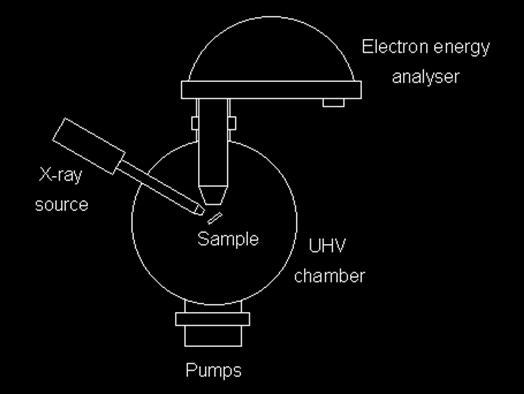
6 Experimental Details The basic requirements for a photoemission experiment (XPS or UPS) are: a source of fixed-energy radiation (an x-ray source for XPS or a He discharge lamp for UPS, or a tunable source such as synchrotron radiation for both) an electron energy analyser (which can disperse the emitted electrons according to their kinetic energy, and thereby measure the flux of emitted electrons of a particular energy) a high vacuum environment (to enable the emitted photoelectrons to be analysed without interference from gas phase collisions)
7
8
9
10
11
12
13 The application of photoemission as a technique to study the chemical and electronic structure of solids has its origins in the work of Kai Siegbahn and coworkers at Uppsala University, Sweden in the fifties. That group made dramatic improvements in the energy resolution and sensitivity of electron spectrometers, enabling a determination of the binding energies of electrons in a wide range of materials. As we ll see, from an analysis of the electronic binding energies it was possible to build up a chemical "fingerprint" of the solid. X-ray photoelectron spectroscopy (XPS) (X-ray photoemission) is thus also commonly referred to as Electron Spectroscopy for Chemical Analysis (ESCA). (Siegbahn s pioneering work earned him the Nobel prize in 1981).
14 The mean free path and surface sensitivity The photoelectrons that are detected in a photoemission experiment originate only from the uppermost layers of a solid. As we ll see, with the correct choice of experimental parameters photoemission can be used to probe just the first few monolayers at the surface of a solid. This surface sensitivity arises from the strong interaction of electrons with matter. An electron travelling through a solid will have a certain inelastic mean free path a characteristic length that it can travel without suffering an energy loss.
15 Thus, electrons ejected from a solid via the photoelectric effect will be of two types: Elastically scattered electrons which have escaped from the solid without suffering an energy loss Inelastically scattered electrons which have lost kinetic energy on their way out of the solid. An electron with energy in the ev range passing through a solid can lose energy via a number of processes.
16 Electron-electron scattering processes. In particular, the photoelectron can excite plasmons in the solid. A plasmon is a collective excitation of the electron gas whose energy is quantised in, typically, the 5 25 ev range. An electron can excite an interband transition, e.g. the excitation of a valence electron to a conduction band state or excite a core-electron to an empty state. The photoelectron on its way out of the solid may have sufficient energy to ionise a core-level with the creation of another photoelectron or an Auger electron
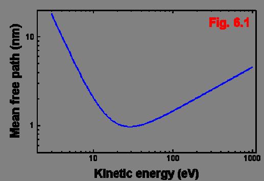
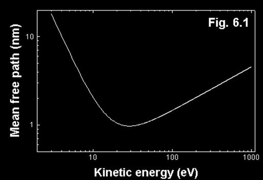
17 The net effect of these processes is that the (inelastic) mean free path of an electron in a solid is strongly dependent on its kinetic energy. At very low kinetic energies the electron simply does not have enough energy to excite the processes listed in 1-3 above so its mean free path is long. At high kinetic energies the electron spends less time passing through a given thickness of solid and thus is less likely to suffer an energy loss. Hence its mean free path in the solid is again quite long. However between these two regions the mean free path passes through a minimum
18
19 Electrons with kinetic energies in the ~ ev range have the shortest mean free paths and at the minimum of the "universal" curve the mean free path is ~ 1 nm. Even for quite high electron energies ( 1-2 kev) the mean free path is still only a few nm. This is a very significant result as regards the use of photoelectron spectroscopy as a surface sensitive probe. It means that even though the penetration depth of the incident X-rays is typically of the order of microns, the electrons that escape from the solid, due to their mean free path, will only have originated from the top few layers.
 so that the electrons are collected at a glancing angle, the electrons have to")

20 Surface sensitivity achieved by experimental geometry If we rotate the spectrometer (or sample) so that the electrons are collected at a glancing angle, the electrons have to traverse a longer distance in the solid (d cos θ ). The larger the angle, the greater the path length for the electrons and, thus, the higher the surface sensitivity of the photoemission measurement
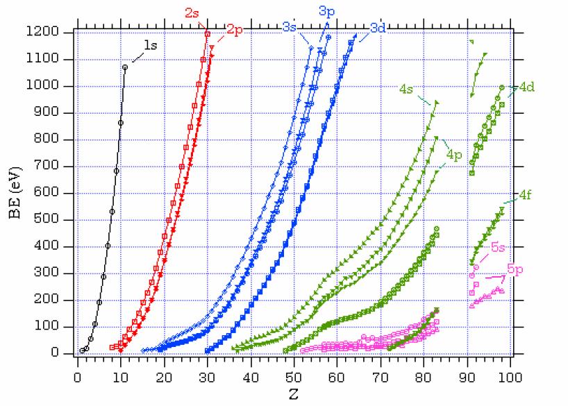
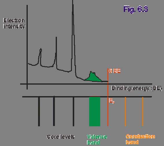
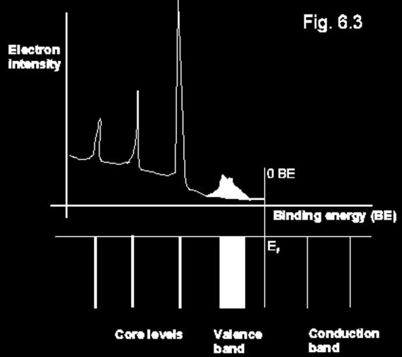
21 Electronic energy levels and the photoemission spectrum As mentioned above, in XPS we are concerned with the excitation of electrons lying in the tightly bound core-levels whereas UPS is used to probe the valence levels. The key equation underlying both processes is Einstein s equation: E KE = hν -E B where E KE is the kinetic energy of the electron ejected from the solid, h ν is the photon energy and E B is the binding energy of the electron in the solid. XPS may be used to provide a chemical "fingerprint" of a surface. This is because the binding energies of the electrons in the core-levels are representative of the atomic species. That is, an electron in a 1s level of oxygen has a particular binding energy which will differ from that of a Si 1s electron, a Ga 3d electron, a C 1s electron etc
22 Although the binding energy will vary depending on the chemical environment of the atom in the solid, the energy differences are generally small enough so that the presence of a particular element may be unequivocally identified from an XPS spectrum. Electrons are excited from filled states out of the solid with a particular kinetic energy. Measurement of the intensities and energies of the outgoing electrons with an electron spectrometer produces a photoemission spectrum that mirrors the distribution of filled levels in the solid.
23
24 Koopman s theorem If we assume that no rearrangement of the electrons either within the atom from which the photoelectron originated or in the neighbouring atoms of the material - occurs following the ejection of the photoelectron (an approximation known as Koopman s theorem) then the binding energy of the electron is simply the negative of the atomic orbital energy (-e k, where the subscript k labels the energy level from which the electron was removed).
25
26
27 Final state effects Koopman s theorem is a severe approximation. The ejection of a photoelectron creates a positively charged core-hole in the atom. Electrons in the vicinity of the positive charge will rearrange to screen it i.e. reduce its energy. The energy reduction is called the relaxation energy and can originate both from the electrons on the atom containing the core-hole (intra-atomic screening) and from those on surrounding atoms (interatomic screening). Relaxation/screening is thus a final state effect. The photoelectron can also interact with other electrons when departing the atom. For example, it may excite a valence electron to an unfilled (conduction band) state and lose an amount of kinetic energy equal to the excitation energy. This is called a shake-up process. Similarly, the departing photoelectron might transfer sufficient energy to the valence electron to remove it entirely from the atom: a shake-off process.
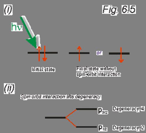
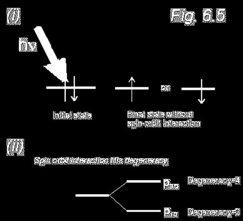
28 Spin-orbit (l-s) coupling A very important initial state effect for any orbital with orbital angular momentum> 0 is spin-orbit coupling/ splitting. This is a magnetic interaction between an electron s spin and its orbital angular momentum. We'll consider a p orbital (though spin/orbit coupling holds equally for d and f orbitals). After removal of an electron from the p orbital through photoemission, the remaining electron can adopt one of two configurations - a spin-up or spin-down state. With no spin-orbit interaction these states would have equal energy. However, spin-orbit coupling lifts the degeneracy and we need to consider the quantum number, j, the total angular momentum quantum number. The value of j is given by l + s where s is the spin quantum number (±½). For a p orbital j=1/2 or 3/2. Thus the final state of the system may be either p 1/2 or p 3/2 and this gives rise to a splitting of the core-level into a doublet.
29
30
31
32
33 Initial state effects: chemical shifts The valence electron distribution at the surface of a solid will differ from that of the bulk solid (sometimes weakly, as for certain metal surfaces, or dramatically, as for a large number of semiconductors Variations in the valence electron distribution will affect the potential a core-level electron feels. Therefore, the binding energy of a core-level electron in an atom at the surface will generally differ from that in a bulk atom. The precise binding energy of a core-level electron depends critically on its chemical environment. Both clean and adsorbate covered surfaces represent chemical environments that differ from that "seen" by an atom in the bulk. The change in the chemical environment produces a shift in the core-level. The magnitude of this chemical shift varies dramatically (from <0.1 ev to ~ 10 ev) from system to system.
34 Chemical shifts are generally interpreted in terms of the initial state of the system (i.e. before the photoelectron has been ejected). Charge transfer will either decrease or increase the charge density of an atom, leading to increased or decreased Coulombic attraction between the nucleus and the core electrons. Thus an atom that has donated a considerable amount of valence charge will produce an XPS peak at a higher binding energy than that of an atom in a lower oxidation state. As an example we can look at the oxidized and clean Si(100) surface. The Si 2p core-level (doublet) peak appears at ~ 100 ev binding energy. However, at approximately 4 ev above the Si 2p peak lies a broad peak due to the oxidised atoms at the Si surface. As we anneal the sample surface we can desorb this oxidised layer and produce a clean Si(100) surface.
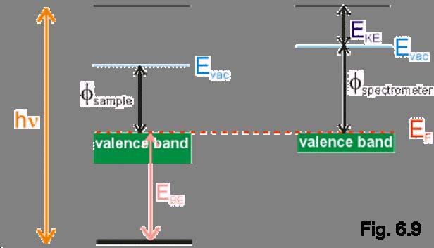
35 Binding energy referencing For this type of core-level analysis we must have accurate measurements of the core-level binding energies. The photoemission measurements are usually for conducting samples (metals or semiconductors). Both the spectrometer and the sample are electrically grounded and, hence their Fermi levels will align. The sample and spectrometer will have different work functions as shown. The binding energy of the photoelectron is referenced to the Fermi level (i.e. the zero of the binding energy scale is at the Fermi level). The binding energy of the photoelectron is given by: E B =hν -E KE - ϕ SP where ϕ SP is the spectrometer work function. Hence, it is the spectrometer and not the sample work function that must be accurately known. The spectrometer is generally calibrated using standard samples prior to the photoemission experiments and its work function determined.
36
37 Auger Electron Spectroscopy (AES) A second important electron spectroscopy used in surface science is Auger electron spectroscopy (AES). Auger electrons are named after their discoverer, Pierre Auger and arise from what is termed an autoionisation process. An electron is ejected from a core-level either by a photon (as in conventional photoemission) or by an incident high energy electron. The core-hole is filled by an electron from a higher energy level. The energy lost by that electron may be given up in the form of an X-ray photon or the quantum of energy is transferred (non-radiatively) to another electron in the atom. That electron (the Auger electron) is thus released from the atom.
38
39
40
41
42
43 The kinetic energy of the Auger electron, unlike that of a photoelectron, is not dependent on the energy of the incident radiation (or electron) that produced the initial core hole. Thus, Auger electrons have energies that are characteristic of the atom from which they arose and may be used for elemental identification. The notation associated with Auger transitions relies on the shell nomenclature. Note that if the valence levels are involved in the Auger process these are denoted by a V.
44
45
46 Generally Auger electron spectroscopy is carried out using an electron gun to produce relatively high energy electrons (in the 2 to 5 kev range) for initial core-level excitation. The Auger peaks are superimposed on the secondary electron background and are generally quite weak. Therefore, the Auger spectrum is usually electronically (sometimes numerically) differentiated to highlight the Auger peaks. In addition to chemical "fingerprinting" of a sample, a very common application of Auger spectroscopy is in the determination of growth modes. An analysis of the attenuation of a substrate Auger peak as a function of coverage enables a determination of whether the growth mode is of Frank-van der Merwe, Vollmer-Weber or Stranski-Krastanov character.
47
X-ray Photoelectron Spectroscopy (XPS)
") X-ray Photoelectron Spectroscopy (XPS) As part of the course Characterization of Catalysts and Surfaces Prof. Dr. Markus Ammann Paul Scherrer Institut markus.ammann@psi.ch Resource for further reading:
X-ray Photoelectron Spectroscopy (XPS) As part of the course Characterization of Catalysts and Surfaces Prof. Dr. Markus Ammann Paul Scherrer Institut markus.ammann@psi.ch Resource for further reading:
Lecture 5. X-ray Photoemission Spectroscopy (XPS)
") Lecture 5 X-ray Photoemission Spectroscopy (XPS) 5. Photoemission Spectroscopy (XPS) 5. Principles 5.2 Interpretation 5.3 Instrumentation 5.4 XPS vs UV Photoelectron Spectroscopy (UPS) 5.5 Auger Electron
Lecture 5 X-ray Photoemission Spectroscopy (XPS) 5. Photoemission Spectroscopy (XPS) 5. Principles 5.2 Interpretation 5.3 Instrumentation 5.4 XPS vs UV Photoelectron Spectroscopy (UPS) 5.5 Auger Electron
Birck Nanotechnology Center XPS: X-ray Photoelectron Spectroscopy ESCA: Electron Spectrometer for Chemical Analysis
 Birck Nanotechnology Center XPS: X-ray Photoelectron Spectroscopy ESCA: Electron Spectrometer for Chemical Analysis Dmitry Zemlyanov Birck Nanotechnology Center, Purdue University Outline Introduction
Introduction to X-ray Photoelectron Spectroscopy (XPS) XPS which makes use of the photoelectric effect, was developed in the mid-1960
 XPS which makes use of the photoelectric effect, was developed in the mid-1960") Introduction to X-ray Photoelectron Spectroscopy (XPS) X-ray Photoelectron Spectroscopy (XPS), also known as Electron Spectroscopy for Chemical Analysis (ESCA) is a widely used technique to investigate
Introduction to X-ray Photoelectron Spectroscopy (XPS) X-ray Photoelectron Spectroscopy (XPS), also known as Electron Spectroscopy for Chemical Analysis (ESCA) is a widely used technique to investigate
Advanced Lab Course. X-Ray Photoelectron Spectroscopy 1 INTRODUCTION 1 2 BASICS 1 3 EXPERIMENT Qualitative analysis Chemical Shifts 7
 Advanced Lab Course X-Ray Photoelectron Spectroscopy M210 As of: 2015-04-01 Aim: Chemical analysis of surfaces. Content 1 INTRODUCTION 1 2 BASICS 1 3 EXPERIMENT 3 3.1 Qualitative analysis 6 3.2 Chemical
X-Ray Photoelectron Spectroscopy (XPS)
") X-Ray Photoelectron Spectroscopy (XPS) Louis Scudiero http://www.wsu.edu/~scudiero; 5-2669 Electron Spectroscopy for Chemical Analysis (ESCA) The basic principle of the photoelectric effect was enunciated
X-Ray Photoelectron Spectroscopy (XPS) Louis Scudiero http://www.wsu.edu/~scudiero; 5-2669 Electron Spectroscopy for Chemical Analysis (ESCA) The basic principle of the photoelectric effect was enunciated
X-Ray Photoelectron Spectroscopy (XPS)-2
-2") X-Ray Photoelectron Spectroscopy (XPS)-2 Louis Scudiero http://www.wsu.edu/~scudiero; 5-2669 Fulmer 261A Electron Spectroscopy for Chemical Analysis (ESCA) The 3 step model: 1.Optical excitation 2.Transport
X-Ray Photoelectron Spectroscopy (XPS)-2 Louis Scudiero http://www.wsu.edu/~scudiero; 5-2669 Fulmer 261A Electron Spectroscopy for Chemical Analysis (ESCA) The 3 step model: 1.Optical excitation 2.Transport
X-Ray Photoelectron Spectroscopy (XPS)-2
-2") X-Ray Photoelectron Spectroscopy (XPS)-2 Louis Scudiero http://www.wsu.edu/~pchemlab ; 5-2669 Fulmer 261A Electron Spectroscopy for Chemical Analysis (ESCA) The 3 step model: 1.Optical excitation 2.Transport
X-Ray Photoelectron Spectroscopy (XPS)-2 Louis Scudiero http://www.wsu.edu/~pchemlab ; 5-2669 Fulmer 261A Electron Spectroscopy for Chemical Analysis (ESCA) The 3 step model: 1.Optical excitation 2.Transport
X-Ray Photoelectron Spectroscopy (XPS)
") X-Ray Photoelectron Spectroscopy (XPS) Louis Scudiero http://www.wsu.edu/~scudiero; 5-2669 Fulmer 261A Electron Spectroscopy for Chemical Analysis (ESCA) The basic principle of the photoelectric effect
X-Ray Photoelectron Spectroscopy (XPS) Louis Scudiero http://www.wsu.edu/~scudiero; 5-2669 Fulmer 261A Electron Spectroscopy for Chemical Analysis (ESCA) The basic principle of the photoelectric effect
IV. Surface analysis for chemical state, chemical composition
 IV. Surface analysis for chemical state, chemical composition Probe beam Detect XPS Photon (X-ray) Photoelectron(core level electron) UPS Photon (UV) Photoelectron(valence level electron) AES electron
5) Surface photoelectron spectroscopy. For MChem, Spring, Dr. Qiao Chen (room 3R506) University of Sussex.
 Surface photoelectron spectroscopy. For MChem, Spring, Dr. Qiao Chen (room 3R506) University of Sussex.") For MChem, Spring, 2009 5) Surface photoelectron spectroscopy Dr. Qiao Chen (room 3R506) http://www.sussex.ac.uk/users/qc25/ University of Sussex Today s topics 1. Element analysis with XPS Binding energy,
For MChem, Spring, 2009 5) Surface photoelectron spectroscopy Dr. Qiao Chen (room 3R506) http://www.sussex.ac.uk/users/qc25/ University of Sussex Today s topics 1. Element analysis with XPS Binding energy,
Energy Spectroscopy. Excitation by means of a probe
 Energy Spectroscopy Excitation by means of a probe Energy spectral analysis of the in coming particles -> XAS or Energy spectral analysis of the out coming particles Different probes are possible: Auger
Methods of surface analysis
 Methods of surface analysis Nanomaterials characterisation I RNDr. Věra Vodičková, PhD. Surface of solid matter: last monoatomic layer + absorbed monolayer physical properties are effected (crystal lattice
PHOTOELECTRON SPECTROSCOPY (PES)
") PHOTOELECTRON SPECTROSCOPY (PES) NTRODUCTON Law of Photoelectric effect Albert Einstein, Nobel Prize 1921 Kaiser-Wilhelm-nstitut (now Max-Planck- nstitut) für Physik Berlin, Germany High-resolution electron
PHOTOELECTRON SPECTROSCOPY (PES) NTRODUCTON Law of Photoelectric effect Albert Einstein, Nobel Prize 1921 Kaiser-Wilhelm-nstitut (now Max-Planck- nstitut) für Physik Berlin, Germany High-resolution electron
Energy Spectroscopy. Ex.: Fe/MgO
 Energy Spectroscopy Spectroscopy gives access to the electronic properties (and thus chemistry, magnetism,..) of the investigated system with thickness dependence Ex.: Fe/MgO Fe O Mg Control of the oxidation
Name: (a) What core levels are responsible for the three photoelectron peaks in Fig. 1?
 What core levels are responsible for the three photoelectron peaks in Fig. 1?") Physics 243A--Surface Physics of Materials: Spectroscopy Final Examination December 16, 2014 (3 problems, 100 points total, open book, open notes and handouts) Name: [1] (50 points), including Figures
Physics 243A--Surface Physics of Materials: Spectroscopy Final Examination December 16, 2014 (3 problems, 100 points total, open book, open notes and handouts) Name: [1] (50 points), including Figures
QUESTIONS AND ANSWERS
 QUESTIONS AND ANSWERS (1) For a ground - state neutral atom with 13 protons, describe (a) Which element this is (b) The quantum numbers, n, and l of the inner two core electrons (c) The stationary state
5.8 Auger Electron Spectroscopy (AES)
") 5.8 Auger Electron Spectroscopy (AES) 5.8.1 The Auger Process X-ray and high energy electron bombardment of atom can create core hole Core hole will eventually decay via either (i) photon emission (x-ray
5.8 Auger Electron Spectroscopy (AES) 5.8.1 The Auger Process X-ray and high energy electron bombardment of atom can create core hole Core hole will eventually decay via either (i) photon emission (x-ray
Ultraviolet Photoelectron Spectroscopy (UPS)
") Ultraviolet Photoelectron Spectroscopy (UPS) Louis Scudiero http://www.wsu.edu/~scudiero www.wsu.edu/~scudiero; ; 5-26695 scudiero@wsu.edu Photoemission from Valence Bands Photoelectron spectroscopy is
Ultraviolet Photoelectron Spectroscopy (UPS) Louis Scudiero http://www.wsu.edu/~scudiero www.wsu.edu/~scudiero; ; 5-26695 scudiero@wsu.edu Photoemission from Valence Bands Photoelectron spectroscopy is
Photon Interaction. Spectroscopy
 Photon Interaction Incident photon interacts with electrons Core and Valence Cross Sections Photon is Adsorbed Elastic Scattered Inelastic Scattered Electron is Emitted Excitated Dexcitated Stöhr, NEXAPS
X- ray Photoelectron Spectroscopy and its application in phase- switching device study
 X- ray Photoelectron Spectroscopy and its application in phase- switching device study Xinyuan Wang A53073806 I. Background X- ray photoelectron spectroscopy is of great importance in modern chemical and
Photoelectron Peak Intensities in Solids
 Photoelectron Peak Intensities in Solids Electronic structure of solids Photoelectron emission through solid Inelastic scattering Other excitations Intrinsic and extrinsic Shake-up, shake-down and shake-off
Photoelectron Spectroscopy. Xiaozhe Zhang 10/03/2014
 Photoelectron Spectroscopy Xiaozhe Zhang 10/03/2014 A conception last time remain Secondary electrons are electrons generated as ionization products. They are called 'secondary' because they are generated
X-Ray Photoelectron Spectroscopy (XPS) Prof. Paul K. Chu
 Prof. Paul K. Chu") X-Ray Photoelectron Spectroscopy (XPS) Prof. Paul K. Chu X-ray Photoelectron Spectroscopy Introduction Qualitative analysis Quantitative analysis Charging compensation Small area analysis and XPS imaging
X-Ray Photoelectron Spectroscopy (XPS) Prof. Paul K. Chu X-ray Photoelectron Spectroscopy Introduction Qualitative analysis Quantitative analysis Charging compensation Small area analysis and XPS imaging
Electron spectroscopy Lecture Kai M. Siegbahn ( ) Nobel Price 1981 High resolution Electron Spectroscopy
 Nobel Price 1981 High resolution Electron Spectroscopy") Electron spectroscopy Lecture 1-21 Kai M. Siegbahn (1918 - ) Nobel Price 1981 High resolution Electron Spectroscopy 653: Electron Spectroscopy urse structure cture 1. Introduction to electron spectroscopies
Electron spectroscopy Lecture 1-21 Kai M. Siegbahn (1918 - ) Nobel Price 1981 High resolution Electron Spectroscopy 653: Electron Spectroscopy urse structure cture 1. Introduction to electron spectroscopies
An Introduction to Diffraction and Scattering. School of Chemistry The University of Sydney
 An Introduction to Diffraction and Scattering Brendan J. Kennedy School of Chemistry The University of Sydney 1) Strong forces 2) Weak forces Types of Forces 3) Electromagnetic forces 4) Gravity Types
X-Ray Photoelectron Spectroscopy (XPS) Auger Electron Spectroscopy (AES)
 Auger Electron Spectroscopy (AES)") X-Ray Photoelectron Spectroscopy (XPS) Auger Electron Spectroscopy (AES) XPS X-ray photoelectron spectroscopy (XPS) is one of the most used techniques to chemically characterize the surface. Also known
X-Ray Photoelectron Spectroscopy (XPS) Auger Electron Spectroscopy (AES) XPS X-ray photoelectron spectroscopy (XPS) is one of the most used techniques to chemically characterize the surface. Also known
Emphasis on what happens to emitted particle (if no nuclear reaction and MEDIUM (i.e., atomic effects)
") LECTURE 5: INTERACTION OF RADIATION WITH MATTER All radiation is detected through its interaction with matter! INTRODUCTION: What happens when radiation passes through matter? Emphasis on what happens
LECTURE 5: INTERACTION OF RADIATION WITH MATTER All radiation is detected through its interaction with matter! INTRODUCTION: What happens when radiation passes through matter? Emphasis on what happens
Probing Matter: Diffraction, Spectroscopy and Photoemission
 Probing Matter: Diffraction, Spectroscopy and Photoemission Anders Nilsson Stanford Synchrotron Radiation Laboratory Why X-rays? VUV? What can we hope to learn? 1 Photon Interaction Incident photon interacts
Inelastic soft x-ray scattering, fluorescence and elastic radiation
 Inelastic soft x-ray scattering, fluorescence and elastic radiation What happens to the emission (or fluorescence) when the energy of the exciting photons changes? The emission spectra (can) change. One
Atomic Structure and Processes
 Chapter 5 Atomic Structure and Processes 5.1 Elementary atomic structure Bohr Orbits correspond to principal quantum number n. Hydrogen atom energy levels where the Rydberg energy is R y = m e ( e E n
XPS o ESCA UPS. Photoemission Spectroscopies. Threshold Spectroscopies (NEXAFS, APS etc ) The physics of photoemission.
 The physics of photoemission.") XPS o ESCA Photoemission Spectroscopies UPS Threshold Spectroscopies (NEXAFS, APS etc ) The physics of photoemission. How are photoemission spectra recorded: sources and analyzers Semi-quantitative analysis.
XPS o ESCA Photoemission Spectroscopies UPS Threshold Spectroscopies (NEXAFS, APS etc ) The physics of photoemission. How are photoemission spectra recorded: sources and analyzers Semi-quantitative analysis.
Low Energy Electrons and Surface Chemistry
 G. Ertl, J. Küppers Low Energy Electrons and Surface Chemistry VCH 1 Basic concepts 1 1.1 Introduction 1 1.2 Principles of ultrahigh vacuum techniques 2 1.2.1 Why is UHV necessary? 2 1.2.2 Production of
Core Level Spectroscopies
 Core Level Spectroscopies Spectroscopies involving core levels are element-sensitive, and that makes them very useful for understanding chemical bonding, as well as for the study of complex materials.
Lecture 23 X-Ray & UV Techniques
 Lecture 23 X-Ray & UV Techniques Schroder: Chapter 11.3 1/50 Announcements Homework 6/6: Will be online on later today. Due Wednesday June 6th at 10:00am. I will return it at the final exam (14 th June).
An introduction to X- ray photoelectron spectroscopy
 An introduction to X- ray photoelectron spectroscopy X-ray photoelectron spectroscopy belongs to a broad class of spectroscopic techniques, collectively called, electron spectroscopy. In general terms,
X-ray Photoemission Spectroscopy (XPS - Ma4)
") Master Laboratory Report X-ray Photoemission Spectroscopy (XPS - Ma4) Supervisor: Andrew Britton Students: Dachi Meurmishvili, Muhammad Khurram Riaz and Martin Borchert Date: November 17th 2016 1 Contents
Master Laboratory Report X-ray Photoemission Spectroscopy (XPS - Ma4) Supervisor: Andrew Britton Students: Dachi Meurmishvili, Muhammad Khurram Riaz and Martin Borchert Date: November 17th 2016 1 Contents
Electron Spettroscopies
 Electron Spettroscopies Spettroscopy allows to characterize a material from the point of view of: chemical composition, electronic states and magnetism, electronic, roto-vibrational and magnetic excitations.
Radiation Detection for the Beta- Delayed Alpha and Gamma Decay of 20 Na. Ellen Simmons
 Radiation Detection for the Beta- Delayed Alpha and Gamma Decay of 20 Na Ellen Simmons 1 Contents Introduction Review of the Types of Radiation Charged Particle Radiation Detection Review of Semiconductor
Physics of Radiotherapy. Lecture II: Interaction of Ionizing Radiation With Matter
 Physics of Radiotherapy Lecture II: Interaction of Ionizing Radiation With Matter Charge Particle Interaction Energetic charged particles interact with matter by electrical forces and lose kinetic energy
X-ray Energy Spectroscopy (XES).
.") X-ray Energy Spectroscopy (XES). X-ray fluorescence as an analytical tool for element analysis is based on 3 fundamental parameters: A. Specificity: In determining an x-ray emission energy E certainty
X-ray Energy Spectroscopy (XES). X-ray fluorescence as an analytical tool for element analysis is based on 3 fundamental parameters: A. Specificity: In determining an x-ray emission energy E certainty
Auger Electron Spectrometry. EMSE-515 F. Ernst
 Auger Electron Spectrometry EMSE-515 F. Ernst 1 Principle of AES electron or photon in, electron out radiation-less transition Auger electron electron energy properties of atom 2 Brief History of Auger
MS482 Materials Characterization ( 재료분석 ) Lecture Note 2: UPS
 Lecture Note 2: UPS") 2016 Fall Semester MS482 Materials Characterization ( 재료분석 ) Lecture Note 2: UPS Byungha Shin Dept. of MSE, KAIST 1 Course Information Syllabus 1. Overview of various characterization techniques (1 lecture)
2016 Fall Semester MS482 Materials Characterization ( 재료분석 ) Lecture Note 2: UPS Byungha Shin Dept. of MSE, KAIST 1 Course Information Syllabus 1. Overview of various characterization techniques (1 lecture)
Lecture 20 Auger Electron Spectroscopy
 Lecture 20 Auger Electron Spectroscopy Auger history cloud chamber Although Auger emission is intense, it was not used until 1950 s. Evolution of vacuum technology and the application of Auger Spectroscopy
ATOMIC STRUCTURE, ELECTRONS, AND PERIODICITY
 ATOMIC STRUCTURE, ELECTRONS, AND PERIODICITY All matter is made of atoms. There are a limited number of types of atoms; these are the elements. (EU 1.A) Development of Atomic Theory Atoms are so small
Interaction of Ionizing Radiation with Matter
 Type of radiation charged particles photonen neutronen Uncharged particles Charged particles electrons (β - ) He 2+ (α), H + (p) D + (d) Recoil nuclides Fission fragments Interaction of ionizing radiation
The Use of Synchrotron Radiation in Modern Research
 The Use of Synchrotron Radiation in Modern Research Physics Chemistry Structural Biology Materials Science Geochemical and Environmental Science Atoms, molecules, liquids, solids. Electronic and geometric
Ma5: Auger- and Electron Energy Loss Spectroscopy
 Ma5: Auger- and Electron Energy Loss Spectroscopy 1 Introduction Electron spectroscopies, namely Auger electron- and electron energy loss spectroscopy are utilized to determine the KLL spectrum and the
Stellar Astrophysics: The Interaction of Light and Matter
 Stellar Astrophysics: The Interaction of Light and Matter The Photoelectric Effect Methods of electron emission Thermionic emission: Application of heat allows electrons to gain enough energy to escape
Chapter Six: X-Rays. 6.1 Discovery of X-rays
 Chapter Six: X-Rays 6.1 Discovery of X-rays In late 1895, a German physicist, W. C. Roentgen was working with a cathode ray tube in his laboratory. He was working with tubes similar to our fluorescent
Photoemission Spectroscopy
 FY13 Experimental Physics - Auger Electron Spectroscopy Photoemission Spectroscopy Supervisor: Per Morgen SDU, Institute of Physics Campusvej 55 DK - 5250 Odense S Ulrik Robenhagen,
1 P a g e h t t p s : / / w w w. c i e n o t e s. c o m / Physics (A-level)
") 1 P a g e h t t p s : / / w w w. c i e n o t e s. c o m / Physics (A-level) Electromagnetic induction (Chapter 23): For a straight wire, the induced current or e.m.f. depends on: The magnitude of the magnetic
1 P a g e h t t p s : / / w w w. c i e n o t e s. c o m / Physics (A-level) Electromagnetic induction (Chapter 23): For a straight wire, the induced current or e.m.f. depends on: The magnitude of the magnetic
Appearance Potential Spectroscopy
 Appearance Potential Spectroscopy Submitted by Sajanlal P. R CY06D009 Sreeprasad T. S CY06D008 Dept. of Chemistry IIT MADRAS February 2006 1 Contents Page number 1. Introduction 3 2. Theory of APS 3 3.
ATOMIC STRUCTURE, ELECTRONS, AND PERIODICITY
 ATOMIC STRUCTURE, ELECTRONS, AND PERIODICITY All matter is made of atoms. There are a limited number of types of atoms; these are the elements. (EU 1.A) Development of Atomic Theory Atoms are so small
MSE 321 Structural Characterization
 Auger Spectroscopy Auger Electron Spectroscopy (AES) Scanning Auger Microscopy (SAM) Incident Electron Ejected Electron Auger Electron Initial State Intermediate State Final State Physical Electronics
Auger Electron Spectroscopy (AES)
") 1. Introduction Auger Electron Spectroscopy (AES) Silvia Natividad, Gabriel Gonzalez and Arena Holguin Auger Electron Spectroscopy (Auger spectroscopy or AES) was developed in the late 1960's, deriving
1. Introduction Auger Electron Spectroscopy (AES) Silvia Natividad, Gabriel Gonzalez and Arena Holguin Auger Electron Spectroscopy (Auger spectroscopy or AES) was developed in the late 1960's, deriving
Auger Electron Spectroscopy (AES) Prof. Paul K. Chu
 Prof. Paul K. Chu") Auger Electron Spectroscopy (AES) Prof. Paul K. Chu Auger Electron Spectroscopy Introduction Principles Instrumentation Qualitative analysis Quantitative analysis Depth profiling Mapping Examples The Auger
Auger Electron Spectroscopy (AES) Prof. Paul K. Chu Auger Electron Spectroscopy Introduction Principles Instrumentation Qualitative analysis Quantitative analysis Depth profiling Mapping Examples The Auger
Lecture 17 Auger Electron Spectroscopy
 Lecture 17 Auger Electron Spectroscopy Auger history cloud chamber Although Auger emission is intense, it was not used until 1950 s. Evolution of vacuum technology and the application of Auger Spectroscopy
Lecture 10. Transition probabilities and photoelectric cross sections
 Lecture 10 Transition probabilities and photoelectric cross sections TRANSITION PROBABILITIES AND PHOTOELECTRIC CROSS SECTIONS Cross section = = Transition probability per unit time of exciting a single
Bonds in molecules are formed by the interactions between electrons.
 CHEM 2060 Lecture 6: Electrostatic Interactions L6-1 PART TWO: Electrostatic Interactions In the first section of this course, we were more concerned with structural aspects of molecules. In this section
Interactions with Matter Photons, Electrons and Neutrons
 Interactions with Matter Photons, Electrons and Neutrons Ionizing Interactions Jason Matney, MS, PhD Interactions of Ionizing Radiation 1. Photon Interactions Indirectly Ionizing 2. Charge Particle Interactions
X-Ray transitions to low lying empty states
 X-Ray Spectra: - continuous part of the spectrum is due to decelerated electrons - the maximum frequency (minimum wavelength) of the photons generated is determined by the maximum kinetic energy of the
7. Electron spectroscopies
 7. Electron spectroscopies 7.1 Energy loss mechanisms - Incoming photons/electrons may excite electronic transitions in the substrate - Spectroscopic techniques focus on obtaining information on this,
4. How can fragmentation be useful in identifying compounds? Permits identification of branching not observed in soft ionization.
 Homework 9: Chapters 20-21 Assigned 12 April; Due 17 April 2006; Quiz on 19 April 2006 Chap. 20 (Molecular Mass Spectroscopy) Chap. 21 (Surface Analysis) 1. What are the types of ion sources in molecular
Photoelectron spectroscopy Instrumentation. Nanomaterials characterization 2
 Photoelectron spectroscopy Instrumentation Nanomaterials characterization 2 RNDr. Věra V Vodičkov ková,, PhD. Photoelectron Spectroscopy general scheme Impact of X-ray emitted from source to the sample
ICTP School on Synchrotron Radiation and Applications 2008 Surface Science, Photoemission and Related Techniques Fadley, Goldoni
 ICTP School on Synchrotron Radiation and Applications 2008 Surface Science, Photoemission and Related Techniques Fadley, Goldoni No. 1 Student background questions and study questions from the lectures.
8.6 Relaxation Processes
 CHAPTER 8. INNER SHELLS 175 Figure 8.17: Splitting of the 3s state in Fe which is missing in Zn. Refs. [12,13]. be aligned parallel or antiparallel with the spins of the 3d electrons of iron. 13 Thus we
MSE 321 Structural Characterization
 Auger Spectroscopy Auger Electron Spectroscopy (AES) Scanning Auger Microscopy (SAM) Incident Electron Ejected Electron Auger Electron Initial State Intermediate State Final State Physical Electronics
Photoelectron Spectroscopy Evidence for Electronic Structure Guided-Inquiry Learning Activity for AP* Chemistry
 Introduction Photoelectron Spectroscopy Evidence for Electronic Structure Guided-Inquiry Learning Activity for AP* Chemistry Catalog No. AP7710 Publication No. 7710AS The chemical properties of elements
Shell Atomic Model and Energy Levels
 Shell Atomic Model and Energy Levels (higher energy, deeper excitation) - Radio waves: Not absorbed and pass through tissue un-attenuated - Microwaves : Energies of Photos enough to cause molecular rotation
MT Electron microscopy Scanning electron microscopy and electron probe microanalysis
 MT-0.6026 Electron microscopy Scanning electron microscopy and electron probe microanalysis Eero Haimi Research Manager Outline 1. Introduction Basics of scanning electron microscopy (SEM) and electron
Lecture 12 Multiplet splitting
 Lecture 12 Multiplet splitting Multiplet splitting Atomic various L and S terms Both valence and core levels Rare earths Transition metals Paramagnetic free molecules Consider 3s level emission from Mn2+
THE NATURE OF THE ATOM. alpha particle source
 chapter THE NATURE OF THE ATOM www.tutor-homework.com (for tutoring, homework help, or help with online classes) Section 30.1 Rutherford Scattering and the Nuclear Atom 1. Which model of atomic structure
Lecture 7 Chemical/Electronic Structure of Glass
 Lecture 7 Chemical/Electronic Structure of Glass Syllabus Topic 6. Electronic spectroscopy studies of glass structure Fundamentals and Applications of X-ray Photoelectron Spectroscopy (XPS) a.k.a. Electron
Molecular Orbital Theory
 Molecular Orbital Theory 1. MO theory suggests that atomic orbitals of different atoms combine to create MOLECULAR ORBITALS 2. Electrons in these MOLECULAR ORBITALS belong to the molecule as whole 3. This
The Photoelectric Effect
 The Photoelectric Effect Light can strike the surface of some metals causing an electron to be ejected No matter how brightly the light shines, electrons are ejected only if the light has sufficient energy
Chapter 8: Electrons in Atoms Electromagnetic Radiation
 Chapter 8: Electrons in Atoms Electromagnetic Radiation Electromagnetic (EM) radiation is a form of energy transmission modeled as waves moving through space. (see below left) Electromagnetic Radiation
EEE4101F / EEE4103F Radiation Interactions & Detection
 EEE4101F / EEE4103F Radiation Interactions & Detection 1. Interaction of Radiation with Matter Dr. Steve Peterson 5.14 RW James Department of Physics University of Cape Town steve.peterson@uct.ac.za March
Explain how Planck resolved the ultraviolet catastrophe in blackbody radiation. Calculate energy of quanta using Planck s equation.
 Objectives Explain how Planck resolved the ultraviolet catastrophe in blackbody radiation. Calculate energy of quanta using Planck s equation. Solve problems involving maximum kinetic energy, work function,
THE EDUCARE (SIROHI CLASSES) TEST SERIES 2018
 TEST SERIES 2018") THE EDUCARE (SIROHI CLASSES) TEST SERIES 2018 XII PHYSICS TEST MODERN PHYSICS NAME-... DATE-.. MM- 25 TIME-1 HR 1) Write one equation representing nuclear fusion reaction. (1) 2) Arrange radioactive radiations
THE EDUCARE (SIROHI CLASSES) TEST SERIES 2018 XII PHYSICS TEST MODERN PHYSICS NAME-... DATE-.. MM- 25 TIME-1 HR 1) Write one equation representing nuclear fusion reaction. (1) 2) Arrange radioactive radiations
X-ray photoelectron spectroscopy - An introduction
 X-ray photoelectron spectroscopy - An introduction Spyros Diplas spyros.diplas@sintef.no spyros.diplas@smn.uio.no SINTEF Materials & Chemistry, Materials Physics -Oslo & Centre of Materials Science and
Film Characterization Tutorial G.J. Mankey, 01/23/04. Center for Materials for Information Technology an NSF Materials Science and Engineering Center
 Film Characterization Tutorial G.J. Mankey, 01/23/04 Theory vs. Experiment A theory is something nobody believes, except the person who made it. An experiment is something everybody believes, except the
Lecture 10. Transition probabilities and photoelectric cross sections
 Lecture 10 Transition probabilities and photoelectric cross sections TRANSITION PROBABILITIES AND PHOTOELECTRIC CROSS SECTIONS Cross section = σ = Transition probability per unit time of exciting a single
MSE 321 Structural Characterization
 r lim = 0 r e + e - mv 2/r e 2 /(4πε 0 r 2 ) KE } W = ½mv 2 - Electrons e =.6022x0-9 C ε 0 = 8.854x0-2 F/m m 0 = 9.094x0-3 kg PE } e 2 4πε 0 r (PE= F d ) e e W = - =( 2 2 -e 2 8πε 0 r 4πε 0 r ) mv 2 e
X-ray Photoelectron Spectroscopy/ Electron spectroscopy for chemical analysis (ESCA), By Francis Chindeka
, By Francis Chindeka") X-ray Photoelectron Spectroscopy/ Electron spectroscopy for chemical analysis (ESCA), By Francis Chindeka X-ray photoelectron spectroscopy (XPS) or Electron spectroscopy for chemical analysis (ESCA), Surface
X-ray Photoelectron Spectroscopy/ Electron spectroscopy for chemical analysis (ESCA), By Francis Chindeka X-ray photoelectron spectroscopy (XPS) or Electron spectroscopy for chemical analysis (ESCA), Surface
Particle nature of light & Quantization
 Particle nature of light & Quantization A quantity is quantized if its possible values are limited to a discrete set. An example from classical physics is the allowed frequencies of standing waves on a
X-ray Spectroscopy. Interaction of X-rays with matter XANES and EXAFS XANES analysis Pre-edge analysis EXAFS analysis
 X-ray Spectroscopy Interaction of X-rays with matter XANES and EXAFS XANES analysis Pre-edge analysis EXAFS analysis Element specific Sensitive to low concentrations (0.01-0.1 %) Why XAS? Applicable under
Physics 100 PIXE F06
 Introduction: Ion Target Interaction Elastic Atomic Collisions Very low energies, typically below a few kev Surface composition and structure Ion Scattering spectrometry (ISS) Inelastic Atomic Collisions
Electron Arrangement - Part 1
 Brad Collins Electron Arrangement - Part 1 Chapter 8 Some images Copyright The McGraw-Hill Companies, Inc. Properties of Waves Wavelength (λ) is the distance between identical points on successive waves.
Planck s Quantum Hypothesis Blackbody Radiation
 Planck s Quantum Hypothesis Blackbody Radiation The spectrum of blackbody radiation has been measured(next slide); it is found that the frequency of peak intensity increases linearly with temperature.
Fig Photoemission process.
 1.1 Photoemission process (Ref. 3.1, P. 43) When a sample surface is irradiated with photons of energy hυ, electrons are emitted from the sample surface. Figure 1.1.1 shows the essence of this photoemission
Chapter 38. Photons Light Waves Behaving as Particles
 Chapter 38 Photons Light Waves Behaving as Particles 38.1 The Photoelectric Effect The photoelectric effect was first discovered by Hertz in 1887, and was explained by Einstein in 1905. The photoelectric
Chapter V: Interactions of neutrons with matter
 Chapter V: Interactions of neutrons with matter 1 Content of the chapter Introduction Interaction processes Interaction cross sections Moderation and neutrons path For more details see «Physique des Réacteurs
Chapter VI: Ionizations and excitations
 Chapter VI: Ionizations and excitations 1 Content Introduction Ionization in gases Ionization in solids Fano factor 2 Introduction (1) Ionizations created by charged particles (incident particles or particles
The photoelectric effect
 The photoelectric effect E K hν-e B E F hν E B A photoemission experiment Lifetime broadening ΔE.Δτ~ħ ΔE~ħ/Δτ + Experimental resolution Hüfner, Photoelectron Spectroscopy (Springer) A photoemission experiment
Auger & X-ray Fluorescence
 At low energies or low temperature gas (plasma) the primary processes are photoionzation or excitation by particles (electron, atom, proton). Recombination takes place with emission of photons. In hot
Chemistry (
 Question 2.1: (i) Calculate the number of electrons which will together weigh one gram. (ii) Calculate the mass and charge of one mole of electrons. Answer 2.1: (i) Mass of one electron = 9.10939 10 31
Outline. Chapter 6 The Basic Interactions between Photons and Charged Particles with Matter. Photon interactions. Photoelectric effect
 Chapter 6 The Basic Interactions between Photons and Charged Particles with Matter Radiation Dosimetry I Text: H.E Johns and J.R. Cunningham, The physics of radiology, 4 th ed. http://www.utoledo.edu/med/depts/radther
4. Inelastic Scattering
 1 4. Inelastic Scattering Some inelastic scattering processes A vast range of inelastic scattering processes can occur during illumination of a specimen with a highenergy electron beam. In principle, many
KMÜ 396 MATERIALS SCIENCE AND TECH. I PRESENTATION ELECTRON ENERGY LOSS SPECTROSCOPY (EELS) TUĞÇE SEZGİN
 TUĞÇE SEZGİN") KMÜ 396 MATERIALS SCIENCE AND TECH. I PRESENTATION ELECTRON ENERGY LOSS SPECTROSCOPY (EELS) TUĞÇE SEZGİN 20970725 HACETTEPE UNIVERSITY DEPARTMENT OF CHEMICAL ENGINEERING, SPRING 2011,APRIL,ANKARA CONTENTS
KMÜ 396 MATERIALS SCIENCE AND TECH. I PRESENTATION ELECTRON ENERGY LOSS SPECTROSCOPY (EELS) TUĞÇE SEZGİN 20970725 HACETTEPE UNIVERSITY DEPARTMENT OF CHEMICAL ENGINEERING, SPRING 2011,APRIL,ANKARA CONTENTS