Ch 3 & 4 Microscopy & Cell Components 1
|
|
|
- Hannah Randall
- 6 years ago
- Views:
Transcription
1 Objectives 1.White book: Read Chap 3 & p & Black book: Read Chap 3 & p75-96 & 106 Objectives: 1. List metric measurement units for microorganisms and convert to other metric units (m, mm, um, nm). 2. Identify parts & functions of the compound light microscope. 3. Define/calculate total magnification & resolution. 4. Compare, contrast, and identify uses (diseases/organisms) for brightfield, darkfield, fluorescent, electron-transmission, and electronscanning microscopy. 5. Differentiate, compare, and explain the appearance and uses of each of the following: acidic & basic dyes, simple, differential & special stains, capsule, endospore, acid-fast and flagella stains. 2/4/2013 Ch 3 & 4 Microscopy & Cell Componenets 1 Objectives, cont d 6. List specific chemicals that are used for each type of stain in the objective above, primary stain, mordant, decolorizer, counterstain. 7. Gram stain: list the steps, purpose, and the appearance of GP & GN cells after each step. 8. Identify the 3 basic shapes of bacteria and secondary arrangements. 9. Describe the structure & function of the glycocalyx, flagella (including arrangement), axial filaments, fimbriae, pili. Identify flagellar arrangements. 10.Compare & contrast the cell walls of GP bacteria, GN bacteria, archaea, mycoplasmas, and mycobacteria. (Including composition, antibiotic & chemical resistance, presence of toxins, staining reactions, effect of penicillin, lysozyme, etc.) 2/4/2013 Ch 3 & 4 Microscopy & Cell Componenets 2 Objectives, Cont d 11. Identify the functions of the cell/plasma membrane, chromatophores/thylakoids, nucleoid, ribosomes, endospores (including location), inclusions. 12. Transport: passive (simple diffusion, osmosis, facilitated diffusion), active transport, hypertonic, hypotonic, isotonic, osmotic lysis, plasmolysis 13. Discuss several pieces of evidence that support the endosymbiotic theory of eukaryotic evolution. 14. Describe the overall structure and defining characteristics of prokaryotes, as compared to eukaryotes. 15. On given slides identify shape, gram reaction, arrangement, type of stain. Measurement Units & Terms 1. Units A. Micrometer (µm) = B. Nanometer (nm) = i. Example: Convert 21.5 nm to m 2. Total Magnification 3. Resolution: Distance apart needed to see (Ability to see ) 2/4/2013 Ch 3 & 4 Microscopy & Cell Componenets 3 2/4/2013 Ch 3 & 4 Microscopy & Cell Componenets 4 Ch 3 & 4 Microscopy & Cell Components 1


2 Resolution & Refractive Index A. Resolving power = N.A. depends on: i. of material between lens & slide. ii. The of most divergent light ray B. To improve resolution: i.. ii.. C. Improve conditions but NOT resolution: i.. ii.. Fig 3.3 Refraction w/ & w/o Oil, p.59 Using oil does improve resolution, as it increases the numerical aperture, which will cause a better (smaller) resolving power number 2/4/2013 Ch 3 & 4 Microscopy & Cell Componenets 5 2/4/2013 Ch 3 & 4 Microscopy & Cell Componenets 6 Types of Scopes-3 subtypes of Light microscopes Scope Enhanced by Advantages Uses Light, Brightfield: Background Visible light Res: Mag: Light, Darkfield: Background & microbes Same Light, Fluorescent: Background & microbes Same & light N/A Fluorescent- dyes: Fluorescent dye on to microbe, microbe fluoresces Inexpensive Easy to use Easier to see microbes directly from specimen, w/o culture Detection of microbes compared to other light microscopy Live specimens (unstained) Stained specimens Bacteria, protozoa Live microbes: When immediate diagnosis needed When cultures aren t avail, or take long 2/4/2013 Ch 3 & 4 Microscopy & Cell Componenets 7 Fig 3.6 Immunofluorescent Staining Technique Demo-Fluorescent marker drawings 2/4/2013 Ch 3 & 4 Microscopy & Cell Componenets 8 Ch 3 & 4 Microscopy & Cell Components 2

 formation 2/4/2013 Ch 3 & 4 Microscopy & Cell Componenets 10 Stains-Slide Prep & Basic Stains Slide Prep: 1. Smear 2.")
 ion A. & stains B. For cell, to detect C.")
3 Scopes-Electron Scope Enhanced by Advantages Uses Electron, Scanning Res; Mag; 3-D Book from U of I Surfaces structures - eukaryote to virus Fig 3.8 Transmission vs. Scanning Electron, Transmission Res Mag Scanned-Probe Res 1/100 of atom Stain w/+ salt of heavy metal res & mag DISADVANTAGE: Need slice as e- can t All e- scopes- due to killing, & fixing under vacuum Res No special prep Virus particles, bacterial flagella, cell structures, protein molecules Map atomic & molecular shapes & processes, ie. DNA, fibrin 2/4/2013 Ch 3 & 4 Microscopy & Cell Componenets 9 (clot) formation 2/4/2013 Ch 3 & 4 Microscopy & Cell Componenets 10 Stains-Slide Prep & Basic Stains Slide Prep: 1. Smear 2. Fix to slide (won t off) B.. C.. D. HOPEFULLY-preserves w/ Staining 1. Basic dye/ stain: Colored ( ) ion of a salt A. Attracted to ( ) bacterial cell; stains B. Crystal violet, methylene blue, safranin, malachite green Acidic Dye / Negative Stain 2. Acidic dye / stain: Colored ( ) ion A. & stains B. For cell, to detect C. Advantage: (no & stain so accurate size & shape) D. Examples: Acid fuchsin, nigrosin 2/4/2013 Ch 3 & 4 Microscopy & Cell Componenets 11 2/4/2013 Ch 3 & 4 Microscopy & Cell Componenets 12 Ch 3 & 4 Microscopy & Cell Components 3


 B. Acid-fast neg = C. ID species, 8. Capsule Stain (w/ stain) A.")
4 Mordant, Simple Stain, Differential Stain 3. Mordant: Substance used to cause more staining NOTE: This is not the stain that gives color, only helps the stain be more intense color 4. Simple stain: basic dye A. All microbes - B. Only for 5. Differential Stain: Use of to groups of bacteria A. Examples: gram stain, acid fast stain Gram Stain 6. Gram Stain: - due to differences A. GP = gram positive,, retain stain i. Us. to penicillin B. GN = gram negative, red, stain & accepts i. to penicillin C. Staining problems i. Need cultures ii. Some bacteria stain iii. timing is iv. Potential -structures/distortions that appear due to prep or staining procedures NOTE: this is potential problem w/all stains 2/4/2013 Ch 3 & 4 Microscopy & Cell Componenets 13 Most common stain in medical microbiology Know procedure-steps, purpose of each step/stain, appearance of cells after each step, how cell wall causes differential staining (Chap 4) 2/4/2013 Ch 3 & 4 Microscopy & Cell Componenets 14 Gram Stain Diagram Stains: Acid Fast & Capsule 7. Acid Fast Stain A. Acid-fast positive = (due to in cell ) B. Acid-fast neg = C. ID species, 8. Capsule Stain (w/ stain) A. Capsule = covering on outside of bacteria B. Variation w/2 stains: i.. Shapes above? GN or GP? Combine? GNR/GNB & GPC 2/4/2013 Ch 3 & 4 Microscopy & Cell Componenets 15 ii.. iii. of capsule left between the stains C. Problems: capsule may 2/4/2013 Ch 3 & 4 Microscopy & Cell Componenets 16 Ch 3 & 4 Microscopy & Cell Components 4


5 Pictures-Acid Fast & Capsule Stains Stains: Endospore 9. Endospore Stain A. Endospore i. Position used to ID species A. Uses to force dye into endospores 2/4/2013 Ch 3 & 4 Microscopy & Cell Componenets 17 2/4/2013 Ch 3 & 4 Microscopy & Cell Componenets 18 Stains; Endospore Pictures Stains; Flagella 10. Flagella Stain A. Flagella = B. used to ID bacteria 1. Discuss vegetative vs. endospores. Free vs. still in cell. 2. Which of the 2 pictures above has been subjected to adverse conditions longer? Explain. 2/4/2013 Ch 3 & 4 Microscopy & Cell Componenets 19 2/4/2013 Ch 3 & 4 Microscopy & Cell Componenets 20 Ch 3 & 4 Microscopy & Cell Components 5


6 Chapter 4: Prokaryotic Cells Prokaryote 1.. Fig 4.5a Prokaryotic Cell Bacteria cell wall 6. Archaea 2/4/2013 Ch 3 & 4 Microscopy & Cell Componenets 21 2/4/2013 Ch 3 & 4 Microscopy & Cell Componenets 22 Fig 6.11a Binary Fission Arrangement Size of bacteria um vs. resolution of light microscope? Arrangement Review: Shapes/Morphology? Arrangements? Other morphology terms: /4/2013 Ch 3 & 4 Microscopy & Cell Componenets 23 2/4/2013 Ch 3 & 4 Microscopy & Cell Componenets 24 Ch 3 & 4 Microscopy & Cell Components 6

 A. Contains 2/4/2013 Ch 3 & 4 Microscopy & Cell Componenets 27 3.")
7 Cell Wall - Bacteria Bacterial Cell Wall 1.. Diagram Cell Wall Diagrams Outside Cell Outside Cell 2. Clinical importance B Penicillin interferes Inside Cell Inside Cell 2/4/2013 Ch 3 & 4 Microscopy & Cell Componenets 25 2/4/2013 Ch 3 & 4 Microscopy & Cell Componenets 26 Table GP vs. GN Cell Wall Characteristics GP Wall GN Wall Contains 2. None Gram Stain & the Cell Wall Cell Wall & gram stain 1. Iodine = 2. Alcohol A. GP: 3. None 3. OUTER Wall Membrane A. Evades B. Contains C.. B. GN: C. GP falsely stain GN when cell wall damaged due to 4. None 4. Periplasm- (where peptidoglycan is) A. Contains 2/4/2013 Ch 3 & 4 Microscopy & Cell Componenets GPR/GPB only: A. : Bacillus & Clostridium B. : Mycobacterium (TB) 2/4/2013 Ch 3 & 4 Microscopy & Cell Componenets 28 Ch 3 & 4 Microscopy & Cell Components 7

8 Chemicals & the Cell Wall Chemical Effects on Cell Wall 1. Lysozyme: A. Most effective on 2. Penicillin Atypical Cell Walls Atypical Cell Walls 1. Mycoplasma species: A. High amount in plasma membrane, from lysis 2. Mycobacteria- High in wall B.. 3. Archea; 2/4/2013 Ch 3 & 4 Microscopy & Cell Componenets 29 2/4/2013 Ch 3 & 4 Microscopy & Cell Componenets 30 Structures External to Cell Wall External Structures 1. Glycocalyx/Capsule: A. EPS (Extracellular polysaccharide) & polypeptide polymer B.. External Filamentous Structures 2. Table: Flagella Axial Filaments Fimbrae Pili C. Negative Stain, but uses 2 dyes i. Basic stains ii. Acidic stains iii. Monotrichous - Spiralled around cell within (AKA endoflagella) 2/4/2013 Ch 3 & 4 Microscopy & Cell Componenets 31 Amphitrichous- Lophotrichous- Peritrichous- 2/4/2013 Ch 3 & 4 Microscopy & Cell Componenets 32 Ch 3 & 4 Microscopy & Cell Components 8


9 Diagram-Axial Filament Photo-Fimbriae 2/4/2013 Ch 3 & 4 Microscopy & Cell Componenets 33 2/4/2013 Ch 3 & 4 Microscopy & Cell Componenets 34 Fig 8.26 Bacterial Conjugation Fig 8.27 Conjugation in E. coli 2/4/2013 Ch 3 & 4 Microscopy & Cell Componenets 35 2/4/2013 Ch 3 & 4 Microscopy & Cell Componenets 36 Ch 3 & 4 Microscopy & Cell Components 9


10 External Filamentous Structures, Cont d 3. NO 4. Taxis: A. Chemotaxis B. Phototaxis Discuss serovars Endospores Structure Internal to Cell Wall 1. Endospores: structures to adverse conditions B. Sporulation / Sporogenesis C. Germination return to state D.. E. Location: F. Survive G. Stains: i. Gram- ii. Endospore Stain: Primary: basic stain Rinse: removes stain from Counterstain: basic stain colors 2/4/2013 Ch 3 & 4 Microscopy & Cell Componenets 37 2/4/2013 Ch 3 & 4 Microscopy & Cell Componenets 38 Fig 4.20a Endospore Formation Endospore Stain Pictures 2/4/2013 Ch 3 & 4 Microscopy & Cell Componenets 39 2/4/2013 Ch 3 & 4 Microscopy & Cell Componenets 40 Ch 3 & 4 Microscopy & Cell Components 10


11 Plasma/Cytoplasmic Membrane 2. Plasma Membrane B.. C. Special: Alcohols, disinfectants, some antibiotics effective here Fig 2.11 Phospholipids 2/4/2013 Ch 3 & 4 Microscopy & Cell Componenets 41 2/4/2013 Ch 3 & 4 Microscopy & Cell Componenets 42 Fig 4.16 Diffusion D. Diffusion: i. Simple diffusion ii. Facilitated diffusion iii. Osmosis E. Active Transport: Diagram on the right: Which type of transport does it represent? Osmosis-Animal vs. Plant Special terms reflect % solute, and therefore affect net direction of osmosis. What do the following prefixes mean? Iso? Hypo? Hyper? Suffix is tonic = tension 2/4/2013 Ch 3 & 4 Microscopy & Cell Componenets 43 2/4/2013 Ch 3 & 4 Microscopy & Cell Componenets 44 Ch 3 & 4 Microscopy & Cell Components 11
 Normal (2) Lysed (3) Shriveled H 2O H Plasma H 2O 2O membrane H 2O iii.")
 Flaccid (5) Turgid (6) Shriveled (plasmolyzed) 2/4/2013 Ch 3 & 4 Microscopy & Cell Componenets 45 2/4/2013 Ch 3 & 4 Microscopy & Cell Componenets 46 Fig 4.")
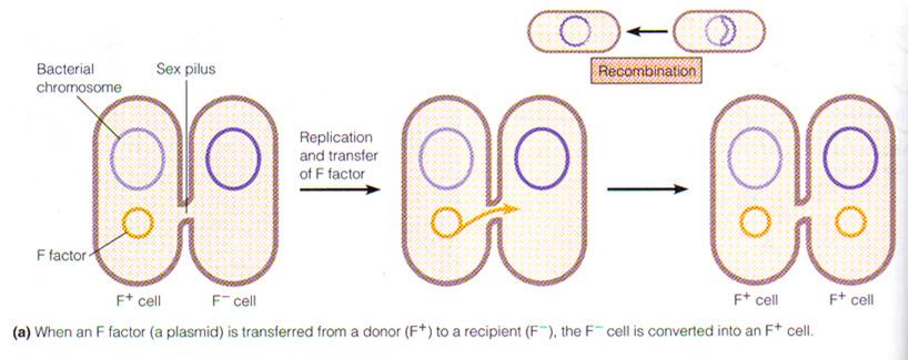
12 Osmosis & Solution Types F. Osmotic Environments i. Isotonic/isoosmotic solution:».» Water movement». ii. Hypotonic solution:» Net H 2 O moves». Diagram from Bio-Osmosis & Plant vs. Animal Isotonic solution Hypotonic solution Hypertonic solution H 2O H 2O H 2O H 2O Animal cell (1) Normal (2) Lysed (3) Shriveled H 2O H Plasma H 2O 2O membrane H 2O iii. Hypertonic solution:» Net H 2 O movement». Plant cell (4) Flaccid (5) Turgid (6) Shriveled (plasmolyzed) 2/4/2013 Ch 3 & 4 Microscopy & Cell Componenets 45 2/4/2013 Ch 3 & 4 Microscopy & Cell Componenets 46 Fig 4.17 cde Osmosis in Varying Osmotic Environments Internal Cell Structures continued 3. Chromatophores/thylakoids: structures 4. Nucleoid/nuclear area: No nuclear membrane A. Contains 5. Plasmids: B. Conjugation: transfer through i. GN- ii. GP- C. Biotech: 2/4/2013 Ch 3 & 4 Microscopy & Cell Componenets 47 2/4/2013 Ch 3 & 4 Microscopy & Cell Componenets 48 Ch 3 & 4 Microscopy & Cell Components 12


13 Fig 8.27a Conjugation-Plasmid Transfer Internal Structures cont d-ribosomes 6. Ribosomes: A. 2 subunits: protein & RNA i. Prokaryotic size: ii. Euk: B. attach to subunit of size 2/4/2013 Ch 3 & 4 Microscopy & Cell Componenets 49 2/4/2013 Ch 3 & 4 Microscopy & Cell Componenets 50 Internal Structures cont d-inclusions 7. Inclusions: B.. Review Structures: Eukaryotes Eukaryotes If cell wall A. Algae: B. Fungi: 2/4/2013 Ch 3 & 4 Microscopy & Cell Componenets 51 2/4/2013 Ch 3 & 4 Microscopy & Cell Componenets 52 Ch 3 & 4 Microscopy & Cell Components 13


14 Endosymbiotic Theory Endosymbiotic Theory 1. Eukaryotes evolved from living inside Table 10.2 Prokaryotic Cells vs. Eukaryotic Organelles 2. Evidence B.. C.. i.. ii.. iii.. 2/4/2013 Ch 3 & 4 Microscopy & Cell Componenets 53 2/4/2013 Ch 3 & 4 Microscopy & Cell Componenets 54 Fig 10.2 Endosymbiotic Theory 2/4/2013 Ch 3 & 4 Microscopy & Cell Componenets 55 Ch 3 & 4 Microscopy & Cell Components 14
Ch 3 & 4 Microscopy & Cell Components 1
 Objectives 1.White book: Read Chap 3 & p 77-98 & 108 2.Black book: Read Chap 3 & p75-96 & 106 Objectives: 1. List metric measurement units for microorganisms and convert to other metric units (m, mm, um,
Ch 3 & 4 Microscopy & Cell Components 1
 Objectives 1.White book: Read Chap 3 & p 77-98 & 108 2.Black book: Read Chap 3 & p75-96 & 106 Objectives: 1. List metric measurement units for microorganisms and convert to other metric units (m, mm, um,
Chap 3 & 4 Microscopy & Cell Components
 Chap 3 & 4 Microscopy & Cell Components Prior Knowledge: Parts of the scope at the right? Type of scope? Proper care and use? Other types of scopes? What are the 3 shapes of bacteria? Preview of smears
chapter one: the history of microbiology
 chapter one: the history of microbiology Revised 6/19/2018 microbes microscopic (small) organisms, viruses, prions prefix sci. notation frac. equivalent dec. equivalent kilo- (k) 1 10 3 1000/1 = 1000 1000
MONTGOMERY COUNTY COMMUNITY COLLEGE BIO 140 CHAPTER 4. Functional Anatomy of Prokaryotic and Eukaryotic Cells
 MONTGOMERY COUNTY COMMUNITY COLLEGE BIO 140 CHAPTER 4 Functional Anatomy of Prokaryotic and Eukaryotic Cells I. PROKARYOTES A. Structure Of The Cell: Chemical Composition And Function 1. Cell Wall a. composition
Ch 2 Part 2. The Microscope
 Ch 2 Part 2 The Microscope SLOs for Microscopic Analysis of Microorganisms Convert among the different units of the metric system. List and describe three elements of good microscopy. Differentiate between
Chapter 3. Observing Organisms Through a Microscope
 Chapter 3 Observing Organisms Through a Microscope Units of Measurement Used n Microbiology Table 3.1 mm Figure 3.2 Figure 3.1 - Overview Compound Light microscopy Have ocular and objective lenses Calculate
Ch 3. Bacteria and Archaea
 Ch 3 Bacteria and Archaea SLOs for Culturing of Microorganisms Compare and contrast the overall cell structure of prokaryotes and eukaryotes. List structures all bacteria possess. Describe three basic
Prokaryotic and Eukaryotic Cells. Structure and Function
 Prokaryotic and Eukaryotic Cells Structure and Function In general microbes or microorganisms may be either prokaryotic (bacteria) or eukaryotic (protists, fungi, and some animals). However, there are
Subject: Staining-Bacterial Cell Structure Lecture Number: 3 Done by: Joud Baki Corrected by: Issa Deir
 Subject: Staining-Bacterial Cell Structure Lecture Number: 3 Done by: Joud Baki Corrected by: Issa Deir 0 Principles of staining: - Revision: Stains can be either simple or differential Gram stains are
Chapter 03 Microscopy and Cell Structure
 Chapter 03 Microscopy and Cell Structure Multiple Choice Questions 1. Eukaryotic cells are A. less complex than prokaryotic cells. B. members of the Domains Bacteria and Archaea. C. defined by the presence
TEST BANK FOR PRESCOTTS MICROBIOLOGY 9TH EDITION BY WILLEY SHERWOOD WOOLVERTON
 TEST BANK FOR PRESCOTTS MICROBIOLOGY 9TH EDITION BY WILLEY SHERWOOD WOOLVERTON Link download full: https://testbankservice.com/download/test-bank-for-prescottsmicrobiology-9th-edition-by-willey-sherwood-woolverton/
Microscopy, Staining, and Classification
 PowerPoint Lecture Presentations prepared by Mindy Miller-Kittrell, North Carolina State University C H A P T E R 4 Microscopy, Staining, and Classification 4. Discuss how microscopy has revealed the structure
Bacterial Morphology and Structure م.م رنا مشعل
 Bacterial Morphology and Structure م.م رنا مشعل SIZE OF BACTERIA Unit for measurement : Micron or micrometer, μm: 1μm=10-3 mm Size: Varies with kinds of bacteria, and also related to their age and external
WHY IS THIS IMPORTANT?
 CHAPTER 9 THE CLINICAL SIGNIFICANCE OF BACTERIAL ANATOMY WHY IS THIS IMPORTANT? Bacterial structures play a significant role in the five steps required for infection OVERVIEW The Clinical Signifcance of
Microbial Genetics, Mutation and Repair. 2. State the function of Rec A proteins in homologous genetic recombination.
 Answer the following questions 1. Define genetic recombination. Microbial Genetics, Mutation and Repair 2. State the function of Rec A proteins in homologous genetic recombination. 3. List 3 types of bacterial
9/8/2010. Chapter 4. Structures Internal to the Cell Wall. The Plasma Membrane. Functional Anatomy of Prokaryotic and Eukaryotic Cells
 Chapter 4 Functional Anatomy of Prokaryotic and Eukaryotic Cells Johana Meléndez Part II slides 39-87 Lectures prepared by Christine L. Case Structures Internal to the Cell Wall Learning Objectives 4-8
By signing below, you acknowledge that you have ensured that you are complying with the above statement.
 Instructions: This exam consists of 31 multiple choice questions on 8 pages, including this one. Please submit your answers on the scantron sheet provided and on this copy of the exam. This exam is closed
LABORATORY 7 ENDOSPORE STAIN AND BACTERIAL MOTILITY
 LABORATORY 7 ENDOSPORE STAIN AND BACTERIAL MOTILITY A. Endospore Stain B. Bacterial Motility A. ENDOSPORE STAIN DISCUSSION A few genera of bacteria, such as Bacillus and Clostridium have the ability to
MORPHOLOGY: the study of form and structure
 MICROBIOLOGY CHAPTER 3 Bacteria Morphology 3:1 Bacteria Structure and Function MORPHOLOGY: the study of form and structure Structure of Bacteria 1. PROKARYOTIC no membrane bound nucleus nor other organelles
NAME: Microbiology BI234 MUST be written and will not be accepted as a typed document. 1.
 Chapter 3 Study Guide Explain the 3 main characteristics that help differentiate prokaryotes from eukaryotes. What are the 7 structures/substances found in all bacterial cells? What are 8 specific structures
Burton's Microbiology for the Health Sciences
 Burton's Microbiology for the Health Sciences Chapter 3. Cell Structure and Taxonomy Chapter 3 Outline Introduction Eucaryotic Cell Structure Procaryotic Cell Structure Summary of Structural Differences
Cell Structure and Function
 PowerPoint Lecture Presentations prepared by Mindy Miller-Kittrell, North Carolina State University C H A P T E R 3 Cell Structure and Function Handout Structure-Function Table Handout Prok vs Euk Table
BACTERIA AND ARCHAEA 10/15/2012
 BACTERIA AND ARCHAEA Chapter 27 KEY CONCEPTS: Structural and functional adaptations contribute to prokaryotic success Rapid reproduction, mutation, and genetic recombination promote genetic diversity in
Shape, Arrangement, and Size. Cocci (s., coccus) bacillus (pl., bacilli) 9/21/2013
 bacillus (pl., bacilli) 9/21/2013") Shape, Arrangement, and Size Cocci (s., coccus) are roughly spherical cells. The other common shape is that of a rod, sometimes called a bacillus (pl., bacilli). Spiral-shaped procaryotes can be either
Shape, Arrangement, and Size Cocci (s., coccus) are roughly spherical cells. The other common shape is that of a rod, sometimes called a bacillus (pl., bacilli). Spiral-shaped procaryotes can be either
INTRODUCTION. Gram Stain
 INTRODUCTION In microbiology, organisms are so small that additional techniques are often required for proper viewing under the microscope. Cytological stains, or dyes that stain cells or cellular features,
= Monera. Taxonomy. Domains (3) BIO162 Page Baluch. Taxonomy: classifying and organizing life
 BIO162 Page Baluch. Taxonomy: classifying and organizing life") Taxonomy BIO162 Page Baluch Taxonomy: classifying and organizing life species Genus Family Order Class Phylum Kingdom Spaghetti Good For Over Came Phillip King Domains (3) DOMAINS 1. Bacteria 2. Archea
Taxonomy BIO162 Page Baluch Taxonomy: classifying and organizing life species Genus Family Order Class Phylum Kingdom Spaghetti Good For Over Came Phillip King Domains (3) DOMAINS 1. Bacteria 2. Archea
TER 26. Preview for 2/6/02 Dr. Kopeny. Bacteria and Archaea: The Prokaryotic Domains. Nitrogen cycle
 Preview for 2/6/02 Dr. Kopeny Bacteria and Archaea: The Prokaryotic Domains TER 26 Nitrogen cycle Mycobacterium tuberculosis Color-enhanced images shows rod-shaped bacterium responsible for tuberculosis
Cells Under the Microscope Measuring Cell Structures
 Copy into Note Packet and Return to Teacher Chapter 3 Cell Structure Section 1: Looking at Cells Objectives Describe how scientists measure the length of objects. Relate magnification and resolution in
Cell Structure and Function. Handout Prok vs Euk Table Handout Structure-Function Table. Prokaryotic Microbes
 PowerPoint Lecture Presentations prepared by Mindy Miller-Kittrell, North Carolina State University C H A P T E R 3 Cell Structure and Function CSLO 1: Describe distinctive characteristics and diverse
General concepts, history. Microscopy and staining. Review Questions-1
 Review Questions-1 General concepts, history What was the technique that Carl Woese used to identify another domain to classify m/o in? How did Pasteur help resolve the debate on spontaneous generation?
Microscopy, Staining, and Classification
 PowerPoint Lecture Presentations prepared by Mindy Miller-Kittrell, North Carolina State University C H A P T E R 4 Microscopy, Staining, and Classification Microscopy Light Microscopy 1) Bright-field
Microscopy, Staining, and Classification. ~10 um. Red Blood Cells = mm 1500 um. Width of penny
 PowerPoint Lecture Presentations prepared by Mindy Miller-Kittrell, North Carolina State University C H A P T E R 4 Microscopy, Staining, and Classification Figure 3.4 Approximate size of various types
Characteristics. Nucleoid Region single circular chromosome plasmids mesosome
 Prokaryotes Characteristics Nucleoid Region single circular chromosome plasmids mesosome No membranebound organelles Ribosomes (70S) Plasma membrane Cell wall peptidoglycan Capsule glycocalyx Flagella
MODULE 2 : FOUNDATIONS IN BIOLOGY
 OCR A LEVEL BIOLOGY MODULE 2 : FOUNDATIONS IN BIOLOGY REVISION NOTES For 2015 onwards specification Miss T Banda All living things are primarily made from 4 key elements: Carbon (C) Hydrogen (H) Oxygen
Microbiology. Definition of a Microorganism. Microorganisms in the Lab. The Study of Microorganisms
 Microbiology The Study of Microorganisms Definition of a Microorganism Derived from the Greek: Mikros, «small» and Organismos, organism Microscopic organism which is single celled (unicellular) or a mass
9/8/2017. Bacteria and Archaea. Three domain system: The present tree of life. Structural and functional adaptations contribute to prokaryotic success
 5 m 2 m 9/8/2017 Three domain system: The present tree of life Bacteria and Archaea Chapter 27 Structural and functional adaptations contribute to prokaryotic success Unicellular Small Variety of shapes
Ch 7: Cell Structure and Functions. AP Biology
 Ch 7: Cell Structure and Functions AP Biology The Cell Theory 1. All living things are made of cells. 2. New cells come from existing cells. 3. Cells are the basic units of structure and function of living
Slide 1. Slide 2. Slide 3. Chapter 4 A Tour of the Cell. State Standards. Introduction to Cells. Standard 1.c. Standard 1.e.
 Slide 1 Chapter 4 A Tour of the Cell Slide 2 State Standards Standard 1.c. Standard 1.e. Slide 3 Introduction to Cells Organisms are either - Single-celled, such as - Multicelled, such as The human body
Chapter 4 Cell Structure and Function Sections 1-6
 Chapter 4 Cell Structure and Function Sections 1-6 4.1 Food For Thought E. coli O157:H7A, strain of bacteria that causes severe illness or death, occasionally contaminates foods such as ground beef and
Cell Structure and Function
 PowerPoint Lecture Presentations prepared by Mindy Miller-Kittrell, North Carolina State University C H A P T E R 3 Cell Structure and Function Processes of Life Growth Reproduction Responsiveness Metabolism
Chapter 3. Cell Structure and Function
 Chapter 3 Cell Structure and Function How do you define life? Growth Reproduction Response to stimulus Metabolism Prokaryotic and Eukaryotic Cells: An Overview Cells Prokaryotes Eukaryotes Figure 3.1 Prokaryotes
KnowIT Questions AQA GCSE Cell Biology
 A. Cell structure part 1 Eukaryotes, prokaryotes and animal and plant cells 1. Where is the genetic material in a prokaryotic cell? 2. Where is the genetic material in a eukaryotic cell? 3. Complete the
Unit 4 Cell Structure, Cell Processes, Cell Reproduction, and Homeostasis. Mrs. Stahl AP Biology
 Unit 4 Cell Structure, Cell Processes, Cell Reproduction, and Homeostasis Mrs. Stahl AP Biology How cells first came about! http://ed.ted.com/lessons/the-wackyhistory-of-cell-theory Robert Hooke 1665 First
BIOL 260-General Microbiology. Instructor: Seana Davidson
 BIOL 260-General Microbiology Instructor: Seana Davidson Welcome to BIOL 260: Microbiology! First day: Review of Syllabus Sign-in Introduce the course, review course expectations Begin with first lab Exercise
AQA Biology Year 1 - Topic 2 - Cells
 Section Topic Description You should be able to: 3.2.1.1 Structure of Label the organelles present in eukaryotic cells. Eukaryotic Cells State the function and structure of the organelles in eukaryotic
Chapter 4 A Tour of the Cell. The human body is made up of trillions of cells many of which are specialized - Muscle cells
 Chapter 4 A Tour of the Cell State Standards Standard 1.c. Standard 1.e. Introduction to Cells Organisms are either - Single-celled, such as - Multicelled, such as The human body is made up of trillions
Overview of Cells. Prokaryotes vs Eukaryotes The Cell Organelles The Endosymbiotic Theory
 Overview of Cells Prokaryotes vs Eukaryotes The Cell Organelles The Endosymbiotic Theory Prokaryotic Cells Archaea Bacteria Come in many different shapes and sizes.5 µm 2 µm, up to 60 µm long Have large
Microbiology Helmut Pospiech
 Microbiology 20.03.2018 Helmut Pospiech The control of what gets in Passive transport along a concentration gradient often inefficient Active transport Requires energy consumption and what gets out ABC
1- Which of the following molecules stores hereditary information? A. ATP B. DNA C. protein D. carbohydrates
 Question 1: Multiple Choice (20 Marks) 1- Which of the following molecules stores hereditary information? A. ATP B. DNA C. protein D. carbohydrates 2- What is the name of the molecule in plants that stores
Take-Home Quiz I. Summer 2005 Semester
 General Instructions and Information: Obtain an answer sheet from the instructor and legibly write your name in the appropriate space. After placing your name, you must enter your Patron ID Number (NOT
1- What are rod-shaped bacteria called? A. cocci B. bacilli C. spirilla D. halophiles
 Question 1: Multiple Choice (20 Marks) 1- What are rod-shaped bacteria called? A. cocci B. bacilli C. spirilla D. halophiles 2- The eukaryotic nucleus houses all of the following except the A. RNA B. DNA
BIODIVERSITY I BIOL 1051 What are Bacteria? INTRODUCTION WHAT ARE MICROORGANISMS? INTRODUCTION WHAT ARE MICROORGANISMS?
 BIODIVERSITY I BIOL 1051 INTRODUCTION WHAT ARE MICROORGANISMS? Professor Marc C. Lavoie marc.lavoie@cavehill.uwi.edu Seen only under the microscope Usually unicellular INTRODUCTION WHAT ARE MICROORGANISMS?
Bacteria. Prepared by. Doua a Hamadi Gellan Ibrahim Rahma Younis Doua a Abdul-Hadi Doua a Amjad Hanin Laith Khamael Dawood
 Bacteria Prepared by Doua a Hamadi Gellan Ibrahim Rahma Younis Doua a Abdul-Hadi Doua a Amjad Hanin Laith Khamael Dawood History of Bacteriology Doua a Hamadi Bacteria were first observed by Antonie van
Classifying Prokaryotes: Eubacteria Plasma Membrane. Ribosomes. Plasmid (DNA) Capsule. Cytoplasm. Outer Membrane DNA. Flagellum.
 Capsule. Cytoplasm. Outer Membrane DNA. Flagellum.") Bacteria The yellow band surrounding this hot spring is sulfur, a waste product of extremophilic prokaryotes, probably of the Domain Archaea, Kingdom Archaebacteria. Bacteria are prokaryotic cells (no
Bacteria The yellow band surrounding this hot spring is sulfur, a waste product of extremophilic prokaryotes, probably of the Domain Archaea, Kingdom Archaebacteria. Bacteria are prokaryotic cells (no
Vocabulary- Bacteria (34 words)
") Biology II BACTERIA Vocabulary- Bacteria (34 words) 1. Prokaryote 21. phototroph 2. Peptidoglycan 22. chemotroph 3. Methanogen 23. obligate anaerobe 4. Halophile 24. facultative anaerobe 5. Thermoacidophile
Biology II BACTERIA Vocabulary- Bacteria (34 words) 1. Prokaryote 21. phototroph 2. Peptidoglycan 22. chemotroph 3. Methanogen 23. obligate anaerobe 4. Halophile 24. facultative anaerobe 5. Thermoacidophile
BACTERIA. CLS 212: Medical Microbiology Miss Zeina Alkudmani
 BACTERIA CLS 212: Medical Microbiology Miss Zeina Alkudmani Prokaryotes Prokaryotic cells possess simpler structures than eukaryotic cells, since they do not have a nucleus or a lot of cytoplasmic organelles.
SCIENCE ROAD TO GOLD. Part 1- Biology Paper 1 Cell Biology Triple Science
 SCIENCE ROAD TO GOLD Part 1- Biology Paper 1 Cell Biology Triple Science 1 Below is a checklist for everything you need to know for this topic 2 A. Cell structure part 1 Eukaryotes, prokaryotes and animal
Chapter 7. Cell Structure & Function
 Chapter 7 Cell Structure & Function Scientists & Discoveries Early 1600 s (Holland): 1st microscope was constructed Anton van Leeuwenhoek (1600 s) used single lens as a microscope to study and very carefully
MICROSCOPY AND CELLS BIO 171 WEEK 3
 MICROSCOPY AND CELLS BIO 171 WEEK 3 MICROSCOPY THE COMPOUND LIGHT MICROSCOPE System of lenses arranged to produce an enlarged, focusable image of a specimen. MICROSCOPY THE MICROSCOPE Illuminating System
SPECIES OF ARCHAEA ARE MORE CLOSELY RELATED TO EUKARYOTES THAN ARE SPECIES OF PROKARYOTES.
 THE TERMS RUN AND TUMBLE ARE GENERALLY ASSOCIATED WITH A) cell wall fluidity. B) cell membrane structures. C) taxic movements of the cell. D) clustering properties of certain rod-shaped bacteria. A MAJOR
Brief history of life on Earth
 Brief history of life on Earth 4.6 Billion Years ago: Earth forms 3.6 Billion Years ago : First life on the planet (Prokaryotes = Bacteria) 2.8 Billion Years ago : First eukaryotic life (also microbial
(A) Exotoxin (B) Endotoxin (C) Cilia (D) Flagella (E) Capsule. A. Incorrect! Only gram-positive bacteria secrete exotoxin.
 Exotoxin (B) Endotoxin (C) Cilia (D) Flagella (E) Capsule. A. Incorrect! Only gram-positive bacteria secrete exotoxin.") College Biology - Problem Drill 13: Prokaryots and Protists Question No. 1 of 10 1. Gram-negative bacteria can cause disease in humans by release of what substance? Question #01 (A) Exotoxin (B) Endotoxin
College Biology - Problem Drill 13: Prokaryots and Protists Question No. 1 of 10 1. Gram-negative bacteria can cause disease in humans by release of what substance? Question #01 (A) Exotoxin (B) Endotoxin
Outline. Viruses, Bacteria, and Archaea. Viruses Structure Classification Reproduction Prokaryotes Structure Reproduction Nutrition Bacteria Archaea
 Viruses, Bacteria, and Archaea Chapter 21 Viruses Structure Classification Reproduction Prokaryotes Structure Reproduction Nutrition Bacteria Archaea Outline The Viruses The Viruses Viruses are noncellular
FUNCTIONAL ANATOMY OF PROKARYOTIC AND EUKARYOTIC CELLS. Lecture 2 By : Norhidayah Abd Aziz
 FUNCTIONAL ANATOMY OF PROKARYOTIC AND EUKARYOTIC CELLS Lecture 2 By : Norhidayah Abd Aziz WHAT IS LIFE? Can grow i.e. increase in size. Can reproduce offspring Responsive to environment survival Metabolism
MICROBE MISSION - SAMPLE TOURNAMENT #1 by Karen L. Lancour
 MICROBE MISSION - SAMPLE TOURNAMENT #1 by Karen L. Lancour STATION A: MICROSCOPY 1. A microscope has an 10 objective and oculars of 4X, 10X, 40X and 100X. What is the range of magnification for this microscope.
Anatomy and Function of Prokaryotes. Dr. Hala Al- Daghistani
 Anatomy and Function of Prokaryotes Dr. Hala Al- Daghistani Bacteria have many sizes and several shapes. Most bacteria range from 0.2 to 2.0 um in diameter and from 2 to 8 um in length. They have a few
CELL LAB OBJECTIVES INTRODUCTION: CELL UNIT. After completing this lab you should be able to:
 AP BIOLOGY CELL UNIT ACTIVITY #3 NAME DATE HOUR CELL LAB OBJECTIVES After completing this lab you should be able to: 1. Compare and contrast prokaryotic and eukaryotic cells, 2. Prepare wet mount slides
Topic 3: Cells Ch. 6. Microscopes pp Microscopes. Microscopes. Microscopes. Microscopes
 Topic 3: Cells Ch. 6 -All life is composed of cells and all cells have a plasma membrane, cytoplasm, and DNA. pp.105-107 - The development of the microscope was the key to understanding that all living
Origins - Three Domain Classification PROKARYOTES
 Bacteria Origins - Three Domain Classification EU PROKARYOTES I. Origins of Bacteria Prokaryotes Eubacteria Archaebacteria A. Prokaryotes = 1. Kingdom Eubacteria 2. Kingdom Archaebacteria 3. Prokaryote
Exercise 6-B STAINING OF MICROORGANISMS GRAM STAIN
 Exercise 6-B STAINING OF MICROORGANISMS GRAM STAIN Introduction The Gram stain, developed by Hans Christian Gram in 1884, is a staining technique allowing different types of microorganisms (usually bacteria)
Practice Questions for Lecture 1, 2, and 3. (Chapters 1, 2 and 3 in book)
") 1) Which choice is NOT an absolute must for an organism to be considered alive versus not-alive? A) adaptation to the environment B) be composed of many cells working together C) have each cell originate
1) Which choice is NOT an absolute must for an organism to be considered alive versus not-alive? A) adaptation to the environment B) be composed of many cells working together C) have each cell originate
NOTE: Questions are written on both sides of the sheets of paper making up this exam booklet!
 Biology 1010 Section A Midterm 1 January 30, 2008 (print): ANSWER KEY Name (signature): Student I.D. #: Time: 50 minutes Read the following instructions: 1. Do not open the examination until you are instructed
Chapter 27: Bacteria and Archaea
 Name Period Overview 1. The chapter opens with amazing tales of life at the extreme edge. What are the masters of adaptation? Describe the one case you thought most dramatic. Concept 27.1 Structural and
STEMscopedia: PLANT AND ANIMAL CELLS
 B.L 14.2 and 14.3 Reflect Take a moment to think about all of the living things on Earth. There is great diversity among organisms, from microscopic bacteria to massive blue whales the largest animals
MICR2208 Lecture 3: Prokaryotic Structure and Function 1
 MICR2208 Lecture 3: Prokaryotic Structure and Function 1 Diversity of Prokaryotes Size Not all prokaryotes are similar in size as they all differ, however, most of the prokaryotes cannot be seen from the
Introduction to Microbiology BIOL 220 Summer Session I, 1996 Exam # 1
 Name I. Multiple Choice (1 point each) Introduction to Microbiology BIOL 220 Summer Session I, 1996 Exam # 1 B 1. Which is possessed by eukaryotes but not by prokaryotes? A. Cell wall B. Distinct nucleus
MICROBIOLOGIA GENERALE. Structure and function of prokaryotic cells 3
 MICROBIOLOGIA GENERALE Structure and function of prokaryotic cells 3 Structure and function of prokaryotic cells: in the cytosol The bacterial chromosome is typically one large circular molecule of DNA
CE 421/521 Environmental Biotechnology. The Cell: The common denominator of all living things Chapter 4 in Vaccari et al. Tim Ellis August 24, 2006
 CE 421/521 Environmental Biotechnology The Cell: The common denominator of all living things Chapter 4 in Vaccari et al. Tim Ellis August 24, 2006 Introduction Cells were discovered around the same time
The cell. The cell theory. So what is a cell? 9/20/2010. Chapter 3
 The cell Chapter 3 The cell theory all living organisms are made up of one or more cells, and all cells arise from other, pre-existing cells So what is a cell? The most basic unit of any organism The smallest
I. Archaeal cell structure. (Chap 2 pg , Supplemental notes 3, 5)
") Thurs, Jan 23, 2003 I. Archaeal cell structure. (Chap 2 pg. 450-453, Supplemental notes 3, 5) The Archaea are a diverse group of prokaryotic organisms that are very different from bacteria and from eucaryotes.
Thurs, Jan 23, 2003 I. Archaeal cell structure. (Chap 2 pg. 450-453, Supplemental notes 3, 5) The Archaea are a diverse group of prokaryotic organisms that are very different from bacteria and from eucaryotes.
Microscopy, Staining, and Classification
 PowerPoint Lecture Presentations prepared by Mindy Miller-Kittrell, North Carolina State University C H A P T E R 4 Microscopy, Staining, and Classification Figure 4.3 The limits of resolution (and some
Archaebacteria and Eubacteria
 Archaebacteria and Eubacteria Bacteria are of immense importance because of their rapid growth, reproduction, and mutation rates, as well as, their ability to exist under adverse conditions. The oldest
The two daughter cells are genetically identical to each other and the parent cell.
 Prokaryote Growth and Reproduction This micrograph shows a bacillus bacteria (probably E. coli) undergoing binary fission. This is a form of asexual reproduction. During prokaryotic binary fission, as
Current evidence indicates that eukaryotes evolved from prokaryotes between 1 and 1.5 billion years ago.
 Current evidence indicates that eukaryotes evolved from prokaryotes between 1 and 1.5 billion years ago. Two theories: 1. Infolding theory 2. Endosymbiotic theory The infolding of the prokaryotic plasma
Exercise VI. Differential Staining: The Gram Stain
 Exercise VI Differential Staining: The Gram Stain The Gram stain, discovered by Dr. Hans Christian Gram in 1884, is the most useful differential stain used to aid in identifying bacteria. It divides bacterial
Prokaryotic and Eukaryotic Cells Lab Activity
 Prokaryotic and Eukaryotic Cells Lab Activity Name: Blk: INTRODUCTION Prokaryotic Cells Cells (the smallest individual units of life) are divided into two basic categories: prokaryotic cells, and eukaryotic
7-1 Life Is Cellular. Copyright Pearson Prentice Hall
 7-1 Life Is Cellular The Discovery of the Cell What is the cell theory? The Discovery of the Cell The cell theory states: All living things are composed of cells. Cells are the basic units of structure
Kharkov National Medical University. Head of Microbiology, Virology and Immunology Department Minukhin Valeriy Vladimirivich
 Kharkov National Medical University Head of Microbiology, Virology and Immunology Department Minukhin Valeriy Vladimirivich Tkachenko Victoria 1, 5, 11, 14, 19, 21, 30 Kovalenko Natalia 2, 12, 25, 29 Siritsa
Biology 112 Practice Midterm Questions
 Biology 112 Practice Midterm Questions 1. Identify which statement is true or false I. Bacterial cell walls prevent osmotic lysis II. All bacterial cell walls contain an LPS layer III. In a Gram stain,
Class IX: Biology Chapter 5: The fundamental unit of life. Chapter Notes. 1) In 1665, Robert Hooke first discovered and named the cells.
 In 1665, Robert Hooke first discovered and named the cells.") Class IX: Biology Chapter 5: The fundamental unit of life. Key learnings: Chapter Notes 1) In 1665, Robert Hooke first discovered and named the cells. 2) Cell is the structural and functional unit of all
Class IX: Biology Chapter 5: The fundamental unit of life. Key learnings: Chapter Notes 1) In 1665, Robert Hooke first discovered and named the cells. 2) Cell is the structural and functional unit of all
B I O. 1. B I O A N A L Y Z E T H E C E L L A S A L I V I N G S Y S T E M.
 Goal 1 B I O. 1. 1 U N D E R S T A N D T H E R E L A T I O N S H I P B E T W E E N T H E S T R U C T U R E S A N D F U N C T I O N S O F C E L L S A N D T H E I R O R G A N E L L E S. B I O. 1. 2 A N A
The Discovery of the Cell
 7-1 Life Is Cellular Review The cell is the basic unit of life! Life began with the first cell! All living things are composed of cells! Cells make up tissues, organs, organ systems and organisms! Understanding
Cell Theory. Cell Structure. Chapter 4. Cell is basic unit of life. Cells discovered in 1665 by Robert Hooke
 Cell Structure Chapter 4 Cell is basic unit of life Cell Theory Cells discovered in 1665 by Robert Hooke Early cell studies conducted by - Mathias Schleiden (1838) - Theodor Schwann (1839) Schleiden &
Lecture one Introduction to the Cell Biology
 Lecture one Introduction to the Cell Biology INTRODUCTION TO THE CELL Both living and non-living things are composed of molecules made from chemical elements such as Carbon, Hydrogen, Oxygen, and Nitrogen.
Cellular Biology. Cells: theory, types, form & function, evolution
 Cellular Biology Cells: theory, types, form & function, evolution The Cell Theory Problems with the Cell Theory? The cell theory has three components: 1. all living organisms are made up of one or more
10/1/2014. Chapter Explain why the cell is considered to be the basic unit of life.
 Chapter 4 PSAT $ by October by October 11 Test 3- Tuesday October 14 over Chapter 4 and 5 DFA- Monday October 20 over everything covered so far (Chapters 1-5) Review on Thursday and Friday before 1. Explain
An Introduction to the Prokaryotic Cells. BIO370 Dr. Ramos
 An Introduction to the Prokaryotic Cells BIO370 Dr. Ramos Characteristics of Cells and Life All living things (single and multicellular) are made of cells that share some common characteristics: Basic
Foundations in Microbiology Seventh Edition
 Lecture PowerPoint to accompany Foundations in Microbiology Seventh Edition Talaro Chapter 4 An Introduction to the Prokaryotic Cell, Its Organization, and Members Copyright The McGraw-Hill Companies,
Biology Unit 3 Exam DO NOT WRITE ON THIS EXAM. Multiple Choice Identify the choice that best completes the statement or answers the question.
 Biology Unit 3 Exam Multiple Choice Identify the choice that best completes the statement or answers the question. 1. Water moves into a cell placed in a(n) solution. a. osmotic c. hypotonic b. hypertonic