Chapter 3. Observing Organisms Through a Microscope
|
|
|
- Chloe Cummings
- 5 years ago
- Views:
Transcription
1 Chapter 3 Observing Organisms Through a Microscope

2 Units of Measurement Used n Microbiology Table 3.1

3 mm Figure 3.2

4 Figure Overview
5 Compound Light microscopy Have ocular and objective lenses Calculate total magnification by: Objective x ocular = total magnification Objective = 5x (scanning), 10x (low), 40x (high), and 100x (oil) Ocular = 10x So, 10x (ocular) with 100x (objective) = 1,000x
6 Compound Light microscopy Resolution = ability to distinguish between two points White light = 0.2um resolution Field of vision decreases as objective power increases 100x objective requires the use of immersion oil Brightfield used for stained smears Darkfield or Phase-contrast used for unstained. Example: Trepomena pallidum
7 Figure 3.3

8 Figure Overview
9 Figure 3.4a (1 of 2)

10 Figure 3.4a (2 of 2)
11 Figure 3.4b (1 of 2)

12 Figure 3.4b (2 of 2)
13 Figure 3.4c (1 of 2)

14 Figure 3.4c (2 of 2)

15 Figure 3.5

16 Table 3.2 (1 of 7)

17 Table 3.2 (2 of 7)

18 Table 3.2 (3 of 7)

19 Table 3.2 (4 of 7)

20 Table 3.5 (5 of 7)

21 Table 3.2 (6 of 7)

22 Table 3.2 (7 of 7)
23 Fluorescence Microscopy Principle substances absorb short wavelengths (ultraviolet UV) and give off light at a longer visable wavelength. Need a microscope with special filters. Good for screening where there are few organisms. Example: Auramine O a fluorochrome for Mycobacterium tuberculosis

24 Figure Overview

25 Figure 3.6a

26 Figure 3.6b

27 Figure 3.7

28 Figure 3.8
29 Electron Microscopy Uses a beam of electron instead of light Get better resolution and can see organisms 100,000 times smaller than with visible light. Have to use to see viruses

30 Figure Overview

31 Figure 3.9a (1 of 2)

32 Figure 3.9a (2 of 2)

33 Figure 3.9b (1 of 2)

34 Figure 3.9b (2 of 2)

35 Figure Overview

36 Figure 3.10a

37 Figure 3.10b
38 Preparing Smears for Staining Fix specimen to slide Have to fix specimen to slide. We will use heat in our lab. Can use methanol to get better preservation. Stains are salts composed of positive and negative ions of which one is colored. Basic dyes= positive colored ion. Acidic dyes = negative colored ion. Bacteria have a slightly negative charge. So, basic dyes are attracted to bacteria. These are crystal violet (purple) and safranin (red) in the gram stain. Apply stain and then view under the microscope
39 Simple Stain Solution of single basic dye Mordant may be added (to intensify stain) Typical lab simple stain = Methylene blue
40 Differential Stains Acts differently with different bacteria and used to distinguish different bacteria. Gram Stain - most important differential stain in microbiology How does it work? Gram positive bacteria retain the purple/blue stain (crystal violet + mordant (iodine)) after the decolorization step; gram negative bacteria do not and thus appear pink/red from the counterstain (safranin). Why? Due to the structural differences in the cell wall: more or less peptidoglycan. Gram positive = purple color Gram negative = pink color

41 Figure Overview

42 Figure 3.11b
43 Acid-Fast Staining Called AFB stain for acid fast bacilli. Binds strongly to waxy cell wall material in some bacteria (Mycobacterium tuberculosis) Acid-fast organisms do not stain by normal Gram methods Mycobacterium tuberculosis, M. avium, other AFB, and Nocardia sp. Retain carbolfuchsin after acid-alcohol decolorization and appear red; non acid-fast organisms take up methylene blue counterstain and appear blue.

44 Figure 3.12
45 Special Stains Stains used to show structures such as capsules, flagella, and endospores. Capsule stain = colloidal solution of dark particles. Example: India ink for Crytococcus neoformans (stain everything but the capsule). Endospore = malachite green as primary and safranin as secondary to detect special resistant, dormant structure that protects bacterium from adverse conditions. Example Clostidium tetani. Flagella = very thin, difficult to see, so use mordant to increase diameter so can see with light microscope.

46 Figure Overview

47 Figure 3.13a

48 Figure 3.13b

49 Figure 3.13c
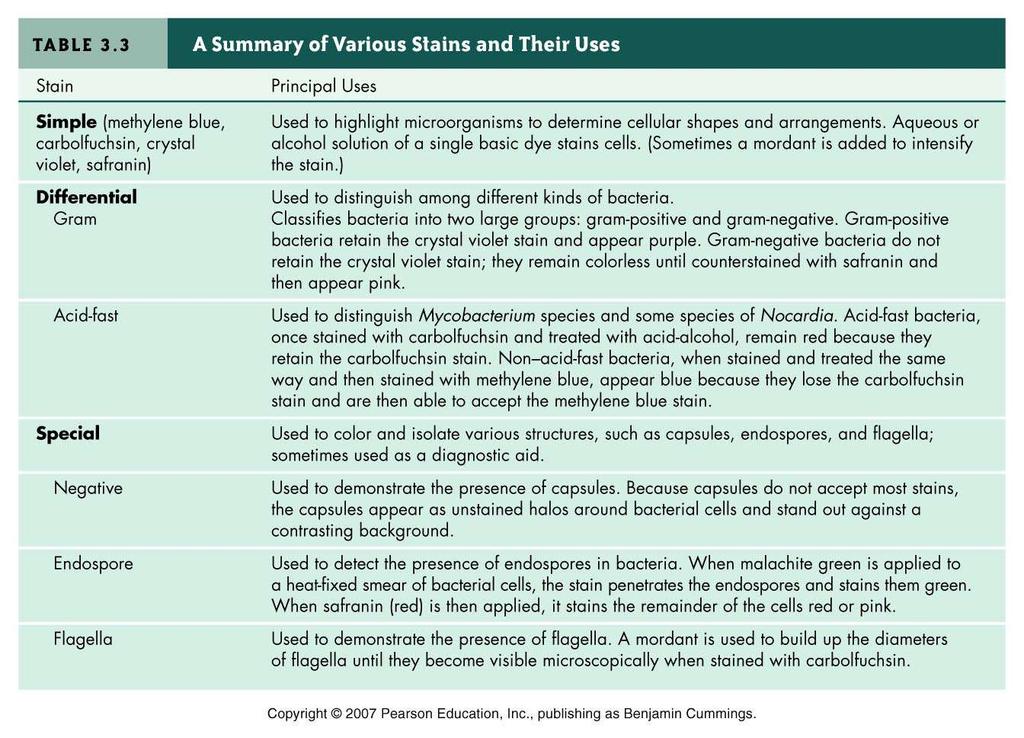
50 Table 3.3
51 GRAM STAIN PROCEDURE 1.The slide should be heat fixed on the heat block, or methanol fixed, prior to staining. 2.Methanol fixation is accomplished by flooding the slide with 95% methanol. Let the slide sit for 2 minutes, drain off the excess methanol, and allow to air dry. 3.Flood the slide with crystal violet for 30 seconds. Rinse with water. 4.Flood the slide with iodine for 60 seconds. Rinse with water. 5.Flood the slide with decolorizer for about ten seconds decolorization is complete when the solution runs clear from the slide. Rinse with water. 6.Flood the slide with safranin for 30 seconds. Rinse with water. 7.Blot the slide dry with absorbent paper and examine the slide under an oil immersion lens. INTERPRETATION gram negative organisms = pink color gram positive organisms = blue color
52 Slide Show Examples of staining techniques
Ch 2 Part 2. The Microscope
 Ch 2 Part 2 The Microscope SLOs for Microscopic Analysis of Microorganisms Convert among the different units of the metric system. List and describe three elements of good microscopy. Differentiate between
TEST BANK FOR PRESCOTTS MICROBIOLOGY 9TH EDITION BY WILLEY SHERWOOD WOOLVERTON
 TEST BANK FOR PRESCOTTS MICROBIOLOGY 9TH EDITION BY WILLEY SHERWOOD WOOLVERTON Link download full: https://testbankservice.com/download/test-bank-for-prescottsmicrobiology-9th-edition-by-willey-sherwood-woolverton/
INTRODUCTION. Gram Stain
 INTRODUCTION In microbiology, organisms are so small that additional techniques are often required for proper viewing under the microscope. Cytological stains, or dyes that stain cells or cellular features,
Exercise VI. Differential Staining: The Gram Stain
 Exercise VI Differential Staining: The Gram Stain The Gram stain, discovered by Dr. Hans Christian Gram in 1884, is the most useful differential stain used to aid in identifying bacteria. It divides bacterial
Ch 3 & 4 Microscopy & Cell Components 1
 Objectives 1.White book: Read Chap 3 & p 77-98 & 108 2.Black book: Read Chap 3 & p75-96 & 106 Objectives: 1. List metric measurement units for microorganisms and convert to other metric units (m, mm, um,
Ch 3 & 4 Microscopy & Cell Components 1
 Objectives 1.White book: Read Chap 3 & p 77-98 & 108 2.Black book: Read Chap 3 & p75-96 & 106 Objectives: 1. List metric measurement units for microorganisms and convert to other metric units (m, mm, um,
Ch 3 & 4 Microscopy & Cell Components 1
 Objectives 1.White book: Read Chap 3 & p 77-98 & 108 2.Black book: Read Chap 3 & p75-96 & 106 Objectives: 1. List metric measurement units for microorganisms and convert to other metric units (m, mm, um,
Microscopy, Staining, and Classification
 PowerPoint Lecture Presentations prepared by Mindy Miller-Kittrell, North Carolina State University C H A P T E R 4 Microscopy, Staining, and Classification 4. Discuss how microscopy has revealed the structure
Exercise 6-B STAINING OF MICROORGANISMS GRAM STAIN
 Exercise 6-B STAINING OF MICROORGANISMS GRAM STAIN Introduction The Gram stain, developed by Hans Christian Gram in 1884, is a staining technique allowing different types of microorganisms (usually bacteria)
Required Materials: immersion oil microscopes Kim-wipes prepared microscope slides
 Microbiology CA/IA Lab Microscopic Examination of Microbes September 10 Objectives: 1. learn how to use a microscope to examine microbes 2. learn to recognize the characteristics of different microbes
BIOL 260-General Microbiology. Instructor: Seana Davidson
 BIOL 260-General Microbiology Instructor: Seana Davidson Welcome to BIOL 260: Microbiology! First day: Review of Syllabus Sign-in Introduce the course, review course expectations Begin with first lab Exercise
Bacterial Gram Staining
 PR021 G-Biosciences 1-800-628-7730 1-314-991-6034 technical@gbiosciences.com A Geno Technology, Inc. (USA) brand name Bacterial Gram Staining Teacher s Guidebook (Cat. # BE 202) think proteins! think G-Biosciences
THE GRAM STAIN OBJECTIVE/RATIONALE KEY POINTS
 THE GRAM STAIN OBJECTIVE/RATIONALE One of the first procedures preformed by the medical microbiologist for the identification of bacteria is the Gram Stain. The student will learn the procedure for performing
chapter one: the history of microbiology
 chapter one: the history of microbiology Revised 6/19/2018 microbes microscopic (small) organisms, viruses, prions prefix sci. notation frac. equivalent dec. equivalent kilo- (k) 1 10 3 1000/1 = 1000 1000
MICROBIOLOGY LAB #1 SAFETY RULES & GRAM STAIN METHOD
 MICROBIOLOGY LAB #1 SAFETY RULES & GRAM STAIN METHOD Precaution processes are extremely important when working with cultures in the lab for the safety of the microbiologist from getting diseases from bacteria
EDUCATIONAL COMMENTARY GRAM STAIN
 EDUCATIONAL COMMENTARY GRAM STAIN Educational commentary is provided through our affiliation with the American Society for Clinical Pathology (ASCP). To obtain FREE CME/CMLE credits click on the Continuing
LABORATORY 7 ENDOSPORE STAIN AND BACTERIAL MOTILITY
 LABORATORY 7 ENDOSPORE STAIN AND BACTERIAL MOTILITY A. Endospore Stain B. Bacterial Motility A. ENDOSPORE STAIN DISCUSSION A few genera of bacteria, such as Bacillus and Clostridium have the ability to
Microbiology. Definition of a Microorganism. Microorganisms in the Lab. The Study of Microorganisms
 Microbiology The Study of Microorganisms Definition of a Microorganism Derived from the Greek: Mikros, «small» and Organismos, organism Microscopic organism which is single celled (unicellular) or a mass
Chapter 03 Microscopy and Cell Structure
 Chapter 03 Microscopy and Cell Structure Multiple Choice Questions 1. Eukaryotic cells are A. less complex than prokaryotic cells. B. members of the Domains Bacteria and Archaea. C. defined by the presence
Mycobacteriology Research Centre, NRITLD 5. Contence
 In The Name Of God Shaheed Beheshti University of Medical sciences and Health Services National Research Institute Tuberculosis and Lung Disease Collaborating Center for Tuberculosis East Mediterranean
Microscopy, Staining, and Classification. ~10 um. Red Blood Cells = mm 1500 um. Width of penny
 PowerPoint Lecture Presentations prepared by Mindy Miller-Kittrell, North Carolina State University C H A P T E R 4 Microscopy, Staining, and Classification Figure 3.4 Approximate size of various types
MODULE 2 : FOUNDATIONS IN BIOLOGY
 OCR A LEVEL BIOLOGY MODULE 2 : FOUNDATIONS IN BIOLOGY REVISION NOTES For 2015 onwards specification Miss T Banda All living things are primarily made from 4 key elements: Carbon (C) Hydrogen (H) Oxygen
Subject: Staining-Bacterial Cell Structure Lecture Number: 3 Done by: Joud Baki Corrected by: Issa Deir
 Subject: Staining-Bacterial Cell Structure Lecture Number: 3 Done by: Joud Baki Corrected by: Issa Deir 0 Principles of staining: - Revision: Stains can be either simple or differential Gram stains are
Chap 3 & 4 Microscopy & Cell Components
 Chap 3 & 4 Microscopy & Cell Components Prior Knowledge: Parts of the scope at the right? Type of scope? Proper care and use? Other types of scopes? What are the 3 shapes of bacteria? Preview of smears
Take-Home Quiz I. Summer 2005 Semester
 General Instructions and Information: Obtain an answer sheet from the instructor and legibly write your name in the appropriate space. After placing your name, you must enter your Patron ID Number (NOT
MONTGOMERY COUNTY COMMUNITY COLLEGE BIO 140 CHAPTER 4. Functional Anatomy of Prokaryotic and Eukaryotic Cells
 MONTGOMERY COUNTY COMMUNITY COLLEGE BIO 140 CHAPTER 4 Functional Anatomy of Prokaryotic and Eukaryotic Cells I. PROKARYOTES A. Structure Of The Cell: Chemical Composition And Function 1. Cell Wall a. composition
Microscopy, Staining, and Classification
 PowerPoint Lecture Presentations prepared by Mindy Miller-Kittrell, North Carolina State University C H A P T E R 4 Microscopy, Staining, and Classification Figure 4.3 The limits of resolution (and some
Microscopy, Staining, and Classification
 PowerPoint Lecture Presentations prepared by Mindy Miller-Kittrell, North Carolina State University C H A P T E R 4 Microscopy, Staining, and Classification Microscopy Light Microscopy 1) Bright-field
Archaebacteria and Eubacteria
 Archaebacteria and Eubacteria Bacteria are of immense importance because of their rapid growth, reproduction, and mutation rates, as well as, their ability to exist under adverse conditions. The oldest
Microbiology Laboratory (BIOL 3702L) Page 1 of 10
 Page 1 of 10") Microbiology Laboratory (BIOL 3702L) Page 1 of 10 Principle and Purpose THE GRAM STAIN The Gram stain is perhaps the most useful test conducted in the clinical microbiology laboratory. It was first developed
Microbiology Laboratory (BIOL 3702L) Page 1 of 10 Principle and Purpose THE GRAM STAIN The Gram stain is perhaps the most useful test conducted in the clinical microbiology laboratory. It was first developed
The Prokaryotes & Viruses
 The Prokaryotes & Viruses Lab Exercise Contents Objectives 1 Introduction 1 Activity.1 Prokaryotic Cell Structure 2 Activity.2 Blue-Green Algae 2 Activity.3 Viruses 3 Activity.4 Gram Staining of Bacteria
Cells and Microscopes Biology Concepts of Biology 2.1
 Cells and Microscopes Biology 100 - Concepts of Biology 2.1 Name Instructor Lab Section Objectives: To gain an understanding of how to: Correctly use the compound light microscope Differentiate between
Cell Shape coccus bacillus spirillum vibrio
 wrong 0 1 2 3 4 5 6 7 8 9 10 11 12 13 14 15 16 17 18 right 56 55 54 53 52 51 50 49 48 47 46 45 44 43 42 41 40 39 38 score 100 98.2 96.4 94.6 92.9 91.1 89.3 87.5 85.7 83.9 82.1 80.4 78.6 76.8 75 73.2 71.4
CELL LAB OBJECTIVES INTRODUCTION: CELL UNIT. After completing this lab you should be able to:
 AP BIOLOGY CELL UNIT ACTIVITY #3 NAME DATE HOUR CELL LAB OBJECTIVES After completing this lab you should be able to: 1. Compare and contrast prokaryotic and eukaryotic cells, 2. Prepare wet mount slides
RELATIONSHIP OF CELL WALL STAINING TO GRAM DIFFERENTIATION'
 RELATONSHP OF CELL WALL STANNG TO GRAM DFFERENTATON' J. W. BARTHOLOMEW AND HAROLD FNKELSTEN Department of Bacteriology, University of Southern California, Los Angeles, California Received for publication
TRACING BACK TO THE BEGINNING
 BACTERIA! TRACING BACK TO THE BEGINNING PROKARYOTES KINGDOM EUBACTERIA KINGDOM ARCHAEBACTERIA CHARACTERISTICS: 1. NO NUCLEUS 2. NO MEMBRANE BOUND ORGANELLES 4. MOST ARE SMALLER THAN EUKARYOTES 5. ARE SINGLE-CELLED
BACTERIA AND ARCHAEA 10/15/2012
 BACTERIA AND ARCHAEA Chapter 27 KEY CONCEPTS: Structural and functional adaptations contribute to prokaryotic success Rapid reproduction, mutation, and genetic recombination promote genetic diversity in
Characteristics. Nucleoid Region single circular chromosome plasmids mesosome
 Prokaryotes Characteristics Nucleoid Region single circular chromosome plasmids mesosome No membranebound organelles Ribosomes (70S) Plasma membrane Cell wall peptidoglycan Capsule glycocalyx Flagella
MICROSCOPY AND CELLS BIO 171 WEEK 3
 MICROSCOPY AND CELLS BIO 171 WEEK 3 MICROSCOPY THE COMPOUND LIGHT MICROSCOPE System of lenses arranged to produce an enlarged, focusable image of a specimen. MICROSCOPY THE MICROSCOPE Illuminating System
Microscopic and macroscopic observation of microorganisms & Gram stain. Mgr. Tomáš Kastl
 Microscopic and macroscopic observation of microorganisms & Gram stain Mgr. Tomáš Kastl MARKS TO NOTICE Morphology of colonies and cells - strructure - size - surface - shape - profile - special organels
Adding the class. Prerequisites. Welcome to Bio 139 General Microbiology. Syllabus. Amy Rogers, M.D., Ph.D.
 Adding the class Welcome to Bio 139 General Microbiology Amy Rogers, M.D., Ph.D. Lectures MW 12:00-1:15 PM (section 8) Labs: MW 1:30-2:45 PM or MW 3:00 PM-4:15 PM I anticipate a large number of students
Obligate anaerobes - cannot grow in the presence of oxygen Facultative anaerobes - can grow with or without oxygen Aerobic - require oxygen
 PROKARYOTES *include bacteria and archaea *singular: bacterium / plural: bacteria PROPERTIES 1. Bacteria are classified into two kingdoms: Eubacteria (true bacteria) and Archaebacteria (Ancient Bacteria).
#2: THE FLOATING PAPER CLIP
 Activity #1: PILE IT ON. Materials: 1 DRY penny, 1 eye dropper, water. Procedure: Make sure the penny is dry. Begin by estimating the number of drops of water that can be piled on the penny before it spills
Origins - Three Domain Classification PROKARYOTES
 Bacteria Origins - Three Domain Classification EU PROKARYOTES I. Origins of Bacteria Prokaryotes Eubacteria Archaebacteria A. Prokaryotes = 1. Kingdom Eubacteria 2. Kingdom Archaebacteria 3. Prokaryote
THE CYTOLOGICAL BASIS FOR THE ROLE OF THE PRIMARY DYE
 THE CYTOLOGICAL BASIS FOR THE ROLE OF THE PRIMARY DYE IN THE GRAM STAIN' CARL LAMANNA AND M. F. MALLETTE Departments of Microbiology and Biochemistry, The Johns Hopkins University School of Hygiene and
By signing below, you acknowledge that you have ensured that you are complying with the above statement.
 Instructions: This exam consists of 31 multiple choice questions on 8 pages, including this one. Please submit your answers on the scantron sheet provided and on this copy of the exam. This exam is closed
Day 2 - Viewing a prepared slide of mixed bacteria on high power.
 Purpose Bacteria Lab To compare the quantity and the different types of bacteria from four different locations within the school. To identify 3 different bacterial colonies on a prepared slide. Materials
Introduction to Microbiology BIOL 220 Summer Session I, 1996 Exam # 1
 Name I. Multiple Choice (1 point each) Introduction to Microbiology BIOL 220 Summer Session I, 1996 Exam # 1 B 1. Which is possessed by eukaryotes but not by prokaryotes? A. Cell wall B. Distinct nucleus
Microbiology Helmut Pospiech
 Microbiology 20.03.2018 Helmut Pospiech The control of what gets in Passive transport along a concentration gradient often inefficient Active transport Requires energy consumption and what gets out ABC
Bacteria. Prepared by. Doua a Hamadi Gellan Ibrahim Rahma Younis Doua a Abdul-Hadi Doua a Amjad Hanin Laith Khamael Dawood
 Bacteria Prepared by Doua a Hamadi Gellan Ibrahim Rahma Younis Doua a Abdul-Hadi Doua a Amjad Hanin Laith Khamael Dawood History of Bacteriology Doua a Hamadi Bacteria were first observed by Antonie van
Module 2: Foundations in biology
 alevelbiology.co.uk Module 2: Foundations in biology SPECIFICATION 2.1.1 Cell structure Learners should be able to demonstrate and apply their knowledge and understanding of: (a) The use of microscopy
Bacillus anthracis. Clostridium botulinum Clostridium perfringens and other, but never Gram-negative microbes
 SPORES (endospores) the spore is formed inside the parent vegetative cell hence the name endospores The spore is a dehydrated, multishelled structure that protects and allows the bacteria to exist in suspended
Western Carolina University. Chem 132 Lab 04 Introduction to Physical Changes and Chemical Reactions Introduction
 Chem 132 Lab 04 Introduction to Physical Changes and Chemical Reactions Introduction This lab serves as an introduction to physical changes. Physical changes involve a change in the form of matter without
Directed Reading A. Section: Bacteria CHARACTERISTICS OF BACTERIA. bacteria? a. cocci b. spirilla c. flagella d. bacilli.
 Skills Worksheet Directed Reading A Section: Bacteria 1 Which of the following is true of bacteria? a All bacteria are the same size b Most bacteria can be seen without a microscope c There are fewer bacteria
BACTERIA. CLS 212: Medical Microbiology Miss Zeina Alkudmani
 BACTERIA CLS 212: Medical Microbiology Miss Zeina Alkudmani Prokaryotes Prokaryotic cells possess simpler structures than eukaryotic cells, since they do not have a nucleus or a lot of cytoplasmic organelles.
(inner dense substance) of the identical bacteria later photographed in the electron
 of the identical bacteria later photographed in the electron") ON THE MICROSCOPIC METHODS OF MEASURING THE DIMENSIONS OF THE BACTERIAL CELL GEORGES KNAYSI Laboratory of Bacteriology, College of Agriculture, Cornell University, Ithaca, New York Received for publication
ON THE MICROSCOPIC METHODS OF MEASURING THE DIMENSIONS OF THE BACTERIAL CELL GEORGES KNAYSI Laboratory of Bacteriology, College of Agriculture, Cornell University, Ithaca, New York Received for publication
KINGDOM MONERA. Bacterial Cell Shape 8/22/2010. The Prokaryotes: Archaebacteria and Eubacteria
 KINGDOM MONERA The Prokaryotes: Archaebacteria and Eubacteria Bacteria are the most organisms living on the Earth. (i.e. 10mL of soil contains 1 x 10 10 bacteria. They are found in nearly every habitat
MICROBE MISSION - SAMPLE TOURNAMENT #1 by Karen L. Lancour
 MICROBE MISSION - SAMPLE TOURNAMENT #1 by Karen L. Lancour STATION A: MICROSCOPY 1. A microscope has an 10 objective and oculars of 4X, 10X, 40X and 100X. What is the range of magnification for this microscope.
dyes under the name of gentian violet. This being the situation, AN INVESTIGATION OF AMERICAN GENTIAN
 AN INVESTIGATION OF AMERICAN GENTIAN VIOLETS REPORT OF COMMIrTTEE ON BACTERIOLOGICAL TECHNIC PREPARED BY H. J. CONN, CHAIRMAN Received for publication June 9, 1922 This report is a continuation of the
Kingdom Monera(Archaebacteria & Eubacteria)
") Kingdom Monera(Archaebacteria & All bacteria are prokaryotes Characteristics: 1. No nucleus Eubacteria) 2. No membrane bound organelles 3. Smaller & less ribosomes 4. Most are smaller than eukaryotes 5.
Kingdom Monera(Archaebacteria & All bacteria are prokaryotes Characteristics: 1. No nucleus Eubacteria) 2. No membrane bound organelles 3. Smaller & less ribosomes 4. Most are smaller than eukaryotes 5.
Slide 1. Slide 2. Slide 3. Chapter 4 A Tour of the Cell. State Standards. Introduction to Cells. Standard 1.c. Standard 1.e.
 Slide 1 Chapter 4 A Tour of the Cell Slide 2 State Standards Standard 1.c. Standard 1.e. Slide 3 Introduction to Cells Organisms are either - Single-celled, such as - Multicelled, such as The human body
Investigation: What Are the Different Types of Cells?
 Name: Date: Investigation: What Are the Different Types of Cells? All living organisms are made of cells. The smallest cells are about 0.001 millimeters in diameter and belong to one of two domains: Bacteria
Ladue Microbe Mission Test SCORE: 90 / 90 Name: Answer Key Date:
 Ladue Microbe Mission Test SCORE: 90 / 90 Name: Answer Key Date: You may not return to previous stations. However, you can move to another station early if you want to do so. I won t judge you for your
Kingdom Monera Bacteria
 Kingdom Monera Bacteria Common bacteria Prokaryotes Strep throat Anthrax Chlamydia E. coli Meningitis Salmonella Micrococcus(intestinal) Streptococcus mutans Haemophilusinfluenzae Cellphonious bacterious
CLASSIFICATION OF BACTERIA
 CLASSIFICATION OF BACTERIA DISCLOSURE Relevant relationships with commercial entities none Potential for conflicts of interest within this presentation none Steps taken to review and mitigate potential
CHAPTER 1 THE SCIENCE OF LIFE
 CHAPTER 1 THE SCIENCE OF LIFE Biology Bio - life Logy- the study of Biology is the study of life or living things Some branches of Biology include- Microbiology, Marine Biology, Botany, Zoology, Ecology
Ultraviolet and Visible Spectroscopy. interaction of materials with light at different electronic levels and the extent, to which such
 Surname 1 Ultraviolet and Visible Spectroscopy Introduction This experiment was carried out to demonstrate the effect of atomic structure on the interaction of materials with light at different electronic
Bacterial Morphology and Structure م.م رنا مشعل
 Bacterial Morphology and Structure م.م رنا مشعل SIZE OF BACTERIA Unit for measurement : Micron or micrometer, μm: 1μm=10-3 mm Size: Varies with kinds of bacteria, and also related to their age and external
Outline. Viruses, Bacteria, and Archaea. Viruses Structure Classification Reproduction Prokaryotes Structure Reproduction Nutrition Bacteria Archaea
 Viruses, Bacteria, and Archaea Chapter 21 Viruses Structure Classification Reproduction Prokaryotes Structure Reproduction Nutrition Bacteria Archaea Outline The Viruses The Viruses Viruses are noncellular
8.1 Life is cellular
 8.1 Life is cellular Early Microscopes In 1665, Englishman Robert Hooke used a microscope to look at a slice of cork. Cork was made of tiny, empty chambers that Hooke called cells. Anton van Leeuwenhoek
Investigating Cells Lab. 1. What major differences do you expect to see between bacterial cells and plant/animal
 Investigating Cells Lab Name 7 th Grade PSI Questions: o How do you use a microscope? o What do bacterial cells look like? o Are there any visible differences between plant cells and animal cells? o What
Acellular Microbe Types
 BIOL 142 Lecture 4 Chapter 4: Microbial Diversity Part 1: Acellular & Prokaryotic Microbes & General Staining Techniques 62 slides 1 Acellular Microbe Types a virion is a complete, infectious viral particle
Use of light microscope and stereomicroscope: measuring microscopic
 Experiment 1 Use of light microscope and stereomicroscope: measuring microscopic objects 1.1 Introduction The microscope is a major tool used by biologists, which was invented about 350 years ago. It is
Scientific Method - the universal approach to solving scientific problems. 1. Problem Statement - Define the problem - ask question
 Biology: 7 Character of Life: 1. Organization of Cells 2. Response to Stimuli 3. Homeostasis 4. Metabolism 5. Growth & Development 6. Reproduction 7. Change Through Time Levels of Organization Atoms molecules
Introduction to the Microscope
 Title: Microscope Mania "Micro" (Greek!) refers to tiny, "scope" refers to view or look. Microscopes are tools used to enlarge images of small objects so they can be studied. The compound light microscope
Vocabulary- Bacteria (34 words)
") Biology II BACTERIA Vocabulary- Bacteria (34 words) 1. Prokaryote 21. phototroph 2. Peptidoglycan 22. chemotroph 3. Methanogen 23. obligate anaerobe 4. Halophile 24. facultative anaerobe 5. Thermoacidophile
Biology II BACTERIA Vocabulary- Bacteria (34 words) 1. Prokaryote 21. phototroph 2. Peptidoglycan 22. chemotroph 3. Methanogen 23. obligate anaerobe 4. Halophile 24. facultative anaerobe 5. Thermoacidophile
Chapter 2 Microbes in Perspective: Of Collectors and Classifiers
 Chapter 2 Microbes in Perspective: Of Collectors and Classifiers Objectives: After reading Chapter Two, you should understand The schemes used throughout history to classify organisms. How microorganisms
Kinetics of Crystal Violet Fading AP* Chemistry Big Idea 4, Investigation 11 An Advanced Inquiry Lab
 Introduction Kinetics of Crystal Violet Fading AP* Chemistry Big Idea 4, Investigation 11 An Advanced Inquiry Lab Catalog o. AP7644S Publication o. 7644S Crystal violet is a common, beautiful purple dye.
Chapter 7 Cell Structure
 Chapter 7 Cell Structure Mr. C. Biology 1 07 Cell Structure Chapter 7 Cell Structure All living things are made of cells. Cells are made up of 3 main parts, Cell Membrane A skin that controls what enters
Lab 1: Using the Microscope & Cell Biology
 Name Lab 1: Using the Microscope & Cell Biology The anatomy of the compound microscope Review or learn the following parts of the compound microscope and their functions. Eyepieces Objectives Arm Stage
EXERCISE 3 COMPARATIVE STUDY OF PROKARYOTIC AND EUKARYOTIC ORGANISMS
 EXERCISE 3 COMPARATIVE STUDY OF PROKARYOTIC AND EUKARYOTIC ORGANISMS 3.1 INTRODUCTION In the previous exercise you studied the ultrastructure of prokaryotic and eukaryotic cells with the help of electron
The invention of the microscope has opened to us a world of extraordinary numbers. A singular drop of pond water reveals countless life forms
 Biology Chapter 19 Notes - Bacteria and Viruses The invention of the microscope has opened to us a world of extraordinary numbers. A singular drop of pond water reveals countless life forms I. Classifying
Gram-negative. No explanation is offered as to why the outer. depends upon the isoelectric point and the ph (Stearn and Stearn,
 THE CELL WALL AND THE GRAM REACTION VICTOR BURKE AND MILDRED WINCHESTER BARNES Bacteriological Laboratories, State College of Washington, Pullman, Washington Received for publication, April 10, 1929 The
Microbe Mission Exam. Princeton Science Olympiad Invitational
 Page 1 Team Number: Team Name: Participant names: 2016-2017 Microbe Mission Exam Princeton Science Olympiad Invitational Page 2 Part 1: Microbial Organisms Matching. Each statement will have ONLY one answer.
GUJARAT UNIVERSITY Syllabus for First Year Microbiology Semester I and II Effective from June 2017
 GUJARAT UNIVERSITY Syllabus for First Year Microbiology Semester I and II Effective from June 2017 1. A student offering Microbiology programme will be offered two theory papers of core course MI 101 and
BACTERIA. Bacteria - small one celled monerans Bacteria like a warm, dark, and moist environment They are found almost everywhere: -on most objects
 Kingdom Monera 1 BACTERIA Bacteria - small one celled monerans Bacteria like a warm, dark, and moist environment They are found almost everywhere: -water -soil -skin -on most objects -air -food -inside
AP Biology Lab 4 PLANT PIGMENTS AND PHOTOSYNTHESIS
 AP Biology Laboratory Date: Name and Period: AP Biology Lab 4 PLANT PIGMENTS AND PHOTOSYNTHESIS OVERVIEW In this lab you will: 1. separate plant pigments using chromatography, and 2. measure the rate of
Chapter 4 A Tour of the Cell. The human body is made up of trillions of cells many of which are specialized - Muscle cells
 Chapter 4 A Tour of the Cell State Standards Standard 1.c. Standard 1.e. Introduction to Cells Organisms are either - Single-celled, such as - Multicelled, such as The human body is made up of trillions
7.1 Life is Cellular. Robert Hooke: Anton van Leeuwenhoek: The smallest unit of any organism- the cell. Robert Hooke
 7.1 Life is Cellular Sunday, December 16, 2012 1:07 PM Vocabulary: Cell: basic unit of all forms of life Cell theory: fundamental concept of biology that states that all living things are composed of cells;
Kinetics of Crystal Violet Fading AP Chemistry Big Idea 4, Investigation 11 An Advanced Inquiry Lab (adapted by Flinn Scientific, Inc.
 Introduction Kinetics of Crystal Violet Fading AP Chemistry Big Idea 4, Investigation 11 An Advanced Inquiry Lab (adapted by Flinn Scientific, Inc.) Crystal violet is a common, beautiful purple dye. In
Form a Hypothesis. Variables in an Experiment Dependent Variable what is being measured (data) Form a Hypothesis 2. Form a Hypothesis 3 15:03 DRY MIX
 Form a Hypothesis 2. Form a Hypothesis 3 15:03 DRY MIX") Scientific Method 1. Ask a question (Make observation) 2. Do some research 3. Form a Hypothesis MUST BE TESTABLE!! A possible explanation for a phenomenon. 4. Test Hypothesis/collect data (experiment time!)
Scientific Method 1. Ask a question (Make observation) 2. Do some research 3. Form a Hypothesis MUST BE TESTABLE!! A possible explanation for a phenomenon. 4. Test Hypothesis/collect data (experiment time!)
Imaging Methods: Breath Patterns
 Imaging Methods: Breath Patterns Breath / condensation pattern: By cooling a substrate below the condensation temperature H 2 O will condense in different rates on the substrate with the nucleation rate
Experiment 1: Thin Layer Chromatography
 Experiment 1: Thin Layer Chromatography Part A: understanding R f values Part B: R f values & solvent polarity Part C: R f values & compound functionality Part D: identification of commercial food dye
10/1/2014. Chapter Explain why the cell is considered to be the basic unit of life.
 Chapter 4 PSAT $ by October by October 11 Test 3- Tuesday October 14 over Chapter 4 and 5 DFA- Monday October 20 over everything covered so far (Chapters 1-5) Review on Thursday and Friday before 1. Explain
5. Move several sections into the second well that contains a few drops of Toluidine Blue.
 HAIGLER PROTOCOL FOR VIEWING CROSS SECTIONS OF PLANTS Materials (per student team): Plants Double-edged razor blades (split into two as demonstrated by the teacher) Masking tape for razor blade handle
Experiment 1: Extraction and Thin Layer Chromatography
 Experiment 1: Extraction and Thin Layer Chromatography Introduction: Chromatography is a useful tool in chemistry and can be very helpful in determining the composition of an unknown sample. In chromatography
Save My Exams! The Home of Revision For more awesome GCSE and A level resources, visit us at Microscopes.
 Microscopes Question Paper 2 Level International Level Subject iology Exam oard IE Topic Microscopes Sub Topic ooklet Multiple hoice Paper Type Question Paper 2 Time llowed : 44 minutes Score : / 36 Percentage
2nd Quarter Lab Assessment Review Packet
 Name Date Period Labs to be Assessed: 2nd Quarter Lab Assessment Review Packet Lab #8 - Using Measuring Tools Lab #9 - Introduction to the Compound Microscope Lab #10 - Using The Compound Microscope Lab
MICR2208 Lecture 3: Prokaryotic Structure and Function 1
 MICR2208 Lecture 3: Prokaryotic Structure and Function 1 Diversity of Prokaryotes Size Not all prokaryotes are similar in size as they all differ, however, most of the prokaryotes cannot be seen from the
Current evidence indicates that eukaryotes evolved from prokaryotes between 1 and 1.5 billion years ago.
 Current evidence indicates that eukaryotes evolved from prokaryotes between 1 and 1.5 billion years ago. Two theories: 1. Infolding theory 2. Endosymbiotic theory The infolding of the prokaryotic plasma