Microscopy, Staining, and Classification
|
|
|
- Eugenia Bryan
- 6 years ago
- Views:
Transcription

1 PowerPoint Lecture Presentations prepared by Mindy Miller-Kittrell, North Carolina State University C H A P T E R 4 Microscopy, Staining, and Classification
2 Figure 4.3 The limits of resolution (and some representative objects within those ranges) of the human eye and of various types of microscopes.
3 Official definitions Magnification, the ratio of an object s image size to its real size Resolution, the measure of the clarity of the image, or the minimum distance of two distinguishable points Contrast, visible differences in brightness between parts of the sample
4 Microscopy General Principles of Microscopy Magnification (enlarge) Resolution (tell apart 2 objects close together) Contrast (Differences in intensity between two objects) How to increase all 3 of these?

5 Figure 4.1 The electromagnetic spectrum. Smaller the object = Use smaller wavelength VIBGYOR
6 General rule for any microscopy/detector Smaller the wavelength smaller the object you can see (small objects need small hands - think about giant fingers and texting)
7 Microscopy Microscopes are used to visualize cells In a light microscope (LM), visible light is passed through a specimen and then through glass lenses Glass lenses focuses light and enlarges and resolves objects

8 Lenses refract (bend) the light, so that the image is magnified Light Air Glass Magnification = 50/5 = 10X Magnify using lenses Focal point 5 50 Specimen Convex lens Inverted, reversed, and Enlarged image
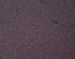
9 Microscopy Light Microscopy Bright-field microscopes Simple single lense Contain a single magnifying lens Similar to magnifying glass Leeuwenhoek used simple microscope to observe microorganisms
10 Microscopy Light Microscopy Bright-field microscopes Compound multiple lenses More than one - Series of lenses for magnification Light passes through specimen into objective lens Have one or two ocular lenses Total magnification = magnification of objective lens X magnification of ocular lens E.g. 10 times X 10 times = 100 times

11 Figure 4.4 A bright-field, compound light microscope. occular lens Line of vision Ocular lens Remagnifies the image formed by the objective lens Body Transmits the image from the objective lens to the ocular lens using prisms Arm Objective lenses Primary lenses that magnify the specimen Stage Holds the microscope slide in position Condenser Focuses light through specimen Diaphragm Controls the amount of light entering the condenser Illuminator Light source Ocular lens Path of light Prism Body Objective lenses Specimen Condenser lenses Illuminator Coarse focusing knob Moves the stage up and down to focus the image Fine focusing knob Base objective lens

12 Figure 4.5 The effect of immersion oil on resolution. Special type of objective lens = oil immersion lens Microscope objective Lenses Microscope objective Refracted light rays lost to lens Glass cover slip More light enters lens Glass cover slip Immersion oil Slide Slide Specimen Light source Light source Without immersion oil With immersion oil Increases magnification and resolution
13 Microscopy General Principles of Microscopy Contrast Differences in intensity between two objects, or an object and its background Increase contrast by staining

 Staining")
14 50 µm Figure 6.3ab Brightfield (unstained specimen) Brightfield (stained specimen) Staining increases contrast
15 Figure 4.16 Simple stains.
16 Figure 4.15 Preparing a specimen for staining.
17 Microscopy Light Microscopy 1) Bright-field microscopes Simple/ Compound

18 Figure 4.8 Four kinds of light microscopy. Nucleus Bacterium Bright field Dark field Dark-field microscopes Best for observing pale objects Only light rays scattered by specimen enter objective lens Specimen appears light against dark background Increases contrast and enables observation of more details

19 Figure 4.6 The light path in a dark-field microscope. Objective Light refracted by specimen Light unrefracted by specimen Specimen Condenser Dark-field stop Dark-field stop
20 Microscopy Light Microscopy 3) Phase microscopes Used to examine living organisms or specimens that would be damaged/altered by attaching them to slides or staining Light rays in phase produce brighter image, while light rays out of phase produce darker image Contrast is created because light waves are out of phase
21 Figure 4.7 Principles of phase microscopy. Rays in phase Rays out of phase Phase plate Bacterium Ray deviated by specimen is 1/4 wavelength out of phase. Deviated ray is now 1/2 Wavelength out of phase.
 Phase-contrast microscope 2) Differential interference")
22 Figure 4.8 Four kinds of light microscopy. Nucleus Two types of Phase microscopes 1) Phase-contrast microscope 2) Differential interference contrast microscope aka Nomarski Phase contrast Nomarski
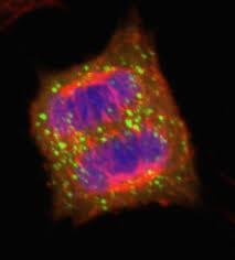
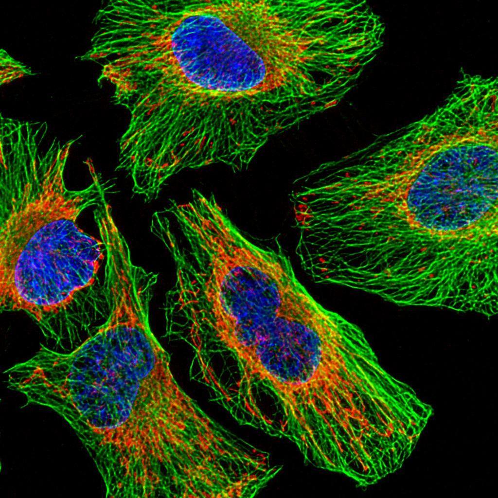
23 Microscopy Light Microscopy Fluorescent microscopes Direct UV light source at specimen Specimen radiates energy back as a longer, visible wavelength UV light increases resolution and contrast Some cells are naturally fluorescent; others must be stained Used in immunofluorescence to identify pathogens and to make visible a variety of proteins

24 Figure 4.9 Fluorescence microscopy. Can help you look through clutter

25 Figure 4.10 Immunofluorescence. Can help you target specific structures Antibodies Fluorescent dye Bacterium Cell-surface antigens Antibodies carrying dye Bacterial cell with bound antibodies carrying dye
26 Microscopy Light Microscopy Confocal microscopes Use fluorescent dyes Use UV lasers to illuminate fluorescent chemicals in a single plane Resolution increased because emitted light passes through pinhole aperture Computer constructs 3-D image from digitized images
27
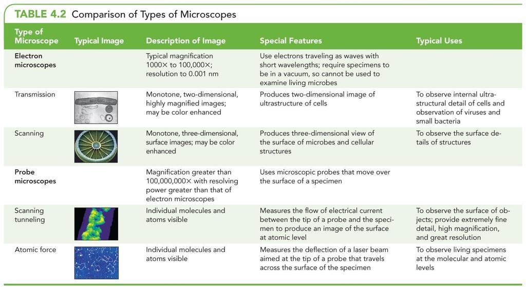
28 Microscopy Electron Microscopy Light microscopes cannot resolve structures closer than 200 nm Electron microscopes have greater resolving power and magnification Magnifies objects 10,000X to 100,000X Detailed views of bacteria, viruses, internal cellular structures, molecules, and large atoms Two types Transmission electron microscopes Scanning electron microscopes
 Eyepiece Projector lens (magnet) Final image seen by eye Final image on")
29 Figure 4.11 A transmission electron microscope (TEM). needs vacuum Light microscope (upside down) Column of transmission electron microscope Live? Lamp Electron gun Condenser lens Specimen Specimen Objective lens Objective lens (magnet) Eyepiece Projector lens (magnet) Final image seen by eye Final image on fluorescent screen need sections

30 Figure 4.12 Scanning electron microscope (SEM). Electron gun Magnetic lenses Beam deflector coil Primary electrons Scanning circuit Secondary electrons Specimen Specimen holder Photomultiplier Detector Monitor Vacuum system

31 Figure 4.13 SEM images.
32 Microscopy Probe Microscopy Magnifies more than 100,000,000 times Two types Scanning tunneling microscopes Atomic force microscopes

33 Figure 4.14 Probe microscopy. DNA Enzyme
34
35 Light Microscopy Staining Simple stains Differential stains Gram stain Acid-fast stain Endospore stain Special stains Negative (capsule) stain Flagellar stain
36 Staining Principles of Staining Dyes used as stains are usually salts Chromophore is the colored portion of the dye Acidic dyes (negatively charged) stain alkaline structures Basic dyes (positively charged) stain acidic structures Basic dyes are more commonly used since inside of most cells is negatively charged
37

38 Bacterium Capsule Background stain
39 Rhodospirillum rubrum


40 Figure 4.21 Flagellar stain of Proteus vulgaris. Flagella
41

42 Gram Staining

43 Gram Staining Mechanism


44 Staining for Electron Microscopy Chemicals containing heavy metals used for transmission electron microscopy
45 Classification and Identification of Microorganisms Taxonomy consists of classification, nomenclature, and identification Organize large amounts of information about organisms Make predictions based on knowledge of similar organisms
46 Classification and Identification of Microorganisms Linnaeus and Taxonomic Categories Linnaeus His system classified organisms based on characteristics in common Grouped organisms that can successfully interbreed into categories called species Used binomial nomenclature
47 Binomial nomenclature naming species of living things by giving each species a name composed of two parts humans belong to the genus Homo and within this genus to the species Homo sapiens. noun-genus adjective-specific epithet

48 Homo naledi (2015)
49 Microbe Naming Rules Each species have 2 names (1)Genus (2)specific epithet Genus specific epithet G. specific epithet Genus specific epithet G. specific epithet Escherichia coli Escherichia coli E. coli E. coli
50 Microbe Naming Rules X X Genus specific epithet Genus specific epithet Y Genus specific epithet Y Genus specific epithet
51 Bacteria have shapes/morphology
52 Figure 3.12 Bacterial shapes and arrangements. 3 Shapes 1) coccus 2) bacillus 3) spirillum 1) staphylo 2) strepto 2 main arrangements
53 Microbe Naming Rules Based on shape and arrangement (Arrangement+shape) (specific epithet) Staphylococcus (specific epithet) Streptobacillus (specific epithet)
54 cocci bacilli shape arrangement Streptococci Streptobacilli Staphylococci
55 Classification and Identification of Microorganisms Linnaeus and Taxonomic Categories Linnaeus's goal was classifying organisms to catalog them Linnaeus - 2 kingdoms Later on - 5 kingdoms Later on 3 domains (genetic material) Goal of modern taxonomy is to reflect phylogenetic hierarchy understanding genetic relationships among organisms Bat vs Bird
56 Figure 4.22 Levels in a Linnaean taxonomic scheme. D K P Class Our For Good So
57 Classification and Identification of Microorganisms Domains Carl Woese compared nucleotide sequences of rrna subunits Proposal of three domains as determined by ribosomal nucleotide sequences Eukarya, Bacteria, and Archaea
58 Classification and Identification of Microorganisms Taxonomic and Identifying Characteristics Physical characteristics Biochemical tests Serological tests Phage typing Analysis of nucleic acids
59 Classification and Identification of Microorganisms Taxonomic and Identifying Characteristics Physical characteristics Can often be used to identify microorganisms Protozoa, fungi, algae, and parasitic worms can often be identified based only on their morphology Some bacterial colonies have distinct appearance used for identification

60 Some bacterial colonies have distinct appearance used for identification

61 Figure 6.8 Characteristics of bacterial colonies. Shape Circular Rhizoid Irregular Filamentous Spindle Margin Entire Undulate Lobate Curled Filiform Elevation Flat Raised Convex Pulvinate Umbonate Size Punctiform Small Moderate Large Colony Texture Smooth or rough Appearance Glistening (shiny) or dull Pigmentation Nonpigmented (e.g., cream, tan, white) Pigmented (e.g., purple, red, yellow) Optical property Opaque, translucent, transparent

62 Figure 4.23 Two biochemical tests for identifying bacteria. Gas bubble Inverted tubes to trap gas Biochemical tests Acid with gas Acid with no gas Inert Hydrogen sulfide produced No hydrogen sulfide

63 Figure 4.24 One tool for the rapid identification of bacteria, the automated MicroScan system. Wells

64 Figure 4.25 An agglutination test, one type of serological test.

65 Figure 4.26 Phage typing. Bacterial lawn Plaques
66 Classification and Identification of Microorganisms Taxonomic and Identifying Characteristics Analysis of nucleic acids Nucleic acid sequence can be used to classify and identify microbes Prokaryotic taxonomy now includes the G + C content of an organism's DNA
67 Classification and Identification of Microorganisms Taxonomic Keys Dichotomous keys Series of paired statements where only one of two "either/or" choices applies to any particular organism Key directs user to another pair of statements, or provides name of organism

68 Figure 4.27 Use of a dichotomous taxonomic key. dichotomous taxonomic key dichotomous taxonomic key activity
Microscopy, Staining, and Classification. ~10 um. Red Blood Cells = mm 1500 um. Width of penny
 PowerPoint Lecture Presentations prepared by Mindy Miller-Kittrell, North Carolina State University C H A P T E R 4 Microscopy, Staining, and Classification Figure 3.4 Approximate size of various types
Microscopy, Staining, and Classification
 PowerPoint Lecture Presentations prepared by Mindy Miller-Kittrell, North Carolina State University C H A P T E R 4 Microscopy, Staining, and Classification 4. Discuss how microscopy has revealed the structure
Microscopy, Staining, and Classification
 PowerPoint Lecture Presentations prepared by Mindy Miller-Kittrell, North Carolina State University C H A P T E R 4 Microscopy, Staining, and Classification Microscopy Light Microscopy 1) Bright-field
Ch 2 Part 2. The Microscope
 Ch 2 Part 2 The Microscope SLOs for Microscopic Analysis of Microorganisms Convert among the different units of the metric system. List and describe three elements of good microscopy. Differentiate between
TEST BANK FOR PRESCOTTS MICROBIOLOGY 9TH EDITION BY WILLEY SHERWOOD WOOLVERTON
 TEST BANK FOR PRESCOTTS MICROBIOLOGY 9TH EDITION BY WILLEY SHERWOOD WOOLVERTON Link download full: https://testbankservice.com/download/test-bank-for-prescottsmicrobiology-9th-edition-by-willey-sherwood-woolverton/
Chapter 4.0 Microscopy, Staining, and Classification
 Chapter 4.0 Microscopy, Staining, and Classification 8/20/2017 MDufilho 1 Classification and Identification of Microorganisms Taxonomy consists of classification, nomenclature, and identification Organize
Chapter 2 Microbes in Perspective: Of Collectors and Classifiers
 Chapter 2 Microbes in Perspective: Of Collectors and Classifiers Objectives: After reading Chapter Two, you should understand The schemes used throughout history to classify organisms. How microorganisms
Ch 10. Classification of Microorganisms
 Ch 10 Classification of Microorganisms Student Learning Outcomes Define taxonomy, taxon, and phylogeny. List the characteristics of the Bacteria, Archaea, and Eukarya domains. Differentiate among eukaryotic,
Microbiology / Active Lecture Questions Chapter 10 Classification of Microorganisms 1 Chapter 10 Classification of Microorganisms
 1 2 Bergey s Manual of Systematic Bacteriology differs from Bergey s Manual of Determinative Bacteriology in that the former a. groups bacteria into species. b. groups bacteria according to phylogenetic
Cells Under the Microscope Measuring Cell Structures
 Copy into Note Packet and Return to Teacher Chapter 3 Cell Structure Section 1: Looking at Cells Objectives Describe how scientists measure the length of objects. Relate magnification and resolution in
Use of light microscope and stereomicroscope: measuring microscopic
 Experiment 1 Use of light microscope and stereomicroscope: measuring microscopic objects 1.1 Introduction The microscope is a major tool used by biologists, which was invented about 350 years ago. It is
chapter one: the history of microbiology
 chapter one: the history of microbiology Revised 6/19/2018 microbes microscopic (small) organisms, viruses, prions prefix sci. notation frac. equivalent dec. equivalent kilo- (k) 1 10 3 1000/1 = 1000 1000
Chapter 3. Observing Organisms Through a Microscope
 Chapter 3 Observing Organisms Through a Microscope Units of Measurement Used n Microbiology Table 3.1 mm Figure 3.2 Figure 3.1 - Overview Compound Light microscopy Have ocular and objective lenses Calculate
Adding the class. Prerequisites. Welcome to Bio 139 General Microbiology. Syllabus. Amy Rogers, M.D., Ph.D.
 Adding the class Welcome to Bio 139 General Microbiology Amy Rogers, M.D., Ph.D. Lectures MW 12:00-1:15 PM (section 8) Labs: MW 1:30-2:45 PM or MW 3:00 PM-4:15 PM I anticipate a large number of students
Microbiology 2320 Spring 2017 Dr. Milind Suraokar CRN# 18168
 Microbiology 2320 Spring 2017 Dr. Milind Suraokar CRN# 18168 PowerPoint Lecture Presentations prepared by Mindy Miller-Kittrell, North Carolina State University C H A P T E R 1 A Brief History of Microbiology
BIOL 260-General Microbiology. Instructor: Seana Davidson
 BIOL 260-General Microbiology Instructor: Seana Davidson Welcome to BIOL 260: Microbiology! First day: Review of Syllabus Sign-in Introduce the course, review course expectations Begin with first lab Exercise
Introductory Microbiology Dr. Hala Al Daghistani
 Introductory Microbiology Dr. Hala Al Daghistani Why Study Microbes? Microbiology is the branch of biological sciences concerned with the study of the microbes. 1. Microbes and Man in Sickness and Health
Chapter 7: Cell Structure and Function 7.1: Life is Cellular
 Chapter 7: Cell Structure and Function 7.1: Life is Cellular Key Questions: 1) What is the cell theory? 2) How do microscopes work? 3) How are prokaryotic and eukaryotic cells different? THINK ABOUT IT
Principles of Biotechnology Lectures of week 4 MICROBIOLOGY AND BIOTECHNOLOGY
 Principles of Biotechnology Lectures of week 4 MICROBIOLOGY AND BIOTECHNOLOGY INTRODUCTION TO MICROBIOLOGY What are microbes? Germs, microbe s s microorganisms are minute living things that individually
Test Bank for Microbiology A Systems Approach 3rd edition by Cowan
 Test Bank for Microbiology A Systems Approach 3rd edition by Cowan Link download full: http://testbankair.com/download/test-bankfor-microbiology-a-systems-approach-3rd-by-cowan/ Chapter 1: The Main Themes
Microscope History Robert Hooke
 1 Microscope History Robert Hooke First described cells in 1665. He viewed thin slices of cork and compared the boxy partitions he observed to the cells (small rooms) in a monastery. (1635 1702) 2 Microscope
Ch 3 & 4 Microscopy & Cell Components 1
 Objectives 1.White book: Read Chap 3 & p 77-98 & 108 2.Black book: Read Chap 3 & p75-96 & 106 Objectives: 1. List metric measurement units for microorganisms and convert to other metric units (m, mm, um,
Ch 3 & 4 Microscopy & Cell Components 1
 Objectives 1.White book: Read Chap 3 & p 77-98 & 108 2.Black book: Read Chap 3 & p75-96 & 106 Objectives: 1. List metric measurement units for microorganisms and convert to other metric units (m, mm, um,
Ch 3 & 4 Microscopy & Cell Components 1
 Objectives 1.White book: Read Chap 3 & p 77-98 & 108 2.Black book: Read Chap 3 & p75-96 & 106 Objectives: 1. List metric measurement units for microorganisms and convert to other metric units (m, mm, um,
Chapter 1. Introduction to Biology. The cell is the basic unit of life 1665-Robert Hooke- 1 st discovered cells in cork. The Seven Properties of Life
 The Science of Life Student Learning Goals - Biology Science and Life SC.912.N.1.1 Goal: Define a problem based on a specific body of knowledge, for example: biology, chemistry, physics, & earth/space
CHAPTER 1 THE SCIENCE OF LIFE
 CHAPTER 1 THE SCIENCE OF LIFE Biology Bio - life Logy- the study of Biology is the study of life or living things Some branches of Biology include- Microbiology, Marine Biology, Botany, Zoology, Ecology
Introduction to Microbiology. CLS 212: Medical Microbiology Miss Zeina Alkudmani
 Introduction to Microbiology CLS 212: Medical Microbiology Miss Zeina Alkudmani Microbiology Micro- means very small (that needs a microscope to see). Microbiology is the study of very small living organisms.
7.1 Life is Cellular. Robert Hooke: Anton van Leeuwenhoek: The smallest unit of any organism- the cell. Robert Hooke
 7.1 Life is Cellular Sunday, December 16, 2012 1:07 PM Vocabulary: Cell: basic unit of all forms of life Cell theory: fundamental concept of biology that states that all living things are composed of cells;
UNIT-5 EM WAVES UNIT-6 RAY OPTICS
 UNIT-5 EM WAVES 2 Marks Question 1. To which regions of electromagnetic spectrum do the following wavelengths belong: (a) 250 nm (b) 1500 nm 2. State any one property which is common to all electromagnetic
object objective lens eyepiece lens
 Advancing Physics G495 June 2015 SET #1 ANSWERS Field and Particle Pictures Seeing with electrons The compound optical microscope Q1. Before attempting this question it may be helpful to review ray diagram
Structure and Function of Plant and Animal Cells
 Science 14 Unit C: From Life to Lifestyle Chapter 8 Structure and Function of Plant and Animal Cells WORKBOOK Name: 8.1 A Closer Look pp. 158-164 Read pp. 158-159 Before the invention of microscope technology,
School. Team Number. Optics
 School Team Number Optics Physical Optics (30%) Proceed to the laser shoot (40%) when your team number is called. 1. What are the four colors used in the CMYK color model? (2 points) 2. Muscae Volitantes
Test Bank for Microbiology A Systems Approach 3rd edition by Cowan
 Test Bank for Microbiology A Systems Approach 3rd edition by Cowan Link download full: https://digitalcontentmarket.org/download/test-bank-formicrobiology-a-systems-approach-3rd-edition-by-cowan Chapter
tip conducting surface
 PhysicsAndMathsTutor.com 1 1. The diagram shows the tip of a scanning tunnelling microscope (STM) above a conducting surface. The tip is at a potential of 1.0 V relative to the surface. If the tip is sufficiently
The Discovery of the Cell
 7-1 Life Is Cellular Review The cell is the basic unit of life! Life began with the first cell! All living things are composed of cells! Cells make up tissues, organs, organ systems and organisms! Understanding
8.1 Life is cellular
 8.1 Life is cellular Early Microscopes In 1665, Englishman Robert Hooke used a microscope to look at a slice of cork. Cork was made of tiny, empty chambers that Hooke called cells. Anton van Leeuwenhoek
MICROSCOPY AND CELLS BIO 171 WEEK 3
 MICROSCOPY AND CELLS BIO 171 WEEK 3 MICROSCOPY THE COMPOUND LIGHT MICROSCOPE System of lenses arranged to produce an enlarged, focusable image of a specimen. MICROSCOPY THE MICROSCOPE Illuminating System
Slide 1. Slide 2. Slide 3. Chapter 4 A Tour of the Cell. State Standards. Introduction to Cells. Standard 1.c. Standard 1.e.
 Slide 1 Chapter 4 A Tour of the Cell Slide 2 State Standards Standard 1.c. Standard 1.e. Slide 3 Introduction to Cells Organisms are either - Single-celled, such as - Multicelled, such as The human body
The Discovery of the Cell
 The Discovery of the Cell The Discovery of the Cell Because there were no instruments to make cells visible, the existence of cells was unknown for most of human history. This changed with the invention
Outline. Viruses, Bacteria, and Archaea. Viruses Structure Classification Reproduction Prokaryotes Structure Reproduction Nutrition Bacteria Archaea
 Viruses, Bacteria, and Archaea Chapter 21 Viruses Structure Classification Reproduction Prokaryotes Structure Reproduction Nutrition Bacteria Archaea Outline The Viruses The Viruses Viruses are noncellular
Biology 160 Cell Lab. Name Lab Section: 1:00pm 3:00 pm. Student Learning Outcomes:
 Biology 160 Cell Lab Name Lab Section: 1:00pm 3:00 pm Student Learning Outcomes: Upon completion of today s lab you will be able to do the following: Properly use a compound light microscope Discuss the
Cells and Microscopes Biology Concepts of Biology 2.1
 Cells and Microscopes Biology 100 - Concepts of Biology 2.1 Name Instructor Lab Section Objectives: To gain an understanding of how to: Correctly use the compound light microscope Differentiate between
Microscopy and the Diversity of Microorganisms
 Microscopy and the Diversity of Microorganisms Today we will learn how to use one of the most important tools a biologist has, the microscope. We will use the microscope to study organisms throughout the
Life is Cellular. At the cellular level, what is the difference between animal cells and bacterial cells? How do microscopes work?
 Life is Cellular At the cellular level, what is the difference between animal cells and bacterial cells? How do microscopes work? Objectives 8a) I can state the cell theory and distinguish between prokaryotes
MICROBE MISSION - SAMPLE TOURNAMENT #1 by Karen L. Lancour
 MICROBE MISSION - SAMPLE TOURNAMENT #1 by Karen L. Lancour STATION A: MICROSCOPY 1. A microscope has an 10 objective and oculars of 4X, 10X, 40X and 100X. What is the range of magnification for this microscope.
UNIT #4: Cell Structure & Func4on
 UNIT #4: Cell Structure & Func4on PART ONE: Microscopes Four Different Types of Microscopes Dissec:ng Microscope Uses natural or electrical light source to illuminate an object Lens for magnifica4on Organisms
Burton's Microbiology for the Health Sciences
 Burton's Microbiology for the Health Sciences Chapter 3. Cell Structure and Taxonomy Chapter 3 Outline Introduction Eucaryotic Cell Structure Procaryotic Cell Structure Summary of Structural Differences
How does your eye form an Refraction
 Astronomical Instruments Eyes and Cameras: Everyday Light Sensors How does your eye form an image? How do we record images? How does your eye form an image? Refraction Refraction is the bending of light
Experiment #4 Nature of Light: Telescope and Microscope and Spectroscope
 Experiment #4 Nature of Light: Telescope and Microscope and Spectroscope In this experiment, we are going to learn the basic principles of the telescope and the microscope that make it possible for us
Chapter 4 A Tour of the Cell. The human body is made up of trillions of cells many of which are specialized - Muscle cells
 Chapter 4 A Tour of the Cell State Standards Standard 1.c. Standard 1.e. Introduction to Cells Organisms are either - Single-celled, such as - Multicelled, such as The human body is made up of trillions
AP5301/ Name the major parts of an optical microscope and state their functions.
 Review Problems on Optical Microscopy AP5301/8301-2015 1. Name the major parts of an optical microscope and state their functions. 2. Compare the focal lengths of two glass converging lenses, one with
Biology First Nine Weeks Review
 Name Date Test Date: November 12, 2009 Biology First Nine Weeks Review Modified True/False Directions: Indicate whether the sentence or statement is true or fals If false, change the identified word to
Student Exploration: Cell Types
 Name: Period: Student Exploration: Cell Types Vocabulary: ATP, bacteria, carbon dioxide (CO 2 ), cell, cellular respiration, compound light microscope, eukaryote, multicellular, muscle cell, neuron, organelle,
Module 2: Foundations in biology
 alevelbiology.co.uk Module 2: Foundations in biology SPECIFICATION 2.1.1 Cell structure Learners should be able to demonstrate and apply their knowledge and understanding of: (a) The use of microscopy
INTRODUCTION prokaryotic eukaryotic pigments
 INTRODUCTION This exercise is intended for you to get familiar and comfortable with using a microscope as well as identifying common microbial groups. Thus, we will observe representatives of all microbes
Lecture one Introduction to the Cell Biology
 Lecture one Introduction to the Cell Biology INTRODUCTION TO THE CELL Both living and non-living things are composed of molecules made from chemical elements such as Carbon, Hydrogen, Oxygen, and Nitrogen.
A. Incorrect! In the binomial naming convention the Kingdom is not part of the name.
 Microbiology Problem Drill 08: Classification of Microorganisms No. 1 of 10 1. In the binomial system of naming which term is always written in lowercase? (A) Kingdom (B) Domain (C) Genus (D) Specific
Lab 1: Using the Microscope & Cell Biology
 Name Lab 1: Using the Microscope & Cell Biology The anatomy of the compound microscope Review or learn the following parts of the compound microscope and their functions. Eyepieces Objectives Arm Stage
Chapter Introduction. of Life. Organisms. Chapter Wrap-Up. Steven P. Lynch
 Steven P. Lynch Chapter Introduction Lesson 1 Lesson 2 Lesson 3 Characteristics of Life Classifying Organisms Chapter Wrap-Up Exploring Life What are living things, and how can they be classified? What
PHYSICS. Ray Optics. Mr Rishi Gopie
 Ray Optics Mr Rishi Gopie Ray Optics Nature of light Light is a form of energy which affects the human eye in such a way as to cause the sensation of sight. Visible light is a range of electromagnetic
SECTION 3 & 4 LIGHT WAVES & INFORMATION TRANSFER
 SECTION 3 & 4 LIGHT WAVES & INFORMATION TRANSFER Light Waves Light is a type of energy that travels as waves. Light is different than other waves because it does not need matter to travel. Light waves
GEOMETRICAL OPTICS Practical 1. Part II. OPTICAL SYSTEMS
 GEOMETRICAL OPTICS Practical 1. Part II. OPTICAL SYSTEMS 1 Introduction Optical systems can consist of a one element (a one lens or a mirror, a magnifying glass), two or three lenses (an eyepiece, theatrical
Chapter 01: Scope of Microbiology VanMeter: Microbiology for the Healthcare Professional, 2nd Edition
 Chapter 01: Scope of Microbiology VanMeter: Microbiology for the Healthcare Professional, 2nd Edition MULTIPLE CHOICE 1. In the sixteenth century a father-and-son team, by the name of, produced a compound
Chapter 4 Cell Structure and Function Sections 1-6
 Chapter 4 Cell Structure and Function Sections 1-6 4.1 Food For Thought E. coli O157:H7A, strain of bacteria that causes severe illness or death, occasionally contaminates foods such as ground beef and
Bacteria are very small
 BACTERIA BACTERIA Bacteria are very small Bacteria are very small compared to cells with nuclei This is a pore in human skin and the yellow spheres are bacteria BACTERIA LIVE ALMOST EVERYWHERE Hot springs
BACTERIA. CLS 212: Medical Microbiology Miss Zeina Alkudmani
 BACTERIA CLS 212: Medical Microbiology Miss Zeina Alkudmani Prokaryotes Prokaryotic cells possess simpler structures than eukaryotic cells, since they do not have a nucleus or a lot of cytoplasmic organelles.
The Prokaryotes & Viruses
 The Prokaryotes & Viruses Lab Exercise Contents Objectives 1 Introduction 1 Activity.1 Prokaryotic Cell Structure 2 Activity.2 Blue-Green Algae 2 Activity.3 Viruses 3 Activity.4 Gram Staining of Bacteria
Biology Slide 1 of 31
 Biology 1 of 31 2 of 31 The Discovery of the Cell The Discovery of the Cell Because there were no instruments to make cells visible, the existence of cells was unknown for most of human history. This changed
= 6 (1/ nm) So what is probability of finding electron tunneled into a barrier 3 ev high?
 So what is probability of finding electron tunneled into a barrier 3 ev high?") STM STM With a scanning tunneling microscope, images of surfaces with atomic resolution can be readily obtained. An STM uses quantum tunneling of electrons to map the density of electrons on the surface
STM STM With a scanning tunneling microscope, images of surfaces with atomic resolution can be readily obtained. An STM uses quantum tunneling of electrons to map the density of electrons on the surface
2.1 CELL STRUCTURE. The cell is the smallest unit of living organisms that shows the characteristics of life.
 2.1.1 Microscopy The cell is the smallest unit of living organisms that shows the characteristics of life. A general introduction to the microscope. The light microscope All cells are microscopic which
Bacteria are very small
 BACTERIA BACTERIA Bacteria are very small Bacteria are very small compared to cells with nuclei (Eukaryotic cells) This is a pore in human skin and the yellow spheres are bacteria CLASSIFICATION OF BACTERIA
Bio10 Practice Lab Exam 1
 Bio10 Practice Lab Exam 1 Instructions: Answer the following questions using the concepts pertaining to laboratory exercises. This information is in your textbook, lab manual, or instructor provided handouts.
Optical Instruments. Chapter 25. Simple Magnifier. Clicker 1. The Size of a Magnified Image. Angular Magnification 4/12/2011
 Optical Instruments Chapter 25 Optical Instruments Analysis generally involves the laws of reflection and refraction Analysis uses the procedures of geometric optics To explain certain phenomena, the wave
KNOW the MICROBES. What are microbes? What are the different types? Who saw them first? How small are they? How do they look?
 KNOW the MICROBES What are microbes? What are the different types? Who saw them first? How small are they? How do they look? - PowerPoint Lecture Presentations prepared by Mindy Miller-Kittrell, North
Telescopes: Portals of Discovery
 Telescopes: Portals of Discovery How do light and matter interact? Emission Absorption Transmission Transparent objects transmit light Opaque objects block (absorb) light Reflection or Scattering Reflection
Einstein Classes, Unit No. 102, 103, Vardhman Ring Road Plaza, Vikas Puri Extn., Outer Ring Road New Delhi , Ph. : ,
 1 O P T I C S 1. Define resolving power of a telescope & microscope and give the expression for its resolving power. 2. Explain briefly the formation of mirage in deserts. 3. The radii of curvature of
Microbial Diversity. Yuzhen Ye I609 Bioinformatics Seminar I (Spring 2010) School of Informatics and Computing Indiana University
 School of Informatics and Computing Indiana University") Microbial Diversity Yuzhen Ye (yye@indiana.edu) I609 Bioinformatics Seminar I (Spring 2010) School of Informatics and Computing Indiana University Contents Microbial diversity Morphological, structural,
Microbial Diversity Yuzhen Ye (yye@indiana.edu) I609 Bioinformatics Seminar I (Spring 2010) School of Informatics and Computing Indiana University Contents Microbial diversity Morphological, structural,
Chapter 2 Review Ms. Oshan
 Ms. Oshan 1. The graph below shows the levels of glucose and insulin in the blood of a human over a period of time. This graph represents A) an allergic reaction B) an antigen-antibody reaction C) maintenance
MEMS Metrology. Prof. Tianhong Cui ME 8254
 MEMS Metrology Prof. Tianhong Cui ME 8254 What is metrology? Metrology It is the science of weights and measures Refers primarily to the measurements of length, weight, time, etc. Mensuration- A branch
A Question. Simple Magnifier. Magnification by a Lens 11/29/2011. The last lecture
 The last lecture Exam: Final: Consult the website, especially room assignments. Makeup: Register with me today. Tea and Cookies: Tuesdays 5PM, NPB 2175 A Question Unpolarized light of intensity I goes
II. Eukaryotic Cell Structure A. Boundaries 1. plasma membrane a. serves as a boundary b/w the cell and its environment b. controls movement of
 I. History of the cell theory A. Anton van Leeuwenhoek (1600s) - dutch lens maker could see things with his lenses that were invisible to the naked eye - developed the simple microscope B. Robert Hooke
Microbial Taxonomy. Classification of living organisms into groups. A group or level of classification
 Lec 2 Oral Microbiology Dr. Chatin Purpose Microbial Taxonomy Classification Systems provide an easy way grouping of diverse and huge numbers of microbes To provide an overview of how physicians think
Prokaryotic and Eukaryotic Cells. Structure and Function
 Prokaryotic and Eukaryotic Cells Structure and Function In general microbes or microorganisms may be either prokaryotic (bacteria) or eukaryotic (protists, fungi, and some animals). However, there are
What are living things, and how can they be classified?
 Classifying Organisms What are living things, and how can they be classified? binomial nomenclature species genus dichotomous key cladogram Classifying Living Things Classification: organizing information
Pre-lab Homework Lab 4: The Cell
 Lab Section: Name: Pre-lab Homework After reading over the lab and the cell chapter in your textbook, answer these questions to be turned in at the beginning of the lab! 1. Define organelle : Two examples
Living Things. Chapter 2
 Living Things Chapter 2 Section 1: What is Life? 6 Characteristics of Living Things: 1. cellular vs. cellular 2. Composed of 5 essential chemicals 1. 2. - main energy source 3. 4. (Fats) 5. - genetic material
Kharkov National Medical University. Head of Microbiology, Virology and Immunology Department Minukhin Valeriy Vladimirivich
 Kharkov National Medical University Head of Microbiology, Virology and Immunology Department Minukhin Valeriy Vladimirivich Tkachenko Victoria 1, 5, 11, 14, 19, 21, 30 Kovalenko Natalia 2, 12, 25, 29 Siritsa
INDIAN INSTITUTE OF TECHNOLOGY ROORKEE NPTEL NPTEL ONLINE CERTIFICATION COURSE. Biomedical Nanotechnology. Lec-05 Characterisation of Nanoparticles
 INDIAN INSTITUTE OF TECHNOLOGY ROORKEE NPTEL NPTEL ONLINE CERTIFICATION COURSE Biomedical Nanotechnology Lec-05 Characterisation of Nanoparticles Dr. P. Gopinath Department of Biotechnology Indian Institute
Cytology: Microscopy
 Cytology: Microscopy Unit Objective I can describe the form and function of prokaryotic and eukaryotic cells and their cellular components. During this unit, we will describe scientific relationships between
MODULE 2 : FOUNDATIONS IN BIOLOGY
 OCR A LEVEL BIOLOGY MODULE 2 : FOUNDATIONS IN BIOLOGY REVISION NOTES For 2015 onwards specification Miss T Banda All living things are primarily made from 4 key elements: Carbon (C) Hydrogen (H) Oxygen
Introduction to Electron Microscopy Andres Kaech. Instrumentation
 Center for Microscopy and Image Analysis Introduction to Electron Microscopy Andres Kaech Instrumentation The types of electron microscopes Transmission electron microscope (TEM) Scanning electron microscope
Reading Preview. Cell Discovery and Theory. History of the Cell Theory. Essential Questions
 Cell Discovery and Theory The invention of the microscope led to the discovery of cells. Real-World Reading Link The different parts of your body might seem to have nothing in common. Your heart, for example,
Nature of Light. What is light? Sources of light. an electromagnetic radiation capable of stimulating the retina of the eye.
 Nature of Light What is light? an electromagnetic radiation capable of stimulating the retina of the eye. electrons Nucleus Electron gains energy When it moves to a higher level Photon bundle (quantum)
CHAPTER 1 BIOLOGY THE SCIENCE OF LIFE
 CHAPTER 1 BIOLOGY THE SCIENCE OF LIFE BIOLOGICAL THEMES 1. Cell Structure & Function cell is the basic unit of life all organisms are composed of at least one cell Unicellular single celled ; bacteria,
Name Date Class. W What I Want to Learn. Lesson 1 Lesson 2 Lesson 3. NEW binomial nomenclature species genus dichotomous key cladogram
 Name Date Class Chapter 9 1 The Practice of Science 2 The Characteristics of Scientific Knowledge 14 Organization and Development of Living Organisms 15 Diversity and Evolution of Living Organisms THINK
Ladue Microbe Mission Test SCORE: 90 / 90 Name: Answer Key Date:
 Ladue Microbe Mission Test SCORE: 90 / 90 Name: Answer Key Date: You may not return to previous stations. However, you can move to another station early if you want to do so. I won t judge you for your
Worksheet for Morgan/Carter Laboratory #13 Bacteriology
 Worksheet for Morgan/Carter Laboratory #13 Bacteriology Ex. 13-1: INVESTIGATING CHARACTERISTICS OF BACTERIA Lab Study A: Colony Morphology Table 13.1 Characteristics of Bacterial Colonies Name of Bacteria
Electromagnetic Waves
 4/15/12 Chapter 26: Properties of Light Field Induction Ok, so a changing magnetic field causes a current (Faraday s law) Why do we have currents in the first place? electric fields of the charges Changing
Eukaryotic microorganisms and viruses
 Eukaryotic microorganisms and viruses 2 1 Heribert Cypionka What is the main difference between the prokaryotic and the eukaryotic cell? Compartimentation >> Separation of reaction rooms >> More complex
Taxonomy. Content. How to determine & classify a species. Phylogeny and evolution
 Taxonomy Content Why Taxonomy? How to determine & classify a species Domains versus Kingdoms Phylogeny and evolution Why Taxonomy? Classification Arrangement in groups or taxa (taxon = group) Nomenclature