Muscle-associated Drosophila Adducin Regulates Larval Neuromuscular Junction Development and the Localization of Draper to the Synapse.
|
|
|
- Brett Dean
- 5 years ago
- Views:
Transcription
1 Muscle-associated Drosophila Adducin Regulates Larval Neuromuscular Junction Development and the Localization of Draper to the Synapse by Mannan Wang B.Sc., Sun Yat-sen University, 2009 Thesis Submitted in Partial Fulfillment of the Requirements for the Degree of Master of Science in the Department of Biomedical Physiology and Kinesiology Faculty of Science Mannan Wang 2013 SIMON FRASER UNIVERSITY Spring 2013 All rights reserved. However, in accordance with the Copyright Act of Canada, this work may be reproduced, without authorization, under the conditions for Fair Dealing. Therefore, limited reproduction of this work for the purposes of private study, research, criticism, review and news reporting is likely to be in accordance with the law, particularly if cited appropriately.
2 Approval Name: Degree: Title of Thesis: Mannan Wang Master of Science Muscle-associated Drosophila Adducin Regulates Larval Neuromuscular Junction Development and the Localization of Draper to the Synapse Examining Committee: Chair: James Wakeling, Associate Professor Wade Parkhouse Senior Supervisor Professor Department of Biomedical Physiology and Kinesiology Charles Krieger Supervisor Professor Department of Biomedical Physiology and Kinesiology Nicholas Harden Supervisor Professor Department of Molecular Biology and Biochemistry Esther Verheyen External Examiner Professor Department of Molecular Biology and Biochemistry Date Defended/Approved: 28 January 2013 ii

3 Partial Copyright Licence iii
4 Abstract Adducin, the cross linker of actin and spectrin, has important regulatory roles in the remodeling of submembranous cytoskeleton during synaptic development. In Drosophila, Drosophila adducins, encoded by hu-li tai shao (hts), are localized to both pre-synaptic and post-synaptic larval neuromuscular junction (NMJ). In animals with muscle-specific knock-down of Hts, NMJs are underdeveloped, whereas overexpression of Hts in the muscle results in NMJ overgrowth. Draper, a transmembrane engulfment receptor, has also been shown to regulate larval NMJ development and may interact with Hts. In vivo, Draper colocalizes with Hts at the postsynaptic region. Moreover, in animals with muscle-specific knock-down of Hts, Draper is more tightly localized to the synapse, whereas overexpression of Hts causes delocalization of Draper immunoreactivity from the synapse. This delocalization of Draper induced by Hts highlights a new avenue by which Hts may be exerting its influence on NMJ development. Keywords: adducin, Hu-li tai shao (Hts), Draper, Drosophila NMJ, synaptic development iv
5 Dedication To my lovely wife Clare Zheng, my parents and friends. v
6 Acknowledgements I would like to express my sincere appreciation to my senior supervisor Dr. Wade Parkhouse for the opportunity to do a Master study at this great university and his continuing support, guidance and encouragements in the past three years; to my committee members: Dr. Charles Krieger and Dr. Nicholas Harden for their support, helpful suggestions and intellectual inputs in my project. My thanks also go to members of the Harden Lab: Simon Wang, Xi Chen, Michael Chou, David Chung, Sharayu Jangam, Byoungjoo Yoo, Nicole Yoo and Mandeep Rau for their friendship and support. I especially thank Simon Wang, for teaching me the lab techniques and guiding me through this project. I also thank the members of the Krieger lab and the Verheyan lab for their support and friendship. Thanks to Timothy Heslip for his technical support on confocal microscopy. I would also like to thank Susie Nugent, the graduate assistant of BPK, for always being there and giving me kind encouragements, thoughtful suggestions and always willing to help. Thanks to my friends Jesse Greiner, Michele Bruner, Tasneem Pirani, Mohammed Hassan-Ali, Clarissa Gilbson, John Manning for peer support during my Master study. Thanks to my wife Clare Zheng and our parents for their constant love and support. None of this would be possible without them. vi
7 Table of Contents Approval... ii Partial Copyright Licence... iii Abstract... iv Dedication... v Acknowledgements... vi Table of Contents... vii List of Tables and Figures... x List of Abbreviations... xii 1. Introduction Amyotrophic Lateral Sclerosis (ALS) Adducin, an membrane cytoskeletal protein, plays a role in synapse development The actin-spectrin cytoskeleton as a key component of synaptic maintenance and plasticity Adducin and its function at actin-spectrin cytoskeleton Adducin and its role in synaptic plasticity and nervous system Drosophila 3 rd instar larval NMJ as a preeminent model for studying synaptic development The formation of Drosophila neuromuscular system Drosophila 3 rd instar larval NMJ as an important model system to study synaptic development Structure and organization of the Drosophila 3 rd instar larval NMJ system Hu-li tai shao (Hts), the Drosophila adducin, regulates larval NMJ development Structure of Hts Hts regulates larval NMJ development Hts regulates larval NMJ development through interactions with synaptic proteins Hts regulates the phosphorylation and localization of Dlg at Drosophila larval NMJ Draper, an engulfment receptor, could be a potential candidate involved in Hts regulation of NMJ development Rationale and research goals Materials and Methods Fly strains and crosses Experimental sample preparation Drosophila 3 rd instar larval body wall preparation and fixation Protein extraction and protein concentration assay Immunohistochemistry Antibody preparation Immunofluorescence staining Mounting of the body wall samples onto the slides Immunostained sample imaging and data analysis vii
8 Confocal fluorescence microscopy Quantitative analysis of NMJ phenotypes Quantification of protein levels in immunohistochemistry Western blotting analysis Results Muscle specific knock-down of Hts in Drosophila causes neuromuscular defect Muscle associated Hts regulates Drosophila larval NMJ development Hts co-localizes with Draper at the post-synaptic region of Drosophila larval NMJ Muscle-associated Hts affects Draper localization at Drosophila larval NMJ Effects of hts mutations on Draper immunoreactivity at Drosophila larval NMJ Muscle-associated Hts regulates Draper localization at Drosophila larval NMJ The delocalization of Draper seen in muscle-associated Hts gain of function larvae is not caused by a general muscle defect Muscle-associated Hts does not affect the protein level of Draper in larval body walls Identifying the positions of Draper protein bands in Western blotting Protein levels of Draper proteins in larval body wall lysates are not affected by muscle-associated Hts variation Dlg is also delocalized from NMJ in a similar pattern to Drpr in larvae overexpressing Drosophila adducin in the muscle Dlg regulates Draper immunoreactivity at the NMJ Draper null mutant larvae exhibit decreased Hts immunoreactivity at the NMJ and possibly affects Hts function Draper regulates Dlg immunoreactivity at the larval NMJ Discussion Muscle-associated Hts is required for normal Drosophila NMJ development Relative contributions of pre- and postsynaptic Hts to NMJ development Loss of presynaptic Hts, but not postsynaptic Hts, causes synaptic retraction Presynaptic Hts restrains NMJ growth, while postsynaptic Hts promotes NMJ growth Postsynaptic Hts regulates NMJ growth via interacting with Draper Draper localization at the Drosophila larval NMJ Muscle-associated Hts regulates Draper localization at postsynaptic region Muscle associated Hts does not affect Draper expression How does muscle-associated Hts regulate Draper localization? Draper delocalization is not caused by general muscle defect Dlg as the speculated mediator of Hts/Draper interaction The potential Hts-Draper interaction could be co-dependent The function of muscle-associated Draper in NMJ development viii
9 Muscle-associated Draper may initiate homotypic interaction during cell-cell recognition events Draper may be involved in the Wg signaling pathway Concluding remarks References Appendix. DVD: Third instar larvae with muscle-specific Hts knock down show severe neuromuscular defect ix
10 List of Tables and Figures Figure 1.1 A schematic model of the physical model of actin-spectrin cytoskeletal complex Figure 1.2 A schematic model of adducin monomer Figure 1.3 Structure and organization of Drosophila 3 rd instar NMJ system Figure 1.4 Domain structure and predicted sizes of four Hts proteins Table 1 Drosophila lines used for experiments Figure 3.1 Figure 3.2 Figure 3.3 mef2>htsrnai flies failed to eclose indicating potential neuromuscular defect caused by muscle-specific Hts knockdown Flies with muscle-specific Hts knockdown have reduced movement and neuromuscular strength Effect of muscle-associated hts on Drosophila larval NMJ morphology Figure 3.4 Muscle-associated hts affects the size of Drosophila larval NMJs Figure 3.5 Draper co-localizes with Hts at postsynaptic NMJ Figure 3.6 Hts regulates the localization of Draper at Drosophila larval NMJ Figure 3.7 Figure 3.8 Figure 3.9 Muscle-specific knockdown of Hts causes tighter localization of Draper at the NMJ Muscle-specific overexpression of Add1 causes delocalization of Draper from postsynaptic NMJ Anti-Pak immunostaining is tightly localized to the postsynaptic NMJ area in muscle-specific hts gain of function larvae Figure 3.10 Anti-GluR-IIb immunostaining is tightly localized to the postsynaptic area in muscle-specific hts gain of function larvae Figure 3.11 Western blotting of Draper identified the band position of every isoform of Draper proteins Figure 3.12 Muscle-associated Hts variation does not affect the protein levels of Draper-I/-III in Drosophila larval body wall lysates Figure 3.13 Dlg is delocalized from the NMJ in a similar pattern to Drpr in larvae overexpressing Drosophila adducin in the muscle x
11 Figure 3.14 Draper localization at the postsynaptic NMJ is affected in larvae with muscle-specific knock-down Figure 3.15 Muslce specific overexpression of Dlg-A, but not Dlg-S97, results in stronger Draper immunoreactivity at the NMJ Figure drpr mutant NMJs show decreased Hts immunoreactivity Figure 3.17 Hts immunoreactivity is slightly decreased at the NMJ in larvae with muscle specific knock-down of Draper Figure 3.18 drpr null mutant NMJ exhibits long, small-caliber neuronal protrusions Figure 3.19 drpr mutant NMJs show slightly decreased Dlg immunoreactivity Figure Muscle-specific overexpression of Draper-I causes a 40% increase of Dlg immunoreactivity at the NMJ xi
12 List of Abbreviations Add ALS Brp BSA CamK CNS DFz2 Dlg Drpr ECL GluR-IIb Hrp Hts MARCKS MHD msod NMJ Pak PBS PKA PKC RNAi SDS SDS-PAGE Ser SSR Adducin Amyotrophic lateral sclerosis Bruchpilot Bovine serum albumin Ca/calmodulin-dependent protein kinase Central nervous system Drosophila Frizzled2 receptor Discs-large Draper Enhanced chemiluminescence Glutamate receptor IIb Horseradish peroxidase Hu-li tai shao Myristoylated alanine-rich C kinase substrate MARCKS-homology domain mutant superoxide dismutase Neuromuscular junction p21-activated kinase Phosphate buffered saline camp-dependent kinase protein kinase C RNA interference Sodium dodecylsulphate SDS-Polyacrylamide gel electrophoresis Serine Subsynaptic reticulum xii
13 1. Introduction 1.1. Amyotrophic Lateral Sclerosis (ALS) ALS, also called motor neuron disease or Lou Gehrig s disease in North America, is the third most common adult-onset neurodegenerative disease. It is characterized by degeneration of a selective group of motor neurons and pathways in the brain and spinal cord, which lead to progressive paralysis of the voluntary muscles (Eisen and Krieger, 1998). The clinical features includes initial muscle spasticity, cramps and fasciculations, and then muscle weakness, atrophy, and eventually paralysis and death within 5 years of diagnosis (Rowland and Shneider, 2001). Approximately 2500~3000 Canadians over 18 live with ALS. The vast majority (at least 90%) of ALS cases are sporadic with no family history. Familial forms of ALS, inherited in autosomal dominant or recessive patterns, account for approximately 5%~10% of all ALS cases (Schymick et al., 2007). Unfortunately the causes of ALS are still poorly understood. Several causal and pathogenetic hypotheses for ALS have been proposed over the years, including toxic protein aggregation in motor neurons (Strong et al., 2005), glutamate excitotoxicity and hyperexcitability at synapses (Martin and Chang, 2012), oxidative stress caused by free radicals (Barber and Shaw, 2010) and axonal transport defect (Chevalier-Larsen and Holzbaur, 2006). In addition, several new pathogenic mutations such as TDP-43 and FUS/TLS have been identified as causatives of ALS (Bolah, 2011). ALS is increasingly recognized as a disorder of multiple etiologies that give rise to a common end-stage disease phenotype. Aberrant expressions and activities of protein kinases and phosphoproteins have also been considered to contribute to neuronal death in ALS (Krieger et al., 2003). Previous work studying protein kinase and phosphoprotein expression in spinal cord tissues obtained from human ALS patients has shown elevated levels of adducin phosphorylation compared to samples from the control population (Hu et al., 2003b). 1
14 Consistently, increased phospho-adducin immunoreactivity was also observed in ventral and dorsal horn spinal cord regions of a murine ALS model (Shan et al., 2005). These results may indicate a close relationship between adducin and ALS Adducin, an membrane cytoskeletal protein, plays a role in synapse development Adducin is a family of membrane cytoskeletal proteins which bind and regulate actin filaments and the actin-spectrin cytoskeletal complex (Matsuoka et al., 2000). In rat brain, adducin is highly enriched in regions with high synapses densities of the hippocampus, cerebral cortex and cerebellum (Seidel et al., 1995). Adducin was also shown to concentrate at the dendritic spines and growing cones of cultured neurons (Matsuoka et al., 1998). Collectively these findings indicate that adducins are constituents of synaptic structures and highly involved in promoting the assembly and dissembly processes underling synaptic plasticity The actin-spectrin cytoskeleton as a key component of synaptic maintenance and plasticity In almost all cell types of metazoan organisms, it has been found that actin and spectrin form a cortical cytoskeletal network which lies beneath the plasma membrane (Bennett, 1990). At synapses, the actin-spectrin cytoskeleton supports the overall synaptic framework and functions as the mediator of synapse dynamics and plasticity. The basic unit of the actin-spectrin skeleton is a heterotetramer composed by α- and β- spectrin subunits (Bennett, 1990; Goeliner and Aberle, 2011), which cross-links to short actin filaments (forming the filamentous network) and tethers the network to the plasma membrane through binding to the membrane protein ankyrin and protein 4.1 (Matsuoka et al., 2000). A schematic model of the actin-spectrin cytoskeleton is shown in Figure

15 Figure 1.1 A schematic model of the physical model of actin-spectrin cytoskeletal complex. The spectrin heterotetramer is composed by two α- and two β-spectrin subunits. The heterotetramers bind actin filaments through the N-terminal actin-binding domain of β- spectrin. β-spectrin also has sites for interacting with the membrane protein ankyrin and protein 4.1, which tethers the actin-spectrin cytoskeleton to the plasma membrane. Adducin is the cross-linker that recruits spectrin to actin filaments. (adapted from Matsuoka et al., 2000) The actin-spectrin cytoskeleton has also been termed a protein accumulation machine because it serves as a scaffold for protein recruitment and interactions. Presynaptically, the spectrin-actin cytoskeleton acts as a scaffold to organize the neurotransmitter release machinery, recruit regulators to the sites of transmitter release, and facilitate vesicle trafficking and endocytosis. Postsynaptcially, the spectrin-actin cytoskeleton organizes the postsynaptic density where neurotransmitter receptors and other signaling machineries are located (Dillon and Goda, 2005). Proper organization and regulation of actin-spectrin cytoskeleton is critical for synaptic stabilization. In addition, actin-spectrin cytoskeleton is also the location where dynamic actin network remodeling facilitates morphological changes of synapses in order to grow or retract in response to activity or injury (Honkura et al., 2008). Regulation of actin polymerization, as well as of the interactions between actin and other cytoskeletal and synaptic proteins underlies a variety of processes that potentially contribute to synaptic plasticity ( Dillon and Goda, 2005; Gu et al., 2010 and Zhou et al., 2011). 3
16 Given the importance of actin-spectrin cytoskeleton in scaffolding synaptic proteins and maintaining synaptic connection, and the fact that proper actin cytoskeletal dynamics is critical for synaptic plasticity, it is intriguing to study the structural proteins localized to the actin-spectrin cytoskeleton and their potential regulatory effects on synaptic integrity and plasticity. Adducin, the cross-linker of spectrin and actin filaments in the actin-spectrin cytoskeletal complex, is the main interest in the present study Adducin and its function at actin-spectrin cytoskeleton Mammalian adducins are a family of submembranous cytoskeletal proteins encoded by three closely related genes: α, β, and γ-adducin. These forms of genes are differently expressed: while α and γ-adducin are expressed ubiquitously, β-adducin is mainly present in the CNS and erythrocytes (Citterio et al., 2003). Proper oligomerization appears to be necessary for adducin activity. Functional adducin is a heterotetramer composed by α/β or α/γ subunit combinations (Matsuoka et al., 2000). Structurally, all three adducin proteins contain an N-terminal globular head domain, a short connecting neck domain, a C-terminal tail domain. The head and neck domains mediate oligomerization, while the C-terminal tail domain contains a 22-residue MARCKShomology domain (MHD), which is named after a similar sequence in the effector domain of myristoylated alanine-rich C kinase subtract (MARCKS) protein (Li et al., 1998). The MARCKS-homology domain embeds key residues and binding sequences, which involve a serine residue that is the target for phosphorylation by protein kinase C (PKC), a sequence for Ca 2+ -dependent calmodulin binding and many other lysine residues of unknown function (Matsuoka et al., 2000). Adducin is essential for the actin-spectrin cytoskeleton because it is the crosslinker of spectrin and actin. By recruiting spectrin to actin filaments, adducin stabilizes a network of short actin filaments connected together by spectrin heterotetramers, which forms the basis of the actin-spectrin cytoskeleton (Matsuoka et al., 2000). Adducin also has regulatory activities in bundling actin filaments and capping the fast-growing (barbed) ends of actin filaments (Kuhlman et al., 1996). All of these functions of adducin are important for structural modification during synapse development. Regulation of adducins provides a switch between dynamically growing actin filaments and the stable spectrin cytoskeleton. The specific binding sites of adducin to actin-spectrin complex are 4

17 not known, however it is shown that the MHD in C-terminal domain is necessary but not sufficient for binding, and the neck domain also contributes to binding (Li et al., 1998) (Figure 1.2). Figure 1.2 A schematic model of adducin monomer. Adducin is composed of an N-terminal globular head domain, a neck domain and a C- terminal tail containing a MARCKS-homology domain. The major phosphorylation sites of PKA and PKC are localized to the MARCKS-homology domain. (adapted from Matsuoka et al., 2000) The activity of adducin is regulated by different protein kinases and signaling molecules. The C-terminal MARCKS domain of adducin contains target sites for phosphorylation by protein kinase C (PKC) and cyclic AMP-dependent protein kinase (PKA) (Matsuoka et al., 1996). Both the actin-capping and actin-spectrin cross-linking activities of adducin are inhibited by PKC/PKA phosphorylation (Matsuoka et al., 1998). The C-terminal MARCKS domain also provides the binding site for Ca 2+ -dependent calmodulin, which has similar inhibitory effects on the activities of adducin (Kuhlman et al., 1996) Adducin and its role in synaptic plasticity and nervous system In Aplysia, Increased phosphorylation of γ-adducin was observed during longterm synaptic facilitation (Gruenbaum et al., 2003). In addition, high levels of phosphorylated adducin have been observed in hippocampal dendritic spines (Matsuoka et al., 1998). These observations suggest that the actin-capping activity of adducin could be regulated during morphological synaptic plasticity by phosphorylation. 5
18 Phosphorylation by PKC/PKA abolishes the actin capping activity of adducin, which as a result releases the barbed end of actin filaments for further actin polymerization (Matsuoka et al., 1996 and 1998). This dynamic growth of actin filaments is hypothesized to give rise to actin-based filopodia extensions at the nerve terminal and promote new synapse formation (Pielage et al., 2011). Adducin loss of function or misregulation has been shown to cause learning and memory defects. β-adducin knock-out mice showed impaired synaptic plasticity and defects in memory and learning (Porro et al., 2010). In a recent study using the nematode Caenorhabditis elegans, it is found that animals lacking the homologue of mammalian adducin failed to consolidate the changes after synaptic remodeling during memory formation, resulting in impaired short- and long-term memory (Vukojevic et al., 2012). These results suggest that during memory formation and learning, adducin not only facilitate enhanced synaptic plasticity, but also is required for the maintenance of the newly formed synapses. Despite the well studied regulatory roles of adducin in memory formation and learning processes, how adducin and its regulation on synaptic plasticity affect other neurological functions remains poorly studied. Aberrant hyperphosphorylation of adducin is observed in spinal cord tissues from both human ALS patients and a murine ALS model (Hu et al., 2003b; Shan et al., 2005). How these observed changes of phosphoadducin levels relate to the etiology of ALS is unknown. Reduced synaptic connectivity and dysregulated synaptic activity are hallmarks of several psychiatric disorders and neurodegenerative diseases (Lin and Koleske, 2010). In ALS, the disassembly of neuromuscular junction is an early pathological feature (Fischer et al., 2004). The loss of muscle-neuronal contact and impaired retrograde uptake/transport in motor neuron axons has also been speculated to contribute to the debility in a murine model of ALS (Parkhouse et al., 2008). In addition, the dysregulation of synaptic activity in the central nervous system has long been proposed to cause motor neuron death in ALS (Martin and Chang, 2012). All the above studies support the study of adducin and its regulatory roles in synapse maintenance and development in order to better understand ALS. In the present study, I used Drosophila 3 rd instar larval neuromuscular junction (NMJ) system as the model to study adducin and its regulatory roles in synaptic development. 6
19 1.3. Drosophila 3 rd instar larval NMJ as a preeminent model for studying synaptic development The Drosophila larval neuromuscular junction (NMJ) system has emerged as an increasingly popular model for studying axonal guidance, synaptic development, synaptic electrophysiology, vesicle trafficking and synaptic plasticity (Jan and Jan, 1976a). Compared to about 25,000 genes on 23 chromosomes for humans, the Drosophila genome encodes only about 13,600 genes on 4 chromosomes (Adams et al., 2000). This much reduced complexity avoids the gene redundancy observed in mammalian subjects (Zhang, 2012). In addition, the Drosophila larval neuromuscular junction (NMJ) is easily accessible for analysis, with a wide range of molecular and genetic tools available to permit sophisticated genetic manipulations The formation of Drosophila neuromuscular system The entire Drosophila metamorphosis takes about 12 days at 25 C. Once an egg is laid, it goes through embryogenesis and takes about 24 hours to hatch, and then 6 days to go through three larval stages (1 st, 2 nd, 3 rd instar larval stage) to turn into an immobile pupa. During the next four days of metamorphosis, most larval tissues in the pupa get destroyed and replaced by adult tissue derived from the imaginal discs, and the pupa finally turn into an adult fly. Adult flies then break the pupa shell and emerge themselves, which is called eclosion. After an adult fly ecloses, it takes another 12 hours for it to become fertile (Miller, 2008). The formation of neuromuscular system happens during embryonic development, when motoneurons in the brain stem start to extend axons along major nerve trunks and then branch into their corresponding muscle area (Johansen et al., 1989b). Meanwhile, each individual muscle is formed from the fusion of a single founder cell with one or more fusion-capable myoblasts (Bate, 1990). The establishment of neuromuscular junctions begins at about embryonic stage 15, around 13 hours after an egg is laid, when axonal growth cones of motoneurons reach their target muscles (Ritzenthaler et al., 2000). Membrane processes (called myopodia) from the muscle firstly make contacts with axonal growth cones and initiate a target-recognizing process (Kohsaka and Nose, 2009). Recognition between myopodia from each muscle and its specific neuronal 7
20 counterpart is mediated by a balance between attractive and repellent cues, and interaction between cell-specific membrane proteins (Rose and Chiba, 2000; Inaki et al., 2010). Myopodia contacts with their specific partner motoneurons are stabilized to form synapses, while those with non-partner motoneurons are retracted and disassembled (Kohsaka and Nose, 2009). By embryonic stage 17, about 18 hours after egg lying, all synapses with mature morphological and functional features will have been established (Broadie and Bate, 1993) Drosophila 3 rd instar larval NMJ as an important model system to study synaptic development One of the most exploited features of Drosophila larval NMJ system is the fact that larval NMJ is a continuously developing synapse. During the growth of a Drosophila larva from hatching to late 3 rd instar stage, its muscle cells undergo a dramatic increase (~150 times) in volume (Ruiz-Canada and Budnik, 2006). Although the general wiring of neuromuscular connections has virtually established by the end of embryogenesis, in response to the dramatic growth of muscle fibers during larval stage, the NMJ needs to continuously expand in size and develop to maintain synaptic efficacy (Ruiz-Canada and Budnik, 2006). It is well established that the development of larval NMJ requires combined activities of synapse formation and retraction (Zito et al., 1999; Eaton et al., 2002). The molecular mechanisms underlying synapse formation have been studied extensively and include target recognition, cell adhesion, modulation of the neuronal cytoskeleton, synapse assembly and stabilization (Luo, 2002; Schuster et al., 1996a; Goda and Davis, 2003). The opposing mechanisms that cause synaptic retraction include modulation of submembranous spectrin/ankyrin skeleton, axonal transport, and growth factor signaling (Pielage et al., 2005; Koch et al., 2008; Eaton et al., 2002; Luo and O Leary, 2005). Drosophila 3 rd instar larval NMJ is an excellent model to study the molecular mechanisms underlying synaptic development. Taking advantage of the well established genetic tools in Drosophila, a wide range of genetic manipulation is available, including stage- and tissue-specific induction or knockdown of gene expression. In addition, Drosophila larval body walls have genetically predetermined layout with well defined 8
21 synapses, which allows consistent and reliable comparisons between animals of different genotypes. Mutations that perturb NMJ development can be easily examined Structure and organization of the Drosophila 3 rd instar larval NMJ system A 3 rd instar larva can be easily dissected to obtain its body wall preparation containing all the abdominal muscles and neuromuscular junctions. 3 rd instar larval body wall is composed of 7 abdominal segments (A1 to A7) of repetitive musculature display. Each abdominal segment has two identical hemisegments located at each side of the ventral midline (Figure 1.3A). Each hemisegment is composed of 30 skeletal, supercontractile muscle fibers appearing in a stereotyped pattern. Individual muscles are uniquely identifiable, based on their sizes, shapes, positions and sites of insertion in the larval cuticle (Cossley, 1978). Muscles in each hemisegment of the larval body wall are innervated by 30 motoneurons located in ventral ganglion. Each motoneuron axon specifically innervates its target muscles with a high degree of accuracy (Keshishian and Chiba, 1993). Most muscles are poly-innervated by no less than two moroneurons, with only one exception, muscle 4 is mono-innervated (Ruiz-Canada and Budnik, 2006). When a motoneuron axon reaches its target muscle, it branches over the muscle and forms a structure called axonal terminal. The axonal terminal consists of branched chains of roughly spherical swollen tips (also called synaptic boutons) to make synaptic connections with the muscle. The synaptic boutons are imbedded into the surface of the muscle tissue but remain interconnected by the axonal tract (Keshishian and Chiba, 1993). Each synaptic bouton contains many presynaptic active zones, where synaptic vesicles fuse and the neurotransmitter is released to the synaptic cleft. The neurotransmitter is then bound by neurotransmitter receptors on the muscle surface clustering at sites that directly appose to the active zones (Figure 1.3D). The size and arrangement of synaptic boutons at each muscle is determined by the innervating motoneurons. There are three classes of motoneurons in Drosophila: type-i, type-ii and type-iii (Jan and Jan, 1976a). Type-I is the most abundant class of motorneurons which gives rise to the largest boutons. Type-I motoneurons can be further subdivided into Type-I big (Type-Ib) motoneurons and Type-I small (Type-Is) 9
22 motoneurons. Type-Ib boutons have bigger size than type-is boutons. The axonal terminal of type-ib tends to be short and minimally branching, whereas that of type-is is usually much longer with more elaborate branching (Jan and Jan, 1976a; Johansen et al., 1989b). Type-II motoneurons form relatively small boutons. Axonal terminals of type- II motoneurons are very long and elaborate (Johansen et al., 1989a; Monastirioti et al., 1995). Type-III motoneurons are rare and only found to innervate muscle 12 with smaller boutons than type I (Hoang and Chiba, 2001) (Figure 1.3B). Type-I motoneurons (both type-ib and type-is) are the primary stimulatory motorneurons that innervate all the larval muscles. Type-I boutons are purely glutamatergic releasing glutamate (the main excitatory neurotransmitter) at active zones (Jan and Jan, 1976b). Type-Is boutons contain vesicles with larger diameter than those in type-ib boutons. Type-Is boutons are likely to release larger quantities of transmitter and have a higher probability of releasing transmitter than type-ib boutons (Atwood et al., 1997; Karunanithi et al., 2002). The other two classes of motoneurons, type-ii and type-iii, mainly form neuromodulatory boutons that release the biogenic amine octopamine (Monastirioti et al., 1995), or a variety of peptides (Gorczyce et al., 1993), though they also contain glutamate-filled vesicles (Johansen et al., 1989b). Different classes of motoneurons also have different corresponding postsynaptic structures. The subsynaptic reticulum (SSR) is an elaborate network of membrane invaginations formed in the plasma membrane of the muscle surrounding the presynaptic boutons (Guan et al., 1996). The specific function of the SSR in unknown, though proper formation of the SSR has been shown to be important for localization of synaptic scaffolding protein discs-large (Dlg) and cytoskeletal protein spectrin (Lahey et al., 1994; Pielage et al., 2006). Type-Ib boutons are surrounded by a larger SSR than type-is boutons. There is no SSR surrounding type-ii and type-iii boutons (Jia et al., 1993). The neuromuscular junction located between the two major ventral longitudinal abdominal muscles (muscle 6/7) has been quite commonly used as the model to study synaptic development (Figure 1.3C), owning to its well-defined structure and the large size of muscle 6/7. Muscle 6 and 7 are only innervated by two types of boutons, type-ib 10


23 and type-is (Figure 1.3B). Type-II and type-iii are absent from muscle 6/7 (Guan et al., 1996; Jan and Jan, 1976a, b). 11
24 Figure 1.3 Structure and organization of Drosophila 3 rd instar NMJ system. (A)---Dissected late third instar larval body wall preparation. The body wall preparation is stained with FITC-phalloidin to show musculature. 3 rd larva has seven abdominal segments (A1-A7). A2 to A7 have identical muscle arrangement and display pattern, A1 is slightly different from the others (Gorczyca and Budnik, 2006). (B)---Schematic model of innervations patterns at muscle pairs 6/7 and 12/13. Muscle 6/7 is only innervated by two types of motoneurons, type-ib and type-is. (adapted from Guan et al., 1996). (C)--- Wild type muscle 6/7 NMJ in A3 segment, immunostained against the neuronal marker Hrp. (D)---The cartoon of a single synaptic bouton (in pink) imbedded in the postsynaptic muscle membrane (in blue). Each bouton contains many active zones (in red), which locate in precise apposition to postsynaptic Glutemate receptor clusters. Synaptic vesicles fuse at active zones and release neurotransmitter (Glutamate) when excitatory signals come. (adapted from Roos and Kelly, 1999) 1.4. Hu-li tai shao (Hts), the Drosophila adducin, regulates larval NMJ development Drosophila orthologs of adducin are encoded by the Hu-li tai shao (hts) locus. Meaning too little nursing in Chinese, hts was firstly characterized during oogenesis where mutant females are sterile and produce egg chambers with fewer than the normal 15 nurse cells (Yue and Spradling, 1992). Data from oogenesis studies suggests that Hts proteins associate with actin and spectrin and may have a conserved role as a regulator of actin-spectrin networks in a manner similar to the mammalian adducins (Zaccai and Lipshitz, 1996a, b) Structure of Hts Alternative splicing of hts transcripts results in four distinct Hts proteins: Add1, Add2, ShAdd and Ovhts. These four protein isoforms share common N-terminal head and neck domains, but differ in C-terminal tail regions (Figure 1.4). Add1 and Add2 exhibit homology to the mammalian adducins as they both contain the MARCKS homology domain (the main regulatory domain) in their C-terminal regions (Petrella et al., 2007). Add1 shares 38% overall identity with vertebrate α-adducin and 64% identity within the MARCKS domain ( Blast NCBI). Add2 only differs from Add1 by having an extra 23 amino acids in the C-terminal region. ShAdd has a truncated C- terminal domain, whereas Ovhts has a novel C-terminal domain termed the Ring Canal (RC) domain (Petrella et al., 2007). 12
25 Four antibodies (htsf, hts1b1, htsm and htsrc) are available to detect different Hts protein domains. htsf antibody (Lin et al., 1994) recognizes the common head and neck domains of all four Hts proteins. hts1b1 antibody (Zaccai and Lipshitz, 1996b) recognizes a portion of the C-terminal tail domain shared by Add1, Add2 and Ovhts. Because the C-terminal domain is truncated in ShAdd, it cannot be detected by hts1b1. As htsm recognizes the MARCKS homology domain it only detects Add1 and Add2. htsrc detects specifically against Ovhts by recognizing its unique RC domain in the C- termial tail (Petrella et al., 2007). Figure 1.4 Domain structure and predicted sizes of four Hts proteins All the four Hts proteins contain the common head (H) and neck (N) domains. Four Hts proteins have distinct C-terminal tail domains: ShAdd contains a truncated tail domain, Ovhts contains a novel Ring Canal domain (in purple), Add1 and Add2 are the only isoforms of Hts with a MARCKS-homology domain (MHD, in red) in their tail domain. Add2 only differs from Add1 by containing an extra exon of 23 amino acids (in yellow). (adapted from Petrella et al., 2007) 13
26 Hts regulates larval NMJ development Two previous studies have identified the localization of Hts in the Drosophila 3 rd instar larval neuromuscular junction (NMJ). Hts has been shown to localize to the postsynaptic membrane of NMJ with staining more specifically localized to type-i glutamatergic boutons (Wang et al., 2011). Three different Hts antibodies were used in the study to detect different Hts isoforms at the NMJ. No immunoreactivity was detected using htsrc antibody, indicating Ovhts is not present to the postsynaptic NMJ. Both hts1b1 (detecting Add1, Add2 and ShAdd) and htsm (only detecting Add1 and Add2) showed similar immunostaining at the postsynaptic NMJ, indicating that Add1 and Add2 (the only Drosophila homologs of adducin) are the predominant isoforms at postsynaptic NMJ (Wang et al., 2011). Pielage et al. (2011) showed that in addition to its postsynaptic localization, Hts is also present in the presynaptic axons. Only one isoform of Hts proteins, Add1, was observed in larval brain, indicating Add1 is the predominant isoform in presynaptic axons (Pielage et al., 2011). Both the above two groups studied Hts regulation on Drosophila larval NMJ development and showed conflicting results. Wang et al. (2011) showed that 3 rd instar larvae which are homozygous mutant for hts 01103, a null allele that contains a P-element insertion upstream of the hts gene, have severely underdeveloped synaptic terminals at muscle 6/7 compared to wild-type control. This underdeveloped phenotype is characterized by decreased branch length and less branch number. However, Pielage et al. (2011) showed that loss of Hts caused overdeveloped NMJs in muscle 4, characterized by an increase in bouton numbers and NMJ span, and the formation of long actin-rich presynaptic protrusions. A possible explanation of this discrepancy is that these two groups were looking at synapses in different muscles. Muscle 6/7 is innervated by both type-ib and type-is motoneurons, whereas muscle 4 is monoinnerved by only type-ib motoneuron. Different muscle properties and innervating profiles may affect the outcomes of hts mutations. Indeed, subgroups of motor neurons and muscles demonstrate a diversity in their susceptibility to an ALS-related gene mutation has been documented in the Drosophila NMJ system (Xia et al., 2012). The regulatory roles of Hts in Drosophila larval NMJ development were further studied using transgenic flies with Hts overexpressions. Wang et al. (2011) 14
27 overexpressed endogenous hts in the muscle by crossing the muscle specific mef2- GAL4 driver to UAS-hts GS13858 (a Gene Search line where UAS sequences were inserted upstream of the endogenous hts gene). Overexpression of endogenous hts in the muscle gave rise to significant NMJ overgrowth at muscle 6/7, characterized by increased bouton number, branch extension and overall NMJ span. On the other side, Pielage et al. (2011) specifically overexpressed the cdna encoding Add1 in the presynaptic neurons and observed its impact on NMJ development at muscle 12/13. Wild type muscle 12 and 13 are innervated by motoneurons that form type-ib boutons as well as small-caliber type-ii and type-iii synaptic terminals. Presynaptic overexpression of hts severely restrained the growth and extensiton of the small-caliber type-ii and type- III synaptic terminals (Pielage et al., 2011). These two findings together suggest that Hts may play distinct roles presynaptically or postsynaptically. In this respect, presynaptic Hts restrains the NMJ growth whereas postsynaptic Hts promotes NMJ growth. More study is needed to better understand Hts and its regulatory roles in the Drosophila larval NMJ system Hts regulates larval NMJ development through interactions with synaptic proteins Adducin/Hts localizes to the actin-spectrin cytoskeleton complex where other synaptic proteins are gathered and assembled. Some synaptic proteins have also been shown to affect synapse development. Exploring the potential interactions between Hts and these synaptic proteins may aid in the understanding of Hts regulation at the NMJ Hts regulates the phosphorylation and localization of Dlg at Drosophila larval NMJ Discs-large (Dlg) is a Drosophila homolog of the mammalian postsynaptic density 95 (PSD-95), which is a member of the membrane-associated guanylate kinase (MAGUK) family of scaffolding proteins. Like other members of MAGUK family, Dlg contains three PSD-95-Discs Large-Zonula Adhesion (PDZ) domains followed by a Src homology 3 (SH3) and a C-terminal guanylate kinase like (GUK) domain (Wood and Bryant, 1991; Budnik et al, 1996). Dlg was originally identified as a tumor suppressor which when mutated causes tumor growth in imaginal discs due to loss of epithelial apicobasal polarity (Wood and 15
28 Bryant, 1991). At the Drosophila larval NMJ, Dlg is concentrated at type I bouton postsynaptic specializations, and to a lesser extent, to the presynaptic bouton border (Lahey et al., 1994). Dlg recruits a variety of synaptic proteins to the postsynaptic membrane and mediates many protein-protein interactions via its three Class-I PDZ repeats and SH3 domain (Chen and Featherstone, 2005; Zito et al., 1997). Dlg also facilitates the accumulation and assembly of Fasciclin2 (Fas2), a homophilic transmembrane cell adhesion molecule, at both postsynaptic and presynaptic membranes to stabilize the synapse (Zito et al., 1997). The breaking and restoration of Fas2/Dlg-mediated adhesion between presynaptic and post-synaptic membranes is likely a critical process underlying synaptic plasticity. Proper localization and regulation of Dlg to the postsynaptic area is required for normal synapse structure, function and development (Lahey et al., 1994; Budnik et al, 1996; Koh et al. 2000). Dlg is regulated by phosphorylation at Ser48 and Ser797 by Ca2+/calmodulin-dependent kinase II (CaMKII) and PAR-1 kinase respectively. Phosphorylation induced by either kinase causes delocalization of Dlg away from the NMJ and impairs its scaffolding function (Koh et al., 1999; Zhang et al., 2007). It has been shown that Hts and Dlg colocalize with each other at postsynaptic NMJ. In addition, Hts and Dlg were shown to co-immunoprecipitate with each other in 3 rd instar larval extracts (Wang et al., 2011). These indicate that Hts and Dlg physically exist in the same protein complex and suggest potential interactions between the two. Consistently, Hts has been found to regulate the phosphorylation and localization of Dlg at postsynaptic NMJ and muscle-specific overexpression of Hts caused Dlg to delocalize form the postsynaptic NMJ in a diffusing pattern. Increased level of phosphorylated Dlg (p-dlg) was detected in cytoplasmic muscle area, along with increased immunoreactivity of both CamKII and PAR-1 detected at the NMJ of larvae with muscle-specific Hts overexpression. These findings indicate that Hts may be regulating Dlg phosphorylation and localization via CamKII and PAR-1, and that interactions between Hts and Dlg may be involved in Hts regulation on NMJ development (Wang et al., 2011) Draper, an engulfment receptor, could be a potential candidate involved in Hts regulation of NMJ development Draper, the Drosophila ortholog of CED-1 in the nematode Caenorhabditis elegans, is an engulfment receptor expressed in phagocytic cells, where it acts to 16
29 recognize cell corpses and initiate engulfment and phagocytosis of the corpses (Zhou et al., 2001). Draper was initially found to be required in embryonic glia for glial clearance of the neuronal cell corpses generated during embryonic neurogenesis (Freeman et al., 2003). Draper also recognizes and engulfs neural debris during axon pruning (Awasake et al., 2006) and removes severed axons in the CNS (MacDonald et al., 2006). Structurally, Draper is a transmembrane receptor containing 15 extracellular atypical epidermal growth factor (EGF) repeats and a single intracellular domain. The extracellular EGFs are important for recognizing the eat me signals sent out from the cell corpses or neuronal debris. A single transmembrane domain (once activated) initiates engulfment events by activating its downstream Drosophila CED-6 (dced-6) and Src family signaling cascade composed of the non-receptor tyrosine Kinases Src42a and Shark (Ziegenfuss et al., 2008). It has been shown that loss of Draper signaling blocks the initial activation of glial responses to axon injury in vivo, which entails upregulation of engulfment genes and extension of glial membranes to injury sites (MacDonald et al., 2006; Ziegenfuss et al., 2008). At the Drosophilal larval NMJ system, Draper localizes to the postsynaptic regions in the muscle and the surrounding glia cells. Interestingly, Draper has also been shown to be involved in Drosophila larval NMJ development (Fuentes-Medel et al., 2009). During normal NMJ development, an excessive number of axonal projections and synaptic connections are initially established. As the appropriate synaptic contacts are strengthened, excessive contacts are destabilized and shed, generating presynaptic debris (neuronally derived membrane and cell fragments) and ghost boutons (immature boutons without postsynaptic membranes surrounding them) (Luo and O Leary, 2005). The clearance of presynaptic debris and ghost boutons are dependent on Draper mediated engulfment and phagocytosis. In draper null mutant larvae (drpr Δ5 ), accumulated presynaptic debris and ghost boutons were observed. Interestingly, impaired synaptic growth (characterized by oversimplified synaptic terminals and decreased type-ib bouton number) was observed at the same time (Fuentes-Medel et al., 2009). This suggests that proper clearance of neuronal debris is critical for synaptic plasticity during larval NMJ development. It has been shown that both glia and muscular Draper are important in the clearance process, with glial Draper mainly mediating the 17
30 clearance of presynaptic debris and muscular Draper mainly clearing ghost boutons away (Fuentes-Medel et al., 2009). Several factors suggest that Draper is a potential candidate to interact with Hts during regulation of NMJ development. Both Draper and Hts have been shown to colocalize with Dlg at the postsynaptic region (Fuentes-Medel et al., 2009; Wang et al., 2011). In addition, a previous yeast two-hybrid based screen of the Drosophila proteome has identified Hts and Draper as putative binding partners (Giot et al., 2003). Thus it is highly possible that Draper and Hts co-localize to the post-synaptic membrane and physically interact with each other. Furthermore, both hts and draper null mutant larvae show similar NMJ defects characterized by severely decreased synaptic terminal span (Fuentes-Medel et al., 2009; Wang et al., 2011). Potential genetic interactions between hts and draper likely exist Rationale and research goals Previous studies have revealed that Hts products are expressed both pre- and postsynaptically at the Drosophila larval NMJ (Wang et al., 2011; Pielage et al., 2011). The function of presynaptic Hts has been well studied. Regulation of presynaptic Hts provides a switch between actin dependent synaptic growth and spectrin dependent synaptic stabilization (Pielage et al., 2011). However, the function of postsynaptic Hts during Drosophila larval NMJ development remains unclear. Homozygous hts null mutant larvae (hts ) were shown to have underdeveloped NMJs. It is unclear if this underdeveloped phenotype is caused by the loss of pre- or postsynaptic Hts. In the present study, the roles of postsynaptic Hts in larval NMJ development will be studied, by examining the effects of muscle-specific Hts knock-down or overexpression on Drosophila larval NMJ development. Hts contains protein interaction domains that initiate binding to other synaptic proteins (Matsuoka et al., 1996) and the interaction between Hts and other synaptic proteins is likely involved in regulating synaptic development (Wang et al., 2011). Draper, a transmembrane engulfment receptor, localizes to the Drosophila larval NMJ and is involved in regulating synaptic development (Fuentes-Medel et al., 2009). Given 18
31 that both Hts and Draper have postsynaptic localization at the Drosophila larval NMJ (Wang et al., 2011; Fuentes-Medel et al., 2009), and that Hts and Draper have been shown to be putative binding partners in a yeast two-hybrid based screen (Giot et al., 2003), it is possible that protein-protein interactions between Hts and Draper exists. To evaluate this possibility, colocalization of Draper and Hts will be first examined using immunohistochemical technique. Then the immunoreactivity of Draper will be further examined at NMJs with different genetic manipulations of Hts. 19
32 2. Materials and Methods 2.1. Fly strains and crosses w 1118 flies were used as wild type controls and flies that are homozygous for hts 01103, a null allele that contains a p-element insertion upstream of the hts gene, were used as hts loss of function flies (Wilson, 2005). UAS-hts GS13858, is a gene search line whereby UAS sequences are inserted upstream of the endogenous hts gene (Toba et al., 1999). Using the UAS-GAL4 system (Duffy, 2002), a muscle specific Hts gain of function line can be made by crossing UAS-htsGS13858 with mef2-gal4 (a musclespecific driver line). Another hts transgene line used in these experiments was UAShts S705S, which is a wild type transgene of htsr1/add1 transcriptional isoform (Whittaker et al., 1999). This transgene was used to study the function of Add1/HtsR1, an isoform of Hts proteins that is found to be highly expressed in the central nervous system of the late stage embryo and suggested to play a role during synapse development (Zaccai and Lipshitz, 1996a, b). CG9325, a UAS-htsRNAi line, was used to cross with mef2- GAL4 to generate a muscle-specific Hts knock-down model. Flies that are homozygous for drpr Δ5, a truncated drpr null allele, are used to study Draper loss of function (Freeman et al., 2003). The fly strains used in this study are summarized in Table 1. w 1118, hts 01103, mef2-gal4 flies were from Bloomington Drosophila Stock Center. hts GS13858 was from Drosophila Genetic Resource Center, Japan. hts F (referred to as hts S705S in the text) was generated by Vincent Chui in the Krieger Lab (Chui, 2011). CG9325 (the hts-rnai stock) and UAS-dlg1RNAi were from Vienna Drosophila RNAi Centre. drpr Δ5, UAS-drprRNAi, UAS-drpr-I were gifts from Dr. Freeman (Fuentes-Medel et al., 2009). UAS-Dlg A and UAS-Dlg S97 were gifts from Dr. Thomas (Ataman et al., 2006). 20
33 Table 1 Drosophila lines used for experiments Fly lines Description Function w 1118 wild type fly line (Bloomington) wild type control hts hts null allele (P element interruption) hts loss of function mef2>gal4 muscle specific driver specifically overexpresses transgenes in the muslce UAShts GS13858 UAShts S705S Gene search line, with UAS sequence inserted upstream of endogenous hts gene wild type transgene of ADD1 transcroptional isoform creating hts gain of function when crossed to mef2>gal4 flies overexpressing ADD1 in the muscle when crossed to mef2>gal4 flies CG9325 a UAS-htsRNAi line initiating muscle-specific hts knock-down when crossed to mef2>gal4 flies drpr Δ5 drpr null allele (P-element induced truncation) drpr loss of function UAS-drpr-I UASdrprRNAi UAS- Dlg1RNAi UAS-Dlg A UAS-Dlg S97 Transgene of Drpr-I transcroptional isoform Transgene of drprrnai Transgene of RNAi that targeting on Dlg Transgene of Dlg A transcroptional isoform Transgene of Dlg S97 transcroptional isoform overexpressing Drpr-1 in the muscle when crossed to mef2>gal4 flies initiating muscle-specific drpr knock-down when crossed to mef2>gal4 flies initiating muscle-specific dlg knock-down when crossed to mef2>gal4 flies overexpressing Dlg A in the muscle when crossed to mef2>gal4 flies overexpressing Dlg S97 in the muscle when crossed to mef2>gal4 flies 21
34 2.2. Experimental sample preparation Drosophila 3 rd instar larval body wall preparation and fixation A modified protocol based on Brent et al., (2009) was performed to make the 3 rd instar larval body wall preparations for immunostaining and visualization of the NMJ. Procedures are briefly described below. The larval body wall dissection was operated under a dissecting microscope with 4X lens. The 3rd instar larva extracted from the cultures was cleaned in phosphatebuffered saline (PBS, ph 7.4). The larva was placed on the dissection platform (made from Sylgard 184 silicone elastomer) with dorsal surface facing up. The larva was stunned with cold PBS buffer and a minutien pin (0.15 mm) was placed between the posterior spiracles of the larva to pin it to the platform. Another pin was placed in the head of the larva near the mouth hook to stretch the animal out lengthwise. A small horizontal incision was made near the posterior pin on the dorsal side of the larva using a pair of fine micro-dissection scissors. The larva was cut open along the dorsal midline and its internal organs were removed. The larval body wall was then stretched with a pin placed to each corner of the body wall. The body wall was rinsed in PBS three times before fixation. Body walls were fixed in Bouin s solution for 15 minites and then rinsed thoroughly with PBT (0.1% w/v Triton in PBS). Fixed body walls were stored in PBT at 4 C until ready for immunostaining Protein extraction and protein concentration assay In order to prepare body wall lysates for western blotting, twelve 3 rd instar larvae of each experimental genotype were dissected and then transferred into a pre-cooled Eppendorf tube containing 150µL NP-40 lysis buffer (150mM NaCl, 1.0% NP-40, 50mM ph8.0 Tris-Cl and protease inhibitor (1 tablet/ 50mL) in H 2 O). Larval body walls were homogenized on ice for 2-3 minutes. Homogenized body wall samples were centrifuged for 10 minutes at 13,000 rpm at 4 C. The supernatant was collected and stored at -80 C for protein concentration assay. 22
35 A Bradford assay was used to determine the protein concentration of body wall lysates as described ( After the concentration of each protein lysate was determined, lysates were diluted in NP-40 lysis buffer, 5x protein loading buffer and 20x reducing agent to get samples with a target concentration (usually 100 µg proteins / 100 µl total solution). Samples can be further diluted accordingly. The diluted samples were stored at -20 C for western blotting Immunohistochemistry Antibody preparation Primary antibodies were diluted in blocking solution (1% w/v bovine serum albumin in PBT). Goat anti-hrp (1:100, Santa Cruz Biotechnology Inc.) was used to label the entire surface of presynaptic neurons by reacting with a neural-specific carbohydrate epitope (Jan and Jan, 1982). The mouse monoclonal 4F3 antibody (1:10, Developmental Studies Hybridoma Bank) was used to label the postsynaptic area by identifying Discs-large (Dlg). The mouse monoclonal nc82 antibody (1:200, Developmental Studies Hybridoma Bank) was used to label the presynaptic active zones by identifying Bruchpilot (Fouquet et al., 2009).The mouse monoclonal 1B1 antibody (1:5, DSHB) was used to label Hts (Wang et al., 2011). Rabbit anti-draper (1:500) was a gift from Marc Freeman (Fuentes-Medel et al., 2009). Rabbit anti-gluriib (1:2500) was a gift from Dr. DiAntonio (DiAntonio et al., 1999). Rabbit anti-pak (1:1000) was made by Dr. Nicholas Harden. Secondary antibodies were all used at a dilution of 1:200. FITC anti-goat, CY3 anti-rabbit, CY5 anti-mouse and Dylight 405 anti-mouse were from Jackson Immuno- Research Laboratories, Inc. FITC anti-mouse and Texas Red anti-rabbit were from Vector Laboratories Immunofluorescence staining Body wall samples were washed with PBT 3 times for 10 minutes each, to wash off the yellow stain from the Bouin s solution. Body walls were then incubated in blocking solution (1% w/v bovine serum albumin in PBT) at room temperature for 1 hour. Body 23
36 walls were then incubated in primary antibody solution at 4 C overnight. After three 10- minute washes in PBT, body walls were incubated in secondary antibody solution in dark at room temperature for 2 hours. After another three 10-minute washes in PBT, body walls were left in Vectashield fluorescent mounting medium (Vector Laboratories, CA, USA) overnight at 4 C prior to mounting Mounting of the body wall samples onto the slides Platform slides were made by gluing a pair of 22x22mm No.1 coverslips with nail polish onto each side of a sample slide, leaving a 13mm gap in between. One or two drops of the Vectashield were pipetted onto the central gap of a platform slide. Body walls were then transferred to and aligned in the gap area of each slide, ensuring that the inner side of the body wall was facing up. A 22x40mm No.1.5 coverslip was then slowly positioned to cover the central gap area. Nail polish was applied to each corner of the coverslip to secure its position. Slides were then stored in slide box at -20 C until ready to be imaged Immunostained sample imaging and data analysis Confocal fluorescence microscopy Immunostained body wall samples were imaged on a Nikon A1R laser scanning confocal microscope. Using Nikon NIS-Elements software, the NMJs at muscles 6/7 in abdominal segment 3 (A3 segment) were selected and imaged using the 40x oilimmersion objective. A 20x oil-immersion objective was used to image the whole area of muscle 6/7. A z-series stack of muscle 6/7 NMJ images of samples and controls were taken using identical exposure parameters. The spacing of successive z-images was set as 0.5 µm. Images were extracted from NIS-Elements software as maximum intensity projections of confocal stacks for analysis Quantitative analysis of NMJ phenotypes To examine effects of Hts expression on morphology of the NMJs, NMJ samples were compared and analyzed in the following four aspects: 1) bouton number, 2) nerve 24
37 branch number, 3) area of individual NMJ standardized by the area of corresponding muscle 6/7. Images taken by 40x objective were used to analyze bouton number and branch number of each NMJ with Nikon NIS-Elements software. Anti-HRP staining was used to define presynaptic domains (including both axons and nerve terminal boutons), anti- Bruchpilot (Brp) staining was used to label active zones on each bouton. The number of anti-brp positive boutons (indicating functional boutons) of each NMJ was counted. The number of branching nerve terminals from the arbor was also counted in each individual NMJ. Images taken with the 20x objective were used to measure the size of each NMJ area and its corresponding muscle 6/7. Analysis was conducted using Adobe Photoshop CS3. The area of each NMJ was selected using the color range selection function. The area of corresponding muscle 6/7 of each NMJ was selected manually using the Lasso Tool. The sizes of selected areas can be determined using the Analysis function. Then a ratio was calculated between the size of each NMJ area and the size of its residing muscle 6/7 to standardize the data. Sample mean of bouton number, branch number and standardized size of NMJ area were compared among different hts variants. Data were expressed as the mean ± the standard error of the mean (SEM). Student s t-test was applied to evaluate statistical significance Quantification of protein levels in immunohistochemistry Images of each NMJs were processed and analyzed using Adobe Photoshop CS3 to determine the fluorescence intensity of the target protein staining. Images were firstly switched into Grayscale mode. Signal at the NMJ was selected using Color Range selection tool and the intensity was determined by measuring the mean gray value. Quantification of target protein levels at each NMJ was calculated as a ratio between the mean gray value of target protein signal and the mean gray value of HRP, which was used as a control to standardize the data. Data were expressed as the mean ± the standard error of the mean (SEM). Student s t-test was applied to evaluate statistical significance. 25
38 2.5. Western blotting analysis To determine if Hts affects Draper expression level, larval body wall lysates made from three different hts genotypes including mef2>wt (Control), mef2>hts 5881 (muscle specific hts gain of function) and mef2>htsrnai (muscle specific hts knockdown) were immunoblotted with anti-draper antibody. Diluted protein lysates were boiled for 5 minutes and then loaded and separated on sodium dodecylsulphate-polyacrylamide gel electrophoresis (SDS-PAGE) gels (5% stacking gels and 8% separating gels). The electrophoresis was conducted at constant voltage of 100V for around 2 hours. After electrophoresis, separated proteins on the separating gel were transferred to a polyvinylidene difluoride (PVDF, Bedford, MA, USA) membrane for 1 hour at constant voltage of 15V. The membrane is then washed with TBS buffer (50mM Tris base, 150 mm NaCl, ph 7.4) three times, 10 minutes each, followed by incubation in blocking solution (5% BSA dissolved in TBST buffer) for 1 hour. After blocking, the membrane was incubated in primary antibody solution (3µL rabbit anti-draper antibody diluted in 5mL 2.5% BSA solution) at 4 C overnight. The membrane was washed with TBST buffer (50mM Tris base, 150 mm NaCl, 0.1% Tween-20 (v/v), ph 7.4) twice, 10 minutes each, followed by two 10-minute incubations in 2.5% BSA solution for blocking. The membrane was incubated in secondary antibodies (2.5 µl HRP anti-rabbit antibody in 5mL 2.5% BSA solution) for 1 hour, followed by four 15- minute washes with TBST buffer. The blots were developed by incubating in enhanced chemiluminescence reagent (ECL, Amersham, Piscataway, NJ, USA) for 1 minute. Films of different exposure time were developed with a Kodak developing machine. Signals were captured and analyzed using Image J software ( Band intensities on immunoblots were determined by performing densitometry as described (http: For quantification of Draper levels, mouse anti-β-actin (1:2000) (DHSB) was used as loading control. Results of normalized mean optical density (Draper/ β-actin) were expressed as mean ± SEM. Statistical comparison of Draper expression level among three genotypes was evaluated by independent t-test. 26
39 3. Results 3.1. Muscle specific knock-down of Hts in Drosophila causes neuromuscular defect To examine the effects of muscle associated Hts on Drosophila neuromuscular function, male UAS-htsRNAi transgenic flies were crossed with virgin female mef2-gal4 driver flies to obtain progeny (referred to as mef2>htsrnai in the text) with musclespecific knock-down of Hts. The effectiveness of hts knock down in mef2>htsrnai was previously examined in the Harden lab, the immunoreactivity of postsynaptic Hts is significant dimished at the 3 rd instar larval muscle6/7 NMJ, whereas the presynaptic stainging of Hts retained (Simon Wang, unpublished data), indicating an effective postsynaptic hts knockdown. Wild type control flies were obtained by crossing male W 1118 flies with virgin female mef2-gal4 driver flies. Parent flies were removed from the food vials 3 days after the crosses were made. Around 5 days after the crosses were made, both wild type control and mef2>htsrnai 3 rd instar larvae crawled out of the food base indicating that they are both larval viable. Around 2-3 days after, all viable larvae developed into pupal stage. Two weeks after the crosses were made, the majority of wild type control flies eclosed from the pupal shells and developed into mature adult flies. However, most mef2>htsrnai flies failed to eclose and were trapped within the pupal shells (Figure 3.1 A-B). This observation suggests that muscle-specific knockdown of Hts in Drosophila causes neuromuscular defects so that the flies were too weak to break the pupa shells and eclose, and eventually starved to death. To test the above hypothesis, late stage fly-filled pupas of each genotype (2 wild type control and 5 mef2>htsrnai ) were picked out from the food vials and ripped open at the anterior side. Flies within the shell were gently pulled out to examine whether the flies were alive and would move. Both wild type control flies were still alive and showed normal crawling and escape behavior. Of five pulled-out mef2>htsrnai flies, three were found already dried out indicating that these flies had died previously. The other two flies 27

40 were still alive. However, these two flies showed severe neuromuscular defects characterized by their inability to crawl and lack of escape behavior. To further evaluate the neuromuscular defects in mef2-htsrnai flies, all live flies were placed dorsal side down on a double-sided tape mounted to a slide. The movement and behavior of the flies were observed and video recorded using a dissecting microscope (see appendix). Compared to wild type control flies, mef2>htsrnai flies showed dramatic decreased movement and escaping behavior. They were only able to partially move their legs with a substantial decrease in range of motion in a shivering pattern. A series of time-frame images captured every 5 seconds from videos of representative flies of each genotype are shown in Figure 3.2 A-B. These results support the hypothesis that muscle-specific knockdown of Hts can cause neuromuscular defects in Drosophila. Figure 3.1 mef2>htsrnai flies failed to eclose indicating potential neuromuscular defect caused by muscle-specific Hts knockdown. Images of food vials of both wild type control and mef2>htsrnai taken two weeks after the original cross date. A) shows food vial culture of wild type control. Arrow heads indicate empty pupal shells which were left in the vial after flies eclosed. B) shows food vial culture of mef2>htsrnai. As indicated by the arrow, most flies were unable to eclose and were trapped in the pupal shells (dark shells). (Images taken by Simon Wang) 28
 and a video of mef2-htsrnai fly (B) that were released from the late stage pupae.")
41 Figure 3.2 Flies with muscle-specific Hts knockdown have reduced movement and neuromuscular strength. Series of images were captured in every 5 seconds from a video of wild type control fly (A) and a video of mef2-htsrnai fly (B) that were released from the late stage pupae. A) The wild type control fly showed normal escape behavior and neuromuscular strength, characterized by a full range of motion of all of its legs, high frequency on moving its thorax and abdomen, and the strength to struggle and move its body for a short distance on the sticky tape surface. B) Compared to wild type control, the mef2>htsrnai fly showed severe neuromuscular weakness, characterized by limited movement of its legs with dramatically decreased range of motion (indicated by the yellow arrow heads) and restrained ability to move its thorax and abdomen. 29
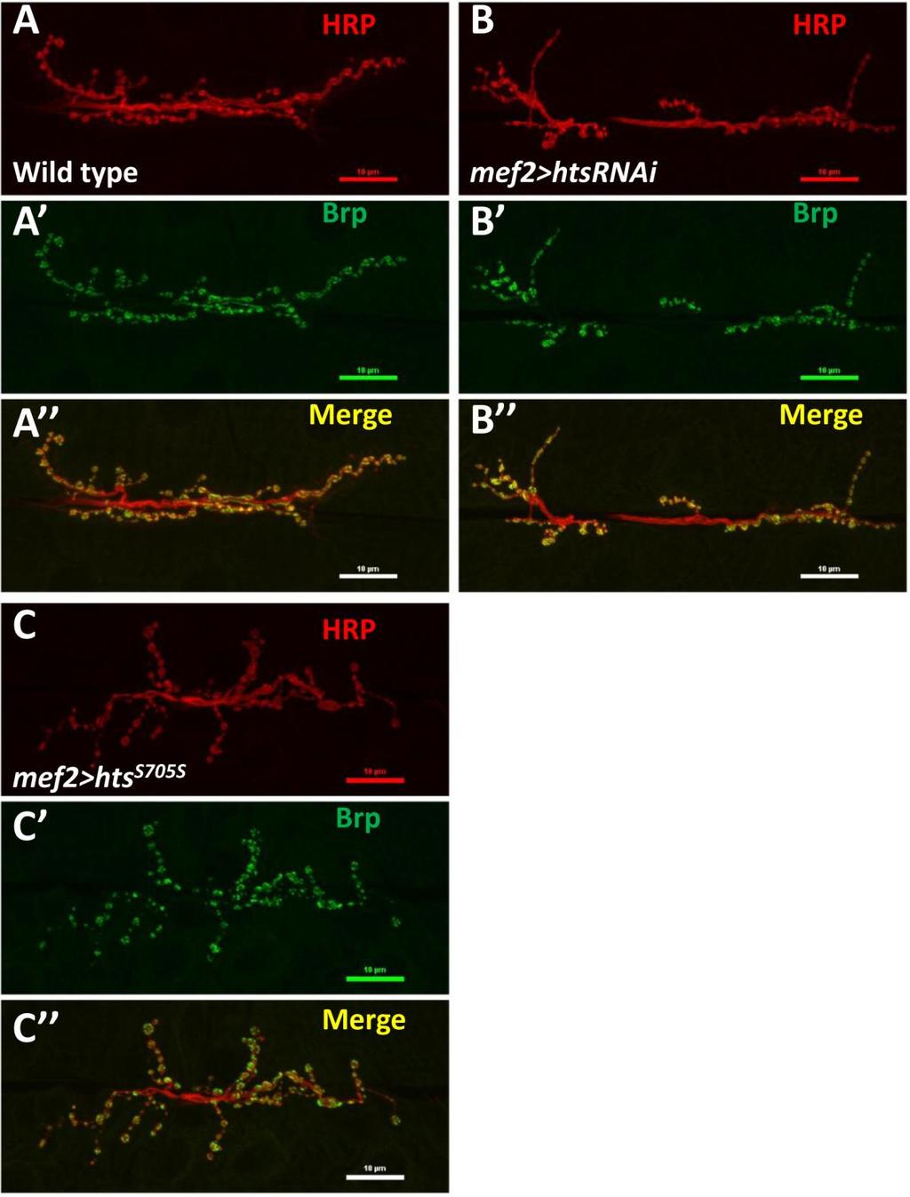
42 3.2. Muscle associated Hts regulates Drosophila larval NMJ development The neuromuscular defect found in mef2>htsrnai flies suggests that muscle associated Hts plays a role in Drosophila neuromuscular junction development. To evaluate the potential effects of muscle-associated Hts on NMJ development, wild type control larvae (wt), larvae with muscle specific hts knockdown (mef2>htsrnai), and larvae with muscle specific hts gain of function (mef2>hts S705S ) were examined. Third instar larvae were dissected to access the NMJs for immunostaining. Anti-HRP antibodies were used to label overall presynaptic regions (Jan and Jan, 1982) and anti- Bruchpilot were used to label active zones which are the sites for neurotransmitter release within each synaptic terminal/bouton (Fouquet et al., 2009). Immunostained images of NMJs at muscle 6/7 show obvious differences of morphology between wild type control, mef2>htsrnai and mef2>hts S705S larvae (Figure 3.3 A-C). Compared to wild type control larvae (Figure 3.3 A-A ), mef2>htsrnai larvae demonstrated an oversimplified NMJ morphology characterized by fewer boutons and less nerve branching into the muscle (Figure 3.3 B-B ). However, mef2>hts S705S larvae had overgrown NMJs characterized by more boutons and highly branched nerve terminals (Figure 3.3 C-C ). Quantitative analysis of average bouton number per NMJ showed that compared to wild type control (57 ± 2.2 boutons; mean ± SEM, n=22), the mean bouton number of mef2>htsrnai NMJs was significantly decreased (41 ± 2.0; mean ± SEM, n=23, p<0.01), and the mean bouton number of mef2>hts S705S NMJs was significantly increased (65 ± 0.4; mean ± SEM, n=22, p<0.05) (Figure 3.3D). Quantitative analysis of average nerve branch number per NMJ showed that compared to wild type control (5.6 ± 0.23 branches; mean ± SEM, n=22), the mean nerve branch number per NMJ in mef2>htsrnai larvae was significantly decreased (3.5 ± 0.28; mean ± SEM, n=23, p<0.05). Whereas the mean nerve branch number per NMJ in mef2>hts S705S larvae was significantly increased (8.6 ± 0.06; mean ± SEM, n=22, p<0.01) (Figure 3.3E). The effect of muscle-associated hts on NMJ development was further examined by conducting a quantitative analysis comparing the sizes of NMJ area. The NMJs were immunolabelled by anti-hrp (neuronal marker) and anti-draper (muscle marker) 30
43 antibodies. Images taken with lower magnification showed not only the NMJ staining, but also the whole area of muscle 6/7 that contains each individual NMJ (Figure 3.4 A-C). To standardize the data, a ratio was calculated between the area of each NMJ and its residing muscle 6/7 area (presented as a percentile). The calculated ratios can then be used to compare sizes of NMJs. This data (Figure 3.4D) shows that compared to sample mean of standardized NMJ area (each NMJ area / muscle 6/7 area, %) of wild type control (1.66% ± 0.07%; mean ± SEM, n=18), the mean of standardized NMJ area in mef2>htsrnai larvae is decreased by ~10% (1.48% ± 0.05%; mean ± SEM, n=18, p<0.05), indicating an underdeveloped phenotype. However, the mean of standardized NMJ area in mef2>hts S705S larvae is increased by ~20% (1.98% ± 0.09%; mean ± SEM, n=18, p<0.01), indicating an overgrown phenotype. These results suggest that muscle-associated Hts regulates Drosophila larval NMJ development. Muscle-specific hts knockdown causes underdeveloped NMJs, whereas muscle-specific hts gain of function causes NMJ overdevelopment. 31
44 32
 Immunostaining of representative neuromuscular synapses on muscle 6/7 (segment A3) in third instar larvae stained with")
45 Figure 3.3 Effect of muscle-associated hts on Drosophila larval NMJ morphology (A to C) Immunostaining of representative neuromuscular synapses on muscle 6/7 (segment A3) in third instar larvae stained with anti-hrp and anti-brp antibodies. All confocal images were taken using the 40x oil lens. Wild type NMJs are shown with immunolabelling against Hrp (A, red) and Brp (A, green). (A) is the merged image of the above two color channels. (B to B ) Muscle-specific hts knockdown (mef2>htsrnai) larvae show abnormal and oversimplified NMJ morphology, characterized by less synaptic boutons and less nerve branches. (C to C ) NMJs from muscle-specific hts overexpression (mef2>hts S705S ) show overgrown NMJ morphology showing more synaptic boutons and highly branched and over-extending nerve terminals. Scale bars, 10 µm (A-C ). (D and E) Quantification of nerve terminal growth of NMJs at muscle 6/7 showing total bouton number per NMJ (D) and total nerve branch number per NMJ (E). Each column (from left to right) represents wild type control (light blue, sample size n= 22), mef2>htsrnai (red, sample size n=23) and mef2>hts S705S (green, sample size n=22) respectively. Error bars represent SEM. Student t-test was used to examine statistical significance. One asterisk (*) indicates significant difference at p<0.05. Double asterisks indicate significant difference at p<
. Anti-Draper (Drpr) was used to label muscle area (red). Scale bar: 100 µm.")
46 Figure 3.4 Muscle-associated hts affects the size of Drosophila larval NMJs. (A to C) Immunostaining of muscle 6/7 NMJs and corresponding muscles. Anti-Hrp antibodies were used to label NMJ area (green). Anti-Draper (Drpr) was used to label muscle area (red). Scale bar: 100 µm. White discontinuous lines indicate the midline between muscle 6 and muscle 7. The muscle above the midline is muscle 6, the muscle below the midline is muscle 7. (D) Quantification of standardized NMJ size demonstrates that mef2>htsrnai larvae (red bar) have smaller NMJs at muscle 6/7 than do wild type control (light blue bar), whereas mef2>hts S705S larvae (green bar) have larger NMJs at muscle 6/7 compared to those in wild type (light blue bar). For each group, sample size is n=18 NMJs. Error bars represent SEM. Student t-test was used to examine statistical significance. One asterisk (*) indicates significant difference at p<0.05. Double asterisks (**) indicate significant difference at p<
47 3.3. Hts co-localizes with Draper at the post-synaptic region of Drosophila larval NMJ To study the mechanism of how hts affects the Drosophila larval NMJ development, it is helpful to search for molecules that may interact with hts in the synaptic compartment. Here I examined the potential interaction between hts and Draper, a synaptic molecule that has been shown to regulate Drosophila larval NMJ development (Fuentes-Medel et al., 2009). Immunostaining of Hts and Draper at muscle 6/7 NMJs showed that Hts immunoreactivity localized to each bouton evenly in donut-like pattern (Figure 3.5A, Figure 3.5B ). The immunoreactivity of Draper localized to each bouton in clusters making a discontinuous donut-like shape (Figure 3.5B ). The clusters of Draper immunostaining overlapped with Hts staining (Figure 3.5B ) suggesting that Draper is co-localized with Hts at the synapse. The Draper immunoreactivity localized predominantly to the peripheral portion of each bouton, suggesting that Draper localization at NMJ was largely post-synaptic. This co-localization of Draper and Hts suggests that Hts may interact with Draper in the postsynaptic area. 35
 is a representative immunostaining image of wild type larval NMJ at muscle 6/7. Scale bar: 10 µm. (B-B ) are higher power images of a bouton indicated by the asterisk in (A).")
48 Figure 3.5 Draper co-localizes with Hts at postsynaptic NMJ. Wild type larval body walls were co-stained with anti-hrp, anti-hts and anti-draper antibodies. (A) is a representative immunostaining image of wild type larval NMJ at muscle 6/7. Scale bar: 10 µm. (B-B ) are higher power images of a bouton indicated by the asterisk in (A). (B) shows anti-hrp staining (green channel). (B ) shows the anti-hts staining (blue channel). (B ) shows anti-draper staining (Red channel). (B ) is the merged image of (B)-(B ), arrow heads indicates co-localization of Hts and Draper at postsynaptic NMJ area. 36
49 3.4. Muscle-associated Hts affects Draper localization at Drosophila larval NMJ Effects of hts mutations on Draper immunoreactivity at Drosophila larval NMJ To study the potential interactions between Hts and Draper (Drpr), Drpr immunoreactivity at NMJ was examined in wild type, hts (null mutation of hts) and mef2>hts GS13858 (muscle-specific overexpression of all endogenous Hts proteins) larvae. Body wall preparations were co-stained using anti-hrp and anti-draper antibodies. As shown in Figure 3.6, immunostaining images revealed that compared to wild type, hts larvae have stronger immunoreactivity of Draper at the NMJ, whereas the immunoreactivity of Draper in mef2>hts GS13858 larvae is de-localized from the NMJ (Figure 3.6 A-C ). Quantification of the relative fluorescence intensity of Draper immunoreactivity was conducted using Adobe Photoshop CS3 software. Fluorescence intensities of both Draper and Hrp immunoreactivity (using Hrp as the control) were captured and measured. A ratio (relative fluorescence intensity of Draper) was then calculated at each NMJ between Draper fluorescence intensity and Hrp fluorescence intensity. The results show that there is about a 25% increase of relative fluorescence intensity of Draper at the NMJ in hts (0.52 ± 0.02; mean ± SEM, n=23, p<0.01) compared to wild-type (0.40 ± 0.01; mean ± SEM, n=23) (Figure 3.6D). Because the distribution of Draper immunoreactivity at NMJ is severely delocalized and weak in mef2>hts GS13858 larvae, the NMJ area cannot be accurately selected by Adobe Photoshop CS3 software (Figure 3.6C ) and the relative fluorescence intensity of Draper cannot be calculated. 37

50 38
 in wild type (A to A ), hts null mutation hts 01103 (B to B ) and muscle-specific endogenous hts overexpression (mef2>hts GS13858 )")
51 D Figure 3.6 Hts regulates the localization of Draper at Drosophila larval NMJ. Representative immunostainings of NMJs (muscle 6/7, A3 segment) in wild type (A to A ), hts null mutation hts (B to B ) and muscle-specific endogenous hts overexpression (mef2>hts GS13858 ) larvae (C to C ). Anti-Hrp staining is shown in green channel (A, B, C ), and anti-draper staining is shown in red channel (A, B, C ). Scale bar: 10 µm. hts null mutant (hts ) larvae show tighter localization of Draper staining at NMJ (B ) compared to wild type (A ). mef2>hts GS13858 larvae show severe loss of Draper immunoreactivity at the NMJ (C ). (D) Quantification of the ratio between Draper and Hrp synaptic fluorescence intensity (relative fluorescence intensity of Draper) shows that hts null mutation hts (orange bar) have about 25% stronger relative fluorescence intensity of Draper compare to in wild type (light blue bar). For both wild type and hts 01103, sample size is n=23 NMJs. Double asterisks (**) indicate significant difference at p<
52 Muscle-associated Hts regulates Draper localization at Drosophila larval NMJ In the previous data, a tighter distribution of Draper immunoreactivity was observed at NMJs in hts null mutant larvae where both presynaptic neuronal Hts and postsynaptic muscular Hts are knocked off. To determine whether this tighter Draper localization at the NMJ is caused by a pre- or postsynaptic loss of Hts, I used mef2>htsrnai to examine the effects of muscle-specific knockdown of Hts on the localization of Draper at the NMJ. Double labeling was performed using anti-hrp and anti-draper antibodies. Compared to wild type, mef2>htsrnai is observed to have stronger Draper immunoreactivity at the NMJ (Figure 3.7A to 3.7B ). Quantitative analysis of fluorescence intensity of the Draper signal supports the above observation by showing that compared to wild type (0.50 ± 0.01; mean ± SEM, n=20), there is a 20% increase of Draper relative fluorescence intensity in mef2>htsrnai NMJs (0.60 ± 0.01; mean ± SEM, n=24, p<0.01) (Figure 3.7C). These results indicate that muscle-specific knockdown of Hts cause tighter localization of Draper at NMJ. The increased level of Draper relative fluorescence intensity in muscle-specific hts knock-down larvae (20%) is very close to the increased level in hts null mutation (~25%), suggesting that muscleassociated Hts is an important regulator of Draper localization at NMJ, not presynaptic Hts. In the same experiment, I also observed that Draper delocalized from the NMJ in mef2>hts GS13858 larvae where endogenous Hts proteins were overexpressed in the muscle. However, the specific isoform(s) of Hts proteins that is/are actually causing the delocalization of Draper is unknown. Given that Add1 is the main isoform of Hts protein during Drosophila synapse development (Pielage et al., 2011), and as Add1 is speculated to be one of the predominant isoforms of Hts at the post-synaptic muscle region (Wang et al, 2011; Petrella et al., 2007), it was intriguing to examine the effect of muscle-specific overexpression of Add1 on the distribution of Draper immunoreactivity at the NMJ. I then crossed the muscle driver mef2-gal4 flies with UAS-hts S705S (Add1 transgene) to obtain larvae with muscle-specific overexpression of Add1. Double immunostaining of anti-hrp and anti-draper shows that compared to the tight localization of Draper at the NMJ of wild type (Figure 3.8A, 3.8A ), the immunostaining of Draper in mef2>hts S705S delocalized from the NMJ and gave a fuzzy appearance 40
53 (Figure 3.8B, 3.8B ). Quantification of relative fluorescence intensity of Draper supports the above observation by showing that compared to in wild type (0.52 ± 0.01; mean ± SEM, n=21), the relative fluorescence intensity of Draper at NMJ is decreased in mef2>hts S705S larvae (0.46 ± 0.01; mean ± SEM, p<0.01, n=22). This indicates that overexpression of Add1, a predominant isoform of Hts proteins in the muscle, can delocalize Draper from the postsynaptic region. All the above results suggest that muscle-associated Hts plays a role in regulating the localization of Draper protein at the postsynaptic NMJ. Loss of Hts in muscle causes tighter localization of Draper at the NMJ, whereas up-regulation of Hts in muscle causes delocalization of Draper from the NMJ. 41
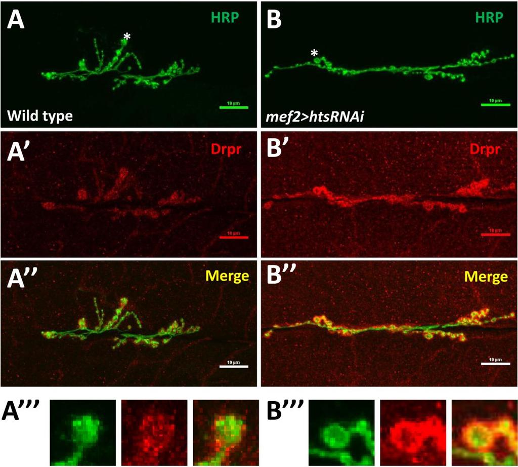
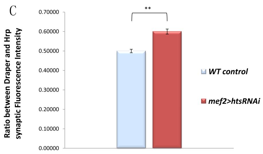
54 42
55 Figure 3.7 Muscle-specific knockdown of Hts causes tighter localization of Draper at the NMJ Representative immunostainings of NMJs (muscle 6/7, A3 segment) in wild type (A to A ) and muscle-specific hts knockdown (mef2>htsrnai) larvae (B to B ). Anti-Hrp staining is shown in green channel (A, B), and anti-draper (Drpr) staining is shown in red channel (A, B ). Merged images of the two channels are (A, B ). Scale bar: 10 µm. (A and B ) are higher power images of the boutons indicated by the asterisks in (A) and (B). Musclespecific hts knockdown (mef2>htsrnai) show tighter localization of Draper staining at NMJ (B, B ) compared to wild type (A, A ). (C) Quantification of the ratio between Draper and Hrp synaptic fluorescence intensity (relative fluorescence intensity of Draper) shows that muscle-specific hts knockdown (mef2>htsrnai) larvae (red bar) have 20% stronger relative fluorescence intensity of Draper at the NMJ compared to in wild type (light blue bar). Sample size of wild type is n=20 NMJs. Sample size of mef2>htsrnai is n=24 NMJs. Double asterisks (**) indicate significant difference at p<

56 44
57 Figure 3.8 Muscle-specific overexpression of Add1 causes delocalization of Draper from postsynaptic NMJ Representative immunostaining of NMJs (muscle 6/7, A3 segment) in wild type (A to A ) and with muscle-specific overexpression of Add1 (mef2>hts S705S ) larvae (B to B ). Anti- Hrp staining is shown in green channel (A, B), and anti-draper staining is shown in red channel (A, B ). Merged images of the two channels are (A, B ). Scale bar: 10 µm. (A ) and (B ) are higher power images of the boutons indicated by the asterisks in (A) and (B). Compared to the tight localization of Draper at NMJ in wild type (A, A ), the immunostaining of Draper in mef2>hts S705S delocalizes from the NMJ and gives a fuzzy appearance (B, B ). (C) Quantification of the ratio between Draper and Hrp synaptic fluorescence intensity (relative fluorescence intensity of Draper) shows that compared to in wild type (light blue bar), the relative fluorescence intensity of Draper at NMJ in mef2>hts S705S larvae (green bar) is significantly decreased, which indicates that Draper is delocalized from NMJ. Sample size of wild type is n=21 NMJs. Sample size of mef2> hts S705S is n=22 NMJs. Double asterisks (**) indicate significant difference at p<
58 The delocalization of Draper seen in muscle-associated Hts gain of function larvae is not caused by a general muscle defect It is possible that muscle-specific hts overexpression could interfere with muscle development. This possible developmental defect of muscle could affect the proper targeting of all the postsynaptic proteins to the NMJ. If this is true, the delocalization of Draper seen in muscle-associated hts gain of function animals might not be caused by specific Hts-Draper interactions. To evaluate this possibility, the distributions of immuno-reactivity of two postsynaptic proteins, Pak and Glutamate receptor IIb (GluR-IIb), were investigated in mef2>hts S705S larvae. The results show that both the immunoreactivity of Pak (Figure 3.9 B, 3.9B ) and GluR-IIb (Figure 3.10B, 3.10B ) are tightly localized to the postsynaptic area in mef2>hts S705S larvae. The relative fluorescence intensity of Pak at NMJ is even slightly stronger in mef2>hts S705S larvae (0.54 ± 0.03; mean ± SEM, n=8, p<0.05) than in wild type larvae (0.48 ± 0.01; mean ± SEM, n=8) (Figure 3.9C). The relative fluorescence intensity of GluR-IIb at NMJ is decreased in mef2>hts S705S larvae (0.44 ± 0.01; mean ± SEM, n=8, p<0.01) compared to wild type larvae (0.58 ± 0.03, mean ± SEM, n=8) (Figure 3.10C). However the immunostaining of GluR-IIb is still tight and sharp at the NMJ. The fuzzy staining pattern of Draper was not observed in GluR- IIb staining. The above data indicate that other postsynaptic proteins were properly targeted to the NMJ area. Therefore the delocalization of Draper seen in larvae with muscle associated hts overexpression is likely not be caused by a general developmental defect of muscle. 46

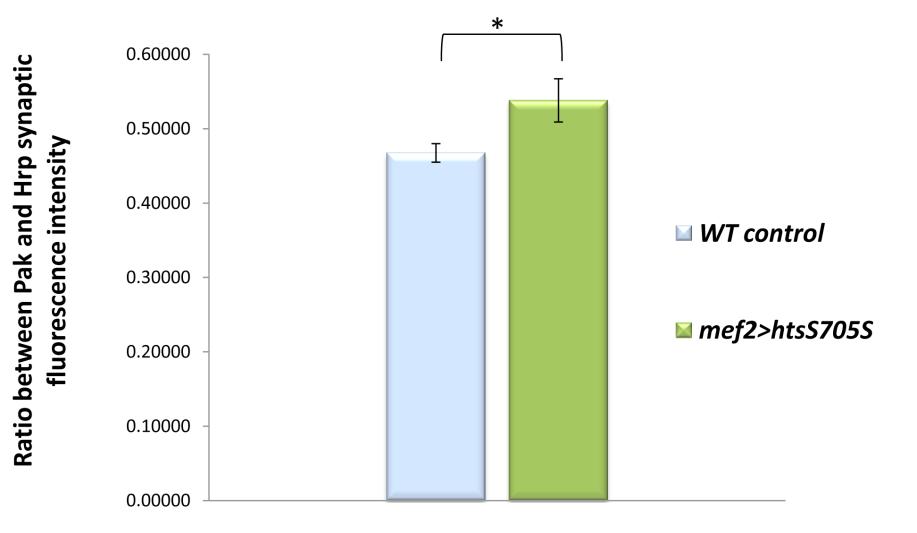
59 C 47
60 Figure 3.9 Anti-Pak immunostaining is tightly localized to the postsynaptic NMJ area in muscle-specific hts gain of function larvae. Representative immunostainings of NMJs (muscle 6/7, A3 segment) in wild type (A to A ) and with muscle-specific hts gain of function (mef2>hts S705S ) larvae (B to B ). Anti-Hrp staining is shown in the green channel (A, B), and anti-pak staining is shown in the red channel (A, B ). Merged images of the two channels are (A, B ). Scale bar: 10 µm. (A ) and (B ) are higher power images of the boutons indicated by the asterisks in (A) and (B). The immunoreactivity of Pak is tightly localized to postysynaptic NMJ in both wild type (A, A ) and mef2>hts S705S larvae (B, B ). (C) Quantification of the ratio between Pak and Hrp synaptic fluorescence intensity (relative fluorescence intensity of Pak) shows that compared to in wild type (light blue bar), the relative fluorescence intensity of Pak at NMJ is increased in mef2>hts S705S larvae. For both wild type and mef2>hts S705S larvae, sample size is n=8 NMJs. One asterisk (*) indicates significant difference at p<
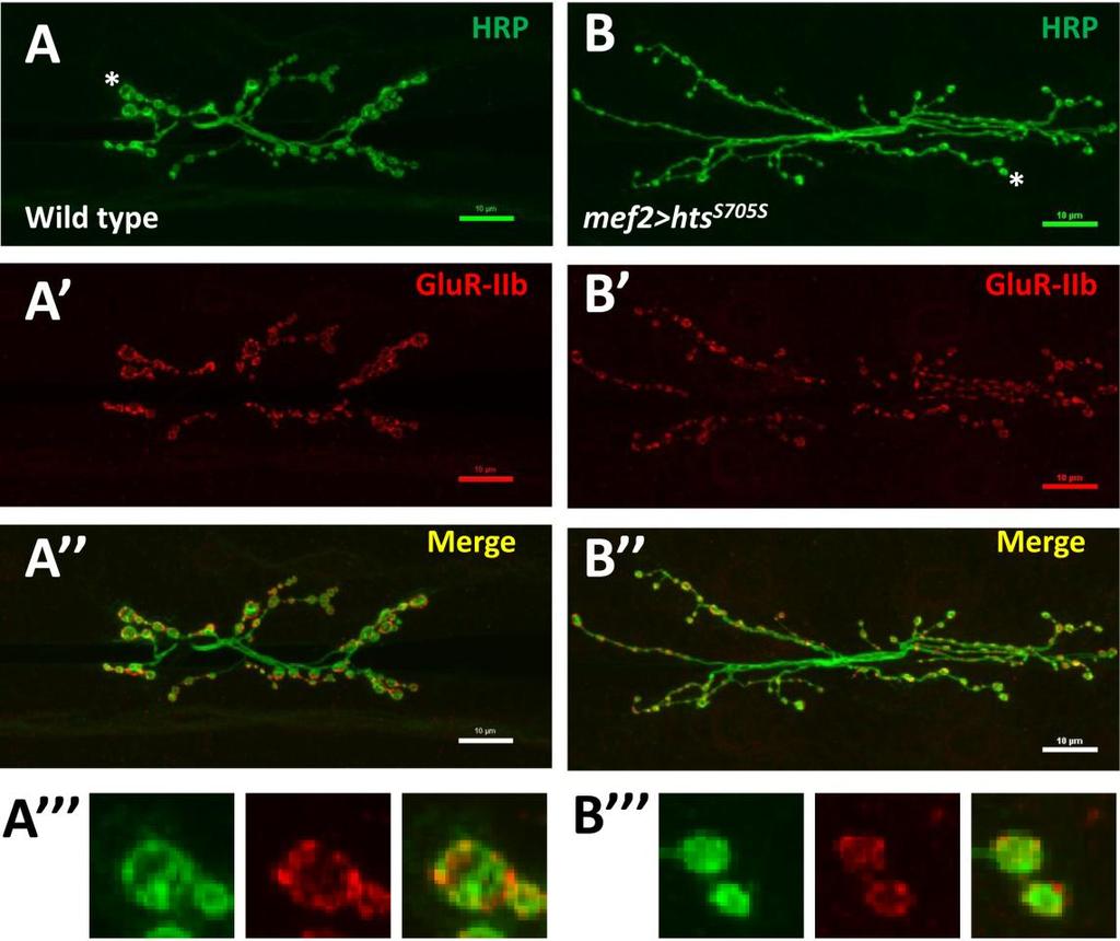
61 C 49
62 Figure 3.10 Anti-GluR-IIb immunostaining is tightly localized to the postsynaptic area in muscle-specific hts gain of function larvae. Representative immunostainings of NMJs (muscle 6/7, A3 segment) in wild type (A to A ) and with muscle-specific hts gain of function (mef2>hts S705S ) larvae (B to B ). Anti-Hrp staining is shown in the green channel (A, B), and anti-glur-iib staining is shown in the red channel (A, B ). Merged images of the two channels are (A, B ).Scale bar: 10 µm. (A ) and (B ) are higher power images of the boutons indicated by the asterisks in (A) and (B).The immunoreactivity of GluR-IIb is tightly localized to postysynaptic NMJ in both wild type (A, A ) and mef2>hts S705S larvae (B, B ). (C) Quantification of the ratio between GluR-IIb and Hrp synaptic fluorescence intensity (relative fluorescence intensity of GluR-IIb) shows that compared to in wild type (light blue bar), the relative fluorescence intensity of GluR-IIb at NMJ is decreased in mef2>hts S705S larvae. For both wild type and mef2>hts S705S larvae, sample size is n=8 NMJs. Double asterisks (**) indicate significant difference at p<
63 3.5. Muscle-associated Hts does not affect the protein level of Draper in larval body walls Identifying the positions of Draper protein bands in Western blotting In order to study whether manipulations of muscle-associated Hts affect the protein level of Draper in larval body walls using Western blotting, it is helpful to firstly identify the band positions of corresponding Drpr proteins. There are three isoforms of Draper proteins (Drpr-I, II, III) in Drosophila, having different structures and molecular weights (Fuentes-Medel et al., 2009). To identify their band positions, Western blotting of larval body wall lysates of wild type control larvae and three transgenic larvae (mef2>drpr-i, mef2>drpr-ii, mef2>drpr-iii) can be performed using the anti-draper antibody (detecting all three Draper isoforms). Because the cdna of each Draper isoform is specifically overexpressed in the muscle of corresponding transgenic larvae, we should be able to identify the band position of each isoform by picking out the band with a significant increase of signal intensity in each drpr transgenic sample. In this experiment, the protein concentration of loading samples needed to be diluted to 10 µg/100 µl to obtain a clean blotting result. When the protein concentration of loading samples was so diluted, the signal of endogenous Draper proteins was below the level of detection in the blots. This explains why no blotting signal was detected in wild type body wall lysates (Figure 3.11). However, the signal of overexpressed Draper isoforms is detectable in the samples of drpr transgenes. As shown in Figure 3.11, the blots demonstrated a protein band of Draper-I (~130KD), a band of Draper-II and a band of Draper-III (both ~70KD) specifically in each corresponding sample of drpr transgenes. The positions of these bands are consistent with a previous observation of Western blotting for Draper in Drosophila salivary glands (McPhee et al., 2010). 51
: wild-type, mef2>drpr-i (where drpr-i is")
64 Figure 3.11 Western blotting of Draper identified the band position of every isoform of Draper proteins Sample lysates loaded into the gels are (from right to left): wild-type, mef2>drpr-i (where drpr-i is specifically overexpressed in muscle), mef2>drpr-ii (where drpr-ii is specifically overexpressed in muscle) and mef2>drpr-iii (where drpr-iii is specifically overexpressed in muscle). The anti-draper antibodies used in this blotting detects all three isoforms of Draper proteins. There is no band signal detected in wild-type, because the sample lysates was too diluted to give detectable signal of endogenous Draper bands. The band of Draper-I is detected (at the molecular weight of about 130kD) in mef2>drpr-i sample lysates. The band of Draper-II is detected (at the molecular weight of around 70kD) in mef2>drpr-ii. The band of Draper-III appears at a similar position to the Draper-II band in mef2>drpr-iii. 52
65 Protein levels of Draper proteins in larval body wall lysates are not affected by muscle-associated Hts variation Western blotting of Draper was performed to compare Draper protein level in wild-type control (mef2>w 1118 ), muscle-associated hts knock-down (mef2>htsrnai) and muscle-specific hts gain of function (mef2>hts S705S ) larval body wall lysates. The body wall lysates of drpr Δ5, a drpr null mutation, was also used as a negative control. β-actin was also blotted using a mouse monoclonal antibody (JLA20, DHSB) to serve as a loading control. A previous study has shown that only drpr-i and drpr-iii are expressed in Drosophila larval body wall tissues, whereas drpr-ii is not expressed (Fuentes-Medel et al., 2009). Thus the band signals appearing at ~70kD in this blotting should correspond to Draper-III only. As shown in Figure 3.12, Western blotting of Draper did not reveal significant difference in either Draper-I or Draper-III protein level among wild type control, mef2>htsrnai and mef2>hts S705S larval body wall lysates. Quantification of normalized relative band densities of Draper-I showed no significant difference among the samples evaluated (wild-type control: ± 0.000; mef2>htsrnai: ± 0.110, p=0.9482; mef2>hts S705S : ± 0.149, p=0.7140; mean ± SEM, n=6). Quantification of normalized relative band intensities of Draper-III did not reveal significant difference among hts variants either (wild-type control: ± 0.000; mef2>htsrnai: 1.021± 0.090, p=0.8202; mef2>hts S705S : ± 0.158, p=0.4059; mean ± SEM, n=6). These results suggest that muscle-associated Hts does not affect the expression level of drpr in Drosophila larval body wall. 53

66 Figure 3.12 Muscle-associated Hts variation does not affect the protein levels of Draper-I/-III in Drosophila larval body wall lysates. Protein levels of Draper-I and Draper-III were compared among the body wall lysates of three hts variants using Western blotting with a-draper antibodies. drpr Δ5, a null mutation of draper, was used as the negative control. All the three hts variants showed detectable protein bands at ~ 130kD indicating Draper-I, and protein bands at ~ 70kD indicating Draper-III. Protein bands of Draper-I and Draper-III were not detected in drpr Δ5. No significant differences were found in the protein level of either Draper-I or Draper-III in body wall lysates from three hts variants. Blots were standardized by loading control β- actin. 54
67 3.6. Dlg is also delocalized from NMJ in a similar pattern to Drpr in larvae overexpressing Drosophila adducin in the muscle Dlg has been previously shown to be delocalized from the NMJ in larvae overexpressing endogenous hts (using Gene search line) in the muscle by Hts overexpression in the muscle (Wang et al., 2011). However, since all the isoforms of Hts protein are overexpressed, it is not cleat if this delocalization of Dlg is caused by adducin-like Hts isoforms. Here I overexpressed Add1 (the adducin-like isoform) in the muscle by crossing UAS-hts S705S transgenic line with mef2-gal4 driver flies, and examined how Add1 overexpression affects Dlg localization to the NMJ. As shown in Figure 3.13 A, B, Dlg is delocalized from the NMJ in mef2>hts S705S larvae. Interesting, the delocalization pattern of Dlg is similar to that of Drpr (Figure 3.13 A, A, B B ), suggesting that Dlg and Drpr may exist in the same protein complex and a potential interactions between Dlg and Drpr. 55
 in wild type (A to A ) and larvae with muscle-specific overexpression of Drosophila adducin (mef2>hts S705S ) (B to B ).")
68 Figure 3.13 Dlg is delocalized from the NMJ in a similar pattern to Drpr in larvae overexpressing Drosophila adducin in the muscle. Representative immunostainings of NMJs (muscle 6/7, A3 segment) in wild type (A to A ) and larvae with muscle-specific overexpression of Drosophila adducin (mef2>hts S705S ) (B to B ). Higer power image of a individual bouton (indicated by the asterisk) is shown at the bottom right corner in each panel. Anti-Hrp staining is shown in the green channel (A, B), anti-drpr staining is shown in the red channel (A, B ), anti-dlg staining is shown in the blue channel (A, B ). Merged images of the three channels are (A, B ). Scale bar: 10 µm. Sample size: wild type=12 NMJs, mef2>hts S705S =12 NMJs. 56
69 3.7. Dlg regulates Draper immunoreactivity at the NMJ Given that overexpression of Hts in the muscle also delocalized Dlg from the NMJ (in the present study and Wang et al., 2011), and that the immunoreactivity of Draper colocalizes with that of Dlg at the postsynaptic NMJ (Fuentes-Medel et al., 2009), it is possible that Dlg interacts with Draper at the postsynaptic NMJ, so that the delocalization of Draper caused by Hts overexpression is mediated by Dlg. To examine the above hypothesis, Draper immunoreactivity at the NMJ was examined in wild type control larvae, and larvae with muscle specific Dlg knockdown (mef2>dlgrnai). In wild type control larvae, Draper immunoreactivity is sharp and tightly localized to the postsynaptic NMJ (Figure 3.14A ). However, in mef2>dlgrnai larvae where Dlg immunoreactivity is absent (Figure 3.14B ), the immunostaining of Draper is fuzzy (Figure 3.14B ) indicating that the localization of Draper to the postsynaptic NMJ is affected. However, Draper immunoreactivity is not totally absent from the NMJ in mef2>dlgrnai larvae, suggesting that Draper targeting to the NMJ is not solely dependent on Dlg. If Dlg interacts with Draper and affects Draper distribution at the NMJ, we should expect an upregulation of Draper immunoreactivity at the NMJ in larvae overexpressing Dlg in the muscle compared to wild type larvae. Two Dlg isoforms (Dlg-A and Dlg-S97) are present at the Drosophila larval NMJ (Ataman et al., 2006). The effects of overexpression of each Dlg isoform on Draper immunoreactivity were examined here. Larvae with muscle-specific overexpression of Dlg-A showed a 20% increase of Draper immunoreactivity at the NMJ compared to wild type control (Figure 3.15A, B and D). Whereas there is no significant change of Draper immunoreactivity detected in larvae overexpressing Dlg-S97 compared to wild type (Figure 3.15A,C and D). These data suggest that only the isoform Dlg-A interacts with Draper at postsynaptic NMJ. 57
 in wild type (A to A ) and larvae with muscle-specific knockdown of Dlg (mef2>dlgrnai) (B to B ).")
70 Figure 3.14 Draper localization at the postsynaptic NMJ is affected in larvae with muscle-specific knock-down. Representative immunostainings of NMJs (muscle 6/7, A3 segment) in wild type (A to A ) and larvae with muscle-specific knockdown of Dlg (mef2>dlgrnai) (B to B ). Anti- Hrp staining is shown in the green channel (A, B), anti-drpr staining is shown in the red channel (A, B ), anti-dlg staining is shown in the blue channel (A, B ). Merged images of the three channels are (A, B ). Scale bar: 10 µm. Sample size: wild type=12 NMJs, mef2>dlgrnai =12 NMJs. 58
71 59
 in wild type (A to A ), larvae with muscle-specific overexpression of Dlg-A (mef2>dlg A ) (B to B ), and larvae with muscle-specific")
72 D Figure 3.15 Muslce specific overexpression of Dlg-A, but not Dlg-S97, results in stronger Draper immunoreactivity at the NMJ. Representative immunostainings of NMJs (muscle 6/7, A3 segment) in wild type (A to A ), larvae with muscle-specific overexpression of Dlg-A (mef2>dlg A ) (B to B ), and larvae with muscle-specific overexpression of Dlg-S97 (mef2>dlg S97 ) (C to C ). Anti-Hrp staining is shown in the green channel (A, B, C), and anti-draper staining is shown in the red channel (A, B, C ). Merged images of the two channels are (A, B, C ). (C) Quantification of the ratio between Draper and Hrp synaptic fluorescence intensity. Sample size: wild type=17 NMJs, mef2>dlg A =18 NMJs, mef2>dlg S97. Double asterisks (**) indicate significant difference at p<
73 3.8. Draper null mutant larvae exhibit decreased Hts immunoreactivity at the NMJ and possibly affects Hts function. Given the morphological similarities between hts and drpr mutant NMJ, their colocalization at the post-synaptic region, and that Hts negatively regulates Draper localization via a specific Hts-Draper interaction, it is intriguing to examine if genetic manipulations of draper affects Hts immunoreactivity. The drpr 5 putative null allele was examined for changes in Hts levels by immunostaining. As shown in Figure 3.16, NMJs from larvae homozygous for drpr null mutant alleles showed similar Hts localization patterns compared to wildtype (Figure 3.16 A and B ), but had a 30% decrease in Hts immunoreactivity (Figure 3.16 A, B and C). Consistently, mild decreases in Hts immunoreactivity (by ~17%) were also observed in two other hypomorphic drpr alleles (Chui, 2011). These results suggest that the expression level of Hts is affected by Drpr loss of function. When the immunoreactivity of Hts was examined in larvae with muscle-specific knock down of Draper (mef2>drprrnai), there is only a slight decrease of Hts immunoreactivity detected compared to wildtype (Figure 3.17 A, B and C). Long, small-caliber neuronal protrusions were observed in hts mutant NMJs (Pielage et al., 2011). In my study, Drpr null mutant NMJs were observed to have a higher frequency on presenting long, small-caliber neuronal protrusions (Figure 3.18). About 44% examined drpr null mutant NMJs (n=18) had neuronal protrusions, whereas no protrusions were observed in wildtype NMJs (n=18). This similar phenotype suggests that Hts function is affected at the drpr null mutant NMJ. 61
74 c 62
75 Figure drpr mutant NMJs show decreased Hts immunoreactivity. Representative immunostainings of NMJs (muscle 6/7, A3 segment) in wild type (A to A ) and drpr null mutant larvae (B to B ). Anti-Hrp staining is shown in the green channel (A, B), and anti-hts staining is shown in the red channel (A, B ). Merged images of the two channels are (A, B ).Scale bar: 10 µm. (C) Quantification of the ratio between Hts and Hrp synaptic fluorescence intensity. Sample size: wild type=14 NMJs, drpr 5 =18 NMJs. Double asterisks (**) indicate significant difference at p<
76 c 64
77 Figure 3.17 Hts immunoreactivity is slightly decreased at the NMJ in larvae with muscle specific knock-down of Draper. Representative immunostainings of NMJs (muscle 6/7, A3 segment) in wild type (A to A ) and larvae with muscle specific Draper knock-down (B to B ). Anti-Hrp staining is shown in the green channel (A, B), and anti-hts staining is shown in the red channel (A, B ). Merged images of the two channels are (A, B ). (C) Quantification of the ratio between Hts and Hrp synaptic fluorescence intensity. Sample size: wild type=17 NMJs, mef2>drprrnai =18 NMJs. 65
 in wild type (A to A ) and drpr null mutant larvae (B to B ).")
78 Figure 3.18 drpr null mutant NMJ exhibits long, small-caliber neuronal protrusions. Representative immunostainings of NMJs (muscle 6/7, A3 segment) in wild type (A to A ) and drpr null mutant larvae (B to B ). Anti-Hrp staining is shown in the green channel (A, B), and anti-dlg staining is shown in the red channel (A, B ). Merged images of the two channels are (A, B ). 44% of the examined NMJs in drpr null mutant larvae showed long, small-caliber neuronal protrusions (indicated by arrows in B-B. Protrusions were not observed in wild type control. Sample size: wild type=18 NMJs, drpr 5 =18 NMJs. 66
79 3.9. Draper regulates Dlg immunoreactivity at the larval NMJ To study whether the interaction between Dlg and Draper is co-dependent, Dlg immunoreactivity at the NMJ was examined in larvae expressing different levels of Draper, including wild type control, drpr 5 null mutant alleles, and larvae with musclespecific overexpression of Draper-I. The results showed that drpr 5 null mutant NMJs had a slight (but not significant) decrease of Dlg immunoreactivity compared to wild type control (Figure 3.19 A, B and C). However, when Draper-I is overexpressed in the muscle, there was a ~40% increase of Draper immunoreactivity at the NMJ compared to wild type control (p<0.01, Figure 3.20 A, B and C). These results suggest that Draper is not required for proper Dlg targeting to the NMJ, however upregulating Draper in the muscle promotes Dlg expression or targeting to the NMJ. 67
 in wild type (A to A ) and drpr null mutant larvae (B to B ).")
. (C) Quantification of the ratio between Dlg and Hrp synaptic fluorescence intensity.")
80 c Figure 3.19 drpr mutant NMJs show slightly decreased Dlg immunoreactivity. Representative immunostainings of NMJs (muscle 6/7, A3 segment) in wild type (A to A ) and drpr null mutant larvae (B to B ). Anti-Hrp staining is shown in the green channel (A, B), and anti-dlg staining is shown in the red channel (A, B ). Merged images of the two channels are (A, B ). (C) Quantification of the ratio between Dlg and Hrp synaptic fluorescence intensity. Sample size: wild type=21 NMJs, drpr 5 =20 NMJs. No statistically significant difference. 68
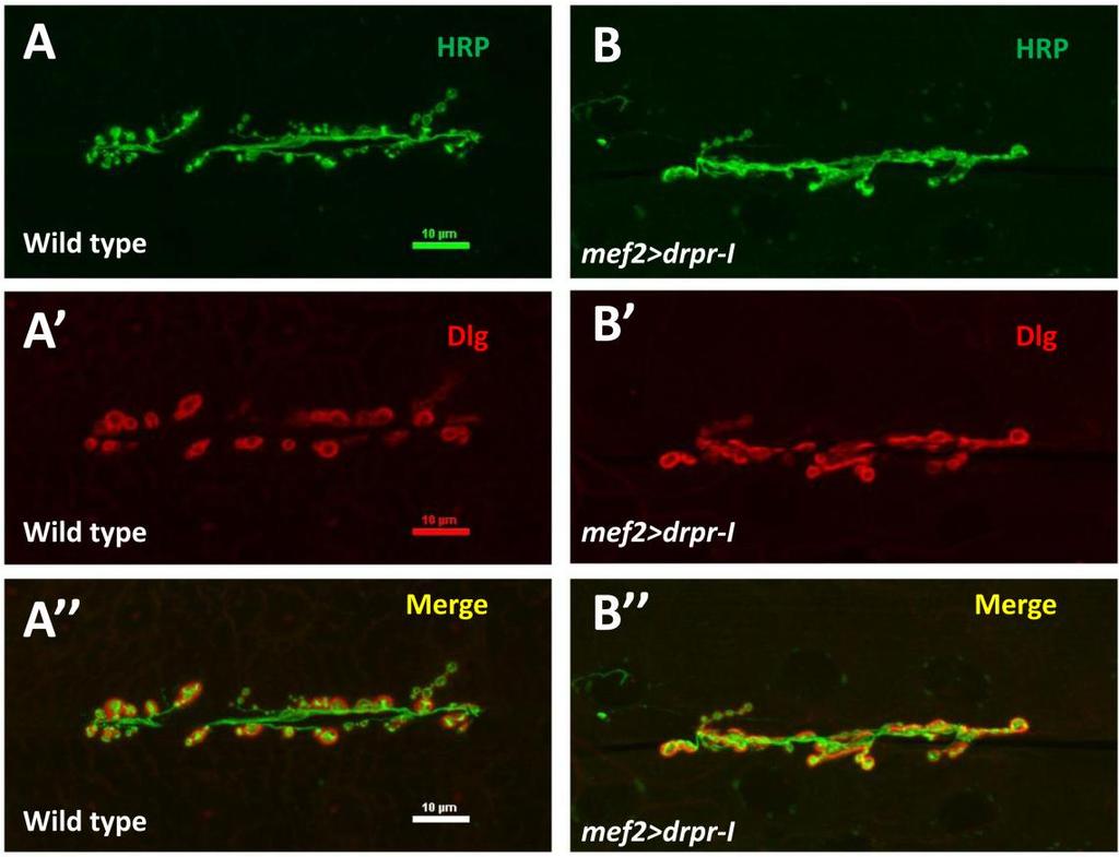
81 c 69
SUPPLEMENTARY INFORMATION
 doi:10.1038/nature10923 Supplementary Figure 1 Ten-a and Ten-m antibody and cell type specificities. a c, Representative single confocal sections of a Drosophila NMJ stained with antibodies to Ten-a (red),
Cells. Steven McLoon Department of Neuroscience University of Minnesota
 Cells Steven McLoon Department of Neuroscience University of Minnesota 1 Microscopy Methods of histology: Treat the tissue with a preservative (e.g. formaldehyde). Dissect the region of interest. Embed
Chapter 4 Evaluating a potential interaction between deltex and git in Drosophila: genetic interaction, gene overexpression and cell biology assays.
 Evaluating a potential interaction between deltex and git in Drosophila: genetic interaction, gene overexpression and cell biology assays. The data described in chapter 3 presented evidence that endogenous
Neurite formation & neuronal polarization
 Neurite formation & neuronal polarization Paul Letourneau letou001@umn.edu Chapter 16; The Cytoskeleton; Molecular Biology of the Cell, Alberts et al. 1 An immature neuron in cell culture first sprouts
Introduction Principles of Signaling and Organization p. 3 Signaling in Simple Neuronal Circuits p. 4 Organization of the Retina p.
 Introduction Principles of Signaling and Organization p. 3 Signaling in Simple Neuronal Circuits p. 4 Organization of the Retina p. 5 Signaling in Nerve Cells p. 9 Cellular and Molecular Biology of Neurons
"Small Brains, Big Ideas: The value of model organisms to science. Dr. Yuly Fuentes-Medel Executive Director Descience
 "Small Brains, Big Ideas: The value of model organisms to science Dr. Yuly Fuentes-Medel Executive Director Descience Outline 1. What is a model organism? 2. Big questions solved by small-brained organisms
Neurite formation & neuronal polarization. The cytoskeletal components of neurons have characteristic distributions and associations
 Mechanisms of neuronal migration & Neurite formation & neuronal polarization Paul Letourneau letou001@umn.edu Chapter 16; The Cytoskeleton; Molecular Biology of the Cell, Alberts et al. 1 The cytoskeletal
Neural development its all connected
 Neural development its all connected How do you build a complex nervous system? How do you build a complex nervous system? 1. Learn how tissue is instructed to become nervous system. Neural induction 2.
Nervous System Organization
 The Nervous System Nervous System Organization Receptors respond to stimuli Sensory receptors detect the stimulus Motor effectors respond to stimulus Nervous system divisions Central nervous system Command
Neuronal Differentiation: Synapse Formation NMJ
 Neuronal Differentiation: Synapse Formation NMJ Approaches to study synapse formation Rodent NMJ (cholinergic) Drosophila NMJ (glutamatergic) Rodent CNS synapse (glutamatergic or GABAergic) C. elegans
Baz, Par-6 and apkc are not required for axon or dendrite specification in Drosophila
 Baz, Par-6 and apkc are not required for axon or dendrite specification in Drosophila Melissa M. Rolls and Chris Q. Doe, Inst. Neurosci and Inst. Mol. Biol., HHMI, Univ. Oregon, Eugene, Oregon 97403 Correspondence
Neurite initiation. Neurite formation begins with a bud that sprouts from the cell body. One or several neurites can sprout at a time.
 Neurite initiation. Neuronal maturation initiation f-actin polarization and maturation tubulin stage 1: "spherical" neuron stage 2: neurons extend several neurites stage 3: one neurite accelerates its
Chapter 37 Active Reading Guide Neurons, Synapses, and Signaling
 Name: AP Biology Mr. Croft Section 1 1. What is a neuron? Chapter 37 Active Reading Guide Neurons, Synapses, and Signaling 2. Neurons can be placed into three groups, based on their location and function.
SUPPLEMENTARY INFORMATION
 DOI: 10.1038/ncb3267 Supplementary Figure 1 A group of genes required for formation or orientation of annular F-actin bundles and aecm ridges: RNAi phenotypes and their validation by standard mutations.
5- Semaphorin-Plexin-Neuropilin
 5- Semaphorin-Plexin-Neuropilin 1 SEMAPHORINS-PLEXINS-NEUROPILINS ligands receptors co-receptors semaphorins and their receptors are known signals for: -axon guidance -cell migration -morphogenesis -immune
Cellular Neurobiology BIPN 140 Fall 2016 Problem Set #8
 Cellular Neurobiology BIPN 140 Fall 2016 Problem Set #8 1. Inductive signaling is a hallmark of vertebrate and mammalian development. In early neural development, there are multiple signaling pathways
Chapter 18 Lecture. Concepts of Genetics. Tenth Edition. Developmental Genetics
 Chapter 18 Lecture Concepts of Genetics Tenth Edition Developmental Genetics Chapter Contents 18.1 Differentiated States Develop from Coordinated Programs of Gene Expression 18.2 Evolutionary Conservation
10/2/2015. Chapter 4. Determination and Differentiation. Neuroanatomical Diversity
 Chapter 4 Determination and Differentiation Neuroanatomical Diversity 1 Neurochemical diversity: another important aspect of neuronal fate Neurotransmitters and their receptors Excitatory Glutamate Acetylcholine
Neurons and Nervous Systems
 34 Neurons and Nervous Systems Concept 34.1 Nervous Systems Consist of Neurons and Glia Nervous systems have two categories of cells: Neurons, or nerve cells, are excitable they generate and transmit electrical
Nervous Tissue. Neurons Neural communication Nervous Systems
 Nervous Tissue Neurons Neural communication Nervous Systems What is the function of nervous tissue? Maintain homeostasis & respond to stimuli Sense & transmit information rapidly, to specific cells and
Cellular Neuroanatomy II The Prototypical Neuron: Neurites. Reading: BCP Chapter 2
 Cellular Neuroanatomy II The Prototypical Neuron: Neurites Reading: BCP Chapter 2 Major Internal Features of a Neuron The neuron is the functional unit of the nervous system. A typical neuron has a soma
Nervous Tissue. Neurons Electrochemical Gradient Propagation & Transduction Neurotransmitters Temporal & Spatial Summation
 Nervous Tissue Neurons Electrochemical Gradient Propagation & Transduction Neurotransmitters Temporal & Spatial Summation What is the function of nervous tissue? Maintain homeostasis & respond to stimuli
Neurons, Synapses, and Signaling
 LECTURE PRESENTATIONS For CAMPBELL BIOLOGY, NINTH EDITION Jane B. Reece, Lisa A. Urry, Michael L. Cain, Steven A. Wasserman, Peter V. Minorsky, Robert B. Jackson Chapter 48 Neurons, Synapses, and Signaling
Anterograde Activin Signaling Regulates Postsynaptic Membrane Potential and GluRIIA/B Abundance at the Drosophila Neuromuscular Junction
 Anterograde Activin Signaling Regulates Postsynaptic Membrane Potential and GluRIIA/B Abundance at the Drosophila Neuromuscular Junction Myung-Jun Kim, Michael B. O Connor* Department of Genetics, Cell
Bypass and interaction suppressors; pathway analysis
 Bypass and interaction suppressors; pathway analysis The isolation of extragenic suppressors is a powerful tool for identifying genes that encode proteins that function in the same process as a gene of
Signal Transduction. Dr. Chaidir, Apt
 Signal Transduction Dr. Chaidir, Apt Background Complex unicellular organisms existed on Earth for approximately 2.5 billion years before the first multicellular organisms appeared.this long period for
Nature Neuroscience: doi: /nn.2662
 Supplementary Figure 1 Atlastin phylogeny and homology. (a) Maximum likelihood phylogenetic tree based on 18 Atlastin-1 sequences using the program Quicktree. Numbers at internal nodes correspond to bootstrap
The neuron as a secretory cell
 The neuron as a secretory cell EXOCYTOSIS ENDOCYTOSIS The secretory pathway. Transport and sorting of proteins in the secretory pathway occur as they pass through the Golgi complex before reaching the
According to the diagram, which of the following is NOT true?
 Instructions: Review Chapter 44 on muscular-skeletal systems and locomotion, and then complete the following Blackboard activity. This activity will introduce topics that will be covered in the next few
Supplementary Materials for
 www.sciencesignaling.org/cgi/content/full/6/301/ra98/dc1 Supplementary Materials for Regulation of Epithelial Morphogenesis by the G Protein Coupled Receptor Mist and Its Ligand Fog Alyssa J. Manning,
Control and Integration. Nervous System Organization: Bilateral Symmetric Animals. Nervous System Organization: Radial Symmetric Animals
 Control and Integration Neurophysiology Chapters 10-12 Nervous system composed of nervous tissue cells designed to conduct electrical impulses rapid communication to specific cells or groups of cells Endocrine
Chapter 16. Cellular Movement: Motility and Contractility. Lectures by Kathleen Fitzpatrick Simon Fraser University Pearson Education, Inc.
 Chapter 16 Cellular Movement: Motility and Contractility Lectures by Kathleen Fitzpatrick Simon Fraser University Two eukaryotic motility systems 1. Interactions between motor proteins and microtubules
Varicose and cheerio collaborate with pebble to mediate semaphorin-1a reverse signaling in Drosophila
 Varicose and cheerio collaborate with pebble to mediate semaphorin-1a reverse signaling in Drosophila Sangyun Jeong a,1, Da-som Yang a, Young Gi Hong a, Sarah P. Mitchell b,c, Matthew P. rown b,c, and
Cellular Neuroanatomy I The Prototypical Neuron: Soma. Reading: BCP Chapter 2
 Cellular Neuroanatomy I The Prototypical Neuron: Soma Reading: BCP Chapter 2 Functional Unit of the Nervous System The functional unit of the nervous system is the neuron. Neurons are cells specialized
Cell Death & Trophic Factors II. Steven McLoon Department of Neuroscience University of Minnesota
 Cell Death & Trophic Factors II Steven McLoon Department of Neuroscience University of Minnesota 1 Remember? Neurotrophins are cell survival factors that neurons get from their target cells! There is a
MEMBRANE STRUCTURE. Lecture 9. Biology Department Concordia University. Dr. S. Azam BIOL 266/
 MEMBRANE STRUCTURE Lecture 9 BIOL 266/4 2014-15 Dr. S. Azam Biology Department Concordia University RED BLOOD CELL MEMBRANE PROTEINS The Dynamic Nature of the Plasma Membrane SEM of human erythrocytes
BIOLOGY. 1. Overview of Neurons 11/3/2014. Neurons, Synapses, and Signaling. Communication in Neurons
 CAMPBELL BIOLOGY TENTH EDITION 48 Reece Urry Cain Wasserman Minorsky Jackson Neurons, Synapses, and Signaling Lecture Presentation by Nicole Tunbridge and Kathleen Fitzpatrick 1. Overview of Neurons Communication
Neurophysiology. Danil Hammoudi.MD
 Neurophysiology Danil Hammoudi.MD ACTION POTENTIAL An action potential is a wave of electrical discharge that travels along the membrane of a cell. Action potentials are an essential feature of animal
Single-Cell Analysis of Drosophila Larval Neuromuscular Synapses
 Developmental Biology 229, 55 70 (2001) doi:10.1006/dbio.2000.9983, available online at http://www.idealibrary.com on Single-Cell Analysis of Drosophila Larval Neuromuscular Synapses Bao Hoang 1 and Akira
Axon guidance I. Paul Garrity March 15, /9.013
 Axon guidance I Paul Garrity March 15, 2004 7.68/9.013 Neuronal Wiring: Functional Framework of the Nervous System Stretch reflex circuit Early theories of axonogenesis Schwann: many neurons link to form
Shavenbaby Couples Patterning to Epidermal Cell Shape Control. Chanut-Delalande H, Fernandes I, Roch F, Payre F, Plaza S (2006) PLoS Biol 4(9): e290
 PLoS Biol 4(9): e290") Shavenbaby Couples Patterning to Epidermal Cell Shape Control. Chanut-Delalande H, Fernandes I, Roch F, Payre F, Plaza S (2006) PLoS Biol 4(9): e290 Question (from Introduction): How does svb control the
Shavenbaby Couples Patterning to Epidermal Cell Shape Control. Chanut-Delalande H, Fernandes I, Roch F, Payre F, Plaza S (2006) PLoS Biol 4(9): e290 Question (from Introduction): How does svb control the
Biology September 2015 Exam One FORM G KEY
 Biology 251 17 September 2015 Exam One FORM G KEY PRINT YOUR NAME AND ID NUMBER in the space that is provided on the answer sheet, and then blacken the letter boxes below the corresponding letters of your
Biology September 2015 Exam One FORM W KEY
 Biology 251 17 September 2015 Exam One FORM W KEY PRINT YOUR NAME AND ID NUMBER in the space that is provided on the answer sheet, and then blacken the letter boxes below the corresponding letters of your
MEMBRANE POTENTIALS AND ACTION POTENTIALS:
 University of Jordan Faculty of Medicine Department of Physiology & Biochemistry Medical students, 2017/2018 +++++++++++++++++++++++++++++++++++++++++++++++++++++++++++++++++++++++++ Review: Membrane physiology
NEURONS, SENSE ORGANS, AND NERVOUS SYSTEMS CHAPTER 34
 NEURONS, SENSE ORGANS, AND NERVOUS SYSTEMS CHAPTER 34 KEY CONCEPTS 34.1 Nervous Systems Are Composed of Neurons and Glial Cells 34.2 Neurons Generate Electric Signals by Controlling Ion Distributions 34.3
Open Syntaxin Docks Synaptic Vesicles
 Marc Hammarlund 1,2[, Mark T. Palfreyman 1,2[, Shigeki Watanabe 1,2[, Shawn Olsen 1,2, Erik M. Jorgensen 1,2* PLoS BIOLOGY 1 Department of Biology, University of Utah, Salt Lake City, Utah, United States
Monitoring neurite morphology and synapse formation in primary neurons for neurotoxicity assessments and drug screening
 APPLICATION NOTE ArrayScan High Content Platform Monitoring neurite morphology and synapse formation in primary neurons for neurotoxicity assessments and drug screening Suk J. Hong and Richik N. Ghosh
CIE Biology A-level Topic 15: Control and coordination
 CIE Biology A-level Topic 15: Control and coordination Notes Neuron structure The nerve cells called neurones play an important role in coordinating communication within the nervous system. The structure
Chapter 9. Nerve Signals and Homeostasis
 Chapter 9 Nerve Signals and Homeostasis A neuron is a specialized nerve cell that is the functional unit of the nervous system. Neural signaling communication by neurons is the process by which an animal
Zool 3200: Cell Biology Exam 5 4/27/15
 Name: Trask Zool 3200: Cell Biology Exam 5 4/27/15 Answer each of the following short answer questions in the space provided, giving explanations when asked to do so. Circle the correct answer or answers
Regulation and signaling. Overview. Control of gene expression. Cells need to regulate the amounts of different proteins they express, depending on
 Regulation and signaling Overview Cells need to regulate the amounts of different proteins they express, depending on cell development (skin vs liver cell) cell stage environmental conditions (food, temperature,
Nervous Systems: Neuron Structure and Function
 Nervous Systems: Neuron Structure and Function Integration An animal needs to function like a coherent organism, not like a loose collection of cells. Integration = refers to processes such as summation
C. elegans L1 cell adhesion molecule functions in axon guidance
 C. elegans L1 cell adhesion molecule functions in axon guidance Biorad Lihsia Chen Dept. of Genetics, Cell Biology & Development Developmental Biology Center C. elegans embryogenesis Goldstein lab, UNC-Chapel
Highwire Regulates Synaptic Growth in Drosophila
 Neuron, Vol. 26, 313 329, May, 2000, Copyright 2000 by Cell Press Highwire Regulates Synaptic Growth in Drosophila Hong I. Wan,* Aaron DiAntonio,* Richard D. Fetter,* Kendra Bergstrom,* Roland Strauss,
Chapter 11. Development: Differentiation and Determination
 KAP Biology Dept Kenyon College Differential gene expression and development Mechanisms of cellular determination Induction Pattern formation Chapter 11. Development: Differentiation and Determination
Dendrites - receives information from other neuron cells - input receivers.
 The Nerve Tissue Neuron - the nerve cell Dendrites - receives information from other neuron cells - input receivers. Cell body - includes usual parts of the organelles of a cell (nucleus, mitochondria)
Drosophila Spastin Regulates Synaptic Microtubule Networks and Is Required for Normal Motor Function
 Drosophila Spastin Regulates Synaptic Microtubule Networks and Is Required for Normal Motor Function Nina Tang Sherwood 1, Qi Sun 1 1, Mingshan Xue 2 2, Bing Zhang 2, Kai Zinn 1 * Open access, freely available
Axon Guidance. Multiple decision points along a growing axon s trajectory Different types of axon guidance cues:
 Axon Guidance Multiple decision points along a growing axon s trajectory Different types of axon guidance cues: Contact mediated - requires direct contact by growth cone Long range - growth cone responds
Shaggy, the Homolog of Glycogen Synthase Kinase 3, Controls Neuromuscular Junction Growth in Drosophila
 The Journal of Neuroscience, July 21, 2004 24(29):6573 6577 6573 Brief Communication Shaggy, the Homolog of Glycogen Synthase Kinase 3, Controls Neuromuscular Junction Growth in Drosophila Bénédicte Franco,
Information processing. Divisions of nervous system. Neuron structure and function Synapse. Neurons, synapses, and signaling 11/3/2017
 Neurons, synapses, and signaling Chapter 48 Information processing Divisions of nervous system Central nervous system (CNS) Brain and a nerve cord Integration center Peripheral nervous system (PNS) Nerves
Midterm 1. Average score: 74.4 Median score: 77
 Midterm 1 Average score: 74.4 Median score: 77 NAME: TA (circle one) Jody Westbrook or Jessica Piel Section (circle one) Tue Wed Thur MCB 141 First Midterm Feb. 21, 2008 Only answer 4 of these 5 problems.
Chapter 48 Neurons, Synapses, and Signaling
 Chapter 48 Neurons, Synapses, and Signaling Concept 48.1 Neuron organization and structure reflect function in information transfer Neurons are nerve cells that transfer information within the body Neurons
Cells of the nervous system
 Cells of the nervous system There are approximately 100 billion neurons in the human brain There are about 100 times as many glial cells in the human brain Similar origin, different functions Other cells
What are mitochondria?
 What are mitochondria? What are mitochondria? An intracellular organelle. There are 100 to 1000s of mitochondria/cell. Most mitochondria come from the mother. Mitochondria have their own DNA Mitochondria
Reading: Chapter 5, pp ; Reference chapter D, pp Problem set F
 Mosaic Analysis Reading: Chapter 5, pp140-141; Reference chapter D, pp820-823 Problem set F Twin spots in Drosophila Although segregation and recombination in mitosis do not occur at the same frequency
Modulation of central pattern generator output by peripheral sensory cells in Drosophila larvae. BioNB4910 Cornell University.
 Modulation of central pattern generator output by peripheral sensory cells in Drosophila larvae BioNB4910 Cornell University Goals 1) Observe the behavioral effects of remotely activating different populations
NOTE: LOOK ON MY WEBSITE FOR THE MUSCLE LABELING POWER POINT/PDF Part I. Identify the parts of the neuron that are labeled below.
 Anatomy & Physiology Nervous System Part I 2/26/16 NOTE: LOOK ON MY WEBSITE FOR THE MUSCLE LABELING POWER POINT/PDF Part I. Identify the parts of the neuron that are labeled below. 1. 2. 3. 5. 4. 6. Part
A role for PS integrins in morphological growth and synaptic function at the postembryonic neuromuscular junction of Drosophila
 Development 126, 5833-5846 (1999) Printed in Great Britain The Company of Biologists Limited 1999 DEV8655 5833 A role for PS integrins in morphological growth and synaptic function at the postembryonic
The majority of cells in the nervous system arise during the embryonic and early post
 Introduction Introduction The majority of cells in the nervous system arise during the embryonic and early post natal period. These cells are derived from population of neural stem cells first shown by
Neurons, Synapses, and Signaling
 CAMPBELL BIOLOGY IN FOCUS URRY CAIN WASSERMAN MINORSKY REECE 37 Neurons, Synapses, and Signaling Lecture Presentations by Kathleen Fitzpatrick and Nicole Tunbridge, Simon Fraser University SECOND EDITION
Positional Cues in the Drosophila Nerve Cord: Semaphorins Pattern the Dorso-Ventral Axis
 Positional Cues in the Drosophila Nerve Cord: Semaphorins Pattern the Dorso-Ventral Axis Marta Zlatic 1,2,3 *, Feng Li 1, Maura Strigini 4, Wesley Grueber 2, Michael Bate 1 * 1 Department of Zoology, University
PI3K signaling and Stat92E converge to modulate glial responsiveness to axonal injury
 University of Massachusetts Medical School escholarship@umms Open Access Articles Open Access Publications by UMMS Authors 11-4-2014 PI3K signaling and Stat92E converge to modulate glial responsiveness
Honors Biology Reading Guide Chapter 11
 Honors Biology Reading Guide Chapter 11 v Promoter a specific nucleotide sequence in DNA located near the start of a gene that is the binding site for RNA polymerase and the place where transcription begins
Lecture 2: mrna localisation and decay in cells and in tissues
 Lecture 2: mrna localisation and decay in cells and in tissues Prof. Ilan Davis, Department of Biochemistry. Senior Research Fellow, Jesus College ilan.davis@bioch.ox.ac.uk http://www.ilandavis.com References
Developmental genetics: finding the genes that regulate development
 Developmental Biology BY1101 P. Murphy Lecture 9 Developmental genetics: finding the genes that regulate development Introduction The application of genetic analysis and DNA technology to the study of
Conclusions. The experimental studies presented in this thesis provide the first molecular insights
 C h a p t e r 5 Conclusions 5.1 Summary The experimental studies presented in this thesis provide the first molecular insights into the cellular processes of assembly, and aggregation of neural crest and
7.06 Problem Set #4, Spring 2005
 7.06 Problem Set #4, Spring 2005 1. You re doing a mutant hunt in S. cerevisiae (budding yeast), looking for temperaturesensitive mutants that are defective in the cell cycle. You discover a mutant strain
37 Neurons, Synapses, and Signaling
 CAMPBELL BIOLOGY IN FOCUS Urry Cain Wasserman Minorsky Jackson Reece 37 Neurons, Synapses, and Signaling Lecture Presentations by Kathleen Fitzpatrick and Nicole Tunbridge Overview: Lines of Communication
Neurochemistry 1. Nervous system is made of neurons & glia, as well as other cells. Santiago Ramon y Cajal Nobel Prize 1906
 Neurochemistry 1 Nervous system is made of neurons & glia, as well as other cells. Santiago Ramon y Cajal Nobel Prize 1906 How Many Neurons Do We Have? The human brain contains ~86 billion neurons and
Supporting Information
 Supporting Information Cao et al. 10.1073/pnas.1306220110 Gram - bacteria Hemolymph Cytoplasm PGRP-LC TAK1 signalosome Imd dfadd Dredd Dnr1 Ikk signalosome P Relish Nucleus AMP and effector genes Fig.
PHYSIOLOGY CHAPTER 9 MUSCLE TISSUE Fall 2016
 PHYSIOLOGY CHAPTER 9 MUSCLE TISSUE Fall 2016 2 Chapter 9 Muscles and Muscle Tissue Overview of Muscle Tissue types of muscle: are all prefixes for muscle Contractility all muscles cells can Smooth & skeletal
NOTES: CH 48 Neurons, Synapses, and Signaling
 NOTES: CH 48 Neurons, Synapses, and Signaling A nervous system has three overlapping functions: 1) SENSORY INPUT: signals from sensory receptors to integration centers 2) INTEGRATION: information from
Introduction. Gene expression is the combined process of :
 1 To know and explain: Regulation of Bacterial Gene Expression Constitutive ( house keeping) vs. Controllable genes OPERON structure and its role in gene regulation Regulation of Eukaryotic Gene Expression
[Frontiers in Bioscience 14, , January 1, 2009] The assembly and maintenance of epithelial junctions in C. elegans
![[Frontiers in Bioscience 14, , January 1, 2009] The assembly and maintenance of epithelial junctions in C. elegans](/thumbs/78/78790227.jpg "[Frontiers in Bioscience 14, , January 1, 2009] The assembly and maintenance of epithelial junctions in C. elegans") [Frontiers in Bioscience 14, 1414-1432, January 1, 2009] The assembly and maintenance of epithelial junctions in C. elegans Allison M. Lynch 1 and Jeff Hardin 1,2 1 Program in Genetics, University of Wisconsin-Madison,
[Frontiers in Bioscience 14, 1414-1432, January 1, 2009] The assembly and maintenance of epithelial junctions in C. elegans Allison M. Lynch 1 and Jeff Hardin 1,2 1 Program in Genetics, University of Wisconsin-Madison,
Cells to Tissues. Peter Takizawa Department of Cell Biology
 Cells to Tissues Peter Takizawa Department of Cell Biology From one cell to ensembles of cells. Multicellular organisms require individual cells to work together in functional groups. This means cells
Visual pigments. Neuroscience, Biochemistry Dr. Mamoun Ahram Third year, 2019
 Visual pigments Neuroscience, Biochemistry Dr. Mamoun Ahram Third year, 2019 References Webvision: The Organization of the Retina and Visual System (http://www.ncbi.nlm.nih.gov/books/nbk11522/#a 127) The
Supplemental table S7.
 Supplemental table S7. GO terms significantly enriched in significantly up-regulated genes of the microarray. K: number of genes from the input cluster in the given category. F: number of total genes in
Types of biological networks. I. Intra-cellurar networks
 Types of biological networks I. Intra-cellurar networks 1 Some intra-cellular networks: 1. Metabolic networks 2. Transcriptional regulation networks 3. Cell signalling networks 4. Protein-protein interaction
HUMAN ANATOMY & PHYSIOLOGY STRUCTURE 30
 HUMAN ANATOMY & PHYSIOLOGY STRUCTURE 30 Description This second-year course in biology emphasizes the workings of the human body. The course is offered in the fall semester and meets six (6) periods per
Big Idea 3: Living systems store, retrieve, transmit and respond to information essential to life processes. Tuesday, December 27, 16
 Big Idea 3: Living systems store, retrieve, transmit and respond to information essential to life processes. Enduring understanding 3.B: Expression of genetic information involves cellular and molecular
Physiology 2 nd year. Neuroscience Optional Lecture
 Academic year 2018/2019 Physiology 2 nd year Semester 1 Curricula Nervous system physiology Blood physiology Acid-base equilibrium Bibliography: Boron & Boulpaep Medical Physiology, 3 rd edition Physiology
Purpose: Perception, Movement, Learning, Memory, Thinking, Communication Functions:
 Nervous System Purpose: Perception, Movement, Learning, Memory, Thinking, Communication Functions: Sensory Input: Obtaining stimulation from the environment (light, heat, pressure, vibration, chemical,
Leucine-rich repeat receptor-like kinases (LRR-RLKs), HAESA, ERECTA-family
, HAESA, ERECTA-family") Leucine-rich repeat receptor-like kinases (LRR-RLKs), HAESA, ERECTA-family GENES & DEVELOPMENT (2000) 14: 108 117 INTRODUCTION Flower Diagram INTRODUCTION Abscission In plant, the process by which a plant
Leucine-rich repeat receptor-like kinases (LRR-RLKs), HAESA, ERECTA-family GENES & DEVELOPMENT (2000) 14: 108 117 INTRODUCTION Flower Diagram INTRODUCTION Abscission In plant, the process by which a plant
Neurons, Synapses, and Signaling
 Chapter 48 Neurons, Synapses, and Signaling PowerPoint Lecture Presentations for Biology Eighth Edition Neil Campbell and Jane Reece Lectures by Chris Romero, updated by Erin Barley with contributions
18.4 Embryonic development involves cell division, cell differentiation, and morphogenesis
 18.4 Embryonic development involves cell division, cell differentiation, and morphogenesis An organism arises from a fertilized egg cell as the result of three interrelated processes: cell division, cell
Nervous System Organization
 The Nervous System Chapter 44 Nervous System Organization All animals must be able to respond to environmental stimuli -Sensory receptors = Detect stimulus -Motor effectors = Respond to it -The nervous
BIOLOGY 11/10/2016. Neurons, Synapses, and Signaling. Concept 48.1: Neuron organization and structure reflect function in information transfer
 48 Neurons, Synapses, and Signaling CAMPBELL BIOLOGY TENTH EDITION Reece Urry Cain Wasserman Minorsky Jackson Lecture Presentation by Nicole Tunbridge and Kathleen Fitzpatrick Concept 48.1: Neuron organization
Caenorhabditis elegans
 Caenorhabditis elegans Why C. elegans? Sea urchins have told us much about embryogenesis. They are suited well for study in the lab; however, they do not tell us much about the genetics involved in embryogenesis.
Domain 6: Communication
 Domain 6: Communication 6.1: Cell communication processes share common features that reflect a shared evolutionary history. (EK3.D.1) 1. Introduction to Communication Communication requires the generation,
PROPERTY OF ELSEVIER SAMPLE CONTENT - NOT FINAL. The Nervous System and Muscle
 The Nervous System and Muscle SECTION 2 2-1 Nernst Potential 2-2 Resting Membrane Potential 2-3 Axonal Action Potential 2-4 Neurons 2-5 Axonal Conduction 2-6 Morphology of Synapses 2-7 Chemical Synaptic