Drosophila Spastin Regulates Synaptic Microtubule Networks and Is Required for Normal Motor Function
|
|
|
- Ralph Jacobs
- 5 years ago
- Views:
Transcription
1 Drosophila Spastin Regulates Synaptic Microtubule Networks and Is Required for Normal Motor Function Nina Tang Sherwood 1, Qi Sun 1 1, Mingshan Xue 2 2, Bing Zhang 2, Kai Zinn 1 * Open access, freely available online PLoS BIOLOGY 1 Broad Center, Division of Biology, California Institute of Technology, Pasadena, California, United States of America, 2 Section of Neurobiology, University of Texas, Austin, Texas, United States of America The most common form of human autosomal dominant hereditary spastic paraplegia (AD-HSP) is caused by mutations in the SPG4 (spastin) gene, which encodes an AAA ATPase closely related in sequence to the microtubule-severing protein Katanin. Patients with AD-HSP exhibit degeneration of the distal regions of the longest axons in the spinal cord. Loss-of-function mutations in the Drosophila spastin gene produce larval neuromuscular junction (NMJ) phenotypes. NMJ synaptic boutons in spastin mutants are more numerous and more clustered than in wild-type, and transmitter release is impaired. spastin-null adult flies have severe movement defects. They do not fly or jump, they climb poorly, and they have short lifespans. spastin hypomorphs have weaker behavioral phenotypes. Overexpression of Spastin erases the muscle microtubule network. This gain-of-function phenotype is consistent with the hypothesis that Spastin has microtubule-severing activity, and implies that spastin loss-of-function mutants should have an increased number of microtubules. Surprisingly, however, we observed the opposite phenotype: in spastin-null mutants, there are fewer microtubule bundles within the NMJ, especially in its distal boutons. The Drosophila NMJ is a glutamatergic synapse that resembles excitatory synapses in the mammalian spinal cord, so the reduction of organized presynaptic microtubules that we observe in spastin mutants may be relevant to an understanding of human Spastin s role in maintenance of axon terminals in the spinal cord. Citation: Sherwood NT, Sun Q, Xue M, Zhang B, Zinn K (2004) Drosophila Spastin regulates synaptic microtubule networks and is required for normal motor function. PLoS Biol 2(12): e429. Introduction Pure autosomal dominant hereditary spastic paraplegia (AD-HSP) is an inherited disease characterized by bilateral spasticity in the absence of other phenotypes (reviewed in Fink 2003; Reid 2003). Afflicted patients experience difficulty in walking and have a distinctive gait. Degeneration of the lateral corticospinal tracts, which contain the axons of cortical neurons that innervate primary limb motoneurons, is observed in the lumbar regions of the spinal cord in patients with AD-HSP. The distal segments of long dorsal root ganglion axons also display degeneration. No evidence is seen for cell death or for primary myelination defects, and the axons of primary motor neurons do not degenerate (Maia and Behan 1974; Wharton et al. 2003). AD-HSP thus appears to selectively affect the distal regions of the longest axons within the spinal cord. Because pathology is usually confined to long spinal cord axons, it has been suggested that the primary defect in pure AD-HSP is in axonal transport or some other process required for maintenance of axon terminals. Perturbation of anterograde or retrograde axonal transport might selectively affect the longest axons, because they would be most vulnerable to a reduction in efficiency of transport of material to or from their terminals. About 40% of cases of pure AD-HSP are caused by mutations in the SPG4 gene, which encodes an AAA ATPase called Spastin (Hazan et al. 1999). AAA ATPases are a large and diverse set of proteins that include an approximately 250 amino acid (aa) conserved domain containing Walker A and B ATP-binding motif sequences (reviewed in Confalonieri and Duguet 1995; Patel and Latterich 1998; Neuwald et al. 1999). They use energy obtained from ATP hydrolysis to catalyze assembly or disassembly of a variety of protein complexes. AAA proteins are involved in many cellular processes, including vesicle trafficking, protein degradation, and microtubule dynamics. Many AAA ATPases form hexameric rings, and it is thought that the ring structures are required for catalytic activity (Vale 2000). Spastin is a member of the meiotic subgroup of AAA ATPases (Frohlich 2001; Frickey and Lupas 2004), which contains proteins involved in vesicle trafficking and micro- Received April 20, 2004; Accepted October 12, 2004; Published November 30, 2004 DOI: /journal.pbio Copyright: Ó 2004 Sherwood et al. This is an open-access article distributed under the terms of the Creative Commons Attribution License, which permits unrestricted use, distribution, and reproduction in any medium, provided the original work is properly cited. Abbreviations: aa, amino acid; AD-HSP, autosomal dominant hereditary spastic paraplegia; CNS, central nervous system; Dlg, Discs-large; EJP, excitatory junction potential; GS, GeneSwitch; LOF, loss of function; mab, monoclonal antibody; mejp, mini excitatory junction potential; NMJ, neuromuscular junction; QC, quantal content; sca, scabrous; spin, spinster; Syt, Synaptotagmin; VNC, ventral nerve cord; WCS, Canton S w Academic Editor: Michael Bate, University of Cambridge *To whom correspondence should be addressed. zinnk@caltech.edu 1 Current address: Computational Biology Service Unit, Cornell Theory Center, Ithaca, New York, United States of America 2 Current address: Division of Neuroscience, Baylor College of Medicine, Houston, Texas, United States of America 0001
2 tubule dynamics. The only member of the subgroup whose activities have been biochemically characterized is Katanin- 60, which is the catalytic subunit of a microtubule-severing protein (McNally and Vale 1993; Hartman et al. 1998). Katanin-60 and Spastin are homologous only within their AAA domains. However, cell culture studies have provided evidence that Spastin is also involved in microtubule dynamics. Expression of wild-type human Spastin in transfected cell lines and cortical neurons caused disassembly of the microtubule cytoskeleton, while a mutant Spastin lacking catalytic activity colocalized with tubulin (Errico et al. 2002; McDermott et al. 2003). The mechanisms by which spastin mutations produce dominant spasticity phenotypes in humans are controversial. A wide variety of nonsense and missense mutations, but no complete gene deletions, have been found in families with AD-HSP. It has been suggested that dominance arises from haploinsufficiency (Charvin et al. 2003). This model, however, would require that the processes in which Spastin participates are vulnerable to a 50% decrease in its enzymatic activity. Another possibility is that truncated or missense mutant Spastins function as dominant negatives. Hexameric AAA ATPase ring complexes might be especially vulnerable to the presence of nonfunctional ( poison ) subunits that assemble into rings but lack catalytic activity. Consistent with this idea, expression of a mutant Spastin that associates with microtubules but cannot catalyze severing altered organelle distribution in transfected cells (McDermott et al. 2003). In the dominant negative model, AD-HSP might only occur when Spastin activity is eliminated or greatly reduced. In this study, we describe the phenotypes arising from mutation of the Drosophila ortholog of human spastin. We initially identified this gene in a gain-of-function screen, in which we found that its overexpression in neurons causes axons in the embryonic central nervous system (CNS) to converge onto the midline (Sun 2000). Overexpression of Spastin in muscles erases their microtubule networks, consistent with the idea that Spastin is a microtubulesevering protein. We made loss-of-function (LOF) spastin mutations, and found that they produce recessive phenotypes affecting the larval neuromuscular system. The Drosophila neuromuscular junction (NMJ) uses glutamate as its neurotransmitter and employs ionotropic glutamate receptors homologous to vertebrate AMPA receptors (Schuster et al. 1991; Petersen et al. 1997; Marrus et al. 2004). It is organized into presynaptic boutons that are surrounded by a postsynaptic scaffold, and its synapses exhibit plastic behavior during development. These properties make the fly NMJ a useful genetic model system for the study of glutamatergic synapses in the mammalian brain and spinal cord (Keshishian et al. 1996; Koh et al. 2000). During the period from larval hatching through the third instar stage, the number of boutons at each NMJ increases by up to 10-fold in order to keep pace with the growth of its muscle target. New boutons are added by a process of budding (Zito et al. 1999). As these boutons mature, their microtubule cytoskeleton is thought to progress through a regulated series of alterations (Roos et al. 2000; Pennetta et al. 2002). In this paper, we show that synaptic growth and function are altered in spastin mutant larval NMJs. Boutons are more numerous than in wild-type larvae, and synaptic transmission is impaired. These changes could result from alterations in synaptic microtubule dynamics, because we find that microtubule bundles are depleted from the distal boutons of NMJs in spastin-null mutants. This is surprising, because the fact that Spastin overexpression destroys microtubule networks might lead one to expect that its removal would increase the number of microtubules. Morphological and microtubule phenotypes are seen only for a total gene deletion, indicating that complete loss of Spastin function is required to alter synaptic microtubules in the fly system. The phenotypes we see are quite different from those described in a recently published study of perturbation of Drosophila spastin using RNAi methods (Trotta et al. 2004). In particular, the changes in synaptic microtubules that occur in spastin LOF mutants are opposite to those reported in the RNAi perturbation paper. spastin is not an essential gene, but mutant adults have severely compromised motor behavior. Null mutants cannot fly or jump, they climb slowly, and they often drag their hind legs. While it is intriguing that spastin mutant flies display such movement phenotypes, further work will be required to determine whether Drosophila can provide a useful organismal model system for human AD-HSP. Nevertheless, insights into the cellular functions of Drosophila Spastin obtained from our work should be relevant to an understanding of Spastin s functions in human neurons. Results The Drosophila spastin Gene We identified spastin in an EP screen for genes involved in embryonic CNS development. EPs are P element derivatives with a block of UAS sites recognized by the yeast transcription factor GAL4 near one end (Rorth 1996). An EP element inserted in the proper orientation upstream of a gene will drive its expression in a cell-specific manner when the insertion line is crossed to the appropriate promoter- GAL4 driver line (Brand and Perrimon 1993). We generated approximately 6,000 new EP insertion lines and screened them by crossing to pan-neuronal (Elav C155 ) and pan-muscle (24B) GAL4 driver lines (Lin and Goodman 1994; Luo et al. 1994). Those lines for which crosses to either driver generated reduced numbers (,20% of expected) of viable adult progeny containing both the EP element and the driver were saved. About 2% of lines (131) exhibited lethality or reduced viability with one of the drivers, and 62 of these were lethal or semilethal with both drivers. The T32 insertion on the Figure 1. Drosophila Spastin: Sequence Alignment and Gene Map (A) Clustal alignment of complete D. melanogaster and H. sapiens Spastin amino acid sequences and the AAA region of Drosophila Katanin-60. Identical and similar residues are highlighted in dark and light gray, respectively. (B) Map of the Drosophila spastin gene, including exons (black boxes) and introns, the position of the T32 EP insertion (nucleotide 58 of the 59 UTR), and the regions deleted by imprecise excision in lines 10-12, 17-7, and The 39 end of the adjacent Rox8 gene is also shown. Arrows indicate direction of transcription. (C) Unrooted phylogenetic tree generated by the neighbor algorithm, showing relationships between the AAA domains of Spastins and their 0002

3 close relatives in human and fly. Dm CG3326 is the counterpart of the human fidgetin/fidlik gene pair, while CG1193 probably encodes a second fly ortholog of human Katanin-60. In the mouse, fidgetin mutations produce inner ear defects that cause head-shaking and circling behaviors (Cox et al. 2000). DOI: /journal.pbio g

4 third chromosome conferred complete lethality when crossed to either driver, and produced a neuronal-driver-dependent axonal phenotype (see below). To identify the gene driven by the T32 element, we cloned a genomic DNA fragment adjacent to the insertion site and used it to identify a full-length cdna encoding a 758-aa protein that is a member of the AAA ATPase family (Figure 1A). The T32 EP element is inserted into the 59 UTR, 222 nucleotides upstream of the predicted ATG start codon (Figure 1B; Sun 2000). The gene driven by T32 is orthologous to the human SPG4 (spastin) gene that is mutated in the most common form of AD-HSP (Hazan et al. 1999). Mammalian Spastins are the only proteins that are homologous to both the C-terminal AAA domain and the N-terminal region of fly Spastin (Figure 1A). Spastin exhibits homology to all other AAA proteins only within its AAA domain (approximately aa of fly Spastin). There are about 30 AAA proteins encoded in the Drosophila genome. The Drosophila Spastin sequence from aa 233 to the C terminus is 49% identical to that of human Spastin (616 aa). The AAA domains of the two proteins are 67% identical. The other region that is conserved between the Spastins (34% identity) corresponds to aa of the fly sequence. The same region is also weakly related (26% identity) to human Spartin, the product of the SPG20 gene mutated in Troyer syndrome, a form of complicated HSP (Patel et al. 2002; Ciccarelli et al. 2003). Spartin is not an AAA ATPase. The AAA protein with a known biochemical function that is most closely related to Spastin (41% identity in the AAA domain) is Katanin-60 (Figure 1A and 1C; McNally and Vale 1993; Hartman et al. 1998). Drosophila Spastin Localizes to the Cytoplasm Human Spastin is thought to be a cytoplasmic protein expressed in many cell types, based on localization of epitope-tagged proteins expressed in transfected cells, and antibody staining of human tissue and neuronal cell lines (Errico et al. 2002, 2004; McDermott et al. 2003; Wharton et al. 2003). However, antibodies generated against Spastin are also reported to stain nuclei in several cell lines and mouse spinal cord neurons (Charvin et al. 2003; Errico et al. 2004). This dual subcellular localization has been proposed to reflect a role for human Spastin in processes involving highly dynamic microtubule states, such as during cell division (reflected in Spastin s nuclear localization) and in axon outgrowth, suggested by the distal cytoplasmic staining of growing axons in culture (Errico et al. 2004). To investigate the subcellular localization of Drosophila Spastin, we generated a variety of antibodies against different regions of the protein (see Materials and Methods). We evaluated these antibodies, by staining stage 16 embryos overexpressing Spastin in the striped Engrailed pattern from an engrailed-gal4 driver. Two different antibodies revealed Spastin expression in the expected striped pattern, which includes a subset of CNS neurons (Figure 2A). Anti-Spastin antibodies stained both the cell bodies and the axons of these neurons (Figure 2B). In the epithelial portions of the Engrailed stripes, where the cells are flat and spread out, we observed that Spastin is expressed uniformly in the cytoplasm, but did not detect any nuclear staining (Figure 2C; see also Figure 7E, showing cytoplasmic expression in muscles). spastin mrna is expressed at low levels within the embryonic ventral nerve cord (VNC) in wild-type embryos (Sun 2000; Kammermeier et al. 2003). Endogenous Spastin Figure 2. Spastin Protein Localizes to the Cytoplasm (A) In embryo fillets in which Spastin overexpression is driven by the engrailed-gal4 driver, a polyclonal antibody, pab1239, generated against the C-terminal half of Spastin (aa ) recognizes the characteristic striped pattern of Engrailed cells. Anterior is up; the CNS is the structure in the center, and the lateral epithelial stripes extend to either side. (B) An enlarged view of the CNS shows Spastin protein in these embryos localizing to neuronal cell bodies (arrow indicates the ventral unpaired midline [VUM] neurons), as well as in commissural and longitudinal axons (arrowheads). (C) A high-magnification view of the Spastin-positive epithelial cells shows that the protein fills the cytoplasm (arrow), and is excluded from the nucleus (arrowhead). Scale bar: (A) 25, (B) 10, and (C) 6.5 lm. DOI: /journal.pbio g
 Anti-Fasciclin II (mab 1D4) staining of filleted late stage 16 control embryos reveals three longitudinal axon")
5 Figure 3. Neuronal Overexpression of Spastin Causes Midline Convergence of Embryonic CNS Axons (A) Anti-Fasciclin II (mab 1D4) staining of filleted late stage 16 control embryos reveals three longitudinal axon bundles (arrow) on each side of the midline. Anterior is up. (B) In sca-gal4/þ; T32/þ embryos raised at room temperature, overexpression of Spastin in neurons causes the ladder to constrict toward the midline (e.g., arrow). (C) Increased Spastin expression at 29 8C causes collapse of the CNS onto the midline (arrow). Longitudinal axon tracts are thin or absent (arrowhead). (D) A phenotype similar to that in (C) is produced by sca-gal4-driven expression of the UAS-spastin cdna insertion at 23 8C. Arrow and arrowhead indicate same as in (C). DOI: /journal.pbio g003 appears to be a very rare protein, and we have not been able to define a staining pattern in wild-type embryos or larvae that disappears in null mutants. An independently generated anti-drosophila Spastin antibody was reported to stain the cytoplasm of both neurons and muscles in wild-type larvae, and staining was also detected at NMJ boutons (Trotta et al. 2004). Taken together, these results suggest that fly Spastin, like human Spastin, is likely to be a widely expressed protein that is primarily localized to the cytoplasm. Spastin Overexpression in Neurons Causes Collapse of the Embryonic CNS Crossing the T32 insertion line to scabrous (sca)-gal4, which is expressed in neuronal precursors and neurons (Klaes et al. 1994), produced very strong CNS phenotypes. In Figure 3, these are visualized by staining with monoclonal antibody (mab) 1D4 (Van Vactor et al. 1993), which labels a set of three longitudinal axon bundles. At 23 8C, embryos displayed abnormal midline crossing of the inner 1D4 bundle, and the entire VNC was narrowed at these crossing sites (Figure 3B). When crosses were performed at 29 8C, a temperature at which GAL4 transactivation is stronger, the VNC collapsed onto the midline and discrete longitudinal bundles were no longer apparent (Figure 3C). We also made transgenic lines bearing a full-length spastin cdna driven by a UAS-containing promoter. After crossing to sca-gal4, such lines produced even stronger phenotypes, in which the VNC collapsed at 23 8C (Figure 3D). Consistent with this observation, we found that more Spastin protein was made in driver 3 UAS-spastin embryos than in driver 3 T32 insertion embryos. spastin LOF Mutations Produce Larval NMJ Phenotypes To evaluate Spastin s functions during development, we generated several deletion mutations from the T32 insertion by imprecise excision. We mapped their breakpoints by sequencing, and these data are displayed in Figure 1B. In line 10-12, about half of the first exon is deleted. The 17-7 deletion ends within the second intron and thus removes the entire first exon (encoding sequence up to aa 251). In both of these lines, DNA encoding the protein region conserved between human and Drosophila Spastin is still present. Deletion 5.75 removes the entire spastin gene, as well as the intergenic region and 129 bp at the 39 end of the sequence of the adjacent predicted gene Rox8. Rox8 contains RRM RNAbinding domains. The 5.75 deletion removes the C-terminal 43 aa of the Rox8 protein, but does not delete into the RRM domains. The function of Rox8 is unknown, and there are no existing Rox8 mutations (Brand and Bourbon 1993). Because the only null spastin mutation also affects Rox8, we relied on rescue experiments (see below) to demonstrate that the phenotypes we describe for the null mutant are due to loss of Spastin. Flies homozygous for spastin and spastin 17-7 have behavioral phenotypes, but they eclose at normal frequencies and are fertile (see below). In contrast, most homozygous spastin 5.75 pupae do not eclose. spastin 5.75 adults have very 0005
 Representative A3 NMJs on muscles 6/7 (A C) or muscle 4 (D F) stained with antibodies against Dlg (green) and")
6 ?1 Figure 4. Synaptic Boutons Are Smaller, More Numerous, and Clustered in spastin LOF Mutants (A F) Representative A3 NMJs on muscles 6/7 (A C) or muscle 4 (D F) stained with antibodies against Dlg (green) and Syt (magenta) are shown for control larvae (WCS; A and D), spastin 5.75 larvae (B and E), and larvae expressing Spastin from the spin-gal4 driver in a spastin 5.75 mutant background (Rescue; C and F). Boutons are arranged in a linear pattern in WCS larvae, whereas in spastin 5.75 larvae their distribution is more clustered and individual boutons are smaller. These phenotypes are rescued by Spastin expression via the spin-gal4 driver. Scale bars, 10 lm. (G) Quantitation of bouton numbers in spastin mutants relative to wild-type and rescued larvae demonstrates complete rescue of the null 0006
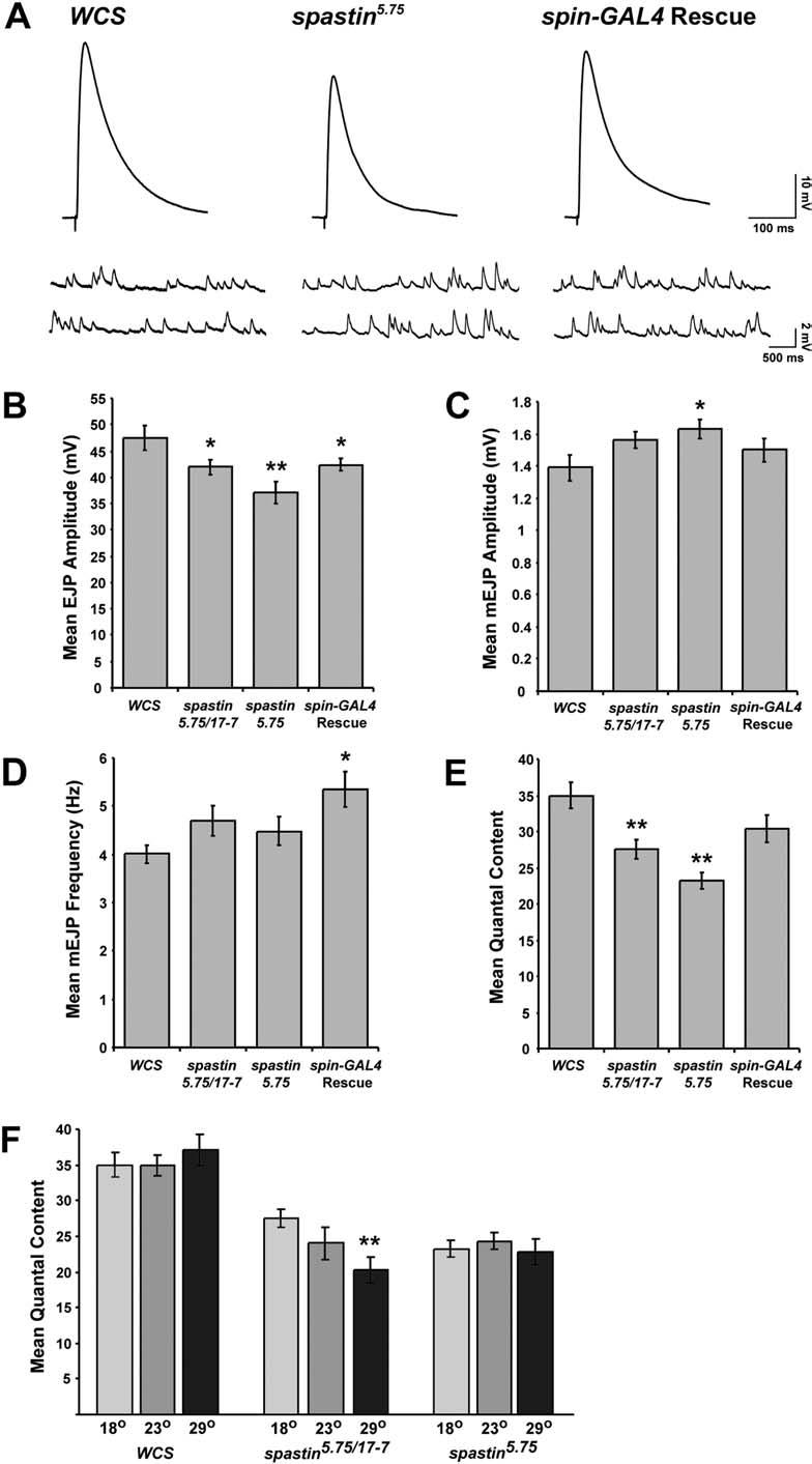
7 phenotype by spin- orelav-gs-gal4-driven expression of Spastin. spastin-null mutants have on average 1.6-fold more type Ib boutons on muscle 4 compared to WCS control larvae. Similarly, spastin-null mutants (of genotype spin-gal4/cyokr-gfp; spastin 5.75 ) have 1.7-fold more boutons compared to their sibling rescued larvae (genotype spin-gal4/uas-spastin; spastin 5.75 ). Boutons are also 1.6-fold more numerous in spastin-null larvae from a neuronal rescue cross (genotype þ/cyokr-gfp; Elav-GS-GAL4, spastin 5.75 /spastin 5.75 ) compared to their siblings in which UAS-spastin is expressed in neurons postembryonically (genotype UAS-spastin /CyOKr-GFP; Elav-GS-GAL, spastin 5.75 /spastin 5.75 ). DOI: /journal.pbio g004 severe behavioral phenotypes, and both sexes are sterile. These results suggest that the and 17-7 alleles are hypomorphic, and that the spastin 5.75 phenotype represents the null condition. RT-PCR analysis of cdna from spastin and spastin 17.7 animals indicated that low levels of truncated spastin transcripts are still produced (data not shown). These may direct synthesis of proteins initiated from internal ATGs that could retain partial function, since they include the entire conserved AAA domain. We could not detect anatomical phenotypes in embryos homozygous for any of the spastin mutations. However, we saw striking morphological changes in the NMJs of spastin 5.75 third instar larvae. In Figure 4, the two predominant types of glutamatergic boutons at the NMJs, Ib (big) and Is (small), are visualized by double-staining larval fillets with antibodies against Synaptotagmin (Syt, magenta; Menon and Zinn 1998) and Discs-large (Dlg, green; Woods and Bryant 1991). Syt is a presynaptic protein involved in neurotransmitter release that is localized to boutons, while Dlg is a primarily postsynaptic scaffold protein localized to the subsynaptic reticulum that surrounds each bouton (Littleton et al. 1993; Lahey et al. 1994). Figure 4A and 4B show that NMJ boutons are smaller and more numerous at the muscle 6/7 NMJ of spastin 5.75 larvae than in Canton S w (WCS) control larvae. (WCS was chosen as a control because, like the lines used to generate our EP insertion mutants, it is derived from a Canton S wild-type background, but it is also w, like the T32 excision derivatives. WCS is also commonly used for behavioral experiments.) Other NMJs are affected in a similar manner (e.g., Figure 4D 4F, showing muscle 4 synapses). The sizes of muscle fibers are normal in spastin mutants. To quantify the NMJ phenotype, we counted the numbers of boutons at the muscle 4 NMJs of segments A2 and A3, where boutons typically form on the internal surface of the muscle and are thus easily imaged. Dlg is expressed at much higher levels at Ib compared to Is boutons, allowing the two types of boutons to be distinguished and counted. Because of the greater variability in Is bouton number between NMJs, we focused our quantitative analysis on the type Ib boutons. However, the numbers of both bouton types were similarly affected in spastin 5.75 larvae. The number of Ib boutons per muscle 4 NMJ was increased by 1.6-fold relative to WCS in spastin mutants at room temperature (approximately 23 8C) (Figure 4G), and the boutons often formed dense clusters, particularly at the ends of NMJ branches (Figure 4E). This morphology was rarely observed in wild-type muscle 4 NMJs, where boutons were arranged more linearly (Figure 4D). The clustered boutons resemble the satellite boutons described by other investigators (Torroja et al. 1999; Franco et al. 2004; Koh et al. 2004; Marie et al. 2004). Hypomorphic spastin and spastin 17-7 mutants had bouton numbers that did not differ significantly from controls. To confirm that loss of Spastin produced the observed NMJ alterations, and to determine whether Spastin is required presynaptically or postsynaptically, we needed to evaluate rescue of the phenotype by expression of Spastin from a UASspastin cdna insertion. This was difficult because of the early lethality produced by expression of Spastin from most drivers. UAS-spastin animals bearing pan-neuronal (Elav- GAL4), motoneuronal (OK6-GAL4), or pan-muscle (24B-GAL4 or G14-GAL4) drivers did not survive to larval stages at 23 8C, and few larvae appeared even at 18 8C. However, third instar larvae in which Spastin expression from the cdna was conferred by spinster (spin)-gal4, a weak driver that functions in both neurons and muscles (Sweeney and Davis 2002), did survive at 23 8C. We were also able to obtain larvae in which Spastin expression was induced in neurons postembryonically. This was done by crossing UAS-spastin to Elav-GeneSwitch (GS)-GAL4, a driver line bearing a neuronally expressed GAL4 derivative that is only active in the presence of the progesterone analog RU486 (Osterwalder et al. 2001; McGuire et al. 2004). Newly hatched larvae from this cross were maintained on RU486-containing food until the third instar stage. To assay rescue, we combined the spin-gal4 and Elav-GS- GAL4 drivers and UAS-spastin insertions separately with spastin 5.75, crossed the driver and UAS-spastin lines together, and assayed NMJ phenotypes in the F1 driver-gal4/uasspastin; spastin 5.75 larvae. In each cross, we compared the rescued larvae to their unrescued spastin mutant siblings (driver-gal4; spastin 5.75 ), because the presence of the driver chromosome had effects on the absolute number of Ib boutons (see Materials and Methods). We observed that the ratio of muscle 4 Ib bouton numbers in spastin mutant controls versus rescued larvae was 1.7 for spin-gal4, and 1.6 for Elav-GS-GAL4 (Figure 4G). Since the ratio of bouton numbers for spastin 5.75 versus WCS was 1.6, this indicated that rescue was essentially complete in both cases. We also observed that the abnormal bouton clustering was eliminated in rescued larvae using either driver (Figures 4E and S1). These results demonstrate that loss of Spastin from neurons during larval development causes the NMJ bouton phenotypes seen in spastin 5.75 mutants. To examine the consequences of driver-dependent postembryonic neuronal expression of Spastin in a wild-type background, we also counted boutons in UAS-spastin; Elav-GS- GAL4 larvae grown on RU486 food (see Materials and Methods). We observed that these larvae had fewer boutons than their siblings ( fold change), and some of their boutons appeared larger (Figure S1C). This phenotype is very mild, but it does suggest that loss and increased expression of Spastin can produce opposite effects on the NMJ. Neurotransmitter Release Is Impaired in spastin Mutants To evaluate whether spastin mutations cause alterations in the electrophysiological properties of the NMJ, we evaluated synaptic transmission at the muscle 6 NMJ in WCS, mutant, and rescued larvae raised at 18 8C. In spastin 5.75 larvae, there was a reduction in the amplitudes of evoked responses 0007
8 0008
9 Figure 5. NMJs in spastin Mutant Larvae Display Reduced QC (A) Representative EJP (upper) and mejp (lower) traces are shown for control (WCS), spastin-null mutant (spastin 5.75 ), and spin-gal4/uas-spastin; spastin 5.75 (Rescue) larvae. All recordings were from the A3 or A4 muscle 6 NMJ. (B) The average EJP amplitude is decreased by about 20% in spastin-null mutants ( mv, n=26) relative to control ( mv, n=14) and Rescue ( mv, n=28) larvae, and is intermediate between control and null levels in hypomorphic spastin 5.75/17-7 transheterozygotes ( mv, n=22). (C) The average amplitude of spontaneous events (mejps) is increased slightly in spastin nulls relative to control and Rescue larvae. (D) The average frequency of spontaneous events is not affected in spastin mutants compared to control. Rescue larvae had a slightly higher mejp frequency. (E) Average QC, a measure of the amount of neurotransmitter released per action potential, is significantly lower in transheterozygotes ( ) versus control ( ), and reduced even further in spastin nulls ( ). This decrease is completely rescued by spin-gal4-driven rescue ( , p ¼ 0.1 compared to WCS). (F) Average QC is temperature dependent in spastin 5.75 /spastin 17-7 transheterozygous larvae, but not in homozygous spastin-null or control larvae. QC measured in transheterozygotes raised at 18 8C (light gray bars) is intermediate between that of control and nulls. At room temperature (dark gray) and 29 8C (black bars), similar QC values are measured in transheterozygotes and null mutants. *, p, 0.05; **, p, DOI: /journal.pbio g005 (excitatory junction potentials [EJPs]) to depolarization of the innervating nerves. Average EJP amplitudes in the null were reduced to 78% of the levels in control (WCS) larvae (Figure 5A and 5B; p, 0.003). We also examined the average amplitude and frequency of responses to single vesicles of spontaneously released neurotransmitter ( mini EJPs [mejps]). mejp amplitude was increased slightly, to 117% of WCS levels (Figure 5A and 5C; p, 0.03). There was no significant change in mejp frequency (Figure 5D; p ¼ 0.3). Quantal content (QC), a measure of the number of vesicles released per evoked event, was calculated by dividing the EJP amplitude by the average mejp amplitude. Because the evoked EJP was decreased and the mejp increased in spastin 5.75 mutants, QC was reduced to 67% of WCS levels in these larvae (Figure 5E; p, ). spastin 17-7 /spastin 5.75 larvae had EJP amplitude, mejp amplitude, and QC values intermediate between those of spastin-null and control larvae. QC in these transheterozygotes was also decreased significantly, to 78% of WCS levels (Figure 5E; p, 0.002). The changes in EJP amplitude and QC observed in spastin 5.75 mutants were completely rescued by spin-gal4-driven Spastin expression. Average QC in rescued larvae was not significantly different from wild-type (p. 0.1), and was 30% greater than in spastin 5.75 mutants (p, 0.005; Figure 5E). We also observed that the electrophysiological phenotypes of the hypomorphic spastin 17-7 /spastin 5.75 larvae were temperature sensitive. While the average QC in transheterozygotes was reduced to 78% of controls at 18 8C, this effect was exacerbated at higher temperatures. At 29 8C, QC was 54% of wild-type (Figure 5F). However, QC in control or in spastin 5.75 null larvae was unaffected by temperature. These results suggest that the N-terminally truncated Spastin protein that is probably made from the spastin 17-7 allele is temperature sensitive. At 29 8C, this hypomorphic allele behaves as a null with respect to NMJ electrophysiology. Finally, we examined escaper larvae (Elav-GAL4/þ; T32/þ) overexpressing Spastin in neurons, and found that synaptic transmission in these larvae was not significantly different from wild-type (data not shown). spastin Mutant Adults Have Severe Behavioral Phenotypes We observed that only approximately 20% of homozygous spastin 5.75 pupae were able to eclose at room temperature compared to 94% of heterozygotes, and the adults that emerged had severe movement defects (see Video S1). They could not fly at all, and did not even appear to move their wings, although the wings inflated and straightened in a normal manner immediately after eclosion. They also did not jump spontaneously, but would jump if persistently prodded in the abdomen. Their legs were weak: when standing, the metathoracic legs often slipped out from underneath them, and during walking they often dragged these legs (see Video S1). They also had difficulty holding on to surfaces when they were upside down. These phenotypes were temperature dependent. Null mutant flies that developed at 18 8C eclosed at much higher rates (56%) than at higher temperatures and moved more normally. Flies homozygous for the hypomorphic mutations, spastin and spastin 17-7, eclosed at normal frequencies at all temperatures. To evaluate these movement defects, we assayed flight and climbing ability in spastin-null and hypomorph flies (Figure 6). The flight assay could only be used for hypomorphs since null mutants were flightless. In this assay, flies were released into the top of a vertical cylinder that had been coated on the inside with oil (Benzer 1973; Atkinson et al. 2000). Poor fliers who took longer to fly fell to the bottom or collided with the lower walls of the cylinder, while good fliers who responded rapidly to being dropped collided with the upper walls. A histogram of the distribution of oil-trapped flies along the height of the cylinder showed that more than half of the spastin hypomorphs did not fly in time to avoid falling to the bottom of the cylinder (Figure 6A). In contrast, the majority (approximately 75%) of the controls, including w 1118 (another Canton S-derived w control) flies and flies homozygous for T32 (in the absence of a GAL4 driver; these have orange eyes), flew well enough to distribute themselves along the sides of the column. Interestingly, for those hypomorphs that did fly out to the sides, their distribution paralleled that of the controls, suggesting that flight responses in the column were relatively normal in this subpopulation of the mutants (Figure 6B). In the climbing assay, flies were tested for their ability to climb up the side of a vial within a limited time period. All WCS and almost all homozygous spastin and spastin 17-7 flies, but only about 40% of spastin 5.75 flies, climbed to the top of the vial within 30 s (Figure 6C). This difference did not reflect a loss of geotactic behavior, since spastin 5.75 flies were typically found at the tops of their vials after several minutes. Overall, mean climbing velocity was approximately 9-fold slower for spastin-null mutants than for wild-type flies, while the hypomorphs were about 2-fold slower than controls (Figure 6D). We also measured the lifespan of the flies. Under our conditions, WCS flies lived an average of 46 d at 25 8C. 0009
 In a flight test assay, over twice as many adult spastin hypomorphs (spastin 10-12 and spastin 17-7 ) fail to fly before")
10 Figure 6. spastin Mutant Flies Have Compromised Motor Behavior and Reduced Lifespans (A) In a flight test assay, over twice as many adult spastin hypomorphs (spastin and spastin 17-7 ) fail to fly before falling to the bottom of a cylinder, in comparison to w 1118 and T32 homozygous controls. (B) Although fewer than half of the hypomorphs fly, compared to more than 70% of controls, the distribution of collision sites of the fliers along the height of the cylinder parallels that of the controls, suggesting that these spastin mutations affect flying ability in some animals but not in others. (C and D) spastin mutants are compromised in their climbing ability. (C) All control (WCS) and nearly all spastin hypomorphs climb to the top of a vial in 30 s, but only 40% of spastin nulls do so. (D) Climbing velocity (measured for those flies that reach the top in 30 s) is cm/s in WCS (n=45), but only and cm/s in spastin (n=28) and spastin 17-7 (n=38) flies, respectively, and cm/s in spastin 5.75 null mutants (n =17; p, for all relative to WCS). (E) Lifespan curves. The curve inflection point at which WCS and hypomorph flies begin to die off at a rapid rate occurs at d after eclosion, and more than 70% of hypomorph flies and 95% of wild-type flies are still alive at 30 d. In contrast, approximately 45% of spastin 5.75 null mutant flies die prior to 4 d after eclosion. However, the majority of the remaining null flies survive more than 25 d, so that the curve inflection point for nulls occurs only a few days before that for controls and hypomorphs. (F) Mean lifespan is d in WCS controls (n =39) compared to and d, respectively, in spastin (n=24, p, 0.02) and spastin 17-7 (n=32, p, 0.006), and d in nulls (n =62, p, ). (G) Only about 10% of spastin 5.75 flies eclosing at room temperature are males, while 40% 45% are males for controls and hypomorphs. DOI: /journal.pbio g006 spastin and spastin 17-7 flies had somewhat shorter lifetimes, surviving an average of 35 d. Lifespan was dramatically reduced in spastin 5.75 flies, which lived an average of only 8 d (Figure 6E and 6F). Examination of mortality curves (Figure 6E), however, revealed that, as in the case of flight ability in the hypomorphs, these flies had a bimodal lifespan distribution. Only 55% of flies were still alive at 4 d post-eclosion, but most of these (37% of the total) then remained alive until about 25 d post-eclosion. After this time, they rapidly died off, and no flies remained alive more than 33 d. Another spastin phenotype observed in adults was malespecific lethality (Figure 6G). For WCS and spastin hypomorphs, more than 40% of eclosed adults were males. However, only approximately 10% of eclosed spastin 5.75 flies were male. We do not understand the origins of this phenotype. In summary, spastin-null adult flies had severely compromised movement behavior and were short-lived, while spastin hypomorphs displayed weaker movement and lifespan phenotypes. We also examined rescue for these behavioral phenotypes. When compared to their non-rescued siblings (spin-gal4/cyokr-gfp; spastin 5.75 ) from the same cross, spin- GAL4/UAS-spastin; spastin 5.75 flies climbed better, were more coordinated, and lived longer (Figure S2; Video S1), indicating that partial rescue was achieved. These flies were still very slow, and it is clear that spin-gal4-driven Spastin expression did not restore behavior to the levels characteristic of control flies such as WCS. However, genetic background effects made the precise efficacy of rescue achieved in this experiment difficult to determine (the non-rescued spastin 5.75 sibling flies bearing the driver and balancer chromosomes used in the rescue cross were much more unhealthy and slow-moving than spastin 5.75 flies without these chromosomes). Spastin Overexpression Erases Microtubule Networks In Vivo To investigate whether Drosophila Spastin affects microtubule networks, we overexpressed it in embryonic muscles using the G14-GAL4 or 24B-GAL4 drivers, and then visualized muscle microtubules in late stage 16 embryos with an anti-b3-0010
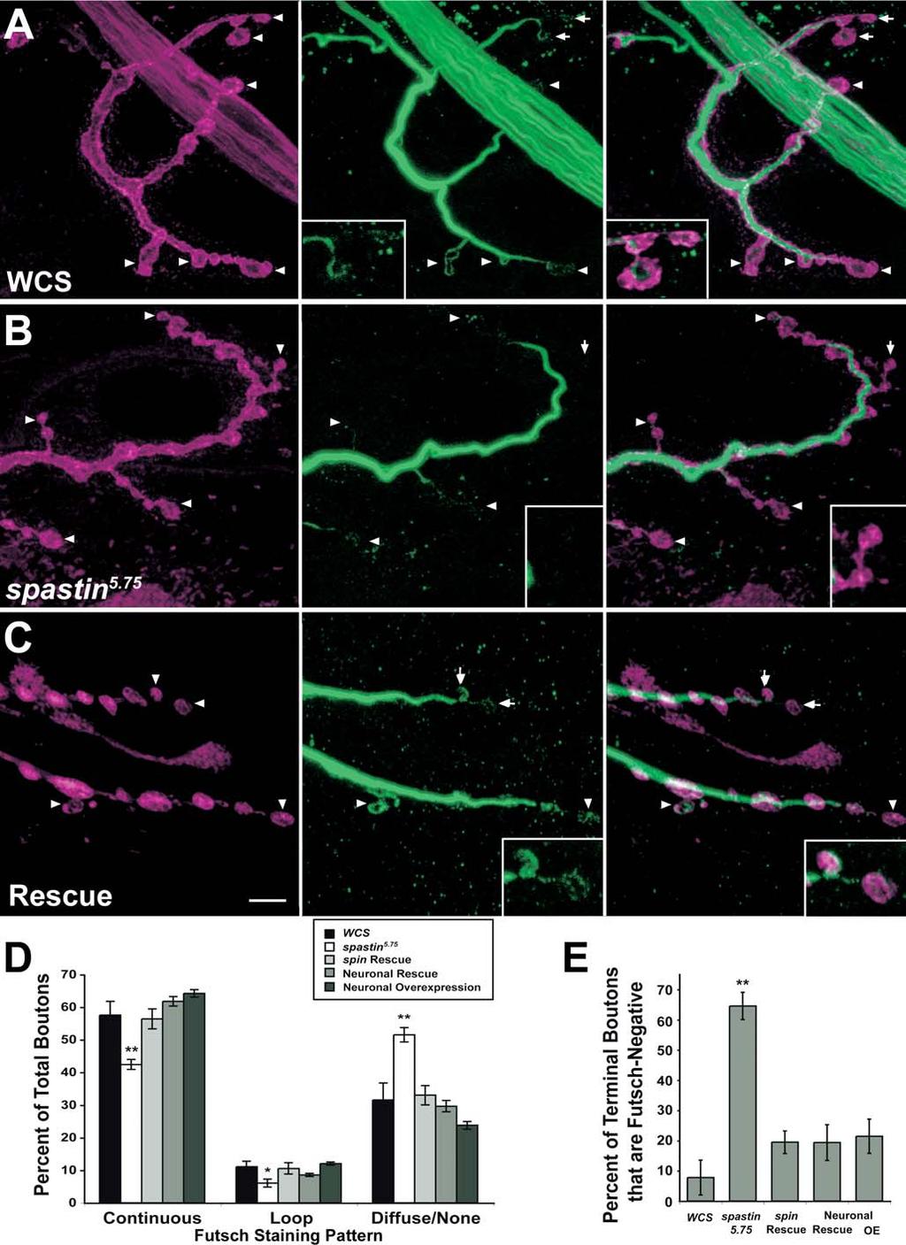
11 tubulin antibody that preferentially stains polymerized tubulin (Buttgereit et al. 1996). In wild-type embryos, a complex network of microtubules aligned along the muscle axes was observed (Figure 7A; two segments are shown). This was clearly seen in both vertically oriented (18 and 21 24) and diagonally oriented (5,11, and 19) muscles. When Spastin was overexpressed in muscles, the muscle microtubule network completely disappeared (Figure 7B). The muscles themselves appeared rounded, and were partially or totally detached from their insertion sites. This detachment may have been a consequence of the dissolution of the microtubule network. Oriented microtubule networks could still be seen within the cap cells of the chordotonal organs in each segment (Figure 7B, brackets); these cells did not express the GAL4 driver and therefore did not overexpress Spastin. Dissolution of the microtubule network was therefore specific to cells in which Spastin was overexpressed. We also overexpressed Spastin in larval muscles by crossing the muscle-specific MHC-GS-GAL4 driver line to UAS-spastin flies. MHC-GS-GAL4 is RU486-inducible, but we could not feed the larvae with RU486 as this was lethal. However, MHC- GS-GAL4 also confers late RU486-independent expression in third instar larvae, and we were able to obtain escaper larvae from the cross and double-stain these for Spastin and a- tubulin. As shown in Figure 7C 7E, larvae that lacked detectable Spastin expression had a dense network of muscle microtubules. In contrast, MHC-GS-GAL4/UAS-spastin larvae that had high levels of muscle Spastin displayed a dramatic reduction in microtubules, so that only faint and sparse microtubules were observed in the muscle fibers (Figure 7F 7H). The strongly staining microtubules still visible in these larvae are those of the neurons and tracheae, which do not overexpress Spastin (Figure 7G 7H). These results also show that this a-tubulin antibody preferentially recognizes polymerized tubulin, since the total amount of tubulin dimers would be the same in both sets of muscles (tubulin dimers are very stable proteins and are unlikely to be proteolyzed after severing). Interestingly, when we overexpressed Spastin in the embryonic or larval CNS, we did not observe an obvious alteration of the axonal microtubule architecture (see also Figure 8). This suggests that Spastin may be unable to disassemble stable axonal microtubule bundles. Nevertheless, the dramatic effects of Spastin overexpression on muscle microtubules suggest that the embryonic CNS collapse phenotype conferred by neuronal overexpression (see Figure 3) may also arise from breakdown of key neuronal microtubules during the axonal growth phase. Microtubule Bundles Are Depleted in Distal NMJ Boutons of spastin LOF Mutants The finding that Spastin overexpression erases the microtubule network in muscles suggested that the spastin LOF NMJ phenotypes could arise from alterations in microtubule networks. To investigate this, we first examined the distribution of Futsch, a microtubule-associated protein related to vertebrate MAP1B (Hummel et al. 2000; Roos et al. 2000). Futsch staining is restricted to stable neuronal microtubule bundles. Because Futsch is not expressed in the underlying muscle, Futsch antibody staining provides the optimal method for quantitatively evaluating stable microtubules within NMJ boutons. At muscle 4 NMJs in wild-type larvae (Figure 8A) we observed continuous microtubule bundles stained by anti- Futsch (mab 22C10; green) within axons and along the axis of each branch of the NMJ (delineated by anti-hrp, which labels neuronal membranes; magenta). The intensity of Futsch staining weakens in the distal portions of the branches. Consistent with earlier findings, we also observed distinctive loops of Futsch staining within some boutons (Roos et al. 2000; Packard et al. 2002; Pennetta et al. 2002). Loops were typically observed in terminal boutons at the ends of branches. In some terminal boutons, however, we detected only punctate staining or no staining at all. This last case may reflect the limits of detection rather than the complete absence of Futsch protein in a bouton. We quantified Futsch distribution by dividing the patterns of Futsch staining in boutons into three classes: continuous (bundles or splayed bundles), looped, and diffuse or undetectable. In spastin 5.75 larvae (Figure 8B), there was a shift in the Futsch pattern toward less organized morphologies (i.e., diffuse/undetectable). At the muscle 4 NMJ of spastin mutant larvae, 74% and 54% as many boutons contained continuous and looped Futsch, respectively, as compared to WCS. In contrast, 63% more boutons in spastin mutants displayed only diffuse or no staining (Figure 8D). These differences were most pronounced at the distal ends of the synaptic branches (Figure 8B, arrows and arrowheads). 65% of terminal boutons in mutants had no detectable Futsch staining, as compared to 8% in WCS (Figure 8E; p, ). The Futsch distribution phenotypes were rescued by expression of Spastin from the spin-gal4 or the RU486- induced, neural-specific Elav-GS-GAL4 drivers. Rescued larvae had Futsch staining patterns very similar to those seen in WCS (Figure 8C 8E). These results show that the reduction in stable synaptic microtubules seen in spastin LOF mutants is due to loss of Spastin from neurons during larval development. We also examined Futsch staining in Elav-GS-GAL4/ UAS-spastin larvae grown on RU486 food, but saw no difference from the pattern in wild-type controls. Thus, stable microtubules at the NMJ do not break down when Spastin is overexpressed at the levels induced by this driver. Having demonstrated statistically significant differences in Futsch localization between control and spastin mutant NMJs, we then directly examined tubulin in muscle 4 NMJ boutons using fixation conditions that reduce muscle microtubule staining (see Materials and Methods). The pattern of NMJ microtubules is complex, and is difficult to quantitatively analyze because of residual signal from microtubules in the underlying muscle. However, using the a-tubulin antibody described above, we were able to clearly visualize looped microtubule structures (Figure 9A, green) within anti-hrplabeled boutons (magenta). These loops were present both along the branches and in the terminal boutons (inset). spastin 5.75 NMJs exhibited weaker a-tubulin staining than wild-type controls, particularly in terminal boutons (Figure 9B). Looped microtubule structures could be seen in some boutons in mutants. However, boutons at the ends of NMJ branches or at the outer edges of the bouton clumps that are characteristic of spastin NMJs often lacked any tubulin staining (inset). Thus, our results indicate that microtubule bundles are selectively depleted from the distal boutons of NMJs in larvae lacking Spastin protein. 0011
 are difficult to distinguish in this panel because of high levels of muscle tubulin staining.")
12 Figure 7. Spastin Overexpression in Muscles Erases the Microtubule Network (A) An antibody against b3-tubulin stains body wall muscles and chordotonal cap cells in stage 16 wild-type embryos. Two abdominal hemisegments are shown; muscle fiber numbers are labeled in one. The cap cells (brackets) are difficult to distinguish in this panel because of high levels of muscle tubulin staining. They extend diagonally from about the middle of muscle 18 to muscle 22. Anterior is to the left, and dorsal is up. (B) When Spastin is overexpressed in muscles (genotype: G14-GAL4/þ; T32/þ), b3-tubulin staining is very weak and has a disorganized pattern in 0012
13 most muscle fibers, but an intact microtubule network is still present in the cap cells, which do not express this driver (brackets). The muscle fibers are misshapen and partially (arrowhead) or completely (arrow) detached from their insertion sites. (C H) Similarly, the microtubule network (recognized by antibodies to a-tubulin) is almost eliminated by high-level Spastin expression in third instar larval muscles. Larvae of genotype UAS-spastin/MHC-GS-GAL ; spastin 5.75 /TM3Ser-ActGFP overexpress Spastin protein specifically in muscles to varying degrees. Wild-type larval muscles had undetectable levels of Spastin using pab 1239 (C) and displayed a dense network of microtubule bundles in the muscle (D and E), as well as in trachea (D, arrow) and neurons (D, arrowhead denotes a terminal arbor). In contrast, larval muscles expressing high levels of Spastin (F) show only faint muscle microtubule staining (G and H), while tracheal (G, arrow) and neuronal (G, arrowhead) staining remain robust. DOI: /journal.pbio g007 Discussion Mutations in the human spastin gene, which encodes an AAA ATPase, are the most common cause of pure AD-HSP. We identified the Drosophila Spastin ortholog (see Figure 1) in a gain-of-function screen (see Figure 3). Drosophila Spastin is a cytoplasmic protein that can also localize to axons (see Figure 2). spastin-null larvae have altered NMJs in which presynaptic boutons are more numerous and smaller than in wild-type, and are organized in dense clusters (see Figure 4). These changes in bouton number and organization are rescued by expression of Spastin in neurons (see Figures 4 and S1). QC, a measure of the number of vesicles of neurotransmitter released in response to an action potential, is reduced at NMJs in both null and hypomorphic spastin mutants (see Figure 5). spastin-null flies have severe movement defects. They cannot fly at all, and do not jump. They climb and walk very slowly, often drag their hind legs when walking (see Video S1), and have greatly reduced lifespans. spastin hypomorphs have milder phenotypes, displaying flying defects and a decrease in climbing speed (see Figure 6). Regulation of Synaptic Microtubule Networks by Spastin The AAA domain of Spastin is quite similar to that of Katanin-60, which is a microtubule-severing protein. To determine whether Spastin might also sever or otherwise alter microtubules in vivo, we overexpressed the protein in embryonic and larval muscles. Strikingly, this overexpression erases or greatly reduces the microtubule network (see Figure 7). These data are consistent with the finding that overexpression of human Spastin in transfected mammalian cells causes microtubule disassembly (Errico et al. 2002; McDermott et al. 2003). Having demonstrated that Spastin can cause disassembly of microtubules in vivo, we then examined how its absence affects the synaptic microtubule cytoskeleton. Based on the overexpression phenotype, one might have expected that microtubules would be more stable or more numerous in spastin LOF mutants. However, our observations indicate the opposite: microtubule bundles are depleted in NMJ boutons when Spastin is absent. At the wild-type muscle 4 NMJ, boutons are arranged along linear axes. Continuous microtubule bundles run along the axes and connect to larger bundles within the innervating axon. Microtubules within boutons are typically arranged in loops and swirls. In spastin-null mutants, boutons are arranged in clumps, and the distal boutons of these clumps often lack any detectable tubulin staining (see Figure 9). Looped microtubule structures are present within some proximal boutons, however, and the bundles connecting the NMJ to the axon are still present. These results suggest that the absence of Spastin selectively affects the construction of the presynaptic microtubule cytoskeleton, and that the severity of the microtubule defects in a bouton are correlated with its distance from the NMJ s axonal branchpoint. We quantitated these defects using an antibody against the microtubule-associated Futsch protein, which defines a subpopulation of stable neuronal microtubule bundles. In wild-type larvae, Futsch staining forms continuous lines along the main branches of the NMJ. Some individual boutons have Futsch loops, while others display only diffuse staining. A comparison of wild-type and spastin-null larvae shows that the distribution of Futsch within boutons shifts from organized structures (bundles and loops) toward diffuse patterns or the absence of detectable staining. This effect is most pronounced at terminal boutons, and is rescued by neuronal expression of Spastin (see Figure 8). If Spastin s function in vivo is to disassemble microtubules, as suggested by our overexpression experiments (see Figure 7), why does its absence produce a paradoxical reduction in microtubules within the NMJ (see Figures 8 and 9)? One possibility is that microtubule severing is required for movement of microtubules into or within the presynaptic region. Some evidence for this idea has been published. In one study, injection of function-blocking anti-katanin-60 antibody into cultured sympathetic neurons reduced process outgrowth, and microtubules were 4- to 5-fold longer in antibody-injected neurons than in control cells (Ahmad et al. 1999). More recent work demonstrated that expression of dominant-negative Katanin-60 reduces axonal outgrowth (Karabay et al. 2004). These results were interpreted as indicating that Katanin is required for severing microtubules to a length that allows their transport along the axon to its growing tip. When Katanin is inhibited, microtubule segments may be too long to be efficiently transported, and this results in a reduction in axon outgrowth. Based on these findings, we suggest that the depletion of microtubules in the distal boutons of spastin mutant NMJs arises because severing of axonal microtubules by Spastin is necessary to generate microtubule polymers that are short enough to be efficiently moved into and through the presynaptic terminals. Perhaps Spastin normally excises sections of microtubules at branchpoints where NMJ branches leave the axon trunk, and these severed microtubule segments (or individual tubulin dimers) are then moved distally into the boutons of the NMJ as it grows. Is Spastin also involved in axon outgrowth or guidance, as suggested by its embryonic gain-of-function phenotype (see Figure 3)? Clearly loss of Spastin activity in Drosophila does not strongly affect outgrowth, since the embryonic CNS axon ladder develops in a normal manner and motor axons reach their appropriate targets in spastin mutants. Furthermore, axonal and muscle microtubules are not detectably altered in spastin-null embryos. Severing of microtubules in vivo, 0013
14 0014
 Anti-Futsch labels stable microtubule bundles in axons, NMJ")
15 ?2 Figure 8. The Distribution of Stable NMJ Microtubule Bundles Marked by the MAP1B-like Protein Futsch Is Altered in spastin Mutant Larvae (A C) Anti-Futsch labels stable microtubule bundles in axons, NMJ boutons, and interbouton regions. The muscle 4 NMJs in segment A3 of third instar wild-type (A), spastin 5.75 (B), and spin-gal4/uas-spastin; spastin 5.75 (Rescue) (C) larvae were immunostained with anti-hrp (A and B) or anti- Syt antibodies (C) to label presynaptic boutons (magenta), and mab 22C10 to label Futsch protein (A C, green). Arrows and arrowheads mark the terminal boutons (those at the ends of synaptic branches); boutons marked by arrows in (A C) are enlarged in insets in the middle and right panels to show examples of Futsch patterns. (A) In control (WCS) larvae, terminal boutons have both looped (arrowheads) and diffuse, punctate (right panel, arrows and inset) patterns of Futsch staining. (B) In spastin mutants, Futsch staining appears similarly strong in axon bundles (not shown) and along the main branches of the bouton arbor. More distal and terminal boutons, however, have diffuse or no Futsch staining (arrows and arrowheads). Note the absence of green staining in insets. (C) The distribution of Futsch staining is restored to the control pattern by spin- GAL4-driven expression of Spastin in the mutant background (arrows and arrowheads indicate loops). Scale bar, 5 lm. (D and E) Quantitative assessment of Futsch staining data. Futsch staining at A2 and A3 muscle 4 NMJs was classified as continuous (bundles or splayed bundles), looped, or diffuse or undetectable (none) for each bouton. (D) The percentage of boutons exhibiting continuous or looped Futsch staining (relative to the total number of boutons for each NMJ) is decreased in spastin mutants relative to controls, while the percentage of boutons having diffuse or no staining is increased. In total, 58% 6 4.2% of boutons in controls have a continuous pattern of Futsch staining, while only 42% 6 1.5% do in mutants. Boutons in this class are predominantly along the major (more proximal) branches of the axon arbor. Similarly, 11% 6 1.7% of wild-type boutons have Futsch loops, but only 6.0% 6 1.1% do in mutants. Most mutant boutons show only diffuse or no Futsch staining (52% 6 2.2%, versus 32% 6 5.2% in controls). Futsch distribution is restored to the control pattern by spin-gal4- orelav- GS-GAL4-driven expression of Spastin. (E) The difference in Futsch distribution is most pronounced at terminal boutons. There is no detectable Futsch staining in the majority of terminal boutons (65% 6 4.5%) in spastin mutants, compared to only 7.8% 6 5.8% of terminal boutons in wild-type larvae (p, ). Futsch staining is restored in most terminal boutons of spin-gal4-orelav-gs-gal4-rescued larvae, with only 20% 6 3.7% and 19% 6 5.9% of boutons, respectively, showing no staining (p ¼ 0.09 compared to WCS). Terminal bouton staining in larvae overexpressing Spastin in neurons was unaffected relative to controls (p ¼ 0.12). **, p, 0.005; *, p, 0.03 relative to WCS; n. 8 NMJs scored in all cases. DOI: /journal.pbio g008 Figure 9. The Microtubule Network in NMJ Boutons Is Altered or Absent in spastin Mutant Larvae (A) In wild-type (WCS) larvae, an antibody against a-tubulin (green) reveals the distribution of the network of microtubule bundles within the A3 muscle 4 NMJ bouton arbor. Presynaptic bouton membranes are labeled by anti-hrp antibody (magenta). The microtubule network has a complex structure and extends into the terminal boutons (arrowheads and inset). Many proximal boutons have loops (arrows). Microtubules are also observed outside of the boundaries of the NMJ; these are within the muscle fiber, which also expresses a-tubulin. Staining of these muscle microtubules is minimized by the use of Bouin s fix. (B) In spastin 5.75 mutants, the microtubule network is much sparser than in controls, particularly in the distal boutons at the edges of the bouton clumps that are characteristic of spastin mutant NMJs (arrowheads and inset). Many of these distal boutons have little or no detectable a-tubulin staining. More proximal boutons still have tubulin loops, however (arrows). Scale bar, 5 lm. DOI: /journal.pbio g
SUPPLEMENTARY INFORMATION
 doi:10.1038/nature10923 Supplementary Figure 1 Ten-a and Ten-m antibody and cell type specificities. a c, Representative single confocal sections of a Drosophila NMJ stained with antibodies to Ten-a (red),
Chapter 4 Evaluating a potential interaction between deltex and git in Drosophila: genetic interaction, gene overexpression and cell biology assays.
 Evaluating a potential interaction between deltex and git in Drosophila: genetic interaction, gene overexpression and cell biology assays. The data described in chapter 3 presented evidence that endogenous
Supplementary Materials for
 www.sciencesignaling.org/cgi/content/full/6/301/ra98/dc1 Supplementary Materials for Regulation of Epithelial Morphogenesis by the G Protein Coupled Receptor Mist and Its Ligand Fog Alyssa J. Manning,
Control and Integration. Nervous System Organization: Bilateral Symmetric Animals. Nervous System Organization: Radial Symmetric Animals
 Control and Integration Neurophysiology Chapters 10-12 Nervous system composed of nervous tissue cells designed to conduct electrical impulses rapid communication to specific cells or groups of cells Endocrine
Nature Neuroscience: doi: /nn.2662
 Supplementary Figure 1 Atlastin phylogeny and homology. (a) Maximum likelihood phylogenetic tree based on 18 Atlastin-1 sequences using the program Quicktree. Numbers at internal nodes correspond to bootstrap
Nature Biotechnology: doi: /nbt Supplementary Figure 1. Overexpression of YFP::GPR-1 in the germline.
 Supplementary Figure 1 Overexpression of YFP::GPR-1 in the germline. The pie-1 promoter and 3 utr were used to express yfp::gpr-1 in the germline. Expression levels from the yfp::gpr-1(cai 1.0)-expressing
Cells. Steven McLoon Department of Neuroscience University of Minnesota
 Cells Steven McLoon Department of Neuroscience University of Minnesota 1 Microscopy Methods of histology: Treat the tissue with a preservative (e.g. formaldehyde). Dissect the region of interest. Embed
Modulation of central pattern generator output by peripheral sensory cells in Drosophila larvae. BioNB4910 Cornell University.
 Modulation of central pattern generator output by peripheral sensory cells in Drosophila larvae BioNB4910 Cornell University Goals 1) Observe the behavioral effects of remotely activating different populations
Highwire Regulates Synaptic Growth in Drosophila
 Neuron, Vol. 26, 313 329, May, 2000, Copyright 2000 by Cell Press Highwire Regulates Synaptic Growth in Drosophila Hong I. Wan,* Aaron DiAntonio,* Richard D. Fetter,* Kendra Bergstrom,* Roland Strauss,
NEURONS, SENSE ORGANS, AND NERVOUS SYSTEMS CHAPTER 34
 NEURONS, SENSE ORGANS, AND NERVOUS SYSTEMS CHAPTER 34 KEY CONCEPTS 34.1 Nervous Systems Are Composed of Neurons and Glial Cells 34.2 Neurons Generate Electric Signals by Controlling Ion Distributions 34.3
Cellular Neuroanatomy II The Prototypical Neuron: Neurites. Reading: BCP Chapter 2
 Cellular Neuroanatomy II The Prototypical Neuron: Neurites Reading: BCP Chapter 2 Major Internal Features of a Neuron The neuron is the functional unit of the nervous system. A typical neuron has a soma
Baz, Par-6 and apkc are not required for axon or dendrite specification in Drosophila
 Baz, Par-6 and apkc are not required for axon or dendrite specification in Drosophila Melissa M. Rolls and Chris Q. Doe, Inst. Neurosci and Inst. Mol. Biol., HHMI, Univ. Oregon, Eugene, Oregon 97403 Correspondence
Midterm 1. Average score: 74.4 Median score: 77
 Midterm 1 Average score: 74.4 Median score: 77 NAME: TA (circle one) Jody Westbrook or Jessica Piel Section (circle one) Tue Wed Thur MCB 141 First Midterm Feb. 21, 2008 Only answer 4 of these 5 problems.
Axis Specification in Drosophila
 Developmental Biology Biology 4361 Axis Specification in Drosophila November 2, 2006 Axis Specification in Drosophila Fertilization Superficial cleavage Gastrulation Drosophila body plan Oocyte formation
The BMP Ligand Gbb Gates the Expression of Synaptic Homeostasis Independent of Synaptic Growth Control
 Article The BMP Ligand Gbb Gates the Expression of Synaptic Homeostasis Independent of Synaptic Growth Control Carleton P. Goold 1 and Graeme W. Davis 1, * 1 Department of Biochemistry and Biophysics,
Chapter 18 Lecture. Concepts of Genetics. Tenth Edition. Developmental Genetics
 Chapter 18 Lecture Concepts of Genetics Tenth Edition Developmental Genetics Chapter Contents 18.1 Differentiated States Develop from Coordinated Programs of Gene Expression 18.2 Evolutionary Conservation
18.4 Embryonic development involves cell division, cell differentiation, and morphogenesis
 18.4 Embryonic development involves cell division, cell differentiation, and morphogenesis An organism arises from a fertilized egg cell as the result of three interrelated processes: cell division, cell
Axis Specification in Drosophila
 Developmental Biology Biology 4361 Axis Specification in Drosophila November 6, 2007 Axis Specification in Drosophila Fertilization Superficial cleavage Gastrulation Drosophila body plan Oocyte formation
Anterograde Activin Signaling Regulates Postsynaptic Membrane Potential and GluRIIA/B Abundance at the Drosophila Neuromuscular Junction
 Anterograde Activin Signaling Regulates Postsynaptic Membrane Potential and GluRIIA/B Abundance at the Drosophila Neuromuscular Junction Myung-Jun Kim, Michael B. O Connor* Department of Genetics, Cell
Nervous System Organization
 The Nervous System Nervous System Organization Receptors respond to stimuli Sensory receptors detect the stimulus Motor effectors respond to stimulus Nervous system divisions Central nervous system Command
Neurons and Nervous Systems
 34 Neurons and Nervous Systems Concept 34.1 Nervous Systems Consist of Neurons and Glia Nervous systems have two categories of cells: Neurons, or nerve cells, are excitable they generate and transmit electrical
Introduction Principles of Signaling and Organization p. 3 Signaling in Simple Neuronal Circuits p. 4 Organization of the Retina p.
 Introduction Principles of Signaling and Organization p. 3 Signaling in Simple Neuronal Circuits p. 4 Organization of the Retina p. 5 Signaling in Nerve Cells p. 9 Cellular and Molecular Biology of Neurons
Bypass and interaction suppressors; pathway analysis
 Bypass and interaction suppressors; pathway analysis The isolation of extragenic suppressors is a powerful tool for identifying genes that encode proteins that function in the same process as a gene of
Nervous Systems: Neuron Structure and Function
 Nervous Systems: Neuron Structure and Function Integration An animal needs to function like a coherent organism, not like a loose collection of cells. Integration = refers to processes such as summation
Fig. S1. Expression pattern of moody-gal4 in third instar. Maximum projection illustrating a dissected moody-gal4>ngfp L3 larva stained for Repo
 Fig. S1. Expression pattern of moody-gal4 in third instar. Maximum projection illustrating a dissected moody-gal4>ngfp L3 larva stained for Repo (magenta), Fas2 (blue) and GFP (green) in overview (A) and
PROPERTY OF ELSEVIER SAMPLE CONTENT - NOT FINAL. The Nervous System and Muscle
 The Nervous System and Muscle SECTION 2 2-1 Nernst Potential 2-2 Resting Membrane Potential 2-3 Axonal Action Potential 2-4 Neurons 2-5 Axonal Conduction 2-6 Morphology of Synapses 2-7 Chemical Synaptic
A complementation test would be done by crossing the haploid strains and scoring the phenotype in the diploids.
 Problem set H answers 1. To study DNA repair mechanisms, geneticists isolated yeast mutants that were sensitive to various types of radiation; for example, mutants that were more sensitive to UV light.
Nervous System Organization
 The Nervous System Chapter 44 Nervous System Organization All animals must be able to respond to environmental stimuli -Sensory receptors = Detect stimulus -Motor effectors = Respond to it -The nervous
Supplementary Figures
 Supplementary Figures Supplementary Fig. S1: Normal development and organization of the embryonic ventral nerve cord in Platynereis. (A) Life cycle of Platynereis dumerilii. (B-F) Axonal scaffolds and
Varicose and cheerio collaborate with pebble to mediate semaphorin-1a reverse signaling in Drosophila
 Varicose and cheerio collaborate with pebble to mediate semaphorin-1a reverse signaling in Drosophila Sangyun Jeong a,1, Da-som Yang a, Young Gi Hong a, Sarah P. Mitchell b,c, Matthew P. rown b,c, and
Shaggy, the Homolog of Glycogen Synthase Kinase 3, Controls Neuromuscular Junction Growth in Drosophila
 The Journal of Neuroscience, July 21, 2004 24(29):6573 6577 6573 Brief Communication Shaggy, the Homolog of Glycogen Synthase Kinase 3, Controls Neuromuscular Junction Growth in Drosophila Bénédicte Franco,
purpose of this Chapter is to highlight some problems that will likely provide new
 119 Chapter 6 Future Directions Besides our contributions discussed in previous chapters to the problem of developmental pattern formation, this work has also brought new questions that remain unanswered.
MCN. Complex Genetic Interactions among Four Receptor Tyrosine Phosphatases Regulate Axon Guidance in Drosophila
 MCN Molecular and Cellular Neuroscience 17, 274 291 (2001) doi:10.1006/mcne.2000.0939, available online at http://www.idealibrary.com on Complex Genetic Interactions among Four Receptor Tyrosine Phosphatases
AP Biology Unit 6 Practice Test 1. A group of cells is assayed for DNA content immediately following mitosis and is found to have an average of 8
 AP Biology Unit 6 Practice Test Name: 1. A group of cells is assayed for DNA content immediately following mitosis and is found to have an average of 8 picograms of DNA per nucleus. How many picograms
Chapter 48 Neurons, Synapses, and Signaling
 Chapter 48 Neurons, Synapses, and Signaling Concept 48.1 Neuron organization and structure reflect function in information transfer Neurons are nerve cells that transfer information within the body Neurons
SUPPLEMENTARY INFORMATION
 DOI: 10.1038/ncb3267 Supplementary Figure 1 A group of genes required for formation or orientation of annular F-actin bundles and aecm ridges: RNAi phenotypes and their validation by standard mutations.
In Vivo Analysis of the Microtubule Severing Protein Katanin-60 in the Drosophila. Nervous System. Lisa M. Pang. Department of Biology Duke University
 In Vivo Analysis of the Microtubule Severing Protein Katanin-60 in the Drosophila Nervous System by Lisa M. Pang Department of Biology Duke University Date: Approved: Nina Tang Sherwood, Supervisor Vann
Positional Cues in the Drosophila Nerve Cord: Semaphorins Pattern the Dorso-Ventral Axis
 Positional Cues in the Drosophila Nerve Cord: Semaphorins Pattern the Dorso-Ventral Axis Marta Zlatic 1,2,3 *, Feng Li 1, Maura Strigini 4, Wesley Grueber 2, Michael Bate 1 * 1 Department of Zoology, University
Chapter 9. Nerve Signals and Homeostasis
 Chapter 9 Nerve Signals and Homeostasis A neuron is a specialized nerve cell that is the functional unit of the nervous system. Neural signaling communication by neurons is the process by which an animal
Systematic Screening of Drosophila Deficiency Mutations for Embryonic Phenotypes and Orphan Receptor Ligands
 Systematic Screening of Drosophila Deficiency Mutations for Embryonic Phenotypes and Orphan Receptor Ligands Ashley P. Wright 1, A. Nicole Fox 1, Karl G. Johnson 2, Kai Zinn 1 * 1 Division of Biology,
Chapter 37 Active Reading Guide Neurons, Synapses, and Signaling
 Name: AP Biology Mr. Croft Section 1 1. What is a neuron? Chapter 37 Active Reading Guide Neurons, Synapses, and Signaling 2. Neurons can be placed into three groups, based on their location and function.
The Transmembrane Semaphorin Sema I Is Required in Drosophila for Embryonic Motor and CNS Axon Guidance
 Neuron, Vol. 20, 207 220, February, 1998, Copyright 1998 by Cell Press The Transmembrane Semaphorin Sema I Is Required in Drosophila for Embryonic Motor and CNS Axon Guidance Hung-Hsiang Yu, Houmam H.
Cellular Neuroanatomy I The Prototypical Neuron: Soma. Reading: BCP Chapter 2
 Cellular Neuroanatomy I The Prototypical Neuron: Soma Reading: BCP Chapter 2 Functional Unit of the Nervous System The functional unit of the nervous system is the neuron. Neurons are cells specialized
Overexpression of Cysteine-String Proteins in Drosophila Reveals Interactions with Syntaxin
 The Journal of Neuroscience, December 1, 1999, 19(23):10270 10279 Overexpression of Cysteine-String Proteins in Drosophila Reveals Interactions with Syntaxin Zhiping Nie, Ravi Ranjan, Julia J. Wenniger,
Cell Death & Trophic Factors II. Steven McLoon Department of Neuroscience University of Minnesota
 Cell Death & Trophic Factors II Steven McLoon Department of Neuroscience University of Minnesota 1 Remember? Neurotrophins are cell survival factors that neurons get from their target cells! There is a
Lecture 7. Development of the Fruit Fly Drosophila
 BIOLOGY 205/SECTION 7 DEVELOPMENT- LILJEGREN Lecture 7 Development of the Fruit Fly Drosophila 1. The fruit fly- a highly successful, specialized organism a. Quick life cycle includes three larval stages
Drosophila Plexin B is a Sema-2a receptor required for axon guidance
 RESEARCH ARTICLE 2125 Development 133, 2125-2135 (2006) doi:10.1242/dev.02380 Drosophila Plexin B is a Sema-2a receptor required for axon guidance Joseph C. Ayoob 1, Jonathan R. Terman 1,2 and Alex L.
Neuronal Differentiation: Synapse Formation NMJ
 Neuronal Differentiation: Synapse Formation NMJ Approaches to study synapse formation Rodent NMJ (cholinergic) Drosophila NMJ (glutamatergic) Rodent CNS synapse (glutamatergic or GABAergic) C. elegans
Open Syntaxin Docks Synaptic Vesicles
 Marc Hammarlund 1,2[, Mark T. Palfreyman 1,2[, Shigeki Watanabe 1,2[, Shawn Olsen 1,2, Erik M. Jorgensen 1,2* PLoS BIOLOGY 1 Department of Biology, University of Utah, Salt Lake City, Utah, United States
Developmental genetics: finding the genes that regulate development
 Developmental Biology BY1101 P. Murphy Lecture 9 Developmental genetics: finding the genes that regulate development Introduction The application of genetic analysis and DNA technology to the study of
The neuron as a secretory cell
 The neuron as a secretory cell EXOCYTOSIS ENDOCYTOSIS The secretory pathway. Transport and sorting of proteins in the secretory pathway occur as they pass through the Golgi complex before reaching the
Cellular Neurobiology BIPN 140 Fall 2016 Problem Set #8
 Cellular Neurobiology BIPN 140 Fall 2016 Problem Set #8 1. Inductive signaling is a hallmark of vertebrate and mammalian development. In early neural development, there are multiple signaling pathways
Genetic Lab 3. Drosophila Fly
 Genetic Lab 3 Drosophila Fly An Introduction to fruit or vinegar fly Drosophila Melanogaster Is a small (about 3mm long), common fly found near unripe and rotted fruit, so that it called fruit or vinegar
Nervous Tissue. Neurons Neural communication Nervous Systems
 Nervous Tissue Neurons Neural communication Nervous Systems What is the function of nervous tissue? Maintain homeostasis & respond to stimuli Sense & transmit information rapidly, to specific cells and
MEMBRANE POTENTIALS AND ACTION POTENTIALS:
 University of Jordan Faculty of Medicine Department of Physiology & Biochemistry Medical students, 2017/2018 +++++++++++++++++++++++++++++++++++++++++++++++++++++++++++++++++++++++++ Review: Membrane physiology
Solutions to Problem Set 4
 Question 1 Solutions to 7.014 Problem Set 4 Because you have not read much scientific literature, you decide to study the genetics of garden peas. You have two pure breeding pea strains. One that is tall
Monitoring neurite morphology and synapse formation in primary neurons for neurotoxicity assessments and drug screening
 APPLICATION NOTE ArrayScan High Content Platform Monitoring neurite morphology and synapse formation in primary neurons for neurotoxicity assessments and drug screening Suk J. Hong and Richik N. Ghosh
Axis Specification in Drosophila
 Developmental Biology Biology 4361 Axis Specification in Drosophila July 9, 2008 Drosophila Development Overview Fertilization Cleavage Gastrulation Drosophila body plan Oocyte formation Genetic control
Neurons, Synapses, and Signaling
 LECTURE PRESENTATIONS For CAMPBELL BIOLOGY, NINTH EDITION Jane B. Reece, Lisa A. Urry, Michael L. Cain, Steven A. Wasserman, Peter V. Minorsky, Robert B. Jackson Chapter 48 Neurons, Synapses, and Signaling
Chapter 11. Development: Differentiation and Determination
 KAP Biology Dept Kenyon College Differential gene expression and development Mechanisms of cellular determination Induction Pattern formation Chapter 11. Development: Differentiation and Determination
SUPPLEMENTARY INFORMATION
 med!1,2 Wild-type (N2) end!3 elt!2 5 1 15 Time (minutes) 5 1 15 Time (minutes) med!1,2 end!3 5 1 15 Time (minutes) elt!2 5 1 15 Time (minutes) Supplementary Figure 1: Number of med-1,2, end-3, end-1 and
Development of Drosophila
 Development of Drosophila Hand-out CBT Chapter 2 Wolpert, 5 th edition March 2018 Introduction 6. Introduction Drosophila melanogaster, the fruit fly, is found in all warm countries. In cooler regions,
Extranuclear Inheritance
 Extranuclear Inheritance Extranuclear Inheritance The past couple of lectures, we ve been exploring exceptions to Mendel s principles of transmission inheritance. Scientists have observed inheritance patterns
1. Draw, label and describe the structure of DNA and RNA including bonding mechanisms.
 Practicing Biology BIG IDEA 3.A 1. Draw, label and describe the structure of DNA and RNA including bonding mechanisms. 2. Using at least 2 well-known experiments, describe which features of DNA and RNA
BIOLOGY 111. CHAPTER 5: Chromosomes and Inheritance
 BIOLOGY 111 CHAPTER 5: Chromosomes and Inheritance Chromosomes and Inheritance Learning Outcomes 5.1 Differentiate between sexual and asexual reproduction in terms of the genetic variation of the offspring.
MEMBRANE STRUCTURE. Lecture 9. Biology Department Concordia University. Dr. S. Azam BIOL 266/
 MEMBRANE STRUCTURE Lecture 9 BIOL 266/4 2014-15 Dr. S. Azam Biology Department Concordia University RED BLOOD CELL MEMBRANE PROTEINS The Dynamic Nature of the Plasma Membrane SEM of human erythrocytes
Reading: Chapter 5, pp ; Reference chapter D, pp Problem set F
 Mosaic Analysis Reading: Chapter 5, pp140-141; Reference chapter D, pp820-823 Problem set F Twin spots in Drosophila Although segregation and recombination in mitosis do not occur at the same frequency
Designer Genes C Test
 Northern Regional: January 19 th, 2019 Designer Genes C Test Name(s): Team Name: School Name: Team Number: Rank: Score: Directions: You will have 50 minutes to complete the test. You may not write on the
Attractive and repulsive functions of Slit are mediated by different receptors in the Drosophila trachea
 Development 129, 4941-4951 (2002) Printed in Great Britain The Company of Biologists Limited 2002 DEV7966 4941 Attractive and repulsive functions of Slit are mediated by different receptors in the Drosophila
Segment boundary formation in Drosophila embryos
 Segment boundary formation in Drosophila embryos Development 130, August 2003 Camilla W. Larsen, Elizabeth Hirst, Cyrille Alexandre and Jean Paul Vincent 1. Introduction: - Segment boundary formation:
According to the diagram, which of the following is NOT true?
 Instructions: Review Chapter 44 on muscular-skeletal systems and locomotion, and then complete the following Blackboard activity. This activity will introduce topics that will be covered in the next few
Multiple Choice Review- Eukaryotic Gene Expression
 Multiple Choice Review- Eukaryotic Gene Expression 1. Which of the following is the Central Dogma of cell biology? a. DNA Nucleic Acid Protein Amino Acid b. Prokaryote Bacteria - Eukaryote c. Atom Molecule
Synaptic Homeostasis Is Consolidated by the Cell Fate Gene gooseberry,adrosophila pax3/7 Homolog
 The Journal of Neuroscience, June 16, 2010 30(24):8071 8082 8071 Development/Plasticity/Repair Synaptic Homeostasis Is Consolidated by the Cell Fate Gene gooseberry,adrosophila pax3/7 Homolog Bruno Marie,
Dendrites - receives information from other neuron cells - input receivers.
 The Nerve Tissue Neuron - the nerve cell Dendrites - receives information from other neuron cells - input receivers. Cell body - includes usual parts of the organelles of a cell (nucleus, mitochondria)
Supporting Information
 Supporting Information Cao et al. 10.1073/pnas.1306220110 Gram - bacteria Hemolymph Cytoplasm PGRP-LC TAK1 signalosome Imd dfadd Dredd Dnr1 Ikk signalosome P Relish Nucleus AMP and effector genes Fig.
Neural development its all connected
 Neural development its all connected How do you build a complex nervous system? How do you build a complex nervous system? 1. Learn how tissue is instructed to become nervous system. Neural induction 2.
Time allowed: 2 hours Answer ALL questions in Section A, ALL PARTS of the question in Section B and ONE question from Section C.
 UNIVERSITY OF EAST ANGLIA School of Biological Sciences Main Series UG Examination 2017-2018 GENETICS BIO-5009A Time allowed: 2 hours Answer ALL questions in Section A, ALL PARTS of the question in Section
Biology: Life on Earth
 Biology: Life on Earth Eighth Edition Lecture for Chapter 11 The Continuity of Life: Cellular Reproduction Cellular Reproduction Intracellular activity between one cell division to the next is the cell
BIOLOGY. 1. Overview of Neurons 11/3/2014. Neurons, Synapses, and Signaling. Communication in Neurons
 CAMPBELL BIOLOGY TENTH EDITION 48 Reece Urry Cain Wasserman Minorsky Jackson Neurons, Synapses, and Signaling Lecture Presentation by Nicole Tunbridge and Kathleen Fitzpatrick 1. Overview of Neurons Communication
Newly made RNA is called primary transcript and is modified in three ways before leaving the nucleus:
 m Eukaryotic mrna processing Newly made RNA is called primary transcript and is modified in three ways before leaving the nucleus: Cap structure a modified guanine base is added to the 5 end. Poly-A tail
Nature Genetics: doi: /ng Supplementary Figure 1. The phenotypes of PI , BR121, and Harosoy under short-day conditions.
 Supplementary Figure 1 The phenotypes of PI 159925, BR121, and Harosoy under short-day conditions. (a) Plant height. (b) Number of branches. (c) Average internode length. (d) Number of nodes. (e) Pods
A Screen of Cell-Surface Molecules Identifies Leucine-Rich Repeat Proteins as Key Mediators of Synaptic Target Selection
 Article A Screen of Cell-Surface Molecules Identifies Leucine-Rich Repeat Proteins as Key Mediators of Synaptic Target Selection Mitsuhiko Kurusu, 1,2, * Amy Cording, 1 Misako Taniguchi, 2 Kaushiki Menon,
BIOLOGY 11/10/2016. Neurons, Synapses, and Signaling. Concept 48.1: Neuron organization and structure reflect function in information transfer
 48 Neurons, Synapses, and Signaling CAMPBELL BIOLOGY TENTH EDITION Reece Urry Cain Wasserman Minorsky Jackson Lecture Presentation by Nicole Tunbridge and Kathleen Fitzpatrick Concept 48.1: Neuron organization
Illegitimate translation causes unexpected gene expression from on-target out-of-frame alleles
 Illegitimate translation causes unexpected gene expression from on-target out-of-frame alleles created by CRISPR-Cas9 Shigeru Makino, Ryutaro Fukumura, Yoichi Gondo* Mutagenesis and Genomics Team, RIKEN
Chapter 18 Regulation of Gene Expression
 Chapter 18 Regulation of Gene Expression Differential gene expression Every somatic cell in an individual organism contains the same genetic information and replicated from the same original fertilized
T H E J O U R N A L O F C E L L B I O L O G Y
 Supplemental material Kasprowicz et al., http://www.jcb.org/cgi/content/full/jcb.201310090/dc1 T H E J O U R N A L O F C E L L B I O L O G Y Figure S1. NMJ morphology of shi 12-12B ; shi-4c not treated
Dimorphism and Homeostasis of Neuromuscular Synaptic Function
 Dimorphism and Homeostasis of Neuromuscular Synaptic Function - Epp m ε Release + + φ - q Sensitivity The nature of the problem Size, Strength, Resistance.. The Power of Drosophila The nature of the problem
ACCELERATE ITS BIOCHEMICAL PROCESSES WHICH WERE SLOWED DOWN BY MITOSIS. THE LENGTH OF THE G1 PHASE CREATES THE DIFFERENCE BETWEEN FAST DIVIDING
 CHAPTER 1: OVERVIEW OF THE CELL CYCLE THE THREE STAGES OF INTERPHASE: INTERPHASE BEFORE A CELL CAN ENTER CELL DIVISION, IT NEEDS TO PREPARE ITSELF BY REPLICATING ITS GENETIC INFORMATION AND ALL OF THE
(Write your name on every page. One point will be deducted for every page without your name!)
") POPULATION GENETICS AND MICROEVOLUTIONARY THEORY FINAL EXAMINATION (Write your name on every page. One point will be deducted for every page without your name!) 1. Briefly define (5 points each): a) Average
POPULATION GENETICS AND MICROEVOLUTIONARY THEORY FINAL EXAMINATION (Write your name on every page. One point will be deducted for every page without your name!) 1. Briefly define (5 points each): a) Average
The Eukaryotic Genome and Its Expression. The Eukaryotic Genome and Its Expression. A. The Eukaryotic Genome. Lecture Series 11
 The Eukaryotic Genome and Its Expression Lecture Series 11 The Eukaryotic Genome and Its Expression A. The Eukaryotic Genome B. Repetitive Sequences (rem: teleomeres) C. The Structures of Protein-Coding
Name 8 Cell Cycle and Meiosis Test Date Study Guide You must know: The structure of the replicated chromosome. The stages of mitosis.
 Name 8 Cell Cycle and Meiosis Test Date Study Guide You must know: The structure of the replicated chromosome. The stages of mitosis. The role of kinases and cyclin in the regulation of the cell cycle.
Axon guidance I. Paul Garrity March 15, /9.013
 Axon guidance I Paul Garrity March 15, 2004 7.68/9.013 Neuronal Wiring: Functional Framework of the Nervous System Stretch reflex circuit Early theories of axonogenesis Schwann: many neurons link to form
Physiology 2 nd year. Neuroscience Optional Lecture
 Academic year 2018/2019 Physiology 2 nd year Semester 1 Curricula Nervous system physiology Blood physiology Acid-base equilibrium Bibliography: Boron & Boulpaep Medical Physiology, 3 rd edition Physiology
A role for PS integrins in morphological growth and synaptic function at the postembryonic neuromuscular junction of Drosophila
 Development 126, 5833-5846 (1999) Printed in Great Britain The Company of Biologists Limited 1999 DEV8655 5833 A role for PS integrins in morphological growth and synaptic function at the postembryonic
CIE Biology A-level Topic 15: Control and coordination
 CIE Biology A-level Topic 15: Control and coordination Notes Neuron structure The nerve cells called neurones play an important role in coordinating communication within the nervous system. The structure
Supplementary Figures Supplementary Tables 1 and 2. Supplementary References
 The Nuclear Import of a Non-Canonical Frizzled2 Signal by Importins-β11 and α2 Promotes Postsynaptic Development Timothy J. Mosca and Thomas L. Schwarz * The F.M. Kirby Neurobiology Center, Children s
Neurons, Synapses, and Signaling
 Chapter 48 Neurons, Synapses, and Signaling PowerPoint Lecture Presentations for Biology Eighth Edition Neil Campbell and Jane Reece Lectures by Chris Romero, updated by Erin Barley with contributions
Report. Functional Diversity of Robo Receptor Immunoglobulin Domains Promotes Distinct Axon Guidance Decisions
 Current Biology 20, 567 572, March 23, 2010 ª2010 Elsevier Ltd All rights reserved DOI 10.1016/j.cub.2010.02.021 Functional Diversity of Robo Receptor Immunoglobulin Domains Promotes Distinct Axon Guidance
Green Fluorescent Protein (GFP) Today s Nobel Prize in Chemistry
 Today s Nobel Prize in Chemistry") In the news: High-throughput sequencing using Solexa/Illumina technology The copy number of each fetal chromosome can be determined by direct sequencing of DNA in cell-free plasma from pregnant women Confession:
In the news: High-throughput sequencing using Solexa/Illumina technology The copy number of each fetal chromosome can be determined by direct sequencing of DNA in cell-free plasma from pregnant women Confession:
MOLECULAR CONTROL OF EMBRYONIC PATTERN FORMATION
 MOLECULAR CONTROL OF EMBRYONIC PATTERN FORMATION Drosophila is the best understood of all developmental systems, especially at the genetic level, and although it is an invertebrate it has had an enormous