Integrin-Linked Kinase, ECM Compostion and Substrate Rigidity Regulate. Focal Adhesion Actin Coupling,
|
|
|
- Barnaby Skinner
- 5 years ago
- Views:
Transcription
1 Integrin-Linked Kinase, ECM Compostion and Substrate Rigidity Regulate Focal Adhesion Actin Coupling, Modulating Survival, Proliferation and Migration: Towards a Biophysical Cancer Biomarker Ashok C. Chander Submitted in partial fulfillment of the requirements for the degree of Doctor of Philosophy under the Executive Committee of the Graduate School of Arts and Sciences COLUMBIA UNIVERSITY 2012
2 2012 Ashok C. Chander All Rights Reserved
3 ABSTRACT Integrin-Linked Kinase, ECM Composition, and Substrate Rigidity Regulate Focal Adhesion - Actin Coupling, Modulating Survival, Proliferation and Migration: Towards a Biophysical Cancer Biomarker Ashok C. Chander The extracellular matrix (ECM) has been implicated in numerous physiological and pathogenic processes. Integrins are thought to be the primary receptors that cells use to transduce biochemical and physical signals from the ECM. Integrin ligand binding is specific for ECM molecules and is regulated by specific protein-protein interactions that further regulate downstream cellular activity such as motility, survival, growth, and proliferation. Termed outside-in signaling, the engagement of integrins results in protein recruitment to sites of cell ECM contacts known as focal adhesions. Focal adhesions (FAs) are central to cell spreading, motility, survival and growth and serve as both physical linkages between the ECM and cytoskeleton as well as signaling centers for a cell on 2D substrates. Termed focal adhesion actin coupling, FAs physically link the cytoskeleton with the ECM via actin binding proteins and are involved in mechanically coupling the cell to the ECM. To date, FAs signaling properties and FA- actin coupling have been unrelated and independent mechanisms. This study provides data that suggests the amount, or level, of focal adhesion coupling in addition to regulating traction force generation, motility events and the rigidity response, also regulates the amount of biochemical signaling towards survival, growth and proliferation. First, via a knockout cell line system I demonstrate that Integrin-Linked
4 Kinase is involved in coupling β1 to collagen and FAs. I then demonstrate that lack of coupling results in altered rigidity sensing, defects in spreading of the cytoplasm, lower force generation and collagen contraction, as well as altered localization and activation of MAP kinases. Specifically, when ILK null cells were plated on collagen coated glass they were unable to reinforce β1 integrin mediated interactions nor spread their cytoplasm or undergo contractile activity. In contrast, when ILK null cells were plated on fibronectin coated glass, ILK null cells progressed to the contractile phase of spreading and then retracted their adhesions, losing the ability to stabilize late stage β1 integrin mediated fibronectin interactions. Moreover, I demonstrate that actin retrograde flow regulates the localization and modification state of FA signaling molecules that regulate survival, growth, and proliferation. Secondly, via changing ECM composition and rigidity of the substrate, I demonstrate that the engagement of both β1 and β3 integrins via collagen type I and fibronectin increases focal adhesion size, focal adhesion actin coupling, and activation of signaling molecules involved in translation, survival, growth, and proliferation. This investigation presents data that supports the idea that the degree of focal adhesion mediated ECM-cytoskeletal coupling correlates with the ability to activate signaling molecules and suggests a model in which focal adhesion-actin coupling regulates the localization and modification state of scaffold and signaling proteins that result in the modulation of survival, growth and proliferation. Finally, I propose the use of an experimentally derived metric to describe ECM-FA-actin coupling and present preliminary data that the proposed metric can also be used as a biomarker for specific disease states such as cance
5 Chapter 1 Introduction The Extracellular Matrix Collagens Collagen Type I Glycoproteins Fibronectin Physical Properties off the ECM Model System ECM-Cell Contacts Integrins Focal Adhesion Proteins Talin Integrin Linked Kinase (ILK) Focal Adhesion Kinase (FAK) Paxillin p130cas Cytoskeletal Activation Actin Nucleation. 56 i
6 1.52 WASP WAVE Formins Myosin II Integrin & Focal Adhesion (FA) Actin Coupling Rigidity Response Integrins, Focal Adhesions & Cancer Summary Preliminary Data & Research Objective Chapter 2 Integrin-Linked Kinase Stabilizes β1 Integrin Adhesions Involved in Rigidity Sensing, Traction Forces, Actin Dynamics and Signaling Abstract Introduction Results Discussion Materials & Methods ii
7 Chapter 3 β1 and β3 Integrin Engagement Rescues Spreading, Growth & Proliferation on Soft Surfaces Abstract Introduction Results Discussion Materials and Methods Chapter 4 Conclusions & Perspective References Appendix Molecular Rationale for Biomarkers 300 iii
8 Diagrams and Figures Diagram 1. Microenvironments a mammalian cell interacts with.. 4 Diagram 2. The different extracellular stimuli a cell interprets: both soluble and insoluble extracellular signals, as well as other cells... 6 Diagram 3. The integrin partners of some ECM proteins Diagram 4. The ECM modeled and idealized as a hydrogel composed of polymers and cross linkers Diagram 5. Four major receptors are known to bind ECM molecules: Integrins, Syndecans, CD44s and the Discoidin Domain Receptor Diagram 6. An example of how different protein stoichiometry can engender different functional properties to focal adhesions iv
9 Diagram 7. As adhesions develop, they take on different protein components and signaling characteristics Diagram 8. As adhesions develop, they take on different morphological characteristics Diagram 9. Schematic diagram depicting general signaling components involved in actin dynamics Diagram 10. An actin-effector-centric, protein-protein interaction network map Diagram 11. ECM-focal adhesion-cytoskeleton coupling..68 Diagram 12. A cell signaling-centric, protein-protein interaction network map...71 Diagram 13. Cartoon model of how focal adhesions contribute to homeostasis and oncogenesis v
10 Diagram 14. Schematic diagram that highlights the physical attributes of the leading edge Preliminary Figure 1. Scanning electron micrographs of collagen type I on hydrogels and glass Preliminary Figure 2. Small molecule screen Figure 1. ILK+/+ and ILK -/- cells exhibit different early integrin-mediated Cell-ECM interactions, dynamics, and phenotype on collagen and fibronectin Figure 2. The spreading defects observed on glass-coated collagen in ILK -/- cells are rescued by expressing ILK-GFP, but not a paxillin-binding mutant form of ILK (ILK- PBS-GFP) Figure 3. Inhibition of myosin rescues endoplasm spreading, while calyculin A reinforces ILK-/- s inability to spread the endoplasm, resulting in the contracted cytoplasm morphology vi
11 Figure 4. ILK+/+ exhibits slower actin and myosin-light-chain (MLC) retrograde flow, and greater MLC persistence at the leading edge compared to ILK-/- cells Figure 5. ILK -/- MEFs cannot reinforce collagen-coated beads and exhibit weaker connections when an oscillatory force of.8nn is exerted on them by magnetic tweezers..106 Figure 6. ILK -/- MEFs do not sense rigidity on collagen, but do sense rigidity on fibronectin while exerting less force on both substrates than wild-type MEFs Figure 7. Adhesions move inward rapidly without ILK Figure 8. ILK-/- exhibit lower levels of phospho-activated MAPKs and exhibit slower proliferation and growth Table 1. Summary of the measurements made in this investigation Figure 9. Western analysis of phosphorylated focal adhesion proteins 117 vii
12 Figure 10. ILK-/- cells form smaller focal adhesions than ILK+/+ cells on 10ug/ml collagen-coated glass.120 Figure 11. ILK-/- cells exhibit slower polarization and wound-closure times Figure 12. ILK -/- cells exhibit slower migration velocities and reduced collagen contraction Figure 13. ILK-/- mislocalize phospho-activated adapter/scaffold proteins and MAPKs, and exhibit less phosphorylation levels Model 1. Model highlights the effect of decrease α2β1 integrin mediated ECMcytoskeleton coupling in ILK-/- cells..133 Figure 14. Coating soft hydrogels with 10ug/ml collagen and 10ug/ml fibronectin rescues spreading and proliferation..148 viii
13 Figure 15. Cells spread on soft collagen + fibronectin-coated substrates exhibit similar focal adhesion size and actin retrograde flow velocity as cells spread on rigid substrates coated with collagen or fibronectin Figure 16. ECM screen and inhibitory antibody treatment show β1 and β3 integrins are necessary for spreading on soft and rigid substrates coated with 10ug/ml collagen, 10ug/ml fibronectin, and 10ug/ml collagen + fibronectin Figure 17. The ratio of focal adhesion size and retrograde flow velocity is inversely proportional to doubling time Figure 18. At early time points, MEFs exert less force on collagen, collagen + fibronectin, and fibronectin-coated soft pillar substrates respectively Figure 19. β1 and β3 integrins co-localize with paxillin and focal adhesions on soft hydrogels coated with collagen + FN ix
14 Figure 20. Phosphorylation of focal adhesion proteins occurs only on soft surfaces coated with 10ug/ml collagen + fibronectin after 30 minutes, and not on soft surfaces coated with 10ug/ml collagen or 10ug/ml fibronectin alone Figure 21. Knockout and inhibitor screen identify molecules involved in the signaling pathway necessary for spreading and growth on collagen and fibronectin Figure 22. Western blot analysis shows cells spread on soft surfaces coated with 10ug/ml collagen + fibronectin activate ERK and the PI3K-AKT pathway, activating the translational regulator p90rsk Figure 23. Spreading on soft substrates is dependent on the availability of β1 and β3 binding sites Figure 24. Effect of myosin inhibition or activation by blebbistatin or calyculin A, on cells spreading on soft and rigid substrates coated with 10ug/ml collagen, 10ug/ml fibronectin, and 10ug/ml collagen + fibronectin x
15 Model 2. Greater FA-Actin coupling enables dpwnstream signaling Figure 25. Focal Adhesion sizes of human wild-type and cancer cell lines Figure 26. Actin retrograde flow velocities of wild-type and cancer cells Figure 27. Calculation of doubling times and Traction Force Index for each cell line Figure 28. Doubling time inversely correlates with the Traction Force Index (TFI) Figure 29. Migration rate correlates with the Traction Force Index (TFI). 319 xi
16 Acknowledgements This research was funded by NIH grant GM Sincere thanks to the members of the Sheetz Laboratory for scientific discussions. ACC sincerely thanks M.P. Sheetz for insights into experimental design and model system. ACC humbly thanks his thesis committee of Dr. John Hunt, Dr. Martin Chalfie, Dr. Lance Kam, Dr. Dan Felsenfeld for their patience and helpful comments. ACC thanks CSC, ACC, and ABC for thoughtful support and discussion. xii
17 Dedication This work is humbly, sincerely, and respectively dedicated to the patients in building 10 at the NIH where I started my first research position. Working in the radiation oncology clinical research lab of NIH introduced me to biochemistry techniques while impressing upon me the immediate relevance and importance of biological research. Seeing patients respond well to experimental therapeutics instilled in me a degree of faith in biomedical research, and its ability to make an immediate positive impact on individual lives. This work was conducted in the spirit of translating basic scientific inquiry to advances in our understanding of disease states. This work is also dedicated to my past research advisors and teachers at NIH, MIT, Dartmouth University, and Georgetown Medical School for their patience, consideration and impassioned scientific insight. Discovery and science is a difficult pursuit that should not be impeded by personal biases. Streamlining the process of discovery by removing idiosyncratic obstacles will translate to faster therapies and societal gains from tax payer- funded research. Finally, this work is dedicated to my friends and family for their support and encouragement during the past seven years. Specifically, I dedicate the diagrams of the following dissertation to my father, Coimbatore S. Chandersekaran, my mother, Achamma C. Chandersekaran, and my fiancé Angela B. Cravens. xiii
18 1 Chapter 1 Introduction
19 2 A cell s microenvironment dictates its physical, chemical, and emergent biological properties. Properties such as cell surface tension (Sheetz & Dai, 1996), membrane potential, cytoskeletal biochemistry (Pollard & Borisy 2003), and lipid dynamics all affect fundamental cellular and physiological processes such as endo/exocytosis (de Camilli et al. 1996), cell shape/polarity (Ingber 2003), cell spreading (Dobereiner et al., 2004), cell motility (Sheetz et al., 1999), as well as higher-order processes - classically described as transcription-dependent processes such as cell survival/apoptosis and differentiation. One major component of a cell s microenvironment, apart from neighboring cells, soluble local signals, and soluble systemic signals, is the extracellular matrix (ECM). The ECM is a dynamic, multi-component scaffold that resides under the epithelial (basement membrane) and surrounds connective tissue (interstitial) (Diagram 1). The extracellular matrix (ECM) is the product of resident cells in tissues, such as fibroblasts, and is composed of at least five major classes of macromolecules: collagens, proteoglycans, glycoproteins, fibrins and elastins. Controlling and modulating the various combinations of these constituents, and their activation state constitute a means for the cell to alter and influence the physical & chemical properties of the ECM - thus engendering specific biological properties to the tissue or organ that the cell resides in. In an effort to be comprehensive and complete, the following will attempt to address and describe a cell s interaction and interpretation of its environment via a top-down, bottom-up approach. I will describe the local microenvironment (top), then the means
20 3 by which a cell interprets that environment (down), focusing on the relevant molecular components and biological mechanisms as they pertain to my investigation. I will then present the results of my investigation (bottom), finally applying the results of my investigation to physiological and pathological events (up). My results are centered towards the delineation of integrin signaling towards motility, survival and growth. I aim to convey relevant theory and observations from my inquiry to describe the reciprocal nature of a cell s interaction with its immediate environment, and the molecular mechanisms that promote survival and proliferation as well as engender relevant cellular, physiological and pathological phenomena. 1.1 The Extracellular Matrix Although the extracellular matrix (ECM) was originally thought to function as a structural scaffold to keep cells together, it is now recognized that the ECM plays a more influential and active role in cellular behavior. Many investigations have shown the ECM to regulate many aspects of, cellular behavior. In the late 1970 s Folkman and colleagues observed that while normal wildtype cells were unable to adhere and proliferate on ECM substrates of soft rigidity. In contrast, some cancer cells were able to adhere and proliferate on soft ECM substrates (Folkman & Moscana, 1978).

21 4
22 5 Moreover, in the 1980s, Bissell and colleagues laid a strong foundation for studies on the role of the ECM in cell behavior by showing that the physical properties of the environment dictate epithelial cell differentiation and tumor induction by Rous sarcoma virus (Bissell et al., 1982). In 2004, McBeath and colleagues showed that human mesenchymal stem cells (hmscs) differentiate into adipocytes or osteoblasts depending on their shape which can be regulated by the density at which they are grown and thus the degree of adhesion to their substrate. The authors also showed that the mechanical cues that drive hmsc differentiation are mediated by the small GTPase RhoA, which was shown by A. Hall and colleagues to signal to the cytoskeleton (Jaffe & Hall, 2005). Soon thereafter, Engler and colleagues found that different degrees of ECM stiffness direct hmsc fate. Engler and colleagues observed that hmscs differentiate into neuron-like cells when cultured on soft matrices, into muscle cells on stiffer matrices and into osteoblasts on even more rigid matrices. They also reported that this information is transmitted by focal adhesions and requires myosin II contractility (Engler et al., 2006). These studies showed that the mechanical properties of the environment are sensed by cells and can even direct lineage specificity, similar to growth factor signaling. More recently, investigations have shown that ECM composition and stiffness affects tumor cell invasion. Specifically this work showed that breast cancer tumorigenesis is

23 6
24 7 accompanied by collagen cross-linking, which makes the ECM more rigid. This in turn promotes the formation of adhesions, which function as signal transducers, interpreting biochemical and physical cues from the microenvironment or immediate. While many components of the fibronectin-sensing pathway have been identified, less is know about the collagen-sensing mechanism. The importance of the ECM in controlling emergent cellular phenotypes, motility, homeostasis and pathological processes has led to many studies aimed at investigating the principles of ECM - cell interactions which are only now beginning to be delineated. Understanding how cells respond to signals from the microenvironment (Diagram 2) hold promising insights into cancer and stem cell therapies, wound healing, and organogenesis, thereby resulting in practical and effective clinical implications and gains. The ECM (Diagram 1) varies over organism, tissue type and microenvironment. The identity, amount, localization, and modification state of different proteins, as well as the way they interact, or are bound to other proteins can vary. These combinatorial permutations of protein composition, modification and localization lead to specific emergent biochemical and physical properties that engender the cellular behavior and phenotype that allow for higher order homeostatic developmental processes such as notochord formation (Hirokawa et al., 2006), organogenesis, limb formation and
25 8 pathological processes such as oncogenesis, and cardiac hypertrophy / myopathy. The ECM can be described as being composed of soluble and insoluble factors, engendering specific physical properties (Diagram 2). Of the insoluble factors, some exist in an evenly distributed net-like architecture, while others form distinct fibers (Diagram 3C), separated by tens of microns. Both types of insoluble factors serve as reservoirs for soluble factors, and are decorated with proteins, carbohydrates, and nucleic acids. Some of the main insoluble factors are collagen, fibronectin, laminin and vitronectin. The two best studied ECM molecules are described below Collagen Collagens are the most abundant ECM protein constituent in Chordates. Currently, ~19 different collagens have been characterized which exhibit different functional properties. Collagens are oligomeric complexes of three polypeptides (α-chains), each with large domains of Gly-X-Y repeats that fold into triple-helices (Burgeson, 1988). Many collagens are specific for a given tissue, such as collagen type II which is found primarily in cartilage. Types I, II, III are the most abundant collagens in the human body that form fibrils largely responsible for the tensile strength of tissues (Prockop & Kivirikko, 1995). Other collagens such as types IV, VII, IX, X and XII are found associated with collagen fibrils and organized in a mesh-like network in the basal lamina (Diagram 1). Exhibiting a fibril supramolecular structure, collagen types I, II, III, V and XI are similar
26 9 in size and contain large triple-helical domains with ~ 330 Gly-X-Y repeats per α-chain. Interestingly, collagens are synthesized as large precursors, or procollagens, that are cleaved at both N- and C- terminals by N- and C- proteinases, respectively. For example, network-forming collagen type IV, VIII, and X self assemble into net-like structures in which monomers associate at the C-termini to form dimers, and at the N-termini to form tetramers. Collagen is implicated in numerous disease models such as osteogenesis imperfecta and osteoporosis (Collagen I mutations), cancer development and a renal disease known as Alport syndrome (Collagen IV) (Prockop & Kivirikko, 1995). Images of collagen coated substrates are presented later in the introduction and collagen type I s structure and self-assembly properties were taken into consideration in the experimental design. Collagens biochemical properties engender many of its physical attributes, regulating specific cellular and physiological processes such as ECM and cellular homeostasis, cell motility, invasion, and development. A better understanding of collagen synthesis, exocytosis, modification and metabolism is of great interest to many disease states.
27 Collagen type I Collagen type I is known to self-assemble in the absence of cells at physiological ph and ionic strength. Collagen type I has also been shown to bind heparin sulfate (Parish, 2006) allowing it to facilitate binding to numerous transmembrane cellular receptors (San Antonio JD et al., 1994). Type I collagen s primary cellular binding partners (Diagram 3) are α1β1, and α2β1 integrins (Xu et al., 2000), the receptor tyrosine kinase, discoidin domain receptor, and the transmembrane proteoglycan syndecan (San Antonio et al., 1994). Little is known about the downstream activation resulting from integrin and syndecan binding to collagen and how that binding event results in cytoskeletal activation or cell survival. Initial evidence suggests that in fibroblasts, collagen-integrin binding results in cytoskeleton-directed, survival and proliferation signals via phosphatidylinositol 3-kinase (PI3K), and focal adhesion kinase (FAK) activation of the Akt survival pathway. Interestingly, on the same collagenous matrices, α1-null fibroblasts fail to recruit and activate the adaptor protein Shc. The failure to activate Shc is accompanied by an inability to recruit Grb2 and mitogen-activated protein kinase (MAPK) activation (Pozzi et al., 1998). Less is known about downstream α2β1 signaling and addressed later on in this investigation.
28 Glycoproteins Glycoproteins are comprised of a diverse array of proteins. A glycoprotein is defined as a molecule composed of a protein and a carbohydrate (an oligosaccharide). The carbohydrate may be attached to the protein in a cotranslational or as a posttranslational modification. The addition of sugar chains often occurs either at asparagine, termed N- glycosylation, or at hydroxylysine, hydroxyproline, serine, or threonine, and is termed O-glycosylation. Monosaccharides commonly found in eukaryotic glycoproteins include glucose, N-acetylglucosamine, galactose, N-acetylgalactosamine, mannose, xylose and N- acetylneuraminic acid (also known as sialic acid) (Lodish et al., Molecular Biology of the Cell). Interestingly, it is thought that the sugar group(s) can assist in protein folding, improve a protein s stability and alter the protein s effecter function. Glycoproteins contain distinct, biochemically active polypeptide domains specialized for binding to specific transmembrane receptors as well as extracellular molecules. Many glycoproteins form oligomers either by formation of interchain disulfide bonds at specific residues, or by non-covalent self association.

29 12
30 Fibronectin Fibronectin (FN), one of the better characterized glycoproteins, is encoded by one gene, exists in over 20 splice variants, and is particularly important for cell adhesion and migration. Understanding the regulation of FN transcriptional control under various ECM conditions may provide a convenient model system to better understand inside-out / outside-in signaling. Ubiquitous in organisms, fibronectin is secreted by cells as a disulfide-bonded dimer and is found both in blood as soluble plasma-fn (0.3mg/mL), and in a fibril, insoluble form in interstitial ECM. It has been observed that cells such as fibroblasts organize FN into extensive fibers and extracellular matrices (Hynes, 1990). Fibronectin is a multidomain molecule with discrete domains that interact with integrins and a number of other cell surface and ECM components. Each monomer of FN consists of three types of repeating units (FN-repeats): type I, II, and III. Each repeat is thought to mediate specific binding events such as self-oligomerization, integrin binding (α5β1, α4β1, α4β7, α-v-β3, α-v-β6), and fibrin, heparin sulfate, collagen and gelatin binding. Importantly, integrin binding is thought to be mediated by a tripeptide RGD, with specificity being conferred by other peptide sequences within the FN repeat (i.e. PHSRN - α5β1) (Hynes, 1992). Glycosylation sites that are either N-linked or O-linked reside mostly within type II repeats and the collagen-binding domain. In vitro, integrin β-1- mediated cell adhesion to fibronectin is particularly efficient in supporting mitogen-
31 14 dependent proliferation of fibroblast, epithelial, and endothelial cells (Pankov & Yamada, 2002). Over the past few years mitogenic signaling pathways that are modulated by integrin-mediated cell adhesion have been identified, including pathways downstream of the Ras and the Rho families of small GTPases, as well as the phosphoinositide 3-OH kinase, PI3K-PKB/Akt pathway in NIH3T3 fibroblasts (Ladeda et al., 2001). Interestingly, while growth factors induce a robust activation of the extracellular signal regulated kinase (ERK)-type mitogen-activated protein kinase (MAPK), cell adhesion to fibronectin induces a milder activation of ERK with slower kinetics (Ladeda et al., 2001). Most importantly, the two pathways do not function independently, but it is thought that crosstalk takes place between integrin signaling and a large variety of growth factor signaling pathways (Ullrich & Schlessinger, 1990; Hehlgans et al., 2007). It is postulated that integrin-mediated adhesion to FN components in vivo modulates the effects of soluble factors present in tissues. In chapter 3 I present an additional example of receptor crosstalk leading to ERK activation sufficient for growth and proliferation. In the case of fibronectin based integrin signaling, it is thought that adhesion of fibroblast cells to FN promotes coupling of the adapter protein Grb2 with FAK, leading to FAK phosphorylation and subsequent MAPK activation in a cytochalasin D sensitive manner (Lipfert et al., 1992). FAK phosphorylation in turn recruits activated tyrosine kinase, Src, to nascent sites of adhesion (FAK and Src). Interestingly, these two tyrosine
32 15 kinases have been implicated in being involved in the activation of three GTPases via various intermediate kinases and GTPases (Price et al., 1998). As I will discuss in more detail later, it is thought that FAK can activate the Cas/Crk/C3G complex, leading to Rap-1 activation of B-Raf, leading to MAPK kinase activation (Vuori et al., 1996; Sawada et al., 2006). FAK has also been shown to activate PLC, PI3K, Rac, and Akt upon FN binding. These interactions play an important role in understanding the downstream effects of observations presented in chapters 2 & 3. Furthermore, FAK, Src and Fyn have been shown to lead to Sos mediated activation of Ras, and subsequent MEK/Erk activation (Schlaepfer et al., 1998). Interestingly, and toward further inquiry, central to these three major pathways is PAK. As multiple integrin based stimuli converge on PAK and lead to the downstream activation of either MEK, ERK, or JNK, thereby regulating motility, growth and proliferation. While better understood than collagen type I, exactly how fibronectin-mediated integrin-signaling activates the actin network and other signaling networks is still unclear and of potential therapeutic interest. In chapters 2 & 3 I present data that implicates ECM-cell interactions in the regulation of MAPKs.
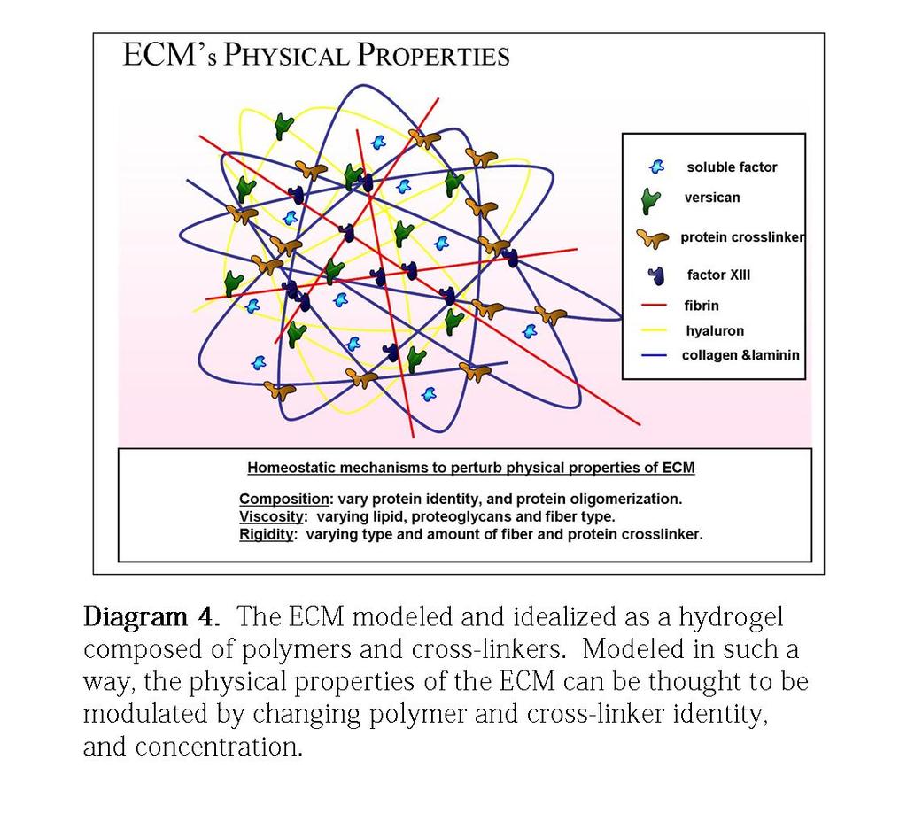
33 Physical Properties of ECM Of all the many mechanical properties inherent to biological systems, stiffness or rigidity is perhaps the most widely investigated. In short, the mechanical rigidity of a material can be determined by measuring its complex modulus or the ratio of stress (force per unit area) to strain (fractional deformation) applied to a material. This value reflects the material s ability to store and dissipate an applied mechanical force. By definition, this is represented by the storage (elastic) modulus and loss (viscous) modulus. Regulated by the biochemical / physical constituents of the ECM, tissues and cells are viscoelastic and exhibit both fluid- and solid-like properties (Diagram 4). Interestingly, the elastic modulus, the measure of the stress required to achieve a specific strain in a substrate without any deformation, has emerged as an important regulator of cellular processes such as growth and differentiation. Physiologically, the elastic moduli of various tissues range over four orders of magnitude from <1 kpa for fat, brain (Gefen et al. 2003), and ~5kPa for mammary tissue (Paszek et al., 2005) to~10 kpa for skeletal muscle (Engler et al., 2004) and 10MPa for bone (Goldstein et al., 1983; Moore, et al., 2010). Individual tissues can also contain significant internal differences in rigidity. For example, there is an approximately threefold variation in rigidity reported within the hippocampus of the brain and epithelial layers in organs such as the skin, lung, prostate and breast. In dramatic contrast, the typical surfaces used to culture cells (e.g., plastic and glass) have stiffnesses on the order of >1 GPa,
34 17
35 18 which is as much as 10 million fold stiffer than a wildtype cellular microenvironment (Moore et al., 2010). In this investigation I utilize bis-acrylamide substrates that provide ECM environments closer to physiological rigidities, better approximating the microenvironments found in tissues. In addition to intrinsic mechanical properties of the microenvironment such as rigidity and extrinsic mechanical perturbations, the application of forces or stresses that induce deformation are important characteristics of cellular microenvironments. Tissue-based examples of mechanical perturbations include stretching and contraction of tendons, ligaments, and musculature, as well as cyclic loading or sheer stress of the vasculature. The mechanically dynamic nature of tissues suggests the potential importance of stress and strain in regulating cell behavior in inactive conditions (Albinsson et al. 2004). Moreover and briefly discussed below, the different means to apply stress in a physiological context, include tensile, compressive, torsional, and shear forces. Interestingly, these forms of stress, in concert and/or individually, are thought to influence cellular phenotype, motility and pathological states in relevant ways. Interestingly, sustained tensile strains have been observed at the cellular level in embryonic systems. Specifically, in Drosophila embryos, experimental compression of cells induced expression of Twist, an important transcription factor regulating germ layer specification and patterning (Farge, 2003). Moreover, stretching has also been
36 19 shown to induce molecular changes in mouse embryonic fibroblasts, where cyclic stretch activated the phosphorylation of a cryptic site on p130cas, which is thought to lead to downstream signaling via MAPK s (Sawada et al., 2006). Given these observations, natural tissue dynamics during development may utilize compression and stretch to promote signaling and induce expression of specific genes responsible for growth, proliferation and differentiation. Another form of dynamic force application is shear flow, which is most often relevant to the circulatory system. Interestingly, sheer stress has been found to be important in regulating non-endothelial cell function as well. Early work demonstrated that shear flow forces facilitates capillary formation of endothelial progenitor cells (Yamamoto et al. 2003). Subsequent studies have found that shear flow forces can induce differentiation of a handful of stem cell types, including murine mesenchymal stem cells and embryonic stem cells, (Yamamoto et al. 2005) into specialized endothelial cells. Mechanical properties (i.e. elastic modulus / rigidity, stress, and strain) play clear roles in regulating growth, proliferation, and differentiation. Other biophysical properties also include structural characteristics such as topography, a substrates surface geometry and shape. Topographical structures such as grooves, ridges, and pits are present in many natural systems at the nanoscale (i.e. in the fibrous structure of collagen and other ECM
37 20 proteins) as well as at the microscale (i.e. in pores in bone marrow and discrete features of the basement membranes in the epidermis). The presence of topographical information in natural systems is a strong motivation to leverage technologies such as soft lithography, microfluidics, electrospinning, and construction of nanostructures in an effort to engineer a material s topography to study cell responses to both nano- and micro-topography. In this investigation I utilize nanofabricated substrates and substrates of different rigidities and ligand availability to better understand the molecular mechanisms that allow cells to interpret the specific physical features of their microenvironment. It has recently been observed that numerous biochemical and physical microenvironment cues regulate cell behavior such as ligand availability and patterning within developing tissues. Some developmental cues include discrete / distinct immobilized adhesive molecules (i.e., amino acid/peptide sequence or protein cleavage products), growth factors [i.e. epidermal growth factor (EGF)], and morphogenic factors (ex. Delta). In addition, steric availability of receptor-ligand binding, cryptic sites exposed by cell-exerted contractile / intermolecular-traction force, and ligand clustering has been recently postulated as being necessary for, or to enhance, classical biochemical signaling mechanisms. In chapter 3 I present data relevant to a cells ability to activate
38 21 translation based on biophysical cues such as the extracellular matrix (ECM) elastic modulus, and ligand availability. 1.3 Model System Cell-spreading serves as an ideal experimental model system to better understand ECMcell interactions. ECM-cell interactions lead to cytoskeleton activation (Dubin-Thaler et al., 2004), focal adhesion ontogeny (Geiger et al., 2009) and inside-out/outside-in signaling. A better understanding of these interactions will lead towards a better understanding of important processes such as development, organogenesis and pathological conditions such as oncogenesis, metastasis, and cardiac hypertrophy. Cell spreading requires the coordinated biochemical activation of integrins (Hynes, 2002), the acto-myosin network (Pollard et al, 2009), coordinated by GTPases (Etienne-Manneville & Hall et al, 2002), Src family kinases (Mitra and Shapaelfer, 2006), lipid species and modifiers (Di Paolo, and De Camilli 2006), and the dynamic equilibrium of numerous other proteins and protein complexes (Geiger et al., 2009). The aforementioned dynamic biochemical states are generally regulated by individual protein expression levels, modification state, and discrete sub-cellular localization - all of which have been under investigation and implicated, in concert, in the context of a wide variety of physiological and pathological phenomenon. Previously, cell spreading in mouse embryonic fibroblasts (MEFs) has been described to occur in 3 phases: 1. initial adhesion, where
39 22 initial integrin-ecm interactions occur and orchestrate the recruitment of the requisite actin polymerization machinery; 2. a fast spreading phase, characterized by the formation of new ECM cell bonds, and 3. a contractile phase, in which focal adhesion formation and acto-myosin contraction occur, allowing for substrate traction and higher order motility events (Döbereiner, et al., 2004). Much of the work described in the following chapters has utilized the cell spreading model system as an assay to better understand ECM-cell interactions, and actin and ECM-cell adhesion dynamics. 1.4 ECM-Cell Contacts Cell extracellular matrix (cell-ecm) interactions are mediated through specialized subcellular sites that contain specific adhesion receptors, cytoskeletal components and a wide variety of intermediate adaptor proteins (Zaidel-Bar et al., 2003 ; Critchley et al, 2004). These adhesion complexes allow cells to sense numerous extracellular signals that signal to the biochemical composition, geometry and physical properties of the ECM (Zaidel-Bar et al., 2003; Bershadsky et al., 2006). Cells can thereby distinguish between different ECM components (Humphries et al., 1990), detect differences in adhesive ligand density, and allow cells to respond to mechanical perturbations and ECM rigidity (Lo et al., 2000; Choquet et al., 1997).

40 23
41 24 Living cells survive, proliferate, and function while being closely associated with the diverse biochemical and physical properties that engender the extracellular matrix milieu. Recently, it has become increasingly clear that the cellular response to ECM signaling involves the ability of the cell to chemically sense specific ECM ligands and interpret a diverse range of physical cues that are generated at, or that act on, the cell- ECM interface. In turn, it is thought that cells can react to internally generated or externally applied forces (Geiger & Bershadsky, 2002; Bershadsky, et al., 2003; Chen, et al., 2008). Moreover, it is thought that cells can sense the topography of the underlying ECM (Spatz, & Geiger, 2007; Vogel & Sheetz, 2006), as well as its rigidity (Engler, et al., 2006 ; Discher, et al, 2005). In this investigation, I present data that demonstrates how cell-ecm contacts both recognize, and are regulated by biochemical and / or physical cues thereby regulating specific activation steps in signaling cascades necessary for transcription and translation. Transmembrane adhesion receptors of the integrin family (Diagram 5) have a primary role in such recognition processes. Studies (Bershadsky et al., 2006) show that the biochemical characteristics of the substrate, as well as its rigidity and spatial organization, are recognized by cells through differential signaling from integrin mediated molecular complexes. Moreover, integrin nucleated complexes are also involved in the sensing and processing of external mechanical stimuli, such as ECM stretching, modification and fluid shear flow. The mechanisms that underlie adhesion
42 25 mediated signaling events elicit many interesting questions. 1) How do adhesion receptors get activated by ECM ligands? 2) How do activated adhesions regulate cytoskeletal dynamics? 3) What spatial, and temporal scales do activated adhesions sense and operate in? 4) How are the molecular interactions at the adhesion site organized and regulated? 5) How do the biochemical and physical features of the ECM activate specific signaling pathways responsible for cytoskeletal activation, survival, growth and proliferation? 1.41 Integrins Alpha and beta (αβ) heterodimeric integrins have been shown to mediate adhesive cellextracellular matrix (ECM) interactions in metazoa that are critical in growth and proliferation, development, homeostasis, and the immune response. It is thought that this is achieved through interactions of the short cytoplasmic integrin tails with intracellular proteins. Moreover, these interactions trigger restructuring of the ligandbinding site through conformational changes in the integrin extracellular domain. Ligand binding in turn elicits conformational changes that are then transmitted back into the cell to regulate numerous cellular responses (Diagram 6). In mammals, 24 integrins have been identified to date, resulting from different pairings among 18 α- and 8 β-subunits (Diagram 3). The extracellular domain of the α- and β- subunits has approximately 1104 residues and 778 residues respectively. Integrins
43 26 recognize a large number of physiologic ligands, including soluble and surface-bound proteins. Integrins bound to soluble or immobilized biochemical ligands form microor macro- clusters (Kim et al., 2004) and transmit mechano-chemical signals inwards (outside-in signaling) that reorganize the cytoskeleton. Integrins, as a result, modulate much of the cell s metabolic and signal transduction machinery (Ingber, 2003; Schwartz & Ginsberg 2002). Interestingly, the dissociation rates of high-affinity integrins ( s 1) responsible for stable adhesion are found to be an order of magnitude less than other ECM receptors (Bhatia et al. 2003). Furthermore it has been postulated that the high dissociation rates (~ s 1) (Shimaoka et al. 2003, Smith et al. 1999) of the low and intermediate-affinity states allow some integrins (i.e. α4 integrins) to mediate cell rolling as in the case of platlets or lymphocytes. Thus depending on their affinity state, such integrins may mediate different forms of migration or motility. Later in this investigation, I present data related to the engagement and activation of integrins, and the down stream effects low activation states have on cytoskeletal dynamics and signal generation. Subcellulary, integrins span the lipid bilayer of cells and promote intracellular signaling, typically in the context of activated cytokine receptors or growth factor receptors (Diagram 6). It has been hypothesized that tumor growth and invasion probably depend on integrin crosstalk with growth factor receptors and other receptors that are known to
44 27 associate with focal adhesion proteins such as G-coupled receptors or cell-cell receptors (Ladeda et al., 2001). Recent investigations suggest that some growth factors and oncogenes require specific integrins for tumor initiation and progression. These studies highlight the importance of integrin signaling, in homeostatic and disease states (Hynes, 2002). The specific binding of the extracellular domains of integrins to ECM proteins or, in some cases, to counter-receptors on adjacent cells, supports cell adhesion and is crucial for embryonic development, tissue maintenance and repair, host defense and homeostasis. As mentioned earlier these processes rely on the binding of integrins to the intracellular cytoskeleton through the relatively short integrin cytoplasmic tails. This linkage permits the bi-directional transmission of force across the plasma membrane (Calderwood et al., 2000; Evans and Calderwood, 2007). In addition to their mechanical roles in adhesion, integrins transmit chemical signals into the cell (outside-in signaling), signaling information on its local environment, adhesive state and surrounding matrix (Hynes, 2002; Miranti and Brugge, 2002). These signals determine cellular responses such as migration, survival, proliferation, differentiation and motility. Moreover, these signals provide a context for responding to other inputs, such as signals generated by growth-factor- or G-protein- coupled receptors. Furthermore, as integrins mediate both force and signaling events, it is of experimental interest to investigate if and how both
45 28 mechanical and biochemical events are coupled. In addition to outside-in signaling, integrins can regulate their affinity for extracellular ligands. They do this by undergoing conformational changes in their extracellular domains that occur in response to signals that propagate to the integrin cytoplasmic tails a process that is termed inside-out signaling (Calderwood, 2004). Outside-in and inside-out signaling require dynamic, spatially and temporally regulated assembly and disassembly of multiple protein complexes (Diagram 7) that form around the cytoplasmic tails of integrins. Geiger and colleagues have recently described a network of 156 components (linked via 690 interactions) that make up the integrin adhesome (Zaidel-Bar et al., 2007). In this investigation, I present data as to how actin dynamics can regulate outside-in signaling, such as signaling that is known to effect transcriptional activation. Moreover, I add information that contributes to our understanding of how cells couple force generation and biochemical signal generation

46 29
47 Focal Adhesion Proteins Focal Adhesions (FAs) as large protein complexes have been described as central in the regulation of motility and growth. Characterized by the initial clustering of α and β integrins (Hynes, 2002), focal adhesions promote the localization of numerous signaling molecules such as focal adhesion kinase (FAK), Src family kinases (Mitra and Schlaepfer, 2006), Akt/PKB and phospho lipase kinases, MAP kinases and numerous adapter or scaffold proteins such as vinculin, talin, and paxillin. Interestingly, the order and modification state of proteins that are recruited to FAs engender the protein complex with different cellular functions (Diagram 6). The extracellular ligands that interact with integrins and anchor these adhesion proteins include collagen, and fibronectin. The best-characterized adhesions are termed focal adhesions, which develop from early forming focal contacts, and subsequently develop into fibrillar adhesions (Diagram 7). Focal-adhesion components have been identified in numerous cell types in vitro, and can be found in many physiological settings in vivo such as adhesions formed by aortic endothelial and epithelial cells with the underlying basement membrane. More than 50 different molecules are found in focal adhesions and other cell matrix adhesions. The ability of focal adhesions to recruit or co-localize different signaling molecules allows for them to coordinate, amplify, and transmit extracellular signals into the intracellular environment as far downstream as, growth and proliferation pathways, and possibly mitochondrial regulation, translational activation and transcriptional activation. In this
48 31 investigation I present data that supports the idea that focal adhesion formation allows for the activation of specific proteins that activate transcription and translation and highlight FA dynamics as a means to regulate such cellular processes. Following the interaction of cooperating proteins with integrin tails, conformational changes are thought to propagate across the membrane to the extracellular domains of integrins, in turn increasing their affinity for ligands. It is thought the binding of individual integrins to talin-mediated interactions between the cytoskeleton and the ECM enables forces to be transmitted, contributing to the reinforcement of the ECMcytoskeleton link and to the recruitment of additional cytoskeletal and signaling proteins (Giannone and Sheetz, 2006; Ginsberg et al., 2005). As adhesions mature (Diagram 8), protein complexes assemble at the cytoplasmic interface of clustered, ligand-bound integrins. These complexes are responsible for connecting integrins to the actin cytoskeleton and transmitting signals into the cell. Many elements of the outside-in integrin signaling cascade have now been identified, but there is considerable variability in the molecular components of integrin-containing adhesions and how the dynamics of their assembly and turnover is regulated still remains largely understood. Moreover, it is still unclear how the clustering of integrins, and the binding of ECM proteins, triggers signaling (Ginsberg et al., 2005). As integrins cluster, and adhesions mature, it is hypothesized that given different molecular constituents populate the focal adhesion,
49 32 focal adhesions can take on different functional roles. A better understanding of these molecular components, their role in cell behavior and the relevant spatial and temporal scales that they operate in is of great experimental interest Talin The binding of talin to the cytoplasmic tail of integrin β subunits has been demonstrated to have a key role in integrin activation (Calderwood, 2004; Ginsberg et al., 2005; Tadokoro et al., 2003). Binding of the phospho-tyrosine binding (PTB) subdomain of the protein 4.1, ezrin, radixin, moesin (FERM) domain of talin to the conserved WxxxNP(I/L)Y motif of the β -integrin tail allows for additional interactions between talin and the membrane-proximal region of the tail that triggers integrin activation (Wegener et al.,2007). The important role of talin in integrin activation in vivo is supported by studies in transgenic mice that show the importance of talin-integrin interactions for platelet aggregation (Nieswandt et al., 2007; Petrich et al., 2007). Talin also binds to actin and to numerous other scaffold and signaling proteins, (Critchley and Gingras, 2008) thereby coupling activated integrins directly to signaling and cytoskeletal protein networks Talin, a 270-kDa F-actin binding protein is thought to play an important role in integrin activation, the initiation of matrix adhesion formation and the linkage of integrin
50 33 receptors to the actin cytoskeleton. Talin, which self-associates via the rod-shaped C- terminus to form a dimer, was one of the first proteins to bind cytoplasmic integrin tails. The globular head of talin contains a 4.1 ezrin, radixin, moesin (FERM) domain which binds with high affinity to the cytoplasmic tails of integrin β1, β2, β3 and β5 (Critchley and Gingras, 2008). It is thought that this interaction involves a conserved NPxY motif in β-integrin tails. The head region also contains binding sites for focal adhesion kinase (FAK), phosphatidylinositol-4,5-biphosphate (PIP2), phosphatidylinositol-4-phosphate 5-kinase type I g (PIPKIg) and for the hyaluronan receptor layilin (Calderwood, 2004). The tail region contains two actin and vinculin binding sites that mediates integrin coupling to the cytoskeleton. In this investigation I present data that demonstrates talin s localization in cells interacting with collagen and its regulation of force generation and collagen contraction. Understanding talins protein domain structure and binding partners may give interesting insight into focal adhesion ontogeny as well as force mediated cellular processes such as wound healing and tissue formation. As an early integrin binding partner, it is thought that talin plays a central role in the first steps of focal complex and adhesion formation following initial integrin engagement to the ECM. Interestingly, it was demonstrated that talin degradation plays a critical role in focal adhesion turnover (Franco et al., 2004). Since talin-deficient mice die at E9 (Monkley et al., 2000), much of the biological function of talin has been elucidated from
51 34 in vitro cell culture experiments. Talin-1-deficient embryonic stem (ES) cells fail to spread on collagen or laminin; however, these cells can spread on fibronectin but are unable to assemble vinculin- or paxillin-containing focal adhesions, or stress fibers (Zhang, 2008). This observation underscores the ability of cells with different FA components to sense individual ECM components differently. Talin knockdown in CHO cells was shown to inhibit activation of integrin αiibβ3, αvβ3 and α5β11 (Tadokoro et al., 2003). Conversely, overexpression of the talin N-terminal head leads to a threefold increase in integrin αiibβ3 activation in CHO cells (Calderwood et al., 1999). By using optical tweezers to obtain force measurements, talin was identified as the important component for maintaining a 2 pn slip bond between fibronectin and the cytoskeleton and is thought that talin is necessary for integrin mediated force generation events (Jiang et al., 2003). In chapters 2 and 3 I explore the different molecular properties of FAs as they interact with either collagen, fibronectin or both, as well as adhesion strenths as measured by magnetic tweezers. In addition to the evidence that talin is required for integrin activation and force generation events, several observations have indicated that other activating factors might cooperate with talin. In recent studies, the proteins of the kindlin family have been identified as important integrin activators (Moser et al., 2008). It is thought that the PTB-like subdomain within the kindlin FERM domain is similar to that of talin (Kloeker
52 35 et al., 2003) but binds to the second NPxY motif in β-integrin tails, whereas talin binds to the first motif. Interestingly, inhibition of kindlin binding inhibits integrin activation, whereas co-expression of kindlin and talin activates integrins. Interestingly, inside-out integrin signaling appears to be a complex process that operates on multiple spatial and temporal scales involving more interactions than those between talin and integrin Integrin-Linked Kinase (ILK) Integrin-linked kinase (ILK) is another key molecule in integrin signaling (Brown, et al., 1998, Legate et al., 2006). Similar to talin, ILK is an essential protein that has a role as a cytoskeletal and signaling scaffold at integrin mediated adhesions. ILK forms a heterotrimeric complex with the LIM-domain protein PINCH and the actin- and paxillin-binding protein parvin. This complex serves as a major scaffold in integrin signaling networks and, in mammals, formation of the complex is required for appropriate targeting of known FA proteins to integrin-mediated adhesions (Legate et al., 2006). ILK contains an N-terminal ankyrin-repeat domain that mediates protein interactions with PINCH1 or PINCH2, and a C-terminal kinase domain that supports interactions with parvins, paxillin, and β-integrin tails (Hannigan et al., 2005; Legate et al., 2006).

53 36
54 37 Interestingly, the kinase domain lacks the catalytic residues that are normally conserved among protein kinases, and whether ILK has kinase activity remains controversial (Hannigan et al., 2005; Legate et al., 2006). Less controversial is ILK s role in integrin signaling and cytoskeletal connections. Importantly, this role is conserved from invertebrates to mammals. In contrast to FAK and other FA proteins, there are only a few structural observations available for ILK. Similar to talin, ILK also interacts with kindlin proteins (Mackinnon et al., 2002), and it is thought that this might account for the observations that implicate ILK in integrin activation (Tucker et al., 2008). In chapter 2 I present data relevant to ILK s role in the activation of β1 integrins and focal adhesion ontogeny. While ILK s kinase function is still controversial, ILK s role as an adaptor continues to be documented. ILK s kinase domain is thought to be important in integrin binding. ILK is composed of three N-terminal ankyrin repeats followed by a linker domain that exhibits homology to a PH domain. The C-terminus of ILK contains a predicted kinase domain and mediates direct ILK binding to β1 and β3 cytoplasmic tails (Hannigan et al., 1996; Pasquet et al., 2002). Many of the known binding partners of ILK also bind to the c- terminus region. Parvins, proteins composed of two calponin homology domains, have been shown to bind to ILK. It is thought that parvins provide a link to paxillin, α-actinin, alpha-pix2 and F-actin. Of the three known parvin isoforms, α-parvin/actopaxin and β-
55 38 parvin/affixin have been shown to bind to ILK (Tu et al., 2001; Yamaji et al., 2001). In addition, paxillin binds directly to ILK. The first ankyrin repeat of ILK binds PINCH-1 and its homologue PINCH-2 (Tu et al., 1999; Zhang et al., 2002). Many of these interactions have been identified in cells interacting with fibronectin, while still not elucidated on collagen. In this investigation I probe ILKs interactions on collagen and identify the localization and dynamics of many ILK s binding partners. Similar to talin, insight into ILK s protein domain architecture and binding proteins will provide interesting information on focal adhesion ontogeny and biochemical signal generation from integrin-ecm contacts. Moreover, quantitative investigations on focal adhesion turnover in cells deficient for components of the ILK-PINCH-parvin complex are required to further elucidate how each individual component contributes to the properties of the complex. Knockout studies have revealed roles for ILK and PINCH in matrix adhesion formation and turnover, and in actin organization. Specifically, deletion of the ilk gene in mice leads to peri-implanation lethality, and ILK-deficient fibroblasts show severely defects in adhesion to fibronectin, vitronectin and laminin (Sakai et al., 2003). In this investigation, I present quantitative analysis of focal adhesion protein dynamics in mouse embryonic fibroblast cells that either express or do not express ILK on collagen and fibronectin.
56 39 Interestingly, organism-wide loss of ILK expression and activity has demonstrated that ILK function is required for eukaryotic development. Gene knock out studies (Mackinnon et al., 2002) have shown that ILK is required to recruit actin filaments to the plasma membrane at muscle attachment points. In vertebrates, loss of-function analysis highlights the importance of ILK in the mediation of protein-protein interactions that regulate cytoskeletal dynamics and act to provide an important node for the activation of signaling pathways (Knoll et al., 2007; Sakai et al., 2003). For example, embryonic lethality was observed in Xenopus laevis and mouse (Sakai et al., 2003) models of ILK ablation, and this can be linked to defects in adhesive and motility mechanics. Similarly, the zebrafish lost-contact mutant exhibited reduced signaling activity, and cardiac dysfunction (Knoll et al., 2007). Taken together, these observations provide a body of evidence that supports the physiological requirement of the adaptor functions of ILK. In this investigation I report the effects on motility and signal generation in mammalian cells interacting with physiologically relevant substrates and present data towards a better understanding of ILKs role in regulating protein dynamics at nodes of interest as well as adaptor dynamics and function. Of the experimental and clinical evidence available, sit is of interest to note that significant defects in normal tissue development and homeostasis occur when ILK is deleted. In contrast, the overexpression of ILK either in cell culture or in transgenic
57 40 mouse models results in oncogenic progression (Dillon et al., 2007; Hannigan et al., 2005; Legate et al., 2006). Interestingly, the expression of ILK is often elevated in human malignancies, and correlates with tumor stage and grade. Importantly, increased ILK expression predicts poor patient survival in several types of cancers (Dai et al., 2003; Graff et al., 2001; Okamura et al., 2007; Takanami, 2005). Given the aforementioned observations, ILK knockout cell lines represent a powerful model system to investigate cancer progression. Mounting evidence, suggests that the oncogenic capacity of ILK derives from its regulation of several downstream targets that promote cell proliferation, survival and migration. In this investigation I leverage an ILK knockout cell line system and I present data that provides novel molecular mechanisms that implicate ILK in regulating the activation state of important regulators of translation, survival and growth. Recently, sirna complementary to ILK have allowed for the investigation of downregulating ILK expression during oncogenic progression. Analogous to sirnamediated downregulation of ILK, ILK antisense-oligonucleotide treatment of glioblastoma cells reduced ILK expression and the phosphorylation of Akt at Ser473 (Edwards et al., 2005; Edwards et al., 2006), and induced apoptosis (Edwards et al., 2005). Furthermore, ILK-antisense treatment of mice harboring established glioblastoma xenografts resulted in stable disease, whereas the tumor volume in control animals

58 41
59 42 increased (Edwards et al., 2005). Interestingly, cell-based studies have been conducted that use ILK-antisense in combination with inhibitors of Raf1 and MEK, thus providing concurrent regulation of survival and proliferative signaling pathways (Edwards et al., 2006). These investigations showed that ILK-antisense and the Ras-MAPK inhibitors had a synergistic effect on glioblastoma-cell survival. Similar results were obtained with ILK sirna in combination with a MEK inhibitor (Edwards et al., 2006). These studies point to a pro-survival function for ILK. In this investigation I present data that provides additional support for a molecular mechanism linking ILK and MAPK signaling Focal Adhesion Kinase (FAK) FAK is a non-receptor protein tyrosine kinase that plays a central role in signaling through integrins and a variety of other receptors (Parsons, 2003). Interestingly, FAK has been implicated in ILK mediated / dependent signaling. An understanding of FAKs protein domain structure offers interesting insight into focal adhesion ontogeny and begins to highlight a pattern of protein domains found in focal adhesion proteins. Structural studies indicate FAK s kinase domain is situated in the central part of the molecule and is flanked by large non-catalytic domains. The N-terminus contains a FERM homology domain which binds has been shown to bind to peptides of the β1- integrin cytoplasmic tail (Schaller et al., 2005). The functional relevance of direct FAK binding to integrin is unclear since the integrin-binding region of FAK is not required to
60 43 target the molecule to focal adhesions (Shen et al., 1999). Finally, the carboxyl terminus of FAK contains the focal adhesion targeting (FAT) domain, a four-helix bundle bearing homology to domains found in p130cas and vinculin (Arold et al., 2002). Activation of FAK in response to integrin activation leads to autophosphorylation of Y397, creating a high-affinity binding site for Src family kinases (SFKs) (Cobb et al., 1995; Schaller et al., 1995). FAK and Src can in turn phosphorylate the linker protein p130 Cas, which binds via its SH3 domain to a proline-rich region in the C-terminal half of FAK. A second proline-rich site is bound by the cytoskeletal effector Rho-GAP GRAF (Hildebrand et al., 1996). Other known ligands of FAK include the survival and proliferation signaling molecules phosphatidylinositol-3-kinase, phospholipase C- gamma and the adaptor protein Grb7. Deletion of the FAK gene in mice leads to embryonic lethality before day E10.5 (Furuta, et al., 1995), while FAK-deficient cells show delayed spreading, increased number of focal adhesions and reduced adhesion turnover (Ilic et al., 1995). One of the first integrin signaling molecules to be identified, and as its binding partners suggest, FAK acts as a phosphorylation-regulated signaling scaffold. Recently FAK has been shown to be important for adhesion turnover, cytoskeletal activation via Rhofamily GTPase activation, and cross-talk between growth-factor signaling and integrins
61 44 (Mitra et al., 2005). In response to integrin clustering, the autophosphorylation of FAK generates docking sites for SH2-domain-containing proteins; these include Src kinases, which in turn become activated and phosphorylate FAK, promoting its kinase activity and its interaction with other proteins. Structural studies have revealed the interaction between FAK and paxillin, and highlights how FAK is inhibited by interactions between its FERM and kinase domains. Interestingly, these structural studies have also elucidated a role for PtdIns(4,5)P2 in FAK activation (Hayashi et al., 2002; Lietha et al., 2007; Mitra et al., 2005). Continuing studies aim to integrate this structural information of FAK into a comprehensive picture of FAK function. A better understand of how FAK interactions are remodeled during adhesion turnover and how those interactions regulate integrin mediated motility and signaling events is of great interest to provide insight into themes or patterns that may describe other FA protein dynamics. In this investigation I provide evidence to support a role for actin dynamics regulating FAK s activation and function, adding to the current understanding of FAK localization and turnover in focal adhesions Paxillin Central to integrin mediated adhesion formation and signal generation, is the recruitment of scaffold or adapter proteins to the sites of ECM-cell interactions. One such scaffold and adapter molecule is paxillin. Paxillin contains five N-terminal LD
62 45 repeats of 13 amino acids each and 4 C-terminal LIM domains that serve as sites for protein-protein interactions. Interestingly, proteins containing LIM domains have been shown to translocate to the nucleus, cycling between focal adhesions and transcription complexes. There are three splice isoforms in mammals, of which only one isoform, paxillin-a, shows broad expression (Mazaki, et al., 1997). Similar to FAK, paxillin coimmunoprecipitates with β1 integrin and can bind synthetic peptides mimicking β1 cytoplasmic tails (Chen et al., 2000). Interestingly, paxillin has been shown to bind α4 cytoplasmic tails with much higher affinity than β1 tails. Paxillin s non-integrin matrix adhesion binding partners are thought to bind through a consensus paxillin binding sequence to at least one LD repeat. Two F-actin binding proteins have been shown to bind to paxillin: 1. vinculin is thought to bind LD1, LD2 and LD4 of paxillin (Brown et al., 1996); 2. α-parvin has been shown to bind LD1 and LD4 (Nikolopoulos et al., 2000). Paxillin has also been shown to bind to the integrin-binding kinases FAK, via LD2 and LD4 (Hashimoto et al., 2001), and ILK, via LD1 (Nikolopoulos et al., 2001), in addition to the p21-activated kinase isoform PAK3 (Hashimoto et al., 2001). Interestingly, both of these proteins link paxillin to the Rac1/Cdc42-specific Gaunine Exchange Factor (GEF)- β-pix/ Cool-1, thereby linking paxillin to the regulation of actin polymerization. Important to its function is paxillin s modification state. Paxillin can be phosphorylated on four N-terminal tyrosines (Schaller et al., 2001), and evidence exists that the
63 46 responsible kinases may be FAK (Richardson et al., 1996) and Src family kinases (Klinghoffer et al., 1999). Tyrosine phosphorylation creates high-affinity binding sites for the SH2 domains of the adaptor protein Crk and for Src (Schaller et al., 1995). As with many focal adhesion proteins, deletion of the paxillin gene in mice is embryonic-lethal (Hagel et al., 2002). The protein appears to be one of the first to be recruited to nascent focal complexes on fibronectin, and its incorporation into αvβ3-containing focal complexes appears to occur simultaneously with talin (Zaidel-Bar et al., 2003). Interestingly, studies have reported that paxillin recruitment is detectable in fibronectin mediated focal complexes before integrin α5β1 (Laukaitis et al., 2001). This is another important example of how during the course of focal adhesion ontogeny, focal adhesions are composed of different proteins at given timepoints, with this composition predicted to dictate focal adhesion function. It is still not clear, how paxillin is recruited into nascent focal complexes, although a C-terminal focal adhesion targeting sequence appears to be required (Brown et al., 1996). The function or binding partners of this sequence, and of all the paxillin LIM domains, have yet to be fully unidentified. The localization and activation state of paxillin, as well as its binding partners, are addressed in the following investigation. Data is presented that details paxillin s role in collagen mediated ECM-cell interactions.
64 47 Interestingly, the complex of paxillin, FAK, Cas and Src is emerging as a potential molecular switch that regulates focal complex turnover and higher order cellular functions such as motility and growth. Interestingly, FAK null and paxillin null cells display a similar phenotype of reduced migration velocity and slow turnover of matrix adhesions (Ilic et al., 1995; Hagel et al., 2002). A detailed study has recently addressed matrix adhesion turnover and FA protein recruitment in mouse embryonic fibroblasts (MEFs) null for FAK, Cas and Src (Hagel et al., 2002). While paxillin localization to focal adhesions does not require FAK, activated Y397-phosphorylated FAK and tyrosinephosphorylated paxillin localize to dynamic matrix adhesions and are thought to be required for high turnover rates. Interestingly, the aforementioned study demonstrates that in the absence of either paxillin, FAK, Src or Cas, fibronectin-matrix adhesion turnover rates are reduced. This investigation details paxillin dynamics at the leading edge and within focal adhesions mediated by integrins bound to collagen. Central to interpreting the results of this investigation, paxillin, as a FAK and ILK binding protein, is an essential signaling scaffold that is recruited early to integrin adhesions (Deakin and Turner, 2008). Paxillin contains several protein-protein interaction modules (leucine-rich repeats, a proline rich region and LIM domains) and its numerous phosphorylation sites provide additional regulated sites of protein-protein interaction. Together, they mediate the binding of kinases (e.g. FAK, Src and ILK),
65 48 phosphatases (e.g. PTP-PEST), actin-binding proteins (e.g. vinculin and the parvins) and regulators and effectors of the Rho family of small GTPases and actin dynamics (e.g. the CrkII-DOCK180-ELMO complex and PIX). As this investigation outlines the dynamics of paxillin at the leading edge, it is of great interest to probe and better understand the dynamics of many of its binding partners at the leading edge as well. Some interactions of paxillin are understood at the structural level such as the FAK-paxillin complex that has been resolved by X-ray crystallography (Hoellerer et al., 2003). Competition between potential binding partners, regulation by conformational changes and signal-dependent phosphorylation may explain the ability of paxillin to coordinate multiple interactions and to regulate dynamic processes such as actin dynamics, adhesion turnover, growth, proliferation and migration (Diagram 6). Phosphorylation of residues in the N- terminus of paxillin by cellular kinases may account for the regulated recruitment of downstream effector molecules such as p130cas. Interestingly, recruitment of p130cas could be potentially important for the transduction of external signals into changes in cell motility and for the regulation of gene expression by the various MAP kinase cascades (Deakin and Turner, 2008). Data relevant to these observations is presented in chapters 2 & 3 and suggests a molecular mechanism that involves paxillin localization, paxillin modification state, binding partner localization, and MAPK activity.
66 p130cas Similar to paxillin, p130cas is a scaffold molecule that has been implicated in numerous cellular processes such as actin polymerization, focal adhesion formation, and mitotic signal generation. p130cas contains an N-terminal SH3 domain, a proline-rich region, a substrate-binding domain- containing 15 repeats of a YxxP sequence, a serine-rich region, and a C-terminal domain. p130cas s most well-known feature is the tyrosine residues in the YxxP sequences. These residues act as substrates of protein tyrosine kinases and, when phosphorylated, provide a binding site for the SH2 binding domains of effector proteins. Again, insight into the domain architecture of p130cas may provide thematic clues as to how cells are able to transduce and amplify extracellular signals. The C-terminal domain is characterized by a binding site that includes a proline-rich region (RPLPSPP), which binds to the Src SH3 domain. P130Cas also contains a tyrosinecontaining sequence (YDYV) which in turn binds to the Src SH2 domain when phosphorylated (Bouton et al., 2001). It is thought upon ECM binding or growth factor and hormone stimulation, integrins, receptor tyrosine kinases, estrogen receptors, and G-protein coupled receptors regulate p130cas through the activation of Src kinase and the formation of a p130cas Src complex. After tyrosine phosphorylation, p130cas and Src recruit adaptors and effectors that activate downstream pathways, resulting in cell survival and increased cell motility
67 50 (Panetti, 2002). p130cas is also required for transformation and metastasis. Interestingly, in response to pro-apoptotic stimuli, p130cas dephosphorylation by eukaryotic phosphatases inhibits the formation of p130cas-dependent signaling complexes and favors cleavage of p130cas by proteases into smaller fragments, which have been shown to translocate to the nucleus and contributes to cell death (Panetti, 2002). The exact regulation and mechanism that allows for p130cas derived peptides to translocate into the nucleus is of great scientific interest. Similar to paxillin and FAK, phosphorylation of tyrosine residues creates binding sites for the SH2 and PTB domains of effecter signaling proteins. Many growth factors and hormones regulate p130cas tyrosine phosphorylation (Bouton et al., 2001). Integrinmediated adhesion also triggers p130cas tyrosine phosphorylation, which correlates with regulation of actin cytoskeleton organization, cell spreading and focal adhesion formation. By contrast, in mitosis, when cells become rounder and temporarily detach from the extracellular matrix during cytokinesis is, p130cas is phosphorylated on serine and threonine residues (Yamakita et al., 1999). Interestingly, at re-entry into G1, this serine/threonine phosphorylation is lost (Yokoyama et al., 2001). It has been hypothesized that the balance of serine/threonine versus tyrosine phosphorylation might be controlled by different phases of the cell cycle or vice versa. Better understand this
68 51 type of molecular switch and signaling circuitry is of interest and data is presented in chapters 2 & 3 that support a molecular mechanism as to how this switch is activated. Recently, mechanical stretch has been shown to increases tyrosine phosphorylation of p130cas and its association with the adaptor protein Crk which results in the activation of the small GTPase Rap1. Therefore, by unfolding p130cas, mechanical forces expose effecter binding sites and phosphorylation sites, providing a potential molecular mechanism to transduce external forces into intracellular biochemical signals (Tamada, et al., 2004; Sawada, et al., 2006). Interestingly, the current model of cell migration suggests that during migration, lamellipodia and filopodia extend from the cell leading edge and create new dynamic adhesions, which form and rapidly disassemble at the base of protrusions. The involvement of p130cas in cell migration has been shown to be dependent on its tyrosine phosphorylation by Src and on the assembly of a p130cas Crk DOCK180 scaffold at adhesion sites (Gustavsso et al., 2004; Webb et al., 2004). It is thought that scaffold formation initiates Rac activation, thereby leading to actin polymerization and the recruitment of integrin receptors necessary for lamellipodia extension and cell migration. Data is presented in this investigation that details how the localization of the p130cas-crk-dock180 complex affects cellular processes such as migration and proliferation.
69 52 In addition to its role in actin dynamics, p130cas is an important transducer of survival signals. Prominent work detailed in some of the available literature suggests that prosurvival signals emanating from the ECM and soluble growth factors and hormones proceed through their respective receptors, then through FAK and Src, to p130cas, activating the small GTPases Ras and Rac, as well as JNK and Erk1/2 MAPK (Giancotti et al., 2002). P130Cas is also required for integrin-dependent EGF-receptor activation, which in turn leads to cell survival. This implies a dual role for p130cas in cell survival and motility as p130cas behaves as a major downstream effecter in integrin and growth factor signaling and as an activator of actin polymerization. Many other important integrin-signaling proteins have been, and continue to be identified and implicated in pro-survival and growth directed events, perhaps even modulating transcription in the nucleus. Experiments directed towards survival and growth signaling pathways, translation, mrna localization, nuclear export / import, and transcription may yield further insights into the regulatory steps in integrin signaling, the roles of different integrins in signaling, focal adhesion assembly, as well as the presumable hierarchy and nature of the hypothetical focal adhesion nucleus signaling axis. In addition, integrins make many important direct or indirect interactions with other transmembrane signaling proteins, including those from the growth-factor receptor, syndecan, discoidin domain receptors, and GPCR families. Understanding

70 53
71 54 how integrins are able to cluster with each other and other receptors is also an area of interest and discussed in part later in Chapter Cytoskeletal Activation Cytoskeletal activation (Diagram 9), namely actin polymerization, branched-filament stabilization, acto-myosin contraction, and stress fiber formation, is necessary for polarization, cell motility, cell division, and higher order processes such as development, wound healing, the immune response and metastasis. Cytoskeletal activation occurs concurrent with focal adhesion ontogeny at the leading edge of cells and as a result both processes are spatially, and biochemically coupled. The interplay between Cdc42, Rac, and Rho GTPase activation is largely thought to regulate this biochemical coupling, as activation of Rac favors actin polymerization, initial focal contact formation, while activation of Rho favors acto-myosin contractility, focal adhesion formation and focal adhesion stabilization. Central to outside-in signaling, and perhaps better understood, is how integrin activation mediates actin dynamics. Actin filaments are connected to integrin rich FA s via a complex of proteins that include talin, vinculin, α-actinin, and paxillin. This complex not only mediates actin polymerization events at the cell s leading edge, or lamellipodia, but mechanically couples the actomyosin contractile apparatus to FAs, and is central in the force transmission between ECM and the cell.
72 55 It has been predicted theoretically (Mogilner and Oster, 1996) and demonstrated in single filament experiments (Kovar and Pollard, 2004) that actin polymerization produces mechanical forces. These polymerization forces in conjunction with the forces generated by the actinomyosin machinery, are thought to be responsible for different forms of cell motility and, in particular, overcoming the cell s inherent membrane tension (Sheetz & Dai, 1996), thereby leading to extension of cellular protrusions (Mogilner and Oster, 2003; Pollard and Borisy, 2003). Cell protrusion is a process in which chemical signaling results in the production of mechanical energy generated from the polymerization, retraction, and net-extension of actin. The polymerization of these filaments is regulated by associated proteins, GTPases, and signaling proteins (Diagram 9). It is thought that protrusion at the leading edge, driven by actin polymerization, coupled with ATP hydrolysis and GTPase activity, is regulated by the dynamic equilibrium between actin nucleators and severing proteins. Some examples of proteins that facilitate actin polymerization or depolymerization are the actin-related protein (Arp)2/3 complex, Spir, profilin, and formins and ADF/cofillin, Gelsolin/villin, and twinfilin (Pollard & Borisy, 2003) (Diagram 9). Interestingly, the elongation of actin bundles and branches has been described with an elastic Brownian ratchet model where actin subunits are added in an ATP dependent manner to the barbed-end of actin filaments as they may be bent and pushed back from
73 56 the membrane, while the restoring force of polymerization pushes the membrane forward (Mogilner and Oster, 1996). Concurrent with polymerization, the actin network (Diagram 10) undergoes retrograde flow away from the leading edge (Vallotton et al., 2003). To date, two types of retrograde flow have been distinguished, fast and slow, fast retrograde flow occurs in the distal most periphery of the lamellipodium while the slow retrograde flow occurs just inside the lamellipodium. Central to this investigation is the reciprocal role actin dynamics and focal adhesion dynamics have on one another Actin Nucleators Actin is one of the most abundant and conserved proteins in eukaryotic cells. A 42kDa monomeric ATP-binding protein, globular (G)-actin, can undergo cycles of selfassembly into filamentous actin, (F)-actin, ATP hydrolysis, and depolymerization. It is thought that actin filaments contain high-affinity barbed ends and less active pointed ends that are differentiated by structural and biochemical characteristics. As mentioned earlier, cell protrusion events, actin polymerization and filament turnover are controlled by many actin-binding proteins, including some that function in monomer sequestration, and others that promote filament nucleation, elongation, capping, severing, or depolymerization (Diagram 9 and 10).
74 57
75 58 The ability of the actin network to generate force, create structural scaffolds, and act as tracks for motor proteins contribute in actin s role in numerous cellular functions, including growth, proliferation, morphogenesis, migration, cytokinesis, and membrane transport. A central aim of this investigation is to better understand the link between actin dynamics and higher-order cellular processes such as migration and proliferation. Evidence is presented in chapters 2 & 3 that support a role for actin retrograde flow in the regulation of signaling molecules necessary for force generation, as well as transcription, translation, survival and proliferation. According to the large body of work geared towards actin polymerization, to initiate actin assembly during motility processes, cells generate free barbed ends that serve as sites for polymerization by uncapping or severing existing filaments, or by nucleating monomers. Interestingly, and accounting for energetic costs associated with actin polymerization, spontaneous actin assembly is thought to be inefficient, as the formation of actin dimers and trimeric nuclei is kinetically unfavorable. It is thought that to overcome this thermodynamic barrier, cells utilize factors that catalyze the nucleation of actin monomers into filaments. Specifically, the Arp2/3 (actin-related protein 2/3) complex, and formins are some of the proteins known to directly participate in nucleation (Goley et al., 2006; Chesarone et al., 2010). The first central actin nucleator to be identified was the Arp2/3 complex, a 220kDa factor composed of seven stablyassociated proteins that are highly conserved across eukaryotic organisms. These include
76 59 Arp2 and Arp3 and five additional subunits, ARPC1-5. It is thought that the Arp2/3 complex is unique in its ability to nucleate filaments as well as organize them into branched networks. In vitro biochemical experiments support the idea that the growing filament is capped by the Arp2/3 complex at its pointed end, while able to elongate at its barbed end. Similar to actin, Arp2 and Arp3 bind ATP (Goley et al., 2004). Efficient nucleation also requires phosphorylation of threonine and tyrosine residues in Arp2. A third contributor to Arp2/3 activation, and the best characterized, involves engagement of the complex by nucleation-promoting factors (NPFs) (Higgs, et al., 2005). Most mammalian NPFs activate Arp2/3 using a WCA domain, which is comprised of one or more WH2 (WASPhomology-2) motifs that bind actin monomers, plus an amphipathic connector region and acidic peptide that collectively bind the Arp2/3 complex. NPFs have been subcategorized into five groups: WASP and N-WASP, three WAVE (WASP-family verprolin homolog; also known as Scar) isoforms, and the recentlyidentified factors WASH (WASP/Scar homolog), WHAMM (WASP homolog associated with actin, membranes and microtubules), and JMY (junction-mediating regulatory protein) (Chesarone, 2009).
77 WASP and N-WASP WASP and N-WASP, the best-characterized NPFs, are conserved across mammals, fungi and protists. Mammalian WASP is expressed specifically in hematopoietic cells, and its mutation results in defective cell migration, phagocytosis, and T-cell signaling, leading to immunodeficiencies in mice and the human disease state termed Wiskott-Aldrich Syndrome (Bosticardo et al., 2009). Another WASP protein, N-WASP is expressed in most cell types, and its deletion results in neurological and cardiac abnormalities, as well as embryonic lethality in mice. N-WASP has been well characterized, and its domain architecture provides insights into how actin polymerization is regulated as well as how actin filaments are able to generate force within the cell and at the leading edge. N- WASP possesses a modular domain organization consisting of an N-terminal WASPhomology-1 (WH1) domain, plus basic, Cdc42/Rac-interactive binding (CRIB), and auto-inhibitory (AI) motifs that are collectively termed the GTPase-binding domain (GBD), and a proline-rich domain (PRD) proximal to its WCA domain. N-WASP is central to cell spreading, and other motility events in that it coordinates the direction of actin protrusion and presumably is heavily regulated by proteins recruited at sites of ECM-cell interaction. A better understanding of N-WASP s activation, membrane localization, recycling, and degradation is an interesting topic of investigation that could yield important therapeutic insights into physiological processes such as wound healing, axon guidance, and organogenesis.
78 WAVE Very similar to the WASPs, the WAVE-family of NPFs are conserved across the metazoa. The three mammalian isoforms are expressed in many cell types, with WAVE1 and WAVE2 distributed ubiquitously. Interestingly, WAVE1 knockout mice have sensorimotor defects, behavioral abnormalities, and reduced viability, whereas WAVE2 ablation is lethal, and knockout embryos exhibit impaired angiogenesis, underdeveloped hearts, hemorrhaging, and brain malformation. Better understanding WASP and WAVE activity and their regulation during motility events may provide important insights into how cells regulate different motility regimes such as rolling, and the invadapodia formation necessary for metastasis. Data is presented in chapter 4 that underscores the relevance of motility events in pathological processes such as uncontrolled growth and metastasis Formins Unlike the Arp2/3 complex, other actin nucleators produce single, unbranched filaments. The most familiar of these are the formins, which are present in most eukaryotes. Their defining feature is the presence of the conserved formin-homology (FH) domains, termed FH1 and FH2 (Higgs et al., 2005). The best characterized mammalian formins are the diaphanous related formins (DRFs) based on their homology to the diaphanous protein from D.melanogaster. In mice and other mammals,
79 62 DRFs are referred to as mdia1-3. Interestingly, the DRFs have a domain organization that can be divided into three functional domains. Their N-termini contain regulatory sequences, including a GBD and an overlapping diaphanous-inhibitory-domain (DID) that participates in autoinhibition (Li et al., 2003). These domains are followed by central coiled-coil (CC) and dimerization domains (DD) that are thought to influence autoregulation. mdia-mediated actin assembly participates in the formation of a variety of cellular structures such as stress fibers, dorsal filaments that emerge from focal adhesions at the leading edge of migrating cells, and the contractile ring that forms during cytokinesis (Watanabe et al., 2008). Relevant to metastasis and wound healing, DRFs also localize to filopodia, where their ability to nucleate actin correlates with the ability to form filopodia (Peng et al., 2003). Interestingly, multiple DRFs are found in lamellipodia and contribute to cell motility (Gupton et al., 2007). Moreover, it has been shown that actin polymerization intitiated by DRF FH2 domains results in activation of serum response factor (SRF)-mediated transcription. This transcriptional circuit is thought to control developmental processes which regulate and rely on cell migration, contractility, or morphogenesis (Young et al., 2008). Delineating the signaling requirements initiated by actin polymerization to transcription activation is an interesting future line of inquiry and preliminary data towards this research aim is presented in the following chapters.
80 Myosin II On the cellular level, actin works in concert with the molecular motor myosin II. Formally, Non-muscle myosin II (NM II) is an actin-binding protein that has actin cross-linking and contractile properties, and is regulated by the phosphorylation of its light and heavy chains and dependent on ATP hydrolysis. The three mammalian NM II isoforms have both specific and redundant properties. Given its position as a regulatory target for many signaling pathways, NM II is poised as a central node in the signaling circuitry that controls cell adhesion, cell migration and tissue architecture. Moreover, myosins constitute a family of motor proteins that play an important role in several cellular processes discussed in the following chapters such as traction force, motility and molecular translocations within the cell. Seminal work has established that myosin molecules walk along, propel the sliding of, and / or produce tension on actin filaments. This contractile activity requires energy, which is provided by the hydrolysis of ATP, and engenders myosin with catalytic sites harboring ATPase activity. Myosin catalytic sites are found in the amino-terminal (head) region of the molecule, and they are activated when myosin binds to actin. It is thought that the carboxy-terminal region of some myosins binds to and moves cargo in a cell, whereas the C-terminal domains of other myosins self-associate into filaments, which allows their heads to tether actin filaments and exert tension within the cell.
81 64 Interestingly, myosins may also act indirectly through actin to bring adhesion-related proteins, such as integrins, or signal transduction molecules into close proximity (Vicente-Manzanares, et al., 2009). In this investigation I present evidence that is consistent with this idea and offer greater detail in the form of spatial and temporal scales of myosin dynamics to the body of evidence that explains stress fiber formation. To understand myosin driven processes, it is important to understand how myosin is regulated. NM II molecules are comprised of three pairs of peptides: two heavy chains of 230 kda, two 20 kda regulatory light chains (RLCs) that regulate NM II activity and two 17 kda essential light chains (ELCs) that in turn stabilize the heavy chain structure. Interestingly, NM II has a fundamental role in processes that require cellular reshaping and movement, such as cell adhesion, cell migration, force generation and cell division. NM II can use its actin cross-linking and contractile functions, which are regulated by phosphorylation and, to regulate the actin cytoskeleton. I present data supporting and extending this concept in chapter 2. As mentioned earlier, NM II is an important regulator of adhesion and polarity in cell migration. These processes involve the dynamic remodeling of the actin cytoskeleton and the interaction of the cell with its environment. In migrating cells, actin organizes into several distinct structures and its polymerization in cellular protrusions drives cell
82 65 migration. It is thought that protrusions generally consist of two physically, and kinetically distinct actin based structures: the lamellipodium and the lamellum. It is thought that the lamellipodium is distinguished by a fast retrograde flow of actin, whereas the lamellum exhibits slower retrograde flow. The convergent zone between the two is characterized by active depolymerization of the dendritic network and the reorganization of actin (Ponti et al., 2004) and is referred to the exo-endoplasmic barrier. This is discussed in more detail later in this investigation. While NM II does not reside in or regulate the physical organization of the lamellipodium, it may affect the net rate of cellular protrusion (Cai et al., 2006). Interestingly when NM II is perturbed, or inhibited via knock out, knock down, by small interfering RNA or with blebbistatin, large actin bundles are no longer observed in the lamellum, while the actin in the lamellipodium remains unchanged. This observation suggests a localization pattern and activation pattern of myosin where myosin plays a larger role in the organization of actin found in the endoplasmic region of cells plated on 2D substrates. In many cells, advancement of the protrusion is interrupted by NM II-generated contractions (Giaonne et al., 2004) that are absent when myosin is inhibited or deleted. One hypothesis for the role of NM II in protrusion formation is that NM II generates the retrograde flow of actin in the lamellum, which is connected to the lamellipodium (Giaonne et al., 2007). Interestingly, inhibition of NM II activity with blebbistatin, or genetic deletion of NM II, greatly decreases the rate of actin retrograde flow in the lamellum (Ponti et al., 2004) and
83 66 inhibits aggregation of actin into small nucleating-bundles at the lamellipodium lamellum interface (Anderson et al., 2008). This lack of aggregation ultimately increases protrusiveness (Even-Ram et al., 2007). These observations support the idea that NM IIgenerated retrograde flow counters the actin polymerization mediated protrusion of the leading edge and thereby reduces the observed protrusion rate dependent on actin polymerization. In chapter 2 & 3, I present data that further explores NMII role in actin retrograde flow and relate it to focal adhesion growth and ontogeny. Interestingly, NM II is dispensable for the assembly and disassembly of nascent adhesions inside the lamellipodium (Choi et al., 2008) and is thought primarily to be required for focal adhesion maturation. Moreover, it has been observed that adhesions mature along thin actin bundles that originate near the transition zone at the lamellipodium and lamellum interface or endo / exo-plasmic barrier (Choi et al., 2008). It is though that integrin actin linkages translate the activity of NM II on adhesions and mediates adhesion formation and maturation. One hypothesis on adhesion maturation is that NM II bundles actin filaments. As a consequence, adhesion proteins at the ends of these actin filaments are thought to be brought together and clustered. This increased molecular interactions between adhesion proteins then results in increased integrin avidity (binding strength) and signaling. For example, it has been observed that the actomyosin-contractility promote binding of vinculin to talin and induces integrin
84 67 clustering (Humphries et al., 2007). A second hypothesis is that NM II-generated force results in conformational changes that expose cryptic binding or activation sites in key adhesion components (Sawada et al, 2006; del Rio et al, 2009). Interestingly, these hypotheses need not be mutually exclusive. In chapter 3, I present data that supports the idea of NMII being necessary for integrin-mediated adhesion formation, supporting both models. NM II is also an important part of the cellular response to mechanical stimulation. As an example of dynamic reciprocity, myosin reacts to mechanical stimuli through cellular signaling pathways that regulate its own activation. One example is that the application of external forces produces post-translational modifications such as phosphorylation, or conformational changes in different signaling molecules, which inhibit protrusion formation and lead to adhesion maturation and actin filament bundling (Galbraith et al., 2002). Conversely, cells in which NM II is inhibited do not respond to external forces (Chen et al., 2008). Based on previous observations, it is thought that NM II influences adhesive signaling through clustering and/or conformational changes, while adhesive signaling also controls NM II activation. For example, integrin activation induces phosphorylation of

85 68
86 69 the adhesion adaptor paxillin on Y118 and of FAK on Y397. These phosphorylation events trigger the activation and recruitment of signaling intermediates such as the p130cas to adhesions and are used as molecular markers later on in this investigation for force generation and focal adhesion mediated signaling events. Moreover I demonstrate the role actin and focal adhesion dynamics have on the activations states of FAK and paxillin. 1.6 Integrins & Focal Adhesion (FA)-Actin Coupling As mentioned before, actin dynamics and focal adhesions are spatially and temporally coordinated. Recently investigations directed at better understanding actin dynamics at the leading edge (Diagram 11 and 14) along with focal adhesions have been motivated by the fact that in order to survive and grow, cells such as fibroblasts (Folkman & Moscana, 1978, Wang et al. 2001; Yang et al. 2006), smooth muscle cells (Tolic-Norrelykke and Wang, 2005), neurons, and cancer cells must attach to spread and maintain stable adhesions in their respective microenvironments. Termed anchorage dependence, once attached, it is thought that these cells generate internal traction forces through actomyosin interactions on intracellular protein complexes proximal to the dynamic actin network at the leading edge. These forces are then thought to be transmitted / propagated via the numerous electrostatic protein-protein interactions within the cellintegrin-ecm interface, leading to the exertion of force or traction on the underlying substrate or ECM. This extracellular cell traction force is essential for focal adhesion
87 70 ontogeny, cell migration, cell shape maintenance, mechanical signal generation, and relevant to physiological and pathological functions such as development, wound healing, the immune response, oncogenesis and metastasis. Physical coupling of the ECM to the actin-myosin cytoskeleton is mediated via focal adhesion proteins, namely actin binding proteins such as talin, vinculin, and paxillin that either directly bind integrins as well or bind integrins through a secondary or tertiary interaction, thereby stabilizing the ECM-cytoskeletal biophysical connection. The ECM-integrin-actin physical coupling along with the GTPase mediated actin dynamics results in intracellular traction forces that have been shown to facilitate focal adhesion formation and modification states of proteins localized to the leading edge. Moving forward, focal Adhesions (FAs) provide a mechanical link between the actin cytoskeleton and the extracellular environment, and in addition act as signaling centers. It is thought that cell adhesions, are mainly coupled to actin bundles called stress fibers which are contracted by non-muscle myosin II motors. In cell migration, FAs are mainly coupled to the retrograde flow of the actin mesh, which is thought to be driven not only by non-muscle myosin II contractility close to the cell body, but also by the ATP driven process of actin polymerization at the leading edge. Moreover, it is thought that the actin cytoskeleton along with the focal adhesions and their interactions are very dynamic, with each component being in a state of continuous flow / turnover. The details of how flow in the actin cytoskeleton is coupled to the dynamics of protein localization and force
88 71 transmission at focal adhesions are not clear. Chapter 2 presents data towards further understanding these interactions. For different cell types, including neurons,
89 72 keratocytes, and fibroblasts it has been shown that the slower the retrograde flow, the faster the cell protrusion, suggesting that the growing actin network pushes the cell envelope forward if coupled at FAs. Interestingly, measurements have shown that an increased rate of cell protrusion occurs when retrograde flow decreases (Lin & Forscher, 1995; Jurado et al., 2005). This observation suggests that adhesions act as a molecular clutch to the extracellular matrix, modulating interactions between the F-actin cytoskeleton and the immobilized extra- cellular matrix (Mitchison and Kirschner,1988). It is hypothesized that when the clutch is engaged, focal adhesion proteins bind strongly to actin and transmit forces to the extracellular matrix via their attachment to integrins - when disengaged, it is thought that focal adhesions bind with low affinity to actin and NMII mediated contractility allows fast retrograde flow of actin and slippage past the focal adhesion. Interestingly, traction force experiments revealed a biphasic relationship between F-actin speed and traction force (Gardel et al., 2008). Near the cell edge where F-actin speed is fast, F-actin speed and traction force are inversely related, consistent with the idea of a molecular clutch. Interestingly, recent experiments and modeling have shown that such a slip/catch clutch could additionally act as a mechanical sensor (Wang, 2007; Chan and Odde, 2008). Future work is needed to determine how the F-actin retrograde flow affects cellular processes in concert with focal adhesions to enable adherent cells to sense the mechanical properties of their surrounding extracellular matrix. A main highlight of this investigation provides evidence and a molecular
90 73 mechanism that involves actin retrograde flow and focal adhesion coupling in the regulation of migration, downstream signal activation and the rigidity response. 1.7 The Rigidity Response As mentioned before the cellular microenvironment (i.e. the ECM) displays both biochemical adhesive ligands important to anchorage-dependent cells as well as important physical cues. One such stimuli, matrix stiffness or rigidity, has recently been recognized as key to cellular processes ranging from spreading (Kostic and Sheetz, 2006) motility (Lo et al., 2000; Pelham and Wang, 1997) to phagocytosis (Beningo and Wang, 2002) and differentiation (Cukierman et al., 2001; Deroanne et al., 2005). Specific cells such as fibroblasts exhibit a preference to spread on stiffer substrates in cell motility a phenomenon referred to as durotaxis (Lo et al., 2000) or the rigidity response (Vogel & Sheetz, 2006). As previous examples are descriptive, the rigidity response can be defined as a phenomenon / mechanism that enable cells to probe and respond to the mechanical property of the substrate they interact with / spread on. To test this hypothesis, it is important to culture cells on substrates with physical properties that can be modulated, while maintaining a constant chemical environment. This is a general theme in the cell spreading system, and discussed further later in this introduction. By maintaining a
91 74 constant total concentration of acrylamide while varying the concentration of bisacrylamide, one obtains a series of chemically identical surfaces / substrates that exhibit a wide range of flexibility (Pelham & Wang, 1998). By using various imaging techniques, and chemical and biochemical methods to perturb a given cell, it is possible to investigate the molecular mechanism that cells need to respond to differences in substrate flexibility - presumably by altering both their adhesion structures and actin-based motile behavior. While the rigidity response, as a phenomenon, has been documented and observed for over 40 years, the underlying molecular mechanism has just begun to be delineated. It is thought the rigidity response is an early response of cells to substrates and correlates with early focal complex formation. Specifically, rigidity of a substrate has been defined as the amount of displacement per unit force on the surface (Moore et al., 2010). It has been postulated by Sheetz et al., that there are at least two major theoretically postulated mechanisms for the rigidity response: kinetic mechanisms where cells measure rigidity by the rate of increase in force as the cell pulls / pushes on the matrix, or positional mechanisms where cells sense the distance that integrins move for a given force (Kostic & Sheetz, 2006). Progress in the Sheetz lab suggests the latter molecular mechanism for rigidity sensing. Recent studies in the lab have aimed to define a molecular pathway responsible for
92 75 sensing rigidity on fibronectin. Recent evidence suggests that rigid fibronectin-integrin binding leads to RPTPα-mediated activation and recruitment of Fyn to the leading edge in a rigidity-dependent manner. Furthermore, it has been postulated that Fyn then phosphorylates p130cas in a force and position-dependent manner. As a result phospho-p130cas catalyzes downstream signaling events by activating SH3 binding domains within the p130cas molecule (Kostic & Sheetz, 2007). Previous work in the Sheetz lab has implicated discrete molecular components of a working rigidity sensing mechanism. Specifically, it was shown that compared to control cells, Shp2 null, integrin β1 null, and talin1 null cell lines all spread to a threefold greater area on fibronectin coated rigid polyacrylamide surfaces than soft fibronectin coated gels. In contrast, RPTPα null cells spread to the same area independent of rigidity on fibronectin surfaces but spread 3 greater on rigid collagen type IV coated surfaces than on soft collagen-type IV surfaces. Interestingly, RPTPα and αvβ3 integrins were shown previously to be colocalized at leading edges and interestingly, antibodies to αvβ3 blocked fibronectin rigidity sensing (Jiang et al., 2006). Taken together, these observations postulated that the RPTPα receptor, and αvβ3 integrin form a rigiditysensitive complex at the leading edge. Subsequently the Sheetz lab showed that increased spreading and growth on rigid fibronectin correlates with leading edge recruitment of Fyn, but not endogenous c-src (Kostic & Sheetz, 2007). Moving forward, more work
93 76 established that the stretch-activated substrate of Fyn and c-src - p130cas, is also required for a rigidity response and it is phosphorylated at the leading edge in a Fyndependent process. Furthermore, work with stretching cells on fibronectin further implicated p130cas in the working model of contraction induced rigidity sensing by demonstrating that tyrosine phosphorylation of p130cas (Cas) is involved in forcedependent activation of the small GTPase Rap1 (Sawada et al., 2006). Toward a clearer understanding of the molecular components in mechanotransduction and perhaps rigidity sensing, the Sheetz lab showed, via bead and magnetic tweezer experiments, that the clustering of fibronectin domains within 40 nm led to integrin α5β1 recruitment, and increased the ability to maintain adhesion forces. Interestingly, this force was supported by α5β1 integrin clusters. Thus, it was inferred that high matrix forces are primarily supported by clustered α5β1 integrins, while less stable αvβ3 integrin linkages initiate mechanotransduction (Roca-Cusachs et al., 2009). Towards a more intimate understanding of rigidity sensing, it is interesting to note that the depletion of talin 2 in talin 1 null cells did not affect the initiation of matrix-activated spreading or Src family kinase (SFK) activation, but abolished the ECM-integrincytoskeleton linkage and sustained cell spreading and adhesion. Specifically, focal adhesion assembly, focal adhesion kinase (FAK) signaling and traction force generation
94 77 on substrates were found to be perturbed. Interestingly, the talin 1 head domain restored β1 integrin activation but only full-length talin 1 restored ECM-cytoskeleton linkages and wild-type cytoskeleton organization. This suggested that talin is not required for initial cell spreading. However, talin provides an important mechanical linkage between ligand-bound integrins and the actin cytoskeleton required to activate focal adhesiondependent and mechano-sensitive mechanisms (Zhang, et al., 2008). Recently the Sheetz lab used magnetic tweezers, total internal reflection fluorescence, and atomic force microscopy to investigate the effect of force on the interaction between talin and vinculin, a focal adhesion protein that is activated by talin binding, leading to reorganization of the cytoskeleton. Application of physiologically relevant forces caused stretching of single talin rods that exposed cryptic binding sites for vinculin. Thus in the talin-vinculin system, molecular mechanotransduction can occur by protein binding after exposure of buried binding sites in the talin-vinculin system (del Rio et al., 2009). While many components of the fibronectin rigidity response have been identified, very little is know about the collagen rigidity sensing mechanism. The data in chapter 2 and 3 I present relevant observations towards the delineation of molecular components involved in the collagen rigidity sensing mechanism. Interestingly, a prominent feature of the rigidity response is that different cells respond to rigidity in different ways.
95 78 Specifically, one of the earlier observations related to the rigidity response was that cancer cells are able to spread and proliferate on soft surfaces while wild-type cells cannot (Stoker et al., 1968). 1.8 Integrins, Focal Adhesions and Cancer As previously mentioned, the integrin signaling adaptors such as p130cas, ILK, and paxillin, are crucial players in oncogenesis and invasion by regulating basic processes such as cell cycle control, survival, cytoskeletal re-organization and migration (Diagram 12). To the point, these scaffold molecules have been implicated in many human cancers and when either their expression and/or function are perturbed, tumorigenesis is inhibited. As discussed earlier, p130cas (also known as BCAR1), the integrin-linked kinase (ILK) and paxillin adaptors are necessary effectors of integrin ECM-cell contact mediated signaling. These adaptor proteins function as multivalent scaffolds that integrate and propagate signals from the ECM to intracellular signaling pathways, thereby promoting cell proliferation, survival and motility (Cabodi et al., 2010). In chapters 2 & 3 I present data that identifies a novel mechanism regulating p130cas localization and p130cas mediated protein interactions. Interestingly, overexpression of p130cas proteins contributes to the development of human cancer. p130cas is necessary for transformation by several oncogenes, such as Src and Her2 (Honda et al., 1998;

96 79
97 80 Korah et al., 2004). Recently, p130cas has been shown to be required for k-ras, b-raf, PTEN and PI3K oncogene-dependent proliferation (Avraham et al., 2003)(Diagram 13). Interestingly, the C-terminal region of p130cas contains the same Src-binding domain implicated in the rigidity response and is required for anchorage-independent growth mediated by constitutively active Src. This observation highlights that the association between p130cas and activated Src kinase is essential for the tyrosine phosphorylation of p130cas and cellular transformation (Huang et al., 2002). In Chapter 2 data about p130cas localization and its effect on cellular function is presented. Interestingly, in Her2-transformed cells, p130cas overexpression confers invasive properties in three-dimensional cultures, sustaining and strengthening PI3K AKT and ERK1, ERK2, ribosomal protein S6 kinase (p70s6k) signaling downstream of Her2, which leads to Rac activation and MMP9 secretion, respectively (Cabodi, et al., 2010). In chapter 3 I present data relevant to how integrins are able to activate these survival and growth pathways. In chapter 2 I detail a molecular mechanism that regulates a cells ability to generate force, migrate, regulate translation and proliferate in cells expressing and null for ILK. The expression of ILK has been analyzed in a large number of human malignancies (Eke et al., 2009; McDonald et al., 2008; Hannigan et al., 2005) and is often found to be increased and associated with tumor progression and shortened survival. As mentioned previously, increased ILK expression has been associated with more differentiated areas
98 81 of malignant gastrointestinal, renal, neural and bone marrow tumors, suggesting that ILK might also be an indicator of differentiated, later stage cancers (Eke et al., 2009; Haase et al., 2008). Chapter 2 presents data that described the molecular effects of ILK ablation and relates it to signaling towards growth, proliferation and motility. Evidence for the role of ILK in breast cancer development was initially observed via the generation of transgenic mice that overexpress ILK in the mammary gland epithelium. ILK overexpression leads to mammary gland hyperplasia and breast tumors of multiple phenotypes (White et al., 2001). Moreover, targeted deletion of ILK in the mammary gland demonstrated that this molecule is required in the initiation of HER2- induced tumors, resulting in delayed tumor growth in vivo and block metastatic properties in vitro (Pontier et al., 2010). Chapter 2 employs an ILK knockout model to better understand the effects of ILK on molecular mechanisms relevant to growth and metastasis. Recently, it has been described that transgenic MMTV Ilk mice have lobuloalveolar hyperplasia and MMTV Ilk; MMTV Wnt1 mice show significant acceleration in mammary tumor incidence and growth (Oloumi et al., 2010), further highlighting ILKs role in cancer progression. With respect to a cell s microenvironment, it has recently been proposed that ECM stiffness modifies the context of signaling and promotes the invasion of oncogenetransformed pre-malignant mammary cells (Yu et al., 2010) and that integrins are
99 82 mechanosensors for matrix and tissue rigidity (Moore et al., 2010). Interestingly, p130cas has been described as a major mechanotransduction protein, which undergoes phosphorylation on force-mediated conformational changes (Sawada et al., 2006) which senses fibronectin, but not collagen, rigidity (Kostic and Sheetz et al., 2006). These data suggest that p130cas might strengthen the intracellular signaling cascades that are activated by the ECM stiffness that is caused by the presence of a tumor. In chapter 2 and chapter 3 data is presented that further delineates the molecular mechanisms of why ECM stiffness might strengthen intracellular signaling cascades. Cytoskeletal mediated cell spreading is closely linked to cell survival and growth. Integrins, focal adhesion proteins, myosin and actin are likely candidates for mediators of this cell spreading requirement (Giannone & Sheetz, 2006; Folkman & Moscana, 1978). Rac and focal adhesion proteins such as p130cas, and paxillin promotes cell cycle progression, and facilitates a cells ability to organize the cytoskeleton and promote spreading (Murga et al., 2002). This suggests that integrins and integrin associated proteins may aid in the localization of important signaling molecules, promoting growth and mitogenic signals. Thus, with regards to the cell spreading model system and the

100 83
101 84 concept of anoiksis, a possible hypothesis to explain the connection between spreading and growth may be that certain cells are unable to grow when spreading is either blocked by the inhibition of actin polymerization, insufficient focal adhesion growth, or integrin clustering due to an inability to localize activated signaling molecules or scaffolds that are thought to confer growth capability. This inability to localize or an altered localization pattern could in turn affect the modification state of focal adhesion proteins necessary for productive protein-protein interactions, and thereby the activity level of important signaling molecules necessary for survival, growth and proliferation. While at least one other alternate hypothesis to explain the connection between spreading and growth exists (i.e. that the physical forces exerted on the nucleus by integrin-induced changes in the cytoskeleton may regulate nuclear events thereby perturbing growth directly (Ezratty, et al., 2005)), the following investigation addresses the former hypothesis. In the following investigation I present data to support the model that ECMmediated cell spreading is important to localize scaffold and signaling molecules, thereby providing a proper localization and activation pattern for proteins inside the cell for adequate generation of signals towards survival, growth and proliferation. Moreover, I highlight actins role in regulating the localization of important proteins involved in migration and growth, thereby modulating the activation state, regulating higher-order processes such as growth, collagen remodeling, and migration.
102 Summary As a cell s microenvironment dictates its physical, chemical and emergent biological properties, recapitulating that microenvironment in a controlled manner and probing the resultant dynamics and kinetics is of potential interest. A cell utilizes integrins and focal adhesion proteins to sense biochemical and physical cues from the ECM, and integrate these external signals into intracellular signals that regulate cytoskeletal dynamics, and signal transduction towards survival and growth. The cell spreading model system is ideal to probe the molecular mechanisms involved in ECM-cell signaling and may lead to the discovery of novel molecular events involved in the reciprocal interaction of a cell s constituent components such as focal adhesion proteins, actin, and myosin with the ECM. As many pathological states are better understood, and the role of the ECM in its regulation of cellular behavior becomes more relevant, the information gleamed from a basic scientific investigation of cell spreading may be translated into powerful therapeutic gains.

103 86
104 Preliminary Data, Methodology & Objectives The region of the cell that is the focus of this inquiry is highlighted in Diagram 14. The data that is presented in the following chapters describes the interactions and dynamics of focal adhesion proteins and actin at the leading edge of the cell (Diagram 14) a thin membrane protrusion composed of spatially segregated protein complexes interacting with a dynamic actin and myosin network. As a means to characterize the experimental system, and identify lead molecules involved in cell spreading as well as how cells sense rigidity on collagen or fibronectin, two main preliminary experiments were performed. First, as collagen coating of polyacrylamide substrates had never been verified in the lab, scanning electron micrographs were generated to confirm uniform collagen coating (Preliminary Figure 1). These micrographs confirmed the ability to adhere polyacrylamide gels to glass coverslips, and coat the polyacrylamide hydrogels with 10ug/ml of collagen type I uniformly. Interestingly, at concentrations greater than 20ug/ml, collagen type I formed fibers instead of the small punctuate aggregates it formed at lower concentrations (Preliminary Figure 1C). To ensure the molecule was covalently attached to the substrate, bis-acrylamide gels were activated with sulfosanpah, a heterobifunctional crosslinking agent containing an N-Hydroxysuccinimide ester (NHS ester) and a photoreactive phenyl azide group. The NHS ester reacts with the surfaces of the hydrogels, resulting in a covalent bond between substrate and ECM protein. This covalent attachment is important and comes to bear when measuring
105 88 force-related interactions between integrins, focal adhesion proteins, actin and the ECM. To identify candidate molecules of interest, and characterize the downstream signaling molecules involved in cell spreading, a small molecule and knockout-cell line screen was employed. The results of the small molecule screen are presented in Preliminary Figure 2. The small molecule screen yielded several interesting and novel results. Interestingly, inhibiting myosin with blebbistatin resulted in rescue of spreading of mouse embryonic fibroblasts on soft substrates (>1 kpa), as did an activator of Adenyl Cyclase, inhibitors of PKB or Akt, transcription and translation on fibronectin (Preliminary Figure2). These results were used to direct further investigations detailed in the following chapters. The result of the knock-out cell line screen yielded only one novel result and is the focus of chapter 2. While I observed that ILK null cells did not sense rigidity on collagen, ILK null cells did sense rigidity on fibronectin, and given ILK s role in oncogenesis, I set out to better understand the molecular mechanism responsible for ILK, collagen sensing and collagen rigidity sensing
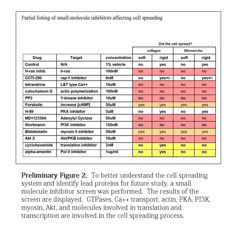
106 89
107 90 While a significant body of work has been done to understand the signaling and motility events initiated by fibronectin-integrin engagement, less work had been done on understanding collagen mediated signaling and motility events. The first aim of this study was to better understand collagen mediated integrin based signaling and to determine if and how collagen-based signaling may be different than fibronectin mediated integrin based signaling. During the course of this study, the concepts of the rigidity response and focal adhesion coupling were new and emerging phenomena. As detailed in the introduction, both phenomena were being investigated using multiple working models and hypotheses. The second and third aims of this investigation were to better understand the molecular components and pathways responsible for the rigidity response and focal adhesion-actin coupling respectively. To better understand collagen-based integrin mediated signaling events I set out to compare the differences between collagen and fibronectin based motility events by spreading mouse embryonic fibroblasts (MEFs) on both collagen and fibronectin, perturb the cell either with small molecule inhibitors, and knock-out cell lines that removed a specific protein from a given population of MEFs. After observing differences in spreading behavior on collagen and fibronectin, I probed the localization and activation state of a given protein using standard and emerging biochemical and microscopy techniques.
108 91 To better understand and delineate the molecular mechanisms involved in the rigidity response. I utilized the ability to create substrates of different rigidities using polyacrylamide gels. By varying the concentration of the polymer- acrylamide and crosslinker- bis, I was able to adjust the rigidity of substrates that the cells interact with, and in some cases spread on. Again, using small molecule inhibitors and knock-out cell lines I probed the molecular pathway involved in rigidity sensing on collagen and fibronectin (Preliminary Figure 2). To better understand the dynamics of the molecular components involved in the rigidity response I used biochemical and microscopy techniques. Finally, as it has been hypothesized that focal adhesion-actin coupling is molecularly linked and perhaps regulates both cell spreading and the rigidity response, I utilized assays such as the pillar assay and collagen contraction to better characterize focal-adhesion actin coupling. In chapter 2 I present results that identify a molecular pathway that is specific to collagen-based integrin mediated signaling, identify a new component necessary for collagen based rigidity sensing, highlight the effects of low focal adhesion-actin coupling on spreading, motility, and downstream signaling that regulates growth and proliferation. Moreover, I propose a model in which actin retrograde flow is involved in activating and regulating the localization of proteins necessary for integrin activation, the rigidity response, force generation, and signal generation necessary for growth and
109 92 proliferation. Modulating ECM composition and substrate rigidity in chapter 3, I present data that highlights the effects of high focal adhesion-actin coupling. Specifically, I present data that supports the idea that increased focal adhesion-actin coupling allows for activation of cytoskeletal and growth signaling pathways on both soft and rigid substrates. The overall objective of this investigation was to better understand collagen mediated rigidity sensing, through the analysis of focal adhesion, and actin dynamics, and the effects actin dynamics and focal adhesion ontogeny might have on downstream signaling.
110 93 Chapter 2 Integrin-Linked Kinase Stabilizes β1 Integrin Adhesions Involved in Rigidity Sensing, Traction Forces, Actin Dynamics and Signaling
111 Abstract Cell spreading and motility involves physical linkages between the extracellular matrix (ECM), integrins and the cytoskeleton. Here we show that integrin-linked kinase (ILK) is necessary for the stabilization of α2/β1 mediated adhesions, traction force and signal generation. Further, ILK-/- cells do not respond to collagen rigidity but do respond to fibronectin rigidity. Expression of full length ILK restores normal spreading but the paxillin binding mutant of ILK does not. On fibronectin, initial spreading that depends upon αv/β3 integrin is minimally affected by the loss of ILK, while mature adhesions that depend upon α5/β1 are affected. In ILK-/- cells, the linkage of the integrin to actin is dramatically weakened leading to increased actin retrograde flow, and reduced traction forces. Although talin, paxillin and p130cas assemble in small adhesions, these molecules move inward at an increased rate, with paxillin moving at the slowest rate. Downstream activation events are also reduced including MAP kinase signaling. Thus, we suggest that ILK is important for β1 integrin-mediated coupling to the cytoskeleton, focal adhesion formation, reinforcement, substrate traction and the localization and activation of signaling proteins.
112 Introduction Integrin-dependent cell adhesions are critical for cell growth, differentiation and the organization of tissues. While many focal adhesion proteins are commonly found in all adhesions, some focal adhesion proteins localize, bind and activate the different integrin heterodimers, as well as bind integrins at different times during the maturation of the focal adhesion (Zaidel-Bar et al., 2004). Moreover, at different stages of motility and growth, different focal adhesion proteins are thought to interact and transduce signals from adhesions. For example, recent work has shown that high forces on cell-fibronectin bonds are supported by α5β1 integrins, while the αv/β3 integrins bind with less strength but are needed for mechanotransduction (Roca-Cusachs et al, 2009). Moreover, certain physiological and pathological responses depend upon specific ligand activation of integrins. For example, cellular responses to rigidity of fibronectin involve a different set of proteins than needed for cellular responses to collagen rigidity (Jiang et al., 2006; Kostic et al., 2006). Integrins are the primary transducers of extracellular matrix (ECM) signals into an intracellular response. Recent studies have shown that not only the biochemical but also the mechanical nature of the ECM can affect the cellular response. In the case of collagen and fibronectin, the β1 and β3 integrins are critical and partner with α v, 1, 2, 5 and 11 in binding to type 1 collagen and fibronectin. Once an integrin binds to its
113 96 matrix partner, it undergoes a conformational change that activates a number of internal binding sites for the cytoskeleton that then tests the physical characteristics of the ECM, localizes focal adhesion proteins, and in turn regulates and is responsive to the cellular and extracellular state. How these cytoplasmic processes are activated is not well understood. In the case of the αvβ3 integrin binding to fibronectin, there is an activation of the receptor-like tyrosine phosphatase, RPTPα, that in turn activates Src family kinases (von Wichert et al., 2003). There may be similar accessory proteins that are involved in the control of β1 interactions with ECM. Although focal adhesions are not prominent in vivo, they do provide a view of the important factors that are involved in cytoskeleton matrix coupling and force generation. Numerous studies have shown that the matrix will signal to small GTPases and locally modulate their activity. In certain cases, GTPase stimulated contraction of the coherent actomyosin network produces traction forces through actin-adhesion-integrin-ecm linkages (Hu et al., 2007); Cai et al. 2010) that further facilitates focal adhesion formation (Zamir et al., 2000). The exact nature of the coupling, and the steps involved in activation have yet to be determined for many of the ECM-cell interactions. It is expected that the control of

114 97
115 98 these interactions will play a critical role in normal tissue function and alterations will be linked to many disease processes. One focal adhesion protein, Integrin-linked kinase (ILK), has recently been the topic of investigation due to its role in numerous pathological states (Radeva et al., 1997; Dai et al., 2003; Hannigan et al., 2005; McDonald et al., 2008). Integrin-linked kinase (ILK) was first identified in a yeast two-hybrid screen based on its interaction with the Β1 integrin cytoplasmic domain (Hannigan et al., 1996). Previous studies have implicated ILK in a range of diverse roles including inhibition of apoptosis (Attwell et al., 2000), and regulation of α5β1 integrin function and fibronectin matrix assembly (Wu et al., 1998). Furthermore, gross deficits in normal development and tissue homeostasis occur when ILK is deleted. Overexpression of ILK either in cell culture or constitutive activation in transgenic mouse models results in oncogenic transformation (Dillon et al., 2007; Hannigan et al., 2005; Legate et al., 2006). Interestingly, the expression of ILK is often elevated in human cancers, and correlates with tumor stage and metastatic potential (Dai et al., 2003; Graff et al., 2001; Okamura et al., 2007; Takanami, 2005). In light of ILK s cellular, physiological and pathological relevance, and to better understand ILK s role in mammalian, ECM-cytoskeleton coupling, we investigated the actin and focal adhesion protein dynamics in embryonic fibroblasts derived from a mouse ILK knock-out line,

116 99
117 100 (Sakai, T, et al., 2003) and have found that the loss of ILK dramatically alters cytoskeleton-β1 integrin coupling in a paxillin-dependent manner. 2.3 Results Spreading on Collagen versus FN (Integrin dependence) To determine if there is an integrin dependence in ILK-/- cells, we analyzed the spreading of ILK-/- and control cells on collagen or fibronectin-coated surfaces. When control suspension cells were added to collagen-coated glass, they spread primarily (~60%) in an isotropic fashion with a significant fraction spreading in an anisotropic fashion (40%) (Dubin-Thaler et al., 2004). In contrast, ILK-/- cells spread almost completely in an isotropic fashion (~90%) and had a greatly expanded lamellipodium (Figure 1). After 5-10 minutes, the ILK-/- cell area gradually decreased whereas the control cells continued to increase in area. If ILK-/- cells were placed on fibronectin, they spread almost normally for the first 5-10 minutes but then retracted their lamellipodia rapidly (Figure 1B). In most cases the central cytoplasm remained spread on fibronectin, unlike on collagen where there was a dramatically contracted central cytoplasm (Figure1A, and 8). The morphology of the ILK-/- cells on collagen was similar to that of talin depleted cells where the central cytoplasm was contracted by myosin because integrins were not

118 101
119 102 mechanically coupled to the actin-myosin network in the periphery and contraction collapsed the cytoplasm (Zhang et al., 2008). To test for the role of myosin contraction in the morphology of ILK null cells, we added the myosin inhibitor blebbistatin which resulted in endoplasm spreading as well as rescue of normal morphology similar to the talin depleted MEF s (Figure 3). Thus, there appears to be a myosin mediated collapse of the actomyosin network due to weakened coupling to adhesions in ILK-/- cells. To determine if the apparent lack of connection between collagen and the cytoskeleton had a functional consequence, we tested the ability of the cells to sense rigidity by determining if the cells spread less on soft surfaces. When the extent of spreading on soft versus rigid polyacrylamide gels was measured, ILK-/- cells showed the same degree of spreading on soft and rigid collagen (Figure 1, E and F). In contrast, the control cells spread to larger areas on rigid collagen than soft. In the ILK-/- cells, similar to when they were spread on collagen-coated glass, there was again a highly contracted core of cytoplasm and broad lamellipodium on both soft and rigid collagen (Figure 1E). With fibronectin-coated acrylamide, both ILK-/- and control cells spread to a larger area on rigid than on soft polyacrylamide. Thus, we see that the rigidity response (rigidity sensing) of collagen was selectively lost in the ILK-/- cells (Figure 1,E and F).
120 103 To determine if ILK was able to restore function to the knockout cells, ILK-GFP was transiently expressed in ILK-/- cells. Normal spreading was restored with full length ILK-GFP but a mutant form of the protein that would not bind to paxillin (Nikolopoulos and Turner, 2001), ILK-PBS was not able to support normal spreading (Figure 2). Further, a mutation of E359K that disrupts the putative kinase C-terminal integrin binding domain, blocked spreading on collagen, but not initial binding of the cells to the surface (Figure 3). Thus, on collagen, ILK function appears to involve both paxillin binding and maintenance of the putative, structured kinase, and integrin binding domain. ILK Localization Supports an Early Role in Collagen Adhesions To better understand ILK s role in collagen adhesion, we determined the localization of ILK during spreading on collagen-coated glass. To do this we transfected ILK null cells with ILK-GFP. Using total internal reflection fluorescence (TIRF) imaging, ILK-GFP localized to early focal contacts in transfected ILK -/- cells during isotropic spreading on collagen-coated glass (Figure 2). Fluorescent foci developed at the late stages of rapid spreading, and moved outward with the velocity of the leading edge protrusion, staying within 0.5 μm of the leading edge (Figure 2). In addition to transfecting a wild-type ILK- GFP fusion protein, we transfected two mutant ILK GFP

121 104
122 105 fusion proteins to better understand what part of ILK might be responsible for its localization. A paxillin binding mutant form of ILK, termed ILK-PBS-GFP exhibited diffuse localization throughout the cell (Figure 2) as well as a fast retrograde flow rate of 8 nm/sec ± 4.3 (n=9) compared to the 0.4 nm/sec ± 0.1 (n=6) of ILK-GFP. Interestingly, the E359K point mutation GFP fusion protein that disrupts the integrin binding domain, termed ILK-E359K-GFP, inhibited spreading completely. Thus the paxillin binding domain was not need for spreading, but was needed for localization to the adhesions. However, the structured, C-terminal integrin binding domain was needed for initiation of spreading (Figure 2, and 3). Increased Actin Flow in ILK-/- cells and Decreased Force If the condensed cytoplasm in the ILK-/- cells was due to a lack of attachment of the actomyosin cytoskeleton with the peripheral adhesions, then the rate of actin flow inward may have been increased as previously observed for talin-depleted cells (Zhang et al., 2008). Several methods were used to measure the actin flow rate, including the rate of dorsal bead transport, of fluorescent actin movement, and of dorsal wave movement. All three methods showed that the rate of movement of actin inward was increased in ILK-/- cells on collagen. As mentioned above, ILK+/+ cells spread faster than ILK-/- cells (86 nm/sec ± 21 (n=16) and 51 nm/sec ± 11 (n=18) for ILK+/+ and ILK-/-, respectively).
123 106
124 107 When we measured actin retrograde flow via the transport of 1 μm diameter beads coated with collagen, the velocity in the ILK+/+ line of cells was 43 nm/sec ± 9 nm/sec (n=7) and in ILK-/- cells it was 128 nm/sec ± 22 (n=7) (Figure 4). Another means of measuring actin retrograde flow was to track actin-based distortions in the membrane by DIC microscopy. In this assay, ILK+/+ cells exhibited an actin rearward flow rate of 37 nm/sec ± 5 nm/sec (n=6) while ILK-/- cells exhibited 117 nm/sec ±15 nm/sec (n=8) (Figure 4). Finally, we measured the rearward flow rate of actin-gfp speckle patterns by TIRF microscopy. In this assay, the actin retrograde flow rate was 44 nm/sec ± 9 nm/sec (n=9) in ILK+/+ cells, versus 143 nm/sec ± 20 nm/sec (n=9) in ILK-/- cells (Figure 4). Taken together, the average actin retrograde flow rates for ILK+/+ and ILK-/- were 43 nm/sec ± 9 nm/sec (n=22) and 128 nm/sec ± 22 nm/sec (n=24), respectively (Figure 4). This increased retrograde flow in ILK-/- cells indicated a defect in the ability of integrins to link to the cytoskeleton in ILK-/- cells, resulting in decreased attachment of actin to the adhesions and greater rate of contraction inward. Myosin-driven Inward Flow Causes Contracted Cytoplasm A corollary of the increased actin flow is that myosin flow should be increased as well and myosin distribution should be altered. MLC-GFP localization was significantly altered in an ILK-/- background (Figure 4). MLC-GFP appeared at the basal membrane earlier in the initial spreading phase in the ILK-/- background, and the contractile ring
125 108 was more prominent. There were also deficits in the formation of transverse stress fibers across the cell. Interestingly, in ILK-/- cells, the contractile ring maintained a position ~ 3.8 μm interior from the leading edge (Figure 4, D-G) compared to ~.9 μm from the leading edge in ILK+/+ cells. Consistent with this observation, the small myosin foci that formed at the leading edge in ILK-/- cells flowed rearward at ~3x the velocity in ILK null cells, 44 nm/sec ±11 nm/sec (n=8) versus 15 nm/sec ±9 nm/sec (n=8) (Figure 4, D-G) in ILK+/+ cells. If the compressed endoplasmic region in ILK-/- cells resulted from the rapid inward transport of the actin cytoskeleton by myosin II, then alteration of myosin contraction activity should alter endoplasm compression. When ILK+/+ cells were treated with calyculina, an activator of myosin II, 29% showed a spread cytoplasm in contrast to 73% in untreated ILK+/+ cells (Figure 3). Furthermore, inhibition of myosin II by blebbistatin increased the percentage of ILK-/- cells with a spread endoplasmic region from 15% to 66% (Figure 3). These observations pointed to the importance of myosin in inhibiting endoplasm spreading when actin was not coupled to peripheral adhesions.

126 109
127 110 β1 Integrin Function But not β3 Appears Altered To test whether ILK was playing a larger role in specific integrin-mediated functions, we compared Β1 to Β3 mediated binding, using inhibitory antibodies to integrins. MEFs were incubated in suspension with inhibitory antibodies and plated on glass substrates coated with 10ug/ml fibronectin or collagen (Figure 1, C and D). Cells were allowed to spread for 30 minutes, fixed and imaged via DIC microscopy. The early time point of fixation ensured that we observed primarily early integrin-ecm interactions. Consistent with previous studies, we observed that inhibition with a cyclic RGD peptide, specific for αvβ3 integrin blocked rapid spreading on fibronectin in both ILK+/+ and ILK-/- cells (Figure 1, C and D). Interestingly, α5β1 blocking antibodies preferentially inhibited spreading of ILK+/+ cells on fibronectin and not ILK null cells, indicating that cells without ILK may preferentially utilized β3 integrin to spread on fibronectin. For cells interacting with collagen, we observed that when treated with inhibitory antibodies to β1 and α2β1 integrins, MEF s were unable to spread on collagen (Figure 1, C and D). These results confirmed the previous observations that early interactions with fibronectin and collagen were mediated by β3 and β1, respectively. We suggest that ILK preferentially mediates coupling of the ECM to the actin cytoskeleton via β1 integrins.

128 111
129 112 Decreased Traction Force Resulting from Decreased Coupling An important consequence of decreased attachment to the integrins is that traction forces may be decreased. A pillar displacement assay was used to measure the traction forces of control and ILK-/- cells. Pillar displacement was determined for spread cells after 60 minutes of spreading. ILK+/+ cells produced an average pillar displacement of 164 nm ± 22 nm (n=17), while ILK-/- cells averaged 62 nm ±24 nm (n= 19) of displacement per pillar on collagen coated pillars (Figure 6). Using a similar method we investigated the pillar displacement by ILK+/+ and ILK null cells on an array of pillars coated with fibronectin. ILK +/+ cells produced an average pillar displacement of 167 nm ±31 nm (n=24), while ILK-/- cells produced an average displacement of 52 nm ±38 nm (n=19) (Figure 6). With collagen- or fibronectin-coated pillars, ILK-/- cells generated considerably lower forces than ILK+/+ cells. These measurements were at later times after spreading where we expected that the fibronectin was bound by α5/β1 integrin. Spread areas were not sufficiently different to account for the differences in force. Thus, it appeared that ILK was selectively needed for the generation of β1 mediated traction forces on collagen and fibronectin.
130 113 Initial Integrin Binding is Strong, but Reinforcement is Weaker without ILK To further confirm our hypothesis that ILK null cells had weakened β1 mediated interactions with the cytoskeleton, we employed a magnetic bead reinforcement assay. Using magnetic tweezers and magnetic beads coated with either fibronectin or collagen (Roca-Cusachs et al. 2009, Tanase et al., 2007), we applied an oscillatory force of.8nn at 1Hz to the bead-cell interface to probe early ECM-integrin interactions. Cells were allowed to interact with beads while spreading on collagen. When a cell bound and moved a bead, the magnetic tweezers probe was placed ~20um away from the bead/cell interface and a 0.8nN oscillatory force was applied to the magnetic bead. ILK +/+ cells were able to maintain a consistent degree of attachment to beads coated with collagen and fibronectin, exhibiting an average displacement of 45 nm ±6 nm (n=10) for collagen coated beads, and 37 nm ±7 nm (n=11) for fibronectin coated beads that was relatively constant over the duration of the applied force (Figure 5). In contrast, ILK-/- cells maintained a constant level of displacement over time for fibronectin coated beads, but were unable to reinforce and maintain a constant level of displacement of collagencoated beads. When subjected to the 0.8 nn oscillatory force, collagen coated beads interacting with ILK-/- cells exhibited an average displacement of 74 nm± 15 nm (n=12), while fibronectin-coated beads exhibited an average displacement of 41 ± 8nm (n=11) when interacting with ILK-/- cells (Figure 5). Because collagen-coated beads were
131 114
132 115
133 116 displaced more on ILK-/- cells by the same oscillatory force at early times, we suggest that ILK preferentially stabilizes β1 mediated ECM- actin cytoskeleton coupling. Adhesion Size is Smaller in the Absence of ILK Previously ILK knockdown and dominant negative studies in Hela cells have shown focal adhesion deficits on fibronectin (Nikolopoulos, S. N. and C. E. Turner (2001)). To determine if adhesion formation was also altered in ILK null cells on collagen, we stained for different adhesion proteins after 1 hour. The ILK null cells, on average, exhibited focal adhesions 5x smaller than wild type MEF s expressing ILK, with adhesion sizes of 1.1 μm 2 ±.4 for ILK+/+ and 0.19 μm 2 ±.06 for ILK-/- cells (Figure 10). Not only actin-binding proteins such as vinculin, and talin were depleted from focal adhesions, but scaffolding and signaling molecules such as p130cas, paxillin and FAK were depleted as well.
134 117
135 118 Adhesions Migrate More Rapidly in the Absence of ILK To determine if smaller adhesions resulted from greater turnover or lower rates of assembly, we observed the dynamics of talin, p130cas and paxillin during the rapid and contractile phases of cell spreading using total internal reflection fluorescence (TIRF) microscopy. Paxillin-GFP localized to focal complexes early on in the rapid spreading phase. During contractile spreading, in ILK+/+ cells, paxillin-gfp persisted at the leading edge and formed two stable fluorescent lines or borders at the leading and interior edge of the lamellipodia, presumably demarcating nascent focal contacts at the leading edge and more mature adhesions toward the interior of the lamellipodia. While paxillin-gfp was stable in adhesions in ILK+/+ cells, paxillin-gfp was remarkably more dynamic at the leading edge in ILK-/- cells (Figure 7, A and D), moving rearward at a velocity of 9 nm/sec ± 3 nm/sec (n=9), paxillin-gfp in ILK+/+ cells moved rearward at 0.2 nm/sec ±.05 nm/sec (n=8) or 45-fold slower (Figure 7, A and D). The retrograde flow of a focal adhesion protein was surprising and we wondered if other focal adhesion proteins behaved similarly. Using the same techniques, we investigated if two other focal adhesion proteins, talin and p130cas, moved at the same or different rates in the ILK-/- background. Talin-GFP was directly bound to integrin as well as actin filaments and was present throughout the basal membrane region in small foci early in spreading. Subsequently, it was
136 119 concentrated in adhesions in early rapid spreading that persisted into late contractile spreading. While talin-gfp in ILK-/- cells was localized to adhesions during early spreading, talin-gfp failed to form sharp, defined focal contacts in fully spread ILK-/- cells. We observed talin formed globular, fluorescent foci in spread ILK-/- cells that moved rearward at ~25 nm/sec ±4 nm/sec (n=5) (Figure 7, C and D). Talin-gfp in spread ILK+/+ cells persisted at the leading edge and moved slowly rearward at 2.4 nm/sec ±.9 nm/sec (n=4) on substrates coated with collagen. For comparison, we measured movements of p130cas, a protein of the CAS (Crk-associated substrate) family that was only indirectly bound to integrins in adhesions. Qualitatively, p130cas-gfp exhibited similar localization and dynamics to paxillin, except that it was recruited to the leading edge later in spreading than ILK and paxillin, concurrent with the start of contraction. p130cas localized to focal adhesions and formed a characteristic fluorescent band, circumscribing the cell, spanning the length of the leading-edge in ILK+/+ cells spread on collagen and moved inward at a low rate of 0.91 nm/sec ±.04 nm/sec (n=9) (Figure 7, B and D). In contrast, p130cas was more dynamic in the ILK-/- background and moved rearward at a rate of 19 nm/sec ± 6.4 nm/sec (n=11) (Figure 7, B and D). Thus, consistent with the observed smaller focal adhesions in ILK null cells, the observed adhesion proteins move more rapidly inward at different rates in ILK-/- cells.

137 120
138 121 Consistent with our observations that paxillin and p130cas move rearward in cells ILK null cells, immunofluorescent staining of phospho-paxillin (Y118) show a diffuse staining pattern in ILK-/- cells as well as significant staining towards the interior of the cell. This is in contrast to the distinct punctate staining pattern found in ILK+/+ cells (Figure 13). Staining of phospho-p130cas (Y165) shows a distinct band ~ 2.5μm interior to the leading edge in ILK-/- with diminished staining at the leading edge, in contrast to the punctate staining at the leading edge in ILK+/+ cells ( Figure 13). MAP Kinase Phosphorylation is Decreased in ILK-/- Cells In contrast to what we found with other signaling molecules such as FAK, p130cas, and src (Figure 9), when we looked at absolute phosphorylation levels of JNK and p44erk via western analysis, a difference was observed when we compared ILK+/+ and ILK-/- lines (Figure 8). In addition to the difference in overall phosphorylation levels of JNK and p44erk, we noticed a significant and consistent difference in the localization of the phosphorylated forms of JNK, and p44erk (Figure 13). Phospho-JNK localizes to small foci at the leading edge in ILK+/+ cells, while it localizes along the entire width of the lamellipodia in ILK -/- cells. Similarly, phospho-p44erk localized to foci at the leading edge of ILK+/+ cells but was unable to localize at any part of the lamellipodia, and remained in the endoplasm of ILK-/- cells (Figure 13). This localization is consistent with the localization of their binding partners phospho-paxillin (Y118) and phospho-
139 122 p130cas (Y165). Finally, we set out to quantify the difference in proliferation between the ILK+/+ and ILK-/- lines using the BrdU assay. After serum starvation to synchronize both cell lines, we incubated each line with BrdU in the presence of serum for 30, 60 and 120 minutes. Upon staining for BrdU incorporation, we observed significantly higher levels of incorporation of BrdU in the ILK+/+ line with an estimated doubling time 640 ±54 minutes for ILK+/+, in contrast to ILK-/- line that had an estimated doubling time of 820 ±52 minutes. Taken with our observations of actin and myosin retrograde flow, these observations suggest that the presence of ILK is critical for activation of the MAPK s involved in stimulation of growth.

140 123
141 124 Collagen Gel Contraction Requires ILK Previous studies on migration of ILK-/- cells have shown decreased rates of movement on 2-D surfaces. We further wished to determine if contraction of 3-D collagen networks would be altered in ILK-/- cells; therefore, we measured 3D type I collagen-gel contraction (Ngo, et al., 2006). An equal number of cells (5K) were seeded in gels of different collagen densities and allowed to grow in culture for 12 days. ILK-/- cells were unable to contract any gel of 1.8 mg/ml, 1.6 mg/ml, or 1.2 mg/ml collagen at day 12, and up to day 21, while ILK+/+ cells were able to contract gels of 1.2 mg/ml and 1.6 mg/ml to 60% and 40% of the original size, respectively, by day 12 (Figure 12). This further underscored ILK s role in the physical linkage of the cytoskeleton to a collagen based ECM. 2.4 Discussion These studies show that ILK-/- cells are particularly deficient in the coupling of matrix to the actin cytoskeleton through the collagen binding integrins and result in increased actin retrograde flow in the lamellipodia. From assays of early spreading to long-term assays of matrix contraction, there is consistently a defect in cytoskeleton-matrix linkage in ILK-/- cells (Figure 1, 3, 6 and 12). The central cytoplasm fails to spread on collagen but spreads on fibronectin (Figure 1). There is a greater rate of actomyosin transport inward and a lower force generated on collagen both on collagen-coated pillars
142 125
143 126 and 3-D collagen fibers (Figure 4, 6 and 12). Although adhesions do form on collagen, they are typically smaller and more dynamic in ILK-/- cells (Figure 7 and 10). Moreover, actin and myosin dynamics are significantly altered with ILK-/- cells exhibiting rapid retrograde flows of actin and myosin. Perhaps the most striking defect is the weakness of the link between talin and integrins bound to collagen in the absence of ILK and the fact that paxillin has the lowest rate of transport inward. One possible explanation is that paxillin is associated with a complex of proteins more tightly coupled to the β1 integrin than talin and ILK would aid in forming a larger, more stable complex. Many of the other changes can be explained as the result of a weakness in the linkage between α2 β1 integrins and talin. Rigidity dependent spreading has been shown previously to involve a different set of components on collagen and fibronectin (Jiang et al., 2006; Kostic et al., 2006; Wang, et al., 2003). In the case of fibronectin, there is evidence of a β3 integrin dependence involving the receptor-like protein phosphatase α (RPTPα) and the Src-family kinase, Fyn (Kostic et al. 2006). RPTPα and Fyn knockout cells spread to the same extent at early times on soft and rigid fibronectin, but they do sense collagen rigidity and spread to a larger area on rigid collagen (Jiang et al., 2006; Kostic et al., 2006). Focal adhesion kinase (FAK) was previously shown to be involved in collagen rigidity sensing since FAK null cells spread to the same area on soft and rigid collagen (Wang, et al., 2000).

144 127
145 128 However, FAK null cells can sense the rigidity of fibronectin-coated surfaces (Jiang, et al., 2006). The behavior of the ILK null cells is similar to FAK-/- in that they have a defect in the sensing of collagen but not fibronectin (Figure 1, E and F). This is all consistent with a role for ILK in the activation of Beta 1 integrin. The role of paxillin is highlighted in the ILK knockout cells. Expression of ILK lacking the paxillin binding domain was not able to restore normal cell behavior and paxillin moved inward in the knockout cells on a collagen-coated surface (Figure 2 and 7). This indicates that ILK stabilization of adhesions involves the anchoring of the LIM domain containing protein, paxillin. Alternatively, this interaction may be influenced by the LIM containing protein complex of PINCH-PARVIN and ILK (Wickström, et al., 2009; Legate, et al., 2006; Wu, 2004). Paxillin s slower rate of inward flow in ILK-/- cells may be due to the observations that paxillin is one of the earliest components of adhesions and it may be bound to other components of the activated integrin complex (Turner, 2000), and unlike talin and p130cas has no direct actin binding domain. An additional linkage to ILK would further stabilize the bond to the integrin and keep the complex from moving inward and would enable the generation of high forces on adhesion contacts. Thus, we suggest that ILK is particularly involved in the stabilization of paxillin linkage to liganded β1 integrin. Previously it was shown that the paxillin binding domain in ILK was necessary for its localization on fibronectin and our results suggest the same for collagen (Nikolopoulos
146 129 and Turner, 2001). Our results with the dominant negative point mutation (Figure 3) suggest that while ILK may not be necessary for spreading on fibronectin and collagen, perturbations in its putative kinase domain, presumably destabilizing the α-helical structure of its C-terminal region and can act as a dominant negative isoform, blocking activation of spreading on collagen. In previous studies of talin-depleted cells on fibronectin, the rapid inward transport of actin was shown to pull the microtubules and vesicles inward and inhibition of myosin enabled the microtubules to extend to the periphery (Zhang, et al., 2008). Both the constricted central region of cytoplasm and the rapid actin movement inward indicate that the assembling actin filament complexes are not strongly coupled to the matrix adhesions and as a result ILK null cells may not be able to extend their microtubules on collagen like talin depleted cells on fibronectin. In contrast, in ILK null cells, the coupling of the early fibronectin, β3 mediated, adhesions to the actin filaments enabled the spreading of the cytoplasm but the later transition to α5β1 integrin caused the release of adhesions and retraction of lamellipodia (Figure 1). Previous studies on rigidity sensing postulate an early connection between the ECM, integrin, and cytoskeleton enabling the cell to sense the rigidity of the substrate as it spreads, mediated by the initial interactions of integrin and substrate.( Giannone and
147 130 Sheetz 2006 ; Danen, et al., 2002). Initial and sustained contacts between the ECM and actin cytoskeleton have been described previously using the mechanical analogy of a clutch (Brown, et al., 2006, Macdonald, et al., 2008; Hu, et al., 2007, Giannone, et al., 2009). The observation that ILK-/- cells exhibit increased retrograde flow and do not sense rigidity on collagen, points to the importance of cytoskeletal coupling in early stages of cell motility, the rigidity response, and ECM sensing (Figure 1). Given the current model, rigidity is thought to be sensed via nascent ECM-integrin-focal adhesioncytoskeleton coupling events that lead to the mechanical stretching of proteins found at nascent focal contacts. The ability of the cell to transduce force across the plasma membrane through the linkage of ECM-integrin-FA-actin-myosin is also linked to chemical signals via mechanotransduction (Vogel & Sheetz, 2006). Since ILK-/- cells did not sense rigidity on collagen (Figure 1E,F), we suggest that the cytoskeletal linkage is necessary to sense, or transduce the rigidity of the substrate and activate the MAPK pathway that is perturbed in ILK-/- cells. Specifically, perhaps it is the weakened actin, focal adhesion and ECM coupling, increased retrograde flow of actin (Figure 4), that results in less transduction by less protein stretching. That is to say that the reduced traction between integrins, focal adhesion proteins and the cytoskeleton in ILK null cells, manifested by increased actin and myosin retrograde flow, allows for the sustained
148 131 activation of actin polymerization characteristic of initial spreading or P1 (Döbereiner, et al., 2004), or initial fast cell spreading. Moreover, the ability of ILK-/- cells to sense rigidity on fibronectin (Figure 1, E and F) coated surfaces indicates that ILK is not necessary for the initial coupling of fibronectin to the cell s cytoskeleton in β3-integrin dependent sensing (Kostic and Sheetz, 2006). Interestingly, when collagen-coated magnetic beads are subjected to 1nN oscillatory forces, they are displaced by twice as much on ILK-/- than on ILK+/+ MEFs (Figure 5). This larger displacement further indicates that the ECM-integrin-FA-actin linkage is weakened. Furthermore, the ability of the cell to stretch the ECM-actin linkage more in the absence of ILK supports the idea that the protein complex at the focal adhesion is stabilized by ILK. Previous studies have described traction force, and clutch dynamics as necessary components of cell motility (Brown, et al., 2006; Macdonald, et al., 2008;, Hu, et al., 2007; Giannone, et al., 2009). It is perhaps not surprising that knockdown of ILK or dominant negative ILK causes defects in late stage fibronectin-mediated focal adhesion formation (Nikolopoulos and Turner, 2001). In line with these previous studies, our observations on collagen and β1-ilk mediated interactions, highlight the fact that significant focal adhesion size deficits and increases in retrograde flow velocities in ILK null cells leads to mislocalization of myosin (Figure 4), focal adhesion proteins (Figure 7), reduced force generation (Figure 6), slower migration and reduced collagen contraction (Figure 11 and
149 132 12). This study points to ILK as an important molecular component in the mechanical linkage between β1 integrins and the adhesion complex. Finally, the decreased strength of the integrin-adhesion protein linkage has important cellular consequences such as reduced levels and mislocalization of phosphorylated scaffold proteins and signaling molecules such as phosphorylated (Y118) paxillin, phosphorylated (Y165) p130cas, phosphorylated (T183/Y185) JNK, and phosphorylated (T204/Y204) ERK (Figure 13). Since phosphorylated paxillin and p130cas (Turner, 2000; Defillippi, et al., 2006; Ishibe, et al., 2004) have previously been shown to act as scaffolds and substrates for MAPK s, among other signaling molecules (Cabodi, et al., 2010; Wu and Dedhar, 2001), the reduced cytoskeleton coupling and increased retrograde flow could explain the effects on downstream signaling and growth. Although previous studies have focused on the abnormalities of ILK null cells moving on fibronectin, the contrast between the early spreading on collagen versus fibronectin suggests that the primary role for ILK is in the linkage of adhesion proteins to β1 integrins. In particular, the stabilization of paxillin binding to the integrin appears weakened and the more rapid rearward movement of talin inward implies that its interaction with β1 integrin is greatly weakened or that it has a stronger coupling to the moving actin filaments. Because there are alternative types of motility that rely upon different integrins and adhesion molecules, the phenotype of the ILK null cells is difficult

150 133
151 134 to fully understand. However, most of the results can be traced to the weakening of β1 integrin binding to adhesion proteins and increased actin retrograde flow. 2.5 Materials and Methods Cell Culture: The ILK +/+ and ILK -/- mouse embryonic fibroblast cell lines were a kind gift for R. Fassler (Sakai, et. Al., 2003), and were maintained in DMEM high glucose (Gibco-Invitrogen) supplemented with 10% fetal bovine serum, 100IU/ml of penicillin- Streptomycin, 2μM of L-Glutamine, and 2μM of HEPES (Gibco-Invitrogen). Cultures were kept at 60% confluence at 37 o C in a 5% CO 2 incubator. Prior to experiments cells were prepared by trypsinizing with Trypsin Like Enzyme (Gibco-Invitrogen), washed with soybean trypsin inhibitor, centrifuged at 1000rpm for 5 minutes, and resuspended in phenol red- and serum-free DMEM (Gibco-Invitrogen). Next, cells were incubated for 20 minutes at 37 o C in suspension, and plated on substrates in serum-free media for observation. Constructs and transfection: ILK-GFP, and ILK-PBS-GFP were generous gifts from C.E. Turner (Nikolopoulos, and Turner, 2001). Actin-GFP, myosin-light chain-gfp, talin- GFP, paxillin-gfp, and p130cas-gfp were described in (Giannone, et al., 2007). DNA constructs were transfected into ILK +/+ and ILK-/- cells using Fugene (Roche)
152 135 transfection system. Roughly 2-3 μg of DNA and 8-12 μl of fugene were used per reaction (10 5 cells). Cover slip-coating and bead coating: Cover glasses were washed 2 hours in 20% nitric acid and then silanized by exposure to gaseous 1,1,1,3,3,3-Hexamethyldisilazane (Sigma). Silanized cover slips or cultures dishes were coated with 10 μg /ml rat tail collagen type I, dissolved in.2% Acetic Acid in 1M PBS overnight at 37 o C. For live-imaging, cells were sealed in a live-imaging chamber and mounted onto a motorized 37 o C stage. DIC and TIRF images were taken on an Olympus BX50 fluorescence microscope with a 60x, N.A objective. Silica beads (100 μl of 2.7-μm) (Dynal Biotech) were washed with 0.1M carbonate buffer and 0.02 M phosphate buffer sequentially before being incubated in 750 μl 2% carbodimide/phosphate buffer for 3 h at room temperature. The beads were then washed with 0.1 M borate buffer. Silica beads were then treated with cyanogen bromide (Technote #205, Bangs Laboratories) and incubated with 10ug/ml collagen, fibronectin or laminin, in.2% Acetic Acid in 1M PBS for collagen and 1M PBS for fibronectin and laminin, overnight at 4 o C and stored in 1 μg/ml of albumin from chicken egg white (Sigma). Magnetic Bead Assay: To apply forces to magnetic beads, a previously described magnetic tweezers apparatus (Tanase et al., 2006) was used. Briefly, an electromagnet
153 136 with a sharpened ferromagnetic core was used to apply a strong magnetic field gradient, generating a force on the beads. The force exerted by the tweezers was calibrated from the velocity of beads in liquids of known viscosity measured as a function of the tip-bead distance and applied current. For force measurements, fibronectin and collagen-coated beads were deposited on cover slips silanized with 1,1,1,3,3,3,-hexamethyldisilazane (Aldrich) and coated with 10 μg/ml collagen (Roche) for 2 h at 37 C. Cells were then trypsinized, resuspended in DMEM high glucose (Gibco-Invitrogen) supplemented with 2μM of L-Glutamine, and 2μM of HEPES (Gibco-Invitrogen) for 30 min at 37 C for recovery, and plated on the coverslips. The system was then mounted on a motorized 37 C stage on an Olympus IX81 fluorescence microscope. DIC images and videos were taken with a 60 objective and a Cascade II CCD camera (Photometrics) at a frequency of 12.8 Hz. TIRF and retrograde velocity measurements: To follow the dynamics of paxillin-gfp, p130casgfp, actin-gfp, MLC-GFP, and talin-gfp in cells, time-lapse images were captured with a cooled CCD camera (Roper Scientific) attached to an Olympus IX81 inverted microscope (objective, Olympus TIRF PIanApo 60 /1.45 oil; imaging software, SimplePCI) coupled to the 488-nm excitation light from an Inova argon-ion laser. Five second time-lapsed images were taken. For retrograde bead measurements, cells were plated onto collagen (10 μg/ml) -coated coverslips preloaded with laminin or collagen-
154 137 coated 2.7-μm silica beads and spread at 37 C. Two-second time-lapse images of beads transported centripetally on the surface of spreading cells were captured on an IX81 Olympus inverted microscope (objective, Olympus PIanApo 60 /1.45 oil; cooled CCD camera, Roper Scientific; imaging software, SimplePCI). Chemicals, and antibodies: The following antibodies were used: a mouse monoclonal antibody (mab) against paxillin (BD Transduction Laboratories), a mouse mab against p130cas (BD Transduction Laboratories), a mouse mab against Src (Upstate Biotechnology), a mouse mab against FAK (Chemicon), a rabbit polyclonal antibody against vinculin (Abcam), an affinity purified polyclonal rabbit phospho-paxillin (Y118), an affinity purified polyclonal rabbit anti-phoshoy165cas antibody (Cell Signaling Technology, Beverly, MA), an affinity-purified rabbit polyclonal phosphoy416-src kinase family antibody (Cell Signaling Technology), an affinity purified rabbit polyclonal phospho Y397-Focal Adhesion Kinase (P-FAK) (Biosource), an affinity purified mouse polyclonal anti-phospho p44/p42 (T202/Y204), an affinity purified mouse polyclonal anti-phospho pjnk (T183/Y185), an affinity purified mouse polyclonal anti-mlc (Cell Signaling Technology), and affinity purified mouse polyclonal anti-phosphomlc (Ser18,19), a goat anti-rabbit immunoglobulin (Ig) conjugated with Alexa 647 (Molecular Probes, a goat anti-rabbit Ig conjugated with Alexa 488 (Molecular Probes), and goat anti-mouse Ig conjugated with Alexa 568 (Molecular Probes),
155 138 horseradish peroxidase conjugated anti-mouse and anti-rabbit antibodies (Amersham). For antibody inhibition experiments, a cyclic-peptide specific for αvβ3 (Bachem) at 7.5ug/ml, an inhibitory β1 antibody, clone 6S6, (Millipore) at 5ug/ml, an inhibitory α2β1 antibody clone Ha1/29 (BD Biosciences) at 5ug/ml, and an inhibitory α5β1 antibody, clone BMC5 (Millipore) at 5ug/ml were used. Cells were trypsinized, counted, and incubated with inhibitory antibody in suspension for 15 minutes and allowed to spread on either 10ug/ml collagen or 10ug/ml fibronectin coated glass. Cells were fixed at 30 minutes after plating, washed with PBS and spread cells were counted. Western blot quantification: Cells in culture were rinsed once with PBS and lysed directly in RIPA buffer. For time-lapse western blotting, transfected cells were trypsinized and incubated in suspension for 20 min before being plated on collagencoated culture dishes. At the indicated times, cells were washed once with PBS and lysed in RIPA. All cell lysates were combined with 4x loading buffer and boiled before loading onto 4 20% gradient bis-acrylamide gels (Lonza). Protein was then transferred to Optitran reinforced nitrocellulose membrane (Whatman). The membrane was blocked with 5% dry milk-pbst, and incubated with primary antibody overnight at 4 C. The membrane was then incubated for 1 hour at room temperature with anti-mouse or antirabbit- HRP (Jackson Laboratories). The signal was detected with ECL western blotting
156 139 detecting reagents (Amersham Biosciences) on Kodak BioMax XAR film. Signal quantification was performed with NIH ImageJ. Immunohistochemistry: Fibroblast cells were plated onto collagen coated coverglasses (10 μg/ml). After incubation for the described time, cells were fixed in 3.7% formaldehyde and permeabilized with 0.1% Triton. Cells were then incubated in PBS- 1%BSA to block non-specific antibody-antigen interaction. Cells were then incubated with primary antibodies for 1 h followed by washing with PBS-1%BSA and incubated with appropriate fluorescent secondary antibodies. Fluorescent signals from all samples were visualized by confocal microscopy, acquired using a Fluoview confocal microscope (Olympus, Melville, NY). DIC images of the cells plated on polyacrylamide substrates were recorded with a cooled CCD camera attached to an Olympus IX81 equipped with a 60 objective. Analysis of acquired images was performed with the image analysis program, ImageJ (by W. Rasband, NIH, Bethesda, MD; Force generation measurements, collagen contraction: The polydimethylsiloxane (PDMS) micro-posts were prepared and characterized as described previously (Cai, et al., 2006). The dimension of the PDMS micro-posts was 2 μm in diameter, 3 μm center-tocenter, and 7 μm in height. To coat posts with collagen, arrays of posts were immersed in 10μg/ml of collagen or fibronectin solution for 1 hr at 37 C and then washed with
157 140 DPBS. Then, cells were plated on the posts in a 37 C incubator for 5-30 min. The tips of the posts were visualized with a LUCPIanFI 40 /0.60 air objective in bright-field mode on an IX71 Olympus inverted microscope (cooled CCD camera, Roper Scientific; imaging software, SimplePCI). A multiple-particle tracking program was used to analyze the displacement of the posts. Briefly, this multiple particle-tracking program calculated the position of each post for an acquired image. This routine was based on the fact that, in bright-field microscopy, micro-posts acted as wave-guides and appeared bright, whereas the background appeared dark. We were able to identify positions of microposts with good accuracy by determining the center of mass of the corresponding bright pixels (Cai, Y., et al., 2006). Collagen gels of specified densities were prepared as to the manufacturers specifications (BD Biosciences). An equal number of cells were seeded and the gel was immersed in culture media for indicated times. Rigidity response assay: The collagen and full-length FN-coated polyacrylamide substrates were prepared as described previously (Pelham and Wang, 1997). The flexibility of the substrate was manipulated by maintaining the total acrylamide concentration at 10% while varying the bis-acrylamide component between 0.4% (rigid) and 0.03% (soft; E =.2 N/m 2 x10 4, and E = 1.8 N/m 2 x10 4, respectively; Engler et al., 2004). The uniformity of FN and collagen coating on the substrate surface was examined using scanning electron microscopy.
158 141 Statistical Analysis: Data sets exhibiting a normal distribution were subjected to an unpaired Student T-test. Data sets displaying non-gaussian distributions were subjected to a Mann-Whitney test. A value of P<.05 was considered statistically significant. All errors are given as ±1 standard deviation (SD).
159 142 Chapter 3 β1 and β3 Integrin Engagement Rescues Spreading, Growth & Proliferation on Soft Surfaces.
160 Abstract Cell function is regulated in large part by extracellular stimuli, including soluble and adhesive factors that bind to cell-surface receptors. Recent evidence suggests that mechanical properties of the extracellular matrix, particularly rigidity, can also mediate cell signaling, proliferation, differentiation and migration. Often pathological states such as oncogenesis, allow cells to circumvent mechanical cues otherwise necessary for the regulation of motility, growth and proliferation. Termed rigidity sensing, or the matrix rigidity response, wild-type fibroblasts require a substrate rigidity of ~3kPa or higher to spread and proliferate. Here we find that engagement of both collagen and fibronectin receptors are sufficient for focal adhesion formation and cell spreading on typically unpermissive soft (~0.5kPa) surfaces. Furthermore, β1 and β3 integrins as well as ILK are necessary for focal adhesion formation on soft surfaces. The recruitment of paxillin and FAK allows for enhanced focal adhesion clustering and increased FA-actin coupling leading to sufficient traction forces at the leading edge on soft surfaces to sustain spreading. We propose engagement of β1 and β3 leads to an increase in focal adhesion coupling to the cytoskeleton, or intracellular traction force. Activation of both integrins enable cells to reinforce and stabilize focal adhesions, activate ERK, the PI3K - Akt pathway and translation on otherwise unpermissive soft substrates.
161 Introduction Through integrins, cells sense physical and biochemical properties of the extracellular matrix (ECM). Cells respond differently to substrates of different rigidity, activating distinct signaling pathways for matrices of given rigidities. Direct integrin signaling and indirect integrin modulation of growth factor, as well as other intracellular signaling pathways, regulate cell behavior and tissue organization. The substrate-rigidity preferences that different cell types exhibit, have been correlated to the rigidities of their native tissues in vivo (Discher et al., 2005), but their rigidity response mechanisms remain unclear. In addition, recent studies have shown that matrix rigidity affects the gene expression and differentiation of stem cells (Engler et al., 2006). Wild-type fibroblasts require a substrate of ~3kPa or higher to spread and proliferate. The rigidity response involves interactions with extracellular matrix (ECM) mainly through integrin receptors, which regulate organization of the actin cytoskeleton (Giancotti and Ruoslahti, 1999). Our previous studies showed that the generation of force on rigid matrix contacts causes reinforcement of the integrin cytoskeleton linkages through recruitment of focal adhesion proteins, increased cell adhesion, and spreading (Choquet et al., 1997; Giannone et al., 2004). Further studies have identified components required for cells to sense rigidity on fibronectin (Jiang et al., 2006; Kostic et al., 2007).
162 145 Previous studies showed that the generation of force on rigid matrix contacts results in reinforcement of the integrin-cytoskeleton linkages through recruitment of focal adhesion proteins, increased cell adhesion, and spreading (Choquet et al., 1997; Giannone et al., 2004). Various cell types respond to matrix rigidity in fundamentally different ways (Yeung et al., 2005). Furthermore, response to increased matrix rigidity can result in tissue disorganization and malignant transformation (Paszek et al., 2005). Integrins function as heterodimers of noncovalently associated transmembrane α and β subunits, (Humphries et al., 2006; Hynes, 2002). The specific binding of the extracellular domains of integrins to extracellular-matrix (ECM) proteins or in some cases, to counter-receptors on adjacent cells, support cell adhesion and is crucial for embryonic development, tissue maintenance, tissue repair, host defense homeostasis, survival and proliferation. These processes rely on the linkage of integrins to the intracellular cytoskeleton through the generally short integrin cytoplasmic tails; such linkage permits the bi-directional transmission of force across the plasma membrane (Calderwood et al., 2000; Evans and Calderwood, 2007). In addition to their mechanical roles in adhesion and anchorage, integrins transmit chemical signals into the cell (outside-in signaling), providing information on its location, local environment, adhesive state and surrounding matrix (Hynes, 2002; Miranti and Brugge, 2002). How integrins contribute to survival and proliferation under physiological conditions is
163 146 poorly understood. Classically, survival and growth pathways activate Akt, ERK and kinases downstream regulating translation and transcription. RNA translation and protein synthesis is a fundamental biological process required for cell growth, survival and proliferation. Translation initiation is the rate limiting and thought to be the most regulated step of translation. In eukaryotes, translation initiation is facilitated by multiple protein factors collectively called eifs (for eukaryotic translation initiation factors). Numerous studies have shown that eif4b is phosphorylated and activated by Ras-MAPK and PI3K-mTOR signaling cascades via the p90rsk1 effector. Interestingly, eif4b regulates translation of proliferative and pro-survival mrnas. Deregulated translational control is believed to play an important role in oncogenic transformation. Indeed, many eifs are classified as proto-oncogenes. In many types of human cancers, eifs are either overexpressed or activated by Ras-MAPK and PI3KmTOR signaling cascades, again via p90rsk1, resulting in increased survival and accelerated proliferation (Carriere et al., 2008). Many of the players in outside-in signaling have been identified, but an improved understanding of the specificity and dynamics of interactions is needed. The effect of matrix organization on signaling is now being investigated, and it is thought that matrices of different rigidities elicit different, and often more physiologically
164 147 relevant, signaling activities in many cell types. In addition, the impact of matrix stiffness and mechanical or shear forces on integrin signaling towards survival, growth, translation and transcription, and the molecular basis for these effects are important areas for research (Evans and Calderwood, 2007; Giannone and Sheetz, 2006). While our previous studies have reported on molecular mechanisms involved in the rigidity response, here we report on a signaling pathway that allows for wild-type fibroblasts to spread, survive, activate translation and proliferate on soft substrates, circumventing the rigidity response. Given the ability of tumor cells to spread and proliferate on soft surfaces, this pathway may be informative as a model to understand integrin mediated signaling events in oncogenesis.

165 148
166 Results Collagen and FN Rescues Cell Spreading on Soft Surfaces, allowing for growth and proliferation. Extracellular matrix (ECM) proteins bind to specific integrin receptors on the cell surface (Giancotti and Ruoslahti, 1999), causing formation of integrin-cytoskeleton bonds that are reinforced by force-dependent recruitment of focal-contact proteins. Reinforcement of the integrin-cytoskeleton bonds is crucial for the response to the matrix rigidity (Choquet et al., 1997; Giannone et al., 2004; Kostic and Sheetz, 2006). To test the hypothesis that increased integrin engagement would rescue spreading of wild-type mouse embryonic fibroblasts (MEF s) on soft surfaces, we coated soft bis-acrylamide hydrogels (~.05kPa) with 10ug/ml collagen type I and 10ug/ml fibronectin (Col type I + FN). In contrast to wildtype MEF s plated on soft hydrogels coated with 10ug/ml collagen type I (Col type I) and soft hydrogels coated with 10ug/ml fibronectin (FN), MEF s plated on Col type I + FN spread to 433 um 2 (± 76um). When MEF s were plated on soft Col type I and soft FN, they spread to a reduced area of 153 um 2 (± 41um), and 132 um 2 (± 34um), respectively (Figure 14, A and C). Further we looked to see if Col type I + FN rescued and sustained growth on soft substrates. Cells plated on soft Col

167 150
168 151 type I + FN exhibited a doubling time of 43hours. Cells coated on soft Col type I or soft FN were unable to grow or proliferate (Figure 14D). β1 and β3 integrins are necessary to rescue spreading on soft substrates To determine if other ECM combinations could rescue spreading on soft surfaces, we coated soft and rigid control surfaces with paired combinations of collagen type I (Col type I), collagen type IV (Col type IV), fibronectin (FN), laminin (LN) (Figure 16A). Interestingly, Col typei + FN was the only ECM combination that rescued spreading on soft surfaces. As the other ECM engagements involved only β1 engagements, and Col type I and FN involved engagement of β1 and β 3 integrins, we hypothesized that it was the engagement of β1 and β3 that enabled spreading on soft surfaces. To further confirm that it was the engagement of both β1 and β3 integrins that rescued spreading on soft surfaces, we coated soft surfaces Col type I + RGD peptide, and Col type I + FNIII (7-10), and found that cells were still able to spread on soft surfaces (Figure 23). We then treated cells with inhibitory antibodies to further confirm that both β1 and β3 were needed to spread on soft surfaces. Interestingly, blocking α2 and αv integrins on collagen and fibronectin, respectively, rescued spreading on soft surfaces, while blocking β1 and β3 integrins inhibited spreading on rigid collagen and fibronectin surfaces

169 152
170 153 respectively. Blocking β1 or β3 integrins inhibited spreading on soft surfaces coated with Col type I + FN as did blocking α2β1 but not α5β1. Moreover, β1 integrin is necessary for spreading on rigid surfaces coated with Col type I and FN, while β3 integrin is not. Cells interacting with Collagen + Fibronectin exhibit enhanced FA-actin coupling. To confirm that engagement of β1 and β3 integrins were involved in focal adhesion formation and development, we stained for paxillin to measured focal adhesion size on soft and rigid surfaces coated with Col type I, FN, and Col type I + FN. Cells plated on soft surfaces coated with Col type I and FN, exhibited average focal adhesion sizes of.09 um 2 (±.02) and.1um 2 (±.02) respectively, while cells spread on soft surfaces coated with Col type I + FN, exhibited average focal adhesion sizes of.9um 2 (±.18), similar to rigid surfaces coated with Col type I or FN alone (Figure 15). Due to the observed large focal adhesion size on Col type I + FN coated substrates we hypothesized that engagement of β1 and β3 integrins resulted in greater FA-actin coupling, and measured actin retrograde flow velocity. Consistent with our observations that focal adhesion size was increased, actin retrograde flow velocity was decreased in cells spread on surfaces coated with Col type I + FN. Cells plated on soft surfaces coated with Col type I, or FN exhibited actin retrograde flow velocities of 189 nm/sec (± 46 nm/sec) and 121 nm/sec (± 45nm/sec) respectively (Figure 15). When cells were plated on Col type I + FN coated soft surfaces cells exhibited similar actin retrograde flow velocities as if they were plated on rigid
171 154 surfaces. Cells plated on soft Col type I + FN exhibited an average actin retrograde flow velocity of 42 nm/sec ± 15 nm/sec. Traction Force Index correlates with spreading and proliferation As we observed the engagement of β1 and β3 integrins resulted in larger focal adhesions and slower actin retrograde flow on soft surfaces, I postulated that the increase in FAactin coupling was responsible for the observed ability to spread and proliferate independent of rigidity. To better understand and quantify FA-actin coupling I introduced an index termed the Traction Force Index (TFI). The TFI is defined as the average focal adhesion size divided by the average actin retrograde flow velocity. By taking DIC and TIRF time-lapse images of MEF s transfected with paxillin-gfp, we were able to measure the actin retrograde flow velocity and focal adhesion size of individual cells. Consistent with our previous observations, cells that were able to spread and proliferate exhibited larger focal adhesions and slower actin retrograde flow velocities - the resulting TFI (focal adhesion size (um2) / actin retrograde flow velocity (um/sec)) for cells that spread and proliferate was greater than 10 um x sec (Figure 17B). Cells with TFIs < 10 um x sec are unable to spread, presumably due to the fact they are not able to generate the sufficient external and internal traction forces necessary to develop and sustain focal adhesion formation (Figure 17B).

172 155
173 156 Early Traction Forces are greater on FN, collagen and FN and collagen respectively To better understand the β1 and β3 mediate interaction, we measured the traction forces generated on soft substrates coated with Col type I, FN, and Col type I + FN. To do this, we employed a pillar assay using PDMS.5um diameter, 1.8um height pillars. Cells were plated after being in suspension for 20 minutes in serum-free media and images were taken at 1Hz. Interestingly, at early time points during the time rigidity is sensed by the cell, pillars coated with Col type I were displaced an average of 81 nm (± 3), pillars coated with Col type I + FN were displaced on average 153 nm (± 41), and pillars coated with FN were displaced an average of 223 nm (± 61) (Figure 18). This result is surprising and suggests that α2β1 integrins transmit less force during early time points of spreading than αvβ3. β1 and β3 co-localize with phospho-focal adhesion proteins To confirm that β1 and β3 integrins are at the leading edge and co-localized, we transfected cells with β3-gfp, fixed and stained for β1 integrins and phospho-(y118)- paxillin (Figure 19). On substrates coated with Col type I, β3 integrins did not localize to focal adhesions and therefore did not colocalize with the β1 integrin and phospho-

174 157
175 158 paxillin rich focal adhesions. Cells plated on substrates coated with FN exhibited focal adhesions enriched with β3 integrins. When cells were plated on Col type I + FN, β1 and β3 integrins colocalized along with phospho-paxillin (Figure 19). P13K-Rock1-Akt pathway and Ca++ signaling are necessary for spreading and proliferation on collagen + FN Using knock-out mouse embryonic fibroblast (MEF) cell lines, we probed what other molecules may be involved in β1, and β3 integrin mediated rigidity-independent signaling. Consistent with our previous results, MEFs with β1 or β3 integrins were unable to spread on Col type I + FN, while cells null for α11 integrin were able to spread. The β1 binding protein Integrin-Linked Kinase (ILK) was also necessary for spreading, as were members of the Src-family kinases, p130cas, and Focal Adhesion Kinase (FAK). PLCγ was not necessary for spreading or proliferation on soft surfaces coated with Col type I + FN (Figure 21 A and B). In addition to the knockout cell line screen we looked at how sensitive spreading and proliferation on soft surfaces coated with Col type I + FN was to small molecule inhibitors of the PI3K-Akt pathway (Figure 21C). Inhibitors of L&T-type Ca++ channel, PI3K, Rock1, and Akt all blocked spreading and proliferation. Surprisingly, an inhibitor of translation also blocked spreading and proliferation.

176 159
177 160 Phosphorylation of FAK and Paxillin is enhanced in cells spread on collagen + FN compared to cells spread on collagen or FN As we saw increased focal adhesion formation on soft substrates coated with Col type I + FN, we aimed to confirm that focal adhesion markers known to be associated with focal adhesion formation and stabilization were activated. By western blot analysis we looked at relative levels of phospho-(y397)-fak and phospho-(y118)-paxillin on soft substrates coated with Col type I, FN, and Col type I + FN. Consistent with our previous observations of focal adhesion size, and the knockout cell line screen, we observed elevated levels of phospho-(y397)-fak and phospho-(y118)-paxillin at 60 minute and 120 minute time points on soft Col type I +FN, but not on soft Col type I or soft FN coated substrates (Figure 20). Engagement of Collagen and Fibronectin activates PI3K - Akt pathway and translation As we saw spreading and proliferation was sensitive to the PI3K-Akt pathway and the translation inhibitor cyclohexamide, we probed activated phospho-species of Akt, p70s6
178 161
179 162 kinase and the translational activator p90rsk1. Interestingly, β1 and β3 integrin engagement was sufficient to activate Akt, p70s6 kinase and p90rsk1 (Figure 22). We observed by western analysis at 12 and 24 hour time points, Phospho-(S473)-Akt, Phospho-(T389)-p70S6K, and Phospho-(T359/S363)-p90RSK1 existed in cells plated on soft substrates coated with Col type I + FN but not in cells plated on soft substrates coated with Col type 1 or FN alone. Finally, the MAP kinase ERK, was also phosphorylated at the 12 hour and 24 hour time points on soft Col type I + FN while it was not on soft Col type 1 or soft FN. 3.4 Discussion Here we report a novel integrin pathway that is sufficient to activate actin-mediated cell spreading and sustain growth and proliferation independent of substrate rigidity. The rigidity response is a coordinated molecular process that underlies many cellular functions including cell growth and transformation. Soft matrices inhibit normal cell growth, while oncogenic transformation enables cells to grow on soft hydrogels. Previous studies have reported on integrin signaling on substrates with far greater rigidities up to 45 GPa (glass). As rigidities relevant to physiological conditions range from.05 kpa (brain), kpa (inside cell layers) 3-7 kpa (connective tissue), ~55 kpa (bone), our study highlights cytoskeletal activation and integrin-mediated signaling under relevant physiological conditions. By various ECM pair combinations, inhibitory

180 163
181 164 antibodies and knock out cell lines we showed that both β1 and β3 integrins were necessary for cytoskeletal activation, growth, and proliferation on 0.5 kpa surfaces (Figure 14 and 16). As cell spreading and proliferation on 2D surfaces requires focal adhesion formation, and focal adhesions (FA) require tension to grow and to be maintained, we postulated that the cells spread on soft substrates coated with Col type I + FN were able to couple FA s with the actin cytoskeleton (Figure 17 and 18). Consistent with this hypothesis we observed larger focal adhesions and slower actin retrograde flow on soft surfaces coated with Col type I + FN than soft surfaces coated with Col type I or FN alone. Moreover, cells plated on soft Col type I + FN exhibited FA sizes and actin retrograde flow speeds similar to cells plated on rigid surfaces coated with Col type I and FN alone (Figure 15 and 17). We conclude that engagement of both β1 and β3 integrins leads to larger focal adhesions and slower actin retrograde flow than when β1 or β3 are engaged with their component ECM molecule alone. As we observed larger focal adhesion size and slower actin retrograde flow velocity are characteristic of increased FA-actin coupling and important for spreading and proliferation on substrates of physiologically relevant moduli, I introduced the Traction Force Index (TFI) as a means to describe the degree of, and quantitatively characterize, FA-actin coupling. I defined the TFI as the ratio of average focal adhesion size (um 2 ) of a cell line on a given substrate divided by average
182 165
183 166 actin retrograde flow speed (um/sec) on a given substrate. On soft substrates coated with Col type I, FN, and Col type I + FN we calculated the respective TFIs to be: 0.27, 0.5, and 25.0 (Figure 17B). As cells were unable to spread and proliferate on soft Col type I and soft FN, we suggest a TFI of less than 10 um x sec is an indicator of low focal adhesionactin coupling and suggestive of unpermissive intracellular spreading and growth biochemical states. Similarly, on rigid substrates coated with Col type I, FN, and Col type I + FN we calculated the respective TFI s to be: 22.4, 31.5, and 78.3 (Figure 17B). We suggest that the greater TFI indicates a greater degree of coupling and correlates with faster growth (Figure 17C). Previously we show that a decrease in FA-actin coupling leads to spreading on soft surfaces and mislocalization of ERK and JNK. In ILK depleted cells, coupling was decreased and as a result leads to increased retrograde flow of paxillin, p130cas, and signaling molecules such as FAK, Src, ERK, and JNK. The mislocalization of FA proteins also led to altered modification states of ERK and JNK. We then postulated that it was the altered FA-actin coupling responsible for slower growth and proliferation of ILK depleted cells. In this report we suggest a correlation with TFI, as a measure of FA-actin coupling, and growth and proliferation. By modulating FA size using substrates coated with different ECM combinations and rigidities, we were able to
184 167
185 168 modulate the level of FA-actin coupling. As focal adhesions grew in size, actin retrograde flow reduced in speed, and growth and proliferation increased. Previously, early and late force measurements for fibronectin have been made, while only measurements in late spreading on collagen have been made. In this investigation we report that the initial traction force generated on collagen is greatly reduced compared to fibronectin (FN) for cells with nascent ECM-FA contacts interacting with pillars. Cells interacting with collagen coated pillars were able to displace.5um diameter pillars an average of 81 nm (±39) while cells interacting with FN coated pillars displace these pillars an average of 223 nm (±61). This suggests that the initial contacts mediated by α2β1 for collagen are coupled less to the actin cytoskeleton or do not activate the acto-myosin networks to the same level as the initial αvβ3 integrins for FN. Interestingly, contrary to our hypothesis, the average pillar displacement for pillars coated with Col type I + FN were no greater than pillars coated with FN alone. Pillars coated with Col type I + FN were displaced an average of 153 nm (±41). This intermediate value could be due to the fact that α2β1 integrins interfere with or cancel out the larger activity of αvβ3 mediated force generation or that pillars were not uniformly coated and the ensemble average is an average of pillars coated with either Col type I or FN. An alternate interpretation of this result takes into account that cells plated on Col type I + FN exhibit slower actin retrograde flow velocities than cells plated on Col type I or FN alone. The observed
186 169
187 170 increase in FA-actin coupling with cells plated on Col type I + FN could suggest that some of the traction force generated by the acto-myosin machinery may be absorbed in the intracellular FA-actin contacts and not transmitted to the external integrin-ecm bond or the acto-myosin network is not activated to the same level as on FN alone. If this is the case, if FA-actin bonds dissipate some of the force generated by the actomyosin contractile machinery, this could also provide an explanation for the engagement of Col type I + FN to rescue spreading. By generating more force on FAs, or engaging the molecular clutch at the appropriate level, the cell is able to achieve the molecular stretch and displacement needed for activation of protein complexes and spreading. We suggest it is the increase in intracellular traction forces that allows for spreading and proliferation as well as the reduced traction force. To verify that other MEFs were able to spread on soft surfaces coated with Col type I + FN, and to identify molecular components that are involved in the signaling pathway, we employed a knockout cell line screen. Of the eight knock out lines we examined, we found that α11 integrins and PLCγ were not necessary for cell spreading and proliferation, while Integrin-Linked Kinase (ILK), Src-family kinases, p130cas, and Focal Adhesion Kinase are necessary for spreading on Col type I + FN. Consistent with our previous data, we also observed β1 and β3 are both necessary for spreading and
188 171 proliferation on soft Col type I + FN, supporting their coordinated role in initiating spreading and proliferation. Previously integrins have been shown to activate numerous pathways. Either acting in concert with growth factor receptors or on substrates with rigidity moduli ~100 fold greater, integrins have been shown to activate MAPK pathways and survival PI3K pathway. In this investigation we show integrins, independent of rigidity, can activate ERK, the PI3K-Akt-p70S6Kinase pathway and the regulator of translation, p90rsk1. Our data supports the idea that engagement of β1 and β3 integrins allows for the activation of survival and growth pathways independent of rigidity, and delineates a novel integrin-mediated-biochemical pathway towards actin polymerization, translation, growth and proliferation. In addition to activating FA proteins such as Phospho-(Y397)-FAK, and Phospho-(118)- paxillin, allowing for focal adhesion turnover and growth, engagement of β1 and β3 integrins allows for activation of phospho-(t202/y204)-erk, phospho-(s473)-akt, phospho-(t389)-p70s6kinase, and the translational activator phospho- (T359/S363)p90RSK1. Consistent with our knock out cell line screen, Integrin-Linked Kinase, a known activator of Akt and substrate for PI3K is necessary for spreading and proliferation on soft Col type I + FN. Furthermore, Akt s downstream activator
189 172 p70s6kinase is activated upon engagement of β1 and β3 integrins. Interestingly, The 90kDa ribosomal S6 kinases (p90s6k) is a member of the family of widely expressed serine/threonine kinases characterized by two unique, functional kinase domains (Smith et al., 1999) and a C-terminal docking site for extracellular signal-regulated kinases (ERKs) (Dalby et al., 1998). Several sites both within and outside of the RSK kinase domain, including Ser380, Thr359, Ser363, and Thr573, are important for kinase activation. RSK1 is activated via coordinated phosphorylation by MAPKs, by autophosphorylation, and by phosphoinositide-3-oh kinase (PI3K) in response to many growth factors, polypeptide hormones, and neurotransmitters (Fisher and Blenis, 1998). Here we show that engaged β1 and β3 integrin is sufficient to activate this regulator of translation. In this investigation we report on a novel integrin signaling pathway. Previous studies have reported on integrin signaling on substrates with rigidities > 8kPa. In this study, we have shown that engagement of α2β1 and αvβ3 integrins is sufficient to activate spreading and proliferation independent of substrate rigidity. As integrins have been shown to be deregulated in pathological states such as cancer, this cell based assay system can be used as a model system to probe and investigate solely integrin mediated signaling. In addition to identifying a model system to explore integrin-based signaling, we have shown that 10ug/ml Col type I + FN is sufficient to induce focal adhesion
190 173
191 174 formation and FA-actin cytoskeleton coupling. In an effort to better characterize the FA-actin coupling necessary for spreading and growth, we introduced the Traction Force Index (focal adhesion size / actin retrograde flow velocity), and find that the Traction Force Index correlates with spreading and growth proliferation. Finally we show that engagement of α2β1 and αvβ3 integrins, leading to increased focal adhesion actin coupling (and a larger TFI) on surfaces, is sufficient to activate the MAPK, ERK, Akt, p90s6k and the translational regulator p70rsk Materials and Methods Cell lines, antibodies and reagents Mouse fibroblast cells (β1 Integrin +/+, β1 Integrin /, β3 Integrin +/+, β3 Integrin /, α11 +/+, α11 /, ILK +/+, ILK /, E507, SYF, RPTPα +/+, p130cas /, FAK +/+, FAK /, PLCγ +/+, PLCγ /,) were maintained at 37 C and 5% CO 2 in Dulbecco's Modified Eagle's medium (Invitrogen) supplemented with 10% fetal bovine serum (Invitrogen), 2 mm L- glutamine, 50 μg/ml streptomycin, and 50 units/ml penicillin. The following antibodies were used: a mouse monoclonal antibody (mab) against paxillin (BD Transduction Laboratories), a mouse mab against FAK (Chemicon), an affinity purified polyclonal rabbit phospho-paxillin (Y118), an affinity purified polyclonal rabbit antiphoshoy165cas antibody (Cell Signaling Technology, Beverly, MA), an affinity-purified rabbit polyclonal phosphoy416-src kinase family antibody (Cell Signaling Technology),
192 175 an affinity purified rabbit polyclonal phospho Y397-Focal Adhesion Kinase (P-FAK) (Biosource), an affinity purified mouse polyclonal anti-phospho p44/p42 (T202/Y204), an affinity purified polyclonal rabbit anti-phospho-(s473)-akt p70s6kinase (Cell Signaling Technology), an affinity purified polyclonal rabbit anti-phospho-(t389)- p70s6kinase (Cell Signaling Technology), an affinity purified polyclonal rabbit antiphospho-(t359/s363)p90rsk1 p70s6kinase (Cell Signaling Technology), a goat antirabbit immunoglobulin (Ig) conjugated with Alexa 647 (Molecular Probes, a goat antirabbit Ig conjugated with Alexa 488 (Molecular Probes), and goat anti-mouse Ig conjugated with Alexa 568 (Molecular Probes), horseradish peroxidase conjugated antimouse and anti-rabbit antibodies (Amersham). For antibody inhibition experiments, a cyclic-peptide specific for αvβ3 (Bachem) at 7.5ug/ml, an inhibitory β1 antibody, clone 6S6, (Millipore) at 5ug/ml, an inhibitory α2β1 antibody clone Ha1/29 (BD Biosciences) at 5ug/ml, and an inhibitory α5β1 antibody, clone BMC5 (Millipore) at 5ug/ml were used. Cells were trypsinized, counted, and incubated with inhibitory antibody in suspension for 15 minutes and allowed to spread on either 10ug/ml collagen or 10ug/ml fibronectin coated glass. Cells were fixed at 30 minutes after plating, washed with PBS and spread cells were counted.
193 176 Cell spreading on polyacrylamide substrates and microscopy The polyacrylamide substrates were prepared as described previously (Discher et al., 2005). The flexibility of the substrate was manipulated by maintaining the total acrylamide concentration at 8% while varying the bis-acrylamide components between 0.4% (rigid surface) and 0.03% (soft surface). The uniformity of FN coating on the substrate surface was examined by coating the gels with Alexa 568 labeled FN and observed by immunofluorescence microscopy. The uniformity of Col type I coating on the surface was examined by SEM. Phase contrast images were recorded with a cooled charge-coupled device camera attached to an Olympus IX81 equipped with a 10 objective. The spread area of individual cells was quantified with Image J software. At least 50 cells were counted for each cell line under each condition. Immunohistochemistry and Confocal microscopy Fibroblast cells were plated onto Col type I, FN, or Col type I + FN -coated polyacrylamide hydrogels (10 μg/ml). After incubation for the described time, cells were fixed in 3.7% formaldehyde and permeabilized with 0.1% Triton. Fixed cells were incubated with primary antibodies (described above) for 1 h followed by washing and incubation with appropriate fluorescent secondary antibodies (also described above). Fluorescent signals from all samples were visualized by confocal microscopy.
194 177 Fibroblasts were transfected with β3 Integrin - GFP and plated as described in Spreading assays. Cells were fixed 30 min after plating in 4% PFA for 20 min and permeabilized with 0.2% Triton X-100. Cells were incubated with monoclonal described above for 1 h followed by detection with Alexa-labeled (488, 568, and 647 nm) secondary antibodies. Samples were further analyzed by confocal microscopy (Fluoview 300; Olympus). DIC and TIRF, and bright-field microscopy of cell spreading Coverslips were prepared as previously described (Cai et al., 2006). Cells were trypsinized, resuspended in serum free culture medium, and then incubated for ~40 minutes at 37 C with or without inhibitor or antibody treatment. For TIRF microscopy of cell-spreading, cells were transfected with paxillin-gfp. TIRF and DIC time-lapse sequential cell images were captured with an Olympus IX81 inverted microscope (objective, Olympus TIRF PIanApo 60 /1.45 oil; imaging software, SimplePCI) coupled to the 488-nm excitation light from an Inova argon-ion laser. Bright-field images of pillar tips were captured with a LUCPlanFI 40 air objective on an Olympus IX81 inverted microscope. All microscopes were equipped with cooled CCD cameras (Roper Scientific) and temperature control boxes. Traction force measurements The polydimethylsiloxane (PDMS) micro-posts were prepared and characterized as described previously (Cai et al., 2006). The dimension of the PDMS micro-posts was 0.5 μm in diameter, 1 μm center-to-center, and 1.8μm in height. To coat posts with
195 178 fibronectin, arrays of posts were immersed in 10 μg/ml of Col type I, FN, or Col type I + FN solution for 1 hr at 37 C and then washed with DPBS. Then, cells were plated on the posts in a 37 C incubator. The tips of the posts were visualized with a LUCPIanFI 40 /0.60 air objective in bright-field mode on an IX71 Olympus inverted microscope (cooled CCD camera, Roper Scientific; imaging software, SimplePCI). A multipleparticle tracking program was used to analyze the displacement of the posts. Briefly, this multiple particle-tracking program calculated the position of each post for an acquired image. This routine was based on the fact that, in bright-field microscopy, micro-posts acted as wave-guides and appeared bright whereas the background appeared dark. We were able to identify positions of micro-posts with good accuracy by determining the center of mass of the corresponding bright pixels. Although each micro-post in a square array would be the intersection of a row and a column of the matrix, each micro-post belonged to three rows with angles of 60 and 120 between them in a hexagonal lattice. To determine the rest position of a given covered micro-post, the computer program located the positions of the uncovered posts belonging to the same row and fitted them linearly. The unbent position of the pillar was estimated as the intersection of two lines, given by the rows to which it belongs.
196 179 Western Blot Analysis Cells in culture were rinsed once with PBS and lysed directly in RIPA buffer. For timelapse western blotting, transfected cells were trypsinized and incubated in suspension for 20 min before being plated on collagen-coated culture dishes. At the indicated times, cells were washed once with PBS and lysed in RIPA. All cell lysates were combined with 4x loading buffer and boiled before loading onto 4 20% gradient bis-acrylamide gels (Lonza). Protein was then transferred to Optitran reinforced nitrocellulose membrane (Whatman). The membrane was blocked with 5% dry milk-pbst, and incubated with primary antibody overnight at 4 C. The membrane was then incubated for 1 hour at room temperature with anti-mouse or anti-rabbit- HRP (Jackson Laboratories). The signal was detected with ECL western blotting detecting reagents (Amersham Biosciences) on Kodak BioMax XAR film. Signal quantification was performed with NIH ImageJ. Image and Statistical Analysis Cell area measurement was performed using the particle analysis function in ImageJ. Force measurement was performed as described previously (Cai et al., 2006). Force mapping was done using a custom function in Igor. All statistical analyses were performed using a Student's t-test tool. All errors are given as ±1 standard deviation (SD).
197 180 Chapter 4 Conclusions & Perspective
198 181 Cell spreading and motility involve physical linkages between the extracellular matrix (ECM), integrins and the cytoskeleton. Numerous proteins compose the subcellular structures termed focal adhesions involved in ECM-cell linkages. Paramount to focal adhesion (FA) formation is integrin activation. This process involves intra-molecular dynamics and inter-molecular interactions resulting in protein oligomerization and focal adhesion formation. Focal adhesions form proximal to the leading edge on similar temporal and spatial scales as the actin cytoskeletal dynamics that regulate protrusive and retraction forces involved in motility events. It is still unclear how FAs and actin interact and what mechanistic function FA-actin interactions may have on motility events. Better understanding how focal adhesion proteins and actin interact has largely been a point of interest with regard to force generation, polarization, and migration. In light of phenomena such as the rigidity response and anchorage-dependent growth, the subject may also be relevant to other cellular processes such as survival, growth and proliferation signal generation, and oncogenesis. In chapter 2 and 3 of this thesis I present data that demonstrates the effects of altered FA-actin dynamics on ECM sensing, rigidity sensing, force generation, and signal generation. The data presented advances the current understanding of these cellular processes by supporting a more dynamic model where catch-slip focal adhesion-actin flow interactions regulate FA protein localization, the physical linkages between the ECM and cell, as well as survival and growth signaling at the leading edge.
199 182 Integrin adaptor or binding proteins are important players in the emergent properties resulting from ECM-cell interactions. ECM-cell interactions are thought to be involved in numerous physiological and disease processes such as development, wound healing, cell transformation, metastasis, and tissue fibrosis (Vogel & Sheetz, 2006). ECM-cell interactions have been shown to play a role is these diverse processes by regulating basic processes such as survival and growth signal generation, traction force generation, cytoskeletal re-organization and migration (Cabodi et al., 2010). The leading edge of the cell is an important zone where ECM-cell contacts form. Mediated by integrins, ECM-cell contacts involve the oligomerization of numerous proteins (Zaidel-Bar, 2007) that form focal adhesions. ECM-cell cytoskeletal linkages are thought to be constructed via intermolecular bonds between ECM molecules integrins FA proteins and actin. Given the current understanding of ECM-actin linkage architecture, it was still unclear as to the exact molecular interactions, order, and dynamics of ECM-FA-actin interactions mediating cellular processes. Previous investigations have proposed a model in which ECM-FA linkages are mediated by stable stress fibers. While this model is consistent with data from long-term or later stage ECM-cell interactions, the architecture and dynamics of early ECM-cell linkages is still unclear. Moreover, at early time points during ECM-cell interactions, or during
200 183 migration, stable stress fibers are not observed at the leading edge suggesting the need for an alternative model. In turn, a growing area of research is the investigation of how early focal adhesions (FAs) and actin networks interact as well as the emergent cellular properties that arise from FA actin interactions. Previous work has shown that actin dynamics are sufficient to alter focal adhesion protein turnover and dynamics (Gupton & Waterman-Storer, 2006). In experiments with migratory cells, actin motion can be identified as the drift of fluorescent speckles of actin-gfp in the actin network proximal to FAs. For different cell types, it has been found that the slower the actin retrograde flow, the faster the cell protrusion, suggesting that the growing actin network pushes the cell forward if sufficiently stalled at FAs (Mitchison et al., 1988; Lin et al., 1995; Jurado et al., 2005). These findings have led to the suggestion that FAs can act as molecular clutches. It is thought that as the clutch engages, retrograde flow slows down and protrusion is increased (Wang 2007; Giannone et al., 2009). It is still uncertain if FAs act as clutches and even more unclear if any cellular mechanisms are regulated by actin flow over FAs and the proposed clutch like behavior. In the previous chapters, I present data that shows that actin rearward flow at the leading edge can regulate focal adhesion protein localization on the order of micrometers within the leading edge. This mislocalization results in altered binding and modification states of these proteins, as well as altered
201 184 rigidity response, altered force generation on collagen, and altered downstream signaling effecting translation, growth, and proliferation. Observations in chapter 2 & 3 highlight how varying levels of clutch engagement regulates the localization and modification of FA proteins and these cellular consequences. The previous chapters present data that contributes to a better temporal and spatial understanding of ECM-cell interactions. The molecular mechanisms that mediate ECM-cell interactions can be described as the following steps: 1. adhesion, 2. rigidity sensing, 3. FA formation and actin polymerization, 4. force generation, and 5. downstream signaling. Many intracellular proteins have been implicated in these processes but the exact role they play, and the temporal and spatial order of their participation in these processes is of interest. Moreover, the dynamics and molecular mechanisms that allow for these steps is of great interest. I discuss advances to the current understanding of these five molecular steps in the following section. Adhesion: ECM Sensing, Integrin Activation and Cooperation Adhesion is thought to involve integrin activation via the binding of integrins to ECM molecules such as collagen or fibronectin. Interestingly, it has been known for sometime that different integrins mediate cell-ecm interactions for different ECM molecules i.e. α2β1 integrins are known to bind collagen while, αvβ3 integrins bind fibronectin (Hynes,
202 ). Moreover, it has been observed that as cells spread on fibronectin, αvβ3 integrins mediate early fibronectin interactions, while α5β1 integrins mediate later stage interactions. In light of this observation, a current area of interest is to better understand the different molecular mechanisms involved in ECM sensing, or in other words, how do cells sense different biochemical components of the ECM. In the previous chapters, I present data that supports the idea that different molecular mechanisms involved in the interactions between cells with different ECM molecules such as collagen and fibronectin. Specifically I observe that ILK null cells are unable to maintain linkages mediated by β1 integrins while they are able to maintain β3 mediated linkages (Figure 1 and 15). When cells are spread on collagen coated substrates, ILK null cells exhibit increased actin rearward flow, an indication of weaker FA-actin coupling (Figure 1A). In contrast when cells are spread on fibronectin, early αvβ3 interactions appear normal, but later stage α5β1 integrin interactions are unable to sustain the contractile forces generated by the cell and adhesions are retracted (Figure 1B). Specifically, the observations that ILK null cells are unable to contract early in spreading on collagen, resulting in an inability to spread their cytoplasm (Figure 1). This is in contrast to their ability to spread their cytoplasm while retracting late forming adhesions on fibronectin, thereby suggesting a functional difference in ILK s role in sensing collagen and fibronectin (Figure 1).
203 186 In chapter 3 I find that engagement of both collagen and fibronectin receptors are sufficient for adhesion formation and cell spreading on typically unpermissive soft (~0.5kPa) surfaces. Furthermore, β1, β3 integrins, and ILK are necessary for focal adhesion formation on soft surfaces. The recruitment of paxillin and FAK allows for enhanced focal adhesion clustering and increased FA-actin coupling, leading to sufficient traction forces at the leading edge on soft surfaces to sustain spreading. Engagement of both α2β1 and αvβ3 integrins leads to an increase in adhesion and FA coupling to the cytoskeleton. Data presented in chapter 3 supports the idea that enhanced FAcytoskeletal coupling enable cells to reinforce and stabilize focal adhesions, activate ERK, the PI3K - Akt pathway and translation on soft substrates. Upon ECM-cell interactions, adhesion and clustering of integrins result in the recruitment of adaptor proteins. Integrin adaptor proteins have been classified into three categories: 1. adaptors that have mainly a structural function, 2. adaptors that fulfill a scaffolding function by providing binding sites for additional focal-adhesion proteins, and 3. adaptors that have catalytic activity. Structural adaptors, including talin, kindling, filamin, and tensin, bind to F-actin and therefore couple integrins to the cytoskeleton directly, allowing for traction force mediated motility events. Catalytic adaptors, such as FAK, Src, and protein phosphatase 2A (PP2A), are thought to be involved in the propagation of signal-transduction pathways from adhesion sites. Data presented in
204 187 chapter 2 identifies integrin-linked kinase (ILK), as a structural adaptor necessary for talin localization on collagen (Figure 5). Furthermore, ILK s structural role is underscored by its involvement in maintaining β1integrin-cytoskeletal linkages, endoplasm spreading, adhesion reinforcement, force generation and collagen contractions (Figure 1, 5, 6, & 13). Interestingly, the observations in chapters 2 & 3 that activated forms of proteins such as Src and FAK are mislocalized towards the interior of the leading edge also identifies integrin-linked kinase as a scaffold adaptor, necessary for catalytic adapter localization (Figures 6, 10, 14, & 16). By definition, FA adaptor proteins bind to other protein partners including integrins. It is thought that there is significant functional crossover between adaptors due to the variety and redundancy of protein interactions they mediate. For example, talin and paxillin can both recruit FAK to focal adhesions (Chen et al., 1995; Hildebrand et al., 1995; Tachibana et al., 1995). FAK can also interface with the actin cytoskeleton through an interaction with the actin-regulatory Arp2-Arp3 (Arp2/3) complex (Serrels et al., 2007). On fibronectin, ILK can connect integrins to F-actin by binding to the actinbinding proteins α- and β-parvin (Nikolopoulos and Turner, 2000; Nikolopoulos and Turner, 2001; Yamaji et al., 2004). The data presented in chapter 2 demonstrates that ILK can connect integrins to f-actin on collagen via paxillin (Figure 2). Moreover, the differences in the way ILK null cells sense collagen versus fibronectin suggest that ILK
205 188 plays a different functional role in forming linkages between fibronectin and collagen (Figure1). The model moving forward is that ILK preferentially mediates β1 integrin interactions, as opposed to β3 integrins. More work is needed to better understand this. This investigation highlights ILK s role in α2β1 integrins binding to collagen and α5β1 integrins binding to fibronectin (Figure 1, 5 & 12). Taken with the observations that ILK null cells are able to sense rigidity on fibronectin, but not collagen, and exhibit different focal adhesion dynamics on collagen and fibronectin (Figure 1), the data support the idea that ILK mediated integrin activation occurs in an ECM specific manner. Rigidity Response After a cell adheres to the ECM via integrin receptors, and integrins are activated, a cell tests the local rigidity of its microenvironment. This mechanism is not clear and work presented in chapters 2 & 3 advances the understanding of this process. Another clue as to the difference in ILK s role in linking β1 interacting collagen but not β3 interacting fibronectin was the observation that ILK null cells spread on soft collagen while they are unable to spread on soft fibronectin (Figure 1). Earlier work in a number of laboratories suggests a role for substrate rigidity in regulating cellular behavior (Ben-Ze ev et al., 1988 ; Burton et al., 1997; Ingber, 2003; Ingber, 2006 ; Lee et al., 1984 ; Li, et al., 1987 ; Lindblad et al., 1991; Lin et al., 1993 ; Jiang et al., 2006). Some consider the first formal study investigating cell ECM stiffness in a controlled system to have been published in
206 In their investigation Pelham and Wang (Pelham et al., 1997) describe changes in cellular properties including shape, spreading, and focal adhesion formation for both normal rat kidney (NRK) epithelial cells and 3T3 fibroblasts with alterations in substrate stiffness. The cytoskeleton of cells is composed of actin myosin stress fibers that actively pull on the surrounding matrix or neighboring cells and generate internal and external traction forces (Bershadsky et al., 2003). Moreover, it is thought that the general purpose of these forces is to provide cells with a means to actively sense and respond to mechanical properties such as the geometry, topography, and rigidity of the environment (Schwarz & Bischofs, 2005; Vogel & Sheetz, 2006). Data presented in chapters 2 and 3 provide evidence for a different model of rigidity sensing. The first clue as to how an alternate model of the rigidity response could be useful is that rigidity sensing is thought to occur early in the cell spreading process before stable stress fibers are present in the cell. The preliminary observation that spreading is rescued on soft surfaces when Mouse Embryonic Fibroblasts are treated with blebbistatin (Preliminary Figure 2) was another clue into better understanding of the rigidity response. This observation suggests that when myosin or contractile activity is inhibited, cells are able to spread on soft surfaces (.05 kpa). Given the observation that reduced coupling and contractile activity in ILK null cells allow cells to spread on soft collagen, the degree of FA actin coupling at the
207 190 leading edge during spreading may be involved in the rigidity response. It is important to note that ILK null cells, while they spread on soft coated collagen, do not reinforce their focal adhesions and do not undergo the transition to a contractile phase of spreading. Importantly, ILK null cells also exhibit large rates of actin rearward flow. I hypothesize that it is the lack of FA-actin coupling that allows ILK null cells to spread on soft surfaces. Conversely, when I increase FA-actin coupling by increasing rigidity or ligand availability, I measured slower actin retrograde flow, resulting in spreading on soft surfaces. By coating soft polyacrylamide gels with 10ug/ml collagen and fibronectin I not only rescue spreading on soft surfaces but rescue contractile activity and growth and proliferation on soft surfaces. Given these observations I propose a model in which the degree of FA-actin coupling during spreading allows for rigidity sensing by the cell. Decreased FA-actin rearward flow coupling as observed in the ILK null cells circumvents the rigidity sensing mechanisms, allowing for spreading but preventing contractile activity and growth. Increased FA-actin coupling at early time points allows for spreading as well as contractile activity and growth on soft surfaces. In other words it is the level of FA-actin coupling to the rearward flow of actin that allows for rigidity sensing and not the physical interaction, and active contraction of stress fibers on FAs. These studies, which used cells grown on ECM-coated polyacrylamide gels, with elastic moduli controlled by varying the amounts of acrylamide monomer or bis-acrylamide
208 191 crosslinker, established a serum free experimental method for investigating cellular responses to integrin interactions and substrate rigidity. A better understanding of cellular mechanosensitivity is of relevance as the process of rigidity sensing plays an important role in the growth, proliferation and function of cells. The work described in this investigation contributes to a better understanding of these processes. Specifically, this investigation implicates ILK as a protein involved in collagen rigidity sensing. Furthermore, this investigation identifies an integrin based mechanism that rescues spreading and growth on soft surfaces (Figure 15) as well as highlights the importance of actin rearward flow and FA-actin coupling with regards to rigidity sensing. Interestingly, these observations contribute to the mechanistic understanding of a cancer cells ability to circumvent the rigidity response. The data presented in chapter 2 & 3 suggest a model where the interaction of nascent FAs with the proximal actin rearward flow regulates the mechanism of the rigidity response (Diagram 14). Increased interaction between FAs and the rearward flowing actin results in intermolecular forces that promotes FA growth and cell spreading. This model is in contrast to the model that supports stable stress fibers at focal adhesions mediating the rigidity response. Interestingly, the observation that cells were able to spread on soft surfaces (Figure 12 14) coated with collagen and fibronectin proved to be dependent on ILK, suggesting that ILK plays a role in the engagement and cooperation of β1 and β3 integrins, and their ability to allow for FAactin coupling, cell spreading and proliferation on collagen & fibronectin coated soft
209 192 surfaces. This observation is consistent with the previous results suggesting that ILK is necessary for β1 integrin function. Finally, the observation that ILK null cells still sense rigidity on fibronectin but do not sense rigidity on collagen, provides more functional evidence supporting ILK s differential role in collagen and fibronectin sensing. Force Generation As mentioned in relation to the rigidity response, cells are constantly probing, pushing and pulling on the surrounding ECM. These cell generated forces drive cell migration, tissue morphogenesis and maintain the intrinsic mechanical structure of tissues and the cells they are composed of (Dembo et al., 1999; Keller et al., 2003). Such forces are thought to guide mechanical and structural events, as well as trigger signaling pathways that promote functions ranging from proliferation to stem cell differentiation (Huang et al., 1998 ; McBeath et al., 2004). Therefore, it is thought that precise measurements of the spatial and temporal nature of these forces are essential to understanding when and where mechanical events come to play in both physiological and pathological settings. At late stage ECM-cytoskeleton interactions it is thought that stress fibers form stable contacts with FAs. These stress fiber FA contacts are thought to mediate force generation events, where contractile forces propagate from stress fiber to FA, and subsequently to the ECM. Interestingly, previous work has hypothesized that stress fiber activation and contraction is involved in the rearward flow of actin at the leading edge.
210 193 This investigation describes conditions of FA protein and actin rearward flow coupling that regulate specific early force generation events and investigates the relationship between early FA-ECM contacts, actin retrograde flow, stress fiber formation and force generation. I observe that actin rearward flow localizes myosin ~ 3 um inward from the leading edge (Figure 4D-G). Interestingly, as observed by live TIRF microscopy, this rearward localization of myosin results in rearward localization of stress fibers (Figure 4D-G). Specifically the observations support a model that at early time points of cell- ECM interaction force can be generated by the dynamic interaction of FAs and actin rearward flow. This observation supports a catch-slip bond model where FA proteins dynamically bind and release actin as it flows rearward, proximal to FAs. For example under periods of small focal adhesion size, actin rearward flow in MEFs exhibit rates approaching 130nm/sec, this rate is indicative of large internal forces within the focal complex or adhesion thereby mechanically disrupting physical bonds within the growing focal complex. These internal forces prevent stable ECM-cytoskeletal interaction and the linkages between integrins and structural adaptors necessary for traction force generation. Moreover, I present data that structural adapter proteins such as talin, under conditions of high actin retrograde flow, also moves rearward at higher rates of retrograde flow (Figure 7). This observation further supports the idea that proteins necessary for ECM- actin linkages are mislocalized and unable to mediate the physical linkages necessary for cellular tractions forces under conditions of high actin
211 194 retrograde flow. These observations move the current body of evidence closer to a detailed spatial and temporal understanding of how actin and FA protein flow in the lamellipodia effect force generation, actin-myosin localization, and stress fiber generation. Given the observations that increased FA size and decreased actin retrograde flow speed result in spreading on soft surfaces (Figure 16-20) a more dynamic picture of ECM-actin linkages emerges in contrast to a model where stable stress fibers are involved in early ECM-cytoskeletal linkages and force generation events. Data from ILK knockout cells show that ILK is necessary to maintain early force bearing contacts with collagen, and late force bearing interactions with fibronectin (Figure 1). Interestingly, paxillin is necessary to maintain contacts with collagen as a paxillinbinding deficient ILK mutant does not rescue spreading on collagen, while wild-type ILK-GFP does (Figure 2). Before this investigation force measurements on collagen have not been reported in mouse embryonic fibroblasts. Cells null for ILK were unable to generate as much force on collagen, displacing collagen coated pillars ~3x less compared to wild-type cells (Figure 6). ILK null cells were also unable to contract collagen gels (Figure 10). Moreover, when an external oscillatory force was applied to magnetic beads coated with collagen or fibronectin, ILK null cells were unable to reinforce collagen coated beads, while fibronectin beads were reinforced, suggesting that the linkage between collagen in force bearing adhesion complexes was disrupted without
212 195 ILK, while the linkage between fibronectin and the adhesion complex was not (Figure 5). Finally, I demonstrate that ILK is necessary for migration, wound closure and collagen contraction, physiological events important for many force mediated processes such as development, wound healing, and ECM maintenance (Figure 10). Focal Adhesions Formation & Signal Generation To date, most of the work done on focal adhesion formation and dynamics has been based on fibronectin integrin interactions. The investigation described in previous chapters, details the interactions and dynamics of focal adhesions and actin on collagen and collagen plus fibronectin. This investigation identifies differences between β1 and β3 integrin interactions, (Figures 1 & 5) FA formation on collagen and fibronectin, and provides evidence to support a more informative model and understanding of integrin activation, focal adhesion formation, and actin dynamics on substrates of physiologically relevant rigidities (Figures 15-22). Interestingly, data presented in chapters 2 & 3 supports a model that actin dynamics can regulate the subcellular localization of focal adhesion proteins, their modification state, and protein interactions necessary for FA formation and cellular processes such as rigidity sensing (Figures 1 & 15), force generation (Figures6 & 18), migration (Figure 12) and signaling events important for translation, growth and proliferation (Figures 8, 20, & 22).
213 196 As actin dynamics at the leading edge are spatially and temporally coordinated with focal adhesion formation and dynamics, one may posit that actin dynamics plays a role in integrin activation, focal adhesion ontogeny and vice versa. Both molecular ensembles, the actin cytoskeleton and focal adhesions, are dynamic with their constituent components in a state of continuous flow and turnover. It is thought that internally or externally derived forces up to a few nanonewtons per adhesion promote the growth of focal adhesions (Balaban et al., 2001, Tan et al., 2003; Riveline 2001). Interestingly, FA size and post-forces were observed to be correlated in fibroblasts (Balaban et al., 2001; Tan et al., 2003). Consistent with these observations, on fibronectin, FA size depends on the local stiffness of the ECM (Katz 2000, Choquet et al., 1997) inhibition of myosin II, and thus contractility of the actin cytoskeletal network, reduces FAs. By varying the rigidity of the substrate experimentally, I confirmed these findings on collagen, and identified ILK as the molecule directly downstream of the β1 mediated molecular pathway involved in that reinforcement process (Figures 11-15). Models that account for force-induced focal adhesion growth have been constructed (Shemesh, et al., 2005; Nicolas et al., 2005) and suggest that tension-induced variations in protein density shift the balance between adsorption of new proteins and the energy cost of elastic distortions at the front and rear of a FA. Nevertheless, there exists an outstanding
214 197 question of how the flow of proteins within cells impacts FA formation, the forces they mediate, and the downstream signals they generate. In this investigation I demonstrated that in the absence of ILK, tension is decreased and focal adhesion proteins such as talin, paxillin, and p130cas flow is increased from <1 nm/sec to 25 nm/sec. The increase in FA protein flow limits their availability to contribute to the functional roles they play in adhesion, force generation, and downstream signaling (Figures 3-9). Specifically, I found that increased actin retrograde flow, as a result of weak integrin actin coupling, resulted in less force generation, migration and growth (Figures 3-5, 12-15). Conversely, when I increased focal adhesion size, and integrin actin coupling by increasing rigidity or ECM composition, I observed a slower actin retrograde flow, greater localization of paxillin, and p130cas in focal adhesions and greater force generation (Figures 5 & 13) as well as growth and proliferation on otherwise unpermissive substrates. Data presented in the previous chapters suggests that an increase in the flow of actin, myosin and FA proteins decreases a cells ability to localize proteins at adhesions (Figure 3-7) and accumulate of proteins necessary for force generation and downstream signaling. Previous work demonstrates that proximal to FAs, the flow of actin is directed from the leading edge to the cell s center (Wang et al., 1985), and its speed tends to be
215 198 significantly higher, closer (within a few microns) to the leading edge of the cell (Ponti et al., 2004). In this investigation, I observed that depending on the level of ECM-FAcytoskeleton coupling, actin retrograde speeds range from ~120 um/sec to ~ 40 um/sec in MEF s interacting with glass coated collagen. Interestingly, I observed differences in retrograde flow speed as cells were plated on different rigidities, suggesting differences in the level of FA-actin coupling. Moreover, my observations provide the first evidence into how actin retrograde flow effects the ability to localize FA proteins necessary for cells to generate forces at ECM-cell contacts, and localize proteins necessary for downstream signaling. Other methods for evaluating the dynamics of individual protein species include fluorescence speckle microscopy (FSM), in which only a small portion of the molecules are tagged and followed, and image correlation spectroscopy, in which distinct directional drifts of fluorescence probes are observed. Both methods indicate that there is a range of protein mobility and rearward dynamics. This was consistent with what I observed with protein dynamics using TIRF on collagen in wild-type mouse embryonic fibroblasts. Moreover, I provided evidence that integrin binding proteins such as ILK are necessary to couple the ECM protein collagen to actin, and the lack of coupling leads to increased flow of actin and subsequently the flow of focal adhesion proteins (Figure 5-7). The observations that reducing coupling can induce increased rearward flow of focal
216 199 adhesion proteins was the first of its kind and adds important information to the current model of focal adhesion mediated force generation and signal generation. In wild type cells, focal adhesion proteins that have a high affinity for integrins are nearly immobile, while those that have a greater affinity to actin filaments are highly mobile (Hu et al., 2007; Brown et al., 2006; Wang, 2007). Upon deletion or inactivation of integrin binding FA proteins, I observed that most FA proteins, either proximal or distal to the integrin in their binding, flow at relatively faster rates depending on their affinity to actin and the level of ECM-FA-actin coupling (Figure 3 & 5). From these observations the data support a catch / slip bond formation model of actin and focal adhesion dynamics at the leading edge. Focal adhesion proteins, with varying affinities to actin, interact with the dynamic actin network and move rearward at increasing speeds as their affinity to actin increases and coupling decreases (Figures 5,6,12, & 14). This working model is supported by other observations that the motions of different molecules in FAs correlate to varying degrees with the motions of actin filaments (Hu et al., 2007). This suggests that layers of FA and cytoskeletal components can slip relative to each other consistent with the model that describes FAs as molecular clutches. The data in chapters 2 & 3 from the ILK cell line system and with substrates of increasing rigidity and ECM concentration support the model of FAs as clutches. As FAs
217 200 increase in size, either with the presence of ILK or increasing rigidity or ECM concentration, actin rearward flow decreases and traction forces increase (Figure 1, 3, 12, & 15). Interestingly, as FAs grow and actin retrograde flow decreases, the localization and activation of signaling molecules in FAs necessary for translation, growth and proliferation increases (Figure 5-8, and 16-18). These observations provide the first evidence that actin retrograde flow can alter the localization and modification state of ERK, JNK, and proteins that regulate translation (i.e. p90rsk1, and p70s6k). Given the observation that mislocalization of paxillin and p130cas by actin results in the mislocalization and altered modification of ERK, JNK (Figure 6) as well as regulators of translation (Figure 15 & 16), my primary hypothesis moving forward is that actin dynamics, regulates the localization of structural adaptors, catalytic adaptors and signaling proteins at the leading edge. By regulating the localization of FA proteins, actin dynamics at the leading edge regulates not only force generation events, but signaling events as well by regulating the activation states of proteins important for cellular functions such as translation and transcription activation. There are many examples of a protein s effecter function being determined by its modification state and sub-cellular localization. Interestingly, Integrin Linked Kinase (ILK) provides an example of a how a protein s modification state and organelle
218 201 localization can alter and dictate its functional role in the cell. Dedhar et al., demonstrated that at the leading edge, ILK is involved in actin mediated polarization and migration, while when localized in the nucleus it is involved in microtubule mediated chromosome dynamics (Fielding et al., 2008; Fielding & Dedhar, 2009). Indeed, there are numerous other examples of how a protein s subcellular localization and modification state affect its function. A major aim of this investigation was to better understand how cytoskeleton induced-micrometer scale displacements in protein localization could effect protein modification as well as cellular functions such as ECM and rigidity sensing, force generation, and downstream signaling towards growth and proliferation. In the previous chapters I demonstrate that increased actin rearward flow at the leading edge can mislocalize proteins necessary for force and signal generation, thereby perturbing force mediated cellular events and signal transduction necessary for growth and proliferation. Further inquiry to better understand and characterize the previous observations can be prioritized in the following way: 1. Understanding integrin specific molecular mechanisms of ECM cell contacts. 2. Further characterizing the molecular interactions involved in actin mediated protein displacement at the leading edge. 3. Further characterizing the effect of protein displacement at the leading edge on protein interactions and signaling that regulate actin polymerization, force generation,
219 202 migration, survival, translation, and proliferation. 4. Characterizing the molecular pathway that is initiated when both β1 and β3 integrins are activated by collagen and fibronectin Towards a better understanding of an integrin specific molecular mechanism of ECMcell contacts, it is of potential interest to investigate the different roles of α and β subunits in focal adhesion formation and composition. One way to investigate the specific roles of α and β subunits would be to create chimera proteins. For example switching α2 and αv intra- and extracellular domains would be an interesting experiment to better understand how α2 integrins mediate collagen interactions and how αv integrins mediate fibronectin interactions. By expressing such chimeras in cells, one may observe different affinities to ECM molecules, altered recruitment of FA proteins, and differences in higher order processes such as rigidity sensing, force generation and migration. As more is understood and documented about β- integrin function, investigation into α- integrin s role in focal adhesion ontogeny would be of interest. While this investigation details Integrin-Linked Kinase s role in FA actin coupling and force generation, more information about the protein domains involved in FA actin coupling would provide important clues as to how cells are able to couple the ECM with the cytoskeleton and regulate force mediated processes such as migration, tissue
220 203 formation, and wound healing. In addition to knock out cell line screens to identify proteins involved in actin retrograde flow velocity, one way to investigate the roles of various protein domains in FA actin coupling is to express truncated forms of proteins, or the specific domains that are found in FA proteins, (i.e. PH, LIM, FAT, ABD, FERM domains) and observe the effect overexpression of a specific domain may have on actin retrograde flow velocities, FA-actin coupling and force generation. Better understanding the molecular players at the resolution of protein domains may add valuable information to the current dynamic slip catch bond model that is currently being investigated to explain motility events. My previous results demonstrated that given high rates of actin rearward flow and low levels of FA-actin coupling, MAPK s are mislocalized rearward toward the interior of the cell (Figure 13). Further investigations into altering ECM-FA-actin coupling will confirm ERK and JNK dynamics. Further confirmation of ERK and JNK localization and activity will provide necessary evidence towards the better understanding how FAs are able to regulate transcriptional processes that ERK and JNK are thought to regulate. Other important regulators of cell growth are the Rho GTPases. Investigating the activation level and localization of Rac, Cdc42, and Rho under different levels of FAactin coupling via GTPase pull-down assays and TIRF microscopy, respectively, may also be informative. This will lead to a better characterization of how actin polymerization
221 204 affects the modification state of these molecules and suggest additional mechanism as to how actin dynamics proximal to FAs can regulate growth and proliferation as well as cellular motility events. Finally, a detailed look at integrin activation when both β1 and β3 integrins are engaged is necessary. To do this one may utilize various fret pairs of GFP-tagged mutants of β1 and β3 integrins to determine if β1 and β3 integrins can interact within the plasma membrane, and what domain(s) mediate that interaction. This experimental design will help to identify portions of each integrin molecule responsible for β1 and β3 oligomerization. The body of work dedicated to understanding ECM-cell interactions is poised to contribute important information on molecular mechanisms related to many cellular and tissue functions such as development, differentiation, and wound healing as well as disease states such as epithelial-born cancers, cardiac hypertrophy, kidney fibrosis and engineering problems such as organogenesis and neuronal regeneration. Understanding the molecular mechanism involved in ECM-cell interactions and its regulation of emerging properties such as rigidity sensing, force generation and growth and proliferation may lead to important clinical advances. Continually leveraging advances in experimental techniques in biochemistry, molecular biology, microscopy, and nanotechnology will facilitate the discovery of (sub)micron / sec scale molecular mechanisms that regulate higher-order cellular, tissue, and organism based processes.
222 205 References Aguirre-Ghiso, J.A. (2001). RalA mediates v-src, v-ras, and v-raf regulation of CD44 and fibronectin expression in NIH3T3 fibroblasts. Biochem. Biophys. Res. Commun. 283: Albinsson, S., I. Nordstrom, and P. Hellstrand. (2004) Stretch of the vascular wall induces smooth muscle differentiation by promoting actin polymerization. J Biol Chem 279: Anderson, T. W., A. N. Vaughan, and L. P. Cramer. (2008). Retrograde flow and myosin II activity within the leading cell edge deliver F-actin to the lamella to seed the formation of graded polarity actomyosin II filament bundles in migrating fibroblasts. Mol Biol Cell 19: Arold, S. T., M. K. Hoellerer, and M. E. Noble. (2002). The structural basis of localization and signaling by the focal adhesion targeting domain. Structure 10: Arthur, W. T., N. K. Noren, and K. Burridge. (2002). Regulation of Rho family GTPases by cell-cell and cell-matrix adhesion. Biol Res 35:
223 206 Arora PD, McCulloch CA Dependence of collagen remodeling on a-smooth muscle actin expression by fibroblasts. J Cell Physiol, 159: Aroush D R and Wagner H D Shear stress profile along a cell focal adhesion Adv. Mater Attwell, S., C. Roskelley, et al. (2000). "The integrin-linked kinase (ILK) suppresses anoikis." Oncogene 19(33): Avraham, H. K., T. H. Lee, Y. Koh, T. A. Kim, S. Jiang, M. Sussman, A. M. Samarel, and S. Avraham. (2003). Vascular endothelial growth factor regulates focal adhesion assembly in human brain microvascular endothelial cells through activation of the focal adhesion kinase and related adhesion focal tyrosine kinase. J Biol Chem 278: Axelrod, D. (2001). "Total internal reflection fluorescence microscopy in cell biology." Traffic 2(11): Axelrod, D., N. L. Thompson, et al. (1983). "Total internal inflection fluorescent microscopy." J Microsc 129(Pt 1):
224 207 Beningo, K. A., C. M. Lo, and Y. L. Wang. (2002). Flexible polyacrylamide substrata for the analysis of mechanical interactions at cell-substratum adhesions. Methods Cell Biol 69: Beningo, K. A., and Y. L. Wang. (2002). Flexible substrata for the detection of cellular traction forces. Trends Cell Biol 12: Bershadsky, A., M. Kozlov, and B. Geiger. (2006). Adhesion-mediated mechanosensitivity: a time to experiment, and a time to theorize. Curr Opin Cell Biol 18: Bershadsky, A. D., N. Q. Balaban, and B. Geiger. (2003). Adhesion-dependent cell mechanosensitivity. Annu Rev Cell Dev Biol 19: Balaban N Q, Schwarz U S, Riveline D, Goichberg P, Tzur G, Sabanay I, Mahalu D, Safran S, Bershadsky A, Addadi L and Geiger B Force and focal adhesion assembly: a close relationship studied using elastic micropatterned substrates Nat. Cell Biol Beningo, K. A., M. Dembo, and Y. L. Wang Responses of fibroblasts to anchorage
225 208 of dorsal extracellular matrix receptors. Proc. Natl Acad. Sci. USA 101: Ben-Ze ev, A., G. S. Robinson, N. L. Bucher, and S. R. Farmer Cell cell and cell matrix interactions differentially regulate the expression of hepatic and cytoskeletal genes in primary cultures of rat hepatocytes. Proc. Natl Acad. Sci. 85(7): Besser A and Safran S A Force-induced adsorption and anisotropic growth of focal adhesions Biophys. J Bershadsky, A. D., C. Ballestrem, L. Carramusa, Y. Zilberman, B. Gilquin, S. Khochbin, A. Y. Alexandrova, A. B. Verkhovsky, T. Shemesh, and M. M. Kozlov. (2006). Assembly and mechanosensory function of focal adhesions: experiments and models. Eur J Cell Biol 85: Berrshadsky, A., Addadi, L., Geiger, B. (2001). Force and focal adhesion assembly: a close relationship studied using elastic micropatterned substrates. Nat. Cell Biol. 3(5): Bershadsky, A.D. (2001). Focal contacts as mechanosensors: externally applied local mechanical force induces growth of focal contacts by an mdia11-dependent and
226 209 ROCK-independent mechanism. J. Cell Biol. 153(6): Baneyx, G., Baugh, L. & Vogel, V. (2002). Fibronectin extension and unfolding within cell matrix fibrils controlled by cytoskeletal tension. PNAS 99(8): Barker, T. H., G. Baneyx, et al. (2005). "SPARC regulates extracellular matrix organization through its modulation of integrin-linked kinase activity." J Biol Chem 280(43): Bhatia, S. K., M. R. King, and D. A. Hammer. (2003). The state diagram for cell adhesion mediated by two receptors. Biophys J 84: Bissell, M.J., Hall, H.G., Parry, G. (1982). How does the extracellular matrix direct gene expression. J. Theor. Biol. 99(1): Boudreau, N., Sympson, C. J., Werb, Z. & Bissell, M. J. (1995). Suppression of ICE and apoptosis in mammary epithelial cells by extracellular matrix. Science 267, Brown, C. M., B. Hebert, et al. (2006). "Probing the integrin-actin linkage using highresolution protein velocity mapping." J Cell Sci 119(Pt 24):
227 210 Brown, M. C. and C. E. Turner (2002). "Roles for the tubulin- and PTP-PEST-binding paxillin LIM domains in cell adhesion and motility." Int J Biochem Cell Biol 34(7): Brown, M. C., J. A. Perrotta, et al. (1996). "Identification of LIM3 as the principal determinant of paxillin focal adhesion localization and characterization of a novel motif on paxillin directing vinculin and focal adhesion kinase binding." J Cell Biol 135(4): Brown, M. C., J. A. Perrotta, et al. (1998). "Serine and threonine phosphorylation of the paxillin LIM domains regulates paxillin focal adhesion localization and cell adhesion to fibronectin." Mol Biol Cell 9(7): Brown, M. C., L. A. Cary, et al. (2005). "Src and FAK kinases cooperate to phosphorylate paxillin kinase linker, stimulate its focal adhesion localization, and regulate cell spreading and protrusiveness." Mol Biol Cell 16(9): Brown, M. T. and J. A. Cooper (1996). "Regulation, substrates and functions of src." Biochim Biophys Acta 1287(2-3):
228 211 Brunton, V. G. and M. C. Frame (2008). "Src and focal adhesion kinase as therapeutic targets in cancer." Curr Opin Pharmacol 8(4): Boonen, K. J., K. Y. Rosaria-Chak, F. P. Baaijens, D. W. van der Schaft, and M. J. Post Essential environmental cues from the satellite cell niche: optimizing proliferation and differentiation. Am. J. Physiol. Cell. Physiol. 296(6):C Boontheekul, T., E. E. Hill, H. J. Kong, and D. J. Mooney Regulating myoblast phenotype through controlled gel stiffness and degradation. Tissue Eng. 13: Brown C M, Hebert B, Kolin D L, Zareno J, Whitmore L, Horwitz A R and Wiseman P W Probing the integrin-actin linkage using high-resolution protein velocity mapping J. Cell Sci Bryant, S. J., R. J. Bender, K. L. Durand, and K. S. Anseth Encapsulating chondrocytes in degrading PEG hydrogels with high modulus: engineering gel structural changes to facilitate cartilaginous tissue production. Biotechnol. Bioeng. 86:
229 212 Bryant, S. J., T. T. Chowdhury, D. A. Lee, D. L. Bader, and K. S. Anseth Crosslinking density influences chondrocyte metabolism in dynamically loaded photocrosslinked poly(ethylene glycol) hydrogels. Ann. Biomed. Eng. 32:407 Burridge K: Are stress fibres contractile? Nature, 294: Burton, K., and D. L. Taylor Traction forces of cytokinesis measured with optically modified elastic substrata. Nature 385: Burgeson, R.E. (1988). New Collagens, new concepts. Annu Rev. Cell Biology. 4: Cance, W. G. and V. M. Golubovskaya (2008). "Focal adhesion kinase versus p53: apoptosis or survival?" Sci Signal 1(20): pe22. Cabodi, S., M. del Pilar Camacho-Leal, P. Di Stefano, and P. Defilippi. (2010) Integrin signalling adaptors: not only figurants in the cancer story. Nat Rev Cancer 10: Cagnol, S., J.C. Chambard. (2010) ERK and cell death: mechanisms of ERK-induced cell death--apoptosis, autophagy and senescence. FEBS J. Jan;277(1):2-21. Epub 2009 Oct
230 Califano, J. P., and C. A. Reinhart-King A balance of substrate mechanics and matrix chemistry regulates endothelial cell network assembly. Cell. Mol. Bioeng. 1: Choquet D, Felsenfeld D P and Sheetz M P Extracellular matrix rigidity causes strengthening of integrin-cytoskeleton linkages Cell Cai, Y., N. Biais, G. Giannone, M. Tanase, G. Jiang, J. M. Hofman, C. H. Wiggins, P. Silberzan, A. Buguin, B. Ladoux, and M. P. Sheetz. (2006). Nonmuscle myosin IIAdependent force inhibits cell spreading and drives F-actin flow. Biophys J 91: Calderwood, D. A Integrin activation. J Cell Sci 117: Calderwood, D. A., R. Zent, R. Grant, D. J. Rees, R. O. Hynes, and M. H. Ginsberg. (1999). The Talin head domain binds to integrin beta subunit cytoplasmic tails and regulates integrin activation. J Biol Chem 274:
231 214 Carriere, A., M. Cargnello, L. A. Julien, H. Gao, E. Bonneil, P. Thibault, and P. P. Roux. (2008). Oncogenic MAPK signaling stimulates mtorc1 activity by promoting RSKmediated raptor phosphorylation. Curr Biol 18: Chalfie, M., Tu, Y., Euskirchen, G., Ward, W.W., Prasher, D.C. (1994). Green fluorescent protein as a marker for gene expression. Science 263(5148): Chan, C. E. and D. J. Odde (2008). "Traction dynamics of filopodia on compliant substrates." Science 322(5908): Chen, L. M., D. Bailey, and C. Fernandez-Valle. (2000). Association of beta 1 integrin with focal adhesion kinase and paxillin in differentiating Schwann cells. J Neurosci 20: Chen, X., K. Pavlish, and J. N. Benoit. (2008). Myosin phosphorylation triggers actin polymerization in vascular smooth muscle. Am J Physiol Heart Circ Physiol 295:H Chesarone, M. A., A. G. DuPage, and B. L. Goode. (2010)Unleashing formins to remodel the actin and microtubule cytoskeletons. Nat Rev Mol Cell Biol 11:62-74.
232 215 Chesarone, M. A., and B. L. Goode. (2009). Actin nucleation and elongation factors: mechanisms and interplay. Curr Opin Cell Biol 21: Choi, C. K., and B. P. Helmke. (2008). Short-Term Shear Stress Induces Rapid Actin Dynamics in Living Endothelial Cells. Mol Cell Biomech 5: Choi, C. K., M. Vicente-Manzanares, J. Zareno, L. A. Whitmore, A. Mogilner, and A. R. Horwitz. (2008). Actin and alpha-actinin orchestrate the assembly and maturation of nascent adhesions in a myosin II motor-independent manner. Nat Cell Biol 10: Choquet, D., D. P. Felsenfeld, and M. P. Sheetz. (1997). Extracellular matrix rigidity causes strengthening of integrin-cytoskeleton linkages. Cell 88: Chrzanowska-Wodnicka, M. and K. Burridge (1996). "Rho-stimulated contractility drives the formation of stress fibers and focal adhesions." J Cell Biol 133(6): Chung, E. and M. Kondo, (2011). Role of Ras/Raf/MEK signaling in physiological hematopoiesis and leukemia development. Immunological Research. Apr; 49(1-3):248-
233 Cobb, M. H., and E. J. Goldsmith. (1995). How MAP kinases are regulated. J Biol Chem 270: Cox, B. D., M. Natarajan, et al. (2006). "New concepts regarding focal adhesion kinase promotion of cell migration and proliferation." J Cell Biochem 99(1): Critchley, D. R. and A. R. Gingras (2008). "Talin at a glance." J Cell Sci 121(Pt 9): Cukierman, E., Mankov, R., Stevens, D.R., Yamada, K.M. (2001). Taking cell-matrix adhesions to the third dimension. Science 284(5547): Dai, D. L., N. Makretsov, et al. (2003). "Increased expression of integrin-linked kinase is correlated with melanoma progression and poor patient survival." Clin Cancer Res 9(12): D'Amico, M., J. Hulit, et al. (2000). "The integrin-linked kinase regulates the cyclin D1
234 217 gene through glycogen synthase kinase 3β and camp-responsive element-binding protein-dependent pathways." J Biol Chem 275(42): Danen, E. H. J., P. Sonneveld, et al. (2002). "The fibronectin-binding integrins α5β1 and αvβ3 differentially modulate RhoAâ GTP loading, organization of cell matrix adhesions, and fibronectin fibrillogenesis." The Journal of Cell Biology 159(6): Deakin, N. O., and C. E. Turner. (2008). Paxillin comes of age. J Cell Sci 121: del Rio, A., R. Perez-Jimenez, R. Liu, P. Roca-Cusachs, J. M. Fernandez, and M. P. Sheetz. (2009). Stretching single talin rod molecules activates vinculin binding. Science 323: Discher, D. E., P. Janmey, and Y. L. Wang. (2005). Tissue cells feel and respond to the stiffness of their substrate. Science 310: Dorssers, L. C., S. Van der Flier, A. Brinkman, T. van Agthoven, J. Veldscholte, E. M. Berns, J. G. Klijn, L. V. Beex, and J. A. Foekens. (2001). Tamoxifen resistance in breast cancer: elucidating mechanisms. Drugs 61:
235 218 Duxbury, M. S., H. Ito, E. Benoit, T. Waseem, S. W. Ashley, and E. E. Whang. (2005). RNA interference demonstrates a novel role for integrin-linked kinase as a determinant of pancreatic adenocarcinoma cell gemcitabine chemoresistance. Clin Cancer Res 11: De Camilli, P., Emr, S.D., McPherson, P.S., & Novick, P. (1996). Phosphoinsitides as regulators in membrane traffic. Science. 271(5255): Defilippi, P., P. Di Stefano, et al. (2006). "p130cas: a versatile scaffold in signaling networks." Trends Cell Biol 16(5): Delcommenne, M., C. Tan, et al. (1998). "Phosphoinositide-3-OH kinase-dependent regulation of glycogen synthase kinase 3 and protein kinase B/AKT by the integrinlinked kinase." Proc Natl Acad Sci U S A 95(19): Dembo M, Wang YL Stresses at the cell-to-substrate interface during locomotion of fibroblasts.biophys J. 76: Deroanne, C.F., Hamelryckx, D., Ho, T.T., Lambert, C.A., Catroux, P., Lapiere, C.M., Nusgens, B.V. (2005). Cdc42 downregulates MMP-1 expression by inhibiting the
236 219 ERK1/2 pathway. J. Cell Sci. 118(Pt6): DeSimone, D.W., Stepp, M.A., Patel, R.S., & Hynes, R.O. (1987). The integrin family of cell surface receptors. Biochem Soc. Trans. 15(5): Dhanasekaran, D. N. and E. P. Reddy (2008). "JNK signaling in apoptosis." Oncogene 27(48): Di Paolo, G. and P. De Camilli (2006). "Phosphoinositides in cell regulation and membrane dynamics." Nature 443(7112): Dillon, R. L., D. E. White, et al. (2007). "The phosphatidyl inositol 3-kinase signaling network: implications for human breast cancer." Oncogene 26(9): Dobereiner, H. G., B. Dubin-Thaler, et al. (2004). "Dynamic phase transitions in cell spreading." Phys Rev Lett 93(10): Dobereiner, H. G., B. J. Dubin-Thaler, et al. (2005). "Force sensing and generation in cell phases: analyses of complex functions." J Appl Physiol 98(4):
237 220 Dobereiner, H. G., B. J. Dubin-Thaler, et al. (2006). "Lateral membrane waves constitute a universal dynamic pattern of motile cells." Phys Rev Lett 97(3): Dobereiner, H.G., Dubin-Thaler, B.J., Giannone, G., & Sheetz, M.P. (2004). Dynamic phase transitions in cell spreading. Phys. Rev. Lett. 93(10): Dubin-Thaler, B. J., G. Giannone, et al. (2004). "Nanometer analysis of cell spreading on matrix-coated surfaces reveals two distinct cell states and STEPs." Biophys J 86(3): Dumesic, P. A., F. A. Scholl, et al. (2009). "Erk1/2 MAP kinases are required for epidermal G2/M progression." J Cell Biol 185(3): Dustin, M. L. and J. A. Cooper (2000). "The immunological synapse and the actin cytoskeleton: molecular hardware for T cell signaling." Nat Immunol 1(1): Edwards, L. A., B. Thiessen, et al. (2005). "Inhibition of ILK in PTEN-mutant human glioblastomas inhibits PKB/Akt activation, induces apoptosis, and delays tumor growth." Oncogene 24(22):
238 221 Eke, I., S. Hehlgans, and N. Cordes. (2009). There's something about ILK. Int J Radiat Biol 85: Engler, A., L. Bacakova, C. Newman, A. Hategan, M. Griffin, and D. Discher. (2004). Substrate compliance versus ligand density in cell on gel responses. Biophys J 86: Engler, A. J., S. Sen, H. L. Sweeney, and D. E. Discher. (2006). Matrix elasticity directs stem cell lineage specification. Cell 126: Even-Ram, S., A. D. Doyle, M. A. Conti, K. Matsumoto, R. S. Adelstein, and K. M. Yamada. (2007). Myosin IIA regulates cell motility and actomyosin-microtubule crosstalk. Nat Cell Biol 9: Elices, M. J. and M. E. Hemler (1989). "The human integrin VLA-2 is a collagen receptor on some cells and a collagen/laminin receptor on others." Proceedings of the National Academy of Sciences of the United States of America 86(24): Etienne-Manneville, S. and A. Hall (2002). "Rho GTPases in cell biology." Nature 420(6916):
239 222 Ezratty, E.J., Partridge, M.A., & Gundersen, G.G. (2005). Micortubule-induced focal adhesion disassembly is mediated by dynamin and focal adhesion kinase. Nat. Cell Biol. 7(6): Farge, E. Mechanical induction of Twist in the Drosophila Foregut/Stomodeal Primordium. (2003) Current Biology, 13(16): Feng, Q., P. Yi, J. Wong, and B. W. O'Malley. (2006). Signaling within a coactivator complex: methylation of SRC-3/AIB1 is a molecular switch for complex disassembly. Mol Cell Biol 26: Folkman, J., and A. Moscona. (1978). Role of cell shape in growth control. Nature 273: Franco, S. J., M. A. Rodgers, B. J. Perrin, J. Han, D. A. Bennin, D. R. Critchley, and A. Huttenlocher. (2004). Calpain-mediated proteolysis of talin regulates adhesion dynamics. Nat Cell Biol 6: Furuta, Y., D. Ilic, S. Kanazawa, N. Takeda, T. Yamamoto, and S. Aizawa. (1995). Mesodermal defect in late phase of gastrulation by a targeted mutation of focal adhesion
240 223 kinase, FAK. Oncogene 11: Fielding, A. B., I. Dobreva, et al. (2008). "Integrin-linked kinase localizes to the centrosome and regulates mitotic spindle organization." J Cell Biol 180(4): Filipenko, N. R., S. Attwell, et al. (2005). "Integrin-linked kinase activity regulates Rac- and Cdc42-mediated actin cytoskeleton reorganization via α-pix." Oncogene 24(38): Fire, A, Xu, S, Montgomery, MK, Kostas, SA, Driver, SE, and Mello, CC (1998). Potent and specific genetic interference by double-stranded RNA in Caenorhabditis elegans. Nature 391: Folkman, J. and A. Moscona (1978). "Role of cell shape in growth control." Nature 273(5661): Frisch, S. M., K. Vuori, et al. (1996). "Control of adhesion-dependent cell survival by focal adhesion kinase." J Cell Biol 134(3): Frisch, S.M., Ruoslahti, E. (1997). Integrins and anoikis. Curr. Opin. Cell Biol. 9(5):
241 Friedrich, E.B., E. Liu, S. Sinha, S. Cook, D.S. Milstone, C.A. MacRae, M. Mariotti, P.J. Kuhlencordt, T. Force and A. Rosenzweiget al., Integrin-linked kinase regulates endothelial cell survival and vascular development, Mol Cell Biol 24 pp Fielding, A. B., I. Dobreva, P. C. McDonald, L. J. Foster, and S. Dedhar Integrin-linked kinase localizes to the centrosome and regulates mitotic spindle organization. J Cell Biol 180: Fielding, A. B., and S. Dedhar The mitotic functions of integrin-linked kinase. Cancer Metastasis Rev 28: Gardel M L, Sabass B, Ji L, Danuser G, Schwarz U and Waterman C M Traction stress in focal adhesions correlates biphasically with actin retrograde flow speed J. Cell Biol Giannone G, Mege R M and Thoumine O Multi-level molecular clutches in motile cell processes Trends Cell Biol
242 225 Gov N S 2006 Modeling the size distribution of focal adhesions Biophys. J Guo W H and Wang Y L Retrograde fluxes of focal adhesion proteins in response to cell migration and mechanical signals Mol. Biol. Cell Gupton SL, Waterman-Storer CM. Spatiotemporal feedback between actomyosin and focal-adhesion systems optimizes rapid cell migration. Cell Jun 30;125(7): Giannone, G. & Sheetz, M.P. (2006). Substrate rigidity and force define form through tyrosine phophatases and kinase pathways. Trends Cell Biol. 16(4): Giannone, G. and M. P. Sheetz (2006). "Substrate rigidity and force define form through tyrosine phosphatase and kinase pathways." Trends in Cell Biology 16(4): Giannone, G., Dubin-Thaler, B.J., Rossier, O., Cai, Y., Chaga, O., Jian, G., Beaver, W., Dobereiner, H.G., Freund, Y., Borisy, G.G., Sheetz, M.P. (2007). Lamellipodial actin mechanically links myosin activity with adhesion-site formation. Cell 128(3): Galbraith, C. G., K. M. Yamada, and M. P. Sheetz. (2002). The relationship between
243 226 force and focal complex development. J Cell Biol 159: Gardel, M. L., B. Sabass, L. Ji, G. Danuser, U. S. Schwarz, and C. M. Waterman. (2008). Traction stress in focal adhesions correlates biphasically with actin retrograde flow speed. J Cell Biol 183: Gefen, A. (2003). Plantar soft tissue loading under the medial metatarsals in the standing diabetic foot. Med Eng Phys 25: Gefen, A., N. Gefen, Q. Zhu, R. Raghupathi, and S. S. Margulies. (2003). Age-dependent changes in material properties of the brain and braincase of the rat. J Neurotrauma 20: Geiger, B., J. P. Spatz, and A. D. Bershadsky. (2009). Environmental sensing through focal adhesions. Nat Rev Mol Cell Biol 10: Giannone, G., R.-M. Mège, et al. (2009). "Multi-level molecular clutches in motile cell processes." Trends in Cell Biology 19(9): Gingras, A.R. et al. (2005) Mapping and consensus sequence identification for multiple
244 227 vinculin binding sites within the talin rod. J. Biol. Chem. 280, Gingras, A.R. et al. (2006) Structural and dynamic characterizationof a vinculin binding site in the talin rod. Biochemistry 45, Ginsberg, M. H., Partridge, A. and Shattil, S. J. (2005). Integrin regulation. Curr. Opin. Cell Biol. 17, Goksoy, E., Ma, Y. Q., Wang, X., Kong, X., Perera, D., Plow, E. F. and Qin, J. (2008). Structural basis for the autoinhibition of talin in regulating integrin activation. Mol. Cell 31, Goldberg, G.S. et al. (2003) Src phosphorylates Cas on tyrosine 253 to promote migration of transformed cells. J. Biol. Chem. 278, Goldmann, W.H. et al. (1998) Differences in elasticity of vinculindeficient F9 cells measured by magnetometry and atomic force microscopy. Exp. Cell Res. 239, Goldmann W. H. (2000) Kinetic determination of focal adhesion protein formation. Biochem. Biophys. Res. Commun. 271:
245 228 Goley ED, Rodenbusch SE, Martin AC, Welch MD. (2004) Critical conformational changes in the Arp2/3 complex are induced by nucleotide and nucleation promoting factor. Mol Cell.16: Goley ED, Welch MD. (2006) The ARP2/3 complex: an actin nucleator comes of age. Nature Rev Mol Cell Biol. 7: Gomez TS, Billadeau DD. (2009) A FAM21-containing WASH complex regulates retromer-dependent sorting. Dev Cell.17: Gonczy P., Echeverri C., Oegema K., Coulson A., Jones S. J., Copley R. R. et al. (2000) Functional genomic analysis of cell division in C. elegans using RNAi of genes on chromosome III. Nature 408: Gotoh, T., et al., (2000) p130cas regulates the activity of AND-34, a novel Ral, Rap1, and R-Ras guanine nucleotide exchange factor, J. Biol. Chem. 275, pp Gotoh, T. et al. (2000) p130cas regulates the activity of AND-34, a novel Ral, Rap1, and R-Ras guanine nucleotide exchange factor. J. Biol. Chem. 275,
246 229 Grashoff C., Aszodi A., Sakai T., Hunziker E. B. and Fassler R. (2003) Integrin-linked kinase regulates chondrocyte shape and proliferation. EMBO Rep. 4: Grashoff C., Aszodi A., Sakai T., Hunziker E. B. and Fassler R. (2003) Integrin-linked kinase regulates chondrocyte shape and proliferation. EMBO Rep. 4: Grashoff C., Thievessen I., Lorenz K., Ussar S. and Fassler R. (2004) Integrin-linked kinase: integrin s mysterious partner. Curr. Opin. Cell Biol. 16: Grashoff C., Thievessen I., Lorenz K., Ussar S. and Fassler R. (2004) Integrin-linked kinase: integrin s mysterious partner. Curr. Opin. Cell Biol. 16: Green, J. A. and Yamada, K. M. (2007). Threedimensional microenvironments modulate fibroblast signaling responses. Adv. Drug Deliv. Rev. 59, Greenwood, J.A. et al. (2000) Restructuring of focal adhesion plaques by PI 3-kinase. Regulation by PtdIns (3,4,5)-p(3) binding to αactinin. J. Cell Biol. 150, Guo W H and Wang Y L (2007) Retrograde fluxes of focal adhesion proteins in
247 230 response to cell migration and mechanical signals Mol. Biol. Cell Gupton SL, Eisenmann K, Alberts AS, Waterman-Storer CM. (2007) mdia2 regulates actin and focal adhesion dynamics and organization in the lamella for efficient epithelial cell migration. J Cell Sci. 120:3475 Gustavsson, A. et al. (2004) Temporal dissection of b1-integrin signaling indicates a role for p130cas-crk in filopodia formation. J. Biol. Chem. 279, Gustavsson, A. et al., (2004) Temporal dissection of β1-integrin signaling indicates a role for p130cas-crk in filopodia formation, J. Biol. Chem. 279, pp Gefen, S., O. Tretiak, and J. Nissanov. (2003). Elastic 3-D alignment of rat brain histological images. IEEE Trans Med Imaging 22: Giancotti, F. G., and E. Ruoslahti. (1999). Integrin signaling. Science 285: Goldstein, S. A., D. L. Wilson, D. A. Sonstegard, and L. S. Matthews. (1983). The mechanical properties of human tibial trabecular bone as a function of metaphyseal location. J Biomech 16:965-9.
248 231 Graff, J. R., J. A. Deddens, B. W. Konicek, B. M. Colligan, B. M. Hurst, H. W. Carter, and J. H. Carter. (2001). Integrin-linked kinase expression increases with prostate tumor grade. Clin Cancer Res 7: Gustavsson, A., M. Yuan, and M. Fallman. (2004). Temporal dissection of beta1- integrin signaling indicates a role for p130cas-crk in filopodia formation. J Biol Chem 279: Hehlgans, S., Haase, M., Cordes, N. (2007). Signalling via integrins: implications for cell survival and anticancer strategies. Biochim Biophys Acta. 1775(1): Hagel M., George E. L., Kim A., Tamimi R., Opitz S. L.,Turner C. E. et al. (2002) The adaptor protein paxillin is essential for normal development in the mouse and is a critical transducer of fibronectin signaling. Mol. Cell Biol. 22: Hamamura, K. et al. (2005) Ganglioside GD3 promotes cell growth and invasion through p130cas and paxillin in malignant melanoma cells. Proc. Natl. Acad. Sci. U. S. A. 102,
249 232 Haase, M. et al. (2008) Expression of integrin-linked kinase is increased in differentiated cells. J. Histochem. Cytochem. 56, Hamid, N. et al. (1999) YopH dephosphorylates Cas and Fyn-binding protein in macrophages. Microb. Pathog. 27, Han D. C. and Guan J. L. (1999) Association of focal adhesion kinase with Grb7 and its role in cell migration. J. Biol. Chem. 274: Han, J., Lim, C. J., Watanabe, N., Soriani, A., Ratnikov, B., Calderwood, D. A., Puzon- McLaughlin, W., Lafuente, E. M., Boussiotis, V. A., Shattil, S. J. et al. (2006). Reconstructing and deconstructing agonistinduced activation of integrin αiiβ3. Curr. Biol. 16, Hannigan G. E., Leung-Hagesteijn C., Fitz-Gibbon L., Coppolino M. G., Radeva G., Filmus J. et al. (1996) Regulation of cell adhesion and anchorage-dependent growth by a new β 1-integrin-linked protein kinase. Nature 379: Hannigan, G., Troussard, A. A. and Dedhar, S. (2005). Integrin-linked kinase: a cancer therapeutic target unique among its ILK. Nat. Rev. Cancer 5,
250 233 Harris ES, Li F, Higgs HN. (2004) The mouse formin, FRLα, slows actin filament barbed end elongation, competes with capping protein, accelerates polymerization from monomers, and severs filaments. J Biol Chem. 279: Harris ES, Rouiller I, Hanein D, Higgs HN. (2006) Mechanistic differences in actin bundling activity of two mammalian formins, FRL1 and mdia2. J Biol Chem. 281: Hashimoto S., Tsubouchi A., Mazaki Y. and Sabe H. (2001) Interaction of paxillin with p21-activated Kinase (PAK). Association of paxillin α with the kinase-inactive and the Cdc42-activated forms of PAK3. J. Biol. Chem. 276: Hayashi, I., Vuori, K. and Liddington, R. C. (2002). The focal adhesion targeting (FAT) region of focal adhesion kinase is a four-helix bundle that binds paxillin. Nat.Struct. Biol. 9, HB, Dembo M, Hanks SK, Wang Y (2001) Focal adhesion kinase is involved in mechanosensing during fibroblast migration. Proc Natl Acad Sci USA 98(20):
251 234 Helsten, T. L., Bunch, T. A., Kato, H., Yamanouchi, J., Choi, S. H., Jannuzi, A. L., Feral, C. C., Ginsberg, M. H., Brower, D. L. and Shattil, S. J. (2008). Differences in regulation of Drosophila and vertebrate integrin affinityby talin. Mol. Biol. Cell 19, Helwani FM, et al. (2004) Cortactin is necessary for E-cadherin-mediated contact formation and actin reorganization. J Cell Biol.164: Hemmings, L. et al. (1996) Talin contains three actin-binding sites each of which is adjacent to a vinculin-binding site. J. Cell Sci. 109, Hertzog M, et al. (2004)The β-thymosin/wh2 domain; structural basis for the switch from inhibition to promotion of actin assembly. Cell. 117: Hibi M., Nagasaki A., Takahashi M., Yamagishi A. and Uyeda T. Q. (2004) Dictyostelium discoideum talin A is crucial for myosin II-independent and adhesiondependent cytokinesis. J. Muscle Res. Cell Motil Higgs HN, Peterson KJ. (2005) Phylogenetic analysis of the formin homology 2 domain. Mol Biol Cell.16:1 13.
252 235 Hildebrand J. D., Schaller M. D. and Parsons J. T. (1995) Paxillin, a tyrosine phosphorylated focal adhesion-associated protein binds to the carboxyl terminal domain of focal adhesion kinase. Mol. Biol. Cell 6: Hildebrand J. D., Taylor G. M. and Parsons J. T. (1996) An SH3 domain-containing GTPase-activating protein for Rho and Cdc42 associates with focal adhesion kinase. Mol. Cell. Biol. 16: Hirokawa N, Tanaka Y, Okada Y, Takeda S. (2006) Nodal flow and the generation of left-right asymmetry. Cell. Apr 7;125(1): Ho HY, et al. Toca-1 mediates Cdc42-dependent actin nucleation by activating the N- WASP-WIP complex. Cell. 2004;118: Hoellerer, M. K., Noble, M. E., Labesse, G., Campbell, I. D., Werner, J. M. and Arold, S. T. (2003). Molecular recognition of paxillin LD motifs by the focal adhesion targeting domain. Structure 11, Honda, H. et al. (1998) Cardiovascular anomaly, impaired actin bundling and resistance to Src-induced transformation in mice lacking p130cas. Nat. Genet. 19,
253 236 Honda, H. et al. (1999) p130(cas), an assembling molecule of actin filaments, promotes cell movement, cell migration, and cell spreading in fibroblasts. Biochem. Biophys. Res. Commun. 262, Hoon Kim, D. et al. (2003) Phosphorylation-dependent cleavage of p130cas in apoptotic rat-1 cells. Biochem. Biophys. Res. Commun. 300, Hotulainen P and Lappalainen P (2006) Stress fibers are generated by two distinct actin assembly mechanisms in motile cells J. Cell Biol Hsia, D.A. et al. (2003) Differential regulation of cell motility andinvasion by FAK. J. Cell Biol. 160, Huang S, Chen CS, Ingber DE Control of cyclin D1, p27(kip1), and cell cycle progression in human capillary endothelial cells by cell shape and cytoskeletal tension. Mol Biol Cell.;9: Hynes RO Integrins: bidirectional, allosteric signaling machines. Cell. Sep 20; 110(6):
254 237 Hu, K., Ji, L., Applegate, K. T., Danuser, G. and Waterman-Storer, C. M. (2007). Differential transmission of actin motion within focal adhesions. Science 315, Huttelmaier, S. et al. (1997) Characterization of two F-actin-binding and oligomerization sites in the cell-contact protein vinculin. Eur. J. Biochem. 247, Huttelmaier, S. et al. (1998) The interaction of the cell-contact proteins VASP and vinculin is regulated by phosphatidylinositol-4,5- bisphosphate. Curr. Biol. 8, Huang, J. et al. (2002) Differential regulation of cell migration, actin stress fiber organization, and cell transformation by functional domains of Crk-associated substrate. J. Biol. Chem. 277, Humphries CL, et al. (2002) Direct regulation of Arp2/3 complex activity and function by the actin binding protein coronin. J Cell Biol. ;159: Humphries MJ. (1990)The molecular basis and specificity of integrin-ligand interactions. J Cell Sci; 97:
255 238 Humphries, J. D. et al. (2007). Vinculin controls focal adhesion formation by direct interactions with talin and actin. J. Cell Biol. 179, Humphries, J. D., Byron, A. and Humphries, M. J. (2006). Integrin ligands at a glance. J. Cell Sci. 119, Humtsoe, J.O. et al. (2005) A streptococcal collagen-like protein interacts with the a2b1 integrin and induces intracellular signaling. J. Biol. Chem. 280, Humtsoe, J.O. et al., (2005) A streptococcal collagen-like protein interacts with the α 2 β 1 integrin and induces intracellular signaling, J. Biol. Chem. 280 pp Hynes, R.O. (1990). Fibronectins. New York: Springer-Verlag. Hynes, R.O. (1992). Integrins: versatility, modulation and signaling in cell adhesion. Cell. 69: Hynes, R.O. (2002). Integrins: bidirectional, allosteric signaling machines. Cell. 110(6):
256 239 Ilic D., Furuta Y., Kanazawa S., Takeda N., Sobue K., Nakatsuji N. et al. (1995) Reduced cell motility and enhanced focal adhesion contact formation in cells from FAK-deficient mice. Nature 377: Ingber DE. (2003) Mechanosensation through integrins: cells act locally but think globally. Proc. Natl. Acad. Sci. USA 100: Innocenti M, et al. (2004) Abi1 is essential for the formation and activation of a WAVE2 signalling complex. Nature Cell Biol.6: Innocenti M, et al. (2005) Abi1 regulates the activity of N-WASP and WAVE in distinct actin-based processes. Nature Cell Biol.7: Ishino, M., Ohba, T., Sasaki, H. & Sasaki, T. (1995) Molecular cloning of a cdna encoding a phosphoprotein, Efs, which contains a Src homology 3 domain and associates with Fyn. Oncogene 11, Ismail AM, Padrick SB, Chen B, Umetani J, Rosen MK. (2009)The WAVE regulatory complex is inhibited.nature Struct Mol Biol.16:
257 240 Ito, H. et al. (2008) Characterization of a multidomain adaptor protein, p140cap, as part of a pre-synaptic complex. J. Neurochem. 107, Ingber, D. E Mechanical control of tissue morphogenesis during embryological development. Int. J. Dev. Biol. 50: Ingber, D. E Tensegrity II. How structural networks influence cellular information processing networks. J. Cell Sci. 116: Ingber, D. E., and J. Folkman Mechanochemical switching between growth and differentiation during fibroblast growth factor-stimulated angiogenesis in vitro: role of extracellular matrix. J. Cell Biol. 109: Ivanov, M.I. et al. (2005) Two substrate-targeting sites in the Yersinia protein tyrosine phosphatase co-operate to promote bacterial virulence. Mol. Microbiol. 55, Izaguirre G., Aguirre L., Hu Y. P., Lee H. Y., Schlaepfer D. D., Aneskievich B. J. et al. (2001) The cytoskeletal/non-muscle isoform of α-actinin is phosphorylated on its actinbinding domain by the focal adhesion kinase. J. Biol. Chem. 276:
258 241 Izard, T. and Vonrhein, C. (2004) Structural basis for amplifying vinculin activation by talin. J. Biol. Chem. 279, Izard, T. et al. (2004) Vinculin activation by talin through helical bundle conversion. Nature 427, Izumchenko, E. et al. (2009) NEDD9 promotes oncogenic signaling in mammary tumor development. Cancer Res. 69, Janssen, M.E. et al. (2006) Three-dimensional structure of vinculin bound to actin filaments. Mol. Cell 21, Iozza, R.V. (2005). Basement membrane proteoglycans: from cellar to ceiling. Nat. Rev. Mol. Cell Biol. 6(8): Jaffe, A. B., and A. Hall. (2005). Rho GTPases: biochemistry and biology. Annu Rev Cell Dev Biol 21: Jay, P. Y., Pham, P. A., Wong, S. A. & Elson, E. L. (1995) A mechanical function of myosin II in cell motility. J. Cell Sci. 108,
259 242 Jiang G., Giannone G., Critchley D. R., Fukumoto E. and Sheetz M. P. (2003) Twopiconewton slip bond between fibronectin and the cytoskeleton depends on talin. Nature 424: Jiang, G., Huang, A. H., Cai, Y., Tanase, M., and Sheetz, M. P. (2006). Rigidity sensing at the leading edge through _v_3 Integrins and RPTP_. Biophys. J. 90, Jiang, G., A. H. Huang, Y. Cai, M. Tanase, and M. P. Sheetz Rigidity sensing at the leading edge through alphavbeta3 integrins and R PTPalpha. Biophys. J. 90: Johnson, R.P. and Craig, S.W. (1994) An intramolecular associationbetween the head and tail domains of vinculinmodulates talin binding. J. Biol. Chem. 269, Johnson, R.P. and Craig, S.W. (1995) F-actin binding site masked bythe intramolecular association of vinculin head and tail domains. Nature 373, Johnson, R.P. and Craig, S.W. (2000) Actin activates a cryptic dimerization potential of the vinculin tail domain. J. Biol. Chem. 275,
260 243 Jurado, C., Haserick, J.R., Lee, J., (2005) Slipping or gripping? Fluorescent speckle microscopy in fish keratocytes reveals two different mechanisms for generating a retrograde flow of actin. Mol. Biol. Cell 16 (2), Kain, K.H. and Klemke, R.L. (2001) Inhibition of cell migration by Abl family tyrosine kinases through uncoupling of Crk-CAS complexes. J. Biol. Chem. 276, Kain, K.H., and R.L. Klemke (2001) Inhibition of cell migration by Abl family tyrosine kinases through uncoupling of Crk-CAS complexes, J. Biol. Chem. 276 pp Katoh K, Kano Y, Amano M, Onishi H, Kaibuchi K, Fujiwara K: Rho-kinasemediated contraction of isolated stress fibers. J Cell Biol, 153: Katz B-Z, Zamir E, Bershadsky A, Kam Z, Yamada K M and Geiger B Physical state of the extracellular matrix regulates the structure and molecular composition of cell matrix adhesions Mol. Biol. Cell Keller R, Davidson LA, Shook DR How we are shaped: the biomechanics of
261 244 gastrulation.differentiation. 71: Kostic, A., and M. P. Sheetz. (2006). Fibronectin rigidity response through Fyn and p130cas recruitment to the leading edge. Mol Biol Cell 17: Karasugi, T. et al. (2009) Association of the tag SNPs in the human SKT gene (KIAA1217) with lumbar disc herniation. J. Bone Miner. Res. 24, Katoh, M. (2004) Evolutionary recombination hotspot around GSDML-GSDM locus is closely linked to the oncogenomic recombination hotspot around the PPP1R1B- ERBB2-GRB7 amplicon. Int. J. Oncol. 24, Kelly AE, Kranitz H, Dotsch V, Mullins RD. (2006) Actin binding to the central domain of WASP/Scar proteins plays a critical role in the activation of the Arp2/3 complex. J Biol Chem. 281: Kelly, D.F. et al. (2006) Structure of the α-actinin-vinculin head domain complex determined by cryo-electron microscopy. J. Mol. Biol. 357, Kempiak SJ, et al. (2005) A neural Wiskott-Aldrich Syndrome protein-mediated
262 245 pathway for localized activation of actin polymerization that is regulated by cortactin. J Biol Chem. 280: Kennedy, S. et al. (2008) SNIP/p140Cap mrna expression is an unfavourable prognostic factor in breast cancer and is not expressed in normal breast tissue. Br. J. Cancer 98, Kiema, T., Lad, Y., Jiang, P., Oxley, C., Baldassarre, M., Wegener, K. L., Campbell, I. D., Ylanne, J. and Calderwood, D. A. (2006). The molecular basis of filamin binding to integrins and competition with talin. Mol. Cell 21, Kim M, Carman CV, Yang W, Salas A, Springer TA. (2004) The primacy of affinity over clustering in regulation of adhesiveness of the integrin αlβ2. J. Cell Biol. 167: Kim M., Carman C. V, and Springer T. A. (2003) Bidirectional transmembrane signaling by cytoplasmic domain separation in integrins. Science 301: Kim, M. et al. (2006) Comparative oncogenomics identifies NEDD9 as a melanoma metastasis gene. Cell 125,
263 246 Kim, W. et al. (2004) The 31-kDa caspase-generated cleavage product of p130cas functions as a transcriptional repressor of E2A in apoptotic cells. J. Biol. Chem. 279, Kim, W., et al., (2004) The 31-kDa caspase-generated cleavage product of p130cas functions as a transcriptional repressor of E2A in apoptotic cells, J. Biol. Chem. 279 pp Kioka, N. et al. (1999) Vinexin: a novel vinculin-binding protein with multiple SH3 domains enhances actin cytoskeletal organization. J. Cell Biol. 144, Kira, M. et al. (2002) STAT3 deficiency in keratinocytes leads to compromised cell migration through hyperphosphorylation of p130(cas). J. Biol. Chem. 277, Kira, M., et al., (2002) STAT3 deficiency in keratinocytes leads to compromised cell migration through hyperphosphorylation of p130(cas), J. Biol. Chem. 277 pp Kirsch, K. et al. (2002) A p130cas tyrosine phosphorylated substrate domain decoy disrupts v-crk signaling. BMC Cell Biol. 3, 18
264 247 Kitzing TM, et al. Positive feedback between Dia1, LARG, and RhoA regulates cell morphology and invasion. Genes Dev. 2007;21: Klinghoffer R. A., Sachsenmaier C., Cooper J. A. and Soriano P. (1999) Src family kinases are required for integrin but not PDGFR signal transduction. EMBO J. 18: Kloeker, S., Major, M. B., Calderwood, D. A., Ginsberg, M. H., Jones, D. A. and Beckerle, M. C. (2003). The Kindler syndrome protein is regulated by TGFΒ and is involved in integrin-mediated adhesion. J. Biol. Chem. 279, Knoll, R., Postel, R., Wang, J., Kratzner, R., Hennecke, G., Vacaru, A. M., Vakeel, P., Schubert, C., Murthy, K., Rana, B. K. et al. (2007). Laminin-α4 and integrinlinked kinase mutations cause human cardiomyopathy via simultaneous defects in cardiomyocytes and endothelial cells. Circulation 116, Kodama, H. et al. (2003) Selective involvement of p130cas/crk/pyk2/c-src in endothelin-1-induced JNK activation. Hypertension 41,
265 248 Kodama, H. et al., (2003) Selective involvement of p130cas/crk/pyk2/c-src in endothelin-1-induced JNK activation, Hypertension 41, pp Kook, S. et al. (2000) Caspase-mediated cleavage of p130cas in etoposide-induced apoptotic Rat-1 cells. Mol. Biol. Cell 11, Kook, S., et al., (2000) Caspase-mediated cleavage of p130cas in etoposide-induced apoptotic Rat-1 cells, Mol. Biol. Cell 11, pp Korah, R. et al., (2004) Expression of FGF-2 alters focal adhesion dynamics in migration-restricted MDA-MB-231 breast cancer cells, Breast Cancer Res. Treat. 88 pp Kovar DR, Harris ES, Mahaffy R, Higgs HN, Pollard TD. (2006) Control of the assembly of ATP- and ADP-actin by formins and profilin. Cell. 124: Kovar, D. R., and T. D. Pollard. (2004). Insertional assembly of actin filament barbed ends in association with formins produces piconewton forces. Proc Natl Acad Sci U S A 101:
266 249 Kovar, D. R., and T. D. Pollard. (2004). Progressing actin: Formin as a processive elongation machine. Nat Cell Biol 6: Kowalski JR, et al. (2005) Cortactin regulates cell migration through activation of N- WASP. JCell Sci. 118: Kreishman-Deitrick M, et al. (2005) NMR analyses of the activation of the Arp2/3 complex by neuronal Wiskott-Aldrich syndrome protein. Biochemistry. 44: Kreitmeier M., Gerisch G., Heizer C. and Muller-Taubenberger A. (1995) A talin homologue of Dictyostelium rapidly assembles at the leading edge of cells in response to chemoattractant. J. Cell Biol. 129: Kruse, K., et al., Contractility and retrograde flow in lamellipodium motion. Phys. Biol. 3 (2), Kueh HY, Charras GT, Mitchison TJ, Brieher WM. (2008) Actin disassembly by cofilin, coronin, and Aip1 occurs in bursts and is inhibited by barbed-end cappers. J Cell Biol. 182:
267 250 Kummer, C. and Ginsberg, M. H. (2006). New approaches to blockade of α4-integrins, proven therapeutic targets in chronic inflammation. Biochem.Pharmacol. 72, Kyaw, M. et al. (2004) Src and Cas are essentially but differentially involved in angiotensin II-stimulated migration of vascular smooth muscle cells via extracellular signal-regulated kinase 1/2 and c-jun NH2-terminal kinase activation. Mol. Pharmacol. 65, Kyaw, M. et al., (2004) Src and Cas are essentially but differentially involved in angiotensin II-stimulated migration of vascular smooth muscle cells via extracellular signal-regulated kinase 1/2 and c-jun NH2-terminal kinase activation, Mol. Pharmacol. 65, pp Ladeda, V., P. Frankel, L. A. Feig, D. A. Foster, E. Bal de Kier Joffe, and J. A. Aguirre- Ghiso. (2001). RalA mediates v-src, v-ras, and v-raf regulation of CD44 and fibronectin expression in NIH3T3 fibroblasts. Biochem Biophys Res Commun 283:
268 251 Lipfert, L., Haimovitch, B., Schaller, M.D., Cobb, B.S., Parsons, J.T., Brugge J.S. (1992). Integrin dependent phophorylation and activation of the protein tyrosine kinase pp125fak. J. Cell Biol. 119: Lai FP, et al. (2009) Cortactin promotes migration and platelet-derived growth factorinduced actin reorganization by signaling to Rho-GTPases. Mol Biol Cell. 20: Lai Y.J. et al., (2005) c-src-mediated phosphorylation of TRIP6 regulates its function in lysophosphatidic acid-induced cell migration, Mol. Cell. Biol. 25, pp Lammers M, Meyer S, Kuhlmann D, Wittinghofer A. (2008) Specificity of interactions between mdia isoforms and Rho proteins. J Biol Chem. 283: Lammers M, Rose R, Scrima A, Wittinghofer A. (2005) The regulation of mdia1 by autoinhibition and its release by Rho*GTP. EMBO J. 24: Lee, E. Y., G. Parry, and M. J. Bissell Modulation of secreted proteins of mouse mammary epithelial cells by the collagenous substrata. J. Cell Biol. 98:
269 252 Li, M. L., J. Aggeler, D. A. Farson, C. Hatier, J. Hassell, and M. J. Bissell. Influence of a reconstituted basement membrane and its components on casein gene expression and secretion in mouse mammary epithelial cells. Proc. Lin, Y. C., and F. Grinnell Decreased level of PDGFstimulated receptor autophosphorylation by fibroblasts in mechanically relaxed collagen matrices. J. Cell Biol. 122: Lindblad, W. J., E. G. Schuetz, K. S. Redford, and P. S. Guzelian Hepatocellular phenotype in vitro is influenced by biophysical features of the collagenous substratum. Hepatology 13: Lau, T. L., Dua, V. and Ulmer, T. S. (2008a). Structure of the integrin IIb transmembrane segment. J. Biol. Chem. 283, Lau, T. L., Partridge, A. W., Ginsberg, M. H. and Ulmer, T. S. (2008b). Structure of the integrin Β3 transmembrane segment in phospholipid bicelles and detergent micelles. Biochemistry 47, Lauffenburger D A and Horwitz A F (1996) Cell migration Cell
270 253 Laukaitis C. M., Webb D. J., Donais K. and Horwitz A. F. (2001) Differential dynamics of α 5 integrin, paxillin and α-actinin during formation and disassembly of adhesions in migrating cells. J. Cell Biol. 153: Le Clainche C, Pantaloni D, Carlier MF. ATP hydrolysis on actin-related protein 2/3 complex causes debranching of dendritic actin arrays. Proc Natl Acad Sci USA. (2003) 100: Lebensohn AM, Kirschner MW. Activation of the WAVE complex by coincident signals controls actin assembly. Mol Cell. (2009) 36: LeClaire LL, 3rd, Baumgartner M, Iwasa JH, Mullins RD, Barber DL. (2008) Phosphorylation of the Arp2/3 complex is necessary to nucleate actin filaments. J Cell Biol. 182: Legate, K. R., Montanez, E., Kudlacek, O. and Fassler, R. (2006). ILK, PINCH and parvin: the tipp of integrin signalling. Nat. Rev. Mol. Cell. Biol. 7, Legg JA, et al. (2007) N-WASP involvement in dorsal ruffle formation in mouse embryonic fibroblasts.mol Biol Cell. 18:
271 254 Lele, T.P. et al. (2006) Mechanical forces alter zyxin unbinding kineticswithin focal adhesions of living cells. J. Cell. Physiol. 207, Leng Y, et al. (2005) Abelson-interactor-1 promotes WAVE2 membrane translocation and Abelson-mediated tyrosine phosphorylation required for WAVE2 activation. Proc Natl Acad Sci USA. 102: Leung-Hagesteijn C., Mahendra A., Naruszewicz I. and Hannigan G. E. (2001) Modulation of integrin signal transduction by ILKAP, a protein phosphatase 2C associating with the integrin- linked kinase, ILK1. EMBO J. 20: Levental, K. R., H. Yu, L. Kass, J. N. Lakins, M. Egeblad, J. T. Erler, S. F. Fong, K. Csiszar, A. Giaccia, W. Weninger, M. Yamauchi, D. L. Gasser, and V. M. Weaver. (2009). Matrix crosslinking forces tumor progression by enhancing integrin signaling. Cell 139: Li F, Higgs HN. (2005) Dissecting requirements for auto-inhibition of actin nucleation by the formin, mdia1. J Biol Chem. 280:
272 255 Li F, Higgs HN. (2003) The mouse Formin mdia1 is a potent actin nucleation factor regulated by autoinhibition. Curr Biol.;13: Lietha, D., Cai, X., Ceccarelli, D. F., Li, Y., Schaller, M. D. and Eck, M. J. (2007). Structural basis for the autoinhibition of focal adhesion kinase. Cell 129, Lin C H and Forscher P (1995) Growth cone advance is inversely proportional to retrograde F-actin flow Neuron Linardopoulou EV, et al. (2007) Human subtelomeric WASH genes encode a new subclass of the WASP family. PLoS Genet. 3:e237. Liu R, et al. (2009) Wash functions downstream of Rho and links linear and branched actin nucleation factors. Development. 136: Liu S., Thomas S. M., Woodside D. G., Rose D. M., Kiosses W. B., Pfaff M. et al. (1999) Binding of paxillin to α4 integrins modifies integrin-dependent biological responses. Nature 402: Liu, S., Calderwood, D. A. and Ginsberg, M. H. (2000). Integrin cytoplasmic domain-
273 256 binding proteins. J. Cell Sci. 113, Lo CM, Wang HB, Dembo M, Wang YL. (2000) Cell movement is guided by the rigidity of the substrate. Biophys J 79: Lock, J. G., Wehrle-Haller, B. & Stromblad, S. (2008) Cell matrix adhesion complexes: master control machinery of cell migration. Semin. Cancer Biol. 18, Lommel S, et al. (2001) Actin pedestal formation by enteropathogenic Escherichia coli and intracellular motility of Shigella flexneri are abolished in N-WASP-defective cells. EMBO Rep. 2: Lu J, et al. (2007) Structure of the FH2 domain of Daam1: implications for formin regulation of actin assembly. J Mol Biol. 369: Lucas, J. T., Salimath, B. P., Slomiany, M. G. & Rosenzweig, S. A. (2010) Regulation of invasive behavior by vascular endothelial growth factor is HEF1-dependent. Oncogene 29, Luo, B. H., Carman, C. V. and Springer, T. A. (2007). Structural basis of integrin
274 257 regulation and signaling. Annu. Rev. Immunol. 25, Mackinnon, A. C., H. Qadota, K. R. Norman, D. G. Moerman, and B. D. Williams. (2002). C. elegans PAT-4/ILK functions as an adaptor protein within integrin adhesion complexes. Curr Biol 12: Martin, A. C., X. P. Xu, I. Rouiller, M. Kaksonen, Y. Sun, L. Belmont, N. Volkmann, D. Hanein, M. Welch, and D. G. Drubin. (2005). Effects of Arp2 and Arp3 nucleotidebinding pocket mutations on Arp2/3 complex function. J Cell Biol 168: McBeath R, Pirone DM, Nelson CM, Bhadriraju K, Chen CS Cell shape, cytoskeletal tension, and RhoA regulate stem cell lineage commitment. Dev Cell. 6: Mitchison T and Kirschner M Cytoskeletal dynamics and Natl Acad. Sci. USA 84: , Mitra, S. K., and D. D. Schlaepfer Integrin-regulated FAK-Src signaling in normal and cancer cells. Curr Opin Cell Biol 18:
275 258 Mitra, S. K., D. A. Hanson, and D. D. Schlaepfer. (2005). Focal adhesion kinase: in command and control of cell motility. Nat Rev Mol Cell Biol 6: Mitchison, T., and M. Kirschner. (1988). Cytoskeletal dynamics and nerve growth. Neuron 1: Miranti, C. K., and J. S. Brugge. (2002). Sensing the environment: a historical perspective on integrin signal transduction. Nat Cell Biol 4:E Mogilner, A., and G. Oster. (1996). Cell motility driven by actin polymerization. Biophys J 71: Monkley, S. J., X. H. Zhou, S. J. Kinston, S. M. Giblett, L. Hemmings, H. Priddle, J. E. Brown, C. A. Pritchard, D. R. Critchley, and R. Fassler. (2000). Disruption of the talin gene arrests mouse development at the gastrulation stage. Dev Dyn 219: Moore, S. W., P. Roca-Cusachs, and M. P. Sheetz. (2010). Stretchy proteins on stretchy substrates: the important elements of integrin-mediated rigidity sensing. Dev Cell 19:
276 259 Moser, M., B. Nieswandt, S. Ussar, M. Pozgajova, and R. Fassler. (2008). Kindlin-3 is essential for integrin activation and platelet aggregation. Nat Med 14: Murga, C., Zohra, M., Teramoto, H., Gutkind, J.S. (2002). Rac1 and RhoG promote cell survival by the activation of PI3K and Akt, independently of their ability to stimulate JNK and NF-kappaB. Oncogene 21(2): Nieswandt, B., M. Moser, I. Pleines, D. Varga-Szabo, S. Monkley, D. Critchley, and R. Fassler Loss of talin1 in platelets abrogates integrin activation, platelet aggregation, and thrombus formation in vitro and in vivo. J Exp Med 204: Nicolas A and Safran S A Limitation of cell adhesion by the elasticity of the extracellular matrix Biophys. J Nicolas A, Geiger B and Safran S A Cell mechanosensitivity controls the anisotropy of focal adhesions Proc. Natl Acad. Sci. USA Nobes, C. D. and A. Hall (1995). "Rho, rac, and cdc42 GTPases regulate the assembly of
277 260 multimolecular focal complexes associated with actin stress fibers, lamellipodia, and filopodia." Cell 81(1): Novak, A., S. C. Hsu, et al. (1998). "Cell adhesion and the integrin-linked kinase regulate the LEF-1 and β-catenin signaling pathways." Proc Natl Acad Sci U S A 95(8): Nelson, C.M., Bissell, M.J., (2002) Of extracellular matrix, scaffolds, and signaling: tissue architecture regulates development, homeostasis, and cancer. Annu. Rev. Cell Dev. Biol. 22: Ngo, P., P. Ramalingam, et al. (2006). "Collagen gel contraction assay." Methods Mol Biol 341: O Neill G. M., Fashena S. J. and Golemis E. A. (2000) Integrin signalling: a new Cas(t) of characters enters the stage. Trends Cell Biol. 10: O Neill, G.M. et al. (2000) Integrin signalling: a new Cas(t) of characters enters the stage. Trends Cell Biol. 10, Bouton, A.H. et al. (2001) Functions of the adapter protein Cas: signal convergence and the determination of cellular responses.
278 261 Oncogene 20, Oikawa T, et al. (2004) PtdIns(3,4,5)P3 binding is necessary for WAVE2-induced formation of lamellipodia. Nature Cell Biol. 6: Okamura, M., S. Yamaji, Y. Nagashima, M. Nishikawa, N. Yoshimoto, Y. Kido, Y. Iemoto, I. Aoki, and Y. Ishigatsubo. (2007). Prognostic value of integrin beta1-ilkpakt signaling pathway in non-small cell lung cancer. Hum Pathol 38: Oktay, M. et al. (1999) Integrin-mediated activation of focal adhesion kinase is required for signaling to Jun NH2-terminal kinase and progression through the G1 phase of the cell cycle. J. Cell Biol. 145, Oktay. M. et al., (1999) Integrin-mediated activation of focal adhesion kinase is required for signaling to Jun NH2-terminal kinase and progression through the G1 phase of the cell cycle, J. Cell Biol. 145, pp Oloumi, A. et al. (2010) Cooperative signaling between Wnt1 and integrin-linked kinase induces accelerated breast tumor development. Breast Cancer Res. 12, R38.
279 262 O'Neill et al., (2000) Integrin signalling: a new Cas(t) of characters enters the stage, Trends Cell Biol. 10, pp Otey C. A. and Carpen O. (2004) Α-actinin revisited: a fresh look at an old player. Cell Motil. Cytoskeleton 58: Otey C. A., Pavalko F. M. and Burridge K. (1990) An interaction between α-actinin and the β 1 integrin subunit in vitro. J. Cell Biol. 111: Otomo T, et al. (2005) Structural basis of actin filament nucleation and processive capping by a formin homology 2 domain. Nature. 433: Otomo T, Otomo C, Tomchick DR, Machius M, Rosen MK. (2005) Structural basis of Rho GTPase-mediated activation of the formin mdia1. Mol Cell. 18: Oxley, C. L., Anthis, N. J., Lowe, E. D., Vakonakis, I., Campbell, I. D. and Wegener, K. L. (2007). An integrin phosphorylation switch: the effect of 3 integrin tail phosphorylation on Dok1 and talin binding. J. Biol. Chem.283, Padrick SB, et al. (2008) Hierarchical regulation of WASP/WAVE proteins. Mol Cell.
280 263 32: Pankov, R., and K. M. Yamada Fibronectin at a glance. J Cell Sci 115: Parizi M, Howard EW, Tomasek JJ Regulation of LPA-promoted myofibroblast contraction: role of Rho, myosin light chain kinase, and myosin light chain phosphatase. Exp Cell Res, 254: Pelham, Jr., R. J., and Y. Wang Cell locomotion and focal adhesions are regulated by substrate flexibility. Proc. Natl Acad. Sci. USA 94: Ponti A, Machacek M, Gupton S L, Waterman-Storer C M and Danuser G Two distinct actin networks drive the protrusion of migrating cells Science Papagrigoriou, E. et al. (2004) Activation of a vinculin-binding site in the talin rod involves rearrangement of a five-helix bundle. EMBO J. 23, Parish, C. R. (2006). The role of heparan sulphate in inflammation. Nat Rev Immunol 6:
281 264 Parsons J. T. (2003) Focal adhesion kinase: the first ten years. J. Cell Sci. 116: Pasquet J. M., Noury M. and Nurden A. T. (2002) Evidence that the platelet integrin αiib β3 is regulated by the integrin- linked kinase, ILK, in a PI3-kinase dependent pathway.thromb. Haemost. 88: Patel, B. et al. (2006) The activity of the vinculin binding sites in talin is influenced by the stability of the helical bundles that make up the talin rod. J. Biol. Chem. 281, Paul AS, Pollard TD. (2008) The role of the FH1 domain and profilin in forminmediated actin-filament elongation and nucleation. Curr Biol. 18:9 19. Pelham RJ Jr (1998) Preparation of a flexible, porous polyacrylamide substrate for mechanical studies of cultured cells. Methods Enzymol 298: Pellegrin S, Mellor H. (2005) The Rho family GTPase Rif induces filopodia through mdia2. Curr Biol. 15: Peng J, et al. (2007) Myeloproliferative defects following targeting of the Drf1 gene
282 265 encoding the mammalian diaphanous related formin mdia1. Cancer Res. 67: Peng J, Wallar BJ, Flanders A, Swiatek PJ, Alberts AS. (2003) Disruption of the Diaphanous-related formin Drf1 gene encoding mdia1 reveals a role for Drf3 as an effector for Cdc42. Curr Biol. 13: Persa d S., Attwell S., Gray V., Mawji N., Deng J. T., Leung D. et al. (2001) Regulation of protein kinase B/Akt-serine 473 phosphorylation by integrin-linked kinase: critical roles for kinase activity and amino acids arginine 211 and serine 343. J. Biol. Chem. 276: Persad S., Attwell S., Gray V., Mawji N., Deng J. T., Leung D. et al. (2001) Regulation of protein kinase B/Akt-serine 473 phosphorylation by integrin-linked kinase: critical roles for kinase activity and amino acids arginine 211 and serine 343. J.Biol. Chem. 276: Persson, C. et al. (1997) The PTPase YopH inhibits uptake of Yersinia,tyrosine phosphorylation of p130cas and FAK, and the associated accumulation of these proteins in peripheral focal adhesions. EMBO J. 16,
283 266 Persson, C. et al., (1997) The PTPase YopH inhibits uptake of Yersinia, tyrosine phosphorylation of p130cas and FAK, and the associated accumulation of these proteins in peripheral focal adhesions, EMBO J. 16, pp Petrich, B. G., Fogelstrand, P., Partridge, A. W., Yousefi, N., Ablooglu, A. J., Shattil, S. J. and Ginsberg, M. H. (2007). The antithrombotic potential of selective blockade of talindependent integrin αiiβ3 (platelet GPIIb-IIIa) activation. J. Clin. Invest. 117, Petrone, A., and Sap, J. (2000). Emerging issues in receptor protein tyrosine phosphatase function: lifting fog or simply shifting? J. Cell Sci. 113(Pt 13), Polte T. R. and Hanks S. K. (1995) Interaction between focal adhesion kinase and Crkassociated tyrosine kinase substrate p130cas. Proc. Natl. Acad. Sci. USA 92: Ponti, A., Machacek, M., Gupton, S. L., Waterman- Storer, C. M. & Danuser, G. (2004) Two distinct actin networks drive the protrusion of migrating cells. Science 305,
284 267 Pontier, S. M. et al. (2010) Integrin-linked kinase has a critical role in ErbB2 mammary tumor progression: implications for human breast cancer. Oncogene 29, Pratt, S.J. et al., (2005) The LIM protein Ajuba influences p130cas localization and Rac1 activity during cell migration, J. Cell Biol. 168, pp Pratt, S.J. et al. (2005) The LIM protein Ajuba influences p130cas localization and Rac1 activity during cell migration. J. Cell Biol. 168, Price, L. S., J. Leng, M. A. Schwartz, and G. M. Bokoch Activation of Rac and Cdc42 by integrins mediates cell spreading. Mol Biol Cell 9: Priddle H., Hemmings L., Monkley S., Woods A., Patel B.,Sutton D. et al. (1998) Disruption of the talin gene compromises focal adhesion assembly in undifferentiated but not differentiated embryonic stem cells. J. Cell Biol. 142: Prockop, D. J., and K. I. Kivirikko. (1995). Collagens: molecular biology, diseases, and potentials for therapy. Annu Rev Biochem 64: Pugacheva E.N. and E.A. Golemis, (2005) The focal adhesion scaffolding protein HEF1
285 268 regulates activation of the Aurora-A and Nek2 kinases at the centrosome, Nat. Cell Biol. 7, pp Pylayeva, Y. et al. (2009) Ras- and PI3K-dependent breast tumorigenesis in mice and humans requires focal adhesion kinase signaling. J. Clin. Invest. 119, Ridley, A.J. et al. (2003) Cell migration: integrating signals from front to back. Science 302, Riggins, R.B. et al. (2003) Synergistic promotion of c-src activation and cell migration by Cas and AND-34/BCAR3. J. Biol. Chem. 278, Riggins, R.B., et al., (2003) Synergistic promotion of c-src activation and cell migration by Cas and AND-34/BCAR3, J. Biol. Chem. 278, pp Rodal AA, et al. (2005) Conformational changes in the Arp2/3 complex leading to actin nucleation. Nature Struct Mol Biol. 12: Rodriguez Fernandez, J.L. et al. (1992) Suppression of tumorigenicity in transformed cells after transfection with vinculin cdna. J. Cell Biol.119,
286 269 Romero S, et al. (2004) Formin is a processive motor that requires profilin to accelerate actin assembly and associated ATP hydrolysis. Cell. 119: Rose R, et al. (2005) Structural and mechanistic insights into the interaction between Rho and mammalian Dia. Nature. 435: Rottner, K. et al. (1999) Interplay between Rac and Rho in the control ofsubstrate contact dynamics. Curr. Biol. 9, Rouiller I, et al. (2008) The structural basis of actin filament branching by the Arp2/3 complex. J Cell Biol. 180: Ryu JR, Echarri A, Li R, Pendergast AM. (2009) Regulation of cell-cell adhesion by Abi/Diaphanous complexes. Mol Cell Biol. 29: Reddy, K. B., Smith, D. M. and Plow, E. F. (2008).Analysis of Fyn function in hemostasis and αiibβ3-integrin signaling. J. Cell Sci. 121, Pollard, T. D. and J. A. Cooper (2009). Actin, a central player in cell shape and
287 270 movement. Science 326(5957): Roca-Cusachs, P., N. C. Gauthier, et al. (2009). "Clustering of α(5)β(1) integrins determines adhesion strength whereas α(v)β(3) and talin enable mechanotransduction." Proc Natl Acad Sci U S A 106(38): Rodriguez, O. C., A. W. Schaefer, et al. (2003). "Conserved microtubule-actin interactions in cell movement and morphogenesis." Nat Cell Biol 5(7): Rosales C, O'Brien V, Kornberg L, Juliano R. (1995). Signal transduction by cell adhesion receptors. Biochim Biophys Acta. 1242(1): Radeva, G., T. Petrocelli, et al. (1997). "Overexpression of the integrin-linked kinase promotes anchorage-independent cell cycle progression." J Biol Chem 272(21): Reif, S., A. Lang, et al. (2003). "The role of focal adhesion kinase-phosphatidylinositol 3- kinase-akt signaling in hepatic stellate cell proliferation and type I collagen expression." J Biol Chem 278(10):
288 271 Riveline, D., E. Zamir, et al. (2001). "Focal contacts as mechanosensors: externally applied local mechanical force induces growth of focal contacts by an mdia1-dependent and ROCK-independent mechanism." J Cell Biol 153(6): Reynolds A.B.,et al., Stable association of activated pp60src with two tyrosine- phosphorylated cellular proteins, Mol. Cell. Biol. 9 (1989), pp Reynolds, A.B. et al. (1989) Stable association of activated pp60srcwith two tyrosine- phosphorylated cellular proteins. Mol. Cell. Biol. 9, Ren, X. D., W. B. Kiosses, D. J. Sieg, C. A. Otey, D. D. Schlaepfer, and M. A. Schwartz. (2000). Focal adhesion kinase suppresses Rho activity to promote focal adhesion turnover. J Cell Sci 113 ( Pt 20): Richardson A. and Parsons T. (1996) A mechanism for regulationof the adhesionassociated protein tyrosine kinase pp125fak. Nature 380: Schaller, M. D. (2001). "Paxillin: a focal adhesion-associated adaptor protein." Oncogene 20(44):
289 272 Schlaepfer, D.D., Hanks, S.K., Hunter, T. (1998). Integrin-mediated signal transduction linked to Ras pathway by GRB2 binding focal adhesion kinase. Nature 372: Schlaepfer, D.D., Hunter, T. (1998). Integrin signaling and tyrosine phosphorylation: just the FAKs? Trends Cell Biol. 8: Schwartz, M. A. (2001). Integrin signaling revisited. Trends Cell Biol 11: Shimaoka, M., and T. A. Springer. (2003). Therapeutic antagonists and conformational regulation of integrin function. Nat Rev Drug Discov 2: Shemesh T, Geiger B, Bershadsky A D and Kozlov M M Focal adhesions as mechanosensors: a physical mechanism Proc. Natl Acad. Sci. USA Smith, A., J. W. Quarmby, M. Collins, S. M. Lockhart, and K. G. Burnand. (1999). Changes in the levels of soluble adhesion molecules and coagulation factors in patients with deep vein thrombosis. Thromb Haemost 82: Schwarzbauer, J.E., and Sechler, J.L. (1999). Fibronectin fibrillogensis: a paradigm for extracellular matrix cellular matrix assembly. Curr. Opin. Cell Biol. 11:
290 273 Servant, M. J., E. Giasson, et al. (1996). "Inhibition of growth factor-induced protein synthesis by a selective MEK inhibitor in aortic smooth muscle cells." J Biol Chem 271(27): Sakai, T., Li, S., Docheva, D., Grashoff, C., Sakai, K., Kostka, G., Braun, A., Pfeifer, A., Yurchenco, P. D. and Fassler, R. (2003). Integrin-linked kinase (ILK) is required for polarizing the epiblast, cell adhesion, and controlling actin accumulation. Genes Dev. 17, Sakakibara, A. et al. (2002) Novel function of Chat in controlling cell adhesion via Cas Crk C3G-pathway-mediated Rap1 activation. J. Cell Sci. 115, Sakata D, et al. (2007) Impaired T lymphocyte trafficking in mice deficient in an actinnucleating protein, mdia1. J Exp Med. 204: Sallee NA, et al. (2008) The pathogen protein EspF(U) hijacks actin polymerization using mimicry and multivalency. Nature. 454: Sampath R., Gallagher P. J. and Pavalko F. M. (1998) Cytoskeletal interactions with the
291 274 leukocyte integrin β2 cytoplasmic tail. Activation-dependent regulation of associations with talin and α-actinin. J. Biol. Chem. 273: Sanders, M.A. and M.D. Basson, (2005) p130cas but not paxillin is essential for Caco-2 intestinal epithelial cell spreading and migration on collagen IV, J. Biol. Chem. 280, pp Sarmiento C, et al. (2008) WASP family members and formin proteins coordinate regulation of cell protrusions in carcinoma cells. J Cell Biol. 180: Saunders, R.M. et al. (2006) Role of vinculin in regulating focal adhesion turnover. Eur. J. Cell Biol. 85, Sawada, Y. et al. (2006) Force sensing by mechanical extension of the Src family kinase substrate p130cas. Cell 127, Schaller M. D. and Schaefer E. M. (2001) Multiple stimuli induce tyrosine phosphorylation of the Crk-binding sites of paxillin. Biochem. J. 360: Schaller M. D., Hildebrand J. D., Shannon J. D., Fox J. W., Vines R. R. and Parsons J. T.
292 275 (1994) Autophosphorylation of the focal adhesion kinase, pp125fak, directs SH2- dependent binding of pp60src. Mol. Cell Biol. 14: Schaller M. D., Otey C. A., Hildebrand J. D. and Parsons J. T. (1995) Focal adhesion kinase and paxillin bind to peptides mimicking β integrin cytoplasmic domains. J. Cell Biol. 130: Schwartz MA, Ginsberg MH. (2002) Networks and crosstalk: integrin signalling spreads. Nat. Cell Biol. 4:E65 68 Sellers, J. R., Eisenberg, E. & Adelstein, R. S. (1982) The binding of smooth muscle heavy meromyosin to actin in the presence of ATP. Effect of phosphorylation. J. Biol. Chem. 257, Semba, K. et al. (2006) A novel murine gene, Sickle tail, linked to the Danforth s short tail locus, is required for normal development of the intervertebral disc. Genetics 172, Senetar, M.A. et al. (2004) Intrasteric inhibition mediates the interaction of the I/LWEQ module proteins Talin1, Talin2, Hip1,and Hip12 with actin. Biochemistry 43, 15418
293 Seth A, Otomo C, Rosen MK. (2006) Autoinhibition regulates cellular localization and actin assembly activity of the diaphanous-related formins FRLα and mdia1. J Cell Biol. 174: Shen Y. and Schaller M. D. (1999) Focal adhesion targeting: the critical determinant of FAK regulation and substrate phosphorylation. Mol. Biol. Cell 10: Shi Y, et al. (2009) The mdial formin is required for neutrophil polarization, migration, and activation of the LARG/RhoA/ROCK signaling axis during chemotaxis. J Immunol. 182: Shi, Q., Bao, S., Song, L., Wu, Q., Bigner, D. D., Hjelmeland, A. B. and Rich, J. N. (2007). Targeting SPARC expression decreases glioma cellular survival and invasion associated with reduced activities of FAK and ILK kinases. Oncogene 26, Shimada A, et al. (2004) The core FH2 domain of diaphanous-related formins is an elongated actin binding protein that inhibits polymerization. Mol Cell. 13:
294 277 Shin, N.Y. et al. (2004) Subsets of the major tyrosine phosphorylation sites in Crkassociated substrate (CAS) are sufficient to promote cell migration. J. Biol. Chem. 279, Shin, N.Y. et al., (2004) Subsets of the major tyrosine phosphorylation sites in Crkassociated substrate (CAS) are sufficient to promote cell migration, J. Biol. Chem. 279, pp Sieg D. J., Hauck C. R. and Schlaepfer D. D. (1999) Required role of focal adhesion kinase (FAK) for integrin-stimulated cell migration. J. Cell Sci. 112 (Pt 16): Singh, M. K. et al. (2008) A novel Cas family member, HEPL, regulates FAK and cell spreading. Mol. Biol. Cell 19, Snapper SB, et al. (2001)N-WASP deficiency reveals distinct pathways for cell surface projections and microbial actin-based motility. Nature Cell Biol. 3: Soderling SH, et al. (2003) Loss of WAVE-1 causes sensorimotor retardation and reduced learning and memory in mice. Proc Natl Acad Sci USA. 100:
295 278 Somlyo, A. P. & Somlyo, A. V. (2003) Ca2+ sensitivity of smooth muscle and nonmuscle myosin II: modulated by G proteins, kinases, and myosin phosphatase.physiol. Rev. 83, Spatz, J. P. & Geiger, B. (2007) Molecular engineering of cellular environments: cell adhesion to nano-digital surfaces. Methods Cell Biol. 83, Steffen A, et al. (2006) Filopodia formation in the absence of functional WAVE- and Arp2/3-complexes.Mol Biol Cell. 17: Steffen A, et al. (2004) Sra-1 and Nap1 link Rac to actin assembly driving lamellipodia formation. EMBO J. 23: Steimle, P.A. et al. (1999) Polyphosphoinositides inhibit the interaction of vinculin with actin filaments. J. Biol. Chem. 274, Stradal TE, Scita G. (2006) Protein complexes regulating Arp2/3-mediated actin assembly. Curr Opin Cell Biol. 18:4 10. Su, J., Muranjan, M., and Sap, J. (1999). Receptor protein tyrosine phosphatase α
296 279 activates Src-family kinases and controls integrin-mediated responses in fibroblasts. Curr. Biol. 9, Su, J., Yang, L. T., and Sap, J. (1996). Association between receptor proteintyrosine phosphatase RPTPα and the Grb2 adaptor. Dual Src homology (SH) 2/SH3 domain requirement and functional consequences. J. Biol. Chem. 271, Subauste, M.C. et al. (2004) Vinculin modulation of paxillin-fak interactions regulates ERK to control survival and motility. J. Cell Biol. 165, Suetsugu S, et al. (2006) Optimization of WAVE2 complex-induced actin polymerization by membrane-bound IRSp53, PIP(3), and Rac. J Cell Biol. 173: Suetsugu S, Yamazaki D, Kurisu S, Takenawa T. (2003) Differential roles of WAVE1 and WAVE2 in dorsal and peripheral ruffle formation for fibroblast cell migration. Dev Cell. 5: Sewing, A., B. Wiseman, et al. (1997). "High-intensity Raf signal causes cell cycle arrest mediated by p21cip1." Mol Cell Biol 17(9):
297 280 Sheetz, M.P. Dai, J. (1996). Modulation of membrane dynamics and cell motility by membrane tension. Trends Cell Biol. 6(5): Sheetz, M.P., Felsenfeld, D., Galbraith, C.G., & Choquet D. (1999). Cell migration as a five-step cycle. Biochem. Soc. Symp. 65: Simons, K. and D. Toomre (2000). "Lipid rafts and signal transduction." Nat Rev Mol Cell Biol 1(1): Small, J. V. and G. P. Resch (2005). "The comings and goings of actin: coupling protrusion and retraction in cell motility." Curr Opin Cell Biol 17(5): Sternlicht, M.D., Werb, Z. (2001). How matrix metalloproteinases regulate cell behavior. Annu. Rev. Cell & Dev. Biol. 17: Stoker, M., O Neill, C., Berryman, S., Waxman, V. (1968). Anchorage and growth regulation in normal and virus transformed cells. Int. J. Cancer 3: Streuli, C. H., Bailey, N. and Bissell, M. J. (1991). Control of mammary epithelial
298 281 differentiation: basement membrane induces tissue-specific gene expression in the absence of cell-cell interaction and morphological polarity. J. Cell Biol. 115, Sunayama, J., F. Tsuruta, et al. (2005). "JNK antagonizes Akt-mediated survival signals by phosphorylating " J Cell Biol 170(2): Tachibana, K., T. Sato, N. D'Avirro, and C. Morimoto. (1995). Direct association of pp125fak with paxillin, the focal adhesion-targeting mechanism of pp125fak. J Exp Med 182: Tadokoro S., Shattil S. J., Eto K., Tai V., Liddington R. C., de Pereda J. M. et al. (2003) Talin binding to integrin β tails: a final common step in integrin activation. Science 302: Takala, H., Nurminen, E., Nurmi, S. M., Aatonen, M., Strandin, T., Takatalo, M., Kiema, T., Gahmberg, C. G., Ylanne, J. and Fagerholm, S. C. (2008). Integrin α2 phosphorylation on THR758 acts as a molecular switch toregulate and filamin binding. Blood 112, Takano K, Toyooka K, Suetsugu S. (2008) EFC/F-BAR proteins and the N-WASP-WIP
299 282 complex induce membrane curvature-dependent actin polymerization. EMBO J. 27: Takino, T. et al. (2003) Tyrosine phosphorylation of the CrkII adaptor protein modulates cell migration. J. Cell Sci. 116, Takino, T. et al. (2003) CrkI adapter protein modulates cell migration and invasion in glioblastoma. Cancer Res.63, Tamada, M. et al. (2004) Activation of a signaling cascade by cytoskeleton stretch. Dev. Cell 7, Tan J L, Tien J, Pirone D M, Gray D S, Bhadriraju K and Chen C S Cells lying on a bed of microneedles: an approach to isolate mechanical force Proc. Natl Acad. Sci. USA Tanase, M., N. Biais, et al. (2007). "Magnetic tweezers in cell biology." Methods Cell Biol 83: Takanami, I. (2005). Increased expression of integrin-linked kinase is associated with
300 283 shorter survival in non-small cell lung cancer. BMC Cancer 5:1. Thomas, S. M. and J. S. Brugge (1997). "Cellular functions regulated by Src family kinases." Annu Rev Cell Dev Biol 13: Tonegawa, S., Brack, C., Hozumi, N., Schuller, R. (1977). Cloning of and immunoglobulin variable region gene from mouse embryo. PNAS 74(8): Tremble, P., Chiquet-Ehrismann, R., Werb, Z. (1994). The extracellular matrix ligands fibronectin and tenascin collaborate in regulating collagenase gene expression in fibroblasts. Mol.Biol.Cell 5(4): Turner, C. E. (2000). "Paxillin interactions." J Cell Sci 113 Pt 23: Ullrich, A. & Schlessinger, J. Signal transduction by receptors with tyrosine kinase activity. Cell 61: Turner, C. E. (2000). Paxillin and focal adhesion signalling. Nat Cell Biol 2:E Turner, C. E. (2000). Paxillin interactions. J Cell Sci 113 Pt 23:
301 284 Tan, C., Mui, A. and Dedhar, S. (2002). Integrin-linked kinase regulates inducible nitricoxide synthase and cyclooxygenase-2 expression in an NF-kappa B-dependent manner. J. Biol. Chem. 277, Tanentzapf, G. and Brown, N. H. (2006). An interaction between integrin and the talin FERM domain mediates integrin activation but not linkage to the cytoskeleton. Nat. Cell Biol. 8, Taylor J. M., Mack C. P., Nolan K., Regan C. P., Owens G. K. and Parsons J. T. (2001) Selective expression of an endogenous inhibitor of FAK regulates proliferation and migration of vascular smooth muscle cells. Mol. Cell Biol. 21: Tehrani S, Tomasevic N, Weed S, Sakowicz R, Cooper JA. (2007) Src phosphorylation of cortactin enhances actin assembly. Proc Natl Acad Sci USA. 104: Terpstra L., Prud homme J., Arabian A., Takeda S., Karsenty G., Dedhar S. et al. (2003) Reduced chondrocyte proliferation and chondrodysplasia in mice lacking the integrinlinked kinase in chondrocytes. J. Cell Biol. 162: Terpstra L., Prud homme J., Arabian A., Takeda S., Karsenty G., Dedhar S. et al. (2003)
302 285 Reduced chondrocyte proliferation and chondrodysplasia in mice lacking the integrinlinked kinase in chondrocytes. J. Cell Biol. 162: Theriot J A and Mitchison T J (1991) Actin microfilament dynamics in locomoting cells Nature Thery, M. et al. (2006) Anisotropy of cell adhesive microenvironment governs cell internal organization and orientation of polarity. Proc. Natl Acad. Sci. USA 103, Tikhmyanova, N., Little, J. L. & Golemis, E. A. (2009) CAS proteins in normal and pathological cell growth control. Cell. Mol. Life Sci. 67, Tilghman, R. W. & Parsons, J. T. (2008) Focal adhesion kinase as a regulator of cell tension in the progression of cancer. Semin. Cancer Biol. 18, Tolic-Norrelykke IM, Wang N (2005) Traction in smooth muscle cells varies with cell spreading. J Biomech 38(7): Tomasevic N, et al. (2007) Differential regulation of WASP and N-WASP by Cdc42, Rac1, Nck, and PI(4,5)P2. Biochemistry. 46:
303 286 Tornillo, G. et al. (2010) p130cas promotes invasiveness of three dimensional ErbB2- transformed mammary acinar structures by enhanced activation of mtor/p70s6k and Rac1. Euro. J. Cell Biol. 18 Oct Torres E, Rosen MK. (2006) Protein-tyrosine kinase and GTPase signals cooperate to phosphorylate and activate Wiskott-Aldrich syndrome protein (WASP)/neuronal WASP. J Biol Chem. 281: Tsujita K, et al. (2006) Coordination between the actin cytoskeleton and membrane deformation by a novel membrane tubulation domain of PCH proteins is involved in endocytosis. J Cell Biol. 172: Tu Y., Huang Y., Zhang Y., Hua Y. and Wu C. (2001) A new focal adhesion protein that interacts with integrin-linked kinase and regulates cell adhesion and spreading. J. Cell Biol. 153: Tu,Y., Li F., Goicoechea S. and Wu C. (1999) The LIM-only protein PINCH directly interacts with integrin-linked kinase and is recruited to integrin-rich sites in spreading cells. Mol. Cell Biol. 19:
304 287 Tu,Y., Li F., Goicoechea S. and Wu C. (1999) The LIM-only protein PINCH directly interacts with integrin-linked kinase and is recruited to integrin-rich sites in spreading cells. Mol.Cell Biol. 19: Tucker, K. L., Sage, T., Stevens, J. M., Jordan, P. A., Jones, S., Barrett, N. E., St Arnaud, R., Frampton, J., Dedhar, S. and Gibbins, J. M. (2008). A dual role for integrin linked kinase in platelets: regulating integrin function and granule secretion. Blood 112, van Agthoven, T. et al. (1998) Identification of BCAR3 by a random search for genes involved in antiestrogen resistance of human breast cancer cells. EMBO J. 17, van Agthoven, T. et al., (1998) Identification of BCAR3 by a random search for genes involved in antiestrogen resistance of human breast cancer cells, EMBO J. 17, pp van der Flier, S. et al. (2000) Bcar1/p130Cas protein and primarybreast cancer: prognosis and response to tamoxifen treatment. J. Natl. Cancer Inst. 92,
305 288 Vallotton, P., Ponti, A., Waterman-Storer, C.M., Salmon, E.D., Danuser, G. (2003). Recovery, visualization, and analysis of actin and tubulin polymer flow in live cells: a fluorescent speckle microscopy study. Biophys. J. 85(20): Van Vactor, D., Wall, D.P., Johnson, K.G. (2006). Heparan sulfate proteoglycans and the emergence of neuronal connectivity. Curr. Opin. Neurobiol. 16(7): Villanueva, J., Y. Yung, et al. (2007). "ERK activity and G1 phase progression: identifying dispensable versus essential activities and primary versus secondary targets." Mol Biol Cell 18(4): Vicente-Manzanares, M., X. Ma, et al. (2009). "Non-muscle myosin II takes centre stage in cell adhesion and migration." Nat Rev Mol Cell Biol 10(11): Vogel, V. and M. P. Sheetz (2009). "Cell fate regulation by coupling mechanical cycles to biochemical signaling pathways." Curr Opin Cell Biol 21(1): Vogel, V. and M. Sheetz (2006). "Local force and geometry sensing regulate cell functions." Nat Rev Mol Cell Biol 7(4):
306 289 Vogel, W., Gish, G.D., Alves, F., Pawson, T. (1997). The discoidin domain receptor tyrosine kinases are activated by collagen. Mol. Cell 1(1): Vuori, K., Hirai, H., Aizawa, S. et al. (1996). Introduction of p130cas signaling complex formation upon integrin-mediated cell adhesion: a role for Src family kinases. Mol. Cell Biol. 16: Vuoro, E., & de Crombrugghe. (1990). The Family of Collgen Genes. Annu. Rev. Biochem. 59: van der Flier, S. et al. (2000) BCAR1/p130Cas expression in untreated and acquired tamoxifen-resistant human breast carcinomas. Int. J. Cancer 89, van der Flier, S. et al. (2001) Immunohistochemical study of the BCAR1/p130Cas protein in non-malignant and malignant human breast tissue. Int. J. Biol. Markers 16, van der Flier, S. et al., (2000) Bcar1/p130Cas protein and primary breast cancer: prognosis and response to tamoxifen treatment, J. Natl. Cancer Inst. 92, pp
307 290 van Horck, F.P. et al. (2002) Essential role of type I(α) phosphatidylinositol 4-phosphate 5-kinase in neurite remodelling. Curr. Biol. 12, Vavylonis D, Kovar DR, O'Shaughnessy B, Pollard TD. (2006) Model of forminassociated actin filament elongation. Mol Cell. 21: Velyvis A., Vaynberg J., Yang Y., Vinogradova O., Zhang Y., Wu C. et al. (2003) Structural and functional insights into PINCH LIM4 domain-mediated integrin signaling. Nat. Struct. Biol. 10: Velyvis A., Vaynberg J., Yang Y., Vinogradova O., Zhang Y., Wu C. et al. (2003) Structural and functional insights into PINCH LIM4 domain-mediated integrin signaling. Nat. Struct. Biol. 10: Velyvis A., Yang Y., Wu C. and Qin J. (2001) Solution structure of the focal adhesion adaptor PINCH LIM1 domain and characterization of its interaction with the integrinlinked kinase ankyrin repeat domain. J. Biol. Chem. 276: Velyvis A., Yang Y., Wu C. and Qin J. (2001) Solution structure of the focal adhesion
308 291 adaptor PINCH LIM1 domain andcharacterization of its interaction with the integrinlinked kinase ankyrin repeat domain. J. Biol. Chem. 276: Viboud, G.I. and Bliska, J.B. (2005) Yersinia outer proteins: role in modulation of host cell signaling responses and pathogenesis. Annu. Rev. Microbiol. 59, Viboud, G.I., and J.B. Bliska, Y (2005)ersinia outer proteins: role in modulation of host cell signaling responses and pathogenesis, Annu. Rev. Microbiol. 59, pp Vinogradova O., Velyvis A., Velyviene A., Hu B., Haas T., Plow E. et al. (2002) A structural mechanism of integrin α(iib)β(3) inside-out activation as regulated by its cytoplasmic face. Cell 110: Von Wichert, G. et al. (2003) Force-dependent integrin-cytoskeleton linkage formation requires downregulation of focal complex dynamics by Shp2. EMBO J. 22, Wachsstock D. H., Wilkins J. A. and Lin S. (1987) Specific interaction of vinculin with α-actinin. Biochem. Biophys. Res. Commun. 146: Wallar BJ, et al. The basic region of the diaphanous-autoregulatory domain (DAD) is
309 292 required for autoregulatory interactions with the diaphanous-related formin inhibitory domain. J Biol Chem.2006;281: Wang, H. B., M. Dembo, S. K. Hanks, and Y. Wang. (2001). Focal adhesion kinase is involved in mechanosensing during fibroblast migration. Proc Natl Acad Sci U S A 98: Wang Y L Exchange of actin subunits at the leading edge of living fibroblasts: possible role of treadmilling J. Cell Biol Wang Y L Flux at focal adhesions: slippage clutch, mechanical gauge, or signal depot Sci. STKE Wang J, Zohar R, McCulloch CA (2006) Multiple roles of αsmooth muscle actin in mechanotransduction. Exp Cell Res 312(3): Wang N, Butler JP, Ingber DE (1993) Mechanotransduction across the cell surface and through the cytoskeleton. Science 260(5111): Wang Y L (2007) Flux at focal adhesions: slippage clutch, mechanical gauge, or signal
310 293 depot Sci. STKE 1 3 Wang, L. et al. (2007) Signaling adaptor protein Crk is indispensable for malignant feature of glioblastoma cell line KMG4. Biochem. Biophys. Res. Commun.362, Wang, X. et al. (2000) Specific inhibition of FGF-induced MAPK activ ation by the receptor-like protein tyrosine phosphatase LAR. Oncogene 19, Wang-Rodriguez, J., Dreilinger, A. D., Alsharabi, G. M. & Rearden, A. (2002) The signaling adapter protein PINCH is up-regulated in the stroma of common cancers, notably at invasive edges. Cancer 95, Watanabe S, et al. (2008) mdia2 induces the actin scaffold for the contractile ring and stabilizes its position during cytokinesis in NIH 3T3 cells. Mol Biol Cell. 19: Watanabe, N., Bodin, L., Pandey, M., Krause, M.,Coughlin, S., Boussiotis, V. A., Ginsberg, M. H. and Shattil, S. J. (2008). Mechanisms and consequences of agonistinduced talin recruitment to platelet integrin αii Β3. J. Cell Biol. 181, Wayner, E. A. and W. G. Carter (1987). "Identification of multiple cell adhesion
311 294 receptors for collagen and fibronectin in human fibrosarcoma cells possessing unique α and common β subunits." The Journal of Cell Biology 105(4): Weaver, V. M., Lelievre, S., Lakins, J. N., Chrenek, M. A., Jones, J. C., Giancotti, F., Werb, Z. and Bissell, M. J. (2002). β4 integrin-dependent formation of polarized threedimensional architecture confers resistance to apoptosis in normal and malignant mammary epithelium. Cancer Cell 2, Willett M, Pollard HJ, Vlasak M, Morley SJ Localization of ribosomes and translation initiation factors to talin/beta3-integrin-enriched adhesion complexes in spreading and migrating mammalian cells. Biol Cell. Feb 17;102(5): Wu, C. (2005). "PINCH, N(i)ck and the ILK: network wiring at cell-matrix adhesions." Trends Cell Biol 15(9): Wu, C. and S. Dedhar (2001). "Integrin-linked kinase (ILK) and its interactors: a new paradigm for the coupling of extracellular matrix to actin cytoskeleton and signaling complexes." J Cell Biol 155(4):
312 295 Wu, C., S. Y. Keightley, et al. (1998). "Integrin-linked protein kinase regulates fibronectin matrix assembly, E-cadherin expression, and tumorigenicity." J Biol Chem 273(1): Webb D. J., Donais K., Whitmore L. A., Thomas S. M., Turner C. E., Parsons J. T. et al. (2004) FAK-Src signalling through paxillin, ERK and MLCK regulates adhesion disassembly. Nat. Cell Biol. 6: Wheeler, J., Gleave, M., Sanghera, J. and Dedhar, S. (2004). Regulation of tumor angiogenesis by integrin-linked kinase (ILK). Cancer Cell 5, White, D. E., R. D. Cardiff, S. Dedhar, and W. J. Muller. (2001). Mammary epithelialspecific expression of the integrin-linked kinase (ILK) results in the induction of mammary gland hyperplasias and tumors in transgenic mice. Oncogene 20: Xu, Y., S. Gurusiddappa, R. L. Rich, R. T. Owens, D. R. Keene, R. Mayne, A. Hook, and M. Hook. (2000). Multiple binding sites in collagen type I for the integrins alpha1beta1 and alpha2beta1. J Biol Chem 275: Yokoyama, N. and W.T. Miller, (2001) Protein phosphatase 2A interacts with the Src
313 296 kinase substrate p130(cas), Oncogene 20, pp Zaidel-Bar R., Ballestrem C., Kam Z. and Geiger B. (2003) Early molecular events in the assembly of matrix adhesions at the leading edge of migrating cells. J. Cell Sci. 116: Zaidel-Bar, R. et al. (2003) Early molecular events in the assembly of matrix adhesions at the leading edge ofmigrating cells. J. Cell Sci. 116, Zaidel-Bar, R. et al. (2005) Polarized downregulation of the paxillinp130cas-rac1 pathway induced by shear flow. J. Cell Sci. 118, Zaidel-Bar, R., Itzkovitz, S., Ma ayan, A., Iyengar, R. and Geiger, B. (2007). Functional atlas of the integrin adhesome. Nat. Cell Biol. 9, Zemljic-Harpf, A.E. et al. (2004) Heterozygous inactivation of thevinculin gene predisposes to stress-induced cardiomyopathy. Am. J. Pathol. 165, Zencheck WD, et al. (2009) Nucleotide- and activator-dependent structural and dynamic changes of arp2/3 complex monitored by hydrogen/deuterium exchange
314 297 and mass spectrometry. J Mol Biol. 390: Zhang X.A., et al., (2003) Requirement of the p130cas-crk coupling for metastasis suppressor KAI1/CD82-mediated inhibition of cell migration, J. Biol. Chem. 278, pp Zhang Y., Chen K., Guo L. and Wu C. (2002) Characterization of PINCH-2, a new focal adhesion protein that regulates the PINCH-1-ILK interaction, cell spreading and migration. J. Biol. Chem. 277: Zhang Y., Chen K., Guo L. and Wu C. (2002) Characterization of PINCH-2, a new focal adhesion protein that regulates the PINCH-1-ILK interaction, cell spreading and migration. J.Biol. Chem. 277: Zhang, X.A. et al. (2003) Requirement of the p130cas-crk coupling for metastasis suppressor KAI1/CD82-mediated inhibition of cell migration. J. Biol. Chem. 278, Zhai, J., H. Lin, Z. Nie, J. Wu, R. Canete-Soler, W. W. Schlaepfer, and D. D. Schlaepfer. (2003). Direct interaction of focal adhesion kinase with p190rhogef. J
315 298 Biol Chem 278: Zhang, X., G. Jiang, Y. Cai, S. J. Monkley, D. R. Critchley, and M. P. Sheetz. (2008). Talin depletion reveals independence of initial cell spreading from integrin activation and traction. Nat Cell Biol 10: Zhang, Y. et al. (2005) Time-resolved mass spectrometry of tyrosine phosphorylation sites in the epidermal growth factor receptor Zhang, (2005), Y. et al., Time-resolved mass spectrometry of tyrosine phosphorylation sites in the epidermal growth factor receptor signaling network reveals dynamic modules, Mol. Cell. Proteomics 4 pp Zhao Z. S., Manser E., Loo T. H. and Lim L. (2000) Coupling of PAK-interacting exchange factor PIX to GIT1 promotes focal complex disassembly. Mol. Cell Biol. 20: Zhou B., et al., (2004) The palmitoylation of metastasis suppressor KAI1/CD82 is important for its motility- and invasiveness-inhibitory activity, Cancer Res. 64, pp
316 299 Zhu J, et al. (2005) Regulation of cortactin/dynamin interaction by actin polymerization during the fission of clathrin-coated pits. J Cell Sci. 118: Zuchero JB, Coutts AS, Quinlan ME, Thangue NB, Mullins RD. (2009) p53-cofactor JMY is a multifunctional actin nucleation factor. Nature Cell Biol. 11: Zwartz G, Chigaev A, Foutz T, Larson RS, Posner R, Sklar LA. (2004). Relationship between molecular and cellular dissociation rates for VLA-4/VCAM-1 interaction in the absence of shear stress. Biophys. J. 86:
317 300 Appendix Molecular Rationale for Biomarkers
318 301 In this section I provide a brief background that supports the suggestion or use of the Traction Force Index as a biomarker for growth and migration. In short, it was the molecular observations that increased actin retrograde flow / decreased focal adhesion coupling resulted in the mislocalization of scaffold proteins as well as mislocalization and altered modification of signaling proteins that engendered altered growth and motility. Conversely, increased coupling facilitated the activation of scaffold and signaling proteins, engendering faster growth and more productive motility events. There are many examples of a proteins effecter function being determined by its modification state and sub-cellular localization. A clear example of this are the numerous proteins that undergoe nucleo-cytoplasmic shuttling. Signaling and scaffold molecules can provide specific functional roles in the cytoplasm before being modified and transported into the nucleus where they regulate different cellular functions. P53, paxillin, FAK, and ILK are just a few of the proteins that act as examples of how a protein s subcellular localization and modification state affect its function. A major aim of this investigation was to better understand how actin-based cytoskeleton inducedmicrometer scale displacements in protein localization could effect protein modification as well as overall larger cellular function such as ECM and rigidity sensing, force generation, and downstream signaling towards growth and proliferation.
319 302 Actin retrograde flow has been described as the net movement of unpolymerized actin from the leading edge towards the interior of the cell on the apical side of the lamella. Previously, knockdown or dominant negative studies implicated ILK in actin mediated processes such as migration, and development. As I observed severe deficits in focal adhesion size in ILK null cells plated on collagen using probes with known actin-binding capability, I decided to investigate the effects small focal adhesions had on actin dynamics. As mentioned before, ILK+/+ cells spread faster than ILK-/- cells (86nm/sec and 31nm/sec for ILK+/+ and ILK-/- respectively) suggesting actin polymerization is faster in ILK+/+ cells. To better understand cytoskeletal perturbations that result from the loss of ILK, I measured actin retrograde flow via three different means. After plating cells on collagen coated glass, we added 1 μm diameter beads coated with collagen and.5 μm beads coated with laminin to the media and observed beads that engaged the cells at the leading edge. The speed of beads that attached to apical-surface integrins was quantified as they moved rearward towards the cell s interior. Actin retrograde flow in the ILK+/+ line of cells was measured as 47.6 nm/sec ± 7.1 nm/sec (n=7) and in ILK-/- cells the actin retrograde flow was determined to be nm/sec ± 21 nm/sec (n=7). Another means of measuring actin retrograde flow is via DIC microscopy by measuring the speeds of actin based distortions, or feature flow made in the membrane to decipher the flow of rearward
320 303 moving actin. By this means of measurement, ILK+/+ exhibited actin rearward flow on the order of 37.2 nm/sec ± 5.1 nm/sec (n=6) while ILK-/- cells exhibited 117.2nm/sec ± 15.2 nm/sec (n=8) (Figure 4). Finally, and perhaps the most direct measure of actin rearward flow utilizes TIRF microscopy and the moderate expression of actin-gfp. By means of this method, ILK+/+ exhibited actin retrograde flow of 44.6 nm/sec ± 9.0 nm/sec (n=9), while ILK-/- exhibited actin retrograde flow of nm/sec ± 20.9 nm/sec (n=9) (Figure 4). Taken together, the average actin retrograde flow for ILK+/+ and ILK-/- is 43.1 nm/sec ± 9 nm/sec (n=22), 128.6nm/sec ± 22 nm/sec (n=24), respectively. The observations that focal adhesions were 5x larger in ILK+/+ cells and actin retrograde flow was ~3x slower than ILK-/- cells supports the idea that the presence of actin binding proteins such as vinculin, talin, and paxillin in FAs facilitate in the coupling of the focal adhesion complex to the proximal actin network. To better understand if perturbations in actin dynamics affect protein localization, I employed live total internal reflection fluorescence microscopy (TIRFM) to assess the dynamics of focal adhesion proteins. Paxillin is a multi-domain scaffold protein that localizes to focal adhesions early during the contractile phase. As mentioned before, through the interactions of its multiple protein-binding domains, paxillin serves as a platform or adapter for the recruitment of numerous regulatory and structural proteins that in concert control the dynamic changes in focal adhesion ontogeny, cytoskeletal
321 304 reorganization, cell signaling and eventually gene expression that are necessary for cell migration, survival and growth. Some of paxillin s recognized binding partners are ILK, FAK, CrkII, ERK, and vinculin. These binding partnes are important in understanding the effects of paxillin localization, as mislocalization of paxilling may result in altered localization of paxillin s binding partners. Paxillin-GFP localizes to focal contacts and adhesions early on in the spreading phase, later than ILK, but before the contractile phase. During the contractile phase in ILK+/+ cells, paxillin-gfp persists at the leading edge and forms two stable fluorescent lines or borders at the leading and interior edge of the lamellipodia. It is thought that these lines demarcate nascent focal contacts at the leading edge and stable focal adhesions toward the interior of the lamellipodia. While paxillin-gfp persists at its points of localization in ILK+/+ cells, paxillin-gfp is more dynamic and its localization more stochastic at the leading edge in ILK-/- cells as compared to ILK+/+ cells. I observed that during the contractile phase in ILK-/- cells, paxillin-gfp undergoes a rearward transposition after transiently localizing to the leading edge. In contrast to never localizing to focal adhesions as the immunofluorescent data suggests, live TIIRFM data supports the idea that paxillin localizes to focal adhesions and then flows rearward concurrent with actin and myosin retrograde flow at a velocity of 9 nm/sec ± 3 nm/sec (n=9) (Figure 4 and 7). This is in contrast to an effective 0.5 nm/sec ±.04 nm/sec (n=9) paxillin-gfp retrograde
322 305 velocity in ILK+/+ cells. This is one of the first reported observations that a focal adhesion protein can move rearward in the same direction as the actin retrograde flow, and exhibits mislocalization and altered dynamics other than perturbations in residentlife time at a given foci. Furthermore, I investigated if other focal adhesion proteins exhibited a similar phenotype in the ILK-/- background. P130Cas exhibited similar localization and dynamics as paxillin. The most notable difference between paxillin and p130cas was that p130cas was recruited to the leading edge later during cell spreading, after ILK and paxillin, and concurrent with the transition to the contractile phase. CAS family members, namely p130cas and CasL, have been most studied in the context of control of cell migration and invasion in the context of cancer. Recently, an increasing body of evidence on specific members of the CAS family supports a broader role for these proteins in influencing cell cycle, differentiation, survival, and its involvement in pathological states such as stroke and the inflammatory response, as well as cancer. I observed that p130cas localized to focal adhesions and formed a characteristic fluorescent band, circumscribing the cell, spanning the circumference of the leadingedge and persisting at this localization throughout the contractile phase in wild type MEF s interacting with a collagen. In contrast to wild type, ILK+/+ MEFs, p130cas s localization in the ILK-/- background is more dynamic, and is pulled rearward in the
323 306 same direction as actin and myosin at a faster rate than paxillin at 17 nm/sec ± 6 nm/sec (n=11) (Figure 7). As discussed in the introduction, the structural characteristics and interacting proteins of p130cas are diverse and growing: the N-terminal SH3 domain binds FAK, FRNK, PTP-PEST, C3G, and Dock180, while a large substrate-binding domain containing 15 repeats of a YxxP sequence, binds Crk, and Nck in a phosphorylation dependent manner. This suggests multiple regulatory roles for p130cas such as regulating actin polymerization, survival, and growth signaling as well as mechanotransduction properties. A serine-rich region has been shown to interact with , while the C-terminal is a site of dimerization, and binding sites for Src family kinases, and PI3K. Given its diverse binding partners of critical adapter proteins, kinases, and phosphatases, one might imagine the downstream consequences of mislocalization and increased rearward flow of p130cas that we observe in the ILK null background. Interestingly, p130cas exhibits a different, more globular fluorescent pattern in ILK null cells, suggesting a perturbation in protein-protein interactions that result in greater aggregation of p130cas in a more dynamic and mobile foci. Importantly, in addition to its mislocalization, I observed mislocalization of phosphorylated forms of p130cas as well as the mislocalization of p130cas s binding partners such as Src, FAK, and ERK.
324 307 Finally I looked at talin-gfp localization and dynamics. Talin (~270 kda) comprises a globular N-terminal head region and a rod domain, which can be cleaved by the protease calpain 2 and is thought to be important for force-dependent motility processes. The talin head domain contains a FERM (ezrin, radixin, moesin, protein 4.1) domain. This domain is thought to harbor binding sites for the cytoplasmic domains of β-integrins, as well as binding sites for filamentous actin (F-actin). These FERM domains also bind two signaling molecules that regulate the dynamics of focal adhesions, namely H-Ras, and focal adhesion kinase (FAK), although it is not clear whether binding to FAK is direct or via an interaction through paxillin s LIM domain. Interestingly, similar to ILK, talin is present throughout the basal membrane region, forms small dynamic foci early during adhesion and spreading, underscoring their importance in integrin activation and subsequent focal adhesion formation. While talin is still present early on during spreading in ILK-/- cells, talin-gfp fails to form sharp, defined focal contacts during the contractile phase, exhibiting similar globular, aggregate, fluorescent foci to p130cas in ILK-/- cells, flowing rearward at ~25 nm/sec ± 4 nm/sec (n=5) (Figure 7). Talin-GFP in ILK+/+ cells persists at the leading edge and exhibits a retrograde flow of 2.38 nm/sec ±.90 (n=4) on substrates coated with collagen. The larger retrograde flow of talin in the ILK-/- background may be attributed to its direct binding to actin, resulting in the larger rearward forces produced by the increased actin retrograde flow.
325 308 Given the observed increases in rearward flow of focal adhesion proteins that are known scaffolds / adaptors, I set out to answer whether this mislocalization had specific regulatory or functional consequences such as further mislocalization of proteins known to bind or modify the scaffold / adapter proteins p130cas, paxillin, and talin. As mentioned previously, paxillin has been shown to be an important adapter protein, not only for focal adhesion proteins but signaling molecules in the ERK pathway (Cabodi et al., 2010). Post-translational modifications such as phosphorylation are important regulatory steps that mediate paxillin s protein-protein interactions. Typically, paxillin and its binding partners are proximal to the leading edge, and that localization near the lamellipodia membrane is important for the function of its binding partners and downstream signaling. Interestingly, mislocalization of this complex could potentially perturb wild-type protein dynamics and signaling. Consistent with the observations that paxillin and p130cas move rearward in cells for ILK, immunofluorescent staining of phospho-paxillin (Y118) show a diffuse staining pattern at the leading edge in ILK-/- cells as well as significant staining towards the interior of ILK-/- cells (Figure 13). This is in contrast to the distinct punctuate staining pattern found in ILK+/+ as well as the lack of staining in the endoplasm of the cell. Staining of phospho-p130cas (Y165, Y410) shows a distinct band ~2.5μm interior to the leading edge in ILK-/- with diminished staining at the leading edge. Similarly, this is in
326 309 contrast to the punctuate staining at the leading edge in ILK+/+ cells (Figure 13). This is also consistent with the observation that p130cas flows rearward from the leading edge in the direction of actin and myosin flow, culminating in a ring of phospho-p130cas (Y165, Y410) at the lamella / endoplasm barrier. These results underscore the importance and relevance of actin dynamics in localizing important adapter proteins. For example, phospho-p130cas is able to bind Crk and Nck allowing for further proteinprotein interactions that could affect nucleation-promoting factors (NPFs), thereby altering actin polymerization. In addition to the altered localization of phospho-paxillin and phospho-p130cas, ILK-/- exhibits reward mislocalization of activated phosphor-src (Y416) and activated phospho-fak (Y397) (Figure 13). This result demonstrates not only the mislocalization of proteins important for focal adhesion ontogeny and cell survival and growth, but also postulates the possibility that these proteins are either modified at the leading edge and then mislocalized, or modified in their mislocalized state. Moreover, this suggests, further mislocalization of other proteins, namely the kinases and phosphatases regulating other protein modification states. Interestingly, western analysis of these phospho-proteins (FAK, paxillin, p130cas) did not indicate any perturbations in absolute phosphorylation level across the ILK+/+ and ILK-/- lines (Figure 9). This suggests that in the absence of ILK, there are compensatory mechanisms within the cell to generate a given level of phosphorylation. Importantly, the general perturbations in actin and myosin dynamics that result in loss of ILK expression is
327 310 sufficient to cause mislocalization of these proteins, modified or otherwise, that may result in the observed downstream physiological deficits such as reduced force generation, collagen contraction, survival and growth and proliferation. MAP kinases are powerful signaling molecules largely thought to regulate cell proliferation and survival and growth. Given the observation that ILK-/- cells grow slower in culture, I looked into possible mechanisms underlying this fact. In contrast to what we found with other signaling molecules, when we looked at absolute phosphorylation levels via western analysis, a difference was observed when we compared ILK+/+ and ILK-/- lines. In addition to the difference in overall phosphorylation levels of JNK and ERK, I noticed a significant and consistent difference in the localization of the phosphorylated forms of JNK, and ERK (Figure 8 and 13). The observations that phospho-jnk localizes to small foci at the leading edge in ILK+/+ cells, in contrast to localizing along the entire width of the lamellipodia in ILK null cells, and our observation that JNK phosphorylation levels are elevated in ILK-/- cells after being plated on 10ug/ml of collagen for 180 minutes, suggests a potential explanation for why ILK -/- cells may grow slower. As a corollary, phospho-erk localized to foci at the leading edge of ILK+/+ cells but was unable to localize at any part of the lamellipodia, and remained in the endoplasm of ILK-/- cells. Consistent with ILK-/- s slow growth, phospho-erk levels were lower in ILK-/- cells, suggesting that its mislocalization due to
328 311 actin, and myosin dynamics results in a lower modification state, thereby resulting in altered downstream signaling dynamics. Finally, I confirmed the difference in proliferation between the ILK+/+ and ILK-/- lines using the BrdU assay. After serum starvation to synchronize both cell lines, I incubated each line with BrdU for 30, 60 and 120 minutes (Figure 8). Upon staining for BrdU incorporation, I observed a time-dose response to BrdU treatment, which served as a internal control, and most notably a significantly higher level of incorporation of BrdU in the ILK+/+ line suggesting that they grow faster on glass coated collagen on the order of doubling every 640 minutes +- 54min, in contrast to ILK-/- that doubles every 820minutes +-52min (Table 1). These results highlight the importance of focal adhesion actin coupling on the localization and modification of proteins involved in down stream signaling. Based on the observed significant focal adhesion size deficits and retrograde flow velocities in ILK null cells and the resultant mislocalization of adapter proteins such as p130cas, FAK, and paxillin, and in an effort to describe and conceptualize the possibly linked phenomena of cell spreading and growth, I introduced a metric that would facilitate the description of ECM - focal adhesion actin cytoskeleton coupling, termed the Traction Force Index (TFI) (Figure 15 and 17). This index, defined as the ratio of focal adhesion size (um 2 ) and actin retrograde velocity (um/sec), yields a quantity with units um x sec, and a magnitude that is proportional to specific physiological phenomena
329 312 such as force generation and collagen contraction. By way of example, for wild type mouse embryonic fibroblasts expressing ILK, the average focal adhesion size was 1.1 square micrometers, and the average actin retrograde flow, or velocity, was.0431 micrometers per second. These values yield a TFI of 25.5 um x sec. Similarly for ILK-/- MEF s the average focal adhesion size is.19 square micrometers, and the average actin retrograde velocity is.1286 micrometers per second yielding a TFI of 1.48 um x sec. As in the ILK background, a large TFI value may correlate with the ability to generate larger forces and displace more collagen. By establishing an index such as the traction force index, one allows for the quantitative description of an important cellular interaction, involved in the regulation and maintenance of numerous cellular, and physiological responses such as polarization, migration, and collagen contraction (Figure 11 and 12). As touched upon in the introduction, these cellular processes are thought to play an important part in the pathological states such as oncogenesis, metastasis and cardiac hypertrophy. Given ILK+/+ cells TFI value of 25.5 um x sec, and ILK-/- cells generating a TFI value of 1.48 um x sec, these two values present a clear quantifiable difference in value (17x greater) to postulate various phenotypic differences between the two cell lines. Furthermore, this index may allow for the prediction of important cellular processes such as force generation and in turn multicellular processes such as wound healing, specific motility events during development or regeneration and pathological states such as cancer progression.
330 313 With future studies deriving the TFI of different wild-type and pathological tissues, one may be able to draw direct correlations between the TFI value and the aforementioned cellular processes. Namely, the proportional constant between the TFI and processes such as force generation, migration velocity and doubling time may be consistent and directly correlate to the phenotypic characteristics of a specific cell type. For example, if one may hypothesize that a cell exhibiting an oncogenic and/or metastatic phenotype would yield or exhibit a larger TFI than a wild-type or non-malignant cell. Furthermore cells derived from pathological tissue would yield significantly different TFI values than cells derived from normal tissue. Similarly I would expect stem-cells to consistently yield different TFI values than differentiated tissues. Preliminary data suggests that the hypothesis that wild-type and cancer cells exhibit different TFI s is worth pursuing. After measuring focal adhesion size (Figure 25), and retrograde flow velocities (Figure 26) for 4 wild-type human cell lines (MCF10A, hmsc, HEK293, Wi38) and 8 human cancer cell lines (MCF7, DU14, SHSY5Y, T47D, PC3, MDA-MB-231, A431, Hec1, lncap, and NCI-NIH460), we calculated their TFI s (Figure 27). Interestingly, when the TFI was plotted against doubling times for each respective cell line, the TFI negatively correlates with doubling time. That is to say, the larger the TFI the faster the cell grows (Figure 28).
331 314 Moreover, after plotting TFI vs. migration rate we see that TFI correlates with migration rate in a different way than TFI with growth. (Figure 29). Because deficits in coupling between the ECM, focal adhesions and actin can be due to many cellular perturbations and anomalies, it may be of value to have a single metric based on two simple and experimentally derived metrics to quantify and initially characterize higher order cellular processes. The data in Chapter 3 further supports the concept of the traction force index exhibiting a relationship with motility events and growth and proliferation. By utilizing surfaces of varying rigidities and / or coated with different ECM compositions, I was able to modulate focal adhesion size, and actin retrograde flow. This is in contrast to the knockout study that modulated focal adhesion size by the removal of a FA component. Modulating the substrate allowed me to control for FA size presumably by increasing integrin engagement.

332 315

333 316
Massachusetts Institute of Technology Harvard Medical School Brigham and Women s Hospital VA Boston Healthcare System 2.79J/3.96J/BE.
 Massachusetts Institute of Technology Harvard Medical School Brigham and Women s Hospital VA Boston Healthcare System 2.79J/3.96J/BE.441/HST522J INTEGRINS I.V. Yannas, Ph.D. and M. Spector, Ph.D. Regulator
Cells to Tissues. Peter Takizawa Department of Cell Biology
 Cells to Tissues Peter Takizawa Department of Cell Biology From one cell to ensembles of cells. Multicellular organisms require individual cells to work together in functional groups. This means cells
Amneh Auben. Abdulrahman Jabr. Diala Abu-Hassan
 21 Amneh Auben Abdulrahman Jabr Diala Abu-Hassan Matrix polysaccharides Extracellular matrix (ECM): It s a collection of components that fills the spaces outside the cell or between the cells. ---------
Signal transduction by Cell-Cell and Cell-Matrix contacts
 Signal transduction by Cell-Cell and Cell-Matrix contacts - Integrins as central adhesion receptors - - Focal Adhesion Kinase (FAK) as a central signal molecule - Structure of Cell/Cell and Cell/Matrix
Cell Adhesion and Signaling
 Cell Adhesion and Signaling mchuang@ntu.edu.tw Institute of Anatomy and Cell Biology 1 Transactivation NATURE REVIEWS CANCER VOLUME 7 FEBRUARY 2007 85 2 Functions of Cell Adhesion cell cycle proliferation
5- Semaphorin-Plexin-Neuropilin
 5- Semaphorin-Plexin-Neuropilin 1 SEMAPHORINS-PLEXINS-NEUROPILINS ligands receptors co-receptors semaphorins and their receptors are known signals for: -axon guidance -cell migration -morphogenesis -immune
6 Mechanotransduction
 6.1 Motivation The process of converting physical forces into biochemical signals and integrating these signals into the cellular response is referred to as mechnotransduction [11, 20]. To fully understand
1. The plasma membrane of eukaryotic cells is supported by a. actin filaments. b. microtubules. c. lamins. d. intermediate filaments.
 ANALYSIS AND MODELING OF CELL MECHANICS Homework #2 (due 1/30/13) This homework involves comprehension of key biomechanical concepts of the cytoskeleton, cell-matrix adhesions, and cellcell adhesions.
Cell-Cell Communication in Development
 Biology 4361 - Developmental Biology Cell-Cell Communication in Development October 2, 2007 Cell-Cell Communication - Topics Induction and competence Paracrine factors inducer molecules Signal transduction
Graduate Institute t of fanatomy and Cell Biology
 Cell Adhesion 黃敏銓 mchuang@ntu.edu.tw Graduate Institute t of fanatomy and Cell Biology 1 Cell-Cell Adhesion and Cell-Matrix Adhesion actin filaments adhesion belt (cadherins) cadherin Ig CAMs integrin
Bio 127 Section I Introduction to Developmental Biology. Cell Cell Communication in Development. Developmental Activities Coordinated in this Way
 Bio 127 Section I Introduction to Developmental Biology Cell Cell Communication in Development Gilbert 9e Chapter 3 It has to be EXTREMELY well coordinated for the single celled fertilized ovum to develop
Supplemental table S7.
 Supplemental table S7. GO terms significantly enriched in significantly up-regulated genes of the microarray. K: number of genes from the input cluster in the given category. F: number of total genes in
Regulation and signaling. Overview. Control of gene expression. Cells need to regulate the amounts of different proteins they express, depending on
 Regulation and signaling Overview Cells need to regulate the amounts of different proteins they express, depending on cell development (skin vs liver cell) cell stage environmental conditions (food, temperature,
 Cell-cell communication in development Review of cell signaling, paracrine and endocrine factors, cell death pathways, juxtacrine signaling, differentiated state, the extracellular matrix, integrins, epithelial-mesenchymal
Signal Transduction Phosphorylation Protein kinases. Misfolding diseases. Protein Engineering Lysozyme variants
 Signal Transduction Phosphorylation Protein kinases Misfolding diseases Protein Engineering Lysozyme variants Cells and Signals Regulation The cell must be able to respond to stimuli Cellular activities
Cell Biology Review. The key components of cells that concern us are as follows: 1. Nucleus
 Cell Biology Review Development involves the collective behavior and activities of cells, working together in a coordinated manner to construct an organism. As such, the regulation of development is intimately
Cell Cell Communication in Development
 Biology 4361 Developmental Biology Cell Cell Communication in Development June 25, 2008 Cell Cell Communication Concepts Cells in developing organisms develop in the context of their environment, including
Chem Lecture 10 Signal Transduction
 Chem 452 - Lecture 10 Signal Transduction 111202 Here we look at the movement of a signal from the outside of a cell to its inside, where it elicits changes within the cell. These changes are usually mediated
Cell Death & Trophic Factors II. Steven McLoon Department of Neuroscience University of Minnesota
 Cell Death & Trophic Factors II Steven McLoon Department of Neuroscience University of Minnesota 1 Remember? Neurotrophins are cell survival factors that neurons get from their target cells! There is a
Signal Transduction. Dr. Chaidir, Apt
 Signal Transduction Dr. Chaidir, Apt Background Complex unicellular organisms existed on Earth for approximately 2.5 billion years before the first multicellular organisms appeared.this long period for
Cell-Cell Communication in Development
 Biology 4361 - Developmental Biology Cell-Cell Communication in Development June 23, 2009 Concepts Cell-Cell Communication Cells develop in the context of their environment, including: - their immediate
Neurite initiation. Neurite formation begins with a bud that sprouts from the cell body. One or several neurites can sprout at a time.
 Neurite initiation. Neuronal maturation initiation f-actin polarization and maturation tubulin stage 1: "spherical" neuron stage 2: neurons extend several neurites stage 3: one neurite accelerates its
Activation of a receptor. Assembly of the complex
 Activation of a receptor ligand inactive, monomeric active, dimeric When activated by growth factor binding, the growth factor receptor tyrosine kinase phosphorylates the neighboring receptor. Assembly
The EGF Signaling Pathway! Introduction! Introduction! Chem Lecture 10 Signal Transduction & Sensory Systems Part 3. EGF promotes cell growth
 Chem 452 - Lecture 10 Signal Transduction & Sensory Systems Part 3 Question of the Day: Who is the son of Sevenless? Introduction! Signal transduction involves the changing of a cell s metabolism or gene
7.013 Problem Set
 7.013 Problem Set 5-2013 Question 1 During a summer hike you suddenly spot a huge grizzly bear. This emergency situation triggers a fight or flight response through a signaling pathway as shown below.
COMPUTER SIMULATION OF DIFFERENTIAL KINETICS OF MAPK ACTIVATION UPON EGF RECEPTOR OVEREXPRESSION
 COMPUTER SIMULATION OF DIFFERENTIAL KINETICS OF MAPK ACTIVATION UPON EGF RECEPTOR OVEREXPRESSION I. Aksan 1, M. Sen 2, M. K. Araz 3, and M. L. Kurnaz 3 1 School of Biological Sciences, University of Manchester,
Richik N. Ghosh, Linnette Grove, and Oleg Lapets ASSAY and Drug Development Technologies 2004, 2:
 1 3/1/2005 A Quantitative Cell-Based High-Content Screening Assay for the Epidermal Growth Factor Receptor-Specific Activation of Mitogen-Activated Protein Kinase Richik N. Ghosh, Linnette Grove, and Oleg
Neurite formation & neuronal polarization
 Neurite formation & neuronal polarization Paul Letourneau letou001@umn.edu Chapter 16; The Cytoskeleton; Molecular Biology of the Cell, Alberts et al. 1 An immature neuron in cell culture first sprouts
Conclusions. The experimental studies presented in this thesis provide the first molecular insights
 C h a p t e r 5 Conclusions 5.1 Summary The experimental studies presented in this thesis provide the first molecular insights into the cellular processes of assembly, and aggregation of neural crest and
Introduction. Gene expression is the combined process of :
 1 To know and explain: Regulation of Bacterial Gene Expression Constitutive ( house keeping) vs. Controllable genes OPERON structure and its role in gene regulation Regulation of Eukaryotic Gene Expression
RANK. Alternative names. Discovery. Structure. William J. Boyle* SUMMARY BACKGROUND
 RANK William J. Boyle* Department of Cell Biology, Amgen, Inc., One Amgen Center Drive, Thousand Oaks, CA 91320-1799, USA * corresponding author tel: 805-447-4304, fax: 805-447-1982, e-mail: bboyle@amgen.com
Website: Selected readings Topics Introduction to Cell Biology Analysis of Cell Mechanics Cell
 Session 1 Website: http://faculty.washington.edu/nsniadec/me599/w13/ Selected readings Topics Introduction to Cell Biology Analysis of Cell Mechanics Cell Mechanics Modeling Measuring Cell Forces Mechanotransduction
Neurite formation & neuronal polarization. The cytoskeletal components of neurons have characteristic distributions and associations
 Mechanisms of neuronal migration & Neurite formation & neuronal polarization Paul Letourneau letou001@umn.edu Chapter 16; The Cytoskeleton; Molecular Biology of the Cell, Alberts et al. 1 The cytoskeletal
Molecular Cell Biology 5068 In Class Exam 1 September 30, Please print your name:
 Molecular Cell Biology 5068 In Class Exam 1 September 30, 2014 Exam Number: Please print your name: Instructions: Please write only on these pages, in the spaces allotted and not on the back. Write your
ADAM FAMILY. ephrin A INTERAZIONE. Eph ADESIONE? PROTEOLISI ENDOCITOSI B A RISULTATO REPULSIONE. reverse. forward
 ADAM FAMILY - a family of membrane-anchored metalloproteases that are known as A Disintegrin And Metalloprotease proteins and are key components in protein ectodomain shedding Eph A INTERAZIONE B ephrin
Zool 3200: Cell Biology Exam 5 4/27/15
 Name: Trask Zool 3200: Cell Biology Exam 5 4/27/15 Answer each of the following short answer questions in the space provided, giving explanations when asked to do so. Circle the correct answer or answers
Chapter 16. Cellular Movement: Motility and Contractility. Lectures by Kathleen Fitzpatrick Simon Fraser University Pearson Education, Inc.
 Chapter 16 Cellular Movement: Motility and Contractility Lectures by Kathleen Fitzpatrick Simon Fraser University Two eukaryotic motility systems 1. Interactions between motor proteins and microtubules
Components of a functional cell. Boundary-membrane Cytoplasm: Cytosol (soluble components) & particulates DNA-information Ribosomes-protein synthesis
 & particulates DNA-information Ribosomes-protein synthesis") Cell (Outline) - Components of a functional cell - Major Events in the History of Earth: abiotic and biotic phases; anaerobic and aerobic atmosphere - Prokaryotic cells impact on the biosphere - Origin
Cell (Outline) - Components of a functional cell - Major Events in the History of Earth: abiotic and biotic phases; anaerobic and aerobic atmosphere - Prokaryotic cells impact on the biosphere - Origin
Biol403 - Receptor Serine/Threonine Kinases
 Biol403 - Receptor Serine/Threonine Kinases The TGFβ (transforming growth factorβ) family of growth factors TGFβ1 was first identified as a transforming factor; however, it is a member of a family of structurally
Reception The target cell s detection of a signal coming from outside the cell May Occur by: Direct connect Through signal molecules
 Why Do Cells Communicate? Regulation Cells need to control cellular processes In multicellular organism, cells signaling pathways coordinate the activities within individual cells that support the function
State Machine Modeling of MAPK Signaling Pathways
 State Machine Modeling of MAPK Signaling Pathways Youcef Derbal Ryerson University yderbal@ryerson.ca Abstract Mitogen activated protein kinase (MAPK) signaling pathways are frequently deregulated in human
MAPK kinase kinase regulation of SAPK/JNK pathways
 MAPK kinase kinase regulation of SAPK/JNK pathways Lisa Stalheim and Gary L. Johnson Abstract SAPK/JNK members of the MAPK family are regulated by at least fourteen known MAPK kinase kinases (MKKKs). In
Enduring understanding 1.A: Change in the genetic makeup of a population over time is evolution.
 The AP Biology course is designed to enable you to develop advanced inquiry and reasoning skills, such as designing a plan for collecting data, analyzing data, applying mathematical routines, and connecting
Cell (Learning Objectives)
") Cell (Learning Objectives) 1. Understand & describe the basic components necessary for a functional cell. 2. Review the order of appearance of cells on earth and explain the endosymbiotic theory. 3. Compare
Cell (Learning Objectives) 1. Understand & describe the basic components necessary for a functional cell. 2. Review the order of appearance of cells on earth and explain the endosymbiotic theory. 3. Compare
Mechanisms of Cell Proliferation
 Mechanisms of Cell Proliferation Cell Cycle G 2 S G 1 Multi-cellular organisms depend on cell division/proliferation; Each organism has a developmental plan that determines its behavior and properties;
Reviewers' comments: Reviewer #1, an expert on cell adhesion and AFM (Remarks to the Author):
:") Reviewers' comments: Reviewer #1, an expert on cell adhesion and AFM (Remarks to the Author): Understanding how cells adhere to extracellular matrix proteins is a very important, yet not fully understood
Reviewers' comments: Reviewer #1, an expert on cell adhesion and AFM (Remarks to the Author): Understanding how cells adhere to extracellular matrix proteins is a very important, yet not fully understood
Big Idea 1: The process of evolution drives the diversity and unity of life.
 Big Idea 1: The process of evolution drives the diversity and unity of life. understanding 1.A: Change in the genetic makeup of a population over time is evolution. 1.A.1: Natural selection is a major
AP Curriculum Framework with Learning Objectives
 Big Ideas Big Idea 1: The process of evolution drives the diversity and unity of life. AP Curriculum Framework with Learning Objectives Understanding 1.A: Change in the genetic makeup of a population over
b. The maximum binding will decrease.
 Cell Signaling Receptors are a. proteins that change conformation upon interaction with a stimulus b. genes that change expression in response to a stimulus c. phosphorylation cascades that control cellular
Mechanisms of Cell Proliferation
 Mechanisms of Cell Proliferation Cell Cycle G 2 S G 1 Multi-cellular organisms depend on cell division/proliferation; Each organism has a developmental plan that determines its behavior and properties;
CHAPTER 1 THE STRUCTURAL BIOLOGY OF THE FGF19 SUBFAMILY
 CHAPTER 1 THE STRUCTURAL BIOLOGY OF THE FGF19 SUBFAMILY Andrew Beenken and Moosa Mohammadi* Department of Pharmacology, New York University School of Medicine, New York, New York, USA. *Corresponding Author:
Lecture 4: viscoelasticity and cell mechanics
 Teaser movie: flexible robots! R. Shepherd, Whitesides group, Harvard 1 Lecture 4: viscoelasticity and cell mechanics S-RSI Physics Lectures: Soft Condensed Matter Physics Jacinta C. Conrad University
Biological Process Term Enrichment
 Biological Process Term Enrichment cellular protein localization cellular macromolecule localization intracellular protein transport intracellular transport generation of precursor metabolites and energy
CEMB summer school. Intro to cell and tissue mechanics July 10, 2017 Paul Janmey
 CEMB summer school. Intro to cell and tissue mechanics July 10, 2017 Paul Janmey janmey@mail.med.upenn.edu Soft Stiff Motivation Soft tissues have well-defined and controlled elastic moduli Cells in vitro
NIH Public Access Author Manuscript Curr Biol. Author manuscript; available in PMC 2010 June 21.
 NIH Public Access Author Manuscript Published in final edited form as: Curr Biol. 2009 September 15; 19(17): R745 R748. doi:10.1016/j.cub.2009.06.034. The mechanical cell Shang-You Tee 1, Andreas Bausch
TENSEGRITY: THE ARCHITECTURAL BASIS OF CELLULAR MECHANOTRANSDUCTION
 Annu. Rev. Physiol. 1997. 59:575 99 Copyright c 1997 by Annual Reviews Inc. All rights reserved TENSEGRITY: THE ARCHITECTURAL BASIS OF CELLULAR MECHANOTRANSDUCTION D. E. Ingber Departments of Pathology
A Model for Integrin Binding in Cells
 A Model for Integrin Binding in Cells By: Kara Huyett Advisor: Doctor Stolarska 31 st of August, 2015 Cell movement, and cell crawling in particular, has implications in various biological phenomena. For
1 GO: regulation of cell size E-04 2 GO: negative regulation of cell growth GO:
 Table S2: The biological modulated by mir-5701 Sr. No Term Id 1 Term Name 2 Hit Gene Number 3 P-Value 4 1 GO:0008361 regulation of cell size 9 4.37E-04 2 GO:0030308 negative regulation of cell growth 8
Enabling Technologies from the Biology Perspective
 Enabling Technologies from the Biology Perspective H. Steven Wiley January 22nd, 2002 What is a Systems Approach in the Context of Biological Organisms? Looking at cells as integrated systems and not as
Lecture 3 13/11/2018
 Lecture 3 13/11/2018 1 Plasma membrane ALL cells have a cell membrane made of proteins and lipids. protein channel Cell Membrane Layer 1 Layer 2 lipid bilayer protein pump Lipid bilayer allows water, carbon
2. In regards to the fluid mosaic model, which of the following is TRUE?
 General Biology: Exam I Sample Questions 1. How many electrons are required to fill the valence shell of a neutral atom with an atomic number of 24? a. 0 the atom is inert b. 1 c. 2 d. 4 e. 6 2. In regards
Anatomy and Physiology. Science Curriculum Framework
 Anatomy and Physiology Science Curriculum Framework Revised 005 Anatomy and Physiology Anatomy and Physiology should develop an understanding of the organization of the human body through studies of body
Muscle regulation and Actin Topics: Tropomyosin and Troponin, Actin Assembly, Actin-dependent Movement
 1 Muscle regulation and Actin Topics: Tropomyosin and Troponin, Actin Assembly, Actin-dependent Movement In the last lecture, we saw that a repeating alternation between chemical (ATP hydrolysis) and vectorial
Molecular Cell Biology 5068 In Class Exam 2 November 8, 2016
 Molecular Cell Biology 5068 In Class Exam 2 November 8, 2016 Exam Number: Please print your name: Instructions: Please write only on these pages, in the spaces allotted and not on the back. Write your
A A A A B B1
 LEARNING OBJECTIVES FOR EACH BIG IDEA WITH ASSOCIATED SCIENCE PRACTICES AND ESSENTIAL KNOWLEDGE Learning Objectives will be the target for AP Biology exam questions Learning Objectives Sci Prac Es Knowl
Delivery. Delivery Processes. Delivery Processes: Distribution. Ultimate Toxicant
 Delivery Ultimate Toxicant The chemical species that reacts with the endogenous target. Toxicity depends on the concentration (dose) of the ultimate toxicant at the target site Delivery Processes Absorption
Semiotic modelling of biological processes: Semiotic processes in the immune system João Queiroz a,b,c & Charbel El-Hani b,c,d
 Semiotic modelling of biological processes: Semiotic processes in the immune system João Queiroz a,b,c & Charbel El-Hani b,c,d a. Institute of Arts and Design, Federal University of Juiz de Fora, Brazil.
Zimmerman AP Biology CBHS South Name Chapter 7&8 Guided Reading Assignment 1) What is resolving power and why is it important in biology?
 What is resolving power and why is it important in biology?") Zimmerman AP Biology CBHS South Name Chapter 7&8 Guided Reading Assignment 1) What is resolving power and why is it important in biology? 2) How does an electron microscope work and what is the difference
Zimmerman AP Biology CBHS South Name Chapter 7&8 Guided Reading Assignment 1) What is resolving power and why is it important in biology? 2) How does an electron microscope work and what is the difference
Mechanisms of Human Health and Disease. Developmental Biology
 Mechanisms of Human Health and Developmental Biology Joe Schultz joe.schultz@nationwidechildrens.org D6 1 Dev Bio: Mysteries How do fertilized eggs become adults? How do adults make more adults? Why and
Overview of Physiology & Homeostasis. Biological explanations Levels of organization Homeostasis
 Overview of Physiology & Homeostasis 1 Biological explanations Levels of organization Homeostasis 2 Biological Explanations Proximate Proximate causation: an explanation of an animal's behavior based on
Advanced Higher Biology. Unit 1- Cells and Proteins 2c) Membrane Proteins
 Membrane Proteins") Advanced Higher Biology Unit 1- Cells and Proteins 2c) Membrane Proteins Membrane Structure Phospholipid bilayer Transmembrane protein Integral protein Movement of Molecules Across Membranes Phospholipid
Advanced Higher Biology Unit 1- Cells and Proteins 2c) Membrane Proteins Membrane Structure Phospholipid bilayer Transmembrane protein Integral protein Movement of Molecules Across Membranes Phospholipid
Gene Control Mechanisms at Transcription and Translation Levels
 Gene Control Mechanisms at Transcription and Translation Levels Dr. M. Vijayalakshmi School of Chemical and Biotechnology SASTRA University Joint Initiative of IITs and IISc Funded by MHRD Page 1 of 9
Proteomics. Areas of Interest
 Introduction to BioMEMS & Medical Microdevices Proteomics and Protein Microarrays Companion lecture to the textbook: Fundamentals of BioMEMS and Medical Microdevices, by Prof., http://saliterman.umn.edu/
Gene regulation I Biochemistry 302. Bob Kelm February 25, 2005
 Gene regulation I Biochemistry 302 Bob Kelm February 25, 2005 Principles of gene regulation (cellular versus molecular level) Extracellular signals Chemical (e.g. hormones, growth factors) Environmental
ADVANCED PLACEMENT BIOLOGY
 ADVANCED PLACEMENT BIOLOGY Description Advanced Placement Biology is designed to be the equivalent of a two-semester college introductory course for Biology majors. The course meets seven periods per week
Model Worksheet Teacher Key
 Introduction Despite the complexity of life on Earth, the most important large molecules found in all living things (biomolecules) can be classified into only four main categories: carbohydrates, lipids,
13-3. Synthesis-Secretory pathway: Sort lumenal proteins, Secrete proteins, Sort membrane proteins
 13-3. Synthesis-Secretory pathway: Sort lumenal proteins, Secrete proteins, Sort membrane proteins Molecular sorting: specific budding, vesicular transport, fusion 1. Why is this important? A. Form and
16 The Cell Cycle. Chapter Outline The Eukaryotic Cell Cycle Regulators of Cell Cycle Progression The Events of M Phase Meiosis and Fertilization
 The Cell Cycle 16 The Cell Cycle Chapter Outline The Eukaryotic Cell Cycle Regulators of Cell Cycle Progression The Events of M Phase Meiosis and Fertilization Introduction Self-reproduction is perhaps
Initiation of translation in eukaryotic cells:connecting the head and tail
 Initiation of translation in eukaryotic cells:connecting the head and tail GCCRCCAUGG 1: Multiple initiation factors with distinct biochemical roles (linking, tethering, recruiting, and scanning) 2: 5
Unit 2: Cells Guided Reading Questions (60 pts total)
") Name: AP Biology Biology, Campbell and Reece, 7th Edition Adapted from chapter reading guides originally created by Lynn Miriello Chapter 6 A Tour of the Cell Unit 2: Cells Guided Reading Questions (60
Name: AP Biology Biology, Campbell and Reece, 7th Edition Adapted from chapter reading guides originally created by Lynn Miriello Chapter 6 A Tour of the Cell Unit 2: Cells Guided Reading Questions (60
CENTRO ESCOLAR UNIVERSITY Biological Sciences Department Manila*Malolos*Makati SYLLABUS
 CENTRO ESCOLAR UNIVERSITY Biological Sciences Department Manila*Malolos*Makati SYLLABUS PRBS 131 BIOSCI131 CELL BIOLOGY 3 units 3 hours lec Course Number Course Title Descriptive Title Credit Unit(s) Hour(s)/Week
1 Introduction to cell biology
 1.1 Motivation Why is the understanding of cell mechancis important? cells need to move and interact with their environment cells have components that are highly dependent on mechanics, e.g., structural
Organization of Genes Differs in Prokaryotic and Eukaryotic DNA Chapter 10 p
 Organization of Genes Differs in Prokaryotic and Eukaryotic DNA Chapter 10 p.110-114 Arrangement of information in DNA----- requirements for RNA Common arrangement of protein-coding genes in prokaryotes=
Organization of Vertebrate Body. Organization of Vertebrate Body
 The Animal Body and Principles of Regulation Chapter 43 There are four levels of organization: 1. Cells 2. Tissues 3. Organs 4. Organ systems Bodies of vertebrates are composed of different cell types
Map of AP-Aligned Bio-Rad Kits with Learning Objectives
 Map of AP-Aligned Bio-Rad Kits with Learning Objectives Cover more than one AP Biology Big Idea with these AP-aligned Bio-Rad kits. Big Idea 1 Big Idea 2 Big Idea 3 Big Idea 4 ThINQ! pglo Transformation
Chemical aspects of the cell. Shape and structure of the cell
 Chemical aspects of the cell Shape and structure of the cell Cellular composition https://www.studyblue.com/ 2 Cellular composition Set of videos with basic information: Cell characteristics: https://www.youtube.com/watch?v=urujd5nexc8
Types of biological networks. I. Intra-cellurar networks
 Types of biological networks I. Intra-cellurar networks 1 Some intra-cellular networks: 1. Metabolic networks 2. Transcriptional regulation networks 3. Cell signalling networks 4. Protein-protein interaction
Examination paper for Bi3016 Molecular Cell Biology
 Department of Biology Examination paper for Bi3016 Molecular Cell Biology Academic contact during examination: Per Winge Phone: 99369359 Examination date: 20 th December 2017 Examination time (from-to):
the spatial arrangement of atoms in a molecule and the chemical bonds that hold the atoms together Chemical structure Covalent bond Ionic bond
 Chemical structure the spatial arrangement of atoms in a molecule and the chemical bonds that hold the atoms together Covalent bond bond formed by the sharing of valence electrons between atoms Ionic bond
Domain 6: Communication
 Domain 6: Communication 6.1: Cell communication processes share common features that reflect a shared evolutionary history. (EK3.D.1) 1. Introduction to Communication Communication requires the generation,
2: CHEMICAL COMPOSITION OF THE BODY
 1 2: CHEMICAL COMPOSITION OF THE BODY CHAPTER OVERVIEW This chapter provides an overview of basic chemical principles that are important to understanding human physiological function and ultimately homeostasis.
targets. clustering show that different complex pathway
 Supplementary Figure 1. CLICR allows clustering and activation of cytoplasmic protein targets. (a, b) Upon light activation, the Cry2 (red) and LRP6c (green) components co-cluster due to the heterodimeric
C. Introduction to Multicellularity
 C. Introduction to Multicellularity 1. Regulation of Organism Size by Cell Mass 2. Regulation of Extracellular Structure 3. Regulation of Cell Adhesion 4. Regulation of the Internal Aqueous Environment
2: CHEMICAL COMPOSITION OF THE BODY
 1 2: CHEMICAL COMPOSITION OF THE BODY Although most students of human physiology have had at least some chemistry, this chapter serves very well as a review and as a glossary of chemical terms. In particular,
Honors Biology Reading Guide Chapter 11
 Honors Biology Reading Guide Chapter 11 v Promoter a specific nucleotide sequence in DNA located near the start of a gene that is the binding site for RNA polymerase and the place where transcription begins
Cell biology: Death drags down the neighbourhood
 Cell biology: Death drags down the neighbourhood The MIT Faculty has made this article openly available. Please share how this access benefits you. Your story matters. Citation As Published Publisher Vasquez,
1. What are the three general areas of the developing vertebrate limb? 2. What embryonic regions contribute to the developing limb bud?
 Study Questions - Lecture 17 & 18 1. What are the three general areas of the developing vertebrate limb? The three general areas of the developing vertebrate limb are the proximal stylopod, zeugopod, and
Elements and Isotopes
 Section 2-1 Notes Atoms Life depends on chemistry. The basic unit of matter is the atom. Atoms are incredibly small The subatomic particles that make up atoms are protons, neutrons, and electrons. Parts
A Monte Carlo study of ligand-dependent integrin signal initiation
 A Monte Carlo study of ligand-dependent integrin signal initiation Federico Felizzi and Dagmar Iber 1 D-BSSE, ETH Zurich, Mattenstrasse 26, CH-4058 Basel, Switzerland 1 Author for correspondance, Dagmar.Iber@bsse.ethz.ch
Untangling the Mechanics of Entangled Biopolymers
 Untangling the Mechanics of Entangled Biopolymers Rae M. Robertson-Anderson Physics Department University of San Diego students/postdocs: Cole Chapman, PhD Tobias Falzone, PhD Stephanie Gorczyca, USD 16