Supplementary Figure 2. Negative stain EM reconstructions. 4
|
|
|
- Alannah Howard
- 5 years ago
- Views:
Transcription
1 Supplementary Information for: EM Structure of human APC/C Cdh1 -EMI1 reveals multimodal mechanism E3 ligase shutdown Item Page Supplementary Figure 1. Analytical Ultracentrifugation of EMI1 DLZT. 2 Supplementary Figure 2. Negative stain EM reconstructions. 4 Supplementary Figure 3. Fitting of APC/C CDH1 kinetic data. 14 Supplementary Figure 4. Characterization of the ubiquitination reactions under initial velocity conditions used to determine the apparent inhibitory constants of EMI1 variants. Supplementary Figure 5. EMI1 is a tight binding inhibitor of both substrate ubiquitination and Ub chain forming activities of the APC/C CDH1. Supplementary Figure 6. Inhibition of APC/C CDH1 and UBCH10-mediated ubiquitination of cycb-nt* at extended reaction times Supplementary Figure N- 1 H HSQC spectra of selected EMI1 ZT variants. 20 Supplementary Figure 8. Representation of prior structural data for APC/CMCC for comparison to APC/C CDH1 -EMI1 DLZT. 21 Supplementary References. 22
2 mechanism of E3 ligase shutdown - 2 Supplemental Figure 1. Analytical Ultracentrifugation (AUC) of EMI1 DLZT. AUC experiments were carried out in a ProteomeLab XL-I analytical ultracentrifuge with a four-hole Beckman An-60 Ti rotor and cells containing sapphire windows and charcoalfilled Epon double-sector centerpieces (Beckman Coulter, Fullerton, CA). The density of the ultracentrifugation buffer, 50 mm Tris ph 7.6, 150 mm NaCl at 20 C were measured with an Anton Paar density meter (Anton Paar USA, Ashland, VA). The loading volume of 400 µl was identical for the reference and sample chambers of the double-sector centerpiece. Following a 1 hour temperature equilibration at 20 C at rest the rotor was accelerated to 60,000 rpm and fringe displacement data at time intervals of 1.0 min were collected with the Rayleigh interference system for 12 hours. (a) Sedimentation velocity profiles (fringe displacement) were fitted to a continuous sedimentation coefficient distribution model c(s). Experiments were conducted at loading protein concentrations of 1.98 (shown above) and 0.68 mg/ml (Figure 1g) in 50 mm Tris, ph 7.6, 150 mm NaCl at 20 C and at a rotor speed of 60,000 rpm. The viscosity of the buffer was calculated from its composition and the partial specific volume at 20 C as well as the molecular weight of the protein was calculated based on its amino acid composition using the software SEDNTERP 1. Buffer eluant from the size-exclusion column was used as an optical reference. Data were analysed with SEDFIT software ( using the model for continuous sedimentation coefficient distribution c(s) with deconvolution of diffusional effects 2,3. (b) Table of best-fit values and estimates of the c(s) analysis of Emi1 in 50 mm Tris ph 7.6, 150 mm NaCl.
3 mechanism of E3 ligase shutdown - 3 The sedimentation coefficient distribution c(s) was calculated with maximum entropy regularization at a confidence level of p = 0.68 and at a resolution of sedimentation coefficients of n = 100. The positions of the meniscus and bottom, as well as time-invariant and radial noises, were fitted. A two-dimensional size-shape distribution, c(s, f/f 0 ) (with the one dimension the s-distribution and the other the f/f 0 -distribution) was also calculated with an equidistant f/f 0 -grid of 0.1 steps that varies from 1.0 to 2.5, a linear s-grid from 0.1 to 5 S with 100 s-values, and Tikhonov-Phillips regularization at one standard deviation (4). The distributions were transformed to c(m,f/f 0 ), c(s,f/f 0 ), c(s,m) and c(s,r) distributions with M the molecular weight, f/f 0 the frictional ratio, s the sedimentation coefficient and R the Stokes radius and plotted as contour plots. This analysis was with regularization. The dotted lines of the c(s,m) distribution plot indicate lines of constant f/f 0. The distributions are not normalized 4. The sedimentation coefficient (s 20 ) was taken from the ordinate maximum of each peak in the best-fit c(s) distribution. Sedimentation coefficient is a measure of the size and shape of a protein in a solution with a specific density and viscosity at a specific temperature. The s 0 20,w -values (standard conditions of 20 C, water as solvent, zero protein concentration), unit in Svedberg, represent converted s-values of the c(s) data. Best-fit weight-average anhydrous frictional ratio (f/f 0 ) and ratio was calculated with SENTERP and standard s 0 20,w values in parenthesis. The molar mass values taken from the ordinate maximum of each peak in the best-fit c(m) distribution was calculated according to the method of Schuck 2. For reference, the theoretical molecular weight is 16,175 Da. The c(s) analysis of the sedimentation velocity data shows the presence of a single sedimenting species with a molecular weight for the low concentration sample to be 16,200 Da (very close to the theoretical molecular mass of 16,175 Da for Emi1) and a f/f 0 value of For the high concentration sample (1.98 g/l) a mass of 17,500 Da with a f/f 0 value of 1.85 was calculated. The best-fit weight-average anhydrous frictional ratio (f/f 0 ) w of 1.73 for the 0.68 mg/ml sample, (close to the 1.72 value calculated with the standard s 0 20,w value) indicates that the molecular shape of Emi1 is very elongated and extended. There may be some folding present in Emi1 because typical values for the hydrated frictional ratios range between 1.3 for nearly globular hydrated proteins to 2.0 for very elongated or glycosylated proteins 3. The hydrodynamic radius of the protein was calculated using the software SEDFIT (
4
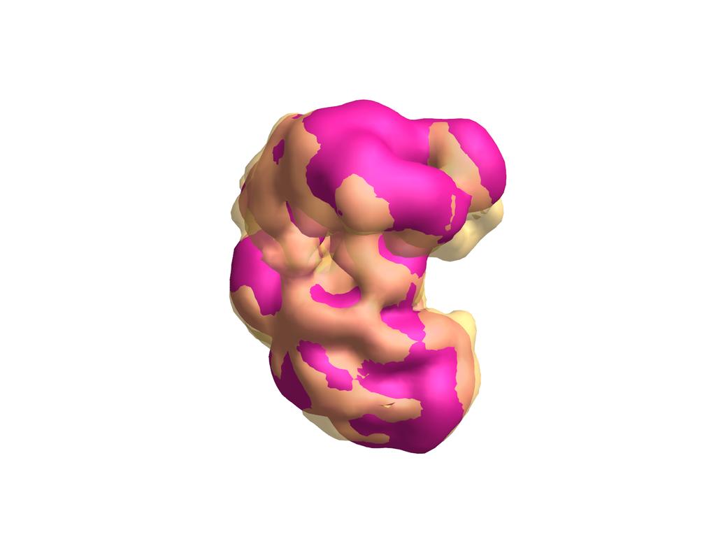
5 mechanism of E3 ligase shutdown - 5 Supplemental Figure 2. Negative stain EM reconstructions. (a) 3-dimensional PDF showing superposition of negative stain EM reconstructions of APC/C CDH1 -EMI1-SKP1 (yellow, transparent) with prior structure of human APC/C CDH1 (magenta, solid) 5. The difference map shown in green reveals the location of EMI1-SKP1 and structural changes propagated throughout the APC/C CDH1 complex. Use of a mouse to rotate the 3D PDF allows visualization of the entire EM map.
6
7 mechanism of E3 ligase shutdown - 7 (b) 3D PDF showing superposition of negative stain EM reconstructions of APC/C CDH1 - EMI1-SKP1 (yellow, transparent) with prior structure of human APC/C CDH1 (magenta, solid) 5.
8
9 mechanism of E3 ligase shutdown - 9 (c) 3D PDF showing superposition of negative stain EM reconstructions of APC/C CDH1 - EMI1 DLZT (yellow, transparent) with prior structure of human APC/C CDH1 (magenta, solid) 5.
10
11 mechanism of E3 ligase shutdown - 11 (d) 3D PDF illustrating EMI1 N-terminal domain localization. The main density not present in APC/C CDH1 -EMI1 DLZT compared to APC/C CDH1 -EMI1-SKP1 is shown in orange (from the APC/C CDH1 -EMI1-SKP1 - APC/C CDH1 -EMI1 DLZT difference map) on the EM reconstruction of APC/C CDH1 -EMI1 DLZT.
12
13 mechanism of E3 ligase shutdown - 13 (e) 3D PDF illustrating MBP from the APC/C CDH1 -EMI1-MBPSKP1 complex. The main density not present in APC/C CDH1 -EMI1-SKP1 compared to APC/C CDH1 -EMI1-MBPSKP1 is shown in red (from the APC/C CDH1 -EMI1-MBPSKP1 - APC/C CDH1 -EMI1-SKP1 difference map) on the EM reconstruction of APC/C CDH1 -EMI1-SKP1.
14 mechanism of E3 ligase shutdown - 14 Supplemental Figure 3. Fitting of APC/C CDH1 kinetic data. Error bars: SEM, n 3. The initial velocities were determined by performing APC/C CDH1 - mediated ubiquitination time courses with UBCH10 as the E2 and cycb-nt* as the substrate, UBCH10 as the E2 and UbcycB-NT* as the substrate, or UBE2S as the E2 and UbcycB-NT* as the substrate. Ubiquitinated products were quantitated and plotted versus time to determine the initial velocities (not shown). The initial velocities were then plotted against concentrations of CDH1, E2, or substrate. The CDH1 concentration was selected to achieve a fully occupied and homogeneous APC/C CDH1 population. An E2 concentration similar to the E2 K m app was chosen to determine the K m app of the substrates. If EMI1 competes with E2 or substrate but the E2 and substrate concentrations were higher than the K m app, the identification of competitive inhibition would be more difficult. Data were fit to a hyperbolic curve by nonlinear regression to determine the ½ max and K m app values. Error was based on three independent measurements for each rate. (a) Titrations of CDH1, with the APC/C, E2, and substrate concentrations fixed at 20 nm, 190 nm, and 200 nm, respectively. (b) Titrations of E2s, with the APC/C, CDH1, and substrate concentrations fixed at 7 nm, 1 µm CDH1, and 200 nm, respectively. (c) Titrations of substrate, with the APC/C, CDH1, UBCH10, and UBE2S concentrations fixed 2 nm, 1 µm, 900 nm and 600 nm, respectively. (d) K m app values for E2s and substrates and ½ max CDH1 concentrations for the indicated APC/C CDH1 -dependent ubiquitination reactions.
15 mechanism of E3 ligase shutdown - 15 Supplemental Figure 4. Characterization of the ubiquitination reactions under initial velocity conditions used to determine the apparent inhibitory constants of EMI1 variants. (a) Representative gels of APC/C-mediated ubiquitination reactions are shown using different E2/substrate combinations. The reactions were carried out at different APC/C concentrations while the concentrations of all other components were fixed as described in Supplementary Fig. 3c. The reactions were then quenched at their respective times and the ubiquitinated products were quantitated and plotted against the APC/C concentration. The linear relationship between the APC/C concentration and ubiquitinated product formation for each E2 and substrate combination suggest that the reactions are under initial velocity conditions. (b) A time-course of the CDH1, D-box substrate-independent Ub chain formation assay is shown to demonstrate the dependence of UBE2S activity on APC/C concentration. In this assay, a fluorescent Ub* that cannot be carried through the E1-E2-E3 cascade was used as a Ub acceptor in APC/C- and UBE2S-dependent Ub chain formation assays.

16 mechanism of E3 ligase shutdown - 16
17 mechanism of E3 ligase shutdown - 17
18 mechanism of E3 ligase shutdown - 18 Supplemental Figure 5. EMI1 is a tight binding inhibitor of both substrate ubiquitination and Ub chain forming activities of the APC/C CDH1. Error bars: SEM, n 3. Representative gels showing concentration dependence of EMI1 FDLZT SKP1 for inhibiting APC/C CDH1 -mediated ubiquitination reactions using three different E2 and substrate combinations, (a) UBCH10 as the E2 and cycb-nt* as the substrate, (b) UBCH10 as the E2 and UbcycB-NT* as the substrate, (c) UBE2S as the E2 and UbcycB- NT* as the substrate. The APC/C, CDH1, UBCH10, and UBE2S concentrations were fixed at 4 nm, 1 µm, 900 nm and 600nM, respectively. As shown in Supplementary Fig. 4, these reactions are under initial velocity conditions and therefore, the enzyme activity is sensitive to EMI1 inhibition. The ubiquitinated products were quantitated and plotted against EMI1 FDLZT concentration. The change in APC/C CDH1 -mediated ubiquitination by such subtle changes in EMI1 concentration around the APC/C concentration demonstrates the tight binding nature of EMI1. (d) The Morrison equation is a quadratic function for tight binding inhibitors that takes into account the depletion of E (Enzyme) and I (Inhibitor) upon the formation of the EI complex 6,7. To assess the lower-limit for accurate K i app determination for EMI1 with this method, data were simulated for a range of theoretical K i values using the tight-binding inhibitor/morrison equation. Representative graphs with a range of theoretical K i values are shown. The curves reveal the requirement to significantly detect enzyme activity at the stoichiometric point ( elbow ) to accurately quantify K i app below 2.5 nm with 5 nm APC/C. However, tight binding versions of EMI1 almost completely deplete the active APC/C at the stoichiometric point, resulting in little remaining activity in this part of the titration. In these cases, instead of an equilibrium curve to determine the K i, EMI1 is merely titrating the active enzyme concentration. The limited fits at the elbow region for our experiments thus set a lower limit of 2.5 nm for apparent K i values that our assays can accurately measure. (e-h) Fitting of the EMI1- mediated inhibition of APC/C-dependent ubiquitination to determine apparent inhibitory constants reported in Table 1. Ubiquitinated products, quantified from gels similar to those shown in (a-c), plotted against EMI1 concentration and fit to Morrison tight binding equation (Eq.1, Methods). (e) APC/C CDH1 - and UBCH10-dependent ubiquitination of cycb-nt*. (f) APC/C CDH1 - and UBCH10-depdendent ubiquitination of UbcycB-NT*. (g) APC/C CDH1 - and UBE2S-dependent ubiquitination of UbcycB-NT*. (h) APC/C- and UBE2S-dependent ubiquitination of Ub*.

19 mechanism of E3 ligase shutdown - 19 Supplemental Figure 6. Inhibition of APC/C CDH1 and UBCH10-mediated ubiquitination of cycb-nt* at extended reaction times. Data are shown for 300 nm (panel a) and 1 µm (panel b) of the indicated EMI1 variants described in Fig. 4.
20 mechanism of E3 ligase shutdown - 20 Supplemental Figure N- 1 H HSQC spectra of selected EMI1 ZT variants. To compare the folding between variants and wild-type EMI1, the most defective alanine mutants (Fig. 5) were 15 N-labeled, purified, and examined by NMR. The L375A and K376A EMI1 ZT variants appear to be properly folded, whereas the R393A EMI1 ZT variant appears to be unfolded based on lack of dispersion for the observed resonances.
 Two views of human APC/C-MCC 8 with density attributed to MCC outlined for comparison to EMI1-bound complexes shown in Fig. 2.")
21 mechanism of E3 ligase shutdown - 21 Supplementary Figure 8. Representation of prior structural data for APC/CMCC for comparison to APC/C CDH1 -EMI1 DLZT. (a) Two views of human APC/C-MCC 8 with density attributed to MCC outlined for comparison to EMI1-bound complexes shown in Fig. 2. Note that MCC does not form the same contact to the catalytic core observed upon APC/C binding to EMI1. (b) Similar views shown with transparent density for MCC over the crystal structure of a portion of S. pombe MCC 9.
22 mechanism of E3 ligase shutdown - 22 Supplemental References 1. Laue, T.M., Shah, B.D., Ridgeway, T.M. & Pelletier, S.L. Computer-aided Interpretation of Analytical Sedimentation Data For Proteins. in Analytical Centrifugation in Biochemistry and Polymer Science (eds. Harding, S.E., Rowe, A.J. & Horton, J.C.) (The Royal Society of Chemistry, Cambridge, UK, 1992). 2. Schuck, P. Size-distribution analysis of macromolecules by sedimentation velocity ultracentrifugation and lamm equation modeling. Biophys J 78, (2000). 3. Brown, P.H., Balbo, A. & Schuck, P. Characterizing protein-protein interactions by sedimentation velocity analytical ultracentrifugation. Curr Protoc Immunol Chapter 18, Unit (2008). 4. Brown, P.H. & Schuck, P. Macromolecular size-and-shape distributions by sedimentation velocity analytical ultracentrifugation. Biophys J 90, (2006). 5. Buschhorn, B.A. et al. Substrate binding on the APC/C occurs between the coactivator Cdh1 and the processivity factor Doc1. Nat Struct Mol Biol 18, 6-13 (2011). 6. Morrison, J.F. Kinetics of the reversible inhibition of enzyme-catalysed reactions by tight-binding inhibitors. Biochim Biophys Acta 185, (1969). 7. Copeland, R.A. Evaluation of enzyme inhibitors in drug discovery. A guide for medicinal chemists and pharmacologists. Methods Biochem Anal 46, (2005). 8. Herzog, F. et al. Structure of the anaphase-promoting complex/cyclosome interacting with a mitotic checkpoint complex. Science 323, (2009). 9. Chao, W.C., Kulkarni, K., Zhang, Z., Kong, E.H. & Barford, D. Structure of the mitotic checkpoint complex. Nature 484, (2012).
SEC MALLs and AUC. 2. Conformation and flexibility Viscometry,
 1. Molecular weight distribution analysis SEC MALLs and AUC 2. Conformation and flexibility Viscometry, AUC, Light scattering Lecture 5 Analytical Ultracentrifugation II: interactions Steve Harding Free
THE SOLUTION CONFORMATION OF NOVEL ANTIBODY FRAGMENTS STUDIED USING THE PROTEOMELAB XL-A ANALYTICAL ULTRACENTRIFUGE
 APPLICATION INFORMATION Peter J. Morgan, Olwyn D. Byron, Stephen E. Harding Department of Applied Biochemistry and Food Science University of Nottingham Sutton Bonington, U. K. Introduction One of the
Diffusion-deconvoluted sedimentation coefficient distributions for the analysis of interacting and non-interacting protein mixtures
 1 Chapter for Modern Analytical Ultracentrifugation: Techniques and Methods (D.J. Scott, S.E. Harding, A.J. Rowe, Editors) The Royal Society of Chemistry, Cambridge (in press) Diffusion-deconvoluted sedimentation
Supplementary figure 1 Application of tmfret in LeuT. (a) To assess the feasibility of using tmfret for distance-dependent measurements in LeuT, a
 To assess the feasibility of using tmfret for distance-dependent measurements in LeuT, a") Supplementary figure 1 Application of tmfret in LeuT. (a) To assess the feasibility of using tmfret for distance-dependent measurements in LeuT, a series of tmfret-pairs comprised of single cysteine mutants
Supplementary figure 1 Application of tmfret in LeuT. (a) To assess the feasibility of using tmfret for distance-dependent measurements in LeuT, a series of tmfret-pairs comprised of single cysteine mutants
Biomolecular hydrodynamics
 Biomolecular hydrodynamics Chem 341, Fall, 2014 1 Frictional coefficients Consider a particle moving with velocity v under the influence of some external force F (say a graviational or electrostatic external
Self-Assembly of Peptide Porphyrin Complexes: Towards the Development of Smart Biomaterials
 Self-Assembly of Peptide Porphyrin Complexes: Towards the Development of Smart Biomaterials Brian C. Kovaric, Bashkim Kokona, Alexander D. Schwab, Margaret A. Twomey, Julio C. de Paula, and Robert Fairman
Analytical Ultracentrifugation Macromolecule Characterization in Solution
 Analytical Ultracentrifugation Macromolecule Characterization in Solution AUC TECH OVERVIEW OPTIMA AUC Analytical Ultracentrifugation Beckman Coulter delivered the first AUC sample characterization tool
By: Ashley and Christine Phy 200 Professor Newman 4/13/12
 By: Ashley and Christine Phy 200 Professor Newman 4/13/12 What is it? Technique used to settle particles in solution against the barrier using centrifugal acceleration Two Types of Centrifuges Analytical
Measuring S using an analytical ultracentrifuge. Moving boundary
 Measuring S using an analytical ultracentrifuge Moving boundary [C] t = 0 t 1 t 2 0 top r bottom 1 dr b r b (t) r b ω 2 = S ln = ω 2 S (t-t dt r b (t o ) o ) r b = boundary position velocity = dr b dt
Analytical Ultracentrifugation in the Study of Protein Selfassociation and Heterogeneous Protein-Protein Interactions:
 Analytical Ultracentrifugation in the Study of Protein Selfassociation and Heterogeneous Protein-Protein Interactions: Protocols for Velocity and Equilibrium Sedimentation Andrea Balbo and Peter Schuck
Joint use of SAS and AUC
 Joint use of SAS and AUC Olwyn Byron School of Life Sciences College of Medical, Veterinary and Life Sciences University of Glasgow, Scotland UK Outline Principles underlying AUC How AUC experiments are
Sedimentation Velocity Analysis of Interacting Systems using c(s) Peter Schuck
 Peter Schuck") Sedimentation Velocity Analysis of Interacting Systems using c(s) Peter Schuck simulated sedimentation of rapidly self-association systems monomer/dimer monomer/trimer 5 concentration (Kd) 4 2 concentration
Sedimentation Velocity Analysis of Interacting Systems using c(s) Peter Schuck simulated sedimentation of rapidly self-association systems monomer/dimer monomer/trimer 5 concentration (Kd) 4 2 concentration
SEC MALLs and AUC. 2. Conformation and flexibility Viscometry,
 1. Molecular weight distribution analysis SEC MALLs and AUC 2. Conformation and flexibility Viscometry, AUC, Light scattering Lecture 4. Analytical l Ultracentrifugation t ti I: Molecular weight and conformation
Supporting Information
 Supporting Information Characterizing the Effect of Salt and Surfactant Concentration on the Counter-ion Atmosphere around Surfactant Stabilized SWCNTs using Analytical Ultracentrifugation Stephanie Lam
THE CRYSTAL STRUCTURE OF THE SGT1-SKP1 COMPLEX: THE LINK BETWEEN
 THE CRYSTAL STRUCTURE OF THE SGT1-SKP1 COMPLEX: THE LINK BETWEEN HSP90 AND BOTH SCF E3 UBIQUITIN LIGASES AND KINETOCHORES Oliver Willhoft, Richard Kerr, Dipali Patel, Wenjuan Zhang, Caezar Al-Jassar, Tina
SEDIMENTATION VELOCITY ANALYTICAL ULTRACENTRIFUGATION
 Analytical Biochemistry, in press ON THE ANALYSIS OF PROTEIN SELF-ASSOCIATION BY SEDIMENTATION VELOCITY ANALYTICAL ULTRACENTRIFUGATION Peter Schuck Protein Biophysics Resource, Division of Bioengineering
NMR study of complexes between low molecular mass inhibitors and the West Nile virus NS2B-NS3 protease
 University of Wollongong Research Online Faculty of Science - Papers (Archive) Faculty of Science, Medicine and Health 2009 NMR study of complexes between low molecular mass inhibitors and the West Nile
Low temperature solution behaviour of Methylophilus methylotrophus electron transferring flavoprotein: a study by analytical ultracentrifugation
 Eur Biophys J (1997) 25: 411 416 Springer-Verlag 1997 ARTICLE Helmut Cölfen Stephen E. Harding Emma K. Wilson Nigel S. Scrutton Donald J. Winzor Low temperature solution behaviour of Methylophilus methylotrophus
Size-Distribution Analysis of Macromolecules by Sedimentation Velocity Ultracentrifugation and Lamm Equation Modeling
 1606 Biophysical Journal Volume 78 March 2000 1606 1619 Size-Distribution Analysis of Macromolecules by Sedimentation Velocity Ultracentrifugation and Lamm Equation Modeling Peter Schuck Molecular Interactions
Structural basis for catalytically restrictive dynamics of a high-energy enzyme state
 Supplementary Material Structural basis for catalytically restrictive dynamics of a high-energy enzyme state Michael Kovermann, Jörgen Ådén, Christin Grundström, A. Elisabeth Sauer-Eriksson, Uwe H. Sauer
Microcalorimetry for the Life Sciences
 Microcalorimetry for the Life Sciences Why Microcalorimetry? Microcalorimetry is universal detector Heat is generated or absorbed in every chemical process In-solution No molecular weight limitations Label-free
Supporting Information
 Supporting Information Band Sedimentation Experiment in Analytical Ultracentrifugation Revisited Cornelia M. Schneider, Dirk Haffke, Helmut Cölfen* Physical Chemistry, University of Konstanz, Universitätsstrasse
Supplementary Information. Overlap between folding and functional energy landscapes for. adenylate kinase conformational change
 Supplementary Information Overlap between folding and functional energy landscapes for adenylate kinase conformational change by Ulrika Olsson & Magnus Wolf-Watz Contents: 1. Supplementary Note 2. Supplementary
Limitations of the ultracentrifugal approach for measuring the effective net charge of a macroion
 ANALYTICAL BIOCHEMISTRY Analytical Biochemistry 333 (2004) 114 118 www.elsevier.com/locate/yabio Limitations of the ultracentrifugal approach for measuring the effective net charge of a macroion Donald
The Approach to Equilibrium in a Buoyant-Density Gradient
 BIOPOLY MERS VOL. 11, 17651769 (1972) The Approach to Equilibrium in a BuoyantDensity Gradient CARL W. SCHMID and JOHN E. HEARST, Department of Chemistry, University of California Berkeley, California
Acta Crystallographica Section D
 Supporting information Acta Crystallographica Section D Volume 70 (2014) Supporting information for article: Structural basis of the heterodimerization of the MST and RASSF SARAH domains in the Hippo signalling
Supplementary Information for
 Supplementary Information for Structural basis for the inhibition of Mycobacterium tuberculosis L,D-transpeptidase by meropenem, a drug effective against extensively drug-resistant strains Hyoun Sook Kim
UltraScan Workshop/Bioc5083 Hydrodynamic Methods
 UltraScan Workshop/Bioc5083 Hydrodynamic Methods Borries Demeler, Ph.D. Department of Biochemistry May/June 2013 Analytical Ultracentrifugation Background What can be learned from AUC? Excellent method
a-chymotrypsin: CHARACTERIZATION OF A SELF-ASSOCIATING SYSTEM IN THE ANALYTICAL ULTRACENTRIFUGE
 APPLICATION INFORMATION Paul Voelker and Don McRorie Beckman Coulter Introduction a-chymotrypsin: CHARACTERIZATION OF A SELF-ASSOCIATING SYSTEM IN THE ANALYTICAL ULTRACENTRIFUGE We have studied the dimerization
Analytical Ultracentrifugation. by: Andrew Rouff and Andrew Gioe
 Analytical Ultracentrifugation by: Andrew Rouff and Andrew Gioe Partial Specific Volume (v) Partial Specific Volume is defined as the specific volume of the solute, which is related to volume increase
Experimental Design and Data Collection
 A. Checks to run to make sure the instrument is in good working condition B. Decide which experiment type is most appropriate, equilibrium or velocity C. Buffer considerations D. Speed selection and length
Supplementary Figures
 1 Supplementary Figures Supplementary Figure 1 Type I FGFR1 inhibitors (a) Chemical structures of a pyrazolylaminopyrimidine inhibitor (henceforth referred to as PAPI; PDB-code of the FGFR1-PAPI complex:
Contents. xiii. Preface v
 Contents Preface Chapter 1 Biological Macromolecules 1.1 General PrincipIes 1.1.1 Macrornolecules 1.2 1.1.2 Configuration and Conformation Molecular lnteractions in Macromolecular Structures 1.2.1 Weak
Supplementary Information. The protease GtgE from Salmonella exclusively targets. inactive Rab GTPases
 Supplementary Information The protease GtgE from Salmonella exclusively targets inactive Rab GTPases Table of Contents Supplementary Figures... 2 Supplementary Figure 1... 2 Supplementary Figure 2... 3
Structure and Interactions of Fish Type III Antifreeze Protein in Solution
 Biophysical Journal, Volume 99 Supporting Material Structure and Interactions of Fish Type III Antifreeze Protein in Solution Andrés G. Salvay, Frank Gabel, Bernard Pucci, Javier Santos, Eduardo I. Howard,
Model Independent Determination of Colloidal Silica Size Distributions via Analytical Ultracentrifugation
 Accelerated Article Subscriber access provided by Universiteit Utrecht Model Independent Determination of Colloidal Silica Size Distributions via Analytical Ultracentrifugation Karel L. Planken, Bonny
Chapter 6. The interaction of Src SH2 with the focal adhesion kinase catalytic domain studied by NMR
 The interaction of Src SH2 with the focal adhesion kinase catalytic domain studied by NMR 103 Abstract The interaction of the Src SH2 domain with the catalytic domain of FAK, including the Y397 SH2 domain
Equilibrium Sedimentation & Sedimentation Velocity: Random Walks in the presence of forces.
 New Posts at the Course Website: Origin Assignment 3 on Analytical Ultracentrifugation (due 3/2/12) Problem Set Data (xls file) Resource Booklet (R7) on Analytical Ultracentrifugation Equilibrium Sedimentation
Marcin Nowotny, Sergei A. Gaidamakov, Rodolfo Ghirlando, Susana M. Cerritelli, Robert J. Crouch, and Wei Yang
 Molecular Cell, Volume 28 Supplemental Data Structure of Human RNase H1 Complexed with an RNA/DNA Hybrid: Insight into HIV Reverse Transcription Marcin Nowotny, Sergei A. Gaidamakov, Rodolfo Ghirlando,
Principles of Physical Biochemistry
 Principles of Physical Biochemistry Kensal E. van Hold e W. Curtis Johnso n P. Shing Ho Preface x i PART 1 MACROMOLECULAR STRUCTURE AND DYNAMICS 1 1 Biological Macromolecules 2 1.1 General Principles
Supporting Information
 Supporting Information Membrane-less compartmentalization facilitates enzymatic cascade reactions and reduces substrate inhibition. Taisuke Kojima 1 and Shuichi Takayama 1,2* [ 1 ] The Wallace H Coulter
Supplemental Materials and Methods
 Supplemental Materials and Methods Time-resolved FRET (trfret) to probe for changes in the Box A/A stem upon complex assembly U3 MINI was folded and the decay of Fl fluorescence was measured at 20 ºC (see
Keywords: dimer dissociation, Streptomyces subtilisin inhibitor, rate constant
 J. Biol. Macromol. 8(2), 38-47 (2008) Article Kinetic study on the dissociation of a dimeric protein, Streptomyces Subtilisin Inhibitor Keiko Momma 1,2, Ben ichiro Tonomura, and Keitaro Hiromi Department
Hydrodynamic Characterisation
 Hydrodynamic Characterisation Viscometry SEC-MALLs Analytical Ultracentrifugation Stephen Harding, NCMH University of Nottingham NCMH at Nottingham: An International Facility for characterising sizes/shapes
Serine-7 but not serine-5 phosphorylation primes RNA polymerase II CTD for P-TEFb recognition
 Supplementary Information to Serine-7 but not serine-5 phosphorylation primes RNA polymerase II CTD for P-TEFb recognition Nadine Czudnochowski 1,2, *, Christian A. Bösken 1, * & Matthias Geyer 1 1 Max-Planck-Institut
According to the manufacture s direction (Pierce), RNA and DNA
, RNA and DNA") Supplementary method Electrophoretic Mobility-shift assay (EMSA) According to the manufacture s direction (Pierce), RNA and DNA oligonuleotides were firstly labeled by biotin. TAVb (1pM) was incubated
Supplementary method Electrophoretic Mobility-shift assay (EMSA) According to the manufacture s direction (Pierce), RNA and DNA oligonuleotides were firstly labeled by biotin. TAVb (1pM) was incubated
Supplementary Figure 1 Crystal contacts in COP apo structure (PDB code 3S0R)
") Supplementary Figure 1 Crystal contacts in COP apo structure (PDB code 3S0R) Shown in cyan and green are two adjacent tetramers from the crystallographic lattice of COP, forming the only unique inter-tetramer
Supplementary Figure 1 Crystal contacts in COP apo structure (PDB code 3S0R) Shown in cyan and green are two adjacent tetramers from the crystallographic lattice of COP, forming the only unique inter-tetramer
Biochemistry 3100 Sample Problems Binding proteins, Kinetics & Catalysis
 (1) Draw an approximate denaturation curve for a typical blood protein (eg myoglobin) as a function of ph. (2) Myoglobin is a simple, single subunit binding protein that has an oxygen storage function
SUPPLEMENTARY INFORMATION
 SUPPLEMENTARY INFORMATION doi:10.1038/nature11524 Supplementary discussion Functional analysis of the sugar porter family (SP) signature motifs. As seen in Fig. 5c, single point mutation of the conserved
17. Biomolecular Interaction
 17. Biomolecular Interaction Methods for characterizing biomolecular interactions Sequence-specific DNA binding ligands Molecular mechanisms of drug action and drug resistance In silico compound design
Nature Structural & Molecular Biology: doi: /nsmb Supplementary Figure 1
 Supplementary Figure 1 Identification of the ScDcp2 minimal region interacting with both ScDcp1 and the ScEdc3 LSm domain. Pull-down experiment of untagged ScEdc3 LSm with various ScDcp1-Dcp2-His 6 fragments.
Supplementary Figure 1. SDS-PAGE analysis of GFP oligomer variants with different linkers. Oligomer mixtures were applied to a PAGE gel containing
 Supplementary Figure 1. SDS-PAGE analysis of GFP oligomer variants with different linkers. Oligomer mixtures were applied to a PAGE gel containing 0.1% SDS without boiling. The gel was analyzed by a fluorescent
Modern analytical ultracentrifugation in protein science: A tutorial review
 REVIEW Modern analytical ultracentrifugation in protein science: A tutorial review JACOB LEBOWITZ, MARC S. LEWIS, AND PETER SCHUCK Molecular Interactions Resource, Division of Bioengineering and Physical
Measuring the size and shape of macromolecules. Hydrodynamics: study of the objects in water How do the move? Translation Rotation
 Measuring the size and shape of macromolecules Hydrodynamics: study of the objects in water How do the move? Translation Rotation 1) Movement with no external forcefree diffusion 2) Movement under the
SUPPLEMENTARY INFORMATION
 doi:10.1038/nature10458 Active Site Remodeling in the Bifunctional Fructose-1,6- bisphosphate aldolase/phosphatase Juan Du, Rafael F. Say, Wei Lü, Georg Fuchs & Oliver Einsle SUPPLEMENTARY FIGURES Figure
Characterization of bovine serum albumin/ chlorogenic acid solution mixtures by analytical ultracentrifugation
 Progr Colloid Polym Sci (2004) 127: 83 88 DOI 10.1007/b98017 Ó Springer-Verlag 2004 A. Seifert H. M. Rawel S. E. Harding J. Kroll Characterization of bovine serum albumin/ chlorogenic acid solution mixtures
Lecture 10: Cyclins, cyclin kinases and cell division
 Chem*3560 Lecture 10: Cyclins, cyclin kinases and cell division The eukaryotic cell cycle Actively growing mammalian cells divide roughly every 24 hours, and follow a precise sequence of events know as
CH 3 CH 2 OH +H 2 O CHO. 2e + 2H + + O 2 H 2 O +HCOOH
 2 4 H CH 3 2e + 2H + + 2 H 2 2 H CH 2 H 2e + 2H + + 2 H 2 2 H +H 2 CH 2e + 2H + + 2 H 2 2 H +HCH Supplemental Figure S. The three-step 4DM reaction, each step requires two reducing equivalents from ADPH
Analytical Ultracentrifugation
 Analytical Ultracentrifugation Stephen McLaughlin Biophysics MRC Laboratory of Molecular Biology Macromolecular Dynamics D Folding N Binding K N-D K D k D agg Aggregation Crystallisation 1 Macromolecular
SUPPLEMENTARY INFORMATION
 doi:1.138/nature1737 Supplementary Table 1 variant Description FSEC - 2B12 a FSEC - 6A1 a K d (leucine) c Leucine uptake e K (wild-type like) K (Y18F) K (TS) K (TSY) K288A mutant, lipid facing side chain
Analysis of nucleotide binding to p97 reveals the properties of a tandem AAA hexameric ATPase
 SUPPLEMENTARY INFORMATION Analysis of nucleotide binding to p97 reveals the properties of a tandem AAA hexameric ATPase Louise C Briggs, Geoff S Baldwin, Non Miyata, Hisao Kondo, Xiaodong Zhang, Paul S
Supporting information
 Supporting information The L-rhamnose Antigen: a Promising Alternative to α-gal for Cancer Immunotherapies Wenlan Chen,, Li Gu,#, Wenpeng Zhang, Edwin Motari, Li Cai, Thomas J. Styslinger, and Peng George
PREPRINT (this article appeared in Analytical Biochemistry 279, , 2000)
") PREPRINT (this article appeared in Analytical Biochemistry 79, 151-163, 000) A Method for Directly Fitting the Time Derivative of Sedimentation Velocity Data and an Alternative Algorithm for Calculating
PREPRINT (this article appeared in Analytical Biochemistry 79, 151-163, 000) A Method for Directly Fitting the Time Derivative of Sedimentation Velocity Data and an Alternative Algorithm for Calculating
Nature Structural and Molecular Biology: doi: /nsmb Supplementary Figure 1. Definition and assessment of ciap1 constructs.
 Supplementary Figure 1 Definition and assessment of ciap1 constructs. (a) ciap1 constructs used in this study are shown as primary structure schematics with domains colored as in the main text. Mutations
The Riboswitch is functionally separated into the ligand binding APTAMER and the decision-making EXPRESSION PLATFORM
 The Riboswitch is functionally separated into the ligand binding APTAMER and the decision-making EXPRESSION PLATFORM Purine riboswitch TPP riboswitch SAM riboswitch glms ribozyme In-line probing is used
Biomolecular hydrodynamics
 Biomolecular hydrodynamics David Case Rutgers, Spring, 2014 January 29, 2014 Why study hydrodynamical methods? The methods we study in this section are low resolution ones one studies diffusional motion
Supporting Text Z = 2Γ 2+ + Γ + Γ [1]
![Supporting Text Z = 2Γ 2+ + Γ + Γ [1]](/thumbs/91/105230015.jpg "Supporting Text Z = 2Γ 2+ + Γ + Γ [1]") Supporting Text RNA folding experiments are typically carried out in a solution containing a mixture of monovalent and divalent ions, usually MgCl 2 and NaCl or KCl. All three species of ions, Mg, M +
Supporting Text RNA folding experiments are typically carried out in a solution containing a mixture of monovalent and divalent ions, usually MgCl 2 and NaCl or KCl. All three species of ions, Mg, M +
BBS501 Section 1 9:00 am 10:00 am Monday thru Friday LRC 105 A & B
 BBS501 Section 1 9:00 am 10:00 am Monday thru Friday LRC 105 A & B Lecturers: Dr. Yie-Hwa Chang Room M130 Phone: #79263 E-mail:changy@slu.edu Dr. Tomasz Heyduk Room M99 Phone: #79238 E-mail: heydukt@slu.edu
Presentation Microcalorimetry for Life Science Research
 Presentation Microcalorimetry for Life Science Research MicroCalorimetry The Universal Detector Heat is either generated or absorbed in every chemical process Capable of thermal measurements over a wide
pyridoxal phosphate synthase
 Supplementary Information 13 C-NMR snapshots of the complex reaction coordinate of pyridoxal phosphate synthase Jeremiah W. Hanes, Ivan Keresztes, and Tadhg P. Begley * Department of Chemistry and Chemical
Characterization of Reversible Kinase Inhibitors using Microfluidic Mobility-Shift Assays
 Application Note 211 Characterization of Reversible Kinase Inhibitors using Microfluidic Mobility-Shift Assays Introduction Current drug discovery efforts typically focus on developing small molecule inhibitors
Supplementary Figure 1. Biochemical and sequence alignment analyses the
 Supplementary Figure 1. Biochemical and sequence alignment analyses the interaction of OPTN and TBK1. (a) Analytical gel filtration chromatography analysis of the interaction between TBK1 CTD and OPTN(1-119).
The Fic protein Doc uses an inverted substrate to phosphorylate and. inactivate EF-Tu
 The Fic protein Doc uses an inverted substrate to phosphorylate and inactivate EF-Tu Daniel Castro-Roa 1, Abel Garcia-Pino 2,3 *, Steven De Gieter 2,3, Nico A.J. van Nuland 2,3, Remy Loris 2,3, Nikolay
Optimization of an Adapta Kinase Assay for CAMK1
 Overview Optimization of an Adapta Kinase Assay for CAMK1 This protocol describes how to perform an Adapta assay with the kinase CAMK1. To maximize the ability of the assay to detect ATP-competitive inhibitors,
The effect of the degree of esterification on the hydrodynamic properties of citrus pectin
 Food Hydrocolloids 14 (2000) 227 235 www.elsevier.com/locate/foodhyd The effect of the degree of esterification on the hydrodynamic properties of citrus pectin G.A. Morris a, *, T.J. Foster b, S.E. Harding
A B C SHORT COLUMN SEDIMENTATION EQUILIBRIUM ANALYSIS FOR RAPID CHARACTERIZATION OF MACROMOLECULES IN SOLUTION. Inches.
 APPLICATION WHITE NOTE PAPER Thomas M. Laue University of New Hampshire Department of Biochemistry Durham, NH 03824 Introduction SHORT COLUMN SEDIMENTATION EQUILIBRIUM ANALYSIS FOR RAPID CHARACTERIZATION
Supplemental data for
 Supplemental data for A Real-Time Guanine Nucleotide Exchange Assay using NMR: Activation of RhoA by PDZ- RhoGEF. Geneviève M.C. Gasmi-Seabrook 1,3, Christopher B. Marshall 1,3, Melissa Cheung 1,3, Bryan
Structure and RNA-binding properties. of the Not1 Not2 Not5 module of the yeast Ccr4 Not complex
 Structure and RNA-binding properties of the Not1 Not2 Not5 module of the yeast Ccr4 Not complex Varun Bhaskar 1, Vladimir Roudko 2,3, Jerome Basquin 1, Kundan Sharma 4, Henning Urlaub 4, Bertrand Seraphin
Sensitive NMR Approach for Determining the Binding Mode of Tightly Binding Ligand Molecules to Protein Targets
 Supporting information Sensitive NMR Approach for Determining the Binding Mode of Tightly Binding Ligand Molecules to Protein Targets Wan-Na Chen, Christoph Nitsche, Kala Bharath Pilla, Bim Graham, Thomas
Supporting Information
 Supporting Information Arai et al. 10.1073/pnas.15179911 SI Text Protein Expression and Purification. Myb3 (mouse, residues 84 315) was expressed in Escherichia coli as a fusion with the B1 domain of protein
CHEM-UA 652: Thermodynamics and Kinetics
 1 CHEM-UA 652: Thermodynamics and Kinetics Notes for Lecture 11 I. PHYSICAL AND CHEMICAL RELEVANCE OF FREE ENERGY In this section, we will consider some examples showing the significance of free energies.
Biomolecular hydrodynamics
 Biomolecular hydrodynamics David Case Rutgers, Spring, 2009 February 1, 2009 Why study hydrodynamical methods? The methods we study in this section are low resolution ones one studies diffusional motion
From Gen. Chem.: 1. WHAT is an ACID? 2. WHAT is a BASE?
 Expt. 1: Biological Buffers Goals: 1. Learn how to use the Henderson-Hasselbach (H-H) eqn. 2. Learn how to prepare buffers. 3. Learn something about physical properties of biological buffers which are
ENZYME KINETICS. Medical Biochemistry, Lecture 24
 ENZYME KINETICS Medical Biochemistry, Lecture 24 Lecture 24, Outline Michaelis-Menten kinetics Interpretations and uses of the Michaelis- Menten equation Enzyme inhibitors: types and kinetics Enzyme Kinetics
State Key Laboratory of Coordination Chemistry, School of Chemistry and Chemical
 Electronic Supplementary Information Evidence for inhibition of HIF-1α Prolyl hydroxylase 3 activity by four biological active tetraazamacrocycles Jing Cao, Zhirong Geng, Xiaoyan Ma, Jinghan Wen, Yuxin
The structure of the deubiquitinase USP15 reveals a misaligned catalytic triad and an open ubiquitin-binding channel
 SUPPORTING INFORMATION The structure of the deubiquitinase USP15 reveals a misaligned catalytic triad and an open ubiquitin-binding channel Stephanie J. Ward, Hayley E. Gratton, Peni Indrayudha #, Camille
Online Supplementary Material. Messenger RNA Interactions in the Decoding Center Control the Rate of Translocation
 Online Supplementary Material Messenger RNA Interactions in the Decoding Center Control the Rate of Translocation Prashant K. Khade and Simpson Joseph Supplementary Figure 1 Dissociation of the f[ 35 S]Met-Phe-tRNA
Introduction to Dynamic Light Scattering with Applications. Onofrio Annunziata Department of Chemistry Texas Christian University Fort Worth, TX, USA
 Introduction to Dynamic Light Scattering with Applications Onofrio Annunziata Department of Chemistry Texas Christian University Fort Worth, TX, USA Outline Introduction to dynamic light scattering Particle
Bacterial protease uses distinct thermodynamic signatures for substrate recognition
 Bacterial protease uses distinct thermodynamic signatures for substrate recognition Gustavo Arruda Bezerra, Yuko Ohara-Nemoto, Irina Cornaciu, Sofiya Fedosyuk, Guillaume Hoffmann, Adam Round, José A. Márquez,
Biochemical / Biophysical Kinetics Made Easy
 Biochemical / Biophysical Kinetics Made Easy Software DYNAFIT in drug discovery research Petr Kuzmič, Ph.D. BioKin, Ltd. 1. Theory: differential equation models - DYNAFIT software 2. Example: lanthascreen
Characterizing Binding Interactions by ITC
 Characterizing Binding Interactions by ITC Christin T. Choma TA Instruments, 19 Lukens Drive, New Castle, DE 1972, USA All biochemical reactions involve recognition, binding and the formation of noncovalent
SUPPLEMENTARY INFORMATION
 Figure S1. Secondary structure of CAP (in the camp 2 -bound state) 10. α-helices are shown as cylinders and β- strands as arrows. Labeling of secondary structure is indicated. CDB, DBD and the hinge are
Table S1. Primers used for the constructions of recombinant GAL1 and λ5 mutants. GAL1-E74A ccgagcagcgggcggctgtctttcc ggaaagacagccgcccgctgctcgg
 SUPPLEMENTAL DATA Table S1. Primers used for the constructions of recombinant GAL1 and λ5 mutants Sense primer (5 to 3 ) Anti-sense primer (5 to 3 ) GAL1 mutants GAL1-E74A ccgagcagcgggcggctgtctttcc ggaaagacagccgcccgctgctcgg
You are advised to spend an equal amount of time on each question.
 UNIVERSITY OF EAST ANGLIA School of Chemistry Main Series UG Examination 2015-16 BIOPHYSICAL CHEMISTRY CHE-5601Y Time allowed: 2 hours Answer THREE questions. You are advised to spend an equal amount of
LanthaScreen Eu Kinase Binding Assay Validation Packet. Optimization of a LanthaScreen Eu Kinase Binding Assay for AURKB
 Page 1 of 18 LanthaScreen Eu Kinase Binding Assay for AURKB Overview This protocol describes how to perform a LanthaScreen Eu Kinase Binding Assay designed to detect and characterize kinase inhibitors.
Chapter 6: Outline-2. Chapter 6: Outline Properties of Enzymes. Introduction. Activation Energy, E act. Activation Energy-2
 Chapter 6: Outline- Properties of Enzymes Classification of Enzymes Enzyme inetics Michaelis-Menten inetics Lineweaver-Burke Plots Enzyme Inhibition Catalysis Catalytic Mechanisms Cofactors Chapter 6:
BSc and MSc Degree Examinations
 Examination Candidate Number: Desk Number: BSc and MSc Degree Examinations 2018-9 Department : BIOLOGY Title of Exam: Molecular Biology and Biochemistry Part I Time Allowed: 1 hour and 30 minutes Marking
Crystal Structure of Fibroblast Growth Factor 9 (FGF9) Reveals Regions. Implicated in Dimerization and Autoinhibition
 Reveals Regions. Implicated in Dimerization and Autoinhibition") JBC Papers in Press. Published on November 1, 2000 as Manuscript M006502200 Crystal Structure of Fibroblast Growth Factor 9 (FGF9) Reveals Regions Implicated in Dimerization and Autoinhibition 1 Copyright
JBC Papers in Press. Published on November 1, 2000 as Manuscript M006502200 Crystal Structure of Fibroblast Growth Factor 9 (FGF9) Reveals Regions Implicated in Dimerization and Autoinhibition 1 Copyright
Determination of Molecular Parameters by Fitting Sedimentation Data to Finite-Element Solutions of the Lamm Equation
 444 Biophysical Journal Volume 74 January 1998 444 454 Determination of Molecular Parameters by Fitting Sedimentation Data to Finite-Element Solutions of the Lamm Equation Borries Demeler* and Hashim Saber
Table S1. Overview of used PDZK1 constructs and their binding affinities to peptides. Related to figure 1.
 Table S1. Overview of used PDZK1 constructs and their binding affinities to peptides. Related to figure 1. PDZK1 constru cts Amino acids MW [kda] KD [μm] PEPT2-CT- FITC KD [μm] NHE3-CT- FITC KD [μm] PDZK1-CT-