SDF1 Antagonism of Axonal Repellents Requires Multiple G Protein Components and an Adam Metalloprotease
|
|
|
- Adam Collins
- 5 years ago
- Views:
Transcription

1 University of Pennsylvania ScholarlyCommons Publicly Accessible Penn Dissertations Spring SDF1 Antagonism of Axonal Repellents Requires Multiple G Protein Components and an Adam Metalloprotease Esther Naomi Twery University of Pennsylvania, entwery@gmail.com Follow this and additional works at: Part of the Developmental Neuroscience Commons Recommended Citation Twery, Esther Naomi, "SDF1 Antagonism of Axonal Repellents Requires Multiple G Protein Components and an Adam Metalloprotease" (2011). Publicly Accessible Penn Dissertations This paper is posted at ScholarlyCommons. For more information, please contact libraryrepository@pobox.upenn.edu.
2 SDF1 Antagonism of Axonal Repellents Requires Multiple G Protein Components and an Adam Metalloprotease { font-family: "Times New Roman"; }p.msonormal, li.msonormal, div.msonormal { margin: 0in 0in pt; font-size: 12pt; font-family: "Times New Roman"; }table.msonormaltable { font-size: 10pt; font-family: "Times New Roman"; }div.section1 { page: Section1; } Growth cones of developing axons navigate by interpreting signals from multiple cues. Some of these are the familiar guidance cues netrin, semaphorin, slit, and ephrin. Growth cones are also influenced by GPCR ligands, including neurotransmitters such as glutamate and chemokines such as SDF1. Previous work from our lab demonstrated that either glutamate or SDF1, acting through their receptors mglur1 or CXCR4, respectively, can reduce growth cone responsiveness to repellent cues. This effect is pertussis toxin-sensitive, implicating Ga i/o proteins, yet dependent on increased camp, implicating Ga s proteins. The antirepellent effect of SDF1 could also be mimicked by inhibition of Rho, suggesting that inhibition of Rho is a component of the antirepellent pathway. Here, I demonstrate that SDF1 antirepellent activity is blocked by peptides or proteins targeting Ga i, Ga q, or Gbg. This suggests that multiple G protein components are required for SDF1 signaling. I also show that SDF1 antirepellent activity is mimicked by constitutively active forms of Ga q, Ga i, or Ga s. This suggests that higher-than-physiological levels of individual G protein components can substitute for a combination of G protein components in antirepellent signaling. A role for Ga q in antirepellent signaling is further supported by the ability of a phospholipase C (PLC) inhibitor to block the SDF1 antirepellent effect, consistent with Ga q s canonical activation of PLC. My work also reveals an alternate mechanism for SDF1-induced antagonism of repellent signaling. I show that the metalloprotease ADAM10 can cleave the repellent receptor neuropilin-1. Further, SDF1 antirepellent activity is blocked by either the metalloprotease inhibitor TAPI-2 or a dominant-negative ADAM10. Thus, inhibitory shedding of repellent receptors may contribute to the antirepellent effect. Previous work has shown that the antirepellent effect is mimicked by pharmacologically increased camp or blocked by a camp antagonist. TAPI-2 does not block the antirepellent effects of a camp analogue, suggesting that ADAM activation belongs to a separate pathway not downstream of camp. This work supports a model wherein SDF1/CXCR4 activates multiple G protein components to both increase camp and activate ADAM10. This would reduce sensitivity to repellents through inactivation of Rho and clearing of repellent receptors from the growth cone surface. Degree Type Dissertation Degree Name Doctor of Philosophy (PhD) Graduate Group Neuroscience First Advisor Jonathan A. Raper This dissertation is available at ScholarlyCommons:
3 Keywords SDF1, axon guidance, G protein, ADAM10, CXCR4, GPCR Subject Categories Developmental Neuroscience This dissertation is available at ScholarlyCommons:
4 SDF1 ANTAGONISM OF AXONAL REPELLENTS REQUIRES MULTIPLE G PROTEIN COMPONENTS AND AN ADAM METALLOPROTEASE Esther Naomi Twery A DISSERTATION in Neuroscience Presented to the Faculties of the University of Pennsylvania in Partial Fulfillment of the Requirements for the Degree of Doctor of Philosophy 2011 SUPERVISOR OF DISSERTATION Jonathan Raper, Professor of Neuroscience GRADUATE GROUP CHAIRPERSON Rita Balice-Gordon, Professor of Neuroscience DISSERTATION COMMITTEE Rita Balice-Gordon, Professor of Neuroscience Jeffrey Golden, Professor of Pathology and Laboratory Medicine David Manning, Professor of Pharmacology Michael Nusbaum, Professor of Neuroscience
5 ACKNOWLEDGMENTS Many thanks to JAR and to my family and friends for their advice and support through this process. --ENT ii
6 ABSTRACT. SDF1 ANTAGONISM OF AXONAL REPELLENTS REQUIRES MULTIPLE G PROTEIN COMPONENTS AND AN ADAM METALLOPROTEASE Esther Naomi Twery Jonathan A. Raper Growth cones of developing axons navigate by interpreting signals from multiple cues. Some of these are the familiar guidance cues netrin, semaphorin, slit, and ephrin. Growth cones are also influenced by GPCR ligands, including neurotransmitters such as glutamate and chemokines such as SDF1. Previous work from our lab demonstrated that either glutamate or SDF1, acting through their receptors mglur1 or CXCR4, respectively, can reduce growth cone responsiveness to repellent cues. This effect is pertussis toxin-sensitive, implicating Gα i/o proteins, yet dependent on increased camp, implicating Gα s proteins. The antirepellent effect of SDF1 could also be mimicked by inhibition of Rho, suggesting that inhibition of Rho is a component of the antirepellent pathway. Here, I demonstrate that SDF1 antirepellent activity is blocked by peptides or proteins targeting Gα i, Gα q, or Gβγ. This suggests that multiple G protein components are required for SDF1 signaling. I also show that SDF1 antirepellent activity is mimicked by constitutively active forms of Gα q, Gα i, or Gα s. This suggests that higherthan-physiological levels of individual G protein components can substitute for a combination of G protein components in antirepellent signaling. A role for Gα q in antirepellent signaling is further supported by the ability of a phospholipase C (PLC) iii
7 inhibitor to block the SDF1 antirepellent effect, consistent with Gα q s canonical activation of PLC. My work also reveals an alternate mechanism for SDF1-induced antagonism of repellent signaling. I show that the metalloprotease ADAM10 can cleave the repellent receptor neuropilin-1. Further, SDF1 antirepellent activity is blocked by either the metalloprotease inhibitor TAPI-2 or a dominant-negative ADAM10. Thus, inhibitory shedding of repellent receptors may contribute to the antirepellent effect. Previous work has shown that the antirepellent effect is mimicked by pharmacologically increased camp or blocked by a camp antagonist. TAPI-2 does not block the antirepellent effects of a camp analogue, suggesting that ADAM activation belongs to a separate pathway not downstream of camp. This work supports a model wherein SDF1/CXCR4 activates multiple G protein components to both increase camp and activate ADAM10. This would reduce sensitivity to repellents through inactivation of Rho and clearing of repellent receptors from the growth cone surface. iv
8 TABLE OF CONTENTS Abstract iii Chapter 1: Introduction. 1 Chapter 2: SDF1-induced antagonism of axonal repulsion requires multiple G protein coupled signaling components that work in parallel. 15 Chapter 3: Involvement of an ADAM metalloprotease in SDF1 antirepellent activity. 36 Chapter 4: Future Directions. 49 References 57 v
9 LIST OF TABLES Table 2-1: Raw Data, Figs vi
10 LIST OF ILLUSTRATIONS Fig. 2-1: Competitive inhibitors of Gα i or Gα q/11, but not Gα s or Gα o, block SDF1-mediated antirepellent activity. 30 Fig. 2-2: Scavengers of Gβγ, Gα i, or Gα q subunits block SDF1 antirepellent activity. 31 Fig. 2-3: Constitutively active Gα q, Gα i, or Gα s mimics SDF1 s antirepellent activity. 31 Fig. 2-4: Inhibiting PLC blocks SDF1 antirepellent activity. 32 Fig. 2-5: Inhibiting PLC blocks antirepellent activity induced by expression of a constitutively active Gα q. 32 Fig. 2-6: A model for antirepellent signaling. 33 Fig. 3-1: Domain structures of ADAM10, ADAM17, DN-ADAM10, and DN-ADAM Fig. 3-2: The metalloprotease inhibitor TAPI-2 blocks SDF1 antirepellent activity. 46 Fig. 3-3: SDF1 antirepellent activity is not blocked by DN-ADAM17 but is blocked by DN- ADAM10 in either DRG or RGC. 47 Fig. 3-4: ADAM10 can cleave NP1. 47 Fig. 3-5: The metalloprotease inhibitor TAPI-2 does not block the SDF1 mimicking effects of a camp analogue or a ROCK inhibitor. 48 Fig. 3-6: A model for ADAM10 involvement in antirepellent signaling. 48 vii
11 Chapter 1: Introduction Axon guidance is a complex process required for the formation of proper connections between neurons and their targets. Guidance is influenced by external cues, signaling initiated by those cues, and the state of the cell when it encounters those cues. In this chapter, I will discuss aspects of the classical guidance molecules, G protein signaling, and modulation of axon guidance. Guidance cues Basic properties of classical guidance cues The first several axon guidance cues identified fall into four major gene families, the semaphorins, slits, ephrins, and netrins. Those four classes of proteins comprise what are sometimes called the classical guidance cues, to distinguish them from the morphogens, chemokines, neurotransmitters, and other molecules which have more recently been identified as helping to guide axons. Semaphorins (semas) are a large family of proteins, divided into six classes in animals and a separate class V, composed of of viral semaphorins (see Fiore and Püschel, 2003). Classes 1, 4, 5, 6, and V are transmembrane, class 7 is GPI-linked, and classes 2 and 3 are secreted (see Fiore and Püschel, 2003). Though different classes of semas have different receptor complexes, plexins are receptors or coreceptors for all except class 2 semas (Yazdani and Terman, 2006). Neuropilins are the primary family of sema coreceptors, 1
12 particularly for class 3 semaphorins, for which they contribute to ligand-receptor specificity (He and Tessier-Lavigne, 1997; Kolodkin et al., 1997). However, neuropilins have short intracellular domains, leaving plexins to transduce most of the intracellular signaling. Sema3A, the first identified semaphorin and the primary repellent which I will discuss in later chapters, was initially identified from chick brain and is a strong repellent for dorsal root ganglion axons (Luo et al., 1993). In some systems, the sema3a receptor complex has been shown to include the cell adhesion molecules L1 (Castellani et al., 2000; Castellani et al., 2002) and TAG-1 (Law et al., 2008) as well as neuropilin-1 and plexin- A1 (Kolodkin et al., 1997; Takahashi et al., 1999). Sema3A can also act as an attractant for dendrites from cortical neurons (Polleux et al., 2000), and other class 3 semas can attract certain populations of axons (Chauvet et al., 2007; Kolk et al., 2009) in addition to their better studied actions as repellents. There is no widely established signaling pathway for repulsion induced by any semaphorin, and the intermediate steps identified differ across semas. Sema3A has been shown to quickly induce actin depolymerization (Fan et al., 1993), which is a likely component of all repellent signaling. Similarly, recent work in fly shows a requirement for the flavoprotein monooxygenase Mical in sema-induced actin rearrangement, and this appears to be a relatively direct pathway (Hung et al., 2010, Terman et al., 2002), but sema signaling in other contexts is more complex. Sema3A-induced growth cone collapse can be enhanced by constitutively active Rac1 or reduced by dominant-negative 2
13 Rac1, suggesting that activation of Rac1 is a component of the sema3a signaling pathway, but a similar dominant-negative RhoA had no effect on sema3a-induced collapse (Jin and Strittmatter, 1997). Similarly, Kuhn et al. (1999) found that dominantnegative Rac1 or dominant-negative Cdc42 could block collapse in response to sema3a, further supporting a role for small G proteins in sema signaling. However, collapse in response to sema4d, which signals through plexin-b1 rather than plexin-a1 (Tamagnone et al., 1999), can be blocked by a dominant-negative PDZ-Rho-GEF, which binds directly to plexin-b1 (Aurandt et al., 2002; Swiercz et al., 2002). In addition to their effects on small G proteins, semas have been shown to increase active GSK-3β (Eickholt et al., 2002), condense growth cone microtubules (Dent et al., 2004), and inactivate cofilin (Aizawa et al., 2001). The slits are a smaller family of secreted repellents, with three main forms in vertebrates and one in invertebrates (see Dickson and Gilestro, 2006). They act through the robo family of receptors (Brose et al., 1999). Slit and robo were first identified in the midline of the fly (Rothberg et al., 1988; Seeger et al., 1993). Mutations in robo cause inappropriate crossing and recrossing of the midline and mutations in slit cause axons to collapse upon the midline. Slit/robo signaling is also important in the vertebrate midline, both at the spinal cord and the optic chiasm. At the chiasm, robo mutations in zebrafish (astray) lead to inappropriate ipsilateral and anterior projections from the optic nerve (Fricke et al., 2001). In the spinal cord, disruption of slit/robo signaling resembles the fly phenotype of axons failing to leave the ventral midline (Long et al., 2004). Slit/robo signaling is somewhat better understood than sema signaling, and it also appears to be 3
14 largely conserved across species. In fly, slit binding to robo induces recruitment of the adaptor protein Dock and the Rac GEF Sos (Fan et al., 2003; Yang and Bashaw, 2006). In vertebrate neurons, slit/robo signaling has been shown to activate the microtubule plus-end tracking protein CLASP (Lee et al., 2004), and robo has been shown to directly bind a family of Rho GAPs (Wong et al., 2001). Netrins are the only classical guidance cues to have major functions as attractants. They were first identified in C. elegans as UNC-6, where mutations disrupted dorsal and ventral pathfinding (Ishii et al., 1992), and soon thereafter in chick, where they were shown to promote outgrowth in vitro (Serafini et al., 1994). There are three classes of netrin receptors, DCC/UNC-40/Frazzled, UNC-5, and DSCAM (Chan et al., 1996; Kolodziej et al., 1996; Leonardo et al., 1997; Ackerman et al., 1997; Ly et al., 2008). Of these, DCC and DSCAM can mediate attraction (Keino-Masu et al., 1996 ; Ly et al., 2008). UNC-5, however, mediates repulsion in both worms and vertebrates (Hedgecock et al., 1990; Colamarino and Tessier-Lavigne, 1995). Netrins are perhaps best known for their role in attracting axons to the midline, whether in worm (Ishii et al., 1992), fly (Harris et al., 1996) or vertebrates (Serafini et al., 1994; Serafini et al., 1996). Netrin signals through camp and PKA (Ming et al., 1997) and activation of the small G proteins Cdc42 and Rac (Shekarabi and Kennedy, 2002; Li et al., 2002). The last major class of classical guidance molecules is the ephrins and Ephs. Ephs are receptor tyrosine kinases, and their activation by ephrins induces receptor dimerization and phosphorylation (Kalo and Pasquale, 1999). Ephrins come in two subtypes, the GPI- 4
15 linked ephrin As and the transmembrane ephrin Bs. Generally, the ephrin As bind EphAtype receptors and ephrin Bs bind EphB-type receptors, but ephrina5 has also been shown to bind EphB2 (Himanen et al., 2004). Ephrin/Eph activity is classically required in topographical patterning in the optic tectum through gradients of A and B-type signals, (Cheng et al., 1995; Marcus et al., 1996) and in the mammalian optic chiasm, where EphB1 is required to send non-crossing RGC axons to the ipsilateral superior colliculus (Williams et al., 2003). Ephs are also involved in the formation of excitatory synapses. EphB2 recruits both NMDA- and AMPA-type glutamate receptors to developing dendritic spines (Dalva et al., 2000; Kayser et al., 2006). In repulsive axon guidance, in addition to Eph autophosphorylation, Ephs can bind SH2- and PDZ-containing proteins (Holland et al., 1997; Dodelet et al., 1999; Torres et al., 1998) as well as GEFs for Rho (Penzes et al., 2003; Sahin et al., 2005; Cowan et al., 2005). In some situations, such as the formation of the anterior commissure in mouse (Kullander et al., 2001), functional Eph signaling does not require kinase activity and must thus work through other Eph effectors. Morphogens as guidance cues Morphogens are proteins that act early in development to pattern the embryo and control cell fate decisions, but several classes of morphogen have also been shown to act as guidance cues later in development. Wnts, for example, are involved in early patterning, during which they provide a caudal signal in somitogenesis and within the neural plate 5
16 (McGrew et al., 1995). Later, Wnt-1 is required for the formation of the caudal midbrain (McMahon and Bradley, 1990; Thomas and Capecchi, 1990). Gradients of Wnts also contribute to the anterior turn made by commissural axons after crossing the midline (Lyuksyutova et al., 2003). BMPs are a major dorsalizing factor in the spinal cord (Liem et al., 1997; Nguyen et al., 2000), where they promote sensory neuronal fates. Once neurons are specified and their axons are extending, BMPs repel commissural axons from the dorsal spinal cord, so they can cross at the floorplate (Augsburger et al., 1999; Butler and Dodd, 2003). Sonic hedgehog (Shh) is also active in early embryonic development, for example, acting as a ventralizing factor in the spinal cord in opposition to BMPs. In axon guidance, Shh acts along with netrin as a floorplate/midline attractant for commissural axons (Charron et al., 2003). Classical guidance cues in other systems Morphogens are not the only cues that function in multiple stages of development. Classical guidance cues also have other functions, both embryonically and in the adult. The most common additional function for guidance cues is guidance in angiogenesis. Slit/robo signaling promotes angiogenesis (Wang et al., 2003; Yang et al., 2010). Netrin, however, has been shown to negatively regulate branching of blood vessels (Lu et al., 2004) and to act as an anti-angiogenic factor (Baioni et al., 2010). Though class 3 semas do not act directly in angiogenesis, their receptors neuropilin-1 and neuropilin-2 are also receptors for vascular endothelial growth factor (VEGF). Neuropilins promote 6
17 angiogenesis upon VEGF stimulation (Gu et al., 2003), and sema/neuropilin signaling is required for development of the heart (Gu et al., 2003). The promotion of angiogenesis, while clearly useful in the developing embryo, can later contribute to tumor angiogenesis and support cancer growth. Recent work has found that inhibition of slit signaling can reduce angiogenesis and tumor growth in melanoma (Wang et al., 2003). Cancer cells are also often motile and can thus be guided by classical axon guidance cues. Semaphorins and netrins have also been linked to several aspects of cancer progression (Flannery and Duman-Scheel, 2009; Duman-Scheel, 2009). Sema4D/Plexin-B1 signaling, in particular, contributes to invasive growth of epithelial cells (Giordano et al., 2002; Basile et al., 2005), and it contributes to the overall and vascular growth of carcinomal tumors in culture (Basile et al., 2006). GPCR signaling Though none of the classical guidance cues signal through GPCRs, neurotransmitters, chemokines, and other signaling molecules do activate GPCRs. This signaling contributes to migration of various cell types and to axon guidance, including the reduction in growth cone responsiveness to repellents which is the focus of my work. 7
18 G protein signaling The G protein alpha subunits are classified into four families based on their most common signaling properties. These families, named for the founding members, are Gα s, Gα q/11, Gα i/o, and Gα 12/13. The Gα s family, comprising Gα s and Gα olf, is best known for its ability to activate adenylate cyclases (Gilman, 1987). The Gα i/o family, which also includes Gα t (transducin) and Gα z, was initially identified for its ability to inhibit adenylate cyclases (Gilman, 1987), but Gα i/o proteins also commonly activate src and Akt (Ram and Iyengar, 2001). The Gα q/11 family includes Gα 14, Gα 15, and Gα 16 as well as Gα q and Gα 11, and it is best known for its activation of PLC and subsequent increases in cytosolic calcium (Birnbaumer 2006). The last family, Gα 12/13, typically activates the small G protein Rho (Buhl et al., 1995), but Gα 12/13 has recently been shown to also activate adenylate cyclase 7, thereby increasing camp (Li et al., 2008). Beta-arrestins It is now clear that GPCRs can signal through mechanisms other than G protein activation. One such pathway uses beta-arrestins, which are scaffolding proteins that bind GPCRs. Beta-arrestins were first identified as negative regulators of GPCRs through ligand-dependent endocytosis and silencing (Lohse et al., 1990). More recently, however, beta-arrestins have been shown to mediate GPCR-dependent signaling, both 8
19 alongside G protein-dependent signaling and independent of G proteins (see DeFea, 2008). Interestingly, beta-arrestin-dependent endocytosis and signaling may be performed by separate beta-arrestins, so that the two processes are independent of each other (Zidar et al., 2009). The best established function of beta-arrestin signaling, whether G protein-dependent or independent, is MAPK activation (Rakhit et al., 2001; Sun et al., 2002, among others). In G protein-dependent beta-arrestin signaling, G proteins typically activate MAPK away from the plasma membrane and beta-arrestin activates MAPK at the cell surface; G protein-independent beta-arrestin signaling includes activation of MAP kinases, inactivation of PI3K, and inactivation of Akt (Shenoy et al., 2006, Wang and DeFea, 2006; Beaulieu et al., 2005). Multiplicity of G proteins Although there are GPCRs which signal through a single type of G protein, many GPCRs work through multiple G proteins, spanning two or more classes. The G proteins activated in any given circumstance may vary by cell type, process, or ligand. In osteoblasts, responses to parathyroid hormone receptor 1 (PTH1R) require Gα s, Gα q/11, and Gα 12/13 for appropriate bone development (Wang et al., 2010). Interestingly, Wang et al. show that different G proteins regulate different subsets of PTH-controlled genes, with most genes being regulated by combinations of G proteins. 9
20 Another receptor that is known to signal through multiple G proteins is the class I metabotropic glutamate receptor mglur1. Most studies focus on the activation of Gα q/11 -type receptors and increases in Ca2+, but activation of Gα i is also widely accepted (see Ferraguti et al., 2008). Some work (Francesconi and Duvoisin, 1998; Tateyama and Kubo, 2006) has suggested that mglur1 can activate Gα s along with Gα q, and others have shown mglur1 activation of Gα q, Gα i, and Gα s in hippocampal cells (Berkeley and Levey, 2003). Previous work from our lab (Kreibich et al, 2004) found a camp increase from mglur1, which we attribute to Gα i/o based on its sensitivity to PTX. Chemokine receptors, including CXCR4, have been shown in various cell types to activate both Gα i/o and Gα q/11 family G proteins (Hall, et al., 1999; Rosenkilde et al., 2004; Kawata et al., 2005; Ignatov et al., 2006; Shi et al., 2007; Tian et al., 2008; Ngai et al., 2009). Further, Soede et al. (2000) found that these G proteins were differentially required in myeloid leukemia cells depending on the type of chemotaxis being assayed. That is, both Gα i and Gα q were required for migration to the liver and spleen, but only Gα q was required for migration to the bone marrow. This suggests that SDF1-induced motility might depend on different processes, which vary depending upon destination or other cell surroundings. 10
21 Efficacy and specificity of minigenes GPCRs activate specific G proteins, and the part of the G protein that enables specific receptor-g protein binding is the C-terminal tail of the alpha subunit. Over the last fifteen years, inhibitory peptides based on those sequences have been employed to study the receptor/g protein pairs involved in various signaling processes. Earlier work used longer peptides, of about 50 amino acids, to block G protein binding. Rasenick et al. (1994), using the longer peptides, found that targeting Gα s proteins blocked beta2- adrenergic-receptor-induced increases in camp in C6 glioma cells. However, much work over the last ten years has found 10-12aa peptides sufficient. These peptides have been shown in multiple systems to specifically block the targeted class of G protein. Gilchrist et al. (1999), demonstrating the functionality of the shorter peptides in transfected HEK293 cells, could block the activity of the M2 acetylcholine receptor, which is known to activate Gα i, with a Gα i -targeting peptide but not with those based on Gα s or Gα q or a scrambled version of the Gα i peptide. Lin et al. (2005) found that short peptides based on Gα 12/13 caused the same gastrulation defects in zebrafish embryos as did morpholinos against these G proteins. Yao et al. (2010) showed that a short peptide targeting Gα q could reduce carbon dioxide sensitivity in Drosophila olfactory receptor neurons, but there was no change in response when they used peptides targeting Gα i, Gα o, or Gα s, or a scrambled version of the Gα q peptide. 11
22 Modulation in axon guidance Work from several labs has shown a role for cyclic nucleotides in controlling growth cones responses to extracellular cues. The Poo lab has shown that using pharmacological reagents to change the levels of camp and cgmp in Xenopus spinal neurons can switch growth cone responses either from attractive to repulsive or from repulsive to attractive, depending on the guidance cue applied (Ming et al., 1997; Song et al., 1998). Their model is that this is due to the ratio of camp to cgmp. Similarly, previous work from our lab (Chalasani et al., 2003) demonstrated that SDF1 s ability to reduce growth cone responsiveness to repellents could be blocked by inhibition of camp and PKA, and that pharmacologically increasing camp levels in chick explants could reduce growth cone responsiveness to repellents, thus mimicking SDF1. Comparable alterations of cgmp levels had no effect on growth cone behavior. In mouse, Imai et al. (2006) showed that the levels of camp in growth cones/axons changed the location to which mouse olfactory sensory neurons project in the olfactory bulb. Mutating olfactory receptors to decrease camp moved the projection anteriorly, while the addition of a constitutively active Gα s moved the projection posteriorly. Though such mutations in olfactory receptor signaling do not quite constitute modulation of extracellular cues, this supports a model for growth cone navigation that is strongly influenced by camp levels. Growth cone responses can also be modulated by alterations in transcription or translation of receptors for various guidance cues. For example, Imai et al. (2006) found 12
23 that neurons expressing a dominant-negative PKA also expressed lower levels of the sema3a receptor neuropilin-1. This, presumably by reducing repulsive responses to sema3a, led to projections anterior to those of neurons expressing that olfactory receptor without the dominant-negative PKA. Recent work from the Fournier lab (Kent et al., 2010) showed that scaffolding molecules other than beta-arrestins can modulate growth cone responses. Specifically, , by binding PKA, promotes a repulsive response to NGF. Since PKA is a major effector of camp, another component of guidance modulation, expression may partly determine the ability of a growth cone to respond to antirepellent cues. Another mechanism for changing growth cone responsiveness to particular cues is alteration of the surface expression of receptors for those cues. This can be achieved either by enhancing or reducing exocytosis or endocytosis (ligand-induced or otherwise) or by cleavage and proteolysis of the receptor. This can resemble the regulation of robomediated repulsion by commissureless, wherein pre-crossing axons have little surface expression of robo because commissureless sequesters it and leads to its degradation (Kidd et al., 1998; Keleman et al., 2002). Guidance cues modulating surface expression of their own receptors can induce shifts in either direction, as with netrin s ability to increase DCC membrane insertion in rat cortical neurons after long-term exposure (Bouchard et al., 2004) or to decrease surface DCC in rat cortical neurons exposed to netrin more briefly (Kim et al., 2005). 13
24 Though not directly related to axon guidance, recent findings from Heinisch et al. (2010) suggest another possible mechanism for signal integration. They showed that, in slice preparations from rat brain, the chemokines SDF1 and CX3CL1 could reduce hyperpolarization induced by morphine, while neither SDF1 nor CX3CL1 alone altered the cells membrane potentials. Axon branching and synaptogenesis are known to be affected by electrical activity (Uesaka et al., 2006; Saneyoshi et al., 2010), and neurotransmitters can act as guidance cues (Lipton et al., 1988; Zheng et al., 1994; Xiang et al., 2002; Berghuis et al., 2007) or as modulators of guidance cues (Kreibich et al., 2004; Bonnin et al., 2007). Thus, alteration of neurons electrophysiological properties might also affect their targeting. In the next chapter, I will show that SDF1 antirepellent signaling requires Gα i, Gα q, and Gβγ in combination, as well as activation of phospholipase C. I will then go on to show that SDF1 antirepellent activity requires the metalloprotease ADAM10, potentially to clear repellent receptors from the growth cone surface. Finally, I will discuss a few important open questions regarding SDF1 and G proteins in modulation of axon guidance. 14
25 SDF1-INDUCED ANTAGONISM OF AXONAL REPULSION REQUIRES MULTIPLE G-PROTEIN COUPLED SIGNALING COMPONENTS THAT WORK IN PARALLEL. E. Naomi Twery 1 and Jonathan A. Raper 2 1 Neuroscience Graduate Group and 2 Department of Neuroscience, University of Pennsylvania School of Medicine, Philadelphia, Pennsylvania, USA Correspondence should be addressed to JAR: raperj@mail.med.upenn.edu Key words: Stromal-cell derived factor 1, SDF1, CXCR4, G protein, GPCR, axon guidance, modulation, semaphorin, sema3a, camp 15
26 Abstract. SDF1 reduces the responsiveness of axonal growth cones to repellent guidance cues in a pertussis-toxin-sensitive, camp-dependent manner. Here, we show that SDF1 s antirepellent effect can be blocked in embryonic chick dorsal root ganglia (DRGs) by expression of peptides or proteins inhibiting either Gα i, Gα q, or Gβγ. SDF1 antirepellent activity is also blocked by pharmacological inhibition of PLC, a common effector protein for Gα q. We also show that SDF1 antirepellent activity can be mimicked by overexpression of constitutively active Gα i, Gα q, or Gα s. These results suggest a model in which multiple G protein components cooperate to produce the camp levels required for SDF1 antirepellent activity. 16
27 Introduction. The development of the nervous system requires the formation of numerous precise connections between neurons and their targets. Growth cones navigate through complex environments in which they are simultaneously exposed to many different guidance cues. Understanding how a growth cone integrates competing cues into a unitary guidance decision is a major challenge. One region of the developing nervous system in which axons are faced with competing guidance information is the developing optic nerve. For example, as axons leave the eye, they are simultaneously exposed to the potent repellent slit2 and to the chemokine SDF1, both of which are expressed along the optic stalk (Niclou et al., 2000; Erskine et al., 2000; Chalasani et al., 2003b; Chalasani et al., 2007; Li et al., 2005). The presence of slit2 might be expected to preclude retinal extension, but SDF1 can mitigate its repellent effects. SDF1, acting through its G-protein coupled receptor CXCR4, has been shown to reduce the sensitivity of growth cones to a variety of repellents in vitro including slit2 (Chalasani et al., 2003a). The signaling pathway through which SDF1 reduces growth cone responses to repellents has been studied using wholly pharmacological approaches (Chalasani et al., 2003a; Kreibich et al., 2004). SDF1's anti-repellent activity in primary neurons is blocked by pertussis toxin, which inhibits Gα i or Gα o, and calmidazolium chloride, which inhibits calmodulin. SDF1 activity is also blocked by the PKA inhibitors PKI and Rp-cAMPs, and mimicked by the camp analogue Sp-cAMPs. Further, SDF1 activity is blocked by knockdown of the calcium/calmodulin-stimulated adenylate cyclase ADCY8 (Xu et al., 2010). These findings suggest that increased camp levels are a component of the SDF1 antirepellent pathway, despite the apparent requirement for G proteins that canonically induce decreased camp levels. Although these studies provide an essential outline of the pathway, they leave many questions unanswered. One of these is how a pertussis toxinsensitive pathway could lead to increased, rather than decreased, camp. 17
28 To better understand how CXCR4 activation increases camp levels, we began by investigating the identities of the G proteins required for antirepellent activity. We transfected primary neuronal cultures with constructs designed to block specific Gα or Gβγ subunits and assayed their effects on antirepellent signaling. Working downstream from these signaling components, we then examined the involvement of phospholipase C (PLC) in SDF1 signaling. Here, we demonstrate that SDF1 s antirepellent activity requires two distinct G alpha subunits, Gα i and Gα q. We also show that anti-repellent signaling is abrogated by a Gβγ scavenger, GRK-CT. These results suggest that Gα i, Gα q, and Gβγ all cooperate to generate SDF1 antirepellent activity. We also show that antirepellent signaling is blocked by PLC inhibitors. Taken together with previous findings, these results are consistent with SDF1/CXCR4 signaling acting through multiple G protein subunits that work together to activate PLC, which in turn ultimately leads to elevated internal calcium levels that stimulate the calcium/calmodulin-dependent adenylate cyclase ADCY8 to produce camp. 18
29 Materials and Methods Ethics statement. Chick embryos were maintained according to University of Pennsylvania Institutional Animal Care and Use Committee (IACUC) guidelines, approved as protocol # Cell culture and explant-based collapse assays. Fertile chicken eggs were purchased from B&E Eggs, York Springs, PA. DRGs were dissected from E7 chick embryos and grown on laminin-coated coverslips in F12 supplemented medium as previously described (Niclou et al., 2000). Explants were cultured for hours before treatment. SDF1 (50nM, Invitrogen), supernatant from sema3a-transfected 293T cells, and/or pharmacological inhibitors as noted were added to wells at the same time. Cells were returned to the incubator for 30 minutes and then fixed for at least 30 minutes with 4% paraformaldehyde plus 10% sucrose in PBS. Growth cones were examined on an Axiovert 35 (Zeiss) with phase optics and scored as collapsed if they had no lamella and no more than two filopodia as described in Kapfhammer et al. (2007). Numbers of collapsed and uncollapsed growth cones from pairs of treatment conditions were compared with a two-tailed Fisher Exact Test and considered significant if p < Statistical comparisons were performed with Prism (GraphPad, La Jolla, CA). Transfection. E7 chick DRGs were dissociated by incubation with 0.25% trypsin-edta (Invitrogen) for 20 minutes at 37ºC and then resuspended in Amaxa nucleofector solution. Cells from 12 ganglia were electroporated with 4µg total plasmid DNA using the G-013 program for the rat neuron kit and the Amaxa nucleofector (Lonza). Plasmid volume varied from 3-10µL, depending on plasmid concentration. Cells were cotransfected with EYFP or Citrine (2µg) and an experimental plasmid (2µg). Transfected cells were cultured as described above for 24 hours before treatment with sema3a supernatant. Plasmid-expressing cells were identified by expression of EYFP or Citrine and counts of brightly green growth cones were analyzed as above. 19
30 Plasmids and Reagents. Expression plasmids for constitutively active G proteins, RGS proteins, and dominant-negative Gα i were obtained from the Missouri Science and Technology cdna Resource Center (Rolla; cdna.org). An expression plasmid containing GRK-CT was provided by P. Alberts (Ghahremani et al., 1999). Expression plasmids encoding G protein interfering peptides were obtained from Cue Biotech (Gilchrist et al., 1999). The PLC inhibitor U73122 (Sigma) was used at 20nM. Immunostaining. Fixed cultures were washed once with PBS and 3 times with PBS + 0.1% Triton-X100, then blocked for half an hour in blocking reagent: PBS + 3% bovine albumin, 1% PVP-10, 1% PVP-40, and 0.1% PVP-360 (Sigma) with 0.2% Triton-X100 added. Goat anti-gfp (Rockland) or mouse anti-ha (Covance) were used at 1:500 and visualized with AlexaFluor secondary antibodies (Invitrogen). Cultured cells were imaged either on a Zeiss Axiovert 35 with a 63x objective or on a (Leica Confocal) with a 63x objective and 3x zoom. Multiple colors were imaged with line-by-line sequential scanning. 20
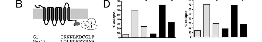
31 Results. Blocking Gα i or Gα q blocks SDF1 antirepellent activity. Semaphorin 3A (sema3a) is a powerful repellent for dorsal root ganglion (DRG) axons (Luo et al., 1993). Bath application of sema3a to DRG growth cones induces them to transition from a spread motile morphology to a collapsed shape without lamellae and few filopodia (Luo et al., 1993). This dramatic change in morphology can be used to measure the strength of repellent cues or to measure the relative susceptibility of growth cones to repellent cues. Using this assay, the repellent responses of DRG growth cones to sema3a, sympathetic growth cones to sema3c, or retinal growth cones to slit2, have all been shown to be greatly reduced in the presence of the chemokine SDF1 (Chalasani et al., 2003a). SDF1 by itself has little discernible effect on these growth cones, but when SDF1 is present, 5 to 8 times more repellent is required to induce half maximal growth cone collapse (Chalasani et al., 2003a). SDF1 acts through its seven transmembrane receptor, CXCR4, to mitigate the ability of repellents to collapse growth cones (Chalasani et al., 2003a). Paradoxically, although its signaling pathway in primary neurons is blocked by the Gα i/o blocker pertussis toxin, SDF1 appears to induce increased camp levels. Previous work from our laboratory showed that SDF1 s antirepellent effects can be blocked by the camp antagonist RpcAMPs or mimicked by the camp analogue SpcAMPs (Chalasani et al., 2003a). An SDF1-induced rise in camp has been observed in cultured primary chick retinal neurons (Xu et al., 2010). To better define the specific G-protein components through which SDF1 acts, dissociated DRGs were co-transfected with expression constructs for EYFP along with plasmids encoding short peptides that selectively block signaling through specific Gα containing G-proteins. These peptides are derived from the C termini of the Gα proteins they target and they selectively compete with the targeted Gα proteins for receptor binding (Gilchrist et al., 1999). Their selectivity and effectiveness has been demonstrated in several other systems, including zebrafish (Lin et al., 2005) and fly (Yao and Carlson, 2010). 21
32 DRG neurons transfected with EYFP alone collapse in response to sema3a (Fig. 1A; compare the first and second grey bars in Fig. 1C, D). The presence of SDF1 makes DRG growth cones resistant to sema3a (Fig. 1A; compare second and third grey bars in Fig.1C, D). For these experiments, transfected DRG cultures were stained for EYFP and only those growth cones that were brightly fluorescent were counted. In EYFP-only conditions, cultures show low background collapse. The percentage of collapsed growth cones increases in the presence of sema3a but increases significantly less when SDF1 is added along with sema3a. Co-transfection of expression plasmids encoding EYFP along with peptides targeting Gα q/11 (Fig. 1C, first panel) or Gα i1/2 (Fig. 1C, second panel) have no effect upon DRG growth cone collapse in the presence of sema3a alone (compare the middle grey bars to the middle black bars). However, the Gα q/11 or Gα i1/2 peptides do block SDF1 s ability to reduce collapse in response to sema3a (compare the third grey bars to the third black bars). This suggests that both Gα q and Gα i mediated G- protein coupled signaling are each required for SDF1 s antirepellent effect. A full-length dominant negative Gα i that has been shown to be effective in transfected CHO cells (Winitz et al., 1994) was tested for its ability to block SDF1-mediated signaling. This construct also blocked the SDF1 antirepellent effect, corroborating the finding with the Gα i based peptide (Fig. 1C, third panel). Co-transfection of EYFP with peptides targeting Gα s or Gα o1 had no effect on DRG responses to sema3a or to SDF1 (Fig. 1D). Because the effectiveness of the Gα s and Gα o1 peptides has been tested in other systems (Rasenick et al., 1994; Vanhauwe et al., 2002), the Gα i1/2 and Gα q peptides were effective, and all of the interfering peptides were expressed from identical expression plasmids, we conclude that Gα s and Gα o are unlikely to be required for antirepellent activity. Both Gβγ and Gα are necessary for SDF1 antirepellent activity. Because the short inhibitory peptides we used block the initial receptor mediated 22
33 dissociation and activation of G proteins, they cannot determine whether SDF1 signaling depends upon alpha or beta-gamma subunits to activate downstream targets. We used the C-terminal portion of GRK2, or GRK-CT, as a Gβγ scavenger that should prevent the complex from stimulating downstream targets (Fig. 2A). Ghahremani et al. (1999) showed that this protein fragment could block Gβγ-specific calcium release in LD2S cells and this construct has since been widely used. Coexpression of GRK-CT with EYFP does not increase background collapse or interfere with growth cones responses to sema3a (Fig. 2B). GRK-CT does, however, block SDF1-induced reduction in sema3amediated growth cone collapse, suggesting that SDF1 antirepellent activity requires Gβγinduced activation of downstream targets. We next set out to determine whether specific Gα subunits activate downstream targets in SDF1 mediated antirepellent signaling. RGS proteins act as GAPs for Gα subunits (Fig 2C). RGS2 specifically binds and inactivates Gα q, and RGS4 primarily binds Gα i but also binds Gα q to a lesser extent (Heximer, 2004; Huang et al., 1997). Coexpression of either RGS2 or RGS4 with EYFP does not affect background levels of collapse, nor does it interfere with sema3a induced collapse (Fig. 2D). Expression of either RGS2 or RGS4 does however, interfere with SDF1's ability to reduce collapse in response to sema3a (Fig. 2D). These results suggest that Gα q, and possibly Gα i, activate downstream targets in the SDF1 mediated antirepellent pathway. Constitutively active Gα subunits. We next asked whether overexpression of specific constitutively active Gα subunits can mimic SDF1 induced antirepellent activity. Gα subunits with a Q to L mutation in the nucleotide-binding region are unable to cleave GTP and are thereby made constitutively active (Graziano and Gilman, 1989; Hermouet et al., 1991; Kroll et al., 1992; Kalinec et al., 1992). Coexpression of QL Gα s with EYFP made DRG growth cones insensitive to sema3a in a manner similar to SDF1, and what is more, SDF1 induced little additional antirepellent effect (Fig. 3A). Similar results were obtained with constitutively active Gα i 23
34 (Fig. 3C) or QL Gα q (Fig. 3D). QL Gα o had no effect on growth cone responses to either sema3a or SDF1 (Fig. 3B). These results suggest that Gα s, Gα q, or Gα i each individually have the capability of initiating signaling events similar to those induced by SDF1, whether or not they participate in SDF1 signaling under normal circumstances. Inhibition of phospholipase C blocks SDF1 antirepellent activity. Because phospholipase C (PLC) is a classical effector of Gα q/11 -class G proteins (Taylor et al., 1991; Smrcka et al., 1991), and since our results show a requirement for Gα q/11 activity in SDF1 mediated antirepellent signaling, we hypothesized that PLC is required for SDF1 s antirepellent activity. The PLC inhibitor U73122 has no effect on background collapse or on growth cone responsiveness to sema3a (Fig. 4, grey bars). U73122 does, however, block SDF1's ability to reduce growth cone responses to sema3a (Fig. 4, black bars). Inhibition of phospholipase C blocks antirepellent effects induced by constitutive Gα q activity. We next tested whether Gα q activation can induce an anti-repellent response through the activation of PLC. As already demonstrated, sensory axons expressing a control Citrine construct collapse in response to sema3a and this collapse is largely mitigated in the presence of SDF1 (Figure 5, empty bars). In contrast, growth cones expressing the constitutively active QL Gα q are insensitive to sema3a. (Figure 5, grey bars). Significant sensitivity to sema3a is restored, however, when PLC is blocked. Growth cones expressing QL Gα s are also insensitive to sema3a, but this insensitivity is not reversed by blocking PLC (Figure 5, black bars). These findings are consistent with the idea that SDF1 induced antirepellent activity is mediated by Gα q activation of PLC, while constitutively active Gα s mediated antirepellent activity is not. As discussed in more detail below, one attractive explanation for these observations is that SDF1 induced activation of PLC indirectly induces elevated camp levels through a separate mechanism from the more traditional direct activation of adenylate cyclases by Gα s. 24
35 Discussion. Although G protein coupled receptors (GPCRs) are often pictured as acting through specific, dedicated G proteins, it is now known that a single GPCR can bind and activate G proteins from more than one G alpha class (see Hermans, 2003). PAR1, a thrombin receptor, can bind to Gα i/o, Gα q, or to Gα 12/13 (Gilchrist et al., 2001). β2-adrenergic receptors, when phosphorylated by PKA, switch affinities from Gα s to Gα i (Daaka et al., 1997). The class I metabotropic glutamate receptor mglur1 has been shown to bind Gα i/o, Gα q/11, and Gα s, at least in certain cell types (Selkirk et al., 2001). Wang et al. (2010) found that parathyroid hormone receptor 1 regulates different genes with different G proteins or combinations of G proteins, suggesting that individual G proteins might be required for some cell behaviors but not for others. These are just a few of the many examples of GPCRs coupling to multiple G proteins. Chemokine receptors as a class are generally thought to signal through Gα i/o -type G proteins to decrease camp, activate PI3K, and activate both p38 and ERK1/2 MAP Kinases (see Rubin, 2009; Teicher and Fricker, 2010, for reviews). PI3K activation leads to activation of a number of other kinases, including Akt. SDF1 signaling through the chemokine receptor CXCR4 is also associated with changes in transcription, usually mediated through MAPK or Akt, that contribute to cell survival (Chalasani et al., 2003b; Suzuki et al., 2001; Vlahakis et al., 2002; Zheng et al., 2008). However, several groups have found that CXCR4 signals through other classes of G proteins. Maghazachi (1997) reported that antibodies targeting Gα o or Gα q, but not Gα i, Gα s, or Gα z, could block SDF1-induced chemotaxis in natural killer cells. Soede et al. (2000) found that CXCR4- dependent migration of myeloid leukemia cells require either the combination of Gαi and Gα q or Gα q alone, depending on the destination tissue. Tan et al. (2006) showed that SDF1/CXCR4-induced migration of Jurkat T cells required both Gα 13, which activated Rho, and Gα i. These and other studies raised the possibility that SDF1/CXCR4 signaling in axon guidance might be more complex than that of the classic chemokine signaling 25
36 pathway. Previous work from our laboratory (Chalasani et al., 2003a) identified several components of SDF1/CXCR4 signaling in the antirepellent pathway, including a pertussis toxin-sensitive G protein, increased camp, and activation of PKA. In addition to the surprising apparent increase in camp levels observed in these previous studies, the effects of SDF1 on axonal responses to repellents were found to be independent of PI3K/Akt signaling and of MAPK. The findings in this study show that SDF1 s antirepellent activity can be blocked separately by Gα i, Gα q/11, or Gβγ specific competitive inhibitors. These data suggest that each is required for the normal function of the antirepellent pathway. However, we also found that overexpression of constitutively active forms of Gα i or of Gα q can mimic application of SDF1. This suggests that either one of these signaling components is capable of stimulating a common downstream element that is sufficient for activation of the pathway. These findings are consistent with the idea that SDF1 stimulates multiple G protein coupled pathways to a degree that is insufficient for any one of them alone to induce a physiological response, but in combination, their actions sum to a level above a threshold for activation to produce an anti-repellent response. We also found that overexpression of a constitutively active Gα s can mimic SDF1 even though a competitive inhibitor of Gα s does not block SDF1 mediated signaling. As Gα s is a canonical stimulator of adenylate cyclase activity and would be expected to elevate camp levels, this finding is consistent with the idea that the common element upon which Gα i, Gα q, and Gβγ all converge downstream from SDF1 activation of CXCR4 is elevated camp levels. Thus, our proposed model of the signaling pathway is that Gα i, Gα q, and their associated βγ subunits all cooperate to increase the local concentration of camp, leading to suppression of axonal repulsion (Fig. 6). The ability of Gα s to accomplish the same thing through a different route raises the possibility that a very wide range of GPCRs could influence axonal responses to repellents and axonal pathfinding. 26
37 Previous work has shown that SDF1 s antirepellent activity requires calmodulin and the calcium/calmodulin-stimulated cyclase ADCY8 (Chalasani et al., 2003a; Xu et al., 2010). Xu (2010) also showed by Förster resonance energy transfer (FRET) that SDF1 stimulates increased camp levels, and that this can be blocked by inhibition of calmodulin. Gα i and Gα q are not ordinarily associated with increases in camp, yet our results show that they are required components in the antirepellent signaling pathway. Gα q and Gβγ activity, through the activation of PLC, can produce diacylglycerol and inositol trisphosphate and thereby increase intracellular calcium (Guttridge et al., 1995). Thus, our present finding that both Gα q/11 and PLC are required for SDF1 antirepellent activity provides a connection between the G proteins activated by SDF1 and the calmodulin and calcium/calmodulin-stimulated cyclase that has been shown to increase camp downstream of SDF1. Our results are consistent with a signaling pathway (Fig. 6) in which multiple G protein components stimulate PLC activity that induces an increase in intracellular calcium levels and leads to the activation of calmodulin. Calmodulin, in turn, activates calcium/calmodulin-stimulated adenylate cyclases, such as ADCY8, and thereby increases camp. Some of the important questions that remain include how elevated camp levels decrease growth cone responses to repellents and the degree to which this modulation of repellent effectiveness is important in axonal pathfinding in vivo. Both SDF1/CXCR4 activity and activity of the calmodulin-activated adenylate cyclases have a strong influence on axonal responses to the repellent slit in vivo (Xu et al., 2010). Our findings in this study suggest that activation of a wide range of GPCRs that signal through Gα i, Gα q, or Gα s could potentially participate in axon guidance decisions. 27
38 Figure 1. Competitive inhibitors of Gα i or Gα q/11, but not Gα s or Gα o, block SDF1- mediated antirepellent activity. (A) Growth cones of dissociated DRGs transfected with EYFP or EYFP + Gα q/11 inhibitory peptide have motile lamellae and filopodia. (B) Specific inhibitory Gα peptides (medium grey) bind selected GPCRs and prevent their association with functional G proteins containing the same Gα peptide sequence. (C,D) Dissociated DRGs were transfected with EYFP-only (grey bars) or with EYFP and an experimental construct (black bars). After 24h in culture, cells were treated for 30 with sema3a or with sema3a + SDF1. (C) The SDF1 antirepellent response is blocked by a by peptides targeting Gα i or Gα q/11, and also by a full-length dominant-negative Gα i. (D) The SDF1 antirepellent response is not affected by peptides targeting Gα s or Gα o. *, p < 0.001; **, p < Figure 2. Scavengers of Gβγ, Gα i, or Gα q subunits block SDF1 antirepellent activity. (A) GRK-CT sequesters βγ subunits while leaving α free to activate downstream effectors. (B) Transfection of dissociated DRGs with GRK-CT blocks the SDF1 antirepellent effect but does not alter background collapse or response to sema3a. (C) RGS proteins sequester specific α subunits and hasten their inactivation while leaving βγ subunits free to activate downstream effectors. (D) Transfection of dissociated DRGs with either RGS2, a α q specific GAP, or RGS4, an α i and to a lesser extent α q specific GAP, block SDF1 antirepellent activity without affecting background collapse or response to sema3a. *, p < 0.05; **, p < 0.005; ***, p < Figure 3. Constitutively active Gα q, Gα i, or Gα s mimics SDF1 s antirepellent effect. (A) Transfection of QL Gα s into DRGs makes them unresponsive to sema3a. (B) Transfection of QL Gα o into DRGs has no effect on their responses to sema3a or SDF1. (C) Transfection of DRGs with QL Gα i or with (D) QL Gα q makes DRGs unresponsive to sema3a. *, p < 0.001, **, p <
39 Figure 4. Inhibiting PLC blocks SDF1 antirepellent activity. DRG explants were treated with 20nM PLC inhibitor U73122 (black bars). U73122 does not alter background collapse or DRG responsiveness to sema3a, but does block the antirepellent effect of SDF1. **, p < Figure 5. Inhibiting PLC blocks antirepellent activity induced by expression of a constitutively active Gα q. DRGs were transfected with expression plasmids for Citrine (control, empty bars), Citrine and QL Gα q (grey bars), or Citrine and QL Gα s (black bars). Expression of QL Gα q makes growth cones insensitive to sema3a unless the PLC blocker U73122 (20nM) is also present. Growth cones expressing QL Gα s are insensitive to sema3a in both the absence and the presence of U *, p< Figure 6. A model for antirepellent signaling. We identify roles for Gα i, Gα q, and Gβγ, as well as PLC, in the antirepellent response to SDF1. Previous work has shown requirements for calmodulin, ADCY8, camp, and PKA, along with inhibition of Rho and ROCK. In our model, CXCR4 activates Gα i and Gα q, and they and their associated Gβγ subunits cooperate to activate PLC. PLC, through generation of diacylglycerol and inositol trisphosphate (Taylor et al., 1991), increases calcium levels. Increased calcium activates calmodulin, which in turn activates ADCY8 and thereby increases camp. Increased camp activates PKA, which phosphorylates MAPK1/2, leading to its activation and function in cell survival. PKA also phosphorylates Rho, which is thereby inactivated. This inactivation and the subsequent inactivation of ROCK are required for the antirepellent response to SDF1. 29


40 30

41 31


42 32
43 33
44 TABLE 1: RAW DATA, FIGS. 1-4 condition collapsed not collapsed condition collapsed not collapsed GTGi Gq/11 mini EYFP ctrl 5 31 EYFP ctrl 5 31 sema sema sema + SDF sema + SDF GTGi ctrl Gq/11 mini ctrl 4 23 sema sema sema + SDF sema + SDF EYFP ctrl 2 28 EYFP ctrl 3 46 sema 11 5 sema sema + SDF sema + SDF GTGi ctrl 1 19 Gq/11 mini ctrl 8 41 sema 19 8 sema sema + SDF sema + SDF EYFP ctrl 2 28 citrine ctrl sema 11 5 sema sema + SDF sema + SDF GTGi ctrl 1 19 Gq/11 mini ctrl sema 19 8 sema sema + SDF sema + SDF Go1 mini Gi mini EYFP ctrl 5 31 EYFP ctrl 5 72 sema sema sema + SDF sema + SDF Go1 mini ctrl Gi mini ctrl sema 20 9 sema sema + SDF sema + SDF EYFP ctrl 5 18 EYFP ctrl 4 61 sema sema sema sema + SDF SDF1 Go1 mini ctrl 5 22 Gi mini ctrl 4 27 sema sema 16 8 sema + SDF sema + SDF citrine ctrl 3 33 EYFP ctrl sema sema sema sema + SDF SDF1 Go1 mini ctrl 5 42 Gi mini ctrl sema sema sema + SDF sema + SDF Gs mini GRK-CT EYFP ctrl 5 72 EYFP ctrl 2 28 sema sema
45 sema + SDF sema + SDF Gs mini ctrl 8 59 GRK-CT ctrl 5 36 sema sema sema + SDF sema + SDF EYFP ctrl 4 54 EYFP ctrl 5 18 sema sema sema sema + SDF SDF1 Gs mini ctrl 3 22 GRK-CT ctrl 7 27 sema sema sema + SDF sema + SDF citrine ctrl citrine ctrl sema sema sema sema + SDF SDF1 Gs mini ctrl GRK-CT ctrl sema sema sema + SDF sema + SDF condition collapsed not collapsed condition collapsed not collapsed RGS2 QLO EYFP ctrl 6 52 citrine ctrl sema sema sema + SDF sema + SDF RGS2 ctrl 3 32 QLO ctrl sema 30 6 sema sema + SDF sema + SDF EYFP ctrl 6 45 citrine ctrl sema sema sema sema + SDF SDF1 RGS2 ctrl QLO ctrl sema sema sema + SDF sema + SDF EYFP ctrl 4 28 EYFP ctrl sema sema sema sema + SDF SDF1 RGS2 ctrl QLO ctrl sema sema sema + SDF sema + SDF RGS4 QLQ EYFP ctrl 4 28 EYFP ctrl 3 21 sema sema sema + SDF sema + SDF RGS4 ctrl QLQ ctrl 6 49 sema sema sema + SDF sema + SDF EYFP ctrl 6 52 EYFP ctrl sema sema
46 sema + SDF sema + SDF RGS4 ctrl QLQ ctrl sema sema sema + SDF sema + SDF EYFP ctrl 8 68 EYFP ctrl 5 22 sema sema sema sema + SDF SDF1 RGS4 ctrl QLQ ctrl 9 48 sema sema sema + SDF sema + SDF QLS QLI EYFP ctrl EYFP ctrl 4 28 sema sema sema + SDF sema + SDF QLS ctrl 6 25 QLI ctrl 9 41 sema sema sema + SDF sema + SDF EYFP ctrl EYFP ctrl 4 58 sema sema sema sema + SDF SDF1 QLS ctrl QLI ctrl 2 35 sema sema sema + SDF sema + SDF citrine ctrl 3 33 EYFP ctrl 3 21 sema sema sema sema + SDF SDF1 QLS ctrl 8 55 QLI ctrl 6 30 sema sema sema + SDF1 - - sema + SDF
47 Chapter 3: Involvement of an ADAM metalloprotease in SDF1 antirepellent activity Our standing model for antirepellent signaling (see Chapter 2, Fig. 5) has been that CXCR4 or other modulatory receptors, through the phosphorylation of Rho, block repellent-induced alterations in the growth cone cytoskeleton. We have identified several steps in the antirepellent pathway upstream of PKA and the inactivation of Rho (chapter 2; Chalasani et al., 2003). However, this is not the only way that antirepellent cues might block repellent activity. For example, work from the Tessier-Lavigne lab (Keino-Masu et al., 1996) showed that a metalloprotease inhibitor could increase both axon outgrowth in response to netrin and expression of the attractive netrin receptor DCC, suggesting that guidance receptors can be regulated by inhibitory shedding. Work in other systems has shown that for ADAM (A Disintegrin And Metalloprotease) proteases are required for normal axon guidance (Schimmelpfeng et al., 2001; Chen et al., 2007). Epithelial growth factor receptor (EGFR) can be activated by ligands other than EGF, such as HB-EGF, which is activated as a ligand by cleavage from the same cell membrane as the receptor. GPCRs can activate ADAMs to produce these EGFR ligands through a shedding mechanism (Asakura et al., 2002; Schafer et al., 2004; Tanida et al., 2004; Mifune et al., 2005). We were therefore interested in the possibility that the inhibitory shedding of repellent receptors might be another mechanism for antirepulsion. We focused on ADAM10 and ADAM17 because they are required for several aspects of neural development, including development of the optic cup and otic pit (Hartmann et al., 2002), formation of the neocortex (Jorissen et al., 2010), and synaptic plasticity (Malinverno et al., 2010), in addition to the guidance roles referenced above. 37
48 The ADAM proteases are named for their unique pair of extracellular protein-interaction domains. ADAMs include a pro domain, which is cleaved off during posttranslational processing, the metalloprotease and disintegrin domains which give their names, a cysteine-rich domain on their extracellular sides, a transmembrane domain, and a cytoplasmic tail (Fig. 1; see Edwards et al. 2008). ADAM17 was originally identified as TACE, TNFα converting enzyme, but it has many other substrates as well. These other substrates include Notch, APP, L1-CAM, N-CAM and neuregulin (see Edwards et al., 2008). ADAM10 is also called α-secretase for its role in processing APP and is known in Drosophila as Kuzbanian (kuz). Aside from APP, ADAM10 has been reported to cleave L1-CAM, EGFR ligands, Notch, and several cadherins, among others (see Edwards et al., 2008). ADAM10/Kuzbanian was initially identified for its involvement in neural fate specification (Rooke et al., 1996) and for facilitating axon extension (Fambrough et al., 1996; Pan and Rubin, 1997). Soon thereafter, ADAM10/Kuzbanian was linked with slit/robo signaling by the finding that kuz, slit, and robo mutants genetically interact in the fly embryo midline, with combinations of mutations yielding more severe phenotypes (Schimmelpfeng et al., 2001). A requirement for proteolytic function in axon guidance is also well-established in the ephrin/eph system. Ephrins are most commonly considered as contact repellents. Both they and their receptors, the Ephs, are attached to the cell membrane, so at least one 38
49 signaling partner must be cleaved for the growth cone to withdraw in response to repulsive signaling. Hattori et al. (2000) first demonstrated this, showing that ADAM10 cleaves ephrina2 when clustered by EphA3-Fc and that an uncleavable ephrina2 slowed axon retraction. Litterst et al. (2007) showed that Eph could be cleaved in two ways. Ligand binding induced a presumably activating cleavage, yielding increased internalization of C-terminal fragments, and this was not sensitive to metalloprotease inhibitors. Calcium and NMDA activation caused a presumably inhibitory cleavage, as shown by shedding of Eph N-termini. This inhibitory cleavage was sensitive to metalloprotease inhibitors but not to the gamma-secretase inhibitor that blocked the activating cleavage. This demonstrates that proteolysis of an individual repellent receptor can either increase or decrease repellent signaling, depending on the protease and cleavage site. Here, we show that SDF1 antirepellent activity in primary chick neurons can be blocked by pharmacological inhibition of metalloproteases or by a dominant negative ADAM10, but not by a dominant negative ADAM17. We also show that shedding of neuropilin-1 overexpressed in 293 cells is enhanced by overexpression of ADAM10 and reduced by expression of a dominant negative ADAM10. Pharmacological inhibition of metalloproteases does not block the SDF1-mimicking effect of a camp analogue, suggesting that ADAM10 activation is not downstream of increased camp. 39
50 Results: We wanted to determine whether SDF1 antirepellent signaling includes an inhibitory cleavage of repellent receptors. To address this question, we began by applying the metalloprotease inhibitor TAPI-2 to explant collapse assays. We saw no effect on background collapse or response to sema3a (Fig. 2, compare first green bar to first black bar and second green bar to second black bar), but TAPI-2 does block the reduction in collapse produced by SDF1 (Fig. 2, compare third green bar to third black bar; **, p < 0.005). TAPI-2, though not a universal metalloprotease inhibitor, blocks several proteases, including ADAM10, ADAM17, and a small number of matrix metalloproteases. Therefore, we wanted to identify which of these proteases might underlie TAPI-2 s SDF1-blocking function. We took advantage of previously published dominant-negative constructs, composed of most of ADAM10 or ADAM17 that are missing their proteolytic domains and/or pro domain (Fig. 1). The effectiveness of dnadam10 has been demonstrated in the Eph/ephrin system in NIH 3T3 cells, where it blocked the cleavage of ephrina2. In the same set of experiments, cleavage of ephrina2 was enhanced by overexpression of full-length ADAM10 (Hattori et al., 2000). The dnadam17 was originally tested in COS-7 cells and HEK293 cells, where it blocked the shedding of TNF-α and FasL (Itai et al., 2001). In collapse assays on dissociated DRGs, neither dn- ADAM construct altered background collapse or response to sema3a (Fig. 3A,B, compare first green bar to first black bar and second green bar to second black bar). 40
51 dnadam10 blocked the influence of SDF1 (Fig. 3A, compare third green bar to third black bar; **, p < 0.001), but dnadam17 did not (Fig. 3B, compare third green bar to third black bar). Expression of the dn-adam constructs was confirmed by staining for the HA tags on the constructs. Recent work in the Drosophila slit-robo system (Coleman et al., 2010) found a prorepulsion role for ADAM10. Since this differed from our finding that blocking ADAM10 had no effect on the repellent response to sema3a, we investigated the effects of dnadam10 on the responses of dissociated chick retina to slit2 and SDF1. Consistent with our DRG results, we found that dnadam10 did not disrupt repellent signaling but did block the antirepellent response to SDF1 (Fig. 3C, compare second green bar to second black bar and third green bar to third black bar; ***, p < ). The ADAM proteases are found at the cell surface, are known to cleave receptors in other systems (EGF, Eph/ephrin), and may have a role in antirepellent signaling. We therefore asked whether ADAM10 could cleave neuropilin-1 (NP1), the ligand-binding component of the sema3a receptor complex. HEK293 cells were transfected with NP1 and ADAM10, dnadam10, or β-galactosidase (as a transfection control). I collected supernatants and lysates from these cells and ran matched Western blots, stained for HAtag (ADAM10 or dnadam10) or myc (N-terminus of NP1), or for tubulin as a celldensity control. Some NP1 N-terminus appeared in the supernatant even in the control condition, in which cells were transfected with NP1 and β-gal (Fig. 4, lane 1), suggesting the existence of endogenous protease activity in the HEK293 cells. Cotransfection of 41
52 NP1 with full-length ADAM10, however, produced a marked increase in cleaved NP1 (Fig. 4, lane 2). Cotransfection of dnadam10 with NP1 blocked the release of cleaved NP1 into the medium (Fig. 4, lane 3). These findings show that ADAM10 can cleave NP1 and that thednadam10 blocks that cleavage. If ADAM10 functions in the SDF1/sema3A interaction the way it works in ephrin/eph signaling (Litterst et al., 2007), then SDF1 may activate ADAM10 to cause a sema3a-inhibitory shedding event. After having found a potential mechanism for ADAM10 function in SDF1 antirepellent activity, we wanted to know where in the SDF1 pathway ADAM10 activation might fall. To that end, we performed explant collapse assays with TAPI-2 and SpcAMPS, a camp analogue that mimics SDF1 s antirepellent effect (Chalasani et al., 2003). We reasoned that, if ADAM10 activation is required downstream of increased camp, TAPI-2 should block the antirepellent effect of SpcAMPS. SpcAMPS reduced collapse with or without TAPI-2 (Fig. 5A, compare third green bar and third black bar). We therefore conclude that ADAM10 activity is not downstream of the SDF1-induced increase in camp. Consistent with this result, preliminary experiments (Fig. 5B, compare third green bar and third black bar) with the ROCK inhibitor Y-27632, which was previously shown to mimic SDF1 s antirepellent effect (Chalasani et al. 2003), showed that TAPI-2 could not block this even further-downstream antirepellent signal. Because ADAM10 activity is not downstream of camp, it must either be upstream of camp or in a parallel pathway. Since other work has shown that ADAM10 can be activated by increased intracellular calcium (Litterst et al., 2007), we hypothesize (Fig. 6) 42
53 that the calcium produced by SDF1-induced PLC activation not only activates calmodulin and ADCY8 but also activates ADAM10. Discussion. There are several possible mechanisms by which antirepellent and repellent signaling pathways might interact. These include opposing effects on the inactivation and activation of small G proteins such as Rho, opposing effects on the growth cone cytoskeleton, or interference with the repellent receptor or receptor complex. The latter possibility can be divided further, into alteration of expression, membrane insertion, membrane localization, endocytosis, and shedding of repellent receptors. Shedding of the receptor may be a more elegant system, or at least an elegant supporting mechanism, than several of the other possibilities, since it would prevent the activation of second messengers and thus require alteration of many fewer proteins. Here, we show that protease activity is required not for the repellent response to sema3a or slit2 but for the reduction in that response by co-application of SDF1. We further demonstrate that the protease requred for SDF1 antirepellent activity is ADAM10 and that ADAM10 can cleave NP1, the component of the sema3a receptor complex which confers ligand specificity. Since ADAM10 has previously been shown to cleave L1 (Maretzky et al., 2005), another component of the sema3a receptor complex, it seems 43
54 likely that ADAM10 s role in SDF1 activity could be to cleave repellent receptors, thereby preventing their activation. Though Coleman et al. (2010) found that Kuz/ADAM10 is required for Robo signaling in the fly, we found that a dominant-negative ADAM10 had no effect on chick RGCs responsiveness to hslit2. Reasons for this difference may include divergences between fly and human Slit or fly and chick Robo, or different signaling machinery in fly embryo midline and chick RGCs. Though previous work had shown requirements for ADAM10 in axon guidance, this is the first association of ADAM10 with a modulatory guidance cue. ADAM10 activation and repellent receptor shedding also comprise a novel mechanism for antirepellent signaling. As the study of signaling in guidance modulation shifts toward in vivo work, the existence of this additional pathway may provide useful insights. Materials and Methods. Plasmids and reagents. TAPI-2 (Peptides International) was used at 100nM. Full-length ADAM10-HA and DNADAM10-HA were provided by J. Flanagan (Harvard Medical School). DNADAM17-HA was provided by S. Nagata (Osaka University). SpcAMPS (Sigma A166) was used at 20µM. Y (Sigma Y0503) was used at 10µM. Antimyc (9E10, Cell Center), anti-ha-tag (MMS-101R, Covance), and anti-tubulin (YL 1/2) 44
55 were each used at 1:1000. Appropriate HRP-tagged secondary antibodies (Jackson Immuno) were used at 1:2000. Cell culture and collapse assays. TAPI-2 experiments were performed on explants. Dominant-negative ADAMs were transfected as described in Chapter 2. Cell culture and assay protocols were as described in Chapter 2. Shedding assay. HEK293T cells plated on polylysine were transfected with the listed plasmids using Lipofectamine Total DNA per transfection was 8µg, per manufacturer s guidelines. Samples of medium (supernatant) or total cell lysate were run on matched 10% SDS-PAGE gels. Blots were stained as noted, developed with ECL- Plus reagents (GE Life Sciences) and exposed to film before being scanned and then analyzed with ImageJ (NIH). 45
56 Figure 1. Domain structures of ADAM10, ADAM17, DN-ADAM10, and DN- ADAM17. ADAM10 and ADAM17 are closely related members of the ADAM family and share the same domain structure. Each contains a pro domain which is cleaved off during posttranslational processing, a protease domain, a cysteine-rich domain, a disintegrin domain, and a transmembrane domain and cytoplasmic tail. DN-ADAM10 lacks the pro domain and protease domain; DN-ADAM17 lacks the protease domain. Figure 2. The metalloprotease inhibitor TAPI-2 blocks SDF1 antirepellent activity. Explants from E7 chick DRGs were treated for 30 with sema3a, SDF1, and TAPI-2, as noted. TAPI-2 (black bars) blocks the SDF1-induced reduction in collapse. **, p < Figure 3. SDF1 antirepellent activity is not blocked by DN-ADAM17 but is blocked by DN-ADAM10 in either DRG or RGC. (A,B) Dissociated E7 chick DRGs were transfected with EYFP, DN-ADAM10, and DN-ADAM17 as noted and then treated with sema3a, SDF1, or sema3a + SDF1. (A) DN-ADAM10 does not block sema3a-induced collapse but does block SDF1 antirepellent activity. **, p < (B) DN-ADAM17 blocks neither sema3a-induced collapse nor SDF1 s antirepellent effect. (C) Dissociated E6 chick RGCs were transfected with EYFP or EYFP + DN-ADAM10 as noted and then treated with slit2, SDF1, or slit2 + SDF1. DN-ADAM10 does not block slit2-induced collapse but does block SDF1 antirepellent activity. ***, p < Figure 4. ADAM10 can cleave NP1. HEK293 cells were transfected with NP1 and β- galactosidase, ADAM10, or DN-ADAM10. Samples from total cell lysate or conditioned medium were run on matched SDS-PAGE gels and blotted for myc (NP1), HA-tag (ADAM10, DN-ADAM10), or tubulin. Overexpression of ADAM10 increases shedding of NP1, whereas DN-ADAM10 reduces shedding of NP1 to below baseline. Figure 5. The metalloprotease inhibitor TAPI-2 does not block the SDF1 mimicking effects of a camp analogue or a ROCK inhibitor. Explants from E7 chick DRGs were treated for 30 with sema3a, TAPI-2, and Sp-cAMPs or the ROCK inhibitor Y-27632, as noted. (A) Sp-cAMPs (third green bar and third black bar) reduces the effect of sema3a in both the absence and presence of TAPI-2. (B) Y (third green bar and third black bar) reduces the effect of sema3a in both the absence and presence of TAPI-2. Figure 6. A model for ADAM10 involvement in antirepellent signaling. In our model, CXCR4 activates Gα i and Gα q, and they and their associated Gβγ subunits cooperate to activate PLC. PLC, through the production of diacylglycerol and inositol trisphosphate, increases calcium levels. Increased calcium activates ADAM10, which blocks repulsion through shedding of repellent receptors, and also activates calmodulin. Calmodulin, in turn, activates ADCY8 and thereby increases camp. Increased camp activates PKA, which phosphorylates MAPK1/2, leading to cell survival. PKA also phosphorylates Rho, which is thereby inactivated. This inactivation and the subsequent inactivation of ROCK are required for the antirepellent response to SDF1. 46


57 47


58 48


59 49
Axon Guidance. Multiple decision points along a growing axon s trajectory Different types of axon guidance cues:
 Axon Guidance Multiple decision points along a growing axon s trajectory Different types of axon guidance cues: Contact mediated - requires direct contact by growth cone Long range - growth cone responds
Axon guidance I. Paul Garrity March 15, /9.013
 Axon guidance I Paul Garrity March 15, 2004 7.68/9.013 Neuronal Wiring: Functional Framework of the Nervous System Stretch reflex circuit Early theories of axonogenesis Schwann: many neurons link to form
Cellular Neurobiology BIPN 140 Fall 2016 Problem Set #8
 Cellular Neurobiology BIPN 140 Fall 2016 Problem Set #8 1. Inductive signaling is a hallmark of vertebrate and mammalian development. In early neural development, there are multiple signaling pathways
5- Semaphorin-Plexin-Neuropilin
 5- Semaphorin-Plexin-Neuropilin 1 SEMAPHORINS-PLEXINS-NEUROPILINS ligands receptors co-receptors semaphorins and their receptors are known signals for: -axon guidance -cell migration -morphogenesis -immune
Reading. Lecture VI. Making Connections 9/17/12. Bio 3411 Lecture VI. Making Connections. Bio 3411 Monday September 17, 2012
 Lecture VI. Making Connections Bio 3411 Monday September 17, 2012!! 1! Reading NEUROSCIENCE: 5 th ed, pp!507?536! 4 th ed, pp 577-609 Bentley, D., & Caudy, M. (1983). Nature, 304(5921), 62-65. Dickson,
THE PROBLEMS OF DEVELOPMENT. Cell differentiation. Cell determination
 We emphasize these points from Kandel in Bi/CNS 150 Bi/CNS/NB 150: Neuroscience Read Lecture Lecture Friday, October 2, 2015 Development 1: pp 5-10 Introduction Brains evolved All higher animals have brains
Heterotrimeric G proteins and the role of lipids in signaling. John Sondek, Ph.D. Depts. of Pharmacology and Biochemistry & Biophyscis
 Heterotrimeric G proteins and the role of lipids in signaling John Sondek, Ph.D. Depts. of Pharmacology and Biochemistry & Biophyscis The GTPase cycle molecular switch A GTPases is NOT a kinase Two major
Signal Transduction Mechanisms in Commissural Axon Guidance: The Role of Intracellular Tyrosine Kinases in Netrin-Dcc/Frazzled Axon Attraction
 University of Pennsylvania ScholarlyCommons Publicly Accessible Penn Dissertations 1-1-2012 Signal Transduction Mechanisms in Commissural Axon Guidance: The Role of Intracellular Tyrosine Kinases in Netrin-Dcc/Frazzled
7.013 Problem Set
 7.013 Problem Set 5-2013 Question 1 During a summer hike you suddenly spot a huge grizzly bear. This emergency situation triggers a fight or flight response through a signaling pathway as shown below.
MCDB 4777/5777 Molecular Neurobiology Lecture 29 Neural Development- In the beginning
 MCDB 4777/5777 Molecular Neurobiology Lecture 29 Neural Development- In the beginning Learning Goals for Lecture 29 4.1 Describe the contributions of early developmental events in the embryo to the formation
Neural development its all connected
 Neural development its all connected How do you build a complex nervous system? How do you build a complex nervous system? 1. Learn how tissue is instructed to become nervous system. Neural induction 2.
Axon Guidance at the Midline: From Mutants to Mechanisms
 Critical Reviews in Biochemistry and Molecular Biology, 39:319 341, 2004 Copyright c Taylor & Francis Inc. ISSN: 1040-9238print / 1549-7798online DOI: 10.1080/10409230490906797 Axon Guidance at the Midline:
Hierarchical Organization of Guidance Receptors: Silencing of Netrin Attraction by Slit Through a Robo/DCC Receptor Complex
 Hierarchical Organization of Guidance Receptors: Silencing of Netrin Attraction by Slit Through a Robo/DCC Receptor Complex Elke Stein and Marc Tessier-Lavigne* Axonal growth cones that cross the nervous
Conclusions. The experimental studies presented in this thesis provide the first molecular insights
 C h a p t e r 5 Conclusions 5.1 Summary The experimental studies presented in this thesis provide the first molecular insights into the cellular processes of assembly, and aggregation of neural crest and
C. elegans L1 cell adhesion molecule functions in axon guidance
 C. elegans L1 cell adhesion molecule functions in axon guidance Biorad Lihsia Chen Dept. of Genetics, Cell Biology & Development Developmental Biology Center C. elegans embryogenesis Goldstein lab, UNC-Chapel
ADAM FAMILY. ephrin A INTERAZIONE. Eph ADESIONE? PROTEOLISI ENDOCITOSI B A RISULTATO REPULSIONE. reverse. forward
 ADAM FAMILY - a family of membrane-anchored metalloproteases that are known as A Disintegrin And Metalloprotease proteins and are key components in protein ectodomain shedding Eph A INTERAZIONE B ephrin
Zool 3200: Cell Biology Exam 5 4/27/15
 Name: Trask Zool 3200: Cell Biology Exam 5 4/27/15 Answer each of the following short answer questions in the space provided, giving explanations when asked to do so. Circle the correct answer or answers
Computational Model Of Axon Guidance
 Mestrado em Engenharia Informática Dissertação Relatório Final arxiv:1508.01537v1 [q-bio.nc] 6 Aug 2015 Computational Model Of Axon Guidance Rui André Ponte Costa racosta@student.dei.uc.pt Orientadores:
Cell Death & Trophic Factors II. Steven McLoon Department of Neuroscience University of Minnesota
 Cell Death & Trophic Factors II Steven McLoon Department of Neuroscience University of Minnesota 1 Remember? Neurotrophins are cell survival factors that neurons get from their target cells! There is a
Visual pigments. Neuroscience, Biochemistry Dr. Mamoun Ahram Third year, 2019
 Visual pigments Neuroscience, Biochemistry Dr. Mamoun Ahram Third year, 2019 References Webvision: The Organization of the Retina and Visual System (http://www.ncbi.nlm.nih.gov/books/nbk11522/#a 127) The
The Molecular Biology of Axon Guidance
 Thursday, January 25, 2001 Science -- Tessier-Lavigne and Goodman 274 (5290): 1123 Page: 1 Institution: HARVARD UNIVERSITY Sign In as Individual FAQ The Molecular Biology of Axon Guidance Marc Tessier-Lavigne
MOLECULAR DRUG TARGETS
 MOLECULAR DRUG TARGETS LEARNING OUTCOMES At the end of this session student shall be able to: List different types of druggable targets Describe forces involved in drug-receptor interactions Describe theories
1. What are the three general areas of the developing vertebrate limb? 2. What embryonic regions contribute to the developing limb bud?
 Study Questions - Lecture 17 & 18 1. What are the three general areas of the developing vertebrate limb? The three general areas of the developing vertebrate limb are the proximal stylopod, zeugopod, and
MIT 9.14 Class The growth of the long extensions of neurons and related topics
 9.14 - Brain Structure and its Origins Spring 2005 Massachusetts Institute of Technology Instructor: Professor Gerald Schneider A sketch of the central nervous system and its origins G. E. Schneider 2005
Name KEY. Biology Developmental Biology Winter Quarter Midterm 3 KEY
 Name KEY 100 Total Points Open Book Biology 411 - Developmental Biology Winter Quarter 2009 Midterm 3 KEY All of the 25 multi-choice questions are single-answer. Choose the best answer. (4 pts each) Place
Reference: Forscher, P., Kaczmarek, L.K., Buchanan, J. and Smith, S.J. (1987) Cyclic AMP induces changes in distribution and transport of organelles
 Cyclic AMP induces changes in distribution and transport of organelles") Reference: Forscher, P., Kaczmarek, L.K., Buchanan, J. and Smith, S.J. (1987) Cyclic AMP induces changes in distribution and transport of organelles within growth cones of Aplysia bag cell neurons. J.
Reference: Forscher, P., Kaczmarek, L.K., Buchanan, J. and Smith, S.J. (1987) Cyclic AMP induces changes in distribution and transport of organelles within growth cones of Aplysia bag cell neurons. J.
Cell-Cell Communication in Development
 Biology 4361 - Developmental Biology Cell-Cell Communication in Development October 2, 2007 Cell-Cell Communication - Topics Induction and competence Paracrine factors inducer molecules Signal transduction
Signal Transduction. Dr. Chaidir, Apt
 Signal Transduction Dr. Chaidir, Apt Background Complex unicellular organisms existed on Earth for approximately 2.5 billion years before the first multicellular organisms appeared.this long period for
STEIN IN-TERM EXAM -- BIOLOGY FEBRUARY 12, PAGE 1 of 7
 STEIN IN-TERM EXAM -- BIOLOGY 3058 -- FEBRUARY 12, 2009 -- PAGE 1 of 7 There are 25 questions in this Biology 3058 exam. All questions are "A, B, C, D, E, F, G, H" questions worth one point each. There
During evolution, organisms have come to perform more
 Vol 436 14 July 2005 doi:10.1038/nature03875 Common mechanisms of nerve and blood vessel wiring Peter Carmeliet 1 & Marc Tessier-Lavigne 2 REVIEWS Blood vessels and nerve fibres course throughout the body
Neurite initiation. Neurite formation begins with a bud that sprouts from the cell body. One or several neurites can sprout at a time.
 Neurite initiation. Neuronal maturation initiation f-actin polarization and maturation tubulin stage 1: "spherical" neuron stage 2: neurons extend several neurites stage 3: one neurite accelerates its
10/2/2015. Chapter 4. Determination and Differentiation. Neuroanatomical Diversity
 Chapter 4 Determination and Differentiation Neuroanatomical Diversity 1 Neurochemical diversity: another important aspect of neuronal fate Neurotransmitters and their receptors Excitatory Glutamate Acetylcholine
Visual pigments. Neuroscience, Biochemistry Dr. Mamoun Ahram Third year, 2015
 Visual pigments Neuroscience, Biochemistry Dr. Mamoun Ahram Third year, 2015 References Photoreceptors and visual pigments Webvision: The Organization of the Retina and Visual System (http://www.ncbi.nlm.nih.gov/books/nbk11522/#a127)
Biol403 - Receptor Serine/Threonine Kinases
 Biol403 - Receptor Serine/Threonine Kinases The TGFβ (transforming growth factorβ) family of growth factors TGFβ1 was first identified as a transforming factor; however, it is a member of a family of structurally
Invariant Sema5A inhibition serves an ensheathing function during optic nerve development
 Development 130, 775-784 2003 The Company of Biologists Ltd doi:10.1242/dev.00299 775 Invariant Sema5A inhibition serves an ensheathing function during optic nerve development Stephen F. Oster, MacDara
Cell Cell Communication in Development
 Biology 4361 Developmental Biology Cell Cell Communication in Development June 25, 2008 Cell Cell Communication Concepts Cells in developing organisms develop in the context of their environment, including
Cell Migration I: Neural Crest Cell Migration. Steven McLoon Department of Neuroscience University of Minnesota
 Cell Migration I: Neural Crest Cell Migration Steven McLoon Department of Neuroscience University of Minnesota 1 Types of Cell Movement passive: active: cell sheets flow cilia or flagella ameboid adhesion
Introduction Principles of Signaling and Organization p. 3 Signaling in Simple Neuronal Circuits p. 4 Organization of the Retina p.
 Introduction Principles of Signaling and Organization p. 3 Signaling in Simple Neuronal Circuits p. 4 Organization of the Retina p. 5 Signaling in Nerve Cells p. 9 Cellular and Molecular Biology of Neurons
Regulation and signaling. Overview. Control of gene expression. Cells need to regulate the amounts of different proteins they express, depending on
 Regulation and signaling Overview Cells need to regulate the amounts of different proteins they express, depending on cell development (skin vs liver cell) cell stage environmental conditions (food, temperature,
Morphogens as conserved axon guidance cues Yimin Zou 1 and Anna I Lyuksyutova 2
 Morphogens as conserved axon guidance cues Yimin Zou 1 and Anna I Lyuksyutova 2 Morphogen family proteins are now widely appreciated as axon guidance cues. Because their roles as morphogens are highly
Cell-Cell Communication in Development
 Biology 4361 - Developmental Biology Cell-Cell Communication in Development June 23, 2009 Concepts Cell-Cell Communication Cells develop in the context of their environment, including: - their immediate
Pioneer midbrain longitudinal axons navigate using a balance of Netrin attraction and Slit repulsion
 Pioneer midbrain longitudinal axons navigate using a balance of Netrin attraction and Slit repulsion Kim et al. Kim et al. Neural Development 2014, 9:17 Kim et al. Neural Development 2014, 9:17 RESEARCH
Bio 127 Section I Introduction to Developmental Biology. Cell Cell Communication in Development. Developmental Activities Coordinated in this Way
 Bio 127 Section I Introduction to Developmental Biology Cell Cell Communication in Development Gilbert 9e Chapter 3 It has to be EXTREMELY well coordinated for the single celled fertilized ovum to develop
The N-terminal Leucine-Rich Regions in Slit Are Sufficient To Repel Olfactory Bulb Axons and Subventricular Zone Neurons
 The Journal of Neuroscience, March 1, 2001, 21(5):1548 1556 The N-terminal Leucine-Rich Regions in Slit Are Sufficient To Repel Olfactory Bulb Axons and Subventricular Zone Neurons Jin-hui Chen, 1 Leng
RabGDI controls axonal midline crossing by regulating Robo1 surface expression
 Philipp et al. Neural Development 2012, 7:36 RESEARCH ARTICLE RabGDI controls axonal midline crossing by regulating Robo1 surface expression Open Access Melanie Philipp 1, Vera Niederkofler 2, Marc Debrunner
Receptors and Ion Channels
 Receptors and Ion Channels Laurie Kellaway Senior Lecturer Department of Human Biology Laurie@curie.uct.ac.za Tel. +27 +21 4066 271 What are the two types of Neurotransmitter receptors Ionotropic receptors
Developmental processes Differential gene expression Introduction to determination The model organisms used to study developmental processes
 Date Title Topic(s) Learning Outcomes: Sept 28 Oct 3 1. What is developmental biology and why should we care? 2. What is so special about stem cells and gametes? Developmental processes Differential gene
RANK. Alternative names. Discovery. Structure. William J. Boyle* SUMMARY BACKGROUND
 RANK William J. Boyle* Department of Cell Biology, Amgen, Inc., One Amgen Center Drive, Thousand Oaks, CA 91320-1799, USA * corresponding author tel: 805-447-4304, fax: 805-447-1982, e-mail: bboyle@amgen.com
13-3. Synthesis-Secretory pathway: Sort lumenal proteins, Secrete proteins, Sort membrane proteins
 13-3. Synthesis-Secretory pathway: Sort lumenal proteins, Secrete proteins, Sort membrane proteins Molecular sorting: specific budding, vesicular transport, fusion 1. Why is this important? A. Form and
9/4/2015 INDUCTION CHAPTER 1. Neurons are similar across phyla Thus, many different model systems are used in developmental neurobiology. Fig 1.
 INDUCTION CHAPTER 1 Neurons are similar across phyla Thus, many different model systems are used in developmental neurobiology Fig 1.1 1 EVOLUTION OF METAZOAN BRAINS GASTRULATION MAKING THE 3 RD GERM LAYER
Role of Organizer Chages in Late Frog Embryos
 Ectoderm Germ Layer Frog Fate Map Frog Fate Map Role of Organizer Chages in Late Frog Embryos Organizer forms three distinct regions Notochord formation in chick Beta-catenin localization How does beta-catenin
Report. Functional Diversity of Robo Receptor Immunoglobulin Domains Promotes Distinct Axon Guidance Decisions
 Current Biology 20, 567 572, March 23, 2010 ª2010 Elsevier Ltd All rights reserved DOI 10.1016/j.cub.2010.02.021 Functional Diversity of Robo Receptor Immunoglobulin Domains Promotes Distinct Axon Guidance
[Frontiers in Bioscience 13, , January 1, 2008] How axons see their way axonal guidance in the visual system. Corinna Haupt, Andrea B.
![[Frontiers in Bioscience 13, , January 1, 2008] How axons see their way axonal guidance in the visual system. Corinna Haupt, Andrea B.](/thumbs/80/82512425.jpg "[Frontiers in Bioscience 13, , January 1, 2008] How axons see their way axonal guidance in the visual system. Corinna Haupt, Andrea B.") [Frontiers in Bioscience 13, 3136-3149, January 1, 2008] How axons see their way axonal guidance in the visual system Corinna Haupt, Andrea B. Huber Institute of Developmental Genetics, GSF - National
[Frontiers in Bioscience 13, 3136-3149, January 1, 2008] How axons see their way axonal guidance in the visual system Corinna Haupt, Andrea B. Huber Institute of Developmental Genetics, GSF - National
Question Set # 4 Answer Key 7.22 Nov. 2002
 Question Set # 4 Answer Key 7.22 Nov. 2002 1) A variety of reagents and approaches are frequently used by developmental biologists to understand the tissue interactions and molecular signaling pathways
Organization of the nervous system. Tortora & Grabowski Principles of Anatomy & Physiology; Page 388, Figure 12.2
 Nervous system Organization of the nervous system Tortora & Grabowski Principles of Anatomy & Physiology; Page 388, Figure 12.2 Autonomic and somatic efferent pathways Reflex arc - a neural pathway that
Sarah Bashiruddin Georgina Lopez Jillian Merica Sarah Wardlaw
 Sarah Bashiruddin Georgina Lopez Jillian Merica Sarah Wardlaw Introduction: Dr. Carol Erickson and her lab study the cellular and molecular mechanisms by which neural crest cells differentiate and migrate
BIOH111. o Cell Biology Module o Tissue Module o Integumentary system o Skeletal system o Muscle system o Nervous system o Endocrine system
 BIOH111 o Cell Biology Module o Tissue Module o Integumentary system o Skeletal system o Muscle system o Nervous system o Endocrine system Endeavour College of Natural Health endeavour.edu.au 1 Textbook
7.013 Spring 2005 Problem Set 4
 MIT Department of Biology 7.013: Introductory Biology - Spring 2005 Instructors: Professor Hazel Sive, Professor Tyler Jacks, Dr. Claudette Gardel NAME TA 7.013 Spring 2005 Problem Set 4 FRIDAY April 8th,
Neurons and Nervous Systems
 34 Neurons and Nervous Systems Concept 34.1 Nervous Systems Consist of Neurons and Glia Nervous systems have two categories of cells: Neurons, or nerve cells, are excitable they generate and transmit electrical
Examination paper for Bi3016 Molecular Cell Biology
 Department of Biology Examination paper for Bi3016 Molecular Cell Biology Academic contact during examination: Per Winge Phone: 99369359 Examination date: 20 th December 2017 Examination time (from-to):
CELL-CELL COMMUNICATION
 CELL-CELL COMMUNICATION paracrine & juxtacrine signalling autocrine & intracrine signalling methods to study cell-cell communication: attraction & repulsion chemotaxis & chemokinesis substrate preference
Multiple B-Class Ephrins and EPH Receptors Regulate Midline Axon Guidance in the Developing Mouse Forebrain
 University of Miami Scholarly Repository Open Access Dissertations Electronic Theses and Dissertations 2006-05-16 Multiple B-Class Ephrins and EPH Receptors Regulate Midline Axon Guidance in the Developing
purpose of this Chapter is to highlight some problems that will likely provide new
 119 Chapter 6 Future Directions Besides our contributions discussed in previous chapters to the problem of developmental pattern formation, this work has also brought new questions that remain unanswered.
The EGF Signaling Pathway! Introduction! Introduction! Chem Lecture 10 Signal Transduction & Sensory Systems Part 3. EGF promotes cell growth
 Chem 452 - Lecture 10 Signal Transduction & Sensory Systems Part 3 Question of the Day: Who is the son of Sevenless? Introduction! Signal transduction involves the changing of a cell s metabolism or gene
Reception The target cell s detection of a signal coming from outside the cell May Occur by: Direct connect Through signal molecules
 Why Do Cells Communicate? Regulation Cells need to control cellular processes In multicellular organism, cells signaling pathways coordinate the activities within individual cells that support the function
Leading the Blind: Guidance of Neuronal and Vascular Growth
 Eukaryon, Vol. 5, March 2009, Lake Forest College Review Article Leading the Blind: Guidance of Neuronal and Vascular Growth Brian Kinsman* Department of Biology Lake Forest College Lake Forest, Illinois
Chem Lecture 10 Signal Transduction
 Chem 452 - Lecture 10 Signal Transduction 111202 Here we look at the movement of a signal from the outside of a cell to its inside, where it elicits changes within the cell. These changes are usually mediated
Neurons, Synapses, and Signaling
 LECTURE PRESENTATIONS For CAMPBELL BIOLOGY, NINTH EDITION Jane B. Reece, Lisa A. Urry, Michael L. Cain, Steven A. Wasserman, Peter V. Minorsky, Robert B. Jackson Chapter 48 Neurons, Synapses, and Signaling
A Theoretical Model of Axon Guidance by the Robo Code
 NOTE Communicated by Arjen van Ooyen A Theoretical Model of Axon Guidance by the Robo Code Geoffrey J. Goodhill geoff@georgetown.edu Department of Neuroscience, Georgetown University Medical Center, Washington,
Molecular Mechanisms of Axon Guidance
 12. S. Ohno, Curr. Opin. Cell Biol. 13, 641 13. D. Bilder et al., Nature Cell Biol. 2, E114 (2000). 14. D. Bilder, M. Li, N. Perrimon, Science 289, 113 (2000). 15. D. Bilder, M. Schober, N. Perrimon, Nature
Neurite formation & neuronal polarization
 Neurite formation & neuronal polarization Paul Letourneau letou001@umn.edu Chapter 16; The Cytoskeleton; Molecular Biology of the Cell, Alberts et al. 1 An immature neuron in cell culture first sprouts
Slit proteins: molecular guidance cues for cells ranging from neurons to leukocytes Kit Wong, Hwan Tae Park*, Jane Y Wu* and Yi Rao
 583 Slit proteins: molecular guidance cues for cells ranging from neurons to leukocytes Kit Wong, Hwan Tae Park*, Jane Y Wu* and Yi Rao Recent studies of molecular guidance cues including the Slit family
Nervous Tissue. Neurons Neural communication Nervous Systems
 Nervous Tissue Neurons Neural communication Nervous Systems What is the function of nervous tissue? Maintain homeostasis & respond to stimuli Sense & transmit information rapidly, to specific cells and
Axon Guidance Pathways and the Control of Gene Expression
 a 00:00 00, 2017 DOI: 10.1002/DVDY.24609 REVIEWS Axon Guidance Pathways and the Control of Gene Expression Samantha A. Russell and Greg J. Bashaw * Department of Neuroscience, Perelman School of Medicine,
Avoidance of posterior tectal membranes by temporal retinal axons
 Development 101. 909-913 (1987) Printed in Great Britain The Company of Biologists Limited 1987 909 Avoidance of posterior tectal membranes by temporal retinal axons JOCHEN WALTER, SIGRID HENKE-FAHLE and
Cells to Tissues. Peter Takizawa Department of Cell Biology
 Cells to Tissues Peter Takizawa Department of Cell Biology From one cell to ensembles of cells. Multicellular organisms require individual cells to work together in functional groups. This means cells
Information processing. Divisions of nervous system. Neuron structure and function Synapse. Neurons, synapses, and signaling 11/3/2017
 Neurons, synapses, and signaling Chapter 48 Information processing Divisions of nervous system Central nervous system (CNS) Brain and a nerve cord Integration center Peripheral nervous system (PNS) Nerves
The majority of cells in the nervous system arise during the embryonic and early post
 Introduction Introduction The majority of cells in the nervous system arise during the embryonic and early post natal period. These cells are derived from population of neural stem cells first shown by
PRACTICE EXAM. 20 pts: 1. With the aid of a diagram, indicate how initial dorsal-ventral polarity is created in fruit fly and frog embryos.
 PRACTICE EXAM 20 pts: 1. With the aid of a diagram, indicate how initial dorsal-ventral polarity is created in fruit fly and frog embryos. No Low [] Fly Embryo Embryo Non-neural Genes Neuroectoderm Genes
Synapses. Electrophysiology and Vesicle release
 Synapses Electrophysiology and Vesicle release Major point Cell theory (cells being separated) implies that cells must communicate with each other through extracellular connections most communication is
3/8/ Complex adaptations. 2. often a novel trait
 Chapter 10 Adaptation: from genes to traits p. 302 10.1 Cascades of Genes (p. 304) 1. Complex adaptations A. Coexpressed traits selected for a common function, 2. often a novel trait A. not inherited from
The sense of smell Outline Main Olfactory System Odor Detection Odor Coding Accessory Olfactory System Pheromone Detection Pheromone Coding
 The sense of smell Outline Main Olfactory System Odor Detection Odor Coding Accessory Olfactory System Pheromone Detection Pheromone Coding 1 Human experiment: How well do we taste without smell? 2 Brief
Cells. Steven McLoon Department of Neuroscience University of Minnesota
 Cells Steven McLoon Department of Neuroscience University of Minnesota 1 Microscopy Methods of histology: Treat the tissue with a preservative (e.g. formaldehyde). Dissect the region of interest. Embed
Supplementary Figures
 Supplementary Figures Supplementary Fig. S1: Normal development and organization of the embryonic ventral nerve cord in Platynereis. (A) Life cycle of Platynereis dumerilii. (B-F) Axonal scaffolds and
Intercellular communication
 Intercellular communication Dewajani Purnomosari Department of Histology and Cell Biology Faculty of Medicine Universitas Gadjah Mada Outline General principle of intercellular communicabon Signal molecules
Bio Section III Organogenesis. The Neural Crest and Axonal Specification. Student Learning Objectives. Student Learning Objectives
 Bio 127 - Section III Organogenesis The Neural Crest and Axonal Specification Gilbert 9e Chapter 10 Student Learning Objectives 1. You should understand that the neural crest is an evolutionary advancement
b. The maximum binding will decrease.
 Cell Signaling Receptors are a. proteins that change conformation upon interaction with a stimulus b. genes that change expression in response to a stimulus c. phosphorylation cascades that control cellular
FARP1 Promotes the Dendritic Growth of Spinal Motor Neuron Subtypes through Transmembrane Semaphorin6A and PlexinA4 Signaling
 Article FARP1 Promotes the Dendritic Growth of Spinal Motor Neuron Subtypes through Transmembrane Semaphorin6A and PlexinA4 Signaling BinQuan Zhuang, 1 YouRong Sophie Su, 1 and Shanthini Sockanathan 1,
The role of ephrins and structured retinal activity in the development of visual map topography
 The role of ephrins and structured retinal activity in the development of visual map topography David Feldheim, UC Santa Cruz KITP Brain08 March 21, 2008 Topographic map development in the mouse visual
The Transmembrane Receptor UNC-40 Directs Muscle Arm Extension in Caenorhabditis elegans
 The Transmembrane Receptor UNC-40 Directs Muscle Arm Extension in Caenorhabditis elegans by Kevin Ka Ming Chan A thesis submitted in conformity with the requirements for the degree of Master of Science
with%dr.%van%buskirk%%%
 with%dr.%van%buskirk%%% How$to$do$well?$ Before$class:$read$the$corresponding$chapter$ Come$to$class$ready$to$par9cipate$in$Top$Hat$ Don t$miss$an$exam!!!!!!!!!!!!!!!!!!!!!!!!!!$ But$I m$not$good$with$science
Name. Biology Developmental Biology Winter Quarter 2013 KEY. Midterm 3
 Name 100 Total Points Open Book Biology 411 - Developmental Biology Winter Quarter 2013 KEY Midterm 3 Read the Following Instructions: * Answer 20 questions (5 points each) out of the available 25 questions
Ligand screening system using fusion proteins of G protein coupled receptors with G protein α subunits
 2 Ligand screening system using fusion proteins of G protein coupled receptors with G protein α subunits G protein coupled receptors A key player of signaling transduction. Cell membranes are packed with
Exam 1 ID#: October 4, 2007
 Biology 4361 Name: KEY Exam 1 ID#: October 4, 2007 Multiple choice (one point each) (1-25) 1. The process of cells forming tissues and organs is called a. morphogenesis. b. differentiation. c. allometry.
!!!!!!!! DB3230 Midterm 2 12/13/2013 Name:
 1. (10 pts) Draw or describe the fate map of a late blastula stage sea urchin embryo. Draw or describe the corresponding fate map of the pluteus stage larva. Describe the sequence of gastrulation events
MBios 401/501: Lecture 14.2 Cell Differentiation I. Slide #1. Cell Differentiation
 MBios 401/501: Lecture 14.2 Cell Differentiation I Slide #1 Cell Differentiation Cell Differentiation I -Basic principles of differentiation (p1305-1320) -C-elegans (p1321-1327) Cell Differentiation II
1- Eph- ephrin. Eph receptors and ephrin ligands. *glycosylphosphatidylinositol. anchored
 1- Eph- ephrin Eph receptors and ephrin ligands * *glycosylphosphatidylinositol membrane anchored 1 Midline guidance in the visual system In animals with binocular vision, most retinal axons (red) cross
Nature Neuroscience: doi: /nn.2717
 Supplementary Fig. 1. Dendrite length is not secondary to body length. Dendrite growth proceeds independently of the rate of body growth and decreases in rate in adults. n 20 on dendrite measurement, n
Drug interactions. (Efficacy is a measure of the size of response produced by receptor activation)
") Drug interactions Receptor A signal transducer, modifying cell function in response to an extracellular signal. - Membrane proteins e.g. AChR, - Cytoplasmic proteins e.g. steroid receptors Drugs usually
Drug interactions Receptor A signal transducer, modifying cell function in response to an extracellular signal. - Membrane proteins e.g. AChR, - Cytoplasmic proteins e.g. steroid receptors Drugs usually
The neuron as a secretory cell
 The neuron as a secretory cell EXOCYTOSIS ENDOCYTOSIS The secretory pathway. Transport and sorting of proteins in the secretory pathway occur as they pass through the Golgi complex before reaching the