Asymmetric activation mechanism of a homodimeric red light
|
|
|
- Theodore Lynch
- 6 years ago
- Views:
Transcription
1 1 2 Asymmetric activation mechanism of a homodimeric red light regulated photoreceptor. 3 G. Gourinchas 1, U. Heintz 2, & A. Winkler 1,*. 4 1 Institute of Biochemistry, Graz University of Technology, Petersgasse 12/II, 8010 Graz, Austria. 5 2 Max Planck Institute for Medical Research, Jahnstrasse 29, Heidelberg, 69120, Germany. 6 *For correspondence: Andreas.Winkler@TUGraz.at Organisms adapt to environmental cues using diverse signaling networks. In order to sense and integrate light for regulating various biological functions, photoreceptor proteins have evolved in a modular way. This modularity is targeted in the development of optogenetic tools enabling the control of cellular events with high spatiotemporal precision. However, the limited understanding of signaling mechanisms impedes the rational design of innovative photoreceptor-effector couples. Here we reveal molecular details of signal transduction in phytochrome-regulated diguanylyl-cyclases. Asymmetric structural changes of the full-length homodimer result in a functional heterodimer featuring two different photoactivation states. Structural changes around the cofactors result in a quasitranslational rearrangement of the distant coiled-coil sensor-effector linker. Eventually, this regulates enzymatic activity by modulating the dimer interface of the output domains. Considering the importance of phytochrome heterodimerization in plant signaling, our mechanistic details of asymmetric photoactivation in a bacterial system reveal novel aspects of the evolutionary adaptation of phytochromes. 1
2 While symmetry is a recurring feature of life on earth, many processes in biology deviate from this general trend. Starting from the selection of specific building blocks for biological systems, i.e. L-amino acids and D-sugars for proteins and nucleic acids, respectively, asymmetry has impacted living organisms ever since the origin of life. The importance of asymmetric processes ranges from the regulation of enzymes 1 and signaling processes 2 4, to the polarity 5 and division 6 of cells, and all the way to phenotypic asymmetry of whole animals 7. Specifically in the field of sensor-effector communication, asymmetry is emerging as a critical aspect of both signal integration 1,3,8,9 and regulation of various output functionalities 1,10, An interesting group of environmental sensors are light-regulated photoreceptors; proteins that interact with various cofactors to convey light responsiveness to different qualities and intensities of light 12. These photoreceptors control complex processes in all kingdoms of life ranging from phototaxis in prokaryotes 13 to intricate regulatory networks of photomorphogenesis in plants 14 or entrainment of circadian rhythms in animals 15. As far as naturally occurring light regulated functionalities are concerned, current evidence for asymmetric properties of the frequently oligomeric photoreceptor systems is mainly observed in the family of plant phytochromes where two different types of asymmetry have been implicated in providing flexible light-regulated systems. The first one is a consequence of the evolutionary duplication of phytochrome genes and the observation that some family members only form mixed heterodimers, e.g. Arabidopsis thaliana phyc and phye only form heterodimers with phyb The second form of asymmetry relates to the functional state of each individual light responsive module in either classical homodimeric or mixed heterodimeric red light photoreceptors. Phytochromes generally interconvert between an inactive red-light absorbing (P r ) and a physiologically active far-red-light absorbing (P fr ) species. Different biophysical properties of functional heterodimers featuring a P fr /P r assembly have been suggested as one plausible model to explain differences in 2
3 morphological development at different photon flux densities of actinic light 19 21, extending previous models that range from differences in degradation 22 to interactions among different phytochrome family members Our characterization of the bacterial phytochrome IsPadC (phytochrome activated diguanylyl cyclase from Idiomarina species A28L) shows that structural rearrangements at the coiled-coil sensor-effector linker that increase enzymatic activity are coupled to heterodimer formation characterized by two different functional phytochrome states. Based on the functional implications of a quasi-translational switching between two coiled-coil conformations coupled to asymmetric changes of the sensory modules, such a mechanism of sensor-effector cross-talk might be more widespread than currently believed, both in phytochrome-like protein signaling and in regulatory mechanisms of dimeric enzymatic functionalities. In canonical phytochromes such asymmetric heterodimers feature one dark-adapted bilin conformation (P r, cofactor in its ZZZssa configuration 24 ) and one activated P fr state with bilin isomerized at the C15-C16 methine bridge (ZZEssa 25 ). Changes in the local environment of the cofactor affect the whole photosensory module (PSM) since the bilin chromophore interacts with different regions of the PSM. In bacterial phytochromes the bilin chromophore is covalently linked via a thioether linkage to a conserved cysteine residue located in the N-terminal extension (NTE), an α-helix preceding the N-terminal Period/ARNT/Single-minded (PAS) domain 26. The main stabilization of the open-chain tetrapyrrole cofactor is mediated by a cgmp phosphodiesterase/adenylyl cyclase/fhla (GAF) domain following the PAS domain. The complete PSM of canonical phytochromes features an additional phytochrome-specific (PHY) domain at the C-terminus that stabilizes the photoactivated P fr state via residues from a domain extension, the PHY-tongue, that interacts with the bilin binding pocket in the GAF domain and undergoes a conformational 69 rearrangement from β-hairpin to α-helix upon P r -to-p fr photoconversion
4 Here we show how structural rearrangements in the vicinity of the cofactors of a functional P fr /P r heterodimer affect additional functional elements of the PSM and how these asymmetric dimers influence downstream signaling partners on a molecular level. Specifically, P fr formation is coupled to a quasi-translational rearrangement of one helix in the coiled-coil sensor-effector linker element that enables switching between two conformational states. While these molecular details are important for the understanding of bacterial diguanylyl cyclase regulation, they also have far reaching implications for the structurally related nucleotidyl cyclases, where the importance of the preceding helical elements has only recently been appreciated and linked to the occurrence of genetic diseases 34. In comparison to functional details of other characterized phytochrome systems that form active P fr /P fr homodimers, our results reveal an interesting molecular mechanism for the evolutionary adaptability of various sensor-effector systems. Considering that the rational design of novel sensor-effector combinations is a critical aspect for the rapidly developing field of optogenetics, the consideration of asymmetric functional states will be beneficial for understanding and optimizing initial designs of promising new lightregulated functionalities Results 87 Screening and expression of IsPadC variants stabilized in different coiled-coil registers Our previous characterization of the IsPadC system revealed that the phytochrome diguanylyl cyclase (DGC) linker region populates two different coiled-coil register conformations that influence the enzymatic activity of the effector 35. To test this hypothesis, amino acids at special positions of the coiled-coil were replaced to either destabilize or stabilize the presumably 4
5 92 stimulating register conformation (Fig. 1a). Previously the corresponding sequence variants of the 93 coiled-coil register were used in screening assays - IsPadC Reg1 (IsPadC D504L A518L; constitutively inactive) and IsPadC Reg2 (IsPadC S505V A526V; constitutively active) and a detailed biochemical characterization was only provided for variants with parts of the coiled-coil deleted 35. This time we focused entirely on the sequence variants with identical linker length and included an additional construct, IsPadC Reg2.a (IsPadC S505V N512V N519V A526V), that is further stabilized in the stimulating register. To this end, we substituted the highly conserved, polar asparagine residues at the generally hydrophobic a positions of the coiled-coil in its stimulating register 2 conformation with valine residues (Figure 1 and Figure 1-figure supplement 1a-c). Employing a congo red-based screening system for cyclic-dimeric-gmp production in E.coli 36, we show that IsPadC Reg2.a, comparable to IsPadC Reg2, is constitutively active under both dark and light conditions, whereas IsPadC Reg1 is always inactive (Fig. 1b). All three constructs were expressed in an E. coli strain coexpressing heme oxygenase 1 to isolate holoproteins loaded with the biliverdin co-factor and to enable a more detailed biochemical characterization. It should be noted that so far none of the IsPadC constructs tested could be purified as apoprotein as they appear to irreversibly aggregate inside the cells in the absence of their cofactor. Therefore, all final protein preparations correspond to fully loaded holoproteins of each construct. A three step purification procedure, consisting of an affinity, a reverse affinity and a size-exclusion chromatography step, allowed isolation of all full-length proteins to apparent homogeneity. Based on the elution volume of the purified proteins in the size exclusion step, all variants form stable dimers in solution. All subsequent biochemical and biophysical experiments were performed with protein samples obtained by this procedure
6 Individual protomers of dimeric IsPadC feature different biliverdin environments upon illumination During our previous characterization of IsPadC we observed only partial formation of the P fr state even under constant red light illumination 35, which clearly differs from absorption spectra of other well characterized bacteriophytochromes, such as e.g. Deinococcus radiodurans phytochrome 37,38 (DrBphP, Fig. 1c), or bathyphytochromes that have P fr as their ground state 28,39. In an attempt to better understand the molecular basis for the incomplete P fr spectrum under steady-state red light conditions, we analyzed the effect of actinic light intensity and the protein concentration on the light-state spectrum. As shown in Fig. 1d decreasing protein concentrations correlate with a reduction of the required light intensity to increase the 750 nm contribution, however, in all cases similar steady-state light-activated spectra of IsPadC are populated. Importantly, the sigmoidal profile obtained for activation of the biliverdin cofactor correlates with the enzymatic readout of the effector domain Since light intensity is apparently not limiting the steady-state P fr population under red light, we denatured IsPadC directly after illumination with saturating red light to address the isomerization state of the biliverdin cofactor. To this end, we followed the protocol of denaturation under acidic conditions described by Thümmler et al. 40 and observed that more than 90 % of the biliverdin population is present as the 15E isomer during constant red light illumination (Fig. 1e). The comparison with DrBphP clearly shows that similar levels of the isomerized biliverdin species are present upon red light activation, however, even complete isomerization of biliverdin apparently does not allow full population of the P fr state in the case of IsPadC (cf. Fig. 1c). This is either due to different biliverdin environments of the two protomers forming an IsPadC dimer or due to a steady-state equilibrium of P fr with preceding intermediates along the activation 6
7 pathway By following the thermal reversion of 15E biliverdin to its 15Z isomer by means of quenching the protein after different dark adaptation times, mean lifetimes and amplitudes are observed that are comparable to those obtained for the native protein (inset of Fig. 1e), which indicates a direct correlation of the native spectral properties with the isomerization state of biliverdin. Interestingly, at the high concentrations of 10 µm required to perform this experiment, fitting of the data required the sum of three exponentials (Table 1 in Supplementary File 1). While this fitting function and the obtained parameters differ from previously reported data for IsPadC 35, we realized that these observations directly correlate with the protein concentration used for the different experiments. At low protein concentrations, fitting with the sum of two exponentials is sufficient to describe the recovery kinetics, however, increasing the protein concentration results in apparently slower recoveries until at some point the inclusion of a third exponential is indicated eventually providing comparable values for the time constants of the first two phases (cf. Table 1 in Supplementary File 1). Importantly, also changing the salt concentration of the samples significantly impacts the obtained recovery kinetics. This, in combination with the effect of protein concentration, points towards an involvement of the protein oligomerization state and/or non-specific interactions of structural elements close to the chromophore binding site in modulating the recovery kinetics of the biliverdin cofactor The spectroscopic characterization of the three IsPadC register variants revealed no major differences compared to the wild type with respect to their dark- and light-state UV/Vis absorption spectra (Figure 1-figure supplement 2a-c). The absorption maxima of the P r states are shifted slightly from 710 nm for the wild-type to 708 nm for both IsPadC Reg1 and IsPadC Reg2.a as well as to 707 nm for IsPadC Reg2. Consequently, the difference spectra of light-dark (Figure 1- figure supplement 2a-c) show analogous changes in maxima of the negative P r signals, whereas the signals associated with P fr formation remain almost unaffected. The thermal recoveries to the 7
8 ground state P r absorption spectra, however, are pronouncedly affected by the amino acid substitutions in the coiled-coil linker (Table 1 in Supplementary File 1 and Figure 1-figure supplement 2d). We also observed that the protein concentration has a pronounced effect on the recovery kinetics of the register variants and have therefore decided to use 2 µm protein samples for the comparison of individual protein variants. Since this concentration differs from that used for the initial IsPadC characterization 35 the reported values in Table 1 in Supplementary File 1 also differ slightly from previously reported data for IsPadC 35. Importantly, recovery kinetics of wild-type IsPadC and the IsPadC Reg2 variant could be fitted with the sum of two exponentials, whereas IsPadC Reg1 and IsPadC Reg2.a required the addition of a third exponential function to provide satisfying fits already at low protein concentrations. Interestingly, the latter variants are presumably extensively stabilized in their coiled-coil registers (inhibiting register 1 and stimulating register 2, respectively) and feature at least one phase with extremely slow recovery, whose contribution to the overall amplitude differs pronouncedly (Figure 1-figure supplement 2d). Especially IsPadC Reg2.a appears to be severely limited in fully recovering to the P r ground state, which shows that the conformation of the coiled-coil register and its dynamics strongly influence the biliverdin environment A closer inspection of the recovery kinetics by comparison of data measured at 710 nm (reformation of P r ) and 750 nm (depletion of P fr and potential non-p fr photoproducts) revealed that the time constants of individual recovery phases match between the 710 and 750 nm analyses. Interestingly, also the relative amplitudes contributing to individual phases at 710 and 750 nm appear to correlate for all constructs. While this observation might suggest that the recoveries do not involve photoproducts that significantly differ in their spectra from the P r or P fr absorbing species, it does not exclude the presence of closely related species such as for example meta-r states 41,42,44. The observation of a non-isosbestic recovery (Figure 1-figure supplement 8
9 e) further supports the involvement of at least one colored species in addition to P r and P fr. In fact, the fairly constant amplitude contributions of the fastest phase of the thermal recoveries (~36 %, cf. Table 1 in Supplementary File 1) suggest that this phase might correspond to the recovery of a biliverdin species with lower extinction coefficient bound to one PadC protomer, whereas the remaining phase(s) reflect the recovery of the second biliverdin species most likely the classical P fr state. A closer inspection of the thermal recoveries in the region of the expected isosbestic points between P r and P fr as well as P r and meta-r (cf. ref 41 ) confirmed that the relative amplitudes of the individual contributions change significantly in this spectral region and that the maximal contribution of the meta-r and P fr species lies close to the isosbestic point of the respective other contribution. Therefore, our initial spectroscopic characterization indicates that two different biliverdin species are stabilized upon red light illumination of IsPadC in the two protomers of the functional parallel phytochrome dimers Such an asymmetric activation of IsPadC and its variants is also supported by the observations made for a construct lacking the PHY-tongue element (IsPadC ::SG ) 35. The spectral characteristics of the illuminated IsPadC ::SG construct are reminiscent of a composite of P r and meta-r states considering meta-r species characterized for D207 and H260 variants of DrBphP 44 or low temperature trapped forms of Agp1 from Agrobacterium tumefaciens 41. The meta-r contribution is also supported by the characteristic changes in the light-dark difference spectrum compared to the other IsPadC variants (Figure 1-figure supplement 2f and Figure figure supplement 3a). Importantly, acidic denaturation of IsPadC ::SG revealed that in comparison to wild-type IsPadC only half of the biliverdin population can be isomerized to the 15E form (Figure 1-figure supplement 3b). The biliverdin cofactor bound to the respective other protomer of the dimeric species radiatively decays to the ground state rather than isomerizing, as 9
10 209 shown by the significantly increased fluorescence of IsPadC ::SG (Figure 1-figure supplemen 3c). Interestingly, the spectral properties of this partial light-activated state are retained over hours (Figure 1-figure supplement 3a) and even over-night incubation does not allow recovery to the P r ground state Therefore, our current working model of IsPadC light activation involves light-induced isomerization of both biliverdin molecules of dimeric IsPadC, however, only one biliverdin moiety can undergo all subsequent structural rearrangements to obtain a classical P fr state. The second biliverdin is most likely trapped in an early meta-r like conformation. This asymmetry is more pronounced for the register variants stabilized in register 2, where the P fr environment is significantly stabilized relative to the wild-type and the IsPadC Reg1 variant. Considering that the stimulating register and activation of enzymatic activity by illumination are coupled in wild-type IsPadC, we also had a closer look at the effect of stabilizing the coiled-coil in the two registers on catalytic turnover of GTP to c-di-gmp The kinetic characterization of IsPadC variants reveals a complex interplay of enzymatic activity and coiled-coil conformations In order to quantify the light regulation capacities of individual IsPadC register variants we employed an HPLC-based assay that enables baseline separation of the substrate GTP, the pppgpg intermediate and the product c-di-gmp. None of the analyzed register variants showed significant levels of intermediate formation as observed for IsPadC homologs or some linker deletion variants characterized previously 35. Similar to IsPadC the conversion of substrate to product tends to be inhibited by increasing substrate concentrations, however, this phenomenon is 10
11 less pronounced for both variants designed to stabilize the stimulating register 2 (Figure 1-figure supplement 4a-c). Table 1 summarizes the obtained rate constants for product formation at a substrate concentration of 200 µm, which corresponds to the maximum of GTP conversion for 234 IsPadC Reg2. At this concentration no light regulation can be observed for IsPadC Reg1 and IsPadC Reg2.a, whereas IsPadC Reg2 still shows residual stimulation of DGC activity by red light (roughly 2-fold). Considering that IsPadC Reg2 activity can be increased whereas IsPadC Reg2.a cannot, indicates that a too strong stabilization of the coiled-coil in the stimulating register can have negative effects for c-di-gmp formation. Apparently, the coiled-coil conformation of register 2 has a positive effect on enzymatic activity, but also the conformational dynamics of this structural element influence overall enzymatic turnover of the IsPadC system The crystal structure of IsPadC Reg2 features an asymmetric P fr /P r heterodimer Addressing the structural implications of the stimulating register 2 conformation was enabled by the crystallization of IsPadC Reg2 under dark conditions. Solving the crystal structure by molecular replacement (MR) using data to 2.85 Å (Table 2 in Supplementary File 1) was only possible after splitting the dark-state wild-type IsPadC structure into individual subdomains, due to substantial structural rearrangements of the overall IsPadC Reg2 structure compared to the wild-type 35. Even though IsPadC Reg2 and wild-type IsPadC form crystals with the same overall lattice, we observed interesting rearrangements in the DGC effector domains that are coupled to the transition of the coiled-coil into the expected register 2 conformation. In addition, one PHY domain and its associated tongue element differs substantially from the dark-state IsPadC structure described previously
12 In fact, already after the initial placement of all IsPadC domains by MR, the density of one protomers PHY-tongue extension did not match the β-hairpin structure characteristic for the darkstate P r environment of biliverdin. Rather the electron density supported an α-helical element that is usually only observed for P fr -conformations 27,28,45 (Fig. 2a-b and Figure 2-figure supplement 1). A closer inspection of the cofactor binding site revealed that also biliverdin in its classical ZZZssa conformation would not fit into the observed electron density. However, models of biliverdin in its isomerized and rotated 15E conformation nicely explain the observed electron 260 density as supported by the polder OMIT maps 46 of the co-factor region (Figure 2-figure supplement 2). The structural rearrangements of the biliverdin environment are, however, only observed for one protomer, whereas the other protomer features the expected dark-state P r environment observed for both protomers in the wild-type IsPadC structure 35. Therefore, the IsPadC Reg2 structure features an asymmetric dimer (Fig. 2a) that is composed of one P fr biliverdin environment (protomer A) and one P r environment (protomer B). In addition to the helical tongue and the ZZEssa biliverdin configuration, also the side chains of residues Y168, H193 and F195 in the proximity of the rotated D-ring change their rotamer positions as described for other phytochrome P fr states 27,28,45 (Fig. 2b and Figure 2-figure supplement 2) The P fr /P r heterodimer of the final IsPadC Reg2 model is further supported by UV/Vis absorption spectra acquired for IsPadC Reg2 crystals in comparison to wild-type IsPadC crystals. Averaging multiple spectra collected at different orientations of crystals relative to the incident measuring light, revealed a clear shoulder around 750 nm for the IsPadC Reg2 crystal spectra (Figure 2-figure supplement 3). While a quantitative comparison to wild-type IsPadC crystal spectra is complicated by the preferred orientation of molecules in the crystal lattice and the resulting effect on extinction coefficients, the almost identical crystal lattices and morphologies of the two crystals enable a qualitative comparison of their spectra after baseline subtraction and scaling to 12
13 their Soret band absorption. The comparison of both crystal spectra clearly demonstrates that the IsPadC Reg2 crystals contain a mixture of P r and P fr populations that reflects the refined P fr /P r heterodimer The asymmetry of the IsPadC Reg2 structure extends from the biliverdin environment and the associated PHY-tongue to the PHY domain itself. Refolding of the tongue hairpin provides the structural flexibility for the PHY domain of chain A to reposition relative to chain B resulting in pronounced rearrangements at the PHY dimer interface. This repositioning of the PHY dimer correlates with the structural rearrangements of the coiled-coil to populate the register 2 conformation. In fact, the terminal PHY-helix directly extends into the coiled-coil sensor-effector linker and the whole structural element undergoes a hinge-like motion that is related to the structural transitions observed for various parallel phytochrome dimers 35 (Fig. 2c). Considering that more than 40 residues are part of this extended helical region, the global structural rearrangements are a complex mixture of individual sub-regions. Especially for the central part of the coiled-coil, the hinge-like separation of the corresponding residues is not very pronounced (Fig. 3 and Figure 3-figure supplement 1) and, due to the absence of any rotational contribution to the transition between registers 1 and 2, is approximated as a quasi-translational movement throughout the manuscript. Interestingly, the hinge-like rearrangement at the central helical spine of the phytochrome module breaks the Arg327-Asp494 salt bridge at the PHY interface. This places the sidechains of Arg327 of both protomers in close proximity potentially contributing to the dynamic nature of the PHY interface upon illumination (Figure 3-figure supplement 1c) The quasi-translational reorientation of the coiled-coil register extends to the very end of the sensor-effector linker, which is directly linked to the strictly conserved DXLT motif of DGCs 47. The structural consequences of the coiled-coil rearrangement are a different spacing of the two 13
14 structural elements corresponding to the functionally important DXLT sequence in the dimeric assembly. Since these structural elements are directly linked to the open active site at the interface of the two GGDEF domains, the coiled-coil register switching apparently has direct implications for DGC activity. In this context it should be emphasized that the IsPadC Reg2 structure has been solved in almost the same crystal lattice as the wild-type IsPadC dark state structure. This implies that the conformational rearrangements of the DGC dimer are associated with the coiled-coil register transition and the asymmetric activation of the sensory module rather than being affected by lattice specific crystal contacts. Interestingly, superimposing the DGC dimer onto itself by aligning one protomer with the corresponding other protomer (residues chain A over chain B) reveals a pronounced asymmetry of the DGC dimer interface in IsPadC Reg2 (Fig. 2c), which significantly differs from the DGC dimer interface of the IsPadC dark state (RMSD over 141 α-carbon and RMSD over 138 α-carbon of chain A superimposed over chain B for IsPadCReg2 and IsPadC, respectively). Overall we observe a rotational movement of the DGC protomer linked to the repositioned coiled-coil linker helix. This leads to an opening of the DGC dimer at one side and a closing at the other side (Fig. 2c and Figure 3-figure supplement 2a-b) In the absence of substrate or product bound to the IsPadC Reg2 structure it is, however, not possible to associate a specific functional state to this DGC architecture. Nevertheless, the fact 318 that IsPadC Reg2 features constitutively high GTP turnover suggests that the conformational opening of the DGC dimer might either increase the affinity for GTP and/or bring the two GTP moieties into an energetically favorable position to catalyze the first phosphodiester bond formation. Interestingly, the DGC dimer architecture does not resemble any deposited GGDEF dimer in an inhibited conformation 48 52, suggesting that the observed dimer might correspond to a functionally important conformation along the catalytic cycle of DGCs. In fact, the relative 14
15 position of the two catalytic GGEEF sites at the center of the DGC dimer is affected by the asymmetric rearrangements in IsPadC Reg2 and the net effect is a shortened distance between the GGEEF sites potentially facilitating the contact between the C3 ribose hydroxyl group of one GTP and the alpha phosphate group of the respective other GTP molecule 48,53 (Figure 3-figure supplement 2c). This could provide the geometry required for the initial phosphodiester bond formation step, generating the pppgpg intermediate. Subsequent structural rearrangements would then be required to complete the second phosphodiester bond formation step and ultimately the release of the c-di-gmp product. That the observed architecture corresponds to one functionally important state along this pathway is further supported by the observed asymmetry of specific conserved residues close to the binding site of the GTP base (e.g. Lys569 and Tyr670) that change their rotamer positions and relative orientation to the other protomer (Figure 3-figure supplement 2d). In fact, these structural rearrangements are similar to changes induced by substrate binding as observed in the GTP soaked wild-type structure Considering the importance of dynamic rearrangements during the catalytic cycle of converting two molecules of GTP to c-di-gmp via the pppgpg intermediate, it is unlikely that a single conformation observed in a crystal structure will correspond to the active conformation of DGCs. To further address the importance of conformational dynamics on DGC activity and on the coupling of the coiled-coil region with both the enzymatic effector and the red-light sensor, we performed hydrogen-deuterium exchange experiments analyzed by mass spectrometry (HDX- MS) The importance of conformational dynamics for the regulation of DGC activity 15
16 Extending our previous HDX-MS characterization of wild-type IsPadC 35, we performed a complementary analysis with the IsPadC Reg2 system, which is almost constitutively active but still shows residual light regulation of enzymatic activity. Comparing just the differences in deuterium incorporation between light- and dark-state conditions for the two systems reveals no major 350 differences. For both the wild-type and the IsPadC Reg2 variant we observed an increase in conformational dynamics in the PHY-tongue region, the central helical spine and the coiled-coil sensor effector linker upon illumination (Fig. 4). This increase in conformational dynamics, especially for the coiled-coil linker, has recently been implicated in increasing DGC activity by providing the conformational flexibility to switch between the two coiled-coil registers 35. A closer inspection of the deuterium exchange kinetics, however, revealed that the conformational dynamics of IsPadC Reg2, compared to the wild-type protein in either the dark-adapted state or during constant red light illumination, are reduced in several functionally important regions. This is especially true for the PHY-tongue region, the coiled-coil and a GGDEF element close to the GTP binding site (Figure 4 and Figure 4-figure supplement 1). The stabilization observed at the DGC dimer interface directly reflects the molecular structure of IsPadC Reg2 with respect to the close interactions between the α0 helices and the DXLT motifs as well as the interaction of the α1/α2 and α4 helices on the closed side of the DGC interface. Obviously the general stabilization of the GGDEF dimer also affects the conformational flexibility of the GGEEF motif Of special interest are the reduced conformational dynamics of the coiled-coil, since we previously correlated an increase in conformational dynamics with an increase in activity. Because IsPadC Reg2 is more active than the wild-type in the dark, the reduced dynamics observed for this variant suggest that it already populates a stable register 2 conformation in solution. The increase in deuterium incorporation upon illumination therefore only increases the dynamics of the system rather than switching to a specific register. Since IsPadC Reg2 features a classical P r 16
17 state in solution, the structural rearrangements in the coiled-coil can apparently occur uncoupled from structural rearrangements in the biliverdin environment, for example linked to P fr formation as observed in the crystal structure. These findings support the proposed switching mechanism between the two coiled-coil registers and show that both a structural component and a dynamic contribution are responsible for regulating enzymatic activity of the output module (Figure 5) As far as the proposed asymmetry of IsPadC upon illumination is concerned, the HDX-data cannot give a decisive answer. However, it is interesting to note that several peptides at the central helical spine, the PHY-tongue and the coiled-coil feature broad isotope distributions during deuterium incorporation (Figure 4-figure supplement 2). These broad distributions are indications for bimodal deuterium uptake that originates from two molecular species with different deuterium exchange kinetics, which would be in line with two biliverdin environments coupled to different PHY-tongue conformations under steady-state light conditions Discussion 384 Negative cooperativity in signal integration upon chromophore isomerization. 385 In the IsPadC 35 system (Fig. 1a), the bacteriophytochrome module is linked to a diguanylyl 386 cyclase (DGC) effector 54 and controls formation of the bacterial second messenger cyclic dimeric-gmp 55 by red light (Fig. 1a-b). A closer inspection of illumination properties of IsPadC revealed red light activation already at low photon flux densities, but also showed only partial P fr formation at saturating light intensities (Fig. 1c-d). Consequently, light-activated absorption spectra differ significantly from those of systems featuring highly stable P fr states 28,30. Nevertheless, acidic denaturation at saturating light conditions revealed more than 90 % of the 17
18 biliverdin to be isomerized to the 15E state (Fig. 1e). In comparison to the D. radiodurans phytochrome (DrBphp) dimer that features two isomerized chromophores under saturating light (Fig. 1e) and allows both phytochrome-tongue (PHY-tongue) elements to be refolded 27,30, we hypothesized that differences in the local environment of the bilin cofactors or at the dimeric interface of IsPadC destabilize the establishment of P fr environments on both protomers. Therefore, a fraction of chromophores, albeit being isomerized, might be trapped in a global P r - like phytochrome environment that does not allow refolding of the tongue to populate the P fr state. Based on the spectral characteristics of IsPadC in comparison to several variants of DrBphP 44 and the importance of P fr /P r heterodimers in plant phytochrome-b signaling 20 the local structural environment of the biliverdin cofactor in the non-p fr protomer most likely reflects a meta-r like state 41 where side chains close to the biliverdin adapt their rotamer positions, however no tongue refolding can take place In general, excitation of the biliverdin chromophore 56,57 is followed by fast isomerization and slower local structural changes around the chromophore binding pocket that are propagated to the effector domains in full-length proteins 35,58. Refolding of the PHY-tongue region from a β- hairpin to an α-helical structure also results in rearrangements at the phytochrome dimer interface and highlights the cross-talk between biliverdin activation and conformational dynamics of the dimeric assembly. Along the line of the relevance of the dimer interface, disruption of the PAS-GAF bidomain dimerization has been shown to impact thermal reversion of P fr in the case of DrBphP 59. Similarly, allosteric effects of the dimer interface were shown to influence the mode of biliverdin interaction in engineered fluorescent proteins 60. The influence of the dimeric interface is further supported by the stabilization of the coiled-coil sensor-effector linker in different constructs of IsPadC. Following thermal relaxation of the variants revealed a pronounced effect of stabilization of register 2 on the recovery of the P fr contribution. At the 18
19 same time, the fast reverting meta-r component is only slightly influenced by the conformational dynamics at the dimer interface. Most likely, the molecular mechanism of meta-r recovery follows a shunt-pathway similar to that described for Agrobacterium fabrum, Agp1 and Agp2 42. The different effects of the dimer interface on the recovery of the different biliverdin species are an additional indication for the structural asymmetry of light-activated IsPadC. In fact, the correlation of increased P fr lifetimes with increased stabilization of the register 2 conformation of the linker at the same time as sustained prevention of P fr formation on the second protomer, i.e. constant amplitudes for the meta-r component (Table 1 in Supplementary File 1), shows that the biliverdin environments are functionally coupled via cooperative effects at the dimeric assembly 20. This special case of negative cooperativity indicates that P fr associated conformational changes in one protomer break the global symmetry of the dimer and result in an asymmetric assembly that prevents reorganization of the second biliverdin binding site and/or associated functional elements such as the PHY-tongue region Further support for the stabilization of heterodimers via the dimer interface originates from the characterization of an IsPadC Δ ::SG variant 35 in which the PHY-tongues have been replaced by a short loop. The increased fluorescence and the fact that only half of the biliverdin population can be isomerized to the 15E state (Figure 1-figure supplement 3b-c) support the importance of the PHY-tongue elements for stabilization of the biliverdin environments. At the same time, the observed asymmetry in the IsPadC Δ ::SG construct shows that structural changes at the dimer interface can stabilize different chromophore environments upon light activation of IsPadC; based on the reduced amplitude and the blue-shifted maximum of the light-dark difference spectrum (Figure 1-figure supplement 3a), we assume a composite of P r and meta-r states to be present upon light activation of IsPadC Δ ::SG. 19
20 Structural rearrangements at the coiled-coil sensor-effector linker modulate DGC activity In IsPadC, structural rearrangements at the dimer interface modulate the conformation of the coiled-coil sensor-effector linker and thereby regulate DGC activity 35. More precisely, the coiledcoil sequence of the linker region adopts two conformational registers leading to either DGC activation or inhibition (Fig. 1b, and Figure 1-figure supplement 1). In fact, the stabilization of the coiled-coil in its inhibited register (IsPadC Reg1 ) led to low, light-independent basal activity comparable to the IsPadC dark state. By contrast, stabilizing the stimulating register (IsPadC Reg2 ) led to a highly active variant with a residual light-induced increase in enzymatic activity. The detailed characterization of variants of IsPadC featuring different degrees of stabilization of the inhibiting or stimulating linker registers revealed significant differences in functional regulation of the enzymatic effector (Fig. 1b, and Table 1). In combination with the effects of the coiled-coil variants on properties of the photocycle described above, their direct influence on the enzymatic functionality of the output module highlights the central role of the coiled coil linker in both integrating the light signal and regulating DGC activity. Interestingly, the IsPadC Reg2 variant features a high dark state activity and can be further stimulated by red light illumination, whereas IsPadC Reg2.a, for which two conserved Asn residues in the coiled-coil element were exchanged by hydrophobic valines, is constitutively active and can no longer be affected by illumination (Fig. 1b, and Table 1). The observed effect of substituting the Asn residues supports their functional 458 relevance at central positions of the heptad units 35,61 and indicates that a conformational switching between coiled-coil registers is required for optimal activation of DGC activity (Figure 5)
21 An asymmetric P fr /P r heterodimer crystal structure reveals the coupling of the stimulating coiled-coil register with P fr state formation The crystal structure of the IsPadC Reg2 variant stabilized in the stimulating register shows that the structural rearrangements of the coiled-coil transition between the two registers, as required for signal integration 35, are compatible with P fr state formation in a single protomer. In combination with the spectral signature of a P fr /meta-r heterodimer observed upon illumination of all IsPadC constructs, this further supports the possibility of an IsPadC heterodimer as a central functional species (Fig. 1d). Upon isomerization and D-ring rotation of one biliverdin the conformational changes induced at the dimer interface might prevent structural reorganization of the PHY-tongue region close to the second biliverdin binding site. Depending on the degree of coupling between various functional elements, different equilibria of P r, lumi-r, meta-r and P fr states can influence the ultimate structural rearrangements in the phytochrome systems. Based on the assignments of meta-r transitions that involve structural rearrangement of the tongue region in other phytochrome systems 58,62,63, we suspect that an early meta-r like state is enriched during steady state light illumination in IsPadC. The different stabilities of P fr states in various dimeric phytochromes might therefore have evolved to optimize specific action spectra as well as to enable the formation of functional heterodimers. Eventually, the possibility to tune the relative amounts of various biliverdin environments by functionally important structural elements might also be involved in the adaptability of the sensory module to different output domains The influence of the dimeric interface on the spectral photoisomerization properties has previously also been observed in inhibited photoconversion of the dimeric chromophore-binding domain of D. radiodurans phytochrome that can partially be reestablished upon monomerization 64. In fact, including the PHY domain in D. radiodurans phytochrome PSM 21
22 constructs or addressing the full-length system results in full conversion to a P fr /P fr homodimer 59,64. Apparently, nature employs both P fr /(meta-r or P r ) heterodimers and P fr /P fr homodimers as functional units for regulating downstream effectors. This appears to be true for bacterial phytochromes as well as evolutionary distant plant phytochromes, where asymmetric properties on the level of different protomers as well as different functional states of individual phytochromes are being employed to regulate complex cellular processes 17,18,20,21,23. However, when considering other organisms, the description of functional asymmetry in photoreceptor oligomers is rather scarce 65,66, although it apparently represents an interesting evolutionary approach to modulate photochromic behavior and signaling diversities. In general, asymmetry is 494 emerging as a critical aspect of signal integration 3,8,9 as well as regulation of output functionalities 1,11. Based on spectral data provided for several phytochrome systems, functional activation through P fr state formation in a single protomer might be a prevalent mechanism conserved in the family of phytochrome-like sensors that has so far been underestimated Detailed comparison of the wild-type and IsPadC Reg2 crystal structures Interestingly, both the wild-type and the IsPadC Reg2 crystal structures have been obtained from crystals grown under constant dark conditions. Even though both constructs display very similar P r state spectra in solution (Fig. 1c and ref. 35 ), IsPadC Reg2 always features a small shoulder around 750 nm indicating a subpopulation of species present in the P fr conformation. Apparently the crystallization conditions favor the assembly of asymmetric P fr /P r heterodimers existing in equilibrium with the predominant P r /P r homodimer in solution. These heterodimers feature the coiled-coil in its stimulating register, which stabilizes one protomer in the P r and the other in the P fr conformation (Fig. 2 and Fig. 3). The enrichment of the P fr species only occurs during crystal 22
23 growth and not in solution, confirming that the amino acid substitutions in IsPadC Reg2 alone are not responsible for the stabilization of P fr under dark conditions, as observed in bathyphytochromes 72. Nevertheless, the P fr /P r heterodimer should not be considered a crystallization artifact, but rather reflects one conformational assembly of the full-length system that is energetically compatible with the stabilization of the coiled-coil in its stimulating register The pronounced asymmetry in the IsPadC Reg2 structure is characterized by one protomer in the P r -state while the second protomer features a P fr -conformation with its biliverdin chromophore in the 15E configuration and the associated PHY-tongue element rearranged into the characteristic P fr α-helical conformation. Obviously in the refolded state, the PHY domains exhibit greater flexibility, leading to a less defined electron density for this region compared to the rest of the protein. The directly linked PHY-tongue elements that are stabilized by interactions with the GAF domains, however, feature a well-defined electron density leaving no ambiguity regarding their differences in secondary structure (Figure 2-figure supplement 1). The P r -state protomer, with its characteristic β-hairpin conformation in the tongue element, superimposes very well with the corresponding protomer of the IsPadC dark state structure (RMSD of 0.26 Ȧ over 347 Cαatoms for the PSM alignment for the respective chains B, Figure 3-figure supplement 1). The superposition is only disturbed by some flexibility towards the end of the coiled-coil linker and the associated DGC. The P fr protomer deviates from the P r state of wild-type IsPadC as well as the P r protomer of IsPadC Reg2 by refolding of the PHY-tongue region into an α-helical structure that leads to a bending at the helical spine region triggering a quasi-translational movement of the associated PHY coiled-coil DGC region (RMSD of 1.56 Ȧ over 402 Cα-atoms for the PSM alignment of IsPadC Reg2 on IsPadC of the respective chains A). The directly linked GGDEF protomer repositions relative to the second DGC protomer increasing the asymmetry within the GGDEF dimer. By calculating the free energy of assembly dissociation (ΔG diss ) by PISA 23
24 analysis, we observed that the energetic stability of the PAS-GAF-PHY interface (residues 8-500) in the IsPadC Reg2 structure is only slightly affected compared to the one calculated for IsPadC 35 (ΔG diss = 6.5 kcal mol -1 and 8.8 kcal mol -1, respectively). Moreover, the stability of the PAS- GAF-PHY-coiled-coil assembly in the IsPadC Reg2 structure is in the range of the same assembly calculated for the IsPadC structure 35 (ΔG diss = 20.0 kcal mol -1, and ΔG diss = 22.6 kcal mol -1, respectively), which suggests that the overall stability of the phytochrome sensor is only slightly affected by the structural rearrangement of the PHY-tongue and the coiled-coil linker. A closer look at the dimeric PHY interface shows that upon linker helix translation the Arg327 - Asp494 intermolecular salt bridges observed in the dark state structure of IsPadC are absent in the IsPadC Reg2 structure thereby enhancing the conformational freedom at the PHY interface (Figure 3-figure supplement 1c). Interestingly, the asymmetry of the IsPadC Reg2 structure is also reflected in several other amino acids at the dimeric interface, for instance Phe132, Gln137, Arg140, Met320 for the PSM interface, and His577, K569, Y670 at the DGC interface. Upon isomerization of the biliverdin cofactor, Tyr168 and Phe195 change their rotamer conformation to accommodate the D-ring rotation as already described for other phytochrome species 28,29,45. Also His193 changes its rotamer to interact with the carbonyl oxygen of the rotated D-ring (Fig. 3b-c). In addition, the tryptophan switch scenario 29,45 is also observed for refolding of the IsPadC PHY-tongue DGC activity is regulated by structural rearrangements and changes in conformational dynamics As far as downstream signaling of the activated PSM heterodimer is concerned, the crystal structure supports the switching mechanism between two coiled-coil registers proposed 24
25 recently 35. In contrast to the anticipated rotation mechanism, we observe a quasi-translational rearrangement (Fig. 3a,d, and Figure 3-figure supplement 1) that is linked to a hinge movement of the whole PHY-coiled-coil linker region (Fig. 2d). This type of functional rearrangement of the phytochrome dimer interface has also been observed by comparison of various phytochrome structures already at the PSM level 35,69. More precisely, the refolding of the PHY-tongue is linked to a tilting of the central helical spine PHY coiled-coil region. This eventually causes the C- terminal parts of the coiled-coil linker to interact more tightly and repositions the directly following DXLT motifs at the dimer interface of the GGDEF domains. This highly conserved motif is directly linked to structural elements that are part of the open active site at the dimer interface of the GGDEF domain 74. In the absence of substrate, the coiled-coil linker rearrangement induces a different DGC dimer architecture due to a displacement of the DGC protomer linked to the photoactivated phytochrome protomer (Fig. 2c). This results in a more open interface at one side of the dimer that potentially facilitates substrate binding or product release. In addition, we observed that the GGEEF motifs of IsPadC Reg2 are positioned closer to each other, indicating that the two GTP substrates could be more closely interacting and potentially initiating the chemical reaction between the two molecules in an asymmetric fashion, as opposed to the symmetric binding of GTP in the dark inhibited conformation 35. It should be noted, however, that the complex reaction mechanism of DGCs involves coordination of two GTP molecules, initial bond formation to form the asymmetric pppgpg intermediate, structural rearrangements to form the second linkage of c-di-gmp and subsequent product dissociation. Therefore, a single crystallographic snapshot cannot correspond to the active conformation of DGCs; the dynamic nature of GGDEF dimerization 35,48,74,75 rather supports a model in which the coiled-coil register switching influences the conformational sampling of different dimeric interfaces. In the case of the stimulating register 2 conformation of the coiled-coil, the sampling 25
26 of dimers compatible with the geometry required for the DGC reaction to proceed is facilitated. Since an additional increase in activity can be achieved by stimulating the conformational dynamics of the coiled-coil by illumination, not a static dimer interface, but rather a dynamic assembly of GGDEF protomers, enables optimal DGC activity. 583 Interestingly, the superposition of the IsPadC Reg2 structure with the GTP soaked IsPadC structure 35 suggests that GTP binding within the DGC dimer induces a slight translation of the coiled-coil linker towards the stimulating register (Figure 3-figure supplement 1a,b). Therefore, the full-length system involves a complex cross-talk between sensor and effector domains that influence each other based on finely tuned dimer interfaces. The coiled-coil as central communication hub integrates the functional state of input and output modules, indicating that the rational design of artificial sensor-effector couples needs to consider this complex interplay of dynamic dimeric interfaces of sensor and effector domains as well as properties of the coiled-coil linker The importance of conformational dynamics was also confirmed by hydrogen-deuterium exchange coupled to mass spectrometry (HDX-MS) experiments (Fig. 4, and Figure 4-figure supplement 1). Interestingly, phytochrome activation by red light results in similar effects for both IsPadC and IsPadC Reg2. Both constructs showed an overall increase in deuterium uptake upon red light illumination indicating a light-induced increase of conformational dynamics. However, the relative amount of deuterium exchanged under dark and light conditions is lower for peptides in functionally important regions of IsPadC Reg2 compared to IsPadC. This general decrease of conformational dynamics of IsPadC Reg2 highlights the molecular cross-talk between different functionally important elements 35. The decrease of deuterium uptake of coiled-coil peptides correlates with the stabilization of the linker in the stimulating register conformation as 26
27 anticipated for the amino acid substitutions in IsPadC Reg2. The fact that this variant has a higher turnover rate constant in the dark compared to IsPadC indicates that the coiled-coil conformation in the stimulating register has a positive effect on enzymatic activity of the DGC (Fig. 5a). However, HDX-MS shows that illumination increases the dynamics of the coiled-coil and thereby destabilizes the coiled-coil arrangement, which results in an additional increase in enzymatic activity; this implies that, in addition to the register conformation, the overall dynamics of the system play an important role in modulating DGC activity (Fig. 5a) Importantly, HDX-MS measurements derive from an ensemble measurement of all peptides in the protein dimer. Therefore, it is not possible to specifically address asymmetric stabilization or destabilization in the context of the full-length protein dimer in solution. However, the broad deuterium uptake distributions of PHY-tongue peptides upon illumination (Fig. 4e) are indicative of a deuterium exchange regime that considers two conformational states with pronounced differences in deuterium uptake kinetics. Even if these observations are no direct proof of a heterodimer in solution, they suggest that different conformational states of the PHY-tongue elements co-exist upon light activation of the PadC system. This conformational coupling is also reflected in the crystal structure of IsPadC Reg2 and its pronounced differences to the inactive wildtype structure. Even though the P fr /P r heterodimer conformation is not the energetically most favorable in solution, it does represent one functional form of the dimeric assembly that highlights the cross-talk between the site of light activation and the central regulatory element for enzymatic activity the coiled-coil linker. Additional experiments to address the directionality of signal transduction by producing apoproteins of IsPadC Reg2 and wild-type IsPadC could provide interesting additional insights, however, the isolation of apoprotein is not feasible in the IsPadC system. 27

28 Figure 5 summarizes the observed influence of the coiled-coil register conformation and the dynamics of the system or regulating output activity of the DGC in a simplified model. To that end, the structure of IsPadC Reg2 and the structural changes associated with activation will help to readdress the activation mechanism of other phytochromes that are commonly used for designing optogenetic tools 76,77. In the context of phytochrome function, our study confirms that functional P fr /meta-r or P fr /P r phytochromes exist and that, while chromophore isomerization coupled to reorganization of the PHY-tongue region appears to be conserved in phytochromes, the extent to which this affects the output domain is tuned by the dimeric phytochrome interface via modulation of the effective population of P fr -states as characterized by the structural rearrangements of the PHY-tongue and the PHY dimer interface. Ultimately, the flexibility to stabilize different functional states via the dimer interface enables phytochromes to be adapted to various effectors and also enables tuning of the action spectra by reducing the cross-section with inactivating far-red light
29 Figure 1. IsPadC constructs and their functional characterization. (a) Schematic representation of IsPadC and coiled-coil linker variants. Individual domains are colored in dark gray, violet, blue, green, orange, and red for the N-terminal extension (NTE), PAS, GAF, PHY, coiled-coil (cc), and DGC domains, respectively. The coiled-coil sequence is shown for each linker variant with red colored residues indicating substituted amino acids. Bold letters refer to the hydrophobic a and d positions of the abcdefg heptad repeat units of the coiled-coil. Wild-type and IsPadC Reg1 are expected to feature the inhibiting coiled coil register 1, whereas IsPadC Reg2 and IsPadC Reg2.a are stabilized in a different coiled-coil register referred to as register 2 (stimulating). (b) In vivo screening of DGC activity. IsPadC shows the expected increased red coloration upon red light illumination activation 35. By contrast the coiled coil variant stabilized in the inhibiting register (IsPadC Reg1 ) shows no activity as reflected by a white coloration, whereas the variants stabilized in the stimulating register (IsPadC Reg2, IsPadC Reg2.a ) show light independent red coloration. (c) UV/Vis characterization of IsPadC showing the dark state spectrum in comparison to one obtained directly after red light illumination. For comparison, spectra acquired under identical light and measurement setting using D. radiodurans phytochrome (DrBphP) are shown (d) Effect of red light photon flux density on enyzmatic activity and on the spectral contribution at 750 nm. To account for the different protein concentrations, all measurements were scaled based on the respective dark state absorption of the P r peak. To account for reduced Pr contributions due to partial light activation by the measuring light at low protein concentrations, the 750 nm contributions were subsequently normalized to saturating red light conditions of the 2µM sample. Full spectra at various flux densities are shown for the 2 µm IsPadC concentration in the inset. (e) Thermal recovery of 15E biliverdin followed by denturation of 10 μm native samples at different time points. For comparison DrBphP has been denaturated in the dark and under illuminated conditions. 29
 Crystal structure of IsPadC Reg2 in cartoon representation with individual domains colored according to Fig. 1a. Within the dimer, the activated protomer is highlighted in bold colors.")
30 Figure 2. Asymmetric activation of a bacteriophytochrome dimer. (a) Crystal structure of IsPadC Reg2 in cartoon representation with individual domains colored according to Fig. 1a. Within the dimer, the activated protomer is highlighted in bold colors. The biliverdin chromophore is colored in dark red and represented in sticks. (b) Close-up view of the biliverdinbinding pocket corresponding to the P fr conformation, highlighting the secondary structure change of the PHY-tongue element. The 2F o -F c electron density map contoured at 1 around the chromophore is shown as light blue mesh. Important residues are shown in sticks. For clarity residues have been hidden from the view. (c) Top view of the asymmetric GGDEF dimer induced by the coiled-coil linker helix translation. The GGDEF dimers are superimposed by aligning protomer A of IsPadC Reg2 to the corresponding protomer B (residues ) and the moved copy of the GGDEF dimer is represented as gray cartoon. (d) Superposition of the P fr protomer of IsPadC Reg2 (colored as in a) and the P r protomer of IsPadC Reg2 (colored in light gray) based on the PAS-GAF core (residues 1-312) of the respective chain A and B. Arrows indicate 30

31 the structural rearrangements associated with P fr formation. The pivot points and the associated lines show the hinge-like rearrangements at the central helical spine and the terminal PHY-helix extending into the coiled-coil linker Figure 3. Structural rearrangements in the dimer conformation upon PHY-tongue refolding. (a) Superposition of the IsPadC dark-state structure (colored in cyan) with the activated IsPadC Reg2 structure (colored in orange) based on the PAS-GAF cores (residues 1-312) of the respective chain B. The superposition shows the quasi-translational rearrangement in the middle of the coiled-coil linker linked to PHY-tongue refolding of one protomer. The superposition of the P r -state phytochrome protomers of IsPadC and IsPadC Reg2 (respective chain B) is almost identical (RMSD of 0.26 Ȧ over 347 Cα-atoms). (b-c) Stick representation of residues around the biliverdin D-ring for the IsPadC P r -state (b) and the IsPadC Reg2 P fr -state (c), 31

32 respectively. The ZZEssa configuration of biliverdin results in rotamer repositioning of Tyr168, Phe195, His193, Tyr255, and results in loss of the Asp199-Arg467 salt bridge between the GAF domain and the PHY-tongue Figure 4. Comparison of the changes in conformational dynamics upon illumination of IsPadC Reg2 IsPadC Reg2 and IsPadC. (a-b) Changes in conformational dynamics upon illumination of (a) and IsPadC (b), respectively, evaluated by HDX-MS. Since time dependent deuterium uptake at the amide positions for different peptides correlates with the stabilization of both β-sheets and α-helices, the deuterium uptake of individual peptides can be related to the stability of the secondary structure elements in a defined region. Closer inspection of individual peptides (panels c-e), revealed that the absolute conformational dynamics of individual peptides differ substantially. The presented structures are colored according to the observed changes in 32

33 relative deuterium incorporation (ΔD rel ) between light-state and dark-adapted state (D rel light D rel dark) after 15 min of deuteration. The changes in ΔD rel are indicated by the scale in the top left corner with blue corresponding to reduced deuterium incorporation and red reflecting increased exchange of amide protons upon red light illumination. The biliverdin chromophore is represented as sticks and colored in gray. (c-e) Comparison of deuterium uptake curves of IsPadC Reg2 and IsPadC peptides in the coiled-coil linker region (c), in the PHY-tongue region (d), and in the GGEEF element of the DGC (e), respectively. D rel is plotted against the deuteration time for light- and dark-state HDX-MS experiments. The error indicators correspond to the sample standard deviation of triplicate measurements. The lower parts show software-estimated abundance distributions of individual deuterated species on a scale from undeuterated to all exchangeable amides deuterated Figure 5. A toggle mechanism between linker conformations modulates DGC activity. (a) Schematic representation of how DGC activity is modulated by the coiled-coil linker register and its intrinsic conformational dynamics. The transition between the inhibiting and the stimulating coiled-coil conformations is represented on the x-axis as the probability to be in the stimulating 33
34 register. Under conditions where a switching between coiled-coil registers is possible the dynamics of the system are increased (blue curve). In combination with the structural contribution of the stimulating register for DGC activity (orange curve), the overall enzymatic activity is approximated as the sum of both contributions (dark curve). A large photodynamic range can only be obtained by a fine-tuned balance of conformational flexibility and structural differences of the coiled coil conformations. (b) Schematic model of PadC activation. In the dark, a stable β-hairpin conformation of the PHY tongue elements maintains the coiled-coil linker in its inhibiting conformational register. Upon red light illumination and isomerization of the biliverdin of one protomer, the dimeric interface of the PSM rearranges leading to more conformational freedom at the PHY interface allowing the population of the stimulating linker register that in turn facilitates GTP conversion at the GGDEF domains Table 1. Comparison of PadC kinetics of substrate conversion and dark-state recoveries. Comparison of initial rates 200 µm GTP (µmol product min -1 µmol -1 enzyme 2 ) Construct Dark state Light state Fold activation IsPadC ± ± x IsPadC Reg ± ± IsPadC Reg2 52 ± ± 5 2.2x IsPadC Reg2.a 53.2 ± ± 3 - * Comparison of product formation between the various constructs was performed for initial reaction rates at 200 µm GTP. Initial rates are quantified from experimental triplicates for three time points, and the sample standard deviation of individual points contributed to the error estimation of the linear fit that is used to calculate the initial rate of product formation. The SE of the estimate from the linear regression is used as error indicator Acknowledgment 34
35 We thank E. Zenzmaier for technical support and U. Vide for cloning, expressing and purifying the IsPadC Δ ::SG variant. We are grateful to I. Schlichting for continuous support. We thank R. Lindner for input during the HDX-MS measurements and during data analysis with the Hexicon 2 software package. We are grateful to K. Gruber for providing access to the crystallization facilities at the Institute of Molecular Biosciences at the University of Graz. We appreciate the efforts of the Strubi-Graz beamline teams that are supported by BAG proposals MX-526 and MX-1939 at various beamlines. Parts of this research were carried out at PETRA III at DESY 78, a member of the Helmholtz Association (HGF). We would like to thank A. Burkhardt for assistance in using beamline P11. We are also grateful to the support of the ESRF staff at the cryobench facility ID29-S 79, specifically A. Royant and G. Gotthard. The authors also acknowledge the platforms of the Grenoble Instruct center (ISBG; UMS 3518 CNRS-CEA-UJF- EMBL) supported by the French Infrastructure for Integrated Structural Biology Initiative FRISBI (ANR-10-INSB-05-02) and GRAL (ANR-10-LABX-49-01) within the Grenoble Partnership for Structural Biology (PSB). Atomic coordinates and structure factors for the reported crystal structure have been deposited with the Protein Data Bank under accession number 6ET7. Funding: G.G. is supported by the by the Austrian Science Fund through the PhD programme DK Molecular Enzymology (W901). The authors gratefully acknowledge support from NAWI Graz. A.W. acknowledges funding by the Austrian Science Fund (FWF): P Author information: The authors declare that they have no financial neither non-financial competing interests Methods 764 Protein preparation. 35
36 Linker register variants were produced by site-directed mutagenesis following the protocol described by Liu et al. 80 using petm-11 PadC based plasmids as templates 35 (used primers are listed in Table 3 in Supplementary File 1). PadC variants were expressed and purified as described in detail previously 35. Briefly, (His) 6 - tagged holoproteins were expressed in BL21 (DE3) containing the previously generated pt7-ho1 helper plasmid 35 to co-express heme oxygenase (HO-1). Cells grown at 37 C in LB medium completed with 0.36% (v/v) glucose and 10 mm MgCl 2 were cooled to 16 C and the media was 772 supplemented with 10 mg liter -1 δ-aminolevulinic acid prior to induction with 0.25 mm isopropyl-β-d-thiogalactopyranoside (IPTG) for ~15 hours. After cell harvesting, the soluble lysates obtained by combined treatment with lysozyme (100µg ml -1 ) and sonication (2 x 5 min, 100 W, continuous mode; Labsonic L, Sartorius, Göttingen, Germany) were separated by ultracentrifugation (206,000g), and affinity-purified on a Ni 2+ -Sepharose matrix (Ni Sepharose 6 Fast Flow, GE Healthcare, Little Chalfont, UK). After a washing step (lysis buffer containing 30 mm imidazole), proteins were eluted (lysis buffer containing 250 mm imidazole) and dialyzed overnight at 4 C in presence of histidine-tagged tobacco etch virus (TEV) protease (~1:16 TEV/substrate). Afterwards TEV and cleaved histidine tag were removed from the protein solution by reloading the dialyzed protein onto the Ni 2+ column and collecting the flow through. An additional step of size-exclusion chromatography on a 16/60 Superdex 200 prep grade column (GE Healthcare) was performed before proteins were concentrated in the buffer listed in Table 3 in in Supplementary File 1 (Amicon Ultra-15; Merck Millipore, Burlington, MA), aliquoted, flash-frozen in liquid nitrogen and conserved at -80 C until needed. For characterization of steady-state kinetics of the PadC variants, the proteins were subject to an additional size-exclusion chromatography run with a 10/300 Superdex 200 increase analytical 36
37 grade column (GE Healthcare) in order to exchange the protein into HPLC buffer (Table 3 in in Supplementary File 1) and to remove potential protein aggregates caused by sample thawing Cell-based diguanylyl cyclase assay To screen for diguanylyl cyclase activity we followed our previously described protocol 35 adapted from the method described by Antoniani et al. 36. E. coli BL21 (DE3) co-expressing the pt7-ho1 plasmid and transformed with petm-11 PadC based plasmids were cultivated at 30 C to OD in YESCA medium (casamino acids 10 mg ml -1 and yeast extract 1.5 mg ml -1 ) supplemented with MgSO 4 (0.05 mg ml -1 ), FeSO 4 (0.5 µg ml -1 ), kanamycin (30 µg ml -1 ), and chloramphenicol (34 µg ml -1 ). After 4 hours induction at 16 C with 0.25 mm IPTG in the presence of 10 mg liter -1 δ-aminolevulinic acid, 3 µl of the induced culture were spotted on YESCA agar plates containing 30 µg ml -1 kanamycin, 34 µg ml -1 chloramphenicol and 0.01 mg 800 ml -1 congo-red, and incubated at 20 C for 16 h in the dark or under constant red light illumination (630 nm, 75 µw cm -2 ). As negative control we used a petm-11 AppA construct 66 that does not show any DGC activity Spectroscopic characterization. UV-Vis absorption spectra were acquired with a CCD-based spectrophotometer Specord S300 Vis spectrophotometer (Analytic Jena, Jena, Germany) using protein samples equilibrated at 20 C and diluted in their corresponding storage buffers (Table 3 in Supplementary File 1). Darkadapted P r -state absorption spectra were measured under non-actinic conditions by minimizing the contact time with measuring light using a neutral density filter (ND=1.0; Thorlabs, Newton, NJ) between the light source and the sample cuvette. P fr -enriched spectra were recorded under 37
38 constant red light irradiation (660 nm, 45 mw cm -2, Thorlabs) in the presence of the same neutral density filter for the measuring light. To record changes in P fr /P r ratio under various photon flux densities, the red LED (660 nm, Thorlabs) was adjusted to different light intensities at a distance of 60 cm from the sample. After establishing a steady-state at each light intensity, spectra were measured and absorbance values at 750 nm were extracted. Each light intensity measurement was acquired from the corresponding lower to the maximal intensity setting before allowing the sample to recover to its ground state and repeating the measurement in a technical triplicate. The sample standard deviation of individual measurements is used as error indicator in Figure 1d. P r -state recovery kinetics were followed at 710 nm and 750 nm after 1 min red light illumination (660 nm, 45 mw cm -2, Thorlabs) using a Specord 200 Plus spectrophotometer (Analytic Jena) with 10 ms integration time sampled every 5 sec and the recorded time constants differ by less than 10 % between 700 nm and 750 nm measurements. To record the UV-Vis absorption spectra of the denatured biliverdin bound proteins, we followed the previously described method from Thümmler et al. 40. The concentrated protein sample was kept in the dark, or irradiated 1 min with saturating red light (660 nm, 45 mw cm -2, Thorlabs), before diluting it 1:10 in quenching buffer (0.1% trichloroacetic acid in methanol) to a final concentration of 1-2 μm. The absorption spectra of the denatured P r - or P fr -states were then directly recorded. Re-illumination of P fr -peptides by red light triggers the isomerization of biliverdin back to the P r -state and therefore P fr -peptides were denatured directly after switching off the red light source and kept in the dark until their measurement. Fluorescence measurements were performed using a spectrofluorophotometer RF-5301 PC (Shimadzu, Kyoto, Japan) equipped with a concave blazed holographic grating excitation and emission monochromator, and containing a 150 W Xenon lamp (Ushio, Cypress, CA) as light 38
39 source. Samples diluted to 2 μm were equilibrated at room temperature and complete darkness for the dark state measurement or 1 min under saturating red light (660 nm, 45 mw cm -2, Thorlabs) for the light state measurement prior to the measurements. A sampling interval of 0.2 nm was used with a slit opening of 1.5 nm for the excitation slit and 5 nm for the emission slit Crystallization and structure elucidation. The IsPadC Reg2 variant was crystallized at 293 K under dark conditions using a sitting-drop vapor diffusion setup. To obtain the best diffracting crystals, 2 µl drops containing equal volumes of protein solution at 5 mg ml -1 and reservoir solution (0.1 M magnesium formate, 12 % (w/v) PEG 3,350), were seeded using the method of streak seeding and equilibrated against 35 µl of reservoir solution. Crystals appeared after overnight incubation and reached final dimensions within 10 days. Crystals were harvested under low intensity non-actinic green light conditions (520 ± 20 nm LED) by transferring the crystals to a cryprotectant solution (reservoir solution containing 30% glycerol) and subsequent flash-freezing in liquid nitrogen. Diffraction data for IsPadC Reg2 were collected at beamline P11 of the Deutsches Elektronen-Synchrotron (DESY). Data were processed using the XDS program package 81 (Table 2 in Supplementary File 1). The crystal structure of IsPadC Reg2 variant was solved by molecular replacement using PHENIX Phaser 82 with the PAS-GAF fragment and the PHY-domain lacking the tongue extensions of the full-length structure of IsPadC (pdb 5LLW), as well as the DGC fragment of the IsPadC GTP soaked structure (pdb 5LLX). Two molecules for the PAS-GAF, PHY, and DGC search model were successfully placed in the asymmetric unit. The initial model of a PAS-GAF-PHY-GGDEF dimer was then manually extended to build the PHY-tongue and coiled-coil linker regions in several rounds of maximum-likelihood refinement of models modified with Coot using A - 39
40 weighted 2mF o DF c and F o F c electron density maps. In addition torsion-ncs restraints and secondary structure restraints were included together with TLS groups for the individual domains of the protein. During the final rounds of refinement reference model restraints were applied on PAS-GAF-PHY excluding the PHY β-hairpin extension using a previously solved high resolution IsPadC PSM structure 35, and on GGDEF domains using the IsPadC dark state structure 35 and optimization of X-ray/ADP weight was performed. Spectral properties of the crystal were recorded at 100 K under a cryostream using a microspectrophotometer at the cryobench ID29S of the ESRF facilities Kinetic analysis. To record the conversion of GTP to c-di-gmp for all the variants we adapted a protocol 35 for high performance liquid chromatography (HPLC) from a previously described method 83. Briefly, purified protein samples were mixed with GTP at various concentrations in reaction buffer (50 mm Hepes ph7.0, 500 mm NaCl, and 50 mm MgCl 2 ) at 20 C under non-actinic light for the dark state measurement, or under constant red light illumination (630 nm, 0.7 mw cm -2, Luminea LED) following a 1 min pre-illumination of the sample for the light state measurements. Samples were then thermally inactivated by 1 min incubation at 95 C. After separating the substrate and products from denatured protein by centrifugation, the nucleotides were separated by a linear 7 minute gradient from 2 to 20% MeOH using a reversed phase HPLC column (SunFire C18 4.6x100; Waters, Milford, MA) equilibrated in 10 mm triethylammonium formate (ph 6.0). For protein variants isolated in a partially activated state and featuring a slow thermal recovery a prolonged incubation of the sample under far-red light (730 nm ± 20 nm, Thorlabs LED) coupled to a far-red light bandpass filter (750 nm, Thorlabs) at room temperature and complete darkness 40
41 was performed to fully populate the P r -state of the sample prior to the dark state measurements. All kinetic data were normalized to the concentration of the dimeric protein, and all samples were corrected for the amount of c-di-gmp formed during the inactivation step Hydrogen-Deuterium exchange coupled to mass spectrometry. Deuterium labeling experiments were performed to address the effect of illumination on the conformational dynamics of IsPadC Reg2 in order to compare it with the previously performed analysis of the IsPadC system 35. Briefly, 200 µm protein sample were equilibrated at 20 C in the dark or under red light illumination (630 nm, 0.7 mw cm -2, Luminea LED) for 1 min prior to starting the deuterium-labelling reaction by diluting the sample 20-fold in D 2 O containing 10 mm Hepes (pd 7.0), 150 mm NaCl, and 10 mm MgCl 2. Reactions were quenched with ice-cold 200 mm ammonium formic acid (ph 2.5) after 10 s, 45 s, 3 min, 15 min, and 60 min and injected into a cooled HPLC setup as described previously 84. To sum up, deuterated samples were digested on an immobilized pepsin column (Poroszyme; Applied Biosystems, Foster City, CA) operated at 10 C. Resulting peptides were desalted on a 2 cm C18 guard column (Discovery Bio C18, Sigma), and separated during a 7 min acetonitrile gradient (15 50%) in the presence of 0.6 % (v/v) formic acid on a reversed phase column (XR ODS 75 mm x 3 mm, 2.2 µm, Shimadzu). Peptides were then infused into a maxis electrospray ionization-ultra high resolution-time-offlight mass spectrometer (Bruker, Billerica, MA). Deuterium incorporation was analyzed and quantified using the Hexicon 2 software package ( References 1. Kim, T. H. et al. The role of dimer asymmetry and protomer dynamics in enzyme catalysis. Science 355, eaag2355 (2017). 41
42 Sysoeva, T. A., Chowdhury, S., Guo, L. & Nixon, B. T. Nucleotide-induced asymmetry within ATPase activator ring drives 54-RNAP interaction and ATP hydrolysis. Genes Dev. 27, (2013). 3. Neiditch, M. B. et al. Ligand-Induced Asymmetry in Histidine Sensor Kinase Complex Regulates Quorum Sensing. Cell 126, (2006). 4. Alvarado, D., Klein, D. E. & Lemmon, M. A. Structural Basis for Negative Cooperativity in Growth Factor Binding to an EGF Receptor. Cell 142, (2010). 5. Li, R. & Gundersen, G. G. Beyond polymer polarity: how the cytoskeleton builds a polarized cell. Nat. Rev. Mol. Cell Biol. 9, 860 (2008). 6. Knoblich, J. A. Asymmetric cell division during animal development. Nat. Rev. Mol. Cell Biol. 2, 11 (2001). 7. Levin, M. Left right asymmetry in embryonic development: a comprehensive review. Mech. Dev. 122, 3 25 (2005). 8. Narayanan, A., Kumar, S., Evrard, A. N., Paul, L. N. & Yernool, D. A. An asymmetric heterodomain interface stabilizes a response regulator DNA complex. Nat. Commun. 5, (2014). 9. Chervitz, S. A. & Falke, J. J. Molecular mechanism of transmembrane signaling by the aspartate receptor: a model. Proc. Natl. Acad. Sci. 93, (1996). 10. Moore, J. O. & Hendrickson, W. A. An Asymmetry-to-Symmetry Switch in Signal Transmission by the Histidine Kinase Receptor for TMAO. Structure 20, (2012). 11. Hu, J. et al. Allosteric Activation of Functionally Asymmetric RAF Kinase Dimers. Cell 154, (2013). 12. Fraikin, G. Y., Strakhovskaya, M. G. & Rubin, A. B. Biological photoreceptors of light-dependent regulatory processes. Biochem. Mosc. 78, (2013). 13. Gomelsky, M. & Hoff, W. D. Light helps bacteria make important lifestyle decisions. Trends Microbiol. 19, (2011). 42
43 Fraser, D. P., Hayes, S. & Franklin, K. A. Photoreceptor crosstalk in shade avoidance. Curr. Opin. Plant Biol. 33, 1 7 (2016). 15. Cashmore, A. R. Cryptochromes: enabling plants and animals to determine circadian time. Cell 114, (2003). 16. Mathews, S. & McBreen, K. Phylogenetic relationships of B-related phytochromes in the Brassicaceae: Redundancy and the persistence of phytochrome D. Mol. Phylogenet. Evol. 49, (2008). 17. Clack, T. et al. Obligate Heterodimerization of Arabidopsis Phytochromes C and E and Interaction with the PIF3 Basic Helix-Loop-Helix Transcription Factor. PLANT CELL ONLINE 21, (2009). 18. Liu, P. & Sharrock, R. A. Directed dimerization: an in vivo expression system for functional studies of type II phytochromes. Plant J. 75, (2013). 19. Casal, J. J., Sánchez, R. A. & Botto, J. F. Modes of action of phytochromes. J. Exp. Bot. 49, (1998). 20. Klose, C. et al. Systematic analysis of how phytochrome B dimerization determines its specificity. Nat. Plants 1, (2015). 21. Hennig, L. & Schäfer, E. Both Subunits of the Dimeric Plant Photoreceptor Phytochrome Require Chromophore for Stability of the Far-red Light-absorbing Form. J. Biol. Chem. 276, (2001). 22. Clough, R. C. & Vierstra, R. D. Phytochrome degradation. Plant Cell Environ. 20, (1997). 23. Sharrock, R. A. & Clack, T. Heterodimerization of type II phytochromes in Arabidopsis. Proc. Natl. Acad. Sci. U. S. A. 101, (2004). 24. Murgida, D. H. et al. The Chromophore Structures of the Pr States in Plant and Bacterial Phytochromes. Biophys. J. 93, (2007). 25. Salewski, J. et al. Structure of the Biliverdin Cofactor in the Pfr State of Bathy and Prototypical 954 Phytochromes. J. Biol. Chem. 288, (2013). 43
44 Lamparter, T. et al. The Biliverdin Chromophore Binds Covalently to a Conserved Cysteine Residue in the N-Terminus of Agrobacterium Phytochrome Agp1. Biochemistry (Mosc.) 43, (2004). 27. Takala, H. et al. Signal amplification and transduction in phytochrome photosensors. Nature 509, (2014). 28. Yang, X., Kuk, J. & Moffat, K. Conformational differences between the Pfr and Pr states in Pseudomonas aeruginosa bacteriophytochrome. Proc. Natl. Acad. Sci. 106, (2009). 29. Anders, K., Daminelli-Widany, G., Mroginski, M. A., von Stetten, D. & Essen, L.-O. Structure of the Cyanobacterial Phytochrome 2 Photosensor Implies a Tryptophan Switch for Phytochrome Signaling. J. Biol. Chem. 288, (2013). 30. Burgie, E. S., Zhang, J. & Vierstra, R. D. Crystal Structure of Deinococcus Phytochrome in the Photoactivated State Reveals a Cascade of Structural Rearrangements during Photoconversion. Structure 24, (2016). 31. Ziegler, M. et al. Characterization of a novel signal transducer element intrinsic to class IIIa/b adenylate cyclases and guanylate cyclases. FEBS J. 284, (2017). 32. Lindner, R. et al. Photoactivation Mechanism of a Bacterial Light-Regulated Adenylyl Cyclase. J. Mol. Biol. 429, (2017). 33. Etzl, S., Lindner, R., Nelson, M. D. & Winkler, A. Structure-guided design and functional characterization of an artificial red light-regulated guanylate/adenylate cyclase for optogenetic applications. J. Biol. Chem. jbc.ra (2018). doi: /jbc.ra Vercellino, I. et al. Role of the nucleotidyl cyclase helical domain in catalytically active dimer formation. Proc. Natl. Acad. Sci. 114, E9821 E9828 (2017). 35. Gourinchas, G. et al. Long-range allosteric signaling in red light regulated diguanylyl cyclases. Sci. Adv. 3, e (2017). 44
45 A to ia i, D., Bo i, P., Ma iąg, A., Raffaelli, N. & La di i, P. Mo itori g of digua ylate y lase activity and of cyclic-di-gmp biosynthesis by whole-cell assays suitable for high-throughput screening of biofilm inhibitors. Appl. Microbiol. Biotechnol. 85, (2010). 37. Wagner, J. R., Brunzelle, J. S., Forest, K. T. & Vierstra, R. D. A light-sensing knot revealed by the structure of the chromophore-binding domain of phytochrome. Nature 438, (2005). 38. Wagner, J. R., Zhang, J., Brunzelle, J. S., Vierstra, R. D. & Forest, K. T. High Resolution Structure of Deinococcus Bacteriophytochrome Yields New Insights into Phytochrome Architecture and Evolution. J. Biol. Chem. 282, (2007). 39. Yang, X., Ren, Z., Kuk, J. & Moffat, K. Temperature-scan cryocrystallography reveals reaction intermediates in bacteriophytochrome. Nature 479, (2011). 40. Thümmler, F., Brandlmeier, T. & Rüdiger, W. Preparation and Properties of Chromopeptides from the Pfr Form of Phytochrome. Z. Für Naturforschung C 36 c, (1981). 41. Borucki, B. et al. Light-induced Proton Release of Phytochrome Is Coupled to the Transient Deprotonation of the Tetrapyrrole Chromophore. J. Biol. Chem. 280, (2005). 42. Hildebrandt, P., Buhrke, D., Michael, N. & Kuhlmann, U. The photoconversion of phytochrome includes an unproductive shunt reaction pathway. ChemPhysChem (2018). doi: /cphc Dasgupta, J., Frontiera, R. R., Taylor, K. C., Lagarias, J. C. & Mathies, R. A. Ultrafast excited-state isomerization in phytochrome revealed by femtosecond stimulated Raman spectroscopy. Proc. Natl. Acad. Sci. 106, (2009). 44. Wagner, J. R. et al. Mutational Analysis of Deinococcus radiodurans Bacteriophytochrome Reveals Key Amino Acids Necessary for the Photochromicity and Proton Exchange Cycle of Phytochromes. J. Biol. Chem. 283, (2008). 45. Burgie, E. S. & Vierstra, R. D. Phytochromes: An Atomic Perspective on Photoactivation and 1002 Signaling. Plant Cell Online 26, (2014). 45
46 Liebschner, D. et al. Polder maps: improving OMIT maps by excluding bulk solvent. Acta Crystallogr. Sect. Struct. Biol. 73, (2017). 47. Römling, U., Liang, Z.-X. & Dow, J. M. Progress in Understanding the Molecular Basis Underlying Functional Diversification of Cyclic Dinucleotide Turnover Proteins. J. Bacteriol. 199, e (2017). 48. Chan, C. et al. Structural basis of activity and allosteric control of diguanylate cyclase. Proc. Natl. Acad. Sci. 101, (2004). 49. Wassmann, P. et al. Stru ture of BeF3 -Modified Response Regulator PleD: Implications for Diguanylate Cyclase Activation, Catalysis, and Feedback Inhibition. Structure 15, (2007). 50. De, N. et al. Phosphorylation-Independent Regulation of the Diguanylate Cyclase WspR. PLoS Biol. 6, e67 (2008). 51. De, N., Navarro, M. V. A. S., Raghavan, R. V. & Sondermann, H. Determinants for the Activation and Autoinhibition of the Diguanylate Cyclase Response Regulator WspR. J. Mol. Biol. 393, (2009). 52. Navarro, M. V. A. S., De, N., Bae, N., Wang, Q. & Sondermann, H. Structural Analysis of the GGDEF- EAL Domain-Containing c-di-gmp Receptor FimX. Structure 17, (2009). 53. Schirmer, T. & Jenal, U. Structural and mechanistic determinants of c-di-gmp signalling. Nat. Rev. Microbiol. 7, 724 (2009). 54. Ryjenkov, D. A., Tarutina, M., Moskvin, O. V. & Gomelsky, M. Cyclic Diguanylate Is a Ubiquitous Signaling Molecule in Bacteria: Insights into Biochemistry of the GGDEF Protein Domain. J. Bacteriol. 187, (2005). 55. Römling, U., Galperin, M. Y. & Gomelsky, M. Cyclic di-gmp: the First 25 Years of a Universal Bacterial Second Messenger. Microbiol. Mol. Biol. Rev. 77, 1 52 (2013). 46
47 Mroginski, M. A., Murgida, D. H. & Hildebrandt, P. The Chromophore Structural Changes during the Photocycle of Phytochrome: A Combined Resonance Raman and Quantum Chemical Approach. Acc. Chem. Res. 40, (2007). 57. Toh, K. C., Stojkovic, E. A., van Stokkum, I. H. M., Moffat, K. & Kennis, J. T. M. Proton-transfer and hydrogen-bond interactions determine fluorescence quantum yield and photochemical efficiency of bacteriophytochrome. Proc. Natl. Acad. Sci. 107, (2010). 58. Björling, A. et al. Structural photoactivation of a full-length bacterial phytochrome. Sci. Adv. 2, e e (2016). 59. Takala, H., Björling, A., Linna, M., Westenhoff, S. & Ihalainen, J. A. Light-induced Changes in the Dimerization Interface of Bacteriophytochromes. J. Biol. Chem. 290, (2015). 60. Stepanenko, O. V. et al. Allosteric effects of chromophore interaction with dimeric near-infrared fluorescent proteins engineered from bacterial phytochromes. Sci. Rep. 6, (2016). 61. Fletcher, J. M. et al. N@ a and N@ d : Oligomer and Partner Specification by Asparagine in Coiled- Coil Interfaces. ACS Chem. Biol. 12, (2017). 62. Stojković, E. A. et al. FTIR Spectroscopy Revealing Light-Dependent Refolding of the Conserved Tongue Region of Bacteriophytochrome. J. Phys. Chem. Lett. 5, (2014). 63. Anders, K., Gutt, A., Gärtner, W. & Essen, L.-O. Phototransformation of the Red Light Sensor Cyanobacterial Phytochrome 2 from Synechocystis Species Depends on Its Tongue Motifs. J. Biol. Chem. 289, (2014). 64. Auldridge, M. E., Satyshur, K. A., Anstrom, D. M. & Forest, K. T. Structure-guided Engineering Enhances a Phytochrome-based Infrared Fluorescent Protein. J. Biol. Chem. 287, (2012). 65. Heintz, U. & Schlichting, I. Blue light-induced LOV domain dimerization enhances the affinity of Aureochrome 1a for its target DNA sequence. Elife 5, e11860 (2016). 66. Winkler, A. et al. A ternary AppA-PpsR-DNA complex mediates light regulation of photosynthesis related gene expression. Nat. Struct. Mol. Biol. 20, (2013). 47
48 Tarutina, M., Ryjenkov, D. A. & Gomelsky, M. An Unorthodox Bacteriophytochrome from Rhodobacter sphaeroides Involved in Turnover of the Second Messenger c-di-gmp. J. Biol. Chem. 281, (2006). 68. Ya g, X., Stojković, E. A., Kuk, J. & Moffat, K. Crystal stru ture of the hro ophore i di g do ai of an unusual bacteriophytochrome, RpBphP3, reveals residues that modulate photoconversion. Proc. Natl. Acad. Sci. 104, (2007). 69. Yang, X. et al. Light Signaling Mechanism of Two Tandem Bacteriophytochromes. Structure 23, (2015). 70. Loughlin, P. C. et al. Spectral properties of bacteriophytochrome AM1_5894 in the chlorophyll d- containing cyanobacterium Acaryochloris marina. Sci. Rep. 6, (2016). 71. Essen, L.-O., Mailliet, J. & Hughes, J. The structure of a complete phytochrome sensory module in the Pr ground state. Proc. Natl. Acad. Sci. 105, (2008). 72. Zienicke, B. et al. Unusual Spectral Properties of Bacteriophytochrome Agp2 Result from a Deprotonation of the Chromophore in the Red-absorbing Form Pr. J. Biol. Chem. 288, (2013). 73. Krissinel, E. & Henrick, K. Inference of Macromolecular Assemblies from Crystalline State. J. Mol. Biol. 372, (2007). 74. Schirmer, T. C-di-GMP Synthesis: Structural Aspects of Evolution, Catalysis and Regulation. J. Mol. Biol. 428, (2016). 75. Zähringer, F., Lacanna, E., Jenal, U., Schirmer, T. & Boehm, A. Structure and Signaling Mechanism of a Zinc-Sensory Diguanylate Cyclase. Structure 21, (2013). 76. Tischer, D. & Weiner, O. D. Illuminating cell signalling with optogenetic tools. Nat. Rev. Mol. Cell Biol. 15, (2014). 48
49 Shcherbakova, D. M., Shemetov, A. A., Kaberniuk, A. A. & Verkhusha, V. V. Natural Photoreceptors as a Source of Fluorescent Proteins, Biosensors, and Optogenetic Tools. Annu. Rev. Biochem. 84, (2015). 78. Burkhardt, A. et al. Status of the crystallography beamlines at PETRA III. Eur. Phys. J. Plus 131, (2016). 79. von Stetten, D. et al. In crystallo optical spectroscopy ( ic OS) as a complementary tool on the macromolecular crystallography beamlines of the ESRF. Acta Crystallogr. D Biol. Crystallogr. 71, (2015). 80. Liu, H. & Naismith, J. H. An efficient one-step site-directed deletion, insertion, single and multiplesite plasmid mutagenesis protocol. BMC Biotechnol. 8, 91 (2008). 81. Kabsch, W. XDS. Acta Crystallogr. D Biol. Crystallogr. 66, (2010). 82. McCoy, A. J. et al. Phaser crystallographic software. J. Appl. Crystallogr. 40, (2007). 83. Enomoto, G. et al. Cyanobacteriochrome SesA Is a Diguanylate Cyclase That Induces Cell Aggregation in Thermosynechococcus. J. Biol. Chem. 289, (2014). 84. Winkler, A. et al. Structural Details of Light Activation of the LOV2-based Photoswitch PA-Rac1. ACS Chem. Biol. 10, (2015). 85. Lindner, R. et al. Hexicon 2: Automated Processing of Hydrogen-Deuterium Exchange Mass Spectrometry Data with Improved Deuteration Distribution Estimation. J. Am. Soc. Mass Spectrom. 25, (2014) Supplementary figure legends Figure 1-figure supplement 1. Coiled-coil linker registers sequences. Representation of the linker heptad units in the context of stimulating and inhibiting linker register conformations for IsPadC Reg1 (a), IsPadC Reg2 (b), and IsPadC Reg2.a (c), respectively. Letters from a to g indicate the 49
50 position of the residue in the abcdefg heptad unit and colors are used to track individual residues in the inhibiting and in the stimulating register conformations Figure 1-figure supplement 2. Characterization of sequence variants of the IsPadC coiledcoil linker. (a-c), UV-Vis spectra of dark-adapted samples and after red light irradiation of IsPadC Reg1 (a), IsPadC Reg2 (b), and IsPadC Reg2.a (c), respectively. The dark state spectra, the light state spectra, and the difference spectra (illuminated minus dark states) are shown as black, red, and blue curves, respectively. All spectra were normalized to the maximum of the P r absorption peak. (d), Thermal recoveries of the various variants visualized as the ratio of the absorbance at 750 nm over the absorbance at 710 nm. (e), Spectral recovery of IsPadC recorded using a diodearray spectrophotometer with a sampling time of 5 sec. Only the main traces until 1 h of measuring time are represented and an inset shows a close up view to show the non-isosbestic recovery. (f), Comparison of difference spectra of illuminated minus dark state contributions of the various IsPadC variants Figure 1-figure supplement 3. Spectral characterization of IsPadC Δ ::SG. (a) Time series of UV-Vis spectra of IsPadC Δ ::SG following 1 min illumination with saturating red light. All recovery spectra overlap almost perfectly indicating no significant recovery to the ground state even after 4 hours in the dark. The difference spectrum of the illuminated minus dark state is represented in blue. Due to a different ratio of the Soret and Q bands relative to tongue containing IsPadC variants, we scaled the spectra to the more constant Soret band of the dark state spectra shown in Figure 1-figure supplement 2. (b) Denaturation of IsPadC Δ ::SG in the dark-adapted state and after illumination with saturating red light in methanol/tca 0.1%. For comparison denatured IsPadC after illumination with saturating red light is included. (c) Fluorescence 50
51 measurements of IsPadC Δ ::SG and IsPadC at 2 μm protein concentration recorded after red light illumination of the sample Figure 1-figure supplement 4. Kinetic characterization of product formation of IsPadC Reg1 (a), IsPadC Reg2 (b), and IsPadC Reg2.a (c), respectively. Product formation was quantified in triplicate for different reaction times and the standard deviation of individual points contributed to the error estimation of the linear fit that is used to calculate the initial rate of product formation. The error of the estimate of the linear fit is represented as error bar for each GTP concentration. Dark-state data are shown in black and light-state data in red Figure 2-figure supplement 1. P r and P fr models and electron density of the PHY-tongue regions of IsPadC Reg2. (a), Polder-map of the omitted PHY-tongue region (residues ) of protomer A featuring a P fr state. The PHY-tongue region adopting a P fr environment is represented in cartoon and colored in orange. The PHY-tongue region of protomer B (colored in light gray) is superimposed based on the PAS-GAF core to highlight the difference between electron density in this region and the P r conformation of the PHY-tongue. (b), Polder-map of the omitted PHY-tongue region (residues ) of protomer B (in light orange) featuring a P r state. The PHY-tongue region of the protomer A is superimposed based on the PAS-GAF core and colored in light gray. The polder-map is a difference electron density map contoured at 3 and shown as green mesh Figure 2-figure supplement 2. Polder-maps generated for the cofactor binding site with chromophores omitted from both chains of IsPadC Reg2 and wild-type IsPadC. (a-b), Poldermap of chromophore binding sites in IsPadC Reg2 chains A and B from left to the right. In each case the chromophore of the corresponding other protomer is superimposed based on the structural alignment of the PAS-GAF core (residues 1-312). The ZZEssa chromophore is colored 51
52 in dark red while the ZZZssa conformation is colored in light red. (c-d), Polder-map of omitted chromophores in IsPadC (PDB: 5LLW) chains A and B from left to the right. The ZZZssa chromophore of protomer A is colored in slate while the ZZZssa chromophore of protomer B is colored in light blue. All polder-maps are F o -F c difference electron density maps contoured at 4 and shown as green mesh. (e), The two chromophores of IsPadC Reg2 protomers A and B are superimposed based on their PAS-GAF domains (residues 1-312). The chromophore in the P fr state is colored in dark red while the chromophore in the P r state is colored in light red. Important residues that change rotamer positions upon chromophore isomerization are highlighted in stick representation. Structural elements obscuring the view on the superimposed chromophores were clipped from the view. (f), The two chromophores of IsPadC protomers A and B are superimposed based on their PAS-GAF domains (residues 1-312). The chromophore of protomer A is colored in slate while the chromophore of protomer B is colored in light blue. Structural elements obscuring the view on the superimposed chromophores were clipped from the view Figure 2-figure supplement 3. Crystal spectra of IsPadC Reg2 and IsPadC at 100K. A spectrum corresponding to IsPadC Reg2 crystals is compared to a spectrum of IsPadC dark state crytals recorded at cryogenic temperatures. To account for preferred orientation of proteins in the crystal lattice various spectra at different positions of the protein crystal with respect to the light path were averaged (20 and 4 individual spectra have been averaged for IsPadC Reg2 and IsPadC, respectively). The two averaged spectra are scaled such that they reflect the isosbestic point of P r and P fr states Figure 3-figure supplement 1. Flexibility of the coiled-coil linker element in response to the molecular cross-talk between sensor and effector. (a), Superposition of IsPadC Reg2 (orange) and IsPadC soaked with GTP (purple) structures onto the IsPadC dark-state (cyan) structure 52
53 based on the PAS-GAF cores (residues 1-312) of the respective chains B. To focus on the coiled coil linker conformation, the DGC dimer was removed for clarity from the rotated view. The superposition shows that GTP binding and the rearrangement of the DGC dimer feature structural changes that are similar to those of the stimulating coiled-coil linker conformation. (b), Close up view on the hinge-like motion of the coiled-coil linker helices upon switching to the stimulating register. For the central coiled-coil region indicated by dashed lines this shows that the structural rearrangement can be approximated by a translation of one helix with respect to the other. (c), Superposition of the IsPadC dark-state structure (cyan) and the activated IsPadC Reg2 structure (orange) based on the PAS-GAF cores (residues 1-312) of the respective chain B, revealed a destabilization of the PHY dimer interface by disrupting the intermolecular Arg327 Asp494 salt bridge Figure 3-figure supplement 2. Allosteric effects of the coiled-coil rearrangement on the conformation of the DGC dimer. (a), Structural alignment of the IsPadC Reg2 (orange) and the IsPadC (cyan) DGC dimers based on superposition of their P r state protomers (chains B) from residues 529 to 683 (RMSD of Ȧ over 148 α-carbons). (b), The rotated view shows the rotational displacement of the DGC dimer due to the associated coiled-coil linker helix translation. (c), Close up view of the GGEEF site shows that, upon coiled-coil linker register switching and rearrangement of the DGC dimer, the relative positioning of the two substrate coordinating sites is changed to potentially facilitate initiation of the catalytic cycle. (d), Close up view of the interface between helices α1/α2 of one DGC protomer and helix α4 of the other protomer. Important residues at the interface are highlighted in stick representation Figure 4-figure supplement 1. Comparison of changes in conformational dynamics of IsPadC Reg2 and IsPadC evaluated by HDX-MS. (a), The presented structures are colored 53
54 according to the observed changes in relative deuterium incorporation (ΔD rel ) between the darkadapted states of IsPadC and IsPadC Reg2 (D rel Dark IsPadC Reg2 D rel Dark IsPadC) (a), and between the light illuminated states of IsPadC and IsPadC Reg2 (D rel Light IsPadC Reg2 D rel Light IsPadC) (b), after 3 min of deuteration. The changes in ΔD rel are indicated by the scale in the top left corner with blue corresponding to reduced deuterium incorporation and red reflecting increased exchange of amide protons in the IsPadC Reg2 variant stabilized in the stimulating register. The biliverdin chromophore is represented as stick model and colored in gray Figure 4-figure supplement 2. Individual deuterium incorporation plots of all evaluated peptides and comparison of common IsPadC Reg2 and IsPadC peptides. Peptide sequences and their corresponding positions are shown on top of each sub-panel. The relative deuterium incorporation is plotted against the labeling time in seconds. IsPadC traces are colored in green and orange for the dark- and light-state, respectively, while IsPadC Reg2 traces are colored in black and red for the dark- and light-state, respectively. D rel values are shown as the mean of three independent measurements and error bars correspond to the standard deviation. The lower part of each sub-panel shows software-estimated abundance distributions of individual deuterated species on a scale from undeuterated to all exchangeable amides deuterated. Please zoom in on the electronic version to visualize all details Supplementary File Table 1. Overview of thermal dark state recoveries for the IsPadC constructs used in this study. * For comparison of individual variants all measurements were done at a protein concentration of 2 μm and initially fitted with the sum of two exponentials. Since we observed 54
55 suboptimal fits of dark-state recoveries of the variants that feature extremely slow recovery contributions we used the sum of three exponentials to fit the recovery of these variants. For variants featuring excessively long recoveries the fit included a fixed known endpoint (y0), which was obtained from the corresponding dark state spectra. After red light illumination of 1 min, changes in absorption at 710 nm were followed, with automatic sampling every 5 s and an integration time of 0.01 s. The contribution of each phase to the thermal recovery is represented as relative amplitude. The SE of the estimate from the nonlinear curve fit corresponding to y = A1*exp( x/ 1) + A2*exp( x/ 2) + y0 for a sum of two exponentials, and to y = A1*exp( x/ 1) + A2*exp( x/ 2) + A3*exp(-x/ 3) + y0 for a sum of three exponentials, were used as error indicators. #1 Recovery time constants measured as described above for IsPadC at 10 μm. Due to a dependence of the recovery kinetics on protein concentration, IsPadC at higher protein concentrations required a fit based on the sum of three exponentials to fit the dark-state recovery. #2 After red light illumination, the 10 μm sample was kept in the dark and aliquots were removed at different time points to follow the changes in the acid denatured spectra of biliverdin linked peptides by diluting the sample 1:10 in methanol/tca 0.1% Table 2. Crystallographic data collection and refinement statistics. Table 3. Overview of oligonucleotides and buffers. (a) Oligonucleotides used in this study. (b) Buffers used for purification and storage of the different IsPadC variants
56
57
58
59
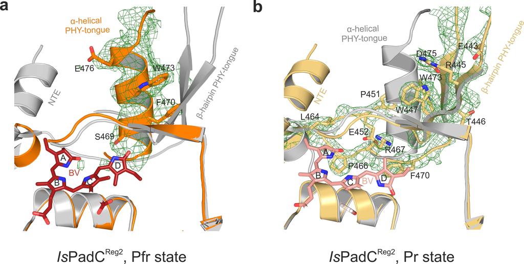
60

61
62

63

64

65
Protein Structure. W. M. Grogan, Ph.D. OBJECTIVES
 Protein Structure W. M. Grogan, Ph.D. OBJECTIVES 1. Describe the structure and characteristic properties of typical proteins. 2. List and describe the four levels of structure found in proteins. 3. Relate
Biotechnology of Proteins. The Source of Stability in Proteins (III) Fall 2015
 Fall 2015") Biotechnology of Proteins The Source of Stability in Proteins (III) Fall 2015 Conformational Entropy of Unfolding It is The factor that makes the greatest contribution to stabilization of the unfolded
Biotechnology of Proteins The Source of Stability in Proteins (III) Fall 2015 Conformational Entropy of Unfolding It is The factor that makes the greatest contribution to stabilization of the unfolded
An Introduction to Metabolism
 An Introduction to Metabolism I. All of an organism=s chemical reactions taken together is called metabolism. A. Metabolic pathways begin with a specific molecule, which is then altered in a series of
What binds to Hb in addition to O 2?
 Reading: Ch5; 158-169, 162-166, 169-174 Problems: Ch5 (text); 3,7,8,10 Ch5 (study guide-facts); 1,2,3,4,5,8 Ch5 (study guide-apply); 2,3 Remember Today at 5:30 in CAS-522 is the second chance for the MB
Detailed description of overall and active site architecture of PPDC- 3dThDP, PPDC-2HE3dThDP, PPDC-3dThDP-PPA and PPDC- 3dThDP-POVA
 Online Supplemental Results Detailed description of overall and active site architecture of PPDC- 3dThDP, PPDC-2HE3dThDP, PPDC-3dThDP-PPA and PPDC- 3dThDP-POVA Structure solution and overall architecture
Chapter 6- An Introduction to Metabolism*
 Chapter 6- An Introduction to Metabolism* *Lecture notes are to be used as a study guide only and do not represent the comprehensive information you will need to know for the exams. The Energy of Life
Visual pigments. Neuroscience, Biochemistry Dr. Mamoun Ahram Third year, 2019
 Visual pigments Neuroscience, Biochemistry Dr. Mamoun Ahram Third year, 2019 References Webvision: The Organization of the Retina and Visual System (http://www.ncbi.nlm.nih.gov/books/nbk11522/#a 127) The
Supplemental Information. Molecular Basis of Spectral Diversity. in Near-Infrared Phytochrome-Based. Fluorescent Proteins
 Chemistry & Biology, Volume 22 Supplemental Information Molecular Basis of Spectral Diversity in Near-Infrared Phytochrome-Based Fluorescent Proteins Daria M. Shcherbakova, Mikhail Baloban, Sergei Pletnev,
Computational Biology 1
 Computational Biology 1 Protein Function & nzyme inetics Guna Rajagopal, Bioinformatics Institute, guna@bii.a-star.edu.sg References : Molecular Biology of the Cell, 4 th d. Alberts et. al. Pg. 129 190
An Introduction to Metabolism
 An Introduction to Metabolism Chapter 8 Objectives Distinguish between the following pairs of terms: catabolic and anabolic pathways; kinetic and potential energy; open and closed systems; exergonic and
BSc and MSc Degree Examinations
 Examination Candidate Number: Desk Number: BSc and MSc Degree Examinations 2018-9 Department : BIOLOGY Title of Exam: Molecular Biology and Biochemistry Part I Time Allowed: 1 hour and 30 minutes Marking
Biology Chemistry & Physics of Biomolecules. Examination #1. Proteins Module. September 29, Answer Key
 Biology 5357 Chemistry & Physics of Biomolecules Examination #1 Proteins Module September 29, 2017 Answer Key Question 1 (A) (5 points) Structure (b) is more common, as it contains the shorter connection
Lecture 2 and 3: Review of forces (ctd.) and elementary statistical mechanics. Contributions to protein stability
 and elementary statistical mechanics. Contributions to protein stability") Lecture 2 and 3: Review of forces (ctd.) and elementary statistical mechanics. Contributions to protein stability Part I. Review of forces Covalent bonds Non-covalent Interactions: Van der Waals Interactions
Lecture 2 and 3: Review of forces (ctd.) and elementary statistical mechanics. Contributions to protein stability Part I. Review of forces Covalent bonds Non-covalent Interactions: Van der Waals Interactions
Signal Transduction Phosphorylation Protein kinases. Misfolding diseases. Protein Engineering Lysozyme variants
 Signal Transduction Phosphorylation Protein kinases Misfolding diseases Protein Engineering Lysozyme variants Cells and Signals Regulation The cell must be able to respond to stimuli Cellular activities
Supplementary Information. Structural basis for precursor protein-directed ribosomal peptide macrocyclization
 Supplementary Information Structural basis for precursor protein-directed ribosomal peptide macrocyclization Kunhua Li 1,3, Heather L. Condurso 1,3, Gengnan Li 1, Yousong Ding 2 and Steven D. Bruner 1*
SUPPLEMENTARY INFORMATION
 doi:10.1038/nature17991 Supplementary Discussion Structural comparison with E. coli EmrE The DMT superfamily includes a wide variety of transporters with 4-10 TM segments 1. Since the subfamilies of the
Introduction to Comparative Protein Modeling. Chapter 4 Part I
 Introduction to Comparative Protein Modeling Chapter 4 Part I 1 Information on Proteins Each modeling study depends on the quality of the known experimental data. Basis of the model Search in the literature
Gene regulation I Biochemistry 302. Bob Kelm February 25, 2005
 Gene regulation I Biochemistry 302 Bob Kelm February 25, 2005 Principles of gene regulation (cellular versus molecular level) Extracellular signals Chemical (e.g. hormones, growth factors) Environmental
MBLG lecture 5. The EGG! Visualising Molecules. Dr. Dale Hancock Lab 715
 MBLG lecture 5 Dr. Dale Hancock D.Hancock@mmb.usyd.edu.au Lab 715 The EGG! Visualising Molecules In molecular biology and biochemistry it is better to view molecules as killer pythons rather than smarties.
Sunhats for plants. How plants detect dangerous ultraviolet rays
 Sunhats for plants How plants detect dangerous ultraviolet rays Anyone who has ever suffered sunburn will know about the effects of too much ultraviolet (UV) radiation, in particular UV-B (from 280-315
Rex-Family Repressor/NADH Complex
 Kasey Royer Michelle Lukosi Rex-Family Repressor/NADH Complex Part A The biological sensing protein that we selected is the Rex-family repressor/nadh complex. We chose this sensor because it is a calcium
SUPPLEMENTARY INFORMATION
 Figure S1. Secondary structure of CAP (in the camp 2 -bound state) 10. α-helices are shown as cylinders and β- strands as arrows. Labeling of secondary structure is indicated. CDB, DBD and the hinge are
Affinity labels for studying enzyme active sites. Irreversible Enzyme Inhibition. Inhibition of serine protease with DFP
 Irreversible Enzyme Inhibition Irreversible inhibitors form stable covalent bonds with the enzyme (e.g. alkylation or acylation of an active site side chain) There are many naturally-occurring and synthetic
CHAPTER 29 HW: AMINO ACIDS + PROTEINS
 CAPTER 29 W: AMI ACIDS + PRTEIS For all problems, consult the table of 20 Amino Acids provided in lecture if an amino acid structure is needed; these will be given on exams. Use natural amino acids (L)
Gene regulation II Biochemistry 302. February 27, 2006
 Gene regulation II Biochemistry 302 February 27, 2006 Molecular basis of inhibition of RNAP by Lac repressor 35 promoter site 10 promoter site CRP/DNA complex 60 Lewis, M. et al. (1996) Science 271:1247
4 Examples of enzymes
 Catalysis 1 4 Examples of enzymes Adding water to a substrate: Serine proteases. Carbonic anhydrase. Restrictions Endonuclease. Transfer of a Phosphoryl group from ATP to a nucleotide. Nucleoside monophosphate
Supplementary Figure 3 a. Structural comparison between the two determined structures for the IL 23:MA12 complex. The overall RMSD between the two
 Supplementary Figure 1. Biopanningg and clone enrichment of Alphabody binders against human IL 23. Positive clones in i phage ELISA with optical density (OD) 3 times higher than background are shown for
Structure of the α-helix
 Structure of the α-helix Structure of the β Sheet Protein Dynamics Basics of Quenching HDX Hydrogen exchange of amide protons is catalyzed by H 2 O, OH -, and H 3 O +, but it s most dominated by base
Isothermal experiments characterize time-dependent aggregation and unfolding
 1 Energy Isothermal experiments characterize time-dependent aggregation and unfolding Technical ote Introduction Kinetic measurements have, for decades, given protein scientists insight into the mechanisms
Supplementary Figure 1. Aligned sequences of yeast IDH1 (top) and IDH2 (bottom) with isocitrate
 and IDH2 (bottom) with isocitrate") SUPPLEMENTARY FIGURE LEGENDS Supplementary Figure 1. Aligned sequences of yeast IDH1 (top) and IDH2 (bottom) with isocitrate dehydrogenase from Escherichia coli [ICD, pdb 1PB1, Mesecar, A. D., and Koshland,
SUPPLEMENTARY FIGURE LEGENDS Supplementary Figure 1. Aligned sequences of yeast IDH1 (top) and IDH2 (bottom) with isocitrate dehydrogenase from Escherichia coli [ICD, pdb 1PB1, Mesecar, A. D., and Koshland,
Visual pigments. Neuroscience, Biochemistry Dr. Mamoun Ahram Third year, 2015
 Visual pigments Neuroscience, Biochemistry Dr. Mamoun Ahram Third year, 2015 References Photoreceptors and visual pigments Webvision: The Organization of the Retina and Visual System (http://www.ncbi.nlm.nih.gov/books/nbk11522/#a127)
CHEM 3653 Exam # 1 (03/07/13)
") 1. Using phylogeny all living organisms can be divided into the following domains: A. Bacteria, Eukarya, and Vertebrate B. Archaea and Eukarya C. Bacteria, Eukarya, and Archaea D. Eukarya and Bacteria
1. Using phylogeny all living organisms can be divided into the following domains: A. Bacteria, Eukarya, and Vertebrate B. Archaea and Eukarya C. Bacteria, Eukarya, and Archaea D. Eukarya and Bacteria
1. What is an ångstrom unit, and why is it used to describe molecular structures?
 1. What is an ångstrom unit, and why is it used to describe molecular structures? The ångstrom unit is a unit of distance suitable for measuring atomic scale objects. 1 ångstrom (Å) = 1 10-10 m. The diameter
Protein Dynamics. The space-filling structures of myoglobin and hemoglobin show that there are no pathways for O 2 to reach the heme iron.
 Protein Dynamics The space-filling structures of myoglobin and hemoglobin show that there are no pathways for O 2 to reach the heme iron. Below is myoglobin hydrated with 350 water molecules. Only a small
Supplemental Information for: Characterizing the Membrane-Bound State of Cytochrome P450 3A4: Structure, Depth of Insertion and Orientation
 Supplemental Information for: Characterizing the Membrane-Bound State of Cytochrome P450 3A4: Structure, Depth of Insertion and Orientation Javier L. Baylon, Ivan L. Lenov, Stephen G. Sligar and Emad Tajkhorshid
Metabolism and enzymes
 Metabolism and enzymes 4-11-16 What is a chemical reaction? A chemical reaction is a process that forms or breaks the chemical bonds that hold atoms together Chemical reactions convert one set of chemical
[Urea] (M) k (s -1 )
![[Urea] (M) k (s -1 )](/thumbs/89/98097251.jpg "[Urea] (M) k (s -1 )") BMB178 Fall 2018 Problem Set 1 Due: 10/26/2018, noon Office hour: 10/25/2018, SFL GSR218 7 9 pm Problem 1. Transition state theory (20 points): Consider a unimolecular reaction where a substrate S is converted
BMB178 Fall 2018 Problem Set 1 Due: 10/26/2018, noon Office hour: 10/25/2018, SFL GSR218 7 9 pm Problem 1. Transition state theory (20 points): Consider a unimolecular reaction where a substrate S is converted
Section Week 3. Junaid Malek, M.D.
 Section Week 3 Junaid Malek, M.D. Biological Polymers DA 4 monomers (building blocks), limited structure (double-helix) RA 4 monomers, greater flexibility, multiple structures Proteins 20 Amino Acids,
Chapter 8. Enzymes: basic concept and kinetics
 Chapter 8 Enzymes: basic concept and kinetics Learning objectives: mechanism of enzymatic catalysis Michaelis -Menton Model Inhibition Single Molecule of Enzymatic Reaction Enzymes: catalysis chemical
2015 AP Biology Unit 2 PRETEST- Introduction to the Cell and Biochemistry
 Name: Class: _ Date: _ 2015 AP Biology Unit 2 PRETEST- Introduction to the Cell and Biochemistry Multiple Choice Identify the choice that best completes the statement or answers the question. 1) In what
Energy, Enzymes, and Metabolism. Energy, Enzymes, and Metabolism. A. Energy and Energy Conversions. A. Energy and Energy Conversions
 Energy, Enzymes, and Metabolism Lecture Series 6 Energy, Enzymes, and Metabolism B. ATP: Transferring Energy in Cells D. Molecular Structure Determines Enzyme Fxn Energy is the capacity to do work (cause
Table 1. Crystallographic data collection, phasing and refinement statistics. Native Hg soaked Mn soaked 1 Mn soaked 2
 Table 1. Crystallographic data collection, phasing and refinement statistics Native Hg soaked Mn soaked 1 Mn soaked 2 Data collection Space group P2 1 2 1 2 1 P2 1 2 1 2 1 P2 1 2 1 2 1 P2 1 2 1 2 1 Cell
Enzyme Enzymes are proteins that act as biological catalysts. Enzymes accelerate, or catalyze, chemical reactions. The molecules at the beginning of
 Enzyme Enzyme Enzymes are proteins that act as biological catalysts. Enzymes accelerate, or catalyze, chemical reactions. The molecules at the beginning of the process are called substrates and the enzyme
Exam I Answer Key: Summer 2006, Semester C
 1. Which of the following tripeptides would migrate most rapidly towards the negative electrode if electrophoresis is carried out at ph 3.0? a. gly-gly-gly b. glu-glu-asp c. lys-glu-lys d. val-asn-lys
NH 2. Biochemistry I, Fall Term Sept 9, Lecture 5: Amino Acids & Peptides Assigned reading in Campbell: Chapter
 Biochemistry I, Fall Term Sept 9, 2005 Lecture 5: Amino Acids & Peptides Assigned reading in Campbell: Chapter 3.1-3.4. Key Terms: ptical Activity, Chirality Peptide bond Condensation reaction ydrolysis
An Introduction to Metabolism
 LECTURE PRESENTATIONS For CAMPBELL BIOLOGY, NINTH EDITION Jane B. Reece, Lisa A. Urry, Michael L. Cain, Steven A. Wasserman, Peter V. Minorsky, Robert B. Jackson Chapter 8 An Introduction to Metabolism
LS1a Fall 2014 Problem Set #2 Due Monday 10/6 at 6 pm in the drop boxes on the Science Center 2 nd Floor
 LS1a Fall 2014 Problem Set #2 Due Monday 10/6 at 6 pm in the drop boxes on the Science Center 2 nd Floor Note: Adequate space is given for each answer. Questions that require a brief explanation should
Supplementary information
 Supplementary information The structural basis of modularity in ECF-type ABC transporters Guus B. Erkens 1,2, Ronnie P-A. Berntsson 1,2, Faizah Fulyani 1,2, Maria Majsnerowska 1,2, Andreja Vujičić-Žagar
A. Reaction Mechanisms and Catalysis (1) proximity effect (2) acid-base catalysts (3) electrostatic (4) functional groups (5) structural flexibility
 proximity effect (2) acid-base catalysts (3) electrostatic (4) functional groups (5) structural flexibility") (P&S Ch 5; Fer Ch 2, 9; Palm Ch 10,11; Zub Ch 9) A. Reaction Mechanisms and Catalysis (1) proximity effect (2) acid-base catalysts (3) electrostatic (4) functional groups (5) structural flexibility B.
(P&S Ch 5; Fer Ch 2, 9; Palm Ch 10,11; Zub Ch 9) A. Reaction Mechanisms and Catalysis (1) proximity effect (2) acid-base catalysts (3) electrostatic (4) functional groups (5) structural flexibility B.
SUPPLEMENTARY INFORMATION
 SUPPLEMENTARY INFORMATION doi:10.1038/nature11524 Supplementary discussion Functional analysis of the sugar porter family (SP) signature motifs. As seen in Fig. 5c, single point mutation of the conserved
Regulation and signaling. Overview. Control of gene expression. Cells need to regulate the amounts of different proteins they express, depending on
 Regulation and signaling Overview Cells need to regulate the amounts of different proteins they express, depending on cell development (skin vs liver cell) cell stage environmental conditions (food, temperature,
THE UNIVERSITY OF MANITOBA. PAPER NO: _1_ LOCATION: 173 Robert Schultz Theatre PAGE NO: 1 of 5 DEPARTMENT & COURSE NO: CHEM / MBIO 2770 TIME: 1 HOUR
 THE UNIVERSITY OF MANITOBA 1 November 1, 2016 Mid-Term EXAMINATION PAPER NO: _1_ LOCATION: 173 Robert Schultz Theatre PAGE NO: 1 of 5 DEPARTMENT & COURSE NO: CHEM / MBIO 2770 TIME: 1 HOUR EXAMINATION:
Dana Alsulaibi. Jaleel G.Sweis. Mamoon Ahram
 15 Dana Alsulaibi Jaleel G.Sweis Mamoon Ahram Revision of last lectures: Proteins have four levels of structures. Primary,secondary, tertiary and quaternary. Primary structure is the order of amino acids
Final Chem 4511/6501 Spring 2011 May 5, 2011 b Name
 Key 1) [10 points] In RNA, G commonly forms a wobble pair with U. a) Draw a G-U wobble base pair, include riboses and 5 phosphates. b) Label the major groove and the minor groove. c) Label the atoms of
Translation. A ribosome, mrna, and trna.
 Translation The basic processes of translation are conserved among prokaryotes and eukaryotes. Prokaryotic Translation A ribosome, mrna, and trna. In the initiation of translation in prokaryotes, the Shine-Dalgarno
Lecture 2-3: Review of forces (ctd.) and elementary statistical mechanics. Contributions to protein stability
 and elementary statistical mechanics. Contributions to protein stability") Lecture 2-3: Review of forces (ctd.) and elementary statistical mechanics. Contributions to protein stability Part I. Review of forces Covalent bonds Non-covalent Interactions Van der Waals Interactions
Lecture 2-3: Review of forces (ctd.) and elementary statistical mechanics. Contributions to protein stability Part I. Review of forces Covalent bonds Non-covalent Interactions Van der Waals Interactions
Biological Chemistry and Metabolic Pathways
 Biological Chemistry and Metabolic Pathways 1. Reaction a. Thermodynamics b. Kinetics 2. Enzyme a. Structure and Function b. Regulation of Activity c. Kinetics d. Inhibition 3. Metabolic Pathways a. REDOX
2013 W. H. Freeman and Company. 6 Enzymes
 2013 W. H. Freeman and Company 6 Enzymes CHAPTER 6 Enzymes Key topics about enzyme function: Physiological significance of enzymes Origin of catalytic power of enzymes Chemical mechanisms of catalysis
Chapter 16 Lecture. Concepts Of Genetics. Tenth Edition. Regulation of Gene Expression in Prokaryotes
 Chapter 16 Lecture Concepts Of Genetics Tenth Edition Regulation of Gene Expression in Prokaryotes Chapter Contents 16.1 Prokaryotes Regulate Gene Expression in Response to Environmental Conditions 16.2
BIOCHEMISTRY Course Outline (Fall, 2011)
") BIOCHEMISTRY 402 - Course Outline (Fall, 2011) Number OVERVIEW OF LECTURE TOPICS: of Lectures INSTRUCTOR 1. Structural Components of Proteins G. Brayer (a) Amino Acids and the Polypeptide Chain Backbone...2
BIOCHEMISTRY 402 - Course Outline (Fall, 2011) Number OVERVIEW OF LECTURE TOPICS: of Lectures INSTRUCTOR 1. Structural Components of Proteins G. Brayer (a) Amino Acids and the Polypeptide Chain Backbone...2
General Biology. The Energy of Life The living cell is a miniature factory where thousands of reactions occur; it converts energy in many ways
 Course No: BNG2003 Credits: 3.00 General Biology 5. An Introduction into Cell Metabolism The Energy of Life The living cell is a miniature factory where thousands of reactions occur; it converts energy
T H E J O U R N A L O F G E N E R A L P H Y S I O L O G Y. jgp
 S u p p l e m e n ta l m at e r i a l jgp Lee et al., http://www.jgp.org/cgi/content/full/jgp.201411219/dc1 T H E J O U R N A L O F G E N E R A L P H Y S I O L O G Y S u p p l e m e n ta l D I S C U S
Hydrogen Bond Switching among Flavin and. Amino Acids Determines the Nature of Proton- Coupled Electron Transfer in BLUF.
 Hydrogen Bond Switching among Flavin and Amino Acids Determines the Nature of Proton- Coupled Electron Transfer in BLUF Photoreceptors Tilo Mathes 1,2, Jingyi Zhu 1, Ivo H.M. van Stokkum 1, M.L. Groot
9/25/2011. Outline. Overview: The Energy of Life. I. Forms of Energy II. Laws of Thermodynamics III. Energy and metabolism IV. ATP V.
 Chapter 8 Introduction to Metabolism Outline I. Forms of Energy II. Laws of Thermodynamics III. Energy and metabolism IV. ATP V. Enzymes Overview: The Energy of Life Figure 8.1 The living cell is a miniature
Transmembrane Domains (TMDs) of ABC transporters
 of ABC transporters") Transmembrane Domains (TMDs) of ABC transporters Most ABC transporters contain heterodimeric TMDs (e.g. HisMQ, MalFG) TMDs show only limited sequence homology (high diversity) High degree of conservation
Transmembrane Domains (TMDs) of ABC transporters Most ABC transporters contain heterodimeric TMDs (e.g. HisMQ, MalFG) TMDs show only limited sequence homology (high diversity) High degree of conservation
Retinal Proteins (Rhodopsins) Vision, Bioenergetics, Phototaxis. Bacteriorhodopsin s Photocycle. Bacteriorhodopsin s Photocycle
 Vision, Bioenergetics, Phototaxis. Bacteriorhodopsin s Photocycle. Bacteriorhodopsin s Photocycle") Molecular chanisms of Photoactivation and Spectral Tuning in Retinal Proteins Emad Tajkhorshid Theoretical and Computational Biophysics Group Beckman Institute University of Illinois at Urbana-Champaign
Molecular chanisms of Photoactivation and Spectral Tuning in Retinal Proteins Emad Tajkhorshid Theoretical and Computational Biophysics Group Beckman Institute University of Illinois at Urbana-Champaign
Introduction to" Protein Structure
 Introduction to" Protein Structure Function, evolution & experimental methods Thomas Blicher, Center for Biological Sequence Analysis Learning Objectives Outline the basic levels of protein structure.
Supplementary Figure 1. Biochemical and sequence alignment analyses the
 Supplementary Figure 1. Biochemical and sequence alignment analyses the interaction of OPTN and TBK1. (a) Analytical gel filtration chromatography analysis of the interaction between TBK1 CTD and OPTN(1-119).
SUPPLEMENTARY INFORMATION
 Supplementary materials Figure S1 Fusion protein of Sulfolobus solfataricus SRP54 and a signal peptide. a, Expression vector for the fusion protein. The signal peptide of yeast dipeptidyl aminopeptidase
Lecture 11: Enzymes: Kinetics [PDF] Reading: Berg, Tymoczko & Stryer, Chapter 8, pp
![Lecture 11: Enzymes: Kinetics [PDF] Reading: Berg, Tymoczko & Stryer, Chapter 8, pp](/thumbs/74/70848503.jpg "Lecture 11: Enzymes: Kinetics [PDF] Reading: Berg, Tymoczko & Stryer, Chapter 8, pp") Lecture 11: Enzymes: Kinetics [PDF] Reading: Berg, Tymoczko & Stryer, Chapter 8, pp. 216-225 Updated on: 2/4/07 at 9:00 pm Key Concepts Kinetics is the study of reaction rates. Study of enzyme kinetics
Lecture 11: Enzymes: Kinetics [PDF] Reading: Berg, Tymoczko & Stryer, Chapter 8, pp. 216-225 Updated on: 2/4/07 at 9:00 pm Key Concepts Kinetics is the study of reaction rates. Study of enzyme kinetics
Packing of Secondary Structures
 7.88 Lecture Notes - 4 7.24/7.88J/5.48J The Protein Folding and Human Disease Professor Gossard Retrieving, Viewing Protein Structures from the Protein Data Base Helix helix packing Packing of Secondary
Introduction to Computational Structural Biology
 Introduction to Computational Structural Biology Part I 1. Introduction The disciplinary character of Computational Structural Biology The mathematical background required and the topics covered Bibliography
Advanced Certificate in Principles in Protein Structure. You will be given a start time with your exam instructions
 BIRKBECK COLLEGE (University of London) Advanced Certificate in Principles in Protein Structure MSc Structural Molecular Biology Date: Thursday, 1st September 2011 Time: 3 hours You will be given a start
Reading Assignments. A. Genes and the Synthesis of Polypeptides. Lecture Series 7 From DNA to Protein: Genotype to Phenotype
 Lecture Series 7 From DNA to Protein: Genotype to Phenotype Reading Assignments Read Chapter 7 From DNA to Protein A. Genes and the Synthesis of Polypeptides Genes are made up of DNA and are expressed
Chapter 1. Topic: Overview of basic principles
 Chapter 1 Topic: Overview of basic principles Four major themes of biochemistry I. What are living organism made from? II. How do organism acquire and use energy? III. How does an organism maintain its
Energy and Cellular Metabolism
 1 Chapter 4 About This Chapter Energy and Cellular Metabolism 2 Energy in biological systems Chemical reactions Enzymes Metabolism Figure 4.1 Energy transfer in the environment Table 4.1 Properties of
SUPPLEMENTARY INFORMATION
 In the format provided by the authors and unedited. SUPPLEMENTARY INFORMATION DOI: 10.1038/NCHEM.2720 1 2 3 Tuning underwater adhesion with cation-π interactions Matthew A. Gebbie, Wei Wei, Alex M. Schrader,
Energy Transformation and Metabolism (Outline)
") Energy Transformation and Metabolism (Outline) - Definitions & Laws of Thermodynamics - Overview of energy flow ecosystem - Biochemical processes: Anabolic/endergonic & Catabolic/exergonic - Chemical reactions
Energy Transformation and Metabolism (Outline) - Definitions & Laws of Thermodynamics - Overview of energy flow ecosystem - Biochemical processes: Anabolic/endergonic & Catabolic/exergonic - Chemical reactions
XV 74. Flouorescence-Polarization-Circular-Dichroism- Jablonski diagram Where does the energy go?
 XV 74 Flouorescence-Polarization-Circular-Dichroism- Jablonski diagram Where does the energy go? 1) Excite system through A Absorbance S 0 S n Excite from ground excited singlet S = 0 could be any of them
Protein Folding Prof. Eugene Shakhnovich
 Protein Folding Eugene Shakhnovich Department of Chemistry and Chemical Biology Harvard University 1 Proteins are folded on various scales As of now we know hundreds of thousands of sequences (Swissprot)
[Urea] (M) k (s -1 )
![[Urea] (M) k (s -1 )](/thumbs/96/126856044.jpg "[Urea] (M) k (s -1 )") BMB178 Fall 2018 Problem Set 1 Due: 10/26/2018, noon Office hour: 10/25/2018, SFL GSR218 7 9 pm Problem 1. Transition state theory (20 points): Consider a unimolecular reaction where a substrate S is converted
BMB178 Fall 2018 Problem Set 1 Due: 10/26/2018, noon Office hour: 10/25/2018, SFL GSR218 7 9 pm Problem 1. Transition state theory (20 points): Consider a unimolecular reaction where a substrate S is converted
C a h p a t p e t r e r 6 E z n y z m y e m s
 Chapter 6 Enzymes 4. Examples of enzymatic reactions acid-base catalysis: give and take protons covalent catalysis: a transient covalent bond is formed between the enzyme and the substrate metal ion catalysis:
Physiochemical Properties of Residues
 Physiochemical Properties of Residues Various Sources C N Cα R Slide 1 Conformational Propensities Conformational Propensity is the frequency in which a residue adopts a given conformation (in a polypeptide)
THE ROLE OF THE PHYTOCHROME B PHOTORECEPTOR IN THE REGULATION OF PHOTOPERIODIC FLOWERING. AnitaHajdu. Thesis of the Ph.D.
 THE ROLE OF THE PHYTOCHROME B PHOTORECEPTOR IN THE REGULATION OF PHOTOPERIODIC FLOWERING AnitaHajdu Thesis of the Ph.D. dissertation Supervisor: Dr. LászlóKozma-Bognár - senior research associate Doctoral
Acta Crystallographica Section D
 Supporting information Acta Crystallographica Section D Volume 70 (2014) Supporting information for article: Structural characterization of the virulence factor Nuclease A from Streptococcus agalactiae
Using Higher Calculus to Study Biologically Important Molecules Julie C. Mitchell
 Using Higher Calculus to Study Biologically Important Molecules Julie C. Mitchell Mathematics and Biochemistry University of Wisconsin - Madison 0 There Are Many Kinds Of Proteins The word protein comes
schematic diagram; EGF binding, dimerization, phosphorylation, Grb2 binding, etc.
 Lecture 1: Noncovalent Biomolecular Interactions Bioengineering and Modeling of biological processes -e.g. tissue engineering, cancer, autoimmune disease Example: RTK signaling, e.g. EGFR Growth responses
Chapter 8 Notes. An Introduction to Metabolism
 Chapter 8 Notes An Introduction to Metabolism Describe how allosteric regulators may inhibit or stimulate the activity of an enzyme. Objectives Distinguish between the following pairs of terms: catabolic
Supplementary Figure 1. SDS-PAGE analysis of GFP oligomer variants with different linkers. Oligomer mixtures were applied to a PAGE gel containing
 Supplementary Figure 1. SDS-PAGE analysis of GFP oligomer variants with different linkers. Oligomer mixtures were applied to a PAGE gel containing 0.1% SDS without boiling. The gel was analyzed by a fluorescent
Biochemistry 462a - Enzyme Kinetics Reading - Chapter 8 Practice problems - Chapter 8: (not yet assigned); Enzymes extra problems
; Enzymes extra problems") Biochemistry 462a - Enzyme Kinetics Reading - Chapter 8 Practice problems - Chapter 8: (not yet assigned); Enzymes extra problems Introduction Enzymes are Biological Catalysis A catalyst is a substance
Biochemistry 462a - Enzyme Kinetics Reading - Chapter 8 Practice problems - Chapter 8: (not yet assigned); Enzymes extra problems Introduction Enzymes are Biological Catalysis A catalyst is a substance
Bacterial protease uses distinct thermodynamic signatures for substrate recognition
 Bacterial protease uses distinct thermodynamic signatures for substrate recognition Gustavo Arruda Bezerra, Yuko Ohara-Nemoto, Irina Cornaciu, Sofiya Fedosyuk, Guillaume Hoffmann, Adam Round, José A. Márquez,
BBS501 Section 1 9:00 am 10:00 am Monday thru Friday LRC 105 A & B
 BBS501 Section 1 9:00 am 10:00 am Monday thru Friday LRC 105 A & B Lecturers: Dr. Yie-Hwa Chang Room M130 Phone: #79263 E-mail:changy@slu.edu Dr. Tomasz Heyduk Room M99 Phone: #79238 E-mail: heydukt@slu.edu
Introduction The gramicidin A (ga) channel forms by head-to-head association of two monomers at their amino termini, one from each bilayer leaflet. Th
 channel forms by head-to-head association of two monomers at their amino termini, one from each bilayer leaflet. Th") Abstract When conductive, gramicidin monomers are linked by six hydrogen bonds. To understand the details of dissociation and how the channel transits from a state with 6H bonds to ones with 4H bonds or
Abstract When conductive, gramicidin monomers are linked by six hydrogen bonds. To understand the details of dissociation and how the channel transits from a state with 6H bonds to ones with 4H bonds or
Charged amino acids (side-chains)
") Proteins are composed of monomers called amino acids There are 20 different amino acids Amine Group Central ydrocarbon N C C R Group Carboxyl Group ALL amino acids have the exact same structure except
Proteins are composed of monomers called amino acids There are 20 different amino acids Amine Group Central ydrocarbon N C C R Group Carboxyl Group ALL amino acids have the exact same structure except
An Introduction to Metabolism
 CAMPBELL BIOLOGY IN FOCUS Urry Cain Wasserman Minorsky Jackson Reece 6 An Introduction to Metabolism Lecture Presentations by Kathleen Fitzpatrick and Nicole Tunbridge Overview: The Energy of Life The
Supplementary Materials for
 advances.sciencemag.org/cgi/content/full/3/4/e1600663/dc1 Supplementary Materials for A dynamic hydrophobic core orchestrates allostery in protein kinases Jonggul Kim, Lalima G. Ahuja, Fa-An Chao, Youlin
Ranjit P. Bahadur Assistant Professor Department of Biotechnology Indian Institute of Technology Kharagpur, India. 1 st November, 2013
 Hydration of protein-rna recognition sites Ranjit P. Bahadur Assistant Professor Department of Biotechnology Indian Institute of Technology Kharagpur, India 1 st November, 2013 Central Dogma of life DNA
An Introduction to Metabolism
 Chapter 8 An Introduction to Metabolism Edited by Shawn Lester PowerPoint Lecture Presentations for Biology Eighth Edition Neil Campbell and Jane Reece Lectures by Chris Romero, updated by Erin Barley
4. The Michaelis-Menten combined rate constant Km, is defined for the following kinetic mechanism as k 1 k 2 E + S ES E + P k -1
 Fall 2000 CH 595C Exam 1 Answer Key Multiple Choice 1. One of the reasons that enzymes are such efficient catalysts is that a) the energy level of the enzyme-transition state complex is much higher than
Biochemistry,530:,, Introduc5on,to,Structural,Biology, Autumn,Quarter,2015,
 Biochemistry,530:,, Introduc5on,to,Structural,Biology, Autumn,Quarter,2015, Course,Informa5on, BIOC%530% GraduateAlevel,discussion,of,the,structure,,func5on,,and,chemistry,of,proteins,and, nucleic,acids,,control,of,enzyma5c,reac5ons.,please,see,the,course,syllabus,and,