Molecular Pathways Mediating Glial Responses during Wallerian Degeneration: A Dissertation
|
|
|
- Abner Heath
- 6 years ago
- Views:
Transcription

1 University of Massachusetts Medical School GSBS Dissertations and Theses Graduate School of Biomedical Sciences Molecular Pathways Mediating Glial Responses during Wallerian Degeneration: A Dissertation Tsai-Yi Lu University of Massachusetts Medical School Follow this and additional works at: Part of the Molecular and Cellular Neuroscience Commons Recommended Citation Lu, T. Molecular Pathways Mediating Glial Responses during Wallerian Degeneration: A Dissertation. (2015). University of Massachusetts Medical School. GSBS Dissertations and Theses. Paper 779. DOI: /M26W2Q. This material is brought to you by escholarship@umms. It has been accepted for inclusion in GSBS Dissertations and Theses by an authorized administrator of escholarship@umms. For more information, please contact Lisa.Palmer@umassmed.edu.
2 MOLECULAR PATHWAYS MEDIATING GLIAL RESPONSES DURING WALLERIAN DEGENERATION A Dissertation Presented By Tsai-Yi Lu Submitted to the Faculty of the University of Massachusetts Graduate School of Biomedical Sciences, Worcester in partial fulfillment of the requirements for the degree of DOCTOR OF PHILOSOPHY May 14 th, 2015 Interdisciplinary Graduate Program
3 ii MOLECULAR PATHWAYS MEDIATING GLIAL RESPONSES DURING WALLERIAN DEGENERATION A Dissertation Presented By Tsai-Yi Lu The signatures of the Dissertation Defense Committee signify completion and approval as to style and content of the Dissertation Marc R. Freeman, Ph.D., Thesis Advisor Vivian Budnik, Ph.D., Member of Committee Carlos Lois, Ph.D., Member of Committee Neal Silverman, Ph.D., Member of Committee Yongjie Yang, Ph.D., Member of Committee The signature of the Chair of the Committee signifies that the written dissertation meets the requirements of the Dissertation Committee Eric H. Baehrecke, Ph.D., Chair of Committee The signature of the Dean of the Graduate School of Biomedical Sciences signifies that the student has met all graduation requirements of the school. Anthony Carruthers, Ph.D., Dean of the Graduate School of Biomedical Sciences Interdisciplinary Graduate Program May 14 th, 2015
4 iii This work is dedicated to Juan Hsiao-Chi, for being my inspiration in the past several years, and to my family, for their unconditional support.
5 iv Acknowledgements I would like to express my greatest appreciation to my advisor, Marc Freeman, for creating the best lab in the world and showing me how to be a great scientist. I would also like to offer my special thanks to my awesome mentor, Johnna Doherty, for patiently taking my questions from fly genetics to what a furry party would look like. My sincere thanks are also extended to all the past and present Freeman lab members, including Mary Logan, Jennifer MacDonald, Jennifer Ziegenfuss, Ozge Tasdemir-Yilmaz, Kimberly Kerr, Yuly Fuentes-Medel, Michelle Avery, Jeannette Osterloh, Allie Muthukamar, Edith Plada, Timothy Rooney, Zhiquo Ma, Lukas Neukomm, Thomas Burdett, Jaeda Coutinho-Budd, Owen Peters, Jemeen Sreedharan, Megan Corty, Jonathan Farley, Sukhee Cho, Lizzy Lewis, Yonca Kushkuly, Jack Wong, Gaynor Smith, Rachel Bradshaw, Tobias Stork, Nicki Fox Tapper, and Amy Sheehan, for giving valuable advice and making life in Worcester so colorful. I am forever grateful to be part of the UMMS GSBS family and would like to thank all the wonderful PhD mentors I met here, including Neal Silverman, Eric Baehrecke, Carlos Lois, Vivian Budnik, Claire Benard and Tony Ip. My special thanks also go to Yongjie Yang, for being my external committee member. Finally, I wish to thank the kindness of the Taiwanese community here at UMMS, especially Chun-Ting Chen, who passed away last summer, for being a respectful and truthful friend since my day 1 at UMMS, and Yung-Chi Huang, for always being on my side and reminding me why I started this journey in the beginning and how far I have come.
6 v Abstract Glia are the understudied brain cells that perform many functions essential to maintain nervous system homeostasis and protect the brain from injury. If brain damage occurs, glia rapidly adopt the reactive state and elicit a series of cellular and molecular events known as reactive gliosis, the hallmark of many neurodegenerative diseases. However, the molecular pathways that trigger and regulate this process remain poorly defined. The fruit fly Drosophila melanogaster has glial cells that are strikingly similar to mammalian glia, and which also exhibit reactive responses after neuronal injury. By exploiting its powerful genetic toolbox, we are uniquely positioned to identify the genes that activate and execute glial responses to neuronal injury in vivo. In this dissertation, I use Wallerian degeneration in Drosophila as a model to characterize molecular pathways responsible for glia to recognize neural injury, become activated, and ultimately engulf and degrade axonal debris. I demonstrate a novel role for the GEF (guanine nucleotide exchange factors) complex DRK/DOS/SOS upstream of small GTPase Rac1 in glial engulfment activity and show that it acts redundantly with previously discovered Crk/Mbc/dCed-12 to execute glial activation after axotomy. In addition, I discovered an exciting new role for the TNF receptor associated factor 4 (TRAF4) in glial response to axon injury. I find that interfering with TRAF4 and the downstream kinase misshapen (msn) function results in impaired glial activation and engulfment of axonal debris. Unexpectedly, I find that TRAF4 physically associates with engulfment receptor Draper making TRAF4 only second factor to bind directly to Draper and show it is essential for Draper-dependent activation of downstream engulfment signaling, including
7 vi transcriptional activation of engulfment genes via the JNK and STAT transcriptional cascades. All of these pathways are highly conserved from Drosophila to mammals and most are known to be expressed in mouse brain glia, suggesting functional conservation. My work should therefore serve as an excellent starting point for future investigations regarding their roles in glial activation/reactive gliosis in various pathological conditions of the mammalian central nervous system.
8 vii Table of Contents Title... i Signature Page... ii Dedication... iii Acknowledgements... iv Abstract... v Table of Contents... vii List of Tables... x List of Figures... xi List of Abbreviations... xiv CHAPTER I: Introduction... 1 What are glia? What do they do?... 2 Reactive gliosis, neural injury and clearance of cellular debris by glia... 3 Glial responses during Wallerian degeneration in the CNS... 4 Drosophila as a model organism to study Wallerian degeneration... 6 Glial responses during Wallerian degeneration in the olfactory nerve injury model... 7 The diversity of glial cell types in Drosophila... 9 Draper-mediated glial engulfment of axonal debris Activation of glial transcriptional responses by JNK and STAT after axon injury... 13
9 viii CHAPTER II: DRK/DOS/SOS Converge with Crk/Mbc/dCed-12 to activate Rac1 during Glial Engulfment of Axonal Debris Introduction Results DRK, DOS, and SOS are required for glial engulfment of axonal debris Glial DRK is recruited to degenerating axons after injury DRK-SOS activation helps glia to internalize axonal debris and activate the phagolysosomal program Rac1, but not Ras, is the main small GTPase effector downstream of SOS in activating the engulfing glia Both DRK/DOS/SOS and Crk/Mbc/dCed-12 activation contribute to Rac1 activity in glial response to axon injury Summary and discussion Materials and methods CHAPTER III: TRAF4 is an Adaptor Protein for Draper to Activate Downstream JNK and STAT Signaling Pathways during Wallerian Degeneration Introduction Results Summary and discussion Materials and methods CHAPTER IV: Summary and Discussion The redundancy of DRK/DOS/SOS and Crk/Mbc/dCed-12 in Rac1 activation The role of DRK in glial engulfment of axonal debris
10 ix The role of DOS in glial engulfment of axonal debris The role of SOS in glial engulfment of axonal debris The role of TRAF4 in glial engulfment of axonal debris The role of MSN in glial engulfment of axonal debris The cross talk between JNK and STAT signaling pathway The axon injury signal and Draper-mediated glial activation Glial subtypes and their responses during Wallerian degeneration The expression of DRK, DOS, SOS, TRAF4, and MSN homologs in the mouse CNS Significance and concluding remarks Reference List
11 x List of Tables Chapter III Table 3.1. Primer sets used for this study.
12 xi List of Figures CHAPTER I Figure 1.1. Using Drosophila olfactory nervous system to study Wallerian degeneration. Figure 1.2. Genes and molecular pathways involved in Drosophila glial responses during Wallerian degeneration. CHAPTER II Figure 2.1. Glial clearance of axonal debris requires DRK, DOS, and SOS. Figure 2.2. The depletion of DRK, DOS, and SOS in glia results in delay of axonal debris clearance after axotomy. Figure 2.3. Alternative RNAi of drk and dos caused similar engulfment defects in adult glia. Figure 2.4. Gain-of-function allele of sos (Sos JC2 ) alone does not change the efficiency of glial clearance of axonal debris. Figure 2.5. DRK is recruited to glial membranes surrounding the injured axons. Figure 2.6. Draper is required for DRK to be recruited to the severed axons. Figure 2.7. DRK protein level in the brain is unaltered 1 day after axonal injury. Figure 2.8. DRK-SOS activation is required for glia to internalize axonal debris. Figure 2.9. DRK-SOS is required for glia to activate phagolysosomal program. Figure Ras has little effect on glial clearance of axonal debris. Figure SOS modulates Rac1 activity in glial response to axonal injury.
13 xii Figure DRK/DOS/SOS and Crk/Mbc/dCed-12 are redundant pathways in activating glial clearance of axonal debris. Figure Glial activation and the clearance of axonal debris require SOS and Mbc. Figure The proposed model of glial response to and clearance of axonal debris after axon injury in Drosophila adult CNS. Figure DRK/DOS/SOS requires Draper to function. CHAPTER III Figure 3.1. TRAF4 is necessary for glia to engulf axonal debris and activate JNK signaling cascade in response to axon injury. Figure 3.2. The C-terminal HA tag does not affect Draper I function in the clearance of axonal debris. Figure 3.3. Glial activation of JNK signaling cascade is specific to the degeneration event. Figure 3.4. MSN is required for glia to engulf axonal debris, activate JNK signaling cascade, and increase Draper expression around the antennal lobe after axotomy. Figure 3.5. The expression of MSN DN did not affect glial membrane hypertrophy after axotomy. Figure 3.6. TRAF4 and JNK signaling cascade is required for STAT transcriptional activity in glia during Wallerian degeneration. Figure 3.7. Draper is cell-autonomously required for glia to activate JNK signaling in response to axon degeneration. Figure 3.8. Astrocytes do not activate JNK signaling cascade after axon injury.
14 xiii Figure 3.9. TRAF4 zinc-finger domain-enriched region physically interacts with Draper I a.a Figure Deletion of a.a , deletion of a.a , and the interruption of Y949 phosphorylation in Draper I do not affect the interaction between Draper I and TRAF4, which is independent of Draper I-Shark association. Figure Src42A, Rac1 and Shark are required for glia to activate JNK signaling cascade in response to axon degeneration. Figure The proposed model of Draper-mediated JNK and STAT signaling pathway activations during glial responses to axon injury. CHAPTER IV Figure 4.1. The newly proposed model of Draper-mediated glial engulfment of axonal debris after injury.
15 xiv List of Abbreviations CNS: Central nervous system DRG: Dorsal root ganglion ORN: Olfactory receptor neuron GFP: Green fluorescent protein MP: Maxillary palp ANT: Antenna/antennal ITAM: Immunoreceptor tyrosine-based activation motif SH2: Src homology 2 SH3: Src homology 3 FcγR: Fc gamma receptor GEF: Guanine nucleotide exchange factor GTP: Guanosine-5 -triphosphate GDP: Guanosine diphosphate MAPK: Mitogen-activation protein kinase MAP4K: Mitogen-activation protein kinase kinase kinase kinase SOG: Subesophageal ganglion GOF: Gain-of-function RTK: Receptor tyrosine kinase BLAST: Basic local alignment search tool FPKM: Fragments per kilobase of transcript sequence per million mapped fragments
16 CHAPTER I: Introduction 1
17 2 What are glia? What do they do? Glia (Greek: glue) is a generic name for a large group of non-neuronal cells in the nervous system. They account for an average about 60% of the cells in the human brain (Azevedo et al., 2009) but not until recently have scientists begun to appreciate their importance in maintaining the homeostasis of the nervous system: they provide nutrition support, modulate synaptic activity, defend the brain from pathogens, and remove cellular debris from the nervous system. The evolutionary origin of glia remains a mystery. Nevertheless, it is known that both arthropods and vertebrates have well-developed glia which can be classified into several subtypes according to their distinct cellular morphologies, molecular features and biological functions (for a comprehensive review regarding the evolutionary origins of glia, see Hartline, 2011). For example, in the mammalian central nervous system (CNS), glia can be categorized into three different major subtypes astrocytes, oligodendrocytes, and microglia. Astrocytes are star-shaped glial cells that are present in both the grey matter (protoplasmic astrocytes) and white matter (fibrous astrocytes) (Andriezen, 1893; Somjen, 1988). They provide energy support to neurons (Tsacopoulos and Magistretti, 1996) and modulate synaptic activity in multiple ways (Clarke and Barres, 2013; Perea et al., 2009). Oligodendrocytes form myelin sheath around axons to facilitate the transmission of action potentials. Microglia, the resident macrophages and professional phagocytes in the brain, are constantly surveying the surrounding microenvironment to detect and eliminate invading pathogens or clear dead neurons and cellular debris (Davalos et al., 2005; Nimmerjahn et al., 2005; Rio-Hortega, 1993). Recent reports also demonstrate the microglial involvement of
18 3 synaptic pruning during normal brain development (Paolicelli et al., 2011; Stevens et al., 2007). Reactive gliosis, neural injury and clearance of cellular debris by glia Microglia are highly sensitive to the brain microenvironment and react rapidly to brain injury by changing dramatically at both the cellular and molecular levels. However, they are not the only type of glial cells that can respond to brain injury. In fact, both astrocytes and a particular type of oligodendrocyte precursor cells, NG2 glia, participate in the multi-cellular response to CNS insults, collectively known as reactive gliosis (Burda and Sofroniew, 2014). Reactive gliosis can be induced by various kinds of brain insults, including ischemic stroke, local traumatic brain injury, pathogen infection, tumor, autoimmune response, neurodegeneration, etc. Whether reactive gliosis is beneficial or detrimental to the recovery of the nervous system has been a long debate. Recent opinions incline to portrait the nature of reactive gliosis as a double-edged sword the glial responses evoked after brain insults help to reduce the damage but also hinder the regenerative power of the nervous system (Barres, 2008; Neumann et al., 2009; Sofroniew, 2005). Neural injury usually gives rise to cellular debris that needs to be cleared from the nervous system. Delayed or hampered clearance of the debris can be observed in the aging brain and in Alzheimer s disease, and efficient removal of neuronal debris has been viewed as a key factor to create a pro-regenerative environment for the nervous system (Dirnagl et al., 2009; Neumann et al., 2009; Vargas and Barres, 2007). In the peripheral
19 4 nervous system, both Schwann cells and macrophages can rapidly remove neural debris after injury. On the contrary, since the CNS is immune-privileged, the clearance of axonal debris relies heavily on the resident professional phagocytes microglia. Live imaging studies of vertebrate microglia have demonstrated their abilities to respond to brain injury within hours and migrate toward the lesion site to perform phagocytosis (Davalos et al., 2005; Nimmerjahn et al., 2005; Peri and Nüsslein-Volhard, 2008). Questions regarding how these glia sense different kinds of injury, what molecular cue(s) are used, what molecular pathways are activated in these glia, and how these glia know when to terminate the responses have fascinated scientists trying to characterize glial responses to brain injury. This dissertation study aims to answer these questions by studying a simple axon injury model called Wallerian degeneration. Glial responses during Wallerian degeneration in the CNS Wallerian degeneration is named after a British neurophysiologist, Augustus V. Waller ( ), who severed nerve fibers in frog tongues and found axons distal to the severe site disintegrated into small particles 5 to 6 days after section (Waller, 1850). Wallerian degeneration had long been thought as a passive process resulted from the loss of cell bodies, thus the nutrition supports, but the characterization of Wld S (Wallerian degeneration-slow) mutant mouse (Lunn et al., 1989; Perry et al., 1990a, 1990b) sparked the idea that axon destruction after axotomy was driven by an axon-autonomous program. Axons expressing Wld S protein preserve axon integrity for more than 7 days in DRG (dorsal root ganglion) cell culture (Buckmaster et al., 1995; Glass et al., 1993), more than
20 5 2 weeks in mice sciatic nerve transection (Lunn et al., 1989), and days in fruit fly olfactory nerve injury model (MacDonald et al., 2006). Wallerian-like degeneration has been described in many murine neurodegenerative disease models, in some cases the expression of Wld S delayed disease progression, suggesting that similar axon destruction program is also deployed in selected pathological axon degeneration (Conforti et al., 2014). During Wallerian degeneration, axon destruction elicits multiple glial reactions in the CNS: Astrocytes near the injury site become reactive and dramatically upregulate GFAP (glial fibrillary acidic protein) (Ludwin, 1990a). At the later stage, astrocytes form glial scar and occasionally undergo mitosis at the injury site (Bignami and Ralston, 1969). Microglia become highly active and are recruited to the injury site to phagocytose axonal debris (Bignami and Ralston, 1969; Ludwin, 1990b). The role of oligodendrocyte during Wallerian degeneration has been elusive but they seem to play little role in clearing axonal debris (Bignami and Ralston, 1969; Chew, 2007; Ludwin, 1990b). However, though these cellular phenomena are well described by the field, the exact molecular pathways/mechanisms regulating glial responses during Wallerian degeneration in the CNS remain unclear and elusive. This is in part due to a lack of glial markers, but also because screening for genes required to drive Wallerian degeneration in mouse is very labor-intensive. A simple model organism that would allow for large-scale genetic screens would greatly facilitate the rapid genetic dissection of the pathways regulating glial response to axotomy. In particular it would be exciting to identify genes required autonomously in glia for their activation after axon injury and clearance of axonal debris.
21 6 To this end, I have used the fruit fly Drosophila melanogaster to study the molecular basis of glial responses during Wallerian degeneration. Drosophila as a model organism to study Wallerian degeneration Drosophila has been used as a model organism to study genetic interactions in various biological processes for more than a hundred years. A cross-genomic analysis revealed that ~75% of human disease genes have fly homologs (Reiter et al., 2001; Rubin et al., 2000), making it an excellent model organism to study the conserved genetic networks activated in a number of pathological conditions. Recent work has demonstrated that Wallerian degeneration is also well conserved in Drosophila (MacDonald et al., 2006; Neukomm et al., 2014; Osterloh et al., 2012; Xiong et al., 2012). First, severed axons distal to the injury site fragmented into small pieces within 24 hours after injury, which is morphologically similar to Wallerian degeneration in mammals (MacDonald et al., 2006; Neukomm et al., 2014). Second, expressing Wld S in axons in vivo suppressed the fragmentation of severed axons (MacDonald et al., 2006; Neukomm et al., 2014), which is the genetic definition of Wallerian-like degeneration. Third, genes that are required to initiate Wallerian degeneration, such as Nmnat/Nmnat1 (nicotinamide mononucleotide adenylyltransferase 1) (Avery et al., 2009; MacDonald et al., 2006; Mack et al., 2001; Xiong et al., 2012), dsarm1/sarm1 (sterile alpha and TIR motif containing 1) (Osterloh et al., 2012) and Highwire/Phr1 (PAM-Highwire-Rpm-1) (Neukomm et al., 2014; Xiong et al., 2012), are all functionally conserved between Drosophila and mammals (Neukomm and Freeman, 2014). Lastly, axonal debris produced after axotomy is also eventually
22 7 cleared from the nervous system by the surrounding glial cells (MacDonald et al., 2006; Neukomm et al., 2014), similar to the microglial response to Wallerian degeneration in mammals (Bignami and Ralston, 1969; Ludwin, 1990b). It therefore appears that Wallerian degeneration in Drosophila is not only morphologically reminiscent of Wallerian degeneration in mammals but that genes and molecular machinery are also very likely to be conserved. As a result, together with the powerful genetic toolbox available for Drosophila biologists, flies present a unique opportunity to study the genetic interactions during Wallerian degeneration in vivo, especially the interactions between degenerating axons and the surrounding glial cells. Glial responses during Wallerian degeneration in the olfactory nerve injury model The robust glial responses during Wallerian degeneration in Drosophila have been best demonstrated in the olfactory nerve injury model (MacDonald et al., 2006). The Drosophila olfactory receptor neurons (ORNs) reside in the third antennal segment and maxillary palps (Figure 1.1A). Each olfactory neuron expresses specific odorant receptor gene (Vosshall et al., 2000) and projects axon into the spatially invariant synaptic field, called glomerulus, to form synapses with other neurons. There are in total 50 glomeruli clustered in a region called antennal lobe. 44 of 50 glomeruli are innervated by ORN axons projected from the antenna, and 6 of them are innervated by maxillary ORN axons projected from the maxillary palps (MacDonald et al., 2006). Different subsets of the olfactory axons can be easily observed under the microscope by using membrane-tethered green fluorescent protein (mcd8::gfp) fused with corresponding
23 8 odorant receptor gene promoter and hence the axon degeneration events can be monitored and easily quantified. For example, OR85e-mCD8::GFP labels a subset of the maxillary palp nerves, as shown in Figure 1.1B, and when maxillary palps are ablated by a noninvasive surgical removal of the entire maxillary palps, OR85e + axons lose their connections to the cell bodies, initiate Wallerian degeneration program and begin to lose membrane integrity within 24 hours after injury (Figure 1.1B, 1dpmp). Glial responses to axon degeneration are immediate and include a transcriptional increase of glial engulfment gene, draper, within the first 1.5 hours after axon injury (Logan et al., 2012). Glial membranes also undergo hypertrophy and can be found at the injured axons as early as 4 hours after axotomy, coinciding with the fragmentation of the axons (MacDonald et al., 2006). At the injured axons, glial membranes are found to form small vesicles with an average radius about 1-2 µm (Ziegenfuss et al., 2012). They only appear after axon injury, and often times (49.85% ± 0.08, Tsai-Yi Lu, unpublished data) contain axonal debris within the enclosed region. The number of vesicles are correlated with the engulfment efficiency of glia, and some of the vesicles can also be stained with Lysotracker, the fluorescent probe that labels acidic organelles, such as lysosomes (Lu et al., 2014; Ziegenfuss et al., 2008). As a consequence, these vesicles are thought to be the internalization structure for glia to engulf axonal debris and later fused with lysosomes in the cytoplasm to degrade the debris (Ziegenfuss et al., 2012). Indeed, 5 days after maxillary palp ablation, very little OR85e + axonal debris remains in the brain, a result from the active glial engulfment of axonal debris during this period of time (Figure 1.1B, 5dpmp).
24 9 The diversity of glial cell types in Drosophila Like mammals, Drosophila also has many different subtypes of glial cells that resemble mammalian glial cells in morphology and biological functions. For example, the antennal lobe region is highly compact with dendrites, axons and glial membranes that ensheath and infiltrate the glomeruli. The major subtypes of glial cells surrounding the antennal lobe astrocytes, ensheathing glia, and cortex glia are functionally and morphologically diverse and have been well characterized in detail in Awasaki et al., (2008) and Doherty et al., (2009) (Figure 1.1A). They all express the glia-specific homeodomain protein, Repo (reverse polarity). Astrocytes, as shown in Figure 1.1A, protrude fine fibrous processes extensively into the antennal lobe and infiltrate nearby glomeruli. It has been shown that astrocytes can promote synapse formation during development and regulate circadian rhythms (Muthukumar et al., 2014; Ng et al., 2011). They exclusively express GABA (gamma-aminobutyric acid) transporter (GAT) (Stork et al., 2014) to facilitate glutamate recycling at the synaptic regions (Muthukumar et al., 2014). Although astrocytes can engulf neural debris after pruning during pupal stage (Tasdemir-Yilmaz and Freeman, 2014), they do not exhibit engulfment activity to clear axonal debris within the antennal lobe after axotomy in the adult (Doherty et al., 2009). Ensheathing glia extend their bulbous processes to wrap axon bundles and glomeruli in the antennal lobe and are the major phagocytic glia that engulf axonal debris during Wallerian degeneration (Doherty et al., 2009). Ensheathing glia are highly responsive to axon injury: their membranes undergo dramatic changes, such as hypertrophy and
25 10 forming small phagocytic vesicle, and they internalize and degrade axonal debris. Draper is strongly expressed in the ensheathing glia and required for all known glial responses to axon injury including ensheathing glial membranes recruitment to the injured glomeruli after maxillary palp ablation (Doherty et al., 2009). The third type of glial cells around the antennal lobe region is cortex glia. They have honeycomb-shaped morphology and form a large mesh-like network with the neuronal cell bodies (mostly interneurons) they enwrap outside the antennal lobe. Adult cortex glia also strongly express Draper, but they do not engulf axonal debris after injury (Doherty et al., 2009). The physiological functions of cortex glia remain largely unclear, although they have been implicated in balancing extracellular ion concentrations and determining seizure susceptibility (Melom and Littleton, 2013). Draper-mediated glial engulfment of axonal debris Drosophila glia also use highly conserved molecular machinery to clear axonal debris during Wallerian degeneration. The first gene identified responsible for glial responses to axon injury is the evolutionarily conserved engulfment receptor Draper (Drpr). In drpr null animals, glia fail to induce membrane hypertrophy, glial membranes are absent from the injured axons 1 day after injury, and >80% of the axonal debris still linger in the brain 5 days after maxillary palp ablation (MacDonald et al., 2006), indicating Draper is required for glia to activate injury response and clear axonal debris. Draper is highly conserved from worms to mammals. Draper homolog in worms, CED-1, is among the first several genes identified in the genetic screen for cell death
26 11 pathways and later found to be required for the neighboring cells to engulf the apoptotic cells (Zhou et al., 2001). The mouse homologs of Draper, MEGF10 and Jedi-1, are required for satellite glial cells to phagocytose neuronal corpses (Wu et al., 2009) and for CNS astrocyte to prune synapses during development (Chung et al., 2013), suggesting that Draper is an ancient molecule that glial cells continue to exploit for engulfment of neuronal debris since early in evolution. Draper is a single-pass transmembrane protein with multiple atypical EGF-like repeats outside the plasma membrane, although their roles in Draper-mediated glial engulfment of axonal debris are not clear yet. In the intracellular domain, Draper has two important signaling motifs: one is the NPXY motif and the other is ITAM (immunoreceptor tyrosine-based activation motif). The NPXY motif is also present in the worm homolog (Caenorhabditis elegans) and the mammalian homolog of Draper. It is thought to be the binding site for dced-6, an evolutionarily conserved engulfment gene that is also required for Drosophila glia to clear axonal debris (Doherty et al., 2009; Fujita et al., 2012). dced-6 is also involved in the engulfment of apoptotic cells in worms, axon pruning in Drosophila, and glial phagocytosis of apoptotic neurons in mouse (Awasaki et al., 2006; Doherty et al., 2009; Liu and Hengartner, 1998; Su et al., 2002; Sullivan et al., 2014). Interestingly, the ITAM found in Draper is not present in CED-1, but both MEGF-10 and Jedi-1 have functional ITAM domains. An ITAM domain is composed of a series of conserved amino acid sequence (typically YxxI/L-X YxxI/L) and commonly found in the immunoreceptors. The phosphorylation of the tyrosine residue(s) within ITAM recruits SH2 (Src homology 2) domain-containing proteins to activate
27 12 downstream pathways and induce phagocytosis. During Fc receptor (FcR)-mediated phagocytosis, the ITAMs of FcγR are phosphorylated by Src family kinase, which then allows the Src family kinase Syk (or Zap-70) to bind. Interestingly, Drosophila Src family kinase, Src42A, can phosphorylate Y934 and Y949 in the ITAM of Draper, and the phosphorylation of Y949 is important for Shark, the Drosophila version of Syk family tyrosine kinase to bind (Ziegenfuss et al., 2008). Knocking down Src42A and Shark in glia respectively affects glial ability to clear axonal debris after injury, implying that Drosophila glia use a mechanism and/or molecular machinery during the engulfment of axonal debris similar to mammalian macrophages when phagocytosing non-self particles. MEGF-10 and Jedi-1 also employ Src24A and Shark mammalian homologs to engulf apoptotic neurons via their ITAMs (Scheib et al., 2012), underlining the highly conserved nature and the importance of the Draper pathway. In addition to Draper, dced-6, Src42A and Shark, many other genes have been reported to be important for glia to clear axonal debris (Doherty et al., 2014; MacDonald et al., 2013; Ziegenfuss et al., 2012). Their roles in glial responses to axon injury are summarized in Figure 1.2, and will be discussed in details in Chapter II and Chapter III. In brief, the axonal debris resulting from axon degeneration signals glial cells via an unknown mechanism, and Draper becomes activated through the ITAM phosphorylation by Src42A and the binding of Shark. The function of dced-6 in Drosophila glial engulfment is not clear yet, but mammalian studies suggest it might be involved in clathrin-dependent phagocytosis (Sullivan et al., 2014). Draper, Src42A, Shark and dced-6 are all required for glial membranes to be recruited to the injured axons (Doherty
28 13 et al., 2009; MacDonald et al., 2006; Ziegenfuss et al., 2008), which is thought to be mediated by the small GTPase Rac1 (Lu et al., 2014; Ziegenfuss et al., 2012). The activation of Rac1 requires GEF (guanine nucleotide exchange factor) to facilitate the binding of Rac1 to GTP, and one of the GEFs known to be upstream of glial Rac1 activation is the non-canonical GEF complex Crk/Mbc/dCed-12 (C. elegans: CED- 2/CED-5/CED-12; mammals: CrkII/Dock180/Elmo) (Ziegenfuss et al., 2012). Knocking down Crk/Mbc/dCed-12 suppresses glial clearance of axonal debris but, unlike rac1 RNAi phenotype, does not completely abolish the recruitment of glial membranes to degenerating axons, leading to the hypothesis that there may be more than one GEF upstream of Rac1 during glial responses to axon injury. I identify this alternative GEF in Chapter II. Activation of glial transcriptional responses by JNK and STAT after axon injury Glia also react to axon injury by changing their gene expression profiles through intracellular signaling pathways. As mentioned earlier, the mrna transcripts of Draper I, the major isoform of Draper that positively regulates glial engulfment, is significantly increased within hours after injury (Logan et al., 2012). Doherty et al. shows that it is due to the activation of the non-canonical STAT (signal transducer and activator of transcription) signaling pathway (Doherty et al., 2014). Normally, after receiving extracellular signals, generally cytokines or growth factors, Janus kinase (JAK) associated with receptor will be autophosphorylated and then recruits STAT. STAT is then phosphorylated by JAK upon tyrosine residues and dimerized to enter into the
29 14 nucleus. Dimerized STATs bind to specific DNA sequences in the genome to initiate transcription of certain genes in response to the extracellular signals. However, during Drosophila glial responses to axon injury, the activation of STAT-dependent transcriptions does not require Hopscotch (Drosophila JAK) but requires Draper, as well as other engulfment genes such as Src42A, Shark, and Rac1(Doherty et al., 2014). The exact mechanism of how Draper regulates STAT signaling activation is unknown, but it appears that glia form a positive-feedback loop to upregulate Draper expression through Draper itself to increase glial engulfment activity during Wallerian degeneration, since increasing Draper expression in glia is sufficient to rescue the engulfment defect caused by the lack of STAT in glia (Doherty et al., 2014). JNK (c-jun N-terminal kinases) signaling cascade is also important in glial clearance of axonal debris. JNK is one of the MAPK (mitogen-activated protein kinase) family members that respond famously to cellular stress. In vivo glial RNAi experiments suggest that glia activate JNK signaling cascade after axon injury through Slipper (Slpr)/TAK1 (TGF-beta activated kinase 1)! MKK4 (mitogen-activated protein kinase kinase 4)! Basket (Drosophila JNK)! dap-1 (Drosophila activator protein 1) and induce downstream dap-1-dependnet gene transcriptions (MacDonald et al., 2013). Depletion of JNK in glia results in axonal debris clearance defect and the lack of Draper upregulation after axon injury (MacDonald et al., 2013). Nevertheless, how glia activate JNK signaling pathway and how JNK pathway regulates Draper expression remains unknown, which will be the main topic in Chapter III.
30 15 To summarize, glia are indispensible brain cells for the development, maintenance, and repair of the nervous system. Glial reactivity has been observed in nearly every pathological condition, and the molecular mechanisms of reactive gliosis are receiving more attention from the field. Yet their intrinsically complicated nature has impeded our unraveling of the molecular mechanisms induced in glia in response to CNS insults. Drosophila has proven a powerful model in which to study fundamental mechanisms of glial roles in Wallerian degeneration. In the work presented in Chapter II, I further expand our understandings of glial engulfment of axonal debris by characterizing a new Rac1 GEF complex, DRK/DOS/SOS, and show that, together with the previously identified Crk/Mbc/dCed-12, these two GEF complexes act redundantly upstream of Rac1 to efficiently activate glia during Wallerian degeneration. In Chapter III, I will describe how the conserved adaptor protein, TRAF4, is an exciting new binding partner for Draper and is required for glia to clear axonal debris and to activate the JNK and STAT signaling pathways in response to axon injury through MSN. TRAF4 therefore provides the missing link between Draper and the activation of glial transcriptional responses after axotomy.
 OR85e + maxillary nerves labeled with membrane tethered GFP (mcd8::gfp) undergo Wallerian degeneration after maxillary palp ablation.")
31 16 Figure 1.1. Using Drosophila olfactory nervous system to study Wallerian degeneration. (A) The wiring diagram of the adult Drosophila ORNs and the architecture of the antennal lobe. See text for details. (B) OR85e + maxillary nerves labeled with membrane tethered GFP (mcd8::gfp) undergo Wallerian degeneration after maxillary palp ablation. Dashed lines roughly circled the antennal lobes. Arrow indicated a glomerulus innervated by OR85e + maxillary nerves. 1dpmp: 1 day after maxillary palp ablation. 5dpmp: 5 days after maxillary palp ablation. Representative z- stack images were shown. Scale bar = 10 µm.
32 17 Figure 1.2. Genes and molecular pathways involved in Drosophila glial responses during Wallerian degeneration. See text for details.
33 18 CHAPTER II: DRK/DOS/SOS Converge with Crk/Mbc/dCed-12 to activate Rac1 during Glial Engulfment of Axonal Debris This chapter contains materials that are reprinted with permission from the Proceedings of the National Academy of Sciences of the United States of America article: DRK/DOS/SOS converge with Crk/Mbc/dCed-12 to activate Rac1 during glial engulfment of axonal debris. Proceedings of the National Academy of Sciences of the United States of America. 2014, 111(34):
34 19 Introduction Activation of glia is a hallmark of nearly all neurodegenerative diseases and neural injuries. When brain insults have occurred, glia rapidly change their morphology and gene expression profiles to invade the injury site and clear pathogens and/or neuronal debris by phagocytic engulfment (Davalos et al., 2005; Nimmerjahn et al., 2005). Failure to clear debris from the CNS can result in prolonged neuroinflammation and hamper the recovery of the CNS (Neumann et al., 2009; Vargas and Barres, 2007). However, the genetic pathways promoting glial activation after neural injuries remain poorly defined. Genetic studies of Wallerian degeneration in Drosophila have provided important insights into glial responses to axotomy (MacDonald et al., 2006). Olfactory neuron axotomy results in the degeneration of axons projecting into the antennal lobe of the fly brain, where local glia sense degenerating axons, and initiate a multistep process of reactivity. Reactive glia up-regulate the transcription of the engulfment receptor Draper (drpr) and extend membranes to degenerating axons (Logan et al., 2012; MacDonald et al., 2013). When at the injury site, glia internalize axonal debris and degrade it through the phagolysosomal pathway (Ziegenfuss et al., 2012). Finally, glia terminate their responses by withdrawing from the injury site and down-regulating Draper, and finally return to a resting state (Logan et al., 2012). Draper is essential for all glial responses to axonal injury. In drpr null mutants, glia fail to respond morphologically to axonal injury, and axonal debris lingers in the brain for weeks after axotomy (MacDonald et al., 2006). Downstream of Draper, the small GTPase Rac1 appears to be critical in executing glial activation to axon injury, as
35 20 loss of Rac1 phenocopies drpr null mutants (Ziegenfuss et al., 2012). The only Rac guanine nucleotide exchange factor (GEF) known to be required for glial engulfment of axonal debris is the noncanonical GEF Crk/Myoblast city (Mbc)/dCed-12. However, in contrast to loss of Rac1, animals lacking Crk/Mbc/dCed-12 signaling exhibit relatively normal activation of glia after axotomy, with glia increasing Draper expression and extending membranes to degenerating axons, but glia then fail to internalize and degrade axonal debris (Ziegenfuss et al., 2012). These data argue for a specific role for the Crk/Mbc/dCed-12 complex at the internalization/degradation phase of the glial response, and suggest that an additional Rac1 GEF must act earlier during initial activation of glial responses to axonal injury. In an RNAi-based screen for new engulfment genes, we identified downstream of receptor kinase (drk) as a gene required for efficient glial clearance of degenerating axons. DRK is best known for its role in signaling downstream of the Sevenless (Sev) receptor tyrosine kinase (RTK), where it functions with daughter of sevenless (dos) and son of sevenless (sos) to activate the small GTPase Ras (Herbst et al., 1999; Olivier et al., 1993; Raabe et al., 1996; Rogge et al., 1991; Simon et al., 1991, 1993). More recent studies have also linked SOS to the activation of Rac1 in the regulation of axon guidance during Drosophila embryonic CNS development (Yang and Bashaw, 2006), indicating that DRK/DOS/SOS can act upstream of multiple small GTPases. Here we show that the DRK/DOS/SOS complex plays a critical role in activation of glial responses to injury and internalization of axonal debris. Moreover, we provide genetic evidence that, at the earliest stage of glial activation, DRK/DOS/SOS
36 21 function redundantly with Crk/Mbc/dCed-12 to promote Rac1 activation and initiate all steps in glial responses to axonal injury. Results DRK, DOS, and SOS are required for glial engulfment of axonal debris To identify new pathways required for glial engulfment of degenerating axons, Johnna Doherty performed an RNAi-based screen for genes required in glia for clearing axonal debris after axotomy. She expressed each of 500 UAS-RNAi constructs from the Vienna Drosophila Resource center (Dietzl et al., 2007) by using the pan-glial driver repo-gal4 (Sepp et al., 2001). For animals that did not survive to adulthood, she further incorporated a temperature-sensitive version of Gal80 (Gal80ts) (McGuire et al., 2004) in the background to temporally control the induction of RNAi exclusively in the adult glia. She ablated maxillary palps in which a subset of olfactory receptor neurons (ORNs) were labeled with membrane-tethered GFP (OR85e-mCD8::GFP) (Couto et al., 2005) and scored axonal debris clearance 5 days after axotomy by quantifying GFP immunoreactivity of OR85e + glomerulus in the antennal lobe, as previously reported (MacDonald et al., 2013). In her primary screen, we found that an RNAi construct (drk RNAi# ) targeting DRK suppressed glial clearance of axonal debris. In control animals, the vast majority of axonal debris was cleared 5 d after axotomy (Figure 2.1A). However, a significant amount of the axonal debris still lingered in the brain of drk RNAi animals at day 5, and
37 22 ultimately cleared in 20 d after axotomy (Figure 2.2A, quantified in Figure 2.2B), which argues for a glial role for DRK in engulfment of axonal debris. DRK is known to physically interact with the adaptor protein DOS (Feller et al., 2002) and the GEF SOS (Olivier et al., 1993), which can activate downstream small GTPase such as Ras and Rac1 (Nimnual et al., 1998; Rogge et al., 1991; Simon et al., 1993; Yang and Bashaw, 2006). She therefore designed a UAS-RNAi construct (dos RNAi#3 ) to knock down DOS in glia and assayed for engulfment defect. Adult glia expressing dos RNAi#3 also exhibited a significant delay in engulfment of axonal debris (Figure 2.1A), arguing that DOS also plays an important role in glial engulfment of degenerating axons. To further validate this result, I repeated this experiment with an additional RNAi line (dos 1044R-3 ), which targets different region of dos mrna from dos RNAi#3, and found similar engulfment defects 5 days after injury (Figure 2.3), arguing that the phenotype was not caused by the off-target effects of RNAi. I next knocked down glial SOS by using RNAi (sos RNAi#42849 ) and found that axonal debris remained uncleared in the CNS for as long as 20 days (Figure 2.1 and Figure 2.2A, 2.2D). I did not observe significant change in the expression of Draper when DRK, DOS, and SOS were knocked down respectively (Figure 2.2E-F), implying that the delayed clearance is not caused by a lower level of Draper expression. Together, these data suggest that, during glial engulfment, similar to Sev RTK signaling, DRK, DOS, and SOS interact with each other to regulate downstream small GTPase activity. During Sev signaling, DRK and DOS couple RTK activation to stimulation of downstream small GTPase activity through SOS. I therefore speculated that increasing SOS activity could potentially compensate for the depletion of
38 23 DRK and DOS. To test this hypothesis, I explored the effect of a gain-of-function (GOF) SOS allele (Sos JC2 ) (Karlovich et al., 1995; Rogge et al., 1991) on the ability of glia to clear axonal debris when DRK or DOS was knocked down. Sos JC2 /+ animals did not exhibit any discernible clearance defect (Figure 2.1), nor did they clear debris faster than controls (Figure 2.4). However, I found the delay in clearance of axonal debris caused by drk RNAi and dos RNAi was completely rescued by Sos JC2 /+ (Figure 2.1). These data indicates that the DRK/DOS/ SOS complex is required for efficient engulfment of axonal debris by glial cells, and that activation of SOS is sufficient to drive glia to engulf axonal debris when DRK or DOS is depleted, consistent with the notion that SOS acts downstream of DRK and DOS. Glial DRK is recruited to degenerating axons after injury After axon injury, Draper is up-regulated in glial cell and recruited to sites where glia actively engulf axonal debris (Logan et al., 2012; MacDonald et al., 2006). I sought to determine whether DRK was also expressed in glia, as our RNAi data would suggest, and whether it was recruited to injury sites during glial engulfment. I used α-drk polyclonal antibodies (Olivier et al., 1993) to detect DRK expression in the adult brain. I first examined DRK localization along the maxillary nerve in the subesophageal ganglion (SOG), through which GFP-labeled mp ORN axons are projected to the antennal lobe. In control brains, I observed widespread DRK expression and, interestingly, 1 day after mp ablation, DRK was enriched along the maxillary nerve which was absent when I drove drk RNAi in glia (Figure 2.5A), indicating that glial DRK is recruited to severed axons.
39 24 Moreover, I found that Draper is required for the recruitment of DRK after injury because I could not detect up-regulation of DRK 1 day after axotomy in drpr null (drpr Δ5 ) animals (Figure 2.6), suggesting that the activation of DRK/DOS/SOS requires Draper. I next sought to determine if DRK expression was increased after antennal ablation, which leads to a dramatic increase in Draper expression and hypertrophy of glial membranes (MacDonald et al., 2006). I labeled glial membranes with mcd8::gfp by using the repo- Gal4 driver, and assayed glial morphology and DRK expression in control animals and animals where the third antennal segments had been ablated 1 day earlier. Consistent with our findings in the SOG, DRK immunoreactivity was dramatically increased around the antennal lobe 1 day after antennal ablation in control flies (Figure 2.5B). The increase in DRK is likely a result of recruitment of DRK to glial membranes at sites of axon injury rather than up-regulation of drk gene transcription and/or translation, as DRK protein levels did not increase significantly after ablation of olfactory organs (Figure 2.7). When we drove drk RNAi in glia, there was a significant decrease of DRK immunoreactivity in the hypertrophic ensheathing glia (Figure 2.5B, arrows) but not in neurons, confirming that the increase of DRK immunoreactivity comes from glia. Together, these results are consistent with the model that DRK acts downstream of Draper to promote engulfment of axonal debris. DRK-SOS activation helps glia to internalize axonal debris and activate the phagolysosomal program
40 25 After axotomy, glial membranes are recruited to severed axons, where they internalize and degrade axonal debris (Ziegenfuss et al., 2012). As the first step to determine where DRK impinged on glial engulfment of axonal debris, I examined glial membrane dynamics after mp injury. In healthy, uninjured animals, glia usually wrap each glomerulus with their fibrous membranes. After axon injury, glia form large numbers of membranous vesicles throughout the field of degenerating axons (Figure 2.8A); these vesicles often contain internalized axonal debris, presumably to become acidic thereafter for degradation (Ziegenfuss et al., 2012). However, I found that, unlike in control animals, knocking down DRK in glia by using repo-gal4 resulted in a failure of glia to efficiently form enclosed vesicles 1 day after injury, with total vesicle number in drk RNAi animals being reduced to 30% of that found in the controls (Figure 2.8B). DRK activity in glial membrane vesicle formation appears to act through SOS, as Sos JC2 /+ was sufficient to suppress the loss of glial membrane vesicles in the drk RNAi background. I next sought to determine whether reduced DRK function affected the ability of glia to internalize axonal debris. I labeled ensheathing glia with UAS-mCD4::tdTomato by using the TIFR-Gal4 driver (Ziegenfuss et al., 2012) and compared internalization of OR85e- mcd8::gfp-labeled ORN axons in control and drk RNAi animals. Internalized axonal debris was scored when GFP + axonal debris was found within tdtomato + membrane-enclosed vesicles. Before injury, tdtomato-labeled glial membrane vesicles were not detectable in the OR85e + glomerulus. However, in controls, 1 day after mp ablation, glial membranes invaded the glomerulus and tdtomato-labeled vesicles containing mcd8::gfp + axonal materials were found throughout this glomerulus
41 26 (Figure 2.8C, arrows). Expression of drk RNAi in TIFR + glia significantly reduced the number of glial vesicles containing axonal debris compared with the control (Figure 2.8D). Activating SOS signaling in the drk RNAi background through GOF Sos JC2 resulted in glial internalization of axonal debris at wild-type levels. Sos JC2 itself did not increase the total number of glial vesicles, nor did it alter the number of vesicles internalizing degenerating axons, arguing that the rescuing effect did not result from a Sos JC2 - dependent general increase in vesicle number. I therefore concluded that DRK-SOS signaling plays an important role in forming phagocytic vesicles during glial engulfment of axonal debris. After internalization of axonal debris, glia activate the phagolysosomal pathway to degrade engulfed axonal materials (Ziegenfuss et al., 2012). To explore if activation of the phagolysosomal pathway was also affected by altered DRK-SOS signaling, I examined the formation of acidified phagolysosomes surrounding severed axons by using LysoTracker red DND-99. In control uninjured brains, the area surrounding OR85e + axons did not exhibit detectable LysoTracker + puncta (Figure 2.9A). One day after injury, LysoTracker + puncta appeared robustly along the degenerating OR85e + axons. Knocking down DRK in ensheathing glia resulted in 75% of reduction in the number of LysoTracker + puncta (Figure 2.9B), indicating that the loss of DRK function severely impedes glial activation of phagolysosomal pathway. I detected a slight increase in the efficiency of phagolysosome maturation with Sos JC2, as revealed by a 1.5-fold increase in the number of LysoTracker + puncta compared with the controls, suggesting that activation of SOS promotes phagolysosome formation. However, I found that Sos JC2 was
42 27 not sufficient to rescue the phagolysosome maturation defect caused by drk RNAi. This lack of rescue by Sos JC2 could indicate that the Sos JC2 allele is not sufficiently strong to overcome the absence of DRK activity during phagolysosome formation, or that another molecule might act in a redundant fashion with DRK during this specific signaling step. In summary, these data argue that DRK-SOS signaling plays a critical role during glial internalization of axonal debris and activation of the phagolysosomal program for degradation of axonal materials. Rac1, but not Ras, is the main small GTPase effector downstream of SOS in activating the engulfing glia SOS is an evolutionarily conserved GEF for the small GTPase Ras, and DRK/DOS/SOS were initially identified as key molecules coupling the Sev RTK to Ras activation (Herbst et al., 1996; Olivier et al., 1993; Raabe et al., 1996; Rogge et al., 1991; Simon et al., 1991, 1993). I therefore explored the possibility that Ras might act as the downstream effector after SOS activation to promote glial clearance of axonal debris. I expressed a dominant-negative (DN) form of Ras (Ras85D N17 ) (Lee et al., 1996) specifically in adult glia and assayed glial engulfment after axotomy. In these animals, 5 days after mp ablation, I found that the vast majority of axonal debris was cleared, as <10% of axonal debris was left 5 days after injury (Figure 2.10). This phenotype is extremely mild compared with what we observed with dos, drk and sos RNAi. Although I could not exclude the possibility that Ras is activated by SOS after injury and plays a minor role in
43 28 clearance of axonal debris, I suspected additional GTPases might be the key downstream effectors of SOS during glial engulfment. The small GTPase Rac1 is a potent regulator of membrane ruffling and lamellipodia formation, and is required for glial phagocytosis of degenerating axons (Ziegenfuss et al., 2012). Interestingly, SOS possesses Rac1-specific GEF activity in mammals and Drosophila through its conserved N-terminal Dbl homology domain (Nimnual et al., 1998; Yang and Bashaw, 2006). I therefore assayed for genetic interactions between Rac1 and SOS. In Drosophila eye development, overexpression of a DN Rac1 (Rac1 N17 ) results in a rough eye phenotype that can be suppressed by supplying excess upstream GEF complex proteins, Mbc and dced-12 (Geisbrecht et al., 2008). I therefore reasoned that over-activation of SOS by Sos JC2 might suppress Rac1 N17 phenotypes during glial engulfment of axonal debris. Adult-specific overexpression of Rac1 N17 in glia resulted in a potent suppression of axonal debris clearance as previously described (Ziegenfuss et al., 2012) (Figure 2.11A). However, when I overexpressed Rac1 N17 in Sos JC2 /+ background, I observed a 50% suppression of clearance defect compared with animals expressing Rac1 N17 alone (Figure 2.11B). As Rac1 N17 also blocks glial membrane extension to the site of injury (Ziegenfuss et al., 2012), I also examined whether Sos JC2 could rescue this defect by scoring recruitment of Draper to severed axons 1 day after axotomy (Figure 2.11C). I found that, consistent with our previous study, glial expression of Rac1 N17 completely suppressed the recruitment of Draper to severed axons, but over-activation of SOS using Sos JC2 partially restored (to 30% control levels) glial recruitment of Draper to axonal debris. The simplest
44 29 interpretation of these data, based on interactions between SOS and RAC1 in other signaling contexts such as the Sevenless pathway, is that the GEF component SOS acts upstream of the small GTPase RAC1. However, I cannot exclude the formal possibility that SOS acts downstream of RAC1 to activate another small GTPase during glial engulfment of axonal debris. Both DRK/DOS/SOS and Crk/Mbc/dCed-12 activation contribute to Rac1 activity in glial response to axon injury Ziegenfuss et al. argued that the GEF complex Crk/Mbc/dCed-12 acts upstream of Rac1 in glial clearance of axonal debris (Ziegenfuss et al., 2012), but there were significant differences in the requirements for Rac1 vs. Crk/ Mbc/dCed-12. Specifically, depletion of Rac1 activity by RNAi or use of DN constructs completely blocked glial activation: there was no recruitment of glial membranes or Draper to sites of axon injury, along with severe defects in axonal debris clearance. In contrast, depletion of Crk, Mbc, or dced-12 resulted in a slight delay in glial recruitment to severed axons, suggesting a complementary GEF acting upstream of Rac1 might also play a role in glial response to axon injury. Based on the similarity of the phenotypes associated with inhibition of Crk/Mbc/dCed-12 and DRK/DOS/SOS (i.e., reduced vesicle formation, failure to activate the phagolysosomal pathway, and an axonal debris clearance defect), I speculated that Crk/Mbc/dCed-12 and DRK/DOS/SOS activation might act redundantly downstream of Draper to activate Rac1 not only at the early phases of the glial response (when glial membranes are recruited to severed axons), but also at later phases (during internalization
45 30 and degradation of axonal debris). This predicts that simultaneous depletion of Crk/Mbc/dCed-12 and DRK/DOS/SOS should result in a complete suppression of glial activation. To test this model, I simultaneously knocked down SOS and MBC, both of which possess the enzymatic GEF activity for Rac1, specifically in adult glia, and examined the recruitment of glial membranes to the severed OR85e + axons 1 day after injury (Figure 2.12A). Interestingly, I found that sos RNAi or mbc RNAi resulted in a slightly reduced recruitment of Draper-decorated glial membranes to severed axons. However, when sos RNAi and mbc RNAi were simultaneously expressed, the recruitment of glial membrane to injured OR85e + glomerulus was completely blocked 1 day after injury (Figure 2.12B). The additive nature of the sos RNAi and mbc RNAi phenotypes also extended to clearance of axonal debris as well as glial hypertrophy response (Figure 2.13). Thus, simultaneous blockade of DRK/DOS/SOS and Crk/Mbc/dCed-12 signaling phenocopied drpr null mutants (MacDonald et al., 2006), and inhibition of Rac1 signaling (Ziegenfuss et al., 2012). I further explored if SOS activation could partially substitute for Crk/Mbc/dCed-12 during glial clearance of axonal debris by crossing Sos JC2 into dced- 12 RNAi background. As previously shown (Ziegenfuss et al., 2012), glial knockdown of dced-12 function strongly suppressed glial clearance of axonal debris 5 days after injury (Figure 2.12C). However, in a Sos JC2 /+ background, the effect of dced-12 RNAi was attenuated (Figure 2.12D), indicating that activation of SOS can partially compensate for the reduced activity from Crk/Mbc/dCed-12 to promote glial clearance of axonal debris. I conclude that Crk/Mbc/dCed-12 and DRK/DOS/ SOS act in a partially redundant
46 31 fashion downstream of Draper to activate Rac1 and thereby glia, and promote glial clearance of axonal debris. Summary and discussion In this chapter, I identify Drosophila DRK, DOS, and SOS as new molecules required for glial responses to axonal injury. I show that glial depletion of DRK, DOS, or SOS results in a delay in glial responses to ORN axotomy and reduced efficiency of glial internalization and digestion of axonal debris. I observe no obvious alterations in glial morphology or expression of engulfment machinery (e.g., Draper), and demonstrate that adult-specific knockdown of DRK/DOS/SOS leads to defects in glial clearance of degenerating axons. These data indicate that DRK/ DOS/SOS promote engulfment signaling in mature glia, and argues against a developmental defect causing the phenotypes I observe. Based on my observation that a dominant GOF allele of SOS (Sos JC2 ) can partially suppress depletion of DRK and DOS, I propose that SOS acts genetically downstream of DRK and DOS. Previously, Ziegenfuss et al. demonstrated a key role for the Drosophila GEF Crk/Mbc/dCed-12 in glial engulfment activity, with elimination of this signaling complex from glia resulting in normal glial activation (e.g., recruitment of glial membranes to axonal debris), but a failure to engulf axonal debris (Ziegenfuss et al., 2012). Here I provide strong evidence that DRK/DOS/SOS and Crk/Mbc/dCed-12 act redundantly downstream of Draper at two key steps in the engulfment process (Figure 2.14). First, based on the fact that simultaneous depletion of
47 32 both signaling complexes phenocopies Rac1 loss of function, I propose that these complexes act redundantly to activate Rac1 and glial responses, including Draper upregulation and extension of glial membranes to degenerating axons. Second, after glia have arrived at axonal debris, both complexes are required for the elimination of axonal debris. At this step, DRK/ DOS/SOS and Crk/Mbc/dCed-12 appear to act in a nonredundant fashion to promote glial internalization of axonal debris and activation of the phagolysosomal program for degradation of internalized axonal material. DRK/DOS/SOS signaling has been studied most intensively for its role downstream of the RTK Sev, where it acts to activate small GTPase Ras (Herbst et al., 1999; Olivier et al., 1993; Raabe et al., 1996; Rogge et al., 1991; Simon et al., 1991, 1993). However, consistent with my findings, in vitro and in vivo studies have also demonstrated a role for SOS in activating Rac1. In cell culture studies, SOS stimulates guanine nucleotide dissociation from Rac1 but not Cdc42 (Nimnual et al., 1998). In some cases, such as axon guidance in the Drosophila embryo, SOS action as a Rac1 GEF is independent of Ras activation (Yang and Bashaw, 2006), but, in other situations SOS activation of Rac1 is coupled to stimulation of Ras (Nimnual et al., 1998). Based on my observation that glial expression of a dominant-negative Ras (Ras85D N17 ) only very weakly suppresses clearance of degenerating axons, SOS activation of Rac1 during glial responses to axonal injury is likely largely independent of Ras activation. The increase of DRK localization to glial membrane processes engulfing axonal debris, and the consequences of DRK depletion by RNAi all argue for an early role for DRK (and likely DOS and SOS) in engulfment events. Although DRK is expressed in neurons and glia
48 33 (see Figure 2.5), our glial-specific RNAi clearly demonstrates that DRK function is required in glia during engulfment. Western blot analysis of the brain lysates before and after axotomy suggests the increase of DRK in the glial membrane processes is likely a result of recruitment of DRK other than up-regulation of gene transcription/translation (Figure 2.7). I also found that Draper is responsible for DRK localization to the site of injury, as DRK does not localize to severed nerves in drpr null animals, suggesting DRK/DOS/SOS activation requires Draper. It seems Draper activation or Drapermediated recruitment of DRK is necessary for DRK/DOS/SOS to execute their functions, as we found Sos JC2 failed to rescue the debris clearance defect in drpr null animals (Figure 2.15). Does DRK interact directly with Draper? Despite considerable efforts, I was unable to detect physical interactions between DRK and Draper. DRK may therefore interact very transiently or indirectly with Draper or DRK might associate with another yet unknown glial receptor that localizes to degenerating axonal materials. The requirements I describe for DRK/DOS/SOS appear to be conserved in mammals in the context of phagocytic activity. Mammalian DRK (growth factor receptor-bound protein 2, Grb2) is accumulated at the phagocytic cup during leukocyte phagocytosis (Kantonen et al., 2011). Mammalian DOS (Grb2-associated binding protein 2, Gab2) also plays a critical role during Fcγ receptor-mediated phagocytosis (Gu et al., 2003). Recently, MEGF10 (mouse Draper) is expressed in mammalian astrocytes and essential for synaptic pruning during refinement of dorsal lateral geniculate nucleus connectivity (Chung et al., 2013). The work I present here also argues that DRK/DOS/SOS and Crk/
49 34 Mbc/dCed-12 are exciting candidates downstream of MEGF10 to promote synaptic pruning.
50 35 Materials and methods Fly strains and antibodies The following Drosophila strains were used: OR85e-mCD8::GFP/CyO (gift from B. Dickson, Research Institute of Molecular Biology, Vienna, Austria) (Couto et al., 2005), UAS-mCD8:: GFP (Lee and Luo, 2001), UAS-mCD4::tdTomato (Han et al., 2011), repo- Gal4/TM3 (Sepp et al., 2001), tub- Gal80 ts (gift from S. Waddell, University of Oxford, Oxford, United Kingdom) (McGuire et al., 2004), TIFR-Gal4/TM3 (gift from H. Hing, University of Illinois, Urbana, IL), Sos JC2 /CyO (gift from G.M. Rubin, Janelia Farm, Ashburn, VA), UAS-Rac1 N17 (gift from L. Luo, Stanford University, Stanford, CA) and UAS-Ras85D N17 (purchased from Bloomington Drosophila Stock Center, Bloomington, IN). The following UAS-RNAi lines were from Vienna Drosophila Resource Center (Vienna, Austria): UAS-drk RNAi#105498, UAS-sos RNAi#42849, UAS-mbc RNAi#16044 and UASdCed-12 RNAi# UAS-dos RNAi#3 was generated by Johnna Doherty, who cloned daughter of sevenless (dos) cdna fragment (nucleotide 1,629 2,112) from Drosophila Genomics Resource Center cdna Stock Center clone SD02517 into pwiz vector. Injection of pwiz-dos RNAi into fly embryos was performed by BestGene (ChinoHills, CA) to make transgenic flies. The following fly stocks were obtained from NIG-FLY Stock Center: UAS-drk 6033R-2 and UAS-dos 1044R-3. To study genetic interactions, the following strains were generated following standard procedure: tub-gal80 ts, OR85emCD8::GFP/CyO; repo-gal4/tm3, tub-gal80 ts ; repo-gal4, UAS- mcd8::gfp/tm3, OR85e-mCD8::GFP, UAS-mCD4::tdTomato/CyO and UAS-drk RNAi#105498, Sos JC2 /CyO.
51 36 Rabbit anti-drk polyclonal antibodies (1:500) was a gift from M. A. Simon (Stanford University, Stanford, CA). Rabbit anti-draper antisera (1:500) was raised as previously described (Freeman et al., 2003). Mouse anti-gfp monoclonal antibody (1:200) was purchased from Molecular Probes (A11120). Cy3 anti-rabbit IgG and FITC anti-mouse IgG were purchased from Jackson ImmunoResearch and used at 1:100. Injury protocols and adult fly brain dissection Standard maxillary palp and antennal ablations were performed as previously described (MacDonald et al., 2006; Wu and Luo, 2006). For experiments that required tub- Gal80 ts,flies were raised at 18 C before eclosion and then transferred to 29 C at least 5 days before injury. After injury, flies were grown at 29 C until the day of dissection. Standard methods were used for dissection, fixation, and antibody staining of Drosophila adult brain (MacDonald et al., 2006). Fly brains were eventually mounted in Vectashield Mounting Medium (H-1000; Vector Labs) and stored at 4 C in the dark before confocal microscopy analysis within 2 weeks. Lysotracker staining in Drosophila adult brains Flies were aged and injured as described in the previous section. Heads, after having been removed from the bodies, were immediately immersed and dissected in chilled PBS solution. Dissected brains were stained by LysoTracker Red DND-99 (L-7528; Molecular Probes)/PBS solution at a dilution of 1:500 at room temperature for 15 min with constant rocking, followed by five quick washes in PBS solution within 15 min, and
52 37 then fixed for another 30 min at room temperature with 4% formal- dehyde/pbs solution/0.1% Triton X-100. To visualize OR85e + axons, fixed brains were further stained with mouse anti-gfp antibody at 1:200. Mounted brains were kept in the dark for 1 h before confocal analysis and imaged on the same day to minimize the decay of LysoTracker signals. Confocal microscopy, image analysis and statistic Confocal microscopy settings were always kept constant throughout the same set of experiments. For axon debris clearance, brains were imaged in 0.85-µm steps with a Zeiss LSM5 Pascal confocal microscope under 63 oil objective lens. Pixel intensity of GFP or Draper immunoreactivity at each OR85e + glomerulus was measured by using ImageJ (National Institutes of Health) as previously described (MacDonald et al., 2006). Glial membrane vesicles and LysoTracker + puncta were detected by spinning-disk confocal microscope (Carl Zeiss) under 63 oil objective lens and analyzed in Volocity (PerkinElmer). Statistics were all carried out in GraphPad Prism 6 (GraphPad Software).
53 38 Figure 2.1. Glial clearance of axonal debris requires DRK, DOS, and SOS. (A) Glia failed to clear OR85e + axonal debris in 5 days after axotomy in drk RNAi, dos RNAi and sos RNAi animals, and the clearance defects in drk RNAi and dos RNAi animals were completely rescued by a GOF allele of sos, Sos JC2. Axons from a subset of maxillary palp (mp) ORNs were labeled by OR85e-mCD8::GFP and the integrity of axons 5 days after mp ablation (5 days after injury) was examined under confocal microscope. Representative images (z-stack) are shown. Scale bar: 30 µm. (B) Quantification of OR85e + axonal debris remaining in the brain 5 days after injury. GFP immunoreactivity of each OR85e + glomerulus was measured and normalized to uninjured, age-matched cohorts (as 100%). Control: OR85e-mCD8::GFP, tub-gal80 ts /+; repo-gal4/+. drk RNAi : OR85emCD8::GFP, tub-gal80 ts /UAS-drk RNAi# ; repo-gal4/+. dos RNAi : OR85emCD8::GFP, tub-gal80 ts /UAS-dos RNAi#3 ; repo-gal4/+. sos RNAi : OR85e-mCD8:: GFP, tub-gal80 ts /UAS-sos RNAi#42849 ; repo-gal4/+. Sos JC2 /+: OR85e-mCD8::GFP, tub- Gal80 ts /Sos JC2 ; repo-gal4/+. drk RNAi, Sos JC2 /+: OR85e-mCD8::GFP, tub-gal80 ts /UASdrk RNAi#105498, Sos JC2 ; repo-gal4/+. Sos JC2 /+; dos RNAi : OR85e-mCD8::GFP, tub-gal80 ts / Sos JC2 ; repo-gal4/uas-dos RNAi#3. Error bars represent SEM throughout. n.s., not significant. (*P < 0.05, **P < 0.005, and ***P < ; one-way ANOVA and Bonferroni post hoc throughout unless otherwise mentioned.)

54 39
55 40 Figure 2.2. The depletion of DRK, DOS, and SOS in glia results in delay of axonal debris clearance after axotomy. (A) Representative images (z- stack) of OR85e + axons from control, drk RNAi, dos RNAi and sos RNAi animals before and after mp ablation (at days 5, 10, and 20). Scale bar: 30 µm. (B D) Quantification of axonal debris remaining in the antennal lobe after axotomy in drk RNAi (B), dos RNAi (C), and sos RNAi (D) animals (n = 10 for all except sos RNAi at day 20, n = 4 because of poor survival of animals). Student t test. Control: OR85e-mCD8::GFP, tub-gal80 ts /+; repo-gal4/+. drk RNAi : OR85e-mCD8::GFP, tub-gal80 ts /UAS-drk RNAi# ; repo-gal4/+. dos RNAi : OR85emCD8::GFP, tub-gal80 ts /+; repo-gal4/uas-dos RNAi#3. sos RNAi : OR85e-mCD8::GFP, tub-gal80 ts /UAS- sos RNAi#42849 ; repo-gal4/+. (E) Draper protein expression does not differ between control, drk RNAi, dos RNAi and sos RNAi animals. Anti-Draper antibody was used to detect the protein level of Draper in western blot with approximately 5 dissected brains per lane. RNAi was induced for at least 5 days at 29 C before dissection. α- Tubulin was used as the internal control. Representative images are shown. The intensity of anti-draper signals were quantified and normalized and were shown in (F) (n = 3 for all). Control: tub-gal80 ts /+; repo-gal4/+. drk RNAi : tub-gal80 ts /UAS-drk RNAi# ; repo- Gal4/+. dos RNAi : tub-gal80 ts /+; repo-gal4/uas-dos RNAi#3. sos RNAi : tub-gal80 ts /UASsos RNAi#42849 ; repo-gal4/+.

56 41
 Quantification of data in (A).")
57 42 Figure 2.3. Alternative RNAi of drk and dos caused similar engulfment defects in adult glia. (A) The clearance assay was performed as described in Figure 2.1. Scale bar: 30 µm. (B) Quantification of data in (A). GFP immunoreactivity of injured OR85e + glomerulus in RNAi animals was normalized to uninjured, age-matched controls (as 100%) as a result of insufficient number of RNAi animals collected during experiment (n = 10 for all). Control: OR85e-mCD8::GFP, tub-gal80 ts /+; repo-gal4/+. drk 6033R-2 : OR85e-mCD8::GFP, tub-gal80 ts /UAS-drk 6033R-2 ; repo-gal4/+. dos 1044R-3 : OR85emCD8::GFP, tub-gal80 ts /+; repo-gal4/uas-dos 1044R-3.
 Representative images of OR85e + axons in control and Sos JC2 /+ animals before and 1 day after mp ablation. Scale bar: 30 µm. (B) Quantification of (A) (n = 10 for all). Student t test.")
58 43 Figure 2.4. Gain-of-function allele of sos (Sos JC2 ) alone does not change the efficiency of glial clearance of axonal debris. (A) Representative images of OR85e + axons in control and Sos JC2 /+ animals before and 1 day after mp ablation. Scale bar: 30 µm. (B) Quantification of (A) (n = 10 for all). Student t test. Control: OR85e- mcd8::gfp, tub- Gal80 ts /+; repo-gal4/+. Sos JC2 /+: OR85e-mCD8::GFP, tub-gal80 ts /Sos JC2 ; repo- Gal4/+.
59 44 Figure 2.5. DRK is recruited to glial membranes surrounding the injured axons. (A) Endogenous DRK(red, α-drk) in glia was recruited to the severed mp nerves labeled with OR85e-mCD8::GFP (green, α-gfp) 1 day after axotomy. Before injury, DRK expression was evenly distributed in the SOG but not co-localized with the OR85e + maxillary nerve. One day after mp injury, DRK expression was increased around the degenerating maxillary nerves (arrowheads), which was not seen in drk RNAi animals. Representative images (single slice) are shown. Scale bar: 10 µm. Control: OR85emCD8::GFP, tub-gal80 ts /+; repo-gal4/+. drk RNAi : OR85e-mCD8::GFP, tub- Gal80 ts /UAS-drk RNAi# ; repo-gal4/+. (B) Endogenous DRK expression in glia was increased 1 day after axotomy. Glial membranes were labeled with mcd8::gfp by gliaspecific repo-gal4 driver. Antennal ablation (removal of the third segment of antennae) was performed to induce a greater extent of axon degeneration in the antennal lobe (AL). Normally, thin glial membranes ensheath (arrows) the antennal lobe and each glomerulus (no injury, control). However, 1 day after antennal ablation, ensheathing glia became hypertrophy (1d after injury, dashed lines), and strong DRK immunoreactivity (red) was found in hypertrophic region of ensheathing glia (yellow), but not when drk RNAi was expressed by repo-gal4, indicating that the increase of DRK is glia-specific. Representative images (single slice) are shown. C, cortex. Scale bar: 10 µm. Control: tub-gal80 ts /+; repo-gal4, UAS-mCD8::GFP/+. drk RNAi : tub-gal80 ts /UAS-drk RNAi# ; repo-gal4, UAS-mCD8::GFP/+.

60 45
 and DRK immunoreactivity before and after injury was determined by α-drk polyclonal antibodies (red).")
61 46 Figure 2.6. Draper is required for DRK to be recruited to the severed axons. Maxillary nerves were labeled with mcd8::gfp (green) and DRK immunoreactivity before and after injury was determined by α-drk polyclonal antibodies (red). Compared with the increase of DRK around the severed maxillary nerves (arrows) 1 day after injury in Draper heterozygous null animal (drpr Δ5 /+), no detectable increase of DRK was found around the severed maxillary nerves in drpr homozygous null animals (drpr Δ5 ). Representative images are shown (single slice). Scale bar: 10 µm. drpr Δ5 /+: OR85emCD8::GFP/CyO, drpr Δ5 /TM6. drpr Δ5 : OR85e-mCD8::GFP/CyO; drpr Δ5.
62 47 Figure 2.7. DRK protein level in the brain is unaltered 1 day after axonal injury. (A) Western blots of adult brain lysates (yw) without injury (no injury) or 1 day after removal of the third segments of the antenna and maxillary palps (1d after injury). No significant change of DRK level was observed. α-tubulin was used as the internal control. Approximately 5 brains were used per lane. (B) Quantification of (A). (n = 3; Student t test)
63 48 Figure 2.8. DRK-SOS activation is required for glia to internalize axonal debris. (A) Glia failed to form membrane vesicles 1 day after axotomy in drk RNAi animals, which was recued by Sos JC2. Glial membranes were labeled by repo-gal4 driving mcd8::gfp. Without injury, glial membranes normally loosely wrap each glomerulus (delineated by dotted lines) in the antennal lobe. However, 1 day after axon injury, glial membranes invaded the field of injured glomerulus and formed enclosed membrane vesicles (arrowhead). These membrane vesicles were largely absent in animals in which DRK expression was knocked down by repo-gal4 (drk RNAi ). Sos JC2 alone did not increase the number of vesicles formed 1 day after injury, but rescued the defect of glial membrane vesicle formation caused by drk RNAi. Representative images (single-slice) are shown. Scale bar: 10 µm. Control: tub-gal80 ts /+; repo-gal4, UAS-mCD8::GFP/+. drk RNAi : tub- Gal80 ts /UAS-drk RNAi# ; repo-gal4, UAS-mCD8::GFP/+. Sos JC2 /+: tub- Gal80 ts /Sos JC2 ; repo-gal4, UAS-mCD8::GFP/+. drk RNAi, Sos JC2 /+: tub-gal80 ts /UASdrk RNAi#105498, Sos JC2 ; repo-gal4, UAS-mCD8::GFP/+. (B) Quantification of the number of glial membrane vesicles in (A). Because there were hardly any detectable glial membrane vesicles without axon injury, we quantified only the number of vesicles in animals 1 day after axon injury (n 3 for all). (C) Internalization of OR85emCD8::GFP-labeled axonal debris was reduced when drk was knocked down in ensheathing glia but restored by Sos JC2. Ensheathing glial membranes were labeled with mcd4::tdtomato (red) driven by TIFR-Gal4. OR85e + axons were labeled with mcd8::gfp (green). Degenerating OR85e + axons were often found inside glial membrane vesicles (arrows). Representative images (single-slice) are shown. Scale bar:

64 49 10 µm. Control: OR85e-mCD8::GFP, UAS-mCD4::tdTomato/+; TIFR-Gal4/+. drk RNAi : OR85e-mCD8::GFP, UAS-mCD4::tdTomato/UAS-drk RNAi# ; TIFR-Gal4/+. Sos JC2 /+: OR85e-mCD8::GFP, UAS-mCD4::tdTomato/Sos JC2 ; TIFR-Gal4/+. drk RNAi, Sos JC2 /+: OR85e-mCD8::GFP, UAS-mCD4::tdTomato/UAS-drk RNAi#105498, Sos JC2 ; TIFR- Gal4/+. (D) Quantification of the number of glial vesicles containing GFP-labeled axonal debris 1 day after axotomy in (C). n 3 for all.
65 50 Figure 2.9. DRK-SOS is required for glia to activate phagolysosomal program. (A) Phagolysosome formation 1 day after axotomy was suppressed when drk was knocked down in ensheathing glia (drk RNAi ), as assayed by LysoTracker staining. Sos JC2 enhanced phagolysosomal activities (Sos JC2 /+), although it was unable to rescue the effect of drk RNAi on phagolysosome formation (drk RNAi, Sos JC2 /+). Representative images (z-stack) are shown. Scale bar: 10 µm. (B) Quantification of the amount of LysoTracker + puncta formed in (A). n 5 for all. Control: OR85e-mCD8::GFP/+; TIFR-Gal4/+. drk RNAi : OR85e-mCD8::GFP/UAS-drk RNAi# ; TIFR-Gal4/+. Sos JC2 /+: OR85emCD8::GFP/Sos JC2 ; TIFR-Gal4/+. drk RNAi, Sos JC2 /+: OR85e-mCD8::GFP/UASdrk RNAi#105498, Sos JC2 ; TIFR-Gal4/+.

66 51
 are shown. Control: +/+; OR85e-mCD8::GFP, tub-gal80 ts /+; repo-gal4/+. Ras85D N17 : UAS-Ras85D N17 /+; OR85e-mCD8::GFP, tub-gal80 ts /+; repo-gal4/+. Scale bar: 30 µm.")
67 52 Figure Ras has little effect on glial clearance of axonal debris. (A) Overexpression of dominant-negative Ras85D (Ras85D N17 ) in adult glia only mildly affected axonal debris clearance. Representative images (z-stack) are shown. Control: +/+; OR85e-mCD8::GFP, tub-gal80 ts /+; repo-gal4/+. Ras85D N17 : UAS-Ras85D N17 /+; OR85e-mCD8::GFP, tub-gal80 ts /+; repo-gal4/+. Scale bar: 30 µm. (B) Quantification of data in (A) (n = 10 for all; Student t test).
68 53 Figure SOS modulates Rac1 activity in glial response to axonal injury. (A) Sos JC2 suppressed the inhibitory effects of dominant-negative Rac1 (Rac1 N17 ) on glial clearance of axonal debris. Scale bar: 30 µm. (B) Quantification of data in (A). n = 10 for all. (C) Sos JC2 partially rescued Rac1 N17 and increase glial membrane (red, α-draper) recruitment to severed OR85e + glomerulus (green, α-gfp) 1 day after axotomy. Scale bar: 10 µm. (D) Quantification of Draper immunoreactivity at OR85e + glomerulus in (C). a.u., arbitrary unit. n = 10 for all. Control: OR85e-mCD8::GFP, tub-gal80 ts /+; repo-gal4/+. Rac1 N17 : OR85e-mCD8::GFP, tub-gal80 ts /+; repo-gal4/uas-rac1 N17. Sos JC2 /+: OR85e-mCD8::GFP, tub-gal80 ts /Sos JC2 ; repo-gal4/+. Sos JC2 /+; Rac1 N17 : OR85e-mCD8::GFP, tub-gal80 ts /Sos JC2 ; repo-gal4/uas-rac1 N17.

69 54
70 55 Figure DRK/DOS/SOS and Crk/Mbc/dCed-12 are redundant pathways in activating glial clearance of axonal debris. (A) Simultaneous knockdown of SOS and Mbc completely blocked glial membrane (red, α-draper) recruitment to injured OR85e + glomerulus (green, α-gfp) 1 day after injury, whereas individual RNAi expression showed mild inhibitory effects. Scale bar: 30 µm. Control: OR85e-mCD8::GFP, tub- Gal80 ts /+; repo-gal4/+. sos RNAi : OR85e-mCD8::GFP, tub-gal80 ts /UAS- sosrnai #42849 ; repo-gal4/+. mbc RNAi : OR85e-mCD8::GFP, tub-gal80 ts /+; repo-gal4/uasmbc RNAi# sos RNAi ; mbc RNAi : OR85e-mCD8::GFP, tub-gal80 ts /UAS-sos RNAi#42849 ; repo-gal4/ UAS-mbc RNAi# (B) Quantification of data in (A). n = 10 for all. (C) Sos JC2 partially rescued the clearance defect cause by dced-12 RNAi. The clearance assay was performed as described in Figure 2.1, and the results were quantified in (D). n = 10 for all. Control: OR85e-mCD8::GFP, tub-gal80 ts /+; repo-gal4/+. dced-12 RNAi : OR85e-mCD8::GFP, tub-gal80 ts /+; repo-gal4/uas-dced-12 RNAi# Sos JC2 /+: OR85e-mCD8::GFP, tub-gal80 ts /Sos JC2 ; repo-gal4/+. Sos JC2 /+. dced-12 RNAi : OR85emCD8::GFP, tub-gal80 ts /Sos JC2 ; repo-gal4/uas-dced-12 RNAi#10455.

71 56
72 57 Figure Glial activation and the clearance of axonal debris require SOS and Mbc. (A) Knocking down sos and mbc simultaneously in adult glia resulted in more severe axonal debris clearance defects compared with single RNAi. Control: OR85emCD8::GFP, tub-gal80 ts /+; repo-gal4/+. sos RNAi : OR85e-mCD8::GFP, tub- Gal80 ts /UAS-sos RNAi#42849 ; repo-gal4/+. mbc RNAi : OR85e-mCD8::GFP, tub-gal80 ts /+; repo-gal4/uas-mbc RNAi# sos RNAi ; mbc RNAi : OR85e-mCD8::GFP, tub-gal80 ts /UASsos RNAi#42849 ; repo-gal4/uas-mbc RNAi# Scale bar: 30 µm. (B) Quantification of data in (A). n 10 for all. (C) Glial hypertrophy was blocked in sos RNAi and mbc RNAi double RNAi animals. Glial membranes (labeled withmcd8::gfp) surrounding the antennal lobe (AL) undergo hypertrophy 1 day after antennal ablation, as indicated by arrows in control. However, glial knockdown of SOS and Mbc slightly reduced the hypertrophy, respectively, and the reduction was more severe when SOS and Mbc were knocked known simultaneously in adult glia. Representative images (single slice) are shown. C: cortex. Scale bar: 10 µm. Control: tub-gal80 ts /+; repo-gal4, UAS-mCD8::GFP/+. sos RNAi : tub-gal80 ts /UAS-sos RNAi#42849 ; repo-gal4, UAS-mCD8::GFP/+. mbc RNAi : tub- Gal80 ts /+; repo-gal4, UAS-mCD8::GFP/UAS-mbc RNAi# sos RNAi ; mbc RNAi : tub- Gal80 ts /UAS-sos RNAi#42849 ; repo-gal4, UAS-mCD8::GFP/UAS-mbc RNAi#16044.

73 58
74 59 Figure The proposed model of glial response to and clearance of axonal debris after axon injury in Drosophila adult CNS. Upon axon injury, glia receive a yet unknown signal(s) to activate a series of events to clear axonal debris from the CNS, which requires Src42A to phosphorylate tyrosine residue(s) in the intracellular domain of Draper, allowing Shark to bind (Ziegenfuss et al., 2008) (step 1). dced-6,which is required for clearance of axonal debris through an undetermined mechanism, can also be recruited (Doherty et al., 2009). After activation, glial membranes become hypertrophic and Draper is up-regulated. Meanwhile, glia start to extend their processes to the injured axons (MacDonald et al., 2006) (step 2). At this step, glia activate DRK/DOS/SOS and Crk/Mbc/dCed-12 pathways to efficiently drive RAC1 function, as knocking down each pathway results in delay of glial membrane recruitment toward the injured axons (Ziegenfuss et al., 2012 and Figure 2.12). The clearance of axonal debris is mediated by glial membrane vesicles, which enclose and internalize axonal debris (step 3). DRK/DOS/SOS and Crk/Mbc/dCed-12 contribute to the formation of glial membrane vesicles (Ziegenfuss et al., 2012 and Figure 2.8), possibly through regulating Rac1 activity. Later, the internalized axonal debris is degraded through phagolysosomal pathway (step 4), which also requires DRK, SOS, and Crk/Mbc/dCed12.
75 60
 Quantification of (A). n = 10 for all. Control: OR85emCD8::GFP/+. Sos JC2 /+: OR85e-mCD8::GFP/Sos JC2. drpr Δ5 /+: OR85emCD8::GFP/+; drpr Δ5 /+.")
76 61 Figure DRK/DOS/SOS requires Draper to function. (A) Sos JC2 failed to rescue the clearance defect in drpr null animals. Representative images (z-stack) are shown. Scale bar: 10 µm. (B) Quantification of (A). n = 10 for all. Control: OR85emCD8::GFP/+. Sos JC2 /+: OR85e-mCD8::GFP/Sos JC2. drpr Δ5 /+: OR85emCD8::GFP/+; drpr Δ5 /+. Sos JC2 /+; drpr Δ5 /+: OR85e-mCD8::GFP/Sos JC2 ; drpr Δ5 /+. drpr Δ5 : OR85e-mCD8::GFP/+; drpr Δ5. Sos JC2 /+; drpr Δ5 : OR85e-mCD8::GFP/Sos JC2 ; drpr Δ5.
77 62 CHAPTER III: TRAF4 is an Adaptor Protein for Draper to Activate Downstream JNK and STAT Signaling Pathways during Wallerian Degeneration The work presented in this chapter was conducted with the helps from Jennifer M. MacDonald, who made the initial discovery, Rachel Bradshaw, who helped with S2 cell culture and co-immunoprecipitation experiments, and Amy Sheehan, who cloned TRAF4 cdna. This chapter is in preparation to be submitted for publication in a peer-reviewed scientific journal.
78 63 Introduction Neural injury in the central nervous system (CNS) elicits reactive gliosis, where the surrounding glia rapidly adopt a proinflammatory state, act as phagocytes, and manage post-injury events. Reactive gliosis is a hallmark of nearly all neurodegenerative diseases, but whether it is beneficial or detrimental to the CNS recovery remains elusive. Also, the identities of the molecular pathways that promote glial activation and reactive functions remain poorly defined. In this chapter I examine molecular pathways downstream of the engulfment receptor Draper in glial activation and clearance of axonal debris during Wallerian degeneration. After axotomy in Drosophila, glia near severed axons become hypertrophic, invade the injury site, and clear axonal debris within 5 days after injury (MacDonald et al., 2006). The conserved glial receptor Draper is a central regulator of glial hypertrophy, infiltration of the injury site, and internalization and degradation of axonal debris (MacDonald et al., 2006). The transcription of Draper is increased within hours after injury (Logan et al., 2012), likely through the JNK (c-jun N-terminal kinase) and STAT (signal transducer and activator of transcription) signaling pathways. For instance, transcriptional reporters of both STAT and dap-1 are strongly activated in glia throughout the brain (Doherty et al., 2014; MacDonald et al., 2013). Moreover, transcriptional activation of the draper gene itself appears to require STAT, and occurs through an enhancer element that is rich in STAT and AP-1 binding sites (Doherty et al., 2014; MacDonald, 2012). Nevertheless, how glia activate JNK and STAT signaling after
79 64 injury downstream of Draper remains unclear. Either pathway could be directly downstream of Draper signaling, or in a parallel genetic pathway. Here I provide evidence that TRAF4 (tumor necrosis factor receptor-associated factor 4) and its downstream MAP kinase kinase kinase kinase (MAP4K), MSN (misshapen), are important for glia to activate JNK and STAT signaling pathways in response to axon degeneration, upregulation of Draper expression, and engulfment axonal debris after axotomy. Depletion of TRAF4 or inhibition of MSN suppressed the activation of JNK signaling cascade, and in turn prevented STAT from activating downstream gene transcriptions. Intriguingly, I found that TRAF4 physically interacts with Draper and directly couples Draper to the activation of JNK signaling pathways through MSN. Thus Draper is capable of regulating both cytoskeletal changes required for engulfment of axonal debris and directly mediates glial activation of both JNK and STAT transcriptional pathways. Results During our candidate screen for genes important for glial engulfment of axonal debris during Wallerian degeneration, Jennifer M. Macdonald found that when UAS-traf4 IR, an RNAi line targeting TRAF4 (Igaki et al., 2006), was driven by the pan-glial driver repo- Gal4, glia failed to clear axonal debris 5 days after maxillary palp ablation (5dpmp) compared to age-matched control animals (Figure 3.1A and B), suggesting TRAF4 is involved in glial engulfment pathway. Interestingly, in traf4 IR animals, Draper
80 65 expression was reduced for ~50% even before axon injury (Figure 3.1C and D), a phenotype similar to stat92e RNAi animals in which the engulfment activity can be restored by co-expressing Draper I cdna in glia (Doherty et al., 2014). However, when I coexpressed Draper I-HA (Draper I cdna C-terminally tagged with HA sequence) (Figure 3.2) in glia with UAS-traf4 IR, no significant rescue of axonal debris clearance was observed in 5 days after maxillary palps ablation (Figure 3.1E and F), arguing that the clearance defect caused by TRAF4 RNAi is not simply due to the insufficient amount of Draper in glia and TRAF4 is necessary for glia to clear axonal debris during Wallerian degeneration. In Drosophila, TRAF4 has been shown to be upstream of JNK signaling pathway (Cha et al., 2003; Liu et al., 1999), which is activated in glia after axon injury and is essential for glial engulfment of axonal debris (MacDonald et al., 2013). I therefore assayed whether TRAF4 is involved in glial activation of JNK signaling pathway by measuring TRE reporter (a transcriptional reporter for dap-1) activity in vivo using control and traf4 IR animals. The TRE reporter is available with either egfp or RFP cdna fused with the binding sequences of AP-1 (tetradecanoylphorbol acetate response element, TRE) and is a standard assay for transcriptional activation downstream of the JNK signaling pathway (Chatterjee and Bohmann, 2012). In the uninjured brains, very little egfp expression was detectable from TREeGFP reporter. However, 1 day after antennal ablation (1dpant), which severed ~90% of the axons in the antennal lobe (MacDonald et al., 2006), robust expression of egfp was seen surrounding the antennal lobe (Figure 3.1G), as previously reported by MacDonald
81 66 et al. (2013). The extrinsic activating signal for JNK signaling to the nucleus remains unknown. To determine whether axon degeneration was required for activation of transcriptional targets of djnk, I assayed the activation of TRE reporter in highwire mutants, where axon degeneration is completely suppressed (Xiong et al., 2012). Remarkably, almost no TRE reporter activity was detected 1 day after antennal ablation in highwire null (hiw ΔN ) animals (Figure 3.3). I next expressed UAS-traf4 IR in glia and found that TRE reporter activation was reduced for ~50% compared to control animals 1 day after antennal ablation (Figure 3.1G and H), arguing that TRAF4 is upstream of dap-1 activation of transcriptional targets in glia during Wallerian degeneration. Liu et al. suggested that TRAF4 activates JNK signaling pathways by binding and then activating MSN (Liu et al., 1999), a Drosophila MAP4K which then phosphorylates the downstream MAP3K Slipper (Garlena et al., 2010). As Slipper is also one of the genes require for glia to engulf axonal debris (MacDonald et al., 2013), I therefore explored the possibility that MSN might also participate in glial responses to axon degeneration. First, I expressed dominant-negative version of MSN (msn DN ) that lacks the central proline-rich region of the gene (Houalla et al., 2005; Mathew et al., 2011) in glia, and found only ~50% of the axonal debris was cleared in 5 days after maxillary palp ablation (Figure 3.4A and B). Second, I found TRE reporter activity was strongly suppressed 1 day after antennal ablation by glial expression of MSN DN (Figure 3.4C and D), arguing that MSN is also upstream of JNK activation in glia during Wallerian degeneration. Taken together, these
82 67 results support the notion that MSN and TRAF4 play important roles in both activating JNK signaling pathway as well as glial engulfment of axonal after injury. The increase of Draper expression that is always found surrounding the edge of antennal lobe was absent when MSN DN was expressed in glia (Figure 3.4E). Since glial membranes still accumulated normally around the antennal lobe (i.e. they were undergoing hypertrophy) (Figure 3.5), I conclude that blocking MSN function affects glial ability to upregulate Draper in response to axon injury. This is consistent with the previous observation that JNK activity is required for glia to upregulate Draper expression after injury (MacDonald et al., 2013). However, unlike in traf4 IR animals, baseline levels of Draper expression were normal when MSN DN was expressed in glia before injury (Figure 3.4C), indicating that MSN does not play a role in establishing baseline levels of Draper and therefore glial responsiveness to axotomy. The fact that TRAF4 affects basal level Draper expression (Figure 3.1C and D and MSN affects only injury-induced Draper upregulation (Figure 3.4E) was surprising in light of the fact that Doherty et al. recently found the transcription of draper is under the control of the non-canonical STAT signaling pathway independent of the canonical STAT signaling receptor Domeless (fly cytokine receptor) and kinase Hopscotch (fly homolog of JAK) (Doherty et al., 2014). Moreover, three STAT92E (fly homology of STAT) binding sites were identified in the enhancer element of draper, and both basal level of Draper expression and injury-induced Draper upregulation require STAT92E (Doherty et al., 2014). I therefore explored the possibility that TRAF4 and MSN might regulate Draper expression upstream of the STAT signaling pathway.
83 68 To quantify STAT92E transcriptional activity, I measured the amount of GFP expression from the STAT transcription reporter 10xSTAT92E-GFP (Bach et al., 2007) in uninjured adult brain lysates. Western blot analysis showed that, consistent with previous study, STAT92E is active even in uninjured adult brains, as GFP was readily detectable from the brain lysates (Figure 3.6A). However, the amount of GFP was significantly decreased in traf4 IR animals (Figure 3.6B), supporting the notion that TRAF4 is required for glia to maintain STAT activity and thus the basal level of Draper expression. Does TRAF4 also affect injury-induced STAT transcriptional activity? To answer this question, I used the degradable GFP reporter 10xSTAT92E-dGFP (Bach et al., 2007) to monitor transient activation of STAT92E. In control brains, a noticeable increase of GFP expression was detected around the antennal lobe 1 day after injury (Figure 3.6C). However, glial expression of traf4 IR strongly suppressed this increase of GFP expression after axon injury (Figure 3.6D), suggesting that TRAF4 is upstream of the injury-induced activation of STAT signaling pathway and the upregulation of Draper after axon injury. Since TRAF4 activates JNK signaling cascade after injury (Figure 3.1G), I went on to test if JNK activation is also required for glia to enhance STAT activity during Wallerian degeneration. Surprisingly, when I knocked down Basket (the only fly homolog of JNK) in glia, there was very little dgfp detectable in the brain 1 day after antennal ablation (Figure 3.6C and D). Furthermore, glial expression of JNK inhibitor, Puckered, also phenocopied basket RNAi with respect to activation of STAT transcriptional reporters (Figure 3.6C and D), suggesting that during glial responses to axon degeneration, phosphorylated JNK is required for glia to activate STAT signaling
84 69 pathway. Does JNK directly activate STAT transcriptional activity or indirectly through AP-1-depedent gene transcription? To answer to this question, I further knocked down Kayak (fly homolog of Fos), one of the components of Drosophila AP-1, in glia and found the injury-induced STAT activity was decreased 1 day after axon injury (Figure 3.6C and D), suggesting that Drosophila AP-1 is important in activating STATdependent gene transcriptions during glial responses to axon degeneration. Taken together, the above results suggest that after axon injury, glia activate JNK signaling cascade through TRAF4 to upregulate Draper expression by increasing STAT activity, which is required for glia to clear axonal debris after injury. Intriguingly, Doherty et al. also showed Draper is upstream of the injury-induced STAT activity, and glia failed to activate the transient 10xSTAT92E-dGFP reporter 1 day after antennal ablation in draper null (drpr Δ5 ) animals (Doherty et al., 2014). To determine whether Draper is also upstream or downstream of JNK activation after axotomy, I compared TRE-eGFP activation after antennal ablation between wild-type (+/+) and draper null animals (Figure 3.7A). While egfp expression was robustly increased in wild-type animals 1 day after injury, animals lacking Draper failed to activate JNK signaling cascade after antennal ablation (Figure 3.7B), suggesting that Draper is required for JNK activation after injury. I next sought to determine whether Draper is required cell-autonomously for glial cells to activate JNK, and therefore used MARCM (mosaic analysis with a repressible cell marker) technique (Lee and Luo, 1999) to generate draper null glial cell clones in the heterozygous animal background. In these animals, cells that lose draper due to
85 70 MARCM recombination were labeled with mcd8::gfp and other GFP - cells retained at least one copy of draper, and therefore should exhibit phenotypes similar to wild-type animals. With this technique, I could also examine JNK signaling activation and the requirement of Draper in different subtypes of glial cells around the antennal lobe, including astrocytes, ensheathing glia, and cortex glia (Doherty et al., 2009). Astrocytes, which express little, if any, Draper after entering the adult stage, did not activate JNK signaling pathway after axotomy (Figure 3.8). On the other hand, both ensheathing (Figure 3.7C, +/+) and cortex glia (Figure 3.7D, +/+) reacted to axon degeneration by robust induction of TRE-dependent RFP expression. When I examined draper null glial cells clones produced by MARCM, ensheathing glia, the major phagocytic glia that undergo membrane hypertrophy and engulf axonal debris after injury (Doherty et al., 2009), failed to undergo hypertrophy and activate JNK signaling cascade in the absence of draper (Figure 3.7C, drpr Δ5 ), suggesting that Draper acts cellautonomously in ensheathing glia during Wallerian degeneration. Moreover, to our surprise, cortex glia that were located some distance from the injured axons (i.e. not in physical contact) also failed to activate TRE reporter 1 day after antennal ablation without Draper (Figure 3.7D, drpr Δ5 ). These data argue that cortex glia are also inherently sensitive to axon injury and use Draper cell-autonomously as the major receptor to sense axon degeneration event even when signaling at some distance. This raise the intriguing possibility that Draper might be activated at some distance by a soluble ligand released from severed axons, or that glia are coupled and capable of
86 71 responding at a population level (through Draper) and activate transcriptional targets of STAT and dap-1. The above findings provided an attractive model whereby Draper, the receptor, might act through TRAF4, an adaptor protein, to activate JNK signaling cascade and in turn STAT-dependent gene transcription in glia. To explore this possibility, I performed co-immunoprecipitation (co-ip) using Drosophila S2 cell lysates to determine if Draper and TRAF4 physically interact. Myc-tagged TRAF4 (TRAF4-myc) and HA-tagged Draper I (Draper I-HA) were co-expressed in S2 cells for 48 hours and then the cell lysates were harvested for co-ip. Lysates were precipitated by anti-ha antibody and then subjected to western blot analysis. Interestingly, I found significant levels of TRAF4- myc in the precipitates, supporting the idea that TRAF4 is physically associated with Draper (Figure 3.9A). To identify the interacting region, I generated two constructs: the N-terminal TRAF4 (ZF, a.a ), which contains 7 zinc-finger domains, and the C-terminal TRAF4 (TD, a.a ) (Liu et al., 1999; Zapata et al., 2000), which encompasses the entire TRAF domain (Figure 3.9B). Both constructs were C-terminally tagged with 6x myc and co-expressed with Draper I-HA in S2 cells, respectively. Co-IP experiment suggested that only the N-terminal, zinc-finger domain-enriched region was capable of binding Draper I, but not the C-terminal TRAF domain (Figure 3.9C), which was thought as the binding site for MSN (Liu et al., 1999). In an attempt to identify the TRAF binding sequences on Draper, I made serial deletions in the intracellular domain of Draper I (Figure 3.9D), and performed co-ip with full-length TRAF4-myc in S2 cells. I
87 72 found that amino acid (a.a.) (Δ ) was critical for Draper I to bind TRAF4 (Figure 3.9E), but the deletion of a.a and a.a , respectively did not interrupt the binding between Draper I and TRAF4 (Figure 3.10A and B). Therefore the evolutionarily conserved NPXY motif, which engulfment protein CED-6 is associated with (Su et al., 2002; Sullivan et al., 2014), is dispensable in Draper-TRAF4 interaction, consistent with the observation that dced-6 did not participate in JNK signaling activation (data not shown). The phosphorylation of Y949 in ITAM (immunoreceptor tyrosine-based activation motif) mediated by Src42A and bound by kinase Shark (Ziegenfuss et al., 2008), was also not required for TRAF4 to bind Draper (Figure 3.10C). However both Src42A RNAi and Shark RNAi in glia interfered with TRE reporter activation 1 day after injury (Figure 3.11), suggesting Src42A and Shark are required for glia to activate JNK signaling pathway. Finally, to determine whether the binding of TRAF4 affects Draper-Shark interaction, I performed co-ip with Draper I (Δ )-HA and Shark-myc, and Shark-myc was still detectable when precipitated with anti-ha antibody (Figure 3.10D), suggesting TRAF4 and Shark bind to Draper independently. Taken together, these data indicate that TRAF4 physically associates with Draper to activate the downstream JNK signaling cascade, likely through its known interaction with MSN, and in turn STAT-dependent transcription during glial responses to axon injury.
88 73 Summary and discussion Here I use both genetic and biochemical analysis to show TRAF4 is an adaptor protein for Draper required activate downstream pathways and engulf axonal debris during Wallerian degeneration. Knocking down TRAF4 and inhibiting the downstream MAP4K MSN function suppress glial engulfment of axonal debris as well as the activation of JNK signaling pathway. Before injury, TRAF4 moderately activates STAT transcriptional activity to maintain basal level Draper expression. Upon axon injury, I propose that Draper-TRAF4-MSN activation turns on JNK signaling cascade, which in turn upregulates Draper expression by boosting STAT transcriptional activity. The interaction between Draper and TRAF4 is mediated through the N-terminal zinc-finger domainenriched region of TRAF4 (a.a ) and the intracellular domain of Draper proximal to the transmembrane region (a.a ). Whether the interaction is mediated through the zinc-finger domains of TRAF4 remains to be determined. Using modified Draper I construct, I demonstrated the TRAF4-Draper interactions is independent of NPXY motif and ITAM, sequences previously identified as the binding sites for engulfment genes dced-6 and Shark, respectively (Su et al., 2002; Ziegenfuss et al., 2008). Nevertheless, Shark is still required for glia to activate JNK signaling cascade, since knocking down Shark in glia results in a failure to induce the TRE reporter. Knocking down Src42A, the kinase that phosphorylates ITAM and thus recruits Shark binding to Draper, also decreases TRE reporter activity after injury, implicating that the recruitment of Shark to Draper is important in JNK signaling activation in glia during Wallerian degeneration. These data are consistent with the previous observation
89 74 that injury-induced activation of STAT requires Shark (Doherty et al., 2014). Moreover, Shark has also been reported to be upstream of JNK activation in Drosophila embryonic dorsal closure and follicle cells engulfment of apoptotic cells (Etchegaray et al., 2012; Fernandez et al., 2000). Although the precise mechanism is still not clear, a simple model would be that Shark activates JNK through Rac1 (Etchegaray et al., 2012) since full activation of Slpr requires dual inputs from MSN and Rac1 (Garlena et al., 2010). Indeed, the preliminary data shows that when Rac1 activity is affected in glial cells by the expression of Rac1 N17, a dominant-negative form of Rac1, TRE reporter activity is significantly suppressed 1 day after injury (Figure 3.11), supporting the notion that Shark is required for JNK signaling activation through Rac1 (model summarized in Figure 3.12). Recent work has demonstrated a conserved role for MEGF10, the mouse ortholog of Draper, in satellite glial cell engulfment of cell corpse (Wu et al., 2009) and astrocyte engulfment of pruned synapses (Chung et al. 2013). Moreover Syk and Src family kinases appear to activate MEGF10 through an ITAM-based signaling mechanism (Scheib et al., 2012). We therefore suspect our model for Draper-Traf4-Shark signaling to the cytoskeleton and nucleus will be functionally conserved in glial responses to neural injury in mammals. Fly strains Materials and methods
90 75 Drosophila strains used in this chapter of study include: OR85e-mCD8::GFP/CyO (Couto et al., 2005), repo-gal4/tm3 (Sepp et al., 2001), UAS-traf4 IR (gift from T. Xu, Yale U) (Igaki et al., 2006), UAS-drprI(24127)-HA (M. Logan, Oregon Health and Science U, unpublished) (Logan et al., 2012), TRE-eGFP-16 (Chatterjee and Bohmann, 2012), TRE-RFP(dsRed.T4)-16 (Chatterjee and Bohmann, 2012), tub-gal80 ts, OR85emCD8::GFP/CyO (Lu et al., 2014), UAS-msn DN (gift from M. Leptin, Cologne U, Cologne) (Mathew et al., 2011), 10xSTAT-GFP; repo-gal4/tm3 (Doherty et al., 2014), 10xSTAT-dGFP; repo-gal4/tm3 (Doherty et al., 2014), UAS-puckered (Bloomington Stock Center), drpr Δ5rec9 (Neukomm et al., 2014), repo-flp 6-2 (II) (Stork et al., 2014), FRT2A, FRT82B (III) (Neukomm et al., 2014), repo-gal4, UAS-mCD8::GFP/TM3 (Doherty et al., 2014), Highwire ΔN (gift from C.A. Collins, U Michigan) (Wu et al., 2005), tub-gal80 ts ; repo-gal4, UAS-mCD8::GFP/TM3 (Lu et al., 2014), and UASshark RNAi#6b (Ziegenfuss et al., 2008). The following Drosophila strains were generated during the study according to standard procedure: TRE-eGFP-16, tub-gal80 ts (II), drpr Δ5rec9, FRT2A/TM6, TRE-RFP-16, UAS-mCD8::GFP (II), tub-gal80, FRT2A, repo- Gal4/TM3, UAS-drprI-HA, drpr Δ5rec9 /TM6 and repo-gal4, drpr Δ5rec9 /TM6. UASsrc42A RNAi#26019, UAS-basket RNAi#34138 and UAS-kayak RNAi#6212 were obtained from Vienna Drosophila Resource Center. Antibodies For primary antibodies used in this study: Mouse anti-gfp (Molecular Probes. 1:200 for IFA; 1:2000 for WB), rabbit anti-draper (1:500 for IFA and 1:1000 for WB after pre-
91 76 absorbed with yw embryos overnight) (Freeman et al., 2003), mouse anti-α-tubulin (DM1A) (Sigma-Aldrich, 1:1000), rat anti-mcherry (Invitrogen, 1:1000), rat anti-ha (3F10) (Roche, 1:2000 for WB), mouse anti-myc (9E10) (Millipore, 1:5000), mouse anti- Highwire (6H4) (Developmental Studies Hybridoma Bank, 1:100) and mouse anti-gat (C-term) 1:5000 (Stork et al., 2014). FITC anti-mouse IgG, Cy3 anti-rabbit IgG, Cy3- anti-rat IgG, Cy3 anti-mouse IgG and Cy5 anti-rabbit IgG antibodies were from Jackson ImmunoResearch Laboratories (all 1:100), as well as HRP (horseradish peroxidase)- conjugated anti-rat antibodies (1:6000). Both HRP-conjugated anti-mouse, and antirabbit IgG were from Abcam (1:6000). Injury protocol and adult brain dissection Drosophila cultures were kept at 25 o C except for the experiments involving UAS-msn DN and UAS-src42A RNAi#26019, due to the lethality they caused when overexpressed using repo-gal4. Temperature-sensitive GAL80 (Gal80 ts ) (McGuire et al., 2004) was used to inhibit the activity of GAL4 at 18 C before fly eclosion and then deactivated at 29 C for at least 5 days before injury. Flies continued to grow at 29 C until the day of dissection. Standard maxillary palp and antennal ablations were performed as previously described (MacDonald et al., 2006). For immunocytochemistry, heads were fixed and dissected following standard procedure (Ziegenfuss et al., 2008). For western blots analysis, heads from adult flies aged at 25 o C for at least 5 days after eclosion were isolated and immediately dissected in chilled PBS. Only the central brains (without optic lobes) were collected and prepared for western blot analysis described in Muthukumar et al. (2014).
92 77 Confocal microcopy and image analysis Confocal microscopy settings were always kept constant throughout the same set of experiments. Imaging of axonal debris clearance was performed as previously described (Lu et al., 2014). Zeiss LSM5 Pascal confocal microscope was used for all axonal debris clearance, TRE-eGFP reporter (except for UAS-msn DN ), and STAT reporter assays using Zeiss Plan-Apochromat 63x/1.4NA oil objective lens. Glial MARCM clones and TREeGFP reporter activity in msn DN animals were analyzed using Zeiss Axio Imager.M2 spinning-disk confocal microscope using Zeiss C-Apochromat 40x/1.2NA water objective lens. All images collected were quantified and processed in ImageJ (National Institutes of Health), as previously described (Doherty et al., 2009). DNA cloning and making deletions in the Draper I intracellular domains To clone TRAF4 cdna, forward primer containing KpnI and reverse primer containing SacII restriction site (all the primer sequences are listed in Table 3.1) were used to amplify TRAF4 cdna from BDGP (Berkeley Drosophila Genome Project) cdna clone LD An approximate 1.6 kb cdna was amplified by Phusion High-Fidelity DNA polymerase (New England BioLabs), purified with QIAquick PCR Purification (QIAGEN) and then digested by KpnI and SacII. Digested DNA fragment was ligated to puast-ct-myc vector that has 6x myc sequence (MEQKLISEEDLNE) at the C- terminus, using T4 DNA ligase (New England BioLabs). The N-terminal (ZF, a.a ) and the C-terminal TRAF domain (TD, a.a ) of TRAF4 were isolated from
93 78 puast-traf4-myc using the primers listed in Table 3.1. Amplified PCR products were digested with both BglII and SacII respectively and then ligated to puast-ct-myc vector. Internal deletions in the Draper I intracellular domain were generated using GeneArt Seamless Cloning and Assembly Enzyme Mix (Life Technologies). See Tabl3 3.1 for the exact sequences of the primers used to amplify Draper I fragments from pac5-draperi-ha. pac5 vector was linearized by KpnI and XbaI, purified with QIAquick PCR Purification (QIAGEN), and then incubated with purified Draper I-HA fragments in 1x Seamless enzyme mix at room temperature (R.T.) for 15 min followed by standard bacterial transformation procedure. To make C-terminally truncated Draper I (Δ ), the forward primer (containing a KpnI restriction site and the sequences matching Draper I nucleotide (nt.) 1-16), and the reverse primer (containing a XbaI restriction site, a stop codon (TAG), 1x HA DNA sequence and Draper I nt ) were used to amplify Draper I cdna from pac5-draper I. PCR product was purified and digested with KpnI and XbaI, and then ligated to pac5 vector. All restrict enzymes used were purchased from New England BioLabs. Schneider 2 (S2) cell culture and co-immunoprecipitation Drosophila S2 cells were cultured and maintained in 10% HyClone TM Fetal Bovine Serine/SFX-Insect TM Media (Fisher Scientific) with 1% Penicillin-Streptomycin solution (Sigma-Aldrich) at 25 C. For immunoprecipitation of Draper I-HA, S2 cells were seeded per well in a 6-well plate the day before DNA transfection, where 100 ng of each appropriate DNA vector were mixed and incubated with Effectene Transfection
94 79 Reagents (QIAGEN), as described in manufacturer s instruction. Two days after transfection, cells were lysed in our modified NP-40 lysis buffer (1% NP-40, 10 mm Tris-HCl ph 7.5, 50 mm NaCl, 30 mm Na 4 P 2 O 7, 50 mm NaF, 100 µm Na 3 VO 4, 5 µm ZnCl 2 and protease inhibitor cocktail purchased from Roche) (Logan et al., 2012). Rat anti-ha monoclonal antibody (0.3 µg per sample) was incubated with Protein G beads (Sigma-Aldrich) at 4 C for 3 hrs before incubated with pre-cleared cell lysates at 4 C overnight. After washed at least 8 times to reduce non-specific binding, proteins on beads were eluted in SDS Loading Buffer (60 mm Tris ph 6.8, 10% glycerol, 2% SDS, 1% β-mercaptoethanol and 0.01% bromophenol blue) and boiled at 100 C for 5 min, following a 2-min centrifugation at 16,000g. Supernatants were loaded into 4-15% gradient Tris-HCl ReadyGel (Bio-Rad). SeeBlue Pre-stained Protein Standard (Life Technologies) was used (10 µl per well) to determine protein molecular weight. Samples were transferred to nitrocellulose membrane (Bio-Rad) after electrophoresis. Before incubated with corresponding primary antibodies at 4 C overnight, membrane was blocked in 5% milk/0.01% Tween-20/PBS at R.T. for 1 hour. Membrane was then washed with 0.01% Tween-20/PBS 2 times in 30 min and then probed with corresponding secondary antibodies at R.T. for 1 hr, followed by another 2 washes in 30 min. Immunoreactivity was detected using Clarity TM Western ECL Substrate (Bio-Rad) or ECL Prime Western Blotting Detection Reagent (Amersham) for TRAF4-myc after co-ip. Image acquisition and protein quantification were conducted using ChemiDoc TM MP system (Bio-Rad) and signal saturation was avoided during acquisition. All results showed were representative blots after at least 3 repeats.
95 80 Statistical analysis All error bars represent S.E.M. Student s t test, one-way ANOVA, and Tukey s post-hoc analysis were carried out using GraphPad Prism 6 (GraphPad Software).
96 81 Figure 3.1. TRAF4 is necessary for glia to engulf axonal debris and activate JNK signaling cascade in response to axon injury. (A) Knocking down TRAF4 in glia (traf4 IR : OR85e-mCD8::GFP/UAS-traf4 IR ; repo-gal4/+) resulted in a delay in axonal debris clearance 5 days after maxillary malp ablation (5dpmp) compared to control animals (OR85e-mCD8::GFP/+; repo-gal4/+). Maxillary nerves were labeled by OR85e-mCD8::GFP. Scale bar = 10 µm throughout. (B) Quantification of OR85e + axon materials remained in (A). 5 days after injury. N = 10 each. ****: P < Student s t. (C) Draper expression was reduced when TRAF4 was knocked down in glia, shown in western blot. Protein samples were extracted from adult central brains. α- tubulin was used as internal control. Control: repo-gal4/+. traf4 IR : UAS-traf4 IR ; repo- Gal4/+. (D) Quantification of (C). **: P < Student s t. N = 3. (E) Overexpression of Draper I did not rescue the clearance defect caused by traf4 IR. Draper immunoreactivity in the cortex region was shown by anti-draper antibody. Control: OR85e-mCD8::GFP/+; repo-gal4/+. traf4 IR : OR85e-mCD8::GFP/UAS-traf4 IR ; repo- Gal4/+. drpri-ha: OR85e-mCD8::GFP/+; repo-gal4/uas-drpri-ha. traf4 IR ; drpri-ha: OR85e-mCD8::GFP/UAS-traf4 IR ; repo-gal4//uas-drpri-ha. (F) Quantification of OR85e + axon materials remained in (E). 5 days after injury. n.s.: not significant. Oneway ANOVA. Tukey s post-hoc. (G) TRE reporter activity was suppressed in TRAF4 RNAi animals (traf4 IR ) 1 day after antennal ablation (1dpant). Anti-GFP antibody was used to detect TRE-eGFP reporter activity. Anti-Draper antibody labeled glial membranes surrounding the antennal lobe. Control: TRE-eGFP-16/+; repo-gal4/+.
 Quantification of the increase of egfp immunoreactivity in (G).")
97 82 traf4 IR : TRE-eGFP-16/UAS-traf4 IR ; repo-gal4/+. (H) Quantification of the increase of egfp immunoreactivity in (G). N = 50 each. Student s t.
 (Lu et al., 2014; MacDonald et al., 2006).")
98 83 Figure 3.2. The C-terminal HA tag does not affect Draper I function in the clearance of axonal debris. (A) Draper I-HA rescued the clearance defect in the draper null (drpr Δ5 ) animals. In drpr Δ5 animals, more than 80% of the OR85e + axonal debris was uncleared 5 days after maxillary palp ablation (5dpmp) (Lu et al., 2014; MacDonald et al., 2006). However, when UAS-Draper I-HA was driven by repo-gal4, the majority amount of axonal debris was cleared within 5 days after injury, comparable to draper heterozygous null animals (drpr Δ5 /+) where no clearance defect was found. drpr Δ5 /+: OR85e-mCD8::GFP/CyO; drpr Δ5 /TM6B, Tb, Hu, e. drpri-ha, drpr Δ5 /+: OR85emCD8::GFP/+; repo-gal4, drpr Δ5 /UAS-drprI-HA, drpr Δ5. (B) Quantification of data in (A). Student s t. N = 10 each.
99 84 Figure 3.3. Glial activation of JNK signaling cascade is specific to the degeneration event. (A) Highwire, the protein that is required for axon degeneration after axotomy, was highly enriched in the synaptic region in the antennal lobe, but not in the surrounding glial cells (repo-gal4, UAS-mCD8::GFP/+). (B) Axotomy by antennal ablation (1dpant) dramatically induced TRE reporter activity (anti-rfp, red) in highwire heterozugous null animals (hiw ΔN /+), but not in highwire null animals (hiw ΔN ). Glial membrane also did not undergo hypertrophy, nor was Draper increased after axotomy (anti-draper). Dotted line delineated the edge of the antennal lobe.

100 85
101 86 Figure 3.4. MSN is required for glia to engulf axonal debris, activate JNK signaling cascade, and increase Draper expression around the antennal lobe after axotomy. (A) The expression of dominant-negative MSN (msn DN ) suppressed glial clearance of axonal debris. Control: OR85e-mCD8::GFP, tub-gal80 ts /+; repo-gal4/+. msn DN : OR85e-mCD8::GFP, tub-gal80 ts /UAS-msn DN ; repo-gal4/+. (B) Quantification of data in (A). Control: N = 10; msn DN : N = 9. Student s t. (C) TRE reporter activity was inhibited 1 day after antennal ablation by glial expression of MSN DN. Control: TREeGFP-16, tub-gal80 ts /+; repo-gal4/+. msn DN : TRE-eGFP-16, tub-gal80 ts /UAS-msn DN ; repo-gal4/+. (D) Quantification of the increase of egfp immunoreactivity in (C). N = 30 each. Student s t. (E) Blowups of the corresponding dashed squares in (C) showed the lack of Draper increase 1 day after antennal ablation in msn DN animals.

102 87
 Glial membranes (mcd8::gfp) were accumulated around the antennal lobe 1 day after antennal ablation in both control (tub-gal80 ts /+; repo-gal4, UAS-mCD8::GFP/+) and msn DN animals (tub-gal80 ts")
103 88 Figure 3.5. The expression of MSN DN did not affect glial membrane hypertrophy after axotomy. (A) Glial membranes (mcd8::gfp) were accumulated around the antennal lobe 1 day after antennal ablation in both control (tub-gal80 ts /+; repo-gal4, UAS-mCD8::GFP/+) and msn DN animals (tub-gal80 ts /UAS-msn DN ; repo-gal4, UASmCD8::GFP/+). Dotted lines delineated the edge of the antennal lobe (AL). The edge of the cortex region (C) was delineated by dash-dot lines (-.) to highlight the expansion of glial membranes around the antennal lobe 1 day after antennal ablation. (B) Quantification of the amount of glial membranes surrounding the antennal lobe. Control (no injury and 1dpant): N = 6. msn DN (no injury): N = 4. msn DN (1dpant): N = 6. Oneway ANOVA. Tukey s post-hoc.
104 89 Figure 3.6. TRAF4 and JNK signaling cascade is required for STAT transcriptional activity in glia during Wallerian degeneration. (A) Knocking down TRAF4 in glia interfered with normal STAT transcriptional activity. Western blots of central brain lysates showed less GFP expression in TRAF4 RNAi animals (10xSTAT-GFP/UAStraf4 IR ; repo-gal4/+) compared to control animals (10xSTAT-GFP/+; repo-gal4/+). (B) Quantification of data in (A). *: P < Student s t. N = 3. (C) Injury-induced glial STAT activation was hindered when JNK signaling cascade was blocked. STATmediated dgfp (degradable GFP) expression was induced 1 day after antennal ablation in control animals (10xSTAT-dGFP/+; repo-gal4/+), but blocked in animals where JNK signaling cascade was interrupted by glial expression of traf4 IR (10xSTAT-dGFP/UAStraf4 IR ; repo-gal4/+), msn DN (10xSTAT-dGFP/UAS-msn DN ; repo-gal4/+), basket RNAi (10xSTAT-dGFP/UAS-basket RNAi#34138 ; repo-gal4/+), and puckered WT (10xSTATdGFP/+; repo-gal4/uas-puckered). Knocking down kayak in glia (kayak RNAi :10xSTATdGFP/+; repo-gal4/uas-kayak RNAi#6212 ) also reduced the STAT-mediated dgfp expression. (D) Quantification of the average increase of dgfp immunoreactivity in (C). traf4 IR : N = 30 each. basket RNAi, puckered WT and kayak RNAi : N = 50 each. Student s t for each group.

105 90
106 91 Figure 3.7. Draper is cell-autonomously required for glia to activate JNK signaling in response to axon degeneration. (A) Glia failed to activate TRE-eGFP reporter 1 day after antennal ablation in draper null (drpr Δ5 ) animals. +/+: TRE-eGFP-16; +/+. drpr Δ5 : TRE-eGFP-16; drpr Δ5. (B) Quantification of the increase of egfp immunoreactivity in (A). N = 30 each. Student s t. (C) Ensheathing glial MARCM clones (arrows, green) responding to axotomy at day 1 post antennal ablation. Control clone (+/+) became hypertrophic and exhibited strong TRE-RFP expression 1 day after injury, while draper null clone (drpr Δ5 ) remained inactive compared to the neighboring ensheathing glia in red. +/+: TRE-RFP-16, UAS-mCD8::GFP/repo-FLP 6-2 ; FRT2A, FRT82B/tub-Gal80, FRT2A, repo-gal4. drpr Δ5 : TRE-RFP-16, UAS-mCD8::GFP/repo-FLP 6-2 ; drpr Δ5, FRT2A/tub- Gal80, FRT2A, repo-gal4. (D) TRE-RFP reporter activity in cortex glial MARCM clones (dotted line, green) 1 day after antennal ablation. Similar to ensheathing glia, cortex glial clone that lacks Draper (drpr Δ5 ) failed to induce TRE-RFP expression compared to the neighboring cortex glial cells. Animal genotypes are the same as in (C).

107 92
 labeled all astrocyte processes. At day 1 post antennal ablation, no TRE- RFP reporter activation (red) was observed in astrocytes.")
108 93 Figure 3.8. Astrocytes do not activate JNK signaling cascade after axon injury. Wild-type astrocyte MARCM clone was labeled with mcd8::gfp (green). Anti-GAT antibody (blue) labeled all astrocyte processes. At day 1 post antennal ablation, no TRE- RFP reporter activation (red) was observed in astrocytes. Animal genotype: TRE-RFP-16, UAS-mCD8::GFP/repo-FLP 6-2 ; FRT2A, FRT82B/tub-Gal80, FRT2A, repo-gal4.
109 94 Figure 3.9. TRAF4 zinc-finger domain-enriched region physically interacts with Draper I a.a (A) Western blots showed TRAF4-myc was associated with Draper I-HA. S2 cells, 2 days after co-transfected with TRAF4-myc and Draper I-HA, were harvested and immunoprecipitated (IP) with anti-ha antibody, followed by western blotting. (B) Schematic drawing of Drosophila TRAF4 and predicted functional domains. ZF: zinc-finger domain-enriched. TD: TRAF domain. (C) Draper I interacted with the ZF region of TRAF4 but not the TRAF domain, as indicated by the western blots where only ZF-myc, but not TD-myc, was identified in the samples after immunoprecipitation by anti-ha antibody. (D) Schematic drawing of Draper I intracellular domain deletions in the corresponding constructs. The extracellular domain was omitted (//) due to space constraint. (E) The deletion of a.a significantly interrupted the association between Draper I and TRAF4. Immmoprecipitation was performed as described in (A). All the Draper I constructs were C-terminally tagged with HA. V: vector alone.
110 95

111 96 Figure Deletion of a.a , deletion of a.a , and the interruption of Y949 phosphorylation in Draper I do not affect the interaction between Draper I and TRAF4, which is independent of Draper I-Shark association. (A) Draper I deletion constructs, Δ and Δ , bound TRAF4-myc equally well in the western blots from S2 cell lysates after immunoprecipitation by anti-ha antibody, suggesting these 2 regions are equally important in mediating the binding between TRAF4 and Draper I. (B) Schematic drawing of the deletions made in Draper I Δ and Δ (C) The phosphorylation of Y949 did not alter the binding ability of Draper I-HA to TRAF4-myc, indicating that the interaction between TRAF4 and Draper I is independent of Shark. (D) The deletion of a.a in Draper I, which disrupts TRAF4-Draper I interaction, was still associated with Shark.
112 97 Figure Src42A, Rac1 and Shark are required for glia to activate JNK signaling cascade in response to axon degeneration. (A) Knocking down Src42A (src42a RNAi ) and blocking Rac1 activity (Rac1 N17 ) in glia suppressed TRE-eGFP reporter activation 1 day after antennal ablation. Control: TRE-eGFP-16, tub-gal80 ts /+; repo-gal4/+. src42a RNAi : TRE-eGFP-16, tub-gal80 ts /UAS-src42A RNAi#26019 ; repo-gal4/+. Rac1 N17 : UAS-Rac1 N17 ; TRE-eGFP-16, tub-gal80 ts /+; repo-gal4/+. (B) Quantification of the increase of egfp immunoreactivity in (A). Control: N = 8. src42a RNAi : N = 7. Rac1N17: N = 6. One-way ANOVA and Bonferroni post hoc. (C) Knocking down Shark in glia (shark RNAi ) almost completely suppressed the activation of TRE-eGFP reporter 1 day after antennal ablation. Control: TRE-eGFP-16/+; repo-gal4/+. shark RNAi : TRE-eGFP- 16/+; repo-gal4/uas-shark RNAi#6b. (D) Quantification of the increase of egfp immunoreactivity in (C). N = 30 each. Student s t.

113 98
114 99 Figure The proposed model of Draper-mediated JNK and STAT signaling pathway activations during glial responses to axon injury. Before injury (black arrows), TRAF4 modulates basal Draper expression through STAT (STAT92E in Drosophila) signaling pathway. Upon axon injury (red arrows), a yet-unknown signal(s) triggers glial responses and activates Draper. TRAF4 recruits MAP4K MSN and then in turn MSN phosphorylates MAP3K Slpr, the activation of which also requires inputs from small GTPase Rac1. Rac1 is believed to be downstream of Shark recruitment to the phosphorylated Draper intracellular ITAM, the target of tyrosine kinase Src42A. Fully activated Slpr induces JNK (Basket in Drosophila) phosphorylation through MAP2K (not shown here for simplicity) and eventually Kayak and Jra (Jun-related antigen) heterodimerizes to form dap-1 to activate downstream gene transcription. The formation of dap-1 somehow is required for the injury-induced STAT activation as the depletion of Kayak blocks injury-induced STAT activation. The upregulation of STAT transcriptional activity increases Draper expression after axon injury to facilitate the engulfment of axonal debris. TRAF4 may have an additional role during Drapermediated engulfment although the mechanism requires further investigation.

115 100
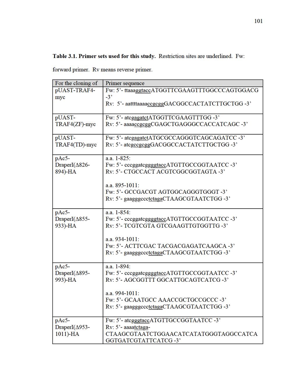
116
PI3K signaling and Stat92E converge to modulate glial responsiveness to axonal injury
 University of Massachusetts Medical School escholarship@umms Open Access Articles Open Access Publications by UMMS Authors 11-4-2014 PI3K signaling and Stat92E converge to modulate glial responsiveness
Eaters of the Dead: How Glial Cells Respond to and Engulf Degenerating Axons in the CNS: A Dissertation
 University of Massachusetts Medical School escholarship@umms GSBS Dissertations and Theses Graduate School of Biomedical Sciences 6-11-2012 Eaters of the Dead: How Glial Cells Respond to and Engulf Degenerating
Cells. Steven McLoon Department of Neuroscience University of Minnesota
 Cells Steven McLoon Department of Neuroscience University of Minnesota 1 Microscopy Methods of histology: Treat the tissue with a preservative (e.g. formaldehyde). Dissect the region of interest. Embed
Cellular and Molecular Mechanisms Driving Glial Engulfment of Degenerating Axons: A Dissertation
 University of Massachusetts Medical School escholarship@umms GSBS Dissertations and Theses Graduate School of Biomedical Sciences 2011-11-14 Cellular and Molecular Mechanisms Driving Glial Engulfment of
Cellular Neuroanatomy II The Prototypical Neuron: Neurites. Reading: BCP Chapter 2
 Cellular Neuroanatomy II The Prototypical Neuron: Neurites Reading: BCP Chapter 2 Major Internal Features of a Neuron The neuron is the functional unit of the nervous system. A typical neuron has a soma
Signal Transduction. Dr. Chaidir, Apt
 Signal Transduction Dr. Chaidir, Apt Background Complex unicellular organisms existed on Earth for approximately 2.5 billion years before the first multicellular organisms appeared.this long period for
Activation of a receptor. Assembly of the complex
 Activation of a receptor ligand inactive, monomeric active, dimeric When activated by growth factor binding, the growth factor receptor tyrosine kinase phosphorylates the neighboring receptor. Assembly
"Small Brains, Big Ideas: The value of model organisms to science. Dr. Yuly Fuentes-Medel Executive Director Descience
 "Small Brains, Big Ideas: The value of model organisms to science Dr. Yuly Fuentes-Medel Executive Director Descience Outline 1. What is a model organism? 2. Big questions solved by small-brained organisms
Dendrites - receives information from other neuron cells - input receivers.
 The Nerve Tissue Neuron - the nerve cell Dendrites - receives information from other neuron cells - input receivers. Cell body - includes usual parts of the organelles of a cell (nucleus, mitochondria)
Cell Death & Trophic Factors II. Steven McLoon Department of Neuroscience University of Minnesota
 Cell Death & Trophic Factors II Steven McLoon Department of Neuroscience University of Minnesota 1 Remember? Neurotrophins are cell survival factors that neurons get from their target cells! There is a
Conclusions. The experimental studies presented in this thesis provide the first molecular insights
 C h a p t e r 5 Conclusions 5.1 Summary The experimental studies presented in this thesis provide the first molecular insights into the cellular processes of assembly, and aggregation of neural crest and
Nervous Tissue. Neurons Neural communication Nervous Systems
 Nervous Tissue Neurons Neural communication Nervous Systems What is the function of nervous tissue? Maintain homeostasis & respond to stimuli Sense & transmit information rapidly, to specific cells and
Richik N. Ghosh, Linnette Grove, and Oleg Lapets ASSAY and Drug Development Technologies 2004, 2:
 1 3/1/2005 A Quantitative Cell-Based High-Content Screening Assay for the Epidermal Growth Factor Receptor-Specific Activation of Mitogen-Activated Protein Kinase Richik N. Ghosh, Linnette Grove, and Oleg
Cell Cell Communication in Development
 Biology 4361 Developmental Biology Cell Cell Communication in Development June 25, 2008 Cell Cell Communication Concepts Cells in developing organisms develop in the context of their environment, including
Introduction Principles of Signaling and Organization p. 3 Signaling in Simple Neuronal Circuits p. 4 Organization of the Retina p.
 Introduction Principles of Signaling and Organization p. 3 Signaling in Simple Neuronal Circuits p. 4 Organization of the Retina p. 5 Signaling in Nerve Cells p. 9 Cellular and Molecular Biology of Neurons
Cell-Cell Communication in Development
 Biology 4361 - Developmental Biology Cell-Cell Communication in Development June 23, 2009 Concepts Cell-Cell Communication Cells develop in the context of their environment, including: - their immediate
Neurons and Nervous Systems
 34 Neurons and Nervous Systems Concept 34.1 Nervous Systems Consist of Neurons and Glia Nervous systems have two categories of cells: Neurons, or nerve cells, are excitable they generate and transmit electrical
Cell-Cell Communication in Development
 Biology 4361 - Developmental Biology Cell-Cell Communication in Development October 2, 2007 Cell-Cell Communication - Topics Induction and competence Paracrine factors inducer molecules Signal transduction
COMPUTER SIMULATION OF DIFFERENTIAL KINETICS OF MAPK ACTIVATION UPON EGF RECEPTOR OVEREXPRESSION
 COMPUTER SIMULATION OF DIFFERENTIAL KINETICS OF MAPK ACTIVATION UPON EGF RECEPTOR OVEREXPRESSION I. Aksan 1, M. Sen 2, M. K. Araz 3, and M. L. Kurnaz 3 1 School of Biological Sciences, University of Manchester,
NEURONS, SENSE ORGANS, AND NERVOUS SYSTEMS CHAPTER 34
 NEURONS, SENSE ORGANS, AND NERVOUS SYSTEMS CHAPTER 34 KEY CONCEPTS 34.1 Nervous Systems Are Composed of Neurons and Glial Cells 34.2 Neurons Generate Electric Signals by Controlling Ion Distributions 34.3
Regulation and signaling. Overview. Control of gene expression. Cells need to regulate the amounts of different proteins they express, depending on
 Regulation and signaling Overview Cells need to regulate the amounts of different proteins they express, depending on cell development (skin vs liver cell) cell stage environmental conditions (food, temperature,
Nervous Tissue. Neurons Electrochemical Gradient Propagation & Transduction Neurotransmitters Temporal & Spatial Summation
 Nervous Tissue Neurons Electrochemical Gradient Propagation & Transduction Neurotransmitters Temporal & Spatial Summation What is the function of nervous tissue? Maintain homeostasis & respond to stimuli
Physiology 2 nd year. Neuroscience Optional Lecture
 Academic year 2018/2019 Physiology 2 nd year Semester 1 Curricula Nervous system physiology Blood physiology Acid-base equilibrium Bibliography: Boron & Boulpaep Medical Physiology, 3 rd edition Physiology
Intro and Homeostasis
 Intro and Homeostasis Physiology - how the body works. Homeostasis - staying the same. Functional Types of Neurons Sensory (afferent - coming in) neurons: Detects the changes in the body. Informations
The majority of cells in the nervous system arise during the embryonic and early post
 Introduction Introduction The majority of cells in the nervous system arise during the embryonic and early post natal period. These cells are derived from population of neural stem cells first shown by
Sensory/ Motor Systems March 10, 2008
 Sensory/ Motor Systems March 10, 2008 Greg Suh greg.suh@med.nyu.edu Title: Chemical Senses- periphery Richard Axel and Linda Buck Win Nobel Prize for Olfactory System Research 2 3 After Leslie Vosshal
Neurochemistry 1. Nervous system is made of neurons & glia, as well as other cells. Santiago Ramon y Cajal Nobel Prize 1906
 Neurochemistry 1 Nervous system is made of neurons & glia, as well as other cells. Santiago Ramon y Cajal Nobel Prize 1906 How Many Neurons Do We Have? The human brain contains ~86 billion neurons and
MEMBRANE POTENTIALS AND ACTION POTENTIALS:
 University of Jordan Faculty of Medicine Department of Physiology & Biochemistry Medical students, 2017/2018 +++++++++++++++++++++++++++++++++++++++++++++++++++++++++++++++++++++++++ Review: Membrane physiology
Course Descriptions Biology
 Course Descriptions Biology BIOL 1010 (F/S) Human Anatomy and Physiology I. An introductory study of the structure and function of the human organ systems including the nervous, sensory, muscular, skeletal,
5- Semaphorin-Plexin-Neuropilin
 5- Semaphorin-Plexin-Neuropilin 1 SEMAPHORINS-PLEXINS-NEUROPILINS ligands receptors co-receptors semaphorins and their receptors are known signals for: -axon guidance -cell migration -morphogenesis -immune
Big Idea 1: The process of evolution drives the diversity and unity of life.
 Big Idea 1: The process of evolution drives the diversity and unity of life. understanding 1.A: Change in the genetic makeup of a population over time is evolution. 1.A.1: Natural selection is a major
Ch 7. The Nervous System 7.1 & 7.2
 Ch 7 The Nervous System 7.1 & 7.2 SLOs Describe the different types of neurons and supporting cells, and identify their functions. Identify the myelin sheath and describe how it is formed in the CNS and
Under the Radar Screen: How Bugs Trick Our Immune Defenses
 Under the Radar Screen: How Bugs Trick Our Immune Defenses Session 2: Phagocytosis Marie-Eve Paquet and Gijsbert Grotenbreg Whitehead Institute for Biomedical Research Salmonella Gram negative bacteria
AP Curriculum Framework with Learning Objectives
 Big Ideas Big Idea 1: The process of evolution drives the diversity and unity of life. AP Curriculum Framework with Learning Objectives Understanding 1.A: Change in the genetic makeup of a population over
Cellular Neuroanatomy I The Prototypical Neuron: Soma. Reading: BCP Chapter 2
 Cellular Neuroanatomy I The Prototypical Neuron: Soma Reading: BCP Chapter 2 Functional Unit of the Nervous System The functional unit of the nervous system is the neuron. Neurons are cells specialized
Geert Geeven. April 14, 2010
 iction of Gene Regulatory Interactions NDNS+ Workshop April 14, 2010 Today s talk - Outline Outline Biological Background Construction of Predictors The main aim of my project is to better understand the
Chapter 48 Neurons, Synapses, and Signaling
 Chapter 48 Neurons, Synapses, and Signaling Concept 48.1 Neuron organization and structure reflect function in information transfer Neurons are nerve cells that transfer information within the body Neurons
Chapter 9. Nerve Signals and Homeostasis
 Chapter 9 Nerve Signals and Homeostasis A neuron is a specialized nerve cell that is the functional unit of the nervous system. Neural signaling communication by neurons is the process by which an animal
purpose of this Chapter is to highlight some problems that will likely provide new
 119 Chapter 6 Future Directions Besides our contributions discussed in previous chapters to the problem of developmental pattern formation, this work has also brought new questions that remain unanswered.
1
 http://photos1.blogger.com/img/13/2627/640/screenhunter_047.jpg 1 The Nose Knows http://graphics.jsonline.com/graphics/owlive/img/mar05/sideways.one0308_big.jpg 2 http://www.stlmoviereviewweekly.com/sitebuilder/images/sideways-253x364.jpg
Developmental genetics: finding the genes that regulate development
 Developmental Biology BY1101 P. Murphy Lecture 9 Developmental genetics: finding the genes that regulate development Introduction The application of genetic analysis and DNA technology to the study of
Supporting Information
 Supporting Information Cao et al. 10.1073/pnas.1306220110 Gram - bacteria Hemolymph Cytoplasm PGRP-LC TAK1 signalosome Imd dfadd Dredd Dnr1 Ikk signalosome P Relish Nucleus AMP and effector genes Fig.
Visual pigments. Neuroscience, Biochemistry Dr. Mamoun Ahram Third year, 2019
 Visual pigments Neuroscience, Biochemistry Dr. Mamoun Ahram Third year, 2019 References Webvision: The Organization of the Retina and Visual System (http://www.ncbi.nlm.nih.gov/books/nbk11522/#a 127) The
Enduring understanding 1.A: Change in the genetic makeup of a population over time is evolution.
 The AP Biology course is designed to enable you to develop advanced inquiry and reasoning skills, such as designing a plan for collecting data, analyzing data, applying mathematical routines, and connecting
Chapter 4 Evaluating a potential interaction between deltex and git in Drosophila: genetic interaction, gene overexpression and cell biology assays.
 Evaluating a potential interaction between deltex and git in Drosophila: genetic interaction, gene overexpression and cell biology assays. The data described in chapter 3 presented evidence that endogenous
Monitoring neurite morphology and synapse formation in primary neurons for neurotoxicity assessments and drug screening
 APPLICATION NOTE ArrayScan High Content Platform Monitoring neurite morphology and synapse formation in primary neurons for neurotoxicity assessments and drug screening Suk J. Hong and Richik N. Ghosh
Axon Death Prevented: Wld s and Other Neuroprotective Molecules: A Dissertation
 University of Massachusetts Medical School escholarship@umms GSBS Dissertations and Theses Graduate School of Biomedical Sciences 2010-12-13 Axon Death Prevented: Wld s and Other Neuroprotective Molecules:
Cellular Neurobiology BIPN 140 Fall 2016 Problem Set #8
 Cellular Neurobiology BIPN 140 Fall 2016 Problem Set #8 1. Inductive signaling is a hallmark of vertebrate and mammalian development. In early neural development, there are multiple signaling pathways
Reception The target cell s detection of a signal coming from outside the cell May Occur by: Direct connect Through signal molecules
 Why Do Cells Communicate? Regulation Cells need to control cellular processes In multicellular organism, cells signaling pathways coordinate the activities within individual cells that support the function
The sense of smell Outline Main Olfactory System Odor Detection Odor Coding Accessory Olfactory System Pheromone Detection Pheromone Coding
 The sense of smell Outline Main Olfactory System Odor Detection Odor Coding Accessory Olfactory System Pheromone Detection Pheromone Coding 1 Human experiment: How well do we taste without smell? 2 Brief
Overview Organization: Central Nervous System (CNS) Peripheral Nervous System (PNS) innervate Divisions: a. Afferent
 Peripheral Nervous System (PNS) innervate Divisions: a. Afferent") Overview Organization: Central Nervous System (CNS) Brain and spinal cord receives and processes information. Peripheral Nervous System (PNS) Nerve cells that link CNS with organs throughout the body.
Overview Organization: Central Nervous System (CNS) Brain and spinal cord receives and processes information. Peripheral Nervous System (PNS) Nerve cells that link CNS with organs throughout the body.
Nervous System Organization
 The Nervous System Nervous System Organization Receptors respond to stimuli Sensory receptors detect the stimulus Motor effectors respond to stimulus Nervous system divisions Central nervous system Command
Domain 6: Communication
 Domain 6: Communication 6.1: Cell communication processes share common features that reflect a shared evolutionary history. (EK3.D.1) 1. Introduction to Communication Communication requires the generation,
13-3. Synthesis-Secretory pathway: Sort lumenal proteins, Secrete proteins, Sort membrane proteins
 13-3. Synthesis-Secretory pathway: Sort lumenal proteins, Secrete proteins, Sort membrane proteins Molecular sorting: specific budding, vesicular transport, fusion 1. Why is this important? A. Form and
Glial cells occupy more than half of the volume of the human
 review doi:10.1038/nature09612 Regulation of synaptic connectivity by glia Cagla Eroglu 1 & Ben A. Barres 2 The human brain contains more than 100 trillion (10 14 ) synaptic connections, which form all
RANK. Alternative names. Discovery. Structure. William J. Boyle* SUMMARY BACKGROUND
 RANK William J. Boyle* Department of Cell Biology, Amgen, Inc., One Amgen Center Drive, Thousand Oaks, CA 91320-1799, USA * corresponding author tel: 805-447-4304, fax: 805-447-1982, e-mail: bboyle@amgen.com
Lecture 7. Development of the Fruit Fly Drosophila
 BIOLOGY 205/SECTION 7 DEVELOPMENT- LILJEGREN Lecture 7 Development of the Fruit Fly Drosophila 1. The fruit fly- a highly successful, specialized organism a. Quick life cycle includes three larval stages
Sensory Encoding of Smell in the Olfactory System of Drosophila
 Sensory Encoding of Smell in the Olfactory System of Drosophila (reviewing Olfactory Information Processing in Drosophila by Masse et al, 2009) Ben Cipollini COGS 160 May 11, 2010 Smell Drives Our Behavior...
10/2/2015. Chapter 4. Determination and Differentiation. Neuroanatomical Diversity
 Chapter 4 Determination and Differentiation Neuroanatomical Diversity 1 Neurochemical diversity: another important aspect of neuronal fate Neurotransmitters and their receptors Excitatory Glutamate Acetylcholine
Axon guidance I. Paul Garrity March 15, /9.013
 Axon guidance I Paul Garrity March 15, 2004 7.68/9.013 Neuronal Wiring: Functional Framework of the Nervous System Stretch reflex circuit Early theories of axonogenesis Schwann: many neurons link to form
Zool 3200: Cell Biology Exam 5 4/27/15
 Name: Trask Zool 3200: Cell Biology Exam 5 4/27/15 Answer each of the following short answer questions in the space provided, giving explanations when asked to do so. Circle the correct answer or answers
Announcements: Test4: Wednesday on: week4 material CH5 CH6 & NIA CAPE Evaluations please do them for me!! ask questions...discuss listen learn.
 Announcements: Test4: Wednesday on: week4 material CH5 CH6 & NIA CAPE Evaluations please do them for me!! ask questions...discuss listen learn. The Chemical Senses: Olfaction Mary ET Boyle, Ph.D. Department
A. Incorrect! The Cell Cycle contains 4 distinct phases: (1) G 1, (2) S Phase, (3) G 2 and (4) M Phase.
 G 1, (2) S Phase, (3) G 2 and (4) M Phase.") Molecular Cell Biology - Problem Drill 21: Cell Cycle and Cell Death Question No. 1 of 10 1. Which of the following statements about the cell cycle is correct? Question #1 (A) The Cell Cycle contains 3
Molecular Cell Biology - Problem Drill 21: Cell Cycle and Cell Death Question No. 1 of 10 1. Which of the following statements about the cell cycle is correct? Question #1 (A) The Cell Cycle contains 3
Neurons, Synapses, and Signaling
 LECTURE PRESENTATIONS For CAMPBELL BIOLOGY, NINTH EDITION Jane B. Reece, Lisa A. Urry, Michael L. Cain, Steven A. Wasserman, Peter V. Minorsky, Robert B. Jackson Chapter 48 Neurons, Synapses, and Signaling
Life Sciences 1a: Section 3B. The cell division cycle Objectives Understand the challenges to producing genetically identical daughter cells
 Life Sciences 1a: Section 3B. The cell division cycle Objectives Understand the challenges to producing genetically identical daughter cells Understand how a simple biochemical oscillator can drive the
NEURONS Excitable cells Therefore, have a RMP Synapse = chemical communication site between neurons, from pre-synaptic release to postsynaptic
 NEUROPHYSIOLOGY NOTES L1 WHAT IS NEUROPHYSIOLOGY? NEURONS Excitable cells Therefore, have a RMP Synapse = chemical communication site between neurons, from pre-synaptic release to postsynaptic receptor
Gene Control Mechanisms at Transcription and Translation Levels
 Gene Control Mechanisms at Transcription and Translation Levels Dr. M. Vijayalakshmi School of Chemical and Biotechnology SASTRA University Joint Initiative of IITs and IISc Funded by MHRD Page 1 of 9
Host-Pathogen Interaction. PN Sharma Department of Plant Pathology CSK HPKV, Palampur
 Host-Pathogen Interaction PN Sharma Department of Plant Pathology CSK HPKV, Palampur-176062 PATHOGEN DEFENCE IN PLANTS A BIOLOGICAL AND MOLECULAR VIEW Two types of plant resistance response to potential
Patrick: An Introduction to Medicinal Chemistry 5e Chapter 04
 01) Which of the following statements is not true about receptors? a. Most receptors are proteins situated inside the cell. b. Receptors contain a hollow or cleft on their surface which is known as a binding
The EGF Signaling Pathway! Introduction! Introduction! Chem Lecture 10 Signal Transduction & Sensory Systems Part 3. EGF promotes cell growth
 Chem 452 - Lecture 10 Signal Transduction & Sensory Systems Part 3 Question of the Day: Who is the son of Sevenless? Introduction! Signal transduction involves the changing of a cell s metabolism or gene
Neurophysiology. Danil Hammoudi.MD
 Neurophysiology Danil Hammoudi.MD ACTION POTENTIAL An action potential is a wave of electrical discharge that travels along the membrane of a cell. Action potentials are an essential feature of animal
ADVANCED PLACEMENT BIOLOGY
 ADVANCED PLACEMENT BIOLOGY Description Advanced Placement Biology is designed to be the equivalent of a two-semester college introductory course for Biology majors. The course meets seven periods per week
Baz, Par-6 and apkc are not required for axon or dendrite specification in Drosophila
 Baz, Par-6 and apkc are not required for axon or dendrite specification in Drosophila Melissa M. Rolls and Chris Q. Doe, Inst. Neurosci and Inst. Mol. Biol., HHMI, Univ. Oregon, Eugene, Oregon 97403 Correspondence
A A A A B B1
 LEARNING OBJECTIVES FOR EACH BIG IDEA WITH ASSOCIATED SCIENCE PRACTICES AND ESSENTIAL KNOWLEDGE Learning Objectives will be the target for AP Biology exam questions Learning Objectives Sci Prac Es Knowl
Cytokines regulate interactions between cells of the hemapoietic system
 Cytokines regulate interactions between cells of the hemapoietic system Some well-known cytokines: Erythropoietin (Epo) G-CSF Thrombopoietin IL-2 INF thrombopoietin Abbas et al. Cellular & Molecular Immunology
BIOLOGY. 1. Overview of Neurons 11/3/2014. Neurons, Synapses, and Signaling. Communication in Neurons
 CAMPBELL BIOLOGY TENTH EDITION 48 Reece Urry Cain Wasserman Minorsky Jackson Neurons, Synapses, and Signaling Lecture Presentation by Nicole Tunbridge and Kathleen Fitzpatrick 1. Overview of Neurons Communication
DOWNLOAD OR READ : THE NEURONAL CYTOSKELETON MOTOR PROTEINS AND ORGANELLE TRAFFICKING IN THE AXON PDF EBOOK EPUB MOBI
 DOWNLOAD OR READ : THE NEURONAL CYTOSKELETON MOTOR PROTEINS AND ORGANELLE TRAFFICKING IN THE AXON PDF EBOOK EPUB MOBI Page 1 Page 2 the neuronal cytoskeleton motor proteins and organelle trafficking in
ADAM FAMILY. ephrin A INTERAZIONE. Eph ADESIONE? PROTEOLISI ENDOCITOSI B A RISULTATO REPULSIONE. reverse. forward
 ADAM FAMILY - a family of membrane-anchored metalloproteases that are known as A Disintegrin And Metalloprotease proteins and are key components in protein ectodomain shedding Eph A INTERAZIONE B ephrin
The neuron as a secretory cell
 The neuron as a secretory cell EXOCYTOSIS ENDOCYTOSIS The secretory pathway. Transport and sorting of proteins in the secretory pathway occur as they pass through the Golgi complex before reaching the
Neural Tissue. PowerPoint Lecture Presentations prepared by Jason LaPres. Lone Star College North Harris Pearson Education, Inc.
 12 Neural Tissue PowerPoint Lecture Presentations prepared by Jason LaPres Lone Star College North Harris An Introduction to the Nervous System The Nervous System Includes all neural tissue in the body
Fig. S1. Expression pattern of moody-gal4 in third instar. Maximum projection illustrating a dissected moody-gal4>ngfp L3 larva stained for Repo
 Fig. S1. Expression pattern of moody-gal4 in third instar. Maximum projection illustrating a dissected moody-gal4>ngfp L3 larva stained for Repo (magenta), Fas2 (blue) and GFP (green) in overview (A) and
S1 Gene ontology (GO) analysis of the network alignment results
 analysis of the network alignment results") 1 Supplementary Material for Effective comparative analysis of protein-protein interaction networks by measuring the steady-state network flow using a Markov model Hyundoo Jeong 1, Xiaoning Qian 1 and
1 Supplementary Material for Effective comparative analysis of protein-protein interaction networks by measuring the steady-state network flow using a Markov model Hyundoo Jeong 1, Xiaoning Qian 1 and
Cell biology: Death drags down the neighbourhood
 Cell biology: Death drags down the neighbourhood The MIT Faculty has made this article openly available. Please share how this access benefits you. Your story matters. Citation As Published Publisher Vasquez,
Astrocyte-Neuron Interactions Regulate Nervous System Assembly and Function: A Dissertation
 University of Massachusetts Medical School escholarship@umms GSBS Dissertations and Theses Graduate School of Biomedical Sciences 1-8-2015 Astrocyte-Neuron Interactions Regulate Nervous System Assembly
Big Idea 3: Living systems store, retrieve, transmit and respond to information essential to life processes. Tuesday, December 27, 16
 Big Idea 3: Living systems store, retrieve, transmit and respond to information essential to life processes. Enduring understanding 3.B: Expression of genetic information involves cellular and molecular
NN-A29932: Supplementary Data 1 Diversity and Wiring Variability of Olfactory Local Interneurons in the Drosophila Antennal Lobe
 NN-A29932: Supplementary Data 1 Diversity and Wiring Variability of Olfactory Local Interneurons in the Drosophila Antennal Lobe Ya-Hui Chou, Maria L. Spletter, Emre Yaksi, Jonathan C. S. Leong, Rachel
Nervous Systems: Neuron Structure and Function
 Nervous Systems: Neuron Structure and Function Integration An animal needs to function like a coherent organism, not like a loose collection of cells. Integration = refers to processes such as summation
Shavenbaby Couples Patterning to Epidermal Cell Shape Control. Chanut-Delalande H, Fernandes I, Roch F, Payre F, Plaza S (2006) PLoS Biol 4(9): e290
 PLoS Biol 4(9): e290") Shavenbaby Couples Patterning to Epidermal Cell Shape Control. Chanut-Delalande H, Fernandes I, Roch F, Payre F, Plaza S (2006) PLoS Biol 4(9): e290 Question (from Introduction): How does svb control the
Shavenbaby Couples Patterning to Epidermal Cell Shape Control. Chanut-Delalande H, Fernandes I, Roch F, Payre F, Plaza S (2006) PLoS Biol 4(9): e290 Question (from Introduction): How does svb control the
PREREQUISITE CHECKLIST
 PREREQUISITE CHECKLIST UNIVERSITY OF CALIFORNIA, BERKELEY SCHOOL OF OPTOMETRY ADMISSIONS AND STUDENT AFFAIRS OFFICE Name: Date: Email: Status (complete, in progress, or planned) Prerequisite Course Requirements
Trophic Factors. Trophic Factors. History 2. History Growth Factors. Giles Plant
 217 - Growth Factors Giles Plant Role in: Growth and Trophic Factors Soluble/diffusible factors - polypeptides Proliferation Differentiation (ie Cancer) Survival (degenerative diseases) Innervation Maintenance
Biol403 - Receptor Serine/Threonine Kinases
 Biol403 - Receptor Serine/Threonine Kinases The TGFβ (transforming growth factorβ) family of growth factors TGFβ1 was first identified as a transforming factor; however, it is a member of a family of structurally
Department Curriculum and Assessment Outline
 Department: Science Year Group: 10 Teaching, learning and assessment during the course: Combined Science 1 2 B1 Key concepts in Biology B2 Cells and control What are the structure and function of cells.
Chem Lecture 10 Signal Transduction
 Chem 452 - Lecture 10 Signal Transduction 111202 Here we look at the movement of a signal from the outside of a cell to its inside, where it elicits changes within the cell. These changes are usually mediated
Old FINAL EXAM BIO409/509 NAME. Please number your answers and write them on the attached, lined paper.
 Old FINAL EXAM BIO409/509 NAME Please number your answers and write them on the attached, lined paper. Gene expression can be regulated at several steps. Describe one example for each of the following:
Map of AP-Aligned Bio-Rad Kits with Learning Objectives
 Map of AP-Aligned Bio-Rad Kits with Learning Objectives Cover more than one AP Biology Big Idea with these AP-aligned Bio-Rad kits. Big Idea 1 Big Idea 2 Big Idea 3 Big Idea 4 ThINQ! pglo Transformation
Types of biological networks. I. Intra-cellurar networks
 Types of biological networks I. Intra-cellurar networks 1 Some intra-cellular networks: 1. Metabolic networks 2. Transcriptional regulation networks 3. Cell signalling networks 4. Protein-protein interaction
with%dr.%van%buskirk%%%
 with%dr.%van%buskirk%%% How$to$do$well?$ Before$class:$read$the$corresponding$chapter$ Come$to$class$ready$to$par9cipate$in$Top$Hat$ Don t$miss$an$exam!!!!!!!!!!!!!!!!!!!!!!!!!!$ But$I m$not$good$with$science
Nervous System Organization
 The Nervous System Chapter 44 Nervous System Organization All animals must be able to respond to environmental stimuli -Sensory receptors = Detect stimulus -Motor effectors = Respond to it -The nervous
Neurons. The Molecular Basis of their Electrical Excitability
 Neurons The Molecular Basis of their Electrical Excitability Viva La Complexity! Consider, The human brain contains >10 11 neurons! Each neuron makes 10 3 (average) synaptic contacts on up to 10 3 other
Chapter 11. Development: Differentiation and Determination
 KAP Biology Dept Kenyon College Differential gene expression and development Mechanisms of cellular determination Induction Pattern formation Chapter 11. Development: Differentiation and Determination