Diffusion imaging of the brain: technical considerations and practical applications
|
|
|
- Gwendoline Cummings
- 6 years ago
- Views:
Transcription
1 Diffusion imaging of the brain: technical considerations and practical applications David G. Norris FC Donders Centre for Cognitive Neuroimaging Nijmegen
2 Sustaining the physiologist in measuring the atomic movements upon which vitality and thought depend after W.C. Roberts-Austen
3 Introduction Diffusion-weighted magnetic resonance imaging has become important in biomedical applications for two reasons: Diagnosis. Particularly of brain infarcts. Fibre-Tracking. Useful both in planning neurosurgery and for mapping connections in the brain.

4 During infarction the signal attenuation in diffusion weighted imaging is reduced and infarcted regions appear bright.


5 3D views and comparison with anatomical preparation Red: Anterior thalamic radiation Light Blue: Posterior thalamic radiation Pink: Superior longitudinal fascicules Blue: Inferior longitudinal fascicules Green: Inferior fronto-occipital fasc. Yellow: Uncinate fiber Courtesy of Susumo Mori (Johns Hopkins)
6 Contents Basic NMR diffusion experiment Diffusion in tissue Anisotropic diffusion Confounds: eddy currents and motion Fibre tracking
7 Basic Theory All MR methods for detecting diffusion are based on the loss of phase coherence resulting from stochastic motion in the presence of a magnetic field gradient.

8 Diffusing spins acquire a phase difference

9 Pulsed Gradient Spin-Echo
10 Attenuation for this experiment is given by: M ( D) ( ) ( ) 2 r,t = M exp γgδ ( Δ ) xy 0 δ 3 The b-value is defined as: ( ) 2 γ δ ( Δ δ 3) b = G

11 Pulsed Gradient Stimulated- Echo
12 Stimulated echo only offers half the signal intensity of a spin echo, but Gives access to very high attenuation factors, and To very long diffusion times
13 Biophysical situation? Diffusion MR allows us to probe tissue microstructure on the micrometer scale. Key relationship is Einstein equation: r 2 = 6Dτ D with typical values of D about 10-3 mm 2 s -1 and diffusion times of about 80 ms (required for a reasonable diffusion attenuation) we obtain mean path length of approx. 20 μm.

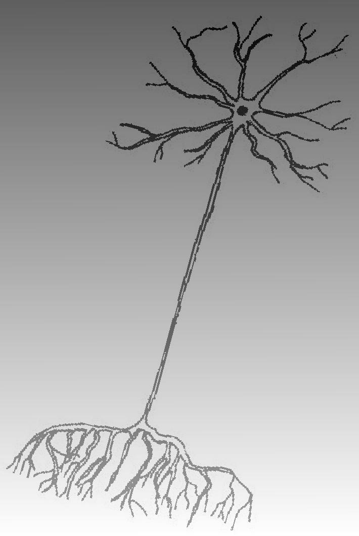
14 Stained microscopic picture of brain tissue A-cell body B-axon and dendrite C-neuroglia
15 Biophysical Situation Brain tissue has an intravascular compartment of about 4% by volume. The remainder is divided between extra- and intracellular spaces in the ratio of 1:4. Water exchanges between these three compartments.
16 Simplest model is two compartments separated by a membrane of limited permeability. Diffusion in the intracellular compartment may be restricted by the cell membrane. In the extracellular space the diffusion length will be reduced by the requirement to diffuse around the cells. Diffusion coefficient often termed ADC: Apparent Diffusion Coefficient

17 Typical modeling approaches
18 Implicit in most models is an assumption that the relative sizes of the intra- and extracellular spaces and the degree of extracellular tortuosity determine ADC. The lack of morphological information makes it difficult to model experimental data exactly.
19 Pathological Models In cerebral infarction a marked reduction in ADC occurs simultaneously with cell swelling and increased tortuosity. A fall in intracellular diffusion is also needed to make models work, but this has not been found experimentally
20 Relevant posters #76 Fast optical tracking of diffusion in brain extracellular space. Hrabe and Hrabetova #77 Dead spaces hinder diffusion and contribute to tortuosity of brain extracellular space. Hrabetova et al #86 Measurement of lcal diffusion properties in brain tissue. Nicholson et al
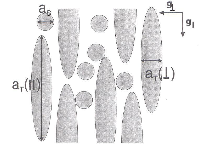
21 2μm Basis of Anisotropy The myelin sheath around axons form an obstacle to water diffusion. myelin sheath diffusion unrestricted diffusion restricted axon
22


23 Electron micrographs of nerve tissue parallel and perpendicular to tract orientation


24 The effect of different orientations of the diffusion weighting gradient on the signal from white matter as a result of anisotropy. (-x,y) (x,y) Slides courtesy: Ted Truard
25 C J
26 The diffusion tensor has the form: D = D D D xx yx zx The on-diagonal elements reflect diffusion parallel to a concentration gradient, the offdiagonal elements the component of diffusion perpendicular to it. D D D xy yy zy D D D xz yz zz
27 To measure the diffusion tensor you need to perform at least six independent measurements as there are six elements in this symmetrical tensor. Also need to know proton density. A regression analysis gives the full tensor information from which a number of scalar values can be obtained.
28 Eigenvalues and eigenvectors As the determinant of the diffusion tensor is positive definite there will always be three positive eigenvalues of the tensor. The three eigenvectors define the principal axes for the diffusion ellipsoid.
29 The diffusion ellipsoid If we consider diffusion along the principal axes and seek the tensor analogue of the Einstein relationship (<r 2> =2Dt) then r T Λ 1 r = 2 x y z 2t or + + = 2t λ 1 λ 2 2 λ 2 3 So the half axes of the ellipsoid are a i = 2tλ i

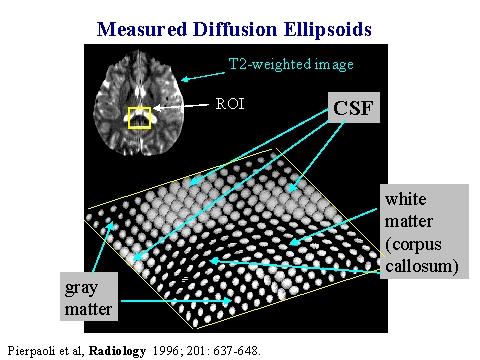
30 The ADC surface of the ellipsoid is a peanut From Frank Magnetic Resonance in Medicine 45, (2001)
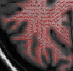
31 Visualisation The full tensor contains a large amount of information. Display of the ellipsoid is difficult but instructive. The orientation of the largest eigenvector is a precursor for fibre tracking. Color coding the axes: RGB for me needs a good knowledge of color mixing.
32
33


34 Colour coding From Masutani, et al. European Journal of Radiology 46, (2003)
35 Combining diffusion weighting and imaging Need pulsed magnetic field gradients for both spatial encoding and diffusion weighting. In human imaging, though not in MR microscopy, the diffusion weighting effect of the imaging gradients is negligible. Cleanest method is building block approach which keeps the diffusion and imaging parts separate from each other.
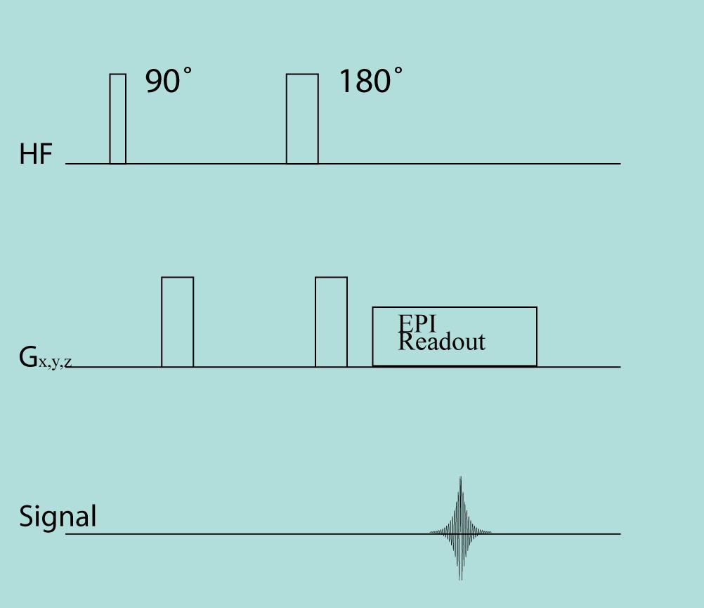
36
37 EPI. Salient features Collects data for complete image after one excitation. Places heavy requirements on gradient systems. Prone to artifacts: shift, scale, shear, voids. Spatial resolution limited to matrices of or maximally Can scan whole brain within seconds
38 Eddy current effects 1 Eddy currents are generally considered as constant during the imaging sequence. In reality they drop-off exponentially, but the approximation is acceptable for EPI. This means that eddy currents can be treated in the same way as effects of main field inhomogeneity.
39 Eddy current effects 2 The eddy current can be considered as producing a constant extra gradient parallel to the direction of the diffusion weighting gradient, and/or a frequency offset. The severity of eddy currents will depend on the strength of the gradients causing them. In tensor imaging multiple orientations of the weighting gradient and multiple strengths are used, and so these effects must be eliminated.
40 Solutions to eddy current artifacts Measure eddy current effects and eliminate in the post-processing. Tailor the diffusion-weighting part of the pulse-sequence to give lower eddy currents.
 2δγG Δ 2")
41 b = a ( ) 2δγG Δ 2 δ
42 General effects of bulk motion Translation causes a global phase shift given by φ = γgvδδ. Rotation causes a phase gradient across the object which, according to the Fourier shift theorem is equivalent to a shift in k-space.
43 Direction of the phase gradient is given by the cross product of the vectors of the diffusion weighting gradient and of the axis of rotation. r φ = γg Ω


44
45 So you are only safe from the effects of rotation if the diffusion-weighting gradient is parallel to the phaseencoding gradient! Multi-excitation experiments require phase-correction between excitations.
46 Pulsatile motion of the brain Common correction methods have implicit assumption that brain moves as a rigid body (no distortion). The pulsatile nature of arterial flow causes non-linear motion during up to about 200 ms post R-wave. Maximum velocities are mm sec -1. Highest velocities are along inferior superior axis. For these reasons it is currently best to use a sensitive single shot method, like EPI.
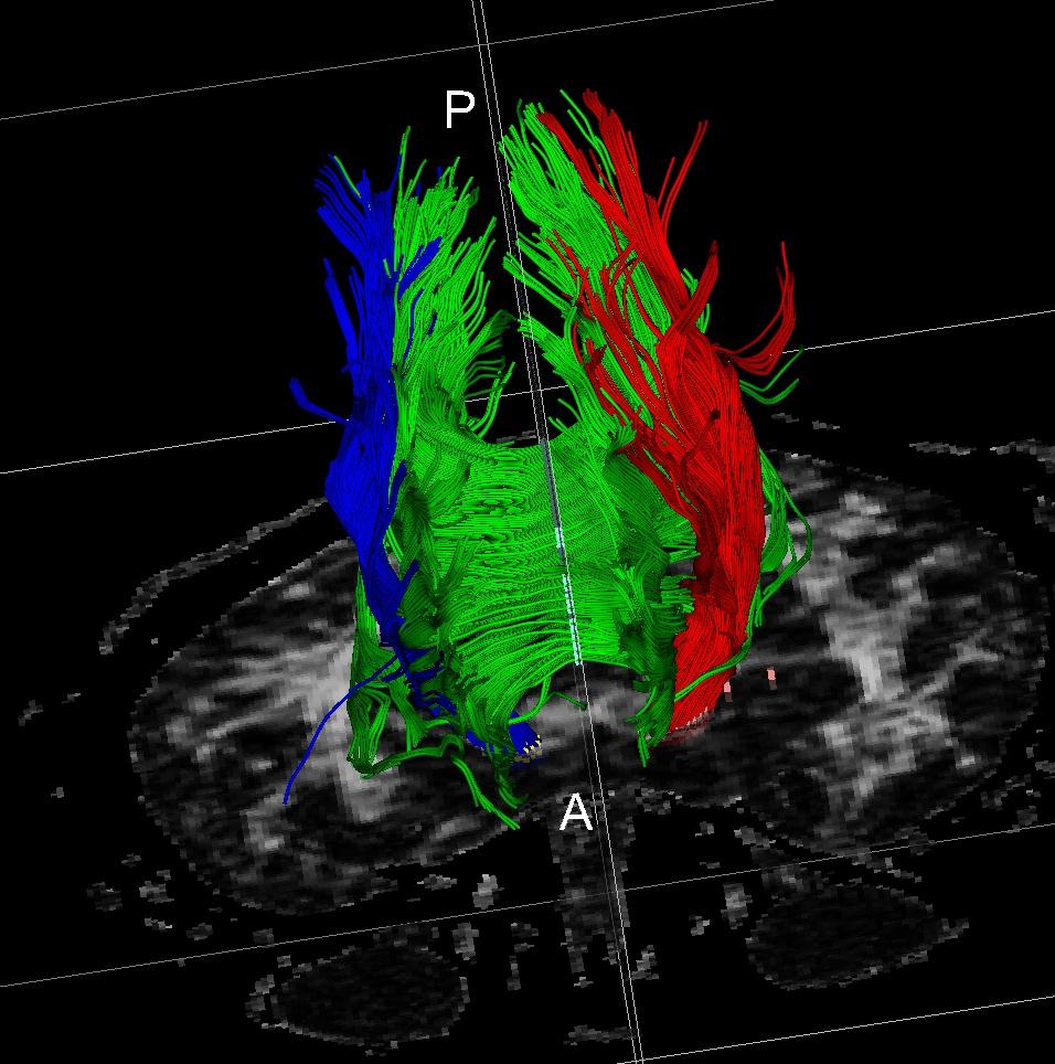
47 Fibre Tracking Surprisingly knowledge of fibre tracts in humans is not perfect. Animals easier to study as active transport in cells can be used. Fibre tracking in humans offers the possibility of combining neuroanatomical with functional information.

48 In all algorithms criteria are developed to connect all the vectors How do we deal with discrete voxels and continuous fibers?
 Tracking through a voxel with constant direction End point defined by the")
49 Line propagation methods Susumu Mori et al. Annals of Neurology 1999 Fiber Assignment by Continuous Tracking (FACT) Tracking through a voxel with constant direction End point defined by the occurrence of sudden transitions in the fiber orientation (low-curvature hypothesis) Mori et al. 1999
50
51 Probability-based based methods Martin Koch et al. Neuroimage 2002 From the seed point a (macroscopic) random walk is performed. The probability of this random walk going in a certain direction is determined by the diffusion tensor This procedure is then repeated in a Monte-Carlo simulation The frequency with which a voxel was encountered in the walk serves as an informal measure of connectivity This measure decreases with distance to seed voxel.

52 Probability-based based methods

53 Jumping particle simulation start
54 Evaluation Streamline-based methods lend themselves to good 3D visualization They don t give information about the certainty with which a tract is calculated Probability-based methods are difficult to visualize in 3D They give information about the likelihood of the tract This likelihood is (mostly) informal Their probability decreases over distance

55 Applications Catani et al. Annals of Neurology 2005 Is there another area connected (other than Broca and Wernicke) via the arcuate fasciculus
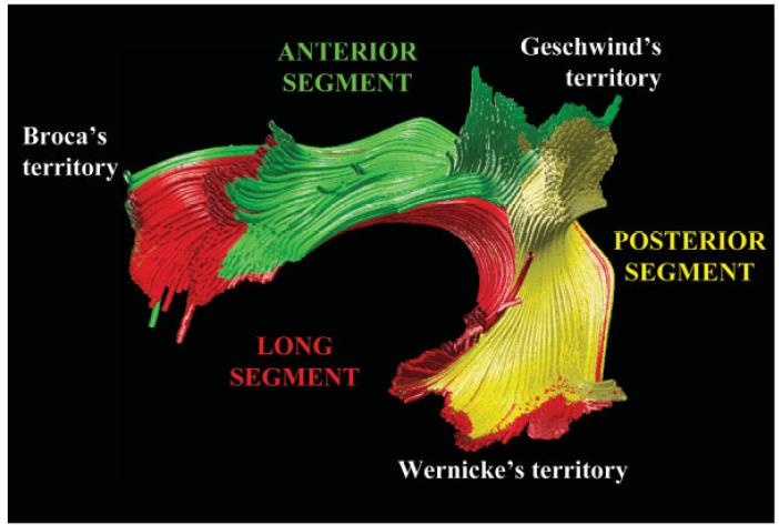
56 Applications
57 Connectivity Neuroscience moving away from which areas activate in task performance to how networks interact. Models based on anatomical hypotheses, many from macaque monkey. Can now start to test in humans
58 PP PP DE LP DE LP V1 V1 ITp ITa ITp ITa V1 = striate cortex DE = dorsal extrastriate visual cortex PP = posterior parietal cortex LP = lateral parietal cortex ITp = posterior inferotemporal cortex ITa = parahippocampal gyrus Legend Proposed connection Non-proposed connection Connection found after reanalysis with low FA Connection found in six subjects Connection found in five subjects Connection found in four subjects Connection found in three subjects Connection found in two subjects Connection found in one subject1
59 Acknowledgements Hubert Fonteijn Martin Koch
DIFFUSION MAGNETIC RESONANCE IMAGING
 DIFFUSION MAGNETIC RESONANCE IMAGING from spectroscopy to imaging apparent diffusion coefficient ADC-Map anisotropy diffusion tensor (imaging) DIFFUSION NMR - FROM SPECTROSCOPY TO IMAGING Combining Diffusion
Advanced Topics and Diffusion MRI
 Advanced Topics and Diffusion MRI Slides originally by Karla Miller, FMRIB Centre Modified by Mark Chiew (mark.chiew@ndcn.ox.ac.uk) Slides available at: http://users.fmrib.ox.ac.uk/~mchiew/teaching/ MRI
醫用磁振學 MRM 擴散張量影像 擴散張量影像原理. 本週課程內容 MR Diffusion 擴散張量造影原理 擴散張量造影應用 盧家鋒助理教授國立陽明大學生物醫學影像暨放射科學系
 本週課程內容 http://www.ym.edu.tw/~cflu 擴散張量造影原理 擴散張量造影應用 醫用磁振學 MRM 擴散張量影像 盧家鋒助理教授國立陽明大學生物醫學影像暨放射科學系 alvin4016@ym.edu.tw MRI The Basics (3rd edition) Chapter 22: Echo Planar Imaging MRI in Practice, (4th edition)
Tensor Visualization. CSC 7443: Scientific Information Visualization
 Tensor Visualization Tensor data A tensor is a multivariate quantity Scalar is a tensor of rank zero s = s(x,y,z) Vector is a tensor of rank one v = (v x,v y,v z ) For a symmetric tensor of rank 2, its
Diffusion Weighted MRI. Zanqi Liang & Hendrik Poernama
 Diffusion Weighted MRI Zanqi Liang & Hendrik Poernama 1 Outline MRI Quick Review What is Diffusion MRI? Detecting Diffusion Stroke and Tumor Detection Presenting Diffusion Anisotropy and Diffusion Tensor
Diffusion Tensor Imaging (DTI): An overview of key concepts
: An overview of key concepts") Diffusion Tensor Imaging (DTI): An overview of key concepts (Supplemental material for presentation) Prepared by: Nadia Barakat BMB 601 Chris Conklin Thursday, April 8 th 2010 Diffusion Concept [1,2]:
Diffusion Tensor Imaging (DTI): An overview of key concepts (Supplemental material for presentation) Prepared by: Nadia Barakat BMB 601 Chris Conklin Thursday, April 8 th 2010 Diffusion Concept [1,2]:
HST.583 Functional Magnetic Resonance Imaging: Data Acquisition and Analysis Fall 2006
 MIT OpenCourseWare http://ocw.mit.edu HST.583 Functional Magnetic Resonance Imaging: Data Acquisition and Analysis Fall 2006 For information about citing these materials or our Terms of Use, visit: http://ocw.mit.edu/terms.
Bayesian multi-tensor diffusion MRI and tractography
 Bayesian multi-tensor diffusion MRI and tractography Diwei Zhou 1, Ian L. Dryden 1, Alexey Koloydenko 1, & Li Bai 2 1 School of Mathematical Sciences, Univ. of Nottingham 2 School of Computer Science and
Ordinary Least Squares and its applications
 Ordinary Least Squares and its applications Dr. Mauro Zucchelli University Of Verona December 5, 2016 Dr. Mauro Zucchelli Ordinary Least Squares and its applications December 5, 2016 1 / 48 Contents 1
Application of diffusion MRI to cancer, heart and brain connectome imaging
 Colloquium @ Department of Physics, NTU Application of diffusion MRI to cancer, heart and brain connectome imaging March 11, 2014 Wen-Yih Isaac Tseng MD, PhD Advanced Biomedical MRI Lab Center for Optoelectronic
Diffusion Tensor Imaging in Humans: Practical Implications for Neuroanatomy
 Diffusion Tensor Imaging in Humans: Practical Implications for Neuroanatomy Collaborators Center for Morphometric Analysis: Nikos Makris Andy Worth Verne S. Caviness George Papadimitriou MGH-NMR Center
Diffusion Imaging II. By: Osama Abdullah
 iffusion Imaging II By: Osama Abdullah Review Introduction. What is diffusion? iffusion and signal attenuation. iffusion imaging. How to capture diffusion? iffusion sensitizing gradients. Spin Echo. Gradient
Applications of Spin Echo and Gradient Echo: Diffusion and Susceptibility Contrast
 Applications of Spin Echo and Gradient Echo: Diffusion and Susceptibility Contrast Chunlei Liu, PhD Department of Electrical Engineering & Computer Sciences and Helen Wills Neuroscience Institute University
Diffusion Tensor Imaging I: The basics. Jennifer Campbell
 Diffusion Tensor Imaging I: The basics Jennifer Campbell Diffusion Tensor Imaging I: The basics Jennifer Campbell Diffusion Imaging MRI: many different sources of contrast T1W T2W PDW Perfusion BOLD DW
Basics of Diffusion Tensor Imaging and DtiStudio
 Basics of Diffusion Tensor Imaging and DtiStudio DTI Basics 1 DTI reveals White matter anatomy Gray matter White matter DTI uses water diffusion as a probe for white matter anatomy Isotropic diffusion
A Neurosurgeon s Perspectives of Diffusion Tensor Imaging(DTI) Diffusion Tensor MRI (DTI) Background and Relevant Physics.
 Diffusion Tensor MRI (DTI) Background and Relevant Physics.") A Neurosurgeon s Perspectives of Diffusion Tensor Imaging(DTI) Kalai Arasu Muthusamy, D.Phil(Oxon) Senior Lecturer & Consultant Neurosurgeon. Division of Neurosurgery. University Malaya Medical Centre.
A Neurosurgeon s Perspectives of Diffusion Tensor Imaging(DTI) Kalai Arasu Muthusamy, D.Phil(Oxon) Senior Lecturer & Consultant Neurosurgeon. Division of Neurosurgery. University Malaya Medical Centre.
Diffusion Tensor Imaging tutorial
 NA-MIC http://na-mic.org Diffusion Tensor Imaging tutorial Sonia Pujol, PhD Surgical Planning Laboratory Harvard University DTI tutorial This tutorial is an introduction to the advanced Diffusion MR capabilities
Medical Visualization - Tensor Visualization. J.-Prof. Dr. Kai Lawonn
 Medical Visualization - Tensor Visualization J.-Prof. Dr. Kai Lawonn Lecture is partially based on the lecture by Prof. Thomas Schultz 2 What is a Tensor? A tensor is a multilinear transformation that
Diffusion MRI. Outline. Biology: The Neuron. Brain connectivity. Biology: Brain Organization. Brain connections and fibers
 Outline Diffusion MRI Alfred Anwander Download of Slides: www.cbs.mpg.de/events/ teaching/brainsignals1112 password: mpi-brain CBSWIKI: Cornet/DiffusionMRI Neuroanatomy Diffusion MRI Diffusion Tensor Imaging
Diffusion tensor imaging (DTI):
:") Diffusion tensor imaging (DTI): A basic introduction to data acquisition and analysis Matthew Cykowski, MD Postdoctoral fellow Research Imaging Center UTHSCSA Room 2.320 cykowski@uthscsa.edu PART I: Acquiring
Diffusion tensor imaging (DTI): A basic introduction to data acquisition and analysis Matthew Cykowski, MD Postdoctoral fellow Research Imaging Center UTHSCSA Room 2.320 cykowski@uthscsa.edu PART I: Acquiring
An Analytical Model of Water Diffusion and Exchange in White Matter from Diffusion MRI and Its Application in Measuring Axon Radii
 An Analytical Model of Water Diffusion and Exchange in White Matter from Diffusion MRI and Its Application in Measuring Axon Radii Wenjin Zhou, Student Member, IEEE, and David H. Laidlaw, Senior Member,
Diffusion-Weighted MRI may be used to measure the apparent diffusion coefficient of water in tissue.
 Specialty Area: MR Physics for Physicists Speaker: Jennifer A. McNab, Ph.D. Assistant Professor, Radiology, Stanford University () Highlights The Bloch-Torrey equation is a generalization of the Bloch
Higher Order Cartesian Tensor Representation of Orientation Distribution Functions (ODFs)
") Higher Order Cartesian Tensor Representation of Orientation Distribution Functions (ODFs) Yonas T. Weldeselassie (Ph.D. Candidate) Medical Image Computing and Analysis Lab, CS, SFU DT-MR Imaging Introduction
Higher Order Cartesian Tensor Representation of Orientation Distribution Functions (ODFs) Yonas T. Weldeselassie (Ph.D. Candidate) Medical Image Computing and Analysis Lab, CS, SFU DT-MR Imaging Introduction
Contrast Mechanisms in MRI. Michael Jay Schillaci
 Contrast Mechanisms in MRI Michael Jay Schillaci Overview Image Acquisition Basic Pulse Sequences Unwrapping K-Space Image Optimization Contrast Mechanisms Static and Motion Contrasts T1 & T2 Weighting,
Diffusion Tensor Imaging (DTI) e Neurite Orientation Dispersion and Density Imaging (NODDI)
 e Neurite Orientation Dispersion and Density Imaging (NODDI)") Diffusion Tensor Imaging (DTI) e Neurite Orientation Dispersion and Density Imaging (NODDI) Claudia AM Gandini Wheeler-Kingshott, PhD Prof. of MRI Physics Overview Diffusion and microstructure NODDI theoretical
Diffusion Tensor Imaging (DTI) e Neurite Orientation Dispersion and Density Imaging (NODDI) Claudia AM Gandini Wheeler-Kingshott, PhD Prof. of MRI Physics Overview Diffusion and microstructure NODDI theoretical
Basic MRI physics and Functional MRI
 Basic MRI physics and Functional MRI Gregory R. Lee, Ph.D Assistant Professor, Department of Radiology June 24, 2013 Pediatric Neuroimaging Research Consortium Objectives Neuroimaging Overview MR Physics
New developments in Magnetic Resonance Spectrocopy and Diffusion MRI. Els Fieremans Steven Delputte Mahir Ozdemir
 New developments in Magnetic Resonance Spectrocopy and Diffusion MRI Els Fieremans Steven Delputte Mahir Ozdemir Overview Magnetic Resonance Spectroscopy (MRS) Basic physics of MRS Quantitative MRS Pitfalls
Diffusion Tensor Imaging I. Jennifer Campbell
 Diffusion Tensor Imaging I Jennifer Campbell Diffusion Imaging Molecular diffusion The diffusion tensor Diffusion weighting in MRI Alternatives to the tensor Overview of applications Diffusion Imaging
EL-GY 6813/BE-GY 6203 Medical Imaging, Fall 2016 Final Exam
 EL-GY 6813/BE-GY 6203 Medical Imaging, Fall 2016 Final Exam (closed book, 1 sheets of notes double sided allowed, no calculator or other electronic devices allowed) 1. Ultrasound Physics (15 pt) A) (9
Tract-Specific Analysis for DTI of Brain White Matter
 Tract-Specific Analysis for DTI of Brain White Matter Paul Yushkevich, Hui Zhang, James Gee Penn Image Computing & Science Lab Department of Radiology University of Pennsylvania IPAM Summer School July
The effect of different number of diffusion gradients on SNR of diffusion tensor-derived measurement maps
 J. Biomedical Science and Engineering, 009,, 96-101 The effect of different number of diffusion gradients on SNR of diffusion tensor-derived measurement maps Na Zhang 1, Zhen-Sheng Deng 1*, Fang Wang 1,
Magnetic Resonance Imaging. Pål Erik Goa Associate Professor in Medical Imaging Dept. of Physics
 Magnetic Resonance Imaging Pål Erik Goa Associate Professor in Medical Imaging Dept. of Physics pal.e.goa@ntnu.no 1 Why MRI? X-ray/CT: Great for bone structures and high spatial resolution Not so great
Lecture 8 Analyzing the diffusion weighted signal. Room CSB 272 this week! Please install AFNI
 Lecture 8 Analyzing the diffusion weighted signal Room CSB 272 this week! Please install AFNI http://afni.nimh.nih.gov/afni/ Next lecture, DTI For this lecture, think in terms of a single voxel We re still
Tensor Visualisation
 Tensor Visualisation Computer Animation and Visualisation Lecture 18 tkomura@ed.ac.uk Institute for Perception, Action & Behaviour School of Informatics Tensors 1 Reminder : Attribute Data Types Scalar
Principles of Nuclear Magnetic Resonance Microscopy
 Principles of Nuclear Magnetic Resonance Microscopy Paul T. Callaghan Department of Physics and Biophysics Massey University New Zealand CLARENDON PRESS OXFORD CONTENTS 1 PRINCIPLES OF IMAGING 1 1.1 Introduction
Tensor Visualisation
 Tensor Visualisation Computer Animation and Visualisation Lecture 16 Taku Komura tkomura@ed.ac.uk Institute for Perception, Action & Behaviour School of Informatics 1 Tensor Visualisation What is tensor
NMR Imaging in porous media
 NMR Imaging in porous media What does NMR give us. Chemical structure. Molecular structure. Interactions between atoms and molecules. Incoherent dynamics (fluctuation, rotation, diffusion). Coherent flow
Problem Set 2 Due Tuesday, September 27, ; p : 0. (b) Construct a representation using five d orbitals that sit on the origin as a basis: 1
 Construct a representation using five d orbitals that sit on the origin as a basis: 1") Problem Set 2 Due Tuesday, September 27, 211 Problems from Carter: Chapter 2: 2a-d,g,h,j 2.6, 2.9; Chapter 3: 1a-d,f,g 3.3, 3.6, 3.7 Additional problems: (1) Consider the D 4 point group and use a coordinate
Problem Set 2 Due Tuesday, September 27, 211 Problems from Carter: Chapter 2: 2a-d,g,h,j 2.6, 2.9; Chapter 3: 1a-d,f,g 3.3, 3.6, 3.7 Additional problems: (1) Consider the D 4 point group and use a coordinate
A Riemannian Framework for Denoising Diffusion Tensor Images
 A Riemannian Framework for Denoising Diffusion Tensor Images Manasi Datar No Institute Given Abstract. Diffusion Tensor Imaging (DTI) is a relatively new imaging modality that has been extensively used
The Diffusion Tensor Imaging Toolbox
 7418 The Journal of Neuroscience, May 30, 2012 32(22):7418 7428 Toolbox Editor s Note: Toolboxes are intended to describe and evaluate methods that are becoming widely relevant to the neuroscience community
Diffusion Magnetic Resonance Imaging Part 1: Theory & Methods
 Diffusion Magnetic Resonance Imaging Part 1: Theory & Methods Benjamin M. Ellingson, Ph.D. Assistant Professor of Radiology, Biomedical Physics and Bioengineering Dept. of Radiological Sciences UCLA Neuro-Oncology
Anisotropy of HARDI Diffusion Profiles Based on the L 2 -Norm
 Anisotropy of HARDI Diffusion Profiles Based on the L 2 -Norm Philipp Landgraf 1, Dorit Merhof 1, Mirco Richter 1 1 Institute of Computer Science, Visual Computing Group, University of Konstanz philipp.landgraf@uni-konstanz.de
The measurement of diffusion and perfusion in biological systems using magnetic resonance imaging
 Phys. Med. Biol. 45 (2000) R97 R138. Printed in the UK PII: S0031-9155(00)99102-4 TOPICAL REVIEW The measurement of diffusion and perfusion in biological systems using magnetic resonance imaging David
Chem8028(1314) - Spin Dynamics: Spin Interactions
 - Spin Dynamics: Spin Interactions") Chem8028(1314) - Spin Dynamics: Spin Interactions Malcolm Levitt see also IK m106 1 Nuclear spin interactions (diamagnetic materials) 2 Chemical Shift 3 Direct dipole-dipole coupling 4 J-coupling 5 Nuclear
Chem8028(1314) - Spin Dynamics: Spin Interactions Malcolm Levitt see also IK m106 1 Nuclear spin interactions (diamagnetic materials) 2 Chemical Shift 3 Direct dipole-dipole coupling 4 J-coupling 5 Nuclear
Understanding brain micro-structure using diffusion magnetic resonance imaging (dmri)
") Understanding brain micro-structure using diffusion magnetic resonance imaging (dmri) Jing-Rebecca Li Equipe DEFI, CMAP, Ecole Polytechnique Institut national de recherche en informatique et en automatique
Understanding brain micro-structure using diffusion magnetic resonance imaging (dmri) Jing-Rebecca Li Equipe DEFI, CMAP, Ecole Polytechnique Institut national de recherche en informatique et en automatique
Chapter 5. The Differential Forms of the Fundamental Laws
 Chapter 5 The Differential Forms of the Fundamental Laws 1 5.1 Introduction Two primary methods in deriving the differential forms of fundamental laws: Gauss s Theorem: Allows area integrals of the equations
Artefact Correction in DTI
 Artefact Correction in DTI (ACID) Wellcome Trust Centre for Neuroimaging, UCL Institute of Neurology, University College London Siawoosh Mohammadi Motivation High-end DTI: tractography Potential problems
Diffusion MR Imaging Analysis (Prac6cal Aspects)
") Diffusion MR Imaging Analysis (Prac6cal Aspects) Jeffry R. Alger Department of Neurology Department of Radiological Sciences Ahmanson- Lovelace Brain Mapping Center UCLA (jralger@ucla.edu) Acknowledgement
Diffusion MR Imaging Analysis (Prac6cal Aspects) Jeffry R. Alger Department of Neurology Department of Radiological Sciences Ahmanson- Lovelace Brain Mapping Center UCLA (jralger@ucla.edu) Acknowledgement
Outlines: (June 11, 1996) Instructor:
 Instructor:") Magnetic Resonance Imaging (June 11, 1996) Instructor: Tai-huang Huang Institute of Biomedical Sciences Academia Sinica Tel. (02) 2652-3036; Fax. (02) 2788-7641 E. mail: bmthh@ibms.sinica.edu.tw Reference:
Magnetic Resonance Imaging (June 11, 1996) Instructor: Tai-huang Huang Institute of Biomedical Sciences Academia Sinica Tel. (02) 2652-3036; Fax. (02) 2788-7641 E. mail: bmthh@ibms.sinica.edu.tw Reference:
CIND Pre-Processing Pipeline For Diffusion Tensor Imaging. Overview
 CIND Pre-Processing Pipeline For Diffusion Tensor Imaging Overview The preprocessing pipeline of the Center for Imaging of Neurodegenerative Diseases (CIND) prepares diffusion weighted images (DWI) and
Introduction to MRI Acquisition
 Introduction to MRI Acquisition James Meakin FMRIB Physics Group FSL Course, Bristol, September 2012 1 What are we trying to achieve? 2 What are we trying to achieve? Informed decision making: Protocols
An introduction to Solid State NMR and its Interactions
 An introduction to Solid State NMR and its Interactions From tensor to NMR spectra CECAM Tutorial September 9 Calculation of Solid-State NMR Parameters Using the GIPAW Method Thibault Charpentier - CEA
FREQUENCY SELECTIVE EXCITATION
 PULSE SEQUENCES FREQUENCY SELECTIVE EXCITATION RF Grad 0 Sir Peter Mansfield A 1D IMAGE Field Strength / Frequency Position FOURIER PROJECTIONS MR Image Raw Data FFT of Raw Data BACK PROJECTION Image Domain
NMR Advanced methodologies to investigate water diffusion in materials and biological systems
 NMR Advanced methodologies to investigate water diffusion in materials and biological systems PhD Candidate _Silvia De Santis PhD Supervisors _dott. Silvia Capuani _prof. Bruno Maraviglia Outlook Introduction:
Problem Set 2 Due Thursday, October 1, & & & & # % (b) Construct a representation using five d orbitals that sit on the origin as a basis:
 Construct a representation using five d orbitals that sit on the origin as a basis:") Problem Set 2 Due Thursday, October 1, 29 Problems from Cotton: Chapter 4: 4.6, 4.7; Chapter 6: 6.2, 6.4, 6.5 Additional problems: (1) Consider the D 3h point group and use a coordinate system wherein
Problem Set 2 Due Thursday, October 1, 29 Problems from Cotton: Chapter 4: 4.6, 4.7; Chapter 6: 6.2, 6.4, 6.5 Additional problems: (1) Consider the D 3h point group and use a coordinate system wherein
1 Diffusion Tensor. x 1, , x n
 Tensor Field Visualization Tensor is the extension of concept of scalar and vector, it is the language of mechanics. Therefore, tensor field visualization is a challenging issue for scientific visualization.
Artificial Intelligence & Neuro Cognitive Systems Fakultät für Informatik. Robot Dynamics. Dr.-Ing. John Nassour J.
 Artificial Intelligence & Neuro Cognitive Systems Fakultät für Informatik Robot Dynamics Dr.-Ing. John Nassour 25.1.218 J.Nassour 1 Introduction Dynamics concerns the motion of bodies Includes Kinematics
Research Article Thalamus Segmentation from Diffusion Tensor Magnetic Resonance Imaging
 Biomedical Imaging Volume 2007, Article ID 90216, 5 pages doi:10.1155/2007/90216 Research Article Thalamus Segmentation from Diffusion Tensor Magnetic Resonance Imaging Ye Duan, Xiaoling Li, and Yongjian
IEICE TRANS. INF. & SYST., VOL.E96 D, NO.6 JUNE
 1387 PAPER An Explanation of Signal Changes in DW-fMRI: Monte Carlo Simulation Study of Restricted Diffusion of Water Molecules Using 3D and Two-Compartment Cortical Cell Models Shizue NAGAHARA, a), Student
6. 3D Kinematics DE2-EA 2.1: M4DE. Dr Connor Myant
 DE2-EA 2.1: M4DE Dr Connor Myant 6. 3D Kinematics Comments and corrections to connor.myant@imperial.ac.uk Lecture resources may be found on Blackboard and at http://connormyant.com Contents Three-Dimensional
Quantitative Metrics for White Matter Integrity Based on Diffusion Tensor MRI Data. Stephanie Lee
 Quantitative Metrics for White Matter Integrity Based on Diffusion Tensor MRI Data Stephanie Lee May 5, 2005 Quantitative Metrics for White Matter Integrity Based on Diffusion Tensor MRI Data ABSTRACT
Introduction to MRI. Spin & Magnetic Moments. Relaxation (T1, T2) Spin Echoes. 2DFT Imaging. K-space & Spatial Resolution.
 Spin Echoes. 2DFT Imaging. K-space & Spatial Resolution.") Introduction to MRI Spin & Magnetic Moments Relaxation (T1, T2) Spin Echoes 2DFT Imaging Selective excitation, phase & frequency encoding K-space & Spatial Resolution Contrast (T1, T2) Acknowledgement:
Introduction to MRI Spin & Magnetic Moments Relaxation (T1, T2) Spin Echoes 2DFT Imaging Selective excitation, phase & frequency encoding K-space & Spatial Resolution Contrast (T1, T2) Acknowledgement:
Diffusion Tensor Processing and Visualization
 NA-MIC National Alliance for Medical Image Computing http://na-mic.org Diffusion Tensor Processing and Visualization Guido Gerig University of Utah NAMIC: National Alliance for Medical Image Computing
Numerical simulation and macroscopic model formulation for diffusion magnetic resonance imaging in the brain
 Numerical simulation and macroscopic model formulation for diffusion magnetic resonance imaging in the brain Jing-Rebecca Li Equipe DEFI, CMAP, Ecole Polytechnique Institut national de recherche en informatique
Introduction to Biomedical Imaging
 Alejandro Frangi, PhD Computational Imaging Lab Department of Information & Communication Technology Pompeu Fabra University www.cilab.upf.edu MRI advantages Superior soft-tissue contrast Depends on among
III, Diffusion, and Susceptibility. August 25, Departments of Mathematics and Applied Math and Computational Science University of Pennsylvania
 III,, and Departments of Mathematics and Applied Math and Computational Science University of Pennsylvania August 25, 2010 Copyright Page All material in this lecture, except as noted within the text,
MRI Physics II: Gradients, Imaging. Douglas C. Noll, Ph.D. Dept. of Biomedical Engineering University of Michigan, Ann Arbor
 MRI Physics II: Gradients, Imaging Douglas C., Ph.D. Dept. of Biomedical Engineering University of Michigan, Ann Arbor Magnetic Fields in MRI B 0 The main magnetic field. Always on (0.5-7 T) Magnetizes
Spin Dynamics Basics of Nuclear Magnetic Resonance. Malcolm H. Levitt
 Spin Dynamics Basics of Nuclear Magnetic Resonance Second edition Malcolm H. Levitt The University of Southampton, UK John Wiley &. Sons, Ltd Preface xxi Preface to the First Edition xxiii Introduction
Improving White Matter Tractography by Resolving the Challenges of Edema
 Improving White Matter Tractography by Resolving the Challenges of Edema Jérémy Lecoeur, Emmanuel Caruyer, Luke Macyszyn, Ragini Verma To cite this version: Jérémy Lecoeur, Emmanuel Caruyer, Luke Macyszyn,
PROTEIN NMR SPECTROSCOPY
 List of Figures List of Tables xvii xxvi 1. NMR SPECTROSCOPY 1 1.1 Introduction to NMR Spectroscopy 2 1.2 One Dimensional NMR Spectroscopy 3 1.2.1 Classical Description of NMR Spectroscopy 3 1.2.2 Nuclear
Doppler echocardiography & Magnetic Resonance Imaging. Doppler echocardiography. History: - Langevin developed sonar.
 1 Doppler echocardiography & Magnetic Resonance Imaging History: - Langevin developed sonar. - 1940s development of pulse-echo. - 1950s development of mode A and B. - 1957 development of continuous wave
Cambridge University Press MRI from A to Z: A Definitive Guide for Medical Professionals Gary Liney Excerpt More information
 Main glossary Aa AB systems Referring to molecules exhibiting multiply split MRS peaks due to spin-spin interactions. In an AB system, the chemical shift between the spins is of similar magnitude to the
Magnetic resonance imaging MRI
 Magnetic resonance imaging MRI Introduction What is MRI MRI is an imaging technique used primarily in medical settings that uses a strong magnetic field and radio waves to produce very clear and detailed
MRI Physics I: Spins, Excitation, Relaxation
 MRI Physics I: Spins, Excitation, Relaxation Douglas C. Noll Biomedical Engineering University of Michigan Michigan Functional MRI Laboratory Outline Introduction to Nuclear Magnetic Resonance Imaging
Advanced MRI: Diffusion MRI 1: DTI and k-space
 k y Advanced MRI: Diffusion MRI 1: DTI and k-space k X Eric Sigmund, PhD February 26th, 2013 LECTURE 1 Neuro Diffusion MRI 3-5 m White matter axons Body 15 m Renal medulla Musculoskeletal 50 m Skeletal
Tensor Visualisation
 Tensor Visualisation Computer Animation and Visualisation Lecture 15 Taku Komura tkomura@ed.ac.uk Institute for Perception, Action & Behaviour School of Informatics 1 Overview Tensor Visualisation What
Improved Correspondence for DTI Population Studies via Unbiased Atlas Building
 Improved Correspondence for DTI Population Studies via Unbiased Atlas Building Casey Goodlett 1, Brad Davis 1,2, Remi Jean 3, John Gilmore 3, and Guido Gerig 1,3 1 Department of Computer Science, University
Diffusion tensor imaging: brain pathway reconstruction
 Neda Sepasian, Jan ten Thije Boonkkamp, Anna Vilanova Diffusion tensor imaging: brain pathway reconstruction NAW 5/6 nr. 4 december 205 259 Neda Sepasian Department of Biomedical Engineering Eindhoven
Connectomics analysis and parcellation of the brain based on diffusion-weighted fiber tractography
 Connectomics analysis and parcellation of the brain based on diffusion-weighted fiber tractography Alfred Anwander Max Planck Institute for Human Cognitive and Brain Sciences Leipzig, Germany What is the
M R I Physics Course. Jerry Allison Ph.D., Chris Wright B.S., Tom Lavin B.S., Nathan Yanasak Ph.D. Department of Radiology Medical College of Georgia
 M R I Physics Course Jerry Allison Ph.D., Chris Wright B.S., Tom Lavin B.S., Nathan Yanasak Ph.D. Department of Radiology Medical College of Georgia M R I Physics Course Spin Echo Imaging Hahn Spin Echo
Noise considerations in the determination of diffusion tensor anisotropy
 Magnetic Resonance Imaging () 659 669 Noise considerations in the determination of diffusion tensor anisotropy Stefan Skare a,b, *, Tie-Qiang Li c, Bo Nordell a,b, Martin Ingvar a a MR Center, Karolinska
Lagrange Multipliers
 Optimization with Constraints As long as algebra and geometry have been separated, their progress have been slow and their uses limited; but when these two sciences have been united, they have lent each
Spatial normalization of diffusion models and tensor analysis
 University of Iowa Iowa Research Online Theses and Dissertations Summer 2009 Spatial normalization of diffusion models and tensor analysis Madhura Aditya Ingalhalikar University of Iowa Copyright 2009
Jan 16: The Visual System
 Geometry of Neuroscience Matilde Marcolli & Doris Tsao Jan 16: The Visual System References for this lecture 1977 Hubel, D. H., Wiesel, T. N., Ferrier lecture 2010 Freiwald, W., Tsao, DY. Functional compartmentalization
Introduction to the Course and the Techniques. Jeffry R. Alger, PhD Ahmanson-Lovelace Brain Mapping Center Department of Neurology
 Introduction to the Course and the Techniques Jeffry R. Alger, PhD Ahmanson-Lovelace Brain Mapping Center Department of Neurology (jralger@ucla.edu) CTSI Neuroimaging April 2013 Rationale for the Course
Cortical diffusion imaging
 Cortical diffusion imaging Alard Roebroeck Maastricht Brain Imaging Center (MBIC) Dept. of Cognitive Neuroscience Faculty of Psychology & Neuroscience Maastricht University Diffusion MRI In vivo & Ex vivo
Basic Pulse Sequences I Saturation & Inversion Recovery UCLA. Radiology
 Basic Pulse Sequences I Saturation & Inversion Recovery Lecture #5 Learning Objectives Explain what the most important equations of motion are for describing spin systems for MRI. Understand the assumptions
A DARK GREY P O N T, with a Switch Tail, and a small Star on the Forehead. Any
 Y Y Y X X «/ YY Y Y ««Y x ) & \ & & } # Y \#$& / Y Y X» \\ / X X X x & Y Y X «q «z \x» = q Y # % \ & [ & Z \ & { + % ) / / «q zy» / & / / / & x x X / % % ) Y x X Y $ Z % Y Y x x } / % «] «] # z» & Y X»
Introduction to Magnetic Resonance Imaging (MRI) Pietro Gori
 Pietro Gori") Introduction to Magnetic Resonance Imaging (MRI) Pietro Gori Enseignant-chercheur Equipe IMAGES - Télécom ParisTech pietro.gori@telecom-paristech.fr September 20, 2017 P. Gori BIOMED 20/09/2017 1 / 76
Introduction to Magnetic Resonance Imaging (MRI) Pietro Gori Enseignant-chercheur Equipe IMAGES - Télécom ParisTech pietro.gori@telecom-paristech.fr September 20, 2017 P. Gori BIOMED 20/09/2017 1 / 76
Field trip: Tuesday, Feb 5th
 Pulse Sequences Field trip: Tuesday, Feb 5th Hardware tour of VUIIIS Philips 3T Meet here at regular class time (11.15) Complete MRI screening form! Chuck Nockowski Philips Service Engineer Reminder: Project/Presentation
EE225E/BIOE265 Spring 2013 Principles of MRI. Assignment 9 Solutions. Due April 29th, 2013
 EE5E/BIOE65 Spring 013 Principles of MRI Miki Lustig This is the last homework in class. Enjoy it. Assignment 9 Solutions Due April 9th, 013 1) In class when we presented the spin-echo saturation recovery
CONTENTS. 2 CLASSICAL DESCRIPTION 2.1 The resonance phenomenon 2.2 The vector picture for pulse EPR experiments 2.3 Relaxation and the Bloch equations
 CONTENTS Preface Acknowledgements Symbols Abbreviations 1 INTRODUCTION 1.1 Scope of pulse EPR 1.2 A short history of pulse EPR 1.3 Examples of Applications 2 CLASSICAL DESCRIPTION 2.1 The resonance phenomenon
Quantitative Analysis of Diffusion Tensor Orientation: Theoretical Framework
 Quantitative Analysis of Diffusion Tensor Orientation: Theoretical Framework Yu-Chien Wu, 1,2 Aaron S. Field, 3 Moo K. Chung, 2,4,5 Benham Badie, 6 and Andrew L. Alexander 1,2,7 * Magnetic Resonance in
IMA Preprint Series # 2298
 RELATING FIBER CROSSING IN HARDI TO INTELLECTUAL FUNCTION By Iman Aganj, Neda Jahanshad, Christophe Lenglet, Arthur W. Toga, Katie L. McMahon, Greig I. de Zubicaray, Margaret J. Wright, Nicholas G. Martin,
NMR and MRI : an introduction
 Intensive Programme 2011 Design, Synthesis and Validation of Imaging Probes NMR and MRI : an introduction Walter Dastrù Università di Torino walter.dastru@unito.it \ Introduction Magnetic Resonance Imaging
MR Advance Techniques. Flow Phenomena. Class I
 MR Advance Techniques Flow Phenomena Class I Flow Phenomena In this class we will explore different phenomenona produced from nuclei that move during the acquisition of data. Flowing nuclei exhibit different
Neuroimage Processing
 Neuroimage Processing Instructor: Moo K. Chung mkchung@wisc.edu Lecture 10-11. Deformation-based morphometry (DBM) Tensor-based morphometry (TBM) November 13, 2009 Image Registration Process of transforming
MRI beyond Fourier Encoding: From array detection to higher-order field dynamics
 MRI beyond Fourier Encoding: From array detection to higher-order field dynamics K. Pruessmann Institute for Biomedical Engineering ETH Zurich and University of Zurich Parallel MRI Signal sample: m γκ,
A model for susceptibility artefacts from respiration in functional echo-planar magnetic resonance imaging
 Phys. Med. Biol. 45 (2000) 3809 3820. Printed in the UK PII: S0031-9155(00)14109-0 A model for susceptibility artefacts from respiration in functional echo-planar magnetic resonance imaging Devesh Raj,
Biomedical Instrumentation
 ELEC ENG 4BD4: Biomedical Instrumentation Lecture 5 Bioelectricity 1. INTRODUCTION TO BIOELECTRICITY AND EXCITABLE CELLS Historical perspective: Bioelectricity first discovered by Luigi Galvani in 1780s