Applications of Spin Echo and Gradient Echo: Diffusion and Susceptibility Contrast
|
|
|
- Aubrie Day
- 5 years ago
- Views:
Transcription
1 Applications of Spin Echo and Gradient Echo: Diffusion and Susceptibility Contrast Chunlei Liu, PhD Department of Electrical Engineering & Computer Sciences and Helen Wills Neuroscience Institute University of California, Berkeley, CA R.F. TE TE 90º º Review of Spin Echo and Gradient Echo 90º 180º Spin Echo G z G y G x TR Readout Readout R.F. θ TE θ Grad Echo G z G y G x TR Readout Readout 1
 contrast Susceptibility weighted imaging (SWI) Quantitative susceptibility mapping")
2 Outline Spin echo: diffusion contrast and quantification Diffusion-weighted imaging (DWI) Diffusion-tensor imaging (DTI) Diffusion fiber tractography Gradient echo: magnetic susceptibility contrast and quantification T2* weighting and blood oxygen level dependent (BOLD) contrast Susceptibility weighted imaging (SWI) Quantitative susceptibility mapping (QSM) 2
3 It s all about phase!!!! Spin Echo 1.1 Diffusion-Weighted Imaging 3


4 Water in Brain and Muscle 75% in skeletal muscle 78% in brain Water molecules are at constant random movement; described by a diffusion coefficient D. x 2 2Dt R.F. G z G y 90º 180º Diffusion Encoding with Single-Shot EPI G G x Start End b = γ 2 G 2 δ 2 ( δ 3) m b = m 0 exp( bd) large diffusion coefficient small diffusion coefficient x 2 2Dt m(0) m(b) 4
5 Spin Echo Without Diffusion Encoding R.F. 90º x 180º x Spin Echo G z G y G x Equilibrium Static spin sees the same Field inhomogeneity: field inhomogeneity: Excitation Dephasing Refocusing Rephasing x z M 0 y M 0 x z m y x z m y M 0 y y y x x x No Diffusion: running at constant speed 90 o 180 o Spin Echo Spin Echo Without Diffusion Encoding spin1 spin2 spin3 5
6 No Diffusion: running at constant speed 90 o 180 o Spin Echo Spin Echo Without Diffusion Encoding spin1 spin2 spin3 Spin Echo With Diffusion Encoding R.F. 90º x 180º x Spin Echo G z G y G x Equilibrium Excitation Dephasing Refocusing Static spins: Rephasing Moving spins: Dephasing x z M 0 y M 0 x z m y x z m y M 0 y y y y x x x x 6
7 With Diffusion: running when drunk 90 o G 180 o G Spin Echo Spin Echo With Diffusion Encoding With Diffusion: running when drunk 90 o G 180 o G Spin Echo Spin Echo With Diffusion Encoding 7

8 Derive Diffusion Signal with Bloch Equation Fick s Second Law t C D 2 C C is spin density Each spin carries magnetic moment. Magnetization is proportional to spin density. M i M j M M t T T M M B k D M 2 1 This equation can be solved using standard methods for solving partial differential equations. For a spin echo sequence, the solution for transverse magnetization is given by b G ( ) 3 m( b) m (0)exp bd Derive Diffusion Signal with Statistics 90 o G 180 o G Spin Echo p() r 1 Gr () t dt left Probability Distribution Function of diffusion 1 e 4 Dt 2 r 2Dt 2 Gr () t dt right B 0 +G r j( 2 1) M M 0 e p( r) dr M 0exp( b D) b: b-value D: diffusion coefficient 8
 Computed diffusion coefficient Need a minimal")


 ~ 1 mm,")
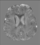
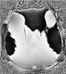
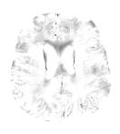
9 Diffusion-Weighted Imaging b = 0 No diffusion weighting b = 1000 s/mm 2 Diffusion weighting D (mm 2 /s) Computed diffusion coefficient Need a minimal of 2 measurements at 2 different b-values to computed D. Diffusion coefficient is commonly referred to as apparent diffusion coefficient (ADC) ADC of free water at room temperature: 2.2x10⁻³ mm 2 /s ADC of brain tissue around 1.0x10⁻³ mm 2 /s Why Single-Shot EPI? G y G x K-Space ifft DWI measures molecular diffusion ~ 10 µm during imaging window; Bulk motion (body motion, breathing, cardiac, brain pulsation) ~ 1 mm, introducing more phase than diffusion; varies from TR to TR. 9
10 G y If Acquire One k-space Line per TR Single-Shot G x m r, b = m(r, 0)e bd e jφ 1(r,TR 1 ) m r, b = m(r, 0)e bd e jφ 2(r,TR 1 ) m r, b = m(r, 0)e bd e jφ 3(r,TR 1 ) How to address it? Inconsistent k-space data causing aliasing Spatial varying signal cancellation Spin Echo 1.2 Diffusion-Tensor Imaging and Tractography 10
. 1.00 D = 0.55x10⁻³ mm 2 /s D = 1.5x10⁻³ mm 2 /s log(s) (a) right splenium corpus callosum (b) right splenium corpus callosum (1 1 0) (1-1 0) 0.")
11 R.F. 90º 180º Anisotropic Diffusion: Orientation Dependent G z G y G x Diffusion encoding gradients can be applied in either one of the three axis or a combination of axis. The gradients are represented by a vector (Gx Gy Gz) D = 0.55x10⁻³ mm 2 /s D = 1.5x10⁻³ mm 2 /s log(s) (a) right splenium corpus callosum (b) right splenium corpus callosum (1 1 0) (1-1 0) b(s/mm 2 ) Mathematical Models of Diffusion Isotropic Anisotropic Scalar D similar molecular displacements in all directions Dxx Dxy Dxz Dxy Dyy Dyz Dxz Dyz D zz greater molecular displacement along cylinders than across 11
12 Diffusion Tensor Signal Model Scalar Diffusion Tensor Diffusion b = γ 2 G 2 δ 2 ( δ 3) m b = m 0 exp( bd) b ij = γ 2 G i G j δ 2 ( δ 3) m b ij = m 0 exp( b ij D ij ) m ( b) m (0)exp bi i Di i Isotropic Diffusion Cerebral Spinal Fluid Instead of a diffusion coefficient, we have a diagonal diffusion tensor, a 3x3 matrix D xx D yy D zz Probability Density Function 12


13 Anisotropic Diffusion m ( b) m (0)exp bi i Di i Instead of a diffusion coefficient, we have a diffusion tensor, a 3x3 matrix D D D D D D D D D xx xy xz xy yy yz xz yz zz Probability Density Function Bloch Equation with Diffusion Term Fick s Second Law C D ijij C t C is spin density; Einstein summation rule. Each spin carries magnetic moment. Magnetization is proportional to spin density. M M B M i M j M M k M t T T Dij ij 2 1 This equation can be solved using standard methods for solving partial differential equations. For a spin echo sequence, the solution for transverse magnetization is given by b G G b b ij i j ( ), ij ji m( b) m(0)exp b D, D D symmetric positive definite ij ij ij ji 13
14 Probability Distribution Function of anisotropic diffusion 1 p() r (4 D t) 32 T 2r Dr t covariance matrix: Σ=2Dt e Statistical Interpretation for Anisotropic Diffusion For diffusion-weighted spin-echo sequence, echo amplitude is M M e p( r) dr M exp( b D ) j( 21) 0 0 b11 b12 b13 D11 D12 D13 b b b b, D D D D b31 b32 b 33 D31 D32 D 33 Tensor Product b : D b D b D ij ij ij ij i, j ij ij Example G G b G b b b : D b( D D D D ) , ( ) b( D 2 D D )
15 Determine Diffusion Tensor Experimentally D is a symmetric tensor. It has six unknowns. A minimal of six non-colinear measurements are required to determine a diffusion tensor. Different measurements are achieved by varying the diffusion encoding gradients including both amplitude and direction. m (0) s ln bijdij b D 2b D 2b D b D 2b D b D mb ( ) D 11 (1) (1) (1) (1) (1) (1) (1) s b11 2b12 2b13 b22 2b23 b D (2) (2) (2) (2) (2) (2) (2) s b11 2b12 2b13 b22 2b23 b D M M M M M M M D22 ( n) ( n) ( n) ( n) ( n) ( n) ( n) s b11 2b12 2b13 b22 2b D 23 b33 23 D 33 Rows have to be independent. (1 1 0) (1 0 1) (0 1 1) (1-1 0) (1 0-1) One Simple Encoding Scheme (0 1-1) x z y R.F. 90º 180º G z G y G x (1 1 0) 15
16 Eigen Decomposition D is coordinate system dependent. If the subject rotates in the magnet, the measured diffusion tensor will be different. Eigen decomposition defines rotation invariant quantities. D = UΛU T 1 2 3, U1, U2, 3 U = U U U U are eigenvectors Λ 0 2 0, 1, 2, 3 are eigenvalues mean diffusivity 3 Matlab function: eig(). U 2 U 3 U 1 1 Diffusion Ellipsoid Fractional Anisotropy (FA) Fractional Anisotropy (FA): a measure of diffusion anisotropy, 0<FA<1 FA 3(( ) ( ) ( ) ) ( ) y z x FA Color-coded FA: based on the orientation of the eigenvector corresponding to the largest eigenvalue 16


17 Fractional Anisotropy (FA) DTI Fiber Tractography x y z y z x Fiber Tractography: a representation of 3D white matter fiber structure. Summary Diffusion-weighted imaging is created by applying diffusion encoding gradients Tissue contrast is based difference in diffusion coefficient Diffusion-tensor imaging measures the orientation dependent diffusion coefficient Major eigenvector of a diffusion tensor is parallel to white matter fiber 17
18 Gradient Echo 2.1 T2*-Weighting and BOLD R.F. θ TE θ G z G y G x TR Readout Readout Magnitude and Phase of Gradient Echo abs(image) angle(image) Phase is due to offset in Larmor frequency. Different voxel has different frequency, consequently, accumulates different phase angle over time. This frequency offset is mainly due to field inhomogeneity caused by magnetic susceptibility variations. 18
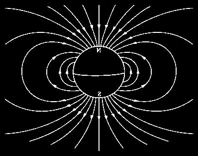
19 What is Magnetic Susceptibility? Magnetic susceptibility is a physical quantity that measures the extent to which a material is magnetized by an applied magnetic field. M magnetization vector H Magnetic field vector χ volume magnetic susceptibility (unitless in SI units) B magnetic flux density vector, or magnetic induction μ 0 vacuum permeability applied H applied H M B M = χ H B = μ 0 (1+χ) H Paramagnetic vs. Diamagnetic H H Paramagnetic χ > 0 M B M = χ H B = μ 0 (1+χ) H H H Diamagnetic χ < 0 M B M = χ H B = μ 0 (1+χ) H 19


20 Magnetic Susceptibility in MRI B 0 = 0 H 0 magnetization m susceptibility H 0 m B B B B 0 B 0 is perturbed by local magnetization induced by susceptibility RF Susceptibility Induced Tissue Contrast Echo 1 Echo 2 Echo n 20
(te+t) e i2πk r dr m r = m k e i2πk r dk If t << TE m r =")
 or B (Tesla) R2")
21 Susceptibility Induces Field Inhomogeneity θ TE Readout t Ideal Case Inhomogeneity Image Recon m k = m r e i2πk r dr t is k dependent m k = m r e iγδb(r)(te+t) e i2πk r dr m r = m k e i2πk r dk If t << TE m r = m(r) e iγδb(r)te phase Sampling m r n = m(r n ) e iγδb(r n)te k max sinc k max r n T 2 decay Voxel Effect of Field Inhomogeneity f B 2 R 2 2 FWHM 2 B 2 Full Width at Half Maximum FWHM (Hz) or B (Tesla) R2 Frequency 21
")


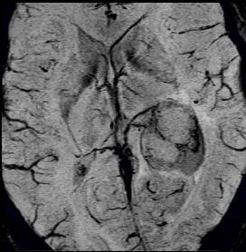
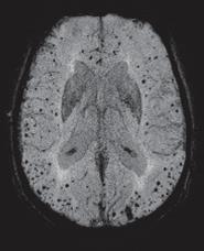
22 Magnitude: T2* Decay S() t S e tt 0 2 OR R2* Mapping and Contrast T2* 25 ms R2* 0 40 Hz White matter Blood vessels Globus pallidus Red nucleus Substatia nigra 22


23 Blood-Oxygen-Level-Dependent Signal Diamagnetic Hb Paramagnetic Hbr Ogawa S. et al, Phase Images of Gradient Echo 23


24 What Is in the Phase? Sources of phase Phase wraps Receiver coil Objects outside the FOV Objects inside the FOV background background tissue Phase Unwrapping Extensively researched; perfect solution still lacking Examples: 3D SRNCP or BP-ASL H. Abdul-Rahamn, et al, "Fast And Robust Three-Dimensional Best Path Phase Unwrapping Algorithm", Applied Optics, Vol. 46, No. 26, pp , 2007 FSL PRELUDE, University of Oxford 24


25 Laplacian Phase Unwrapping Totally automatic; Fast; Guarantee continuity Remove phase originated from sources outside FOV Li W. et al, NeuroImage 2011; 55: Filter Background: Sphere mean value property Harmonic function Mean phase over a sphere S Phase at the center 25


26 Summary of Phase Processing unwrapping Filtering 2.3 Susceptibility Weighted Imaging (SWI) 26
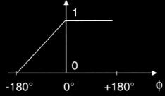
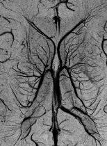
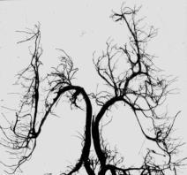
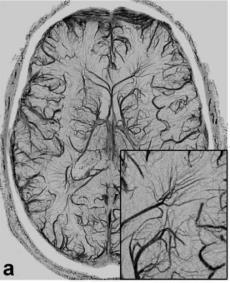
27 Susceptibility Weighted Imaging GRE Phase GRE Magn SWI = Magn*(PhaseMask) 4 SWI MIP SWI X Unwrapping Filtering Phase Mask Susceptibility Weighted Imaging Courtesy of Juergen Reichenbach 27


")


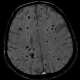
28 Pitfalls of SWI high sensitivity (micro-lesions) venous blood, iron, calcium low specificity (hypointense) qualitative, not quantitative Bo Phase is orientation dependent Bo Phase Is this bleeding? SWI 2.4 Quantitative Susceptibility Mapping (QSM) 28


29 Susceptibility Source Magnetic Field Change B m H 0 m B 0 Susceptibility Source Magnetic Field Change B m H 0 m B 0 29
30 Susceptibility Source Magnetic Field Change? B m B 0 Find the Demagnetizing Field h Magnetic flux density distribution in a first order approximation B (1 χ)( H h) H 0 B 0 0 H ( χ ) h 0 h? h = FT 1 k k z k 2χ k H 0 30
31 Magnetic Field Observed By a Spin Magnetic flux density seen by a spin B = μ 0 ( χ)(h 0 + h) H 0 Susceptibility inclusion B h Magnetic susceptibility Applied field vector Demagnetizing field vector Magnetic Field Observed By a Spin B z = μ χ H 0z + h z = μ χ H 0 FT 1 k z 2 k 2χ k H 0 μ 0 H χμ 0H 0 μ 0 FT 1 k z 2 k 2χ k H 0 δb z (r) = B z μ 0 H 0 = FT 1 ( 1 3 k z 2 k 2)χ k μ 0H 0 δb z (k) = ( 1 3 k z 2 k 2)χ k B 0 For simplicity, write δb z as δb B 0 = μ 0 H 0 31


32 Step 3: Solve A Deconvolution Problem Measurements Unknown Convolution Deconvolution k z Dividing by zero! k y k x 32


")

33 Quantitative Susceptibility Mapping Filter Background Phase Solve Inverse Problem Raw Phase Unwrapped Phase Tissue Phase Susceptibility (ppm) 3 T Tissue Phase ppm Susceptibility 33

34 Susceptibility Is Orientation Dependent B 0 Magnetic Susceptibility Is Anisotropic Susceptibility at Orientation # Magnetic susceptibility is orientation dependent, MRM 2010; 63:


35 Susceptibility Tensor Imaging H 0 k x k z k k y T 1 ( ) ˆ ( ) ˆ ˆ ˆ T k χ k H k 0 H χ( k) H H k Ht 2 3 k Susceptibility tensor is symmetric; 6 unknowns, MRM 2010; 63: Susceptibility Tensor Imaging = 35
36 Summary Magnetic susceptibility causes field perturbation Field perturbation results in frequency shift that can be measured by gradient echo phase images High-passed filtered phase is used to generate SWI images Background phase can be removed with sphere mean value filter The relation between field perturbation and susceptibility is a convolution QSM solves the deconvolution problem STI treats susceptibility as a tensor instead of a scalar 36
Quantitative Susceptibility Mapping and Susceptibility Tensor Imaging. Magnetization and Susceptibility
 Quantitative Susceptibility Mapping and Susceptibility Tensor Imaging 1, Chunlei Liu, Ph.D. 1 Brain Imaging and Analysis Center Department of Radiology Duke University, Durham, NC, USA 1 Magnetization
Advanced Topics and Diffusion MRI
 Advanced Topics and Diffusion MRI Slides originally by Karla Miller, FMRIB Centre Modified by Mark Chiew (mark.chiew@ndcn.ox.ac.uk) Slides available at: http://users.fmrib.ox.ac.uk/~mchiew/teaching/ MRI
Contrast Mechanisms in MRI. Michael Jay Schillaci
 Contrast Mechanisms in MRI Michael Jay Schillaci Overview Image Acquisition Basic Pulse Sequences Unwrapping K-Space Image Optimization Contrast Mechanisms Static and Motion Contrasts T1 & T2 Weighting,
Diffusion Tensor Imaging (DTI): An overview of key concepts
: An overview of key concepts") Diffusion Tensor Imaging (DTI): An overview of key concepts (Supplemental material for presentation) Prepared by: Nadia Barakat BMB 601 Chris Conklin Thursday, April 8 th 2010 Diffusion Concept [1,2]:
Diffusion Tensor Imaging (DTI): An overview of key concepts (Supplemental material for presentation) Prepared by: Nadia Barakat BMB 601 Chris Conklin Thursday, April 8 th 2010 Diffusion Concept [1,2]:
Introduction to MRI. Spin & Magnetic Moments. Relaxation (T1, T2) Spin Echoes. 2DFT Imaging. K-space & Spatial Resolution.
 Spin Echoes. 2DFT Imaging. K-space & Spatial Resolution.") Introduction to MRI Spin & Magnetic Moments Relaxation (T1, T2) Spin Echoes 2DFT Imaging Selective excitation, phase & frequency encoding K-space & Spatial Resolution Contrast (T1, T2) Acknowledgement:
Introduction to MRI Spin & Magnetic Moments Relaxation (T1, T2) Spin Echoes 2DFT Imaging Selective excitation, phase & frequency encoding K-space & Spatial Resolution Contrast (T1, T2) Acknowledgement:
Basics of Diffusion Tensor Imaging and DtiStudio
 Basics of Diffusion Tensor Imaging and DtiStudio DTI Basics 1 DTI reveals White matter anatomy Gray matter White matter DTI uses water diffusion as a probe for white matter anatomy Isotropic diffusion
Physics of MR Image Acquisition
 Physics of MR Image Acquisition HST-583, Fall 2002 Review: -MRI: Overview - MRI: Spatial Encoding MRI Contrast: Basic sequences - Gradient Echo - Spin Echo - Inversion Recovery : Functional Magnetic Resonance
EL-GY 6813/BE-GY 6203 Medical Imaging, Fall 2016 Final Exam
 EL-GY 6813/BE-GY 6203 Medical Imaging, Fall 2016 Final Exam (closed book, 1 sheets of notes double sided allowed, no calculator or other electronic devices allowed) 1. Ultrasound Physics (15 pt) A) (9
DIFFUSION MAGNETIC RESONANCE IMAGING
 DIFFUSION MAGNETIC RESONANCE IMAGING from spectroscopy to imaging apparent diffusion coefficient ADC-Map anisotropy diffusion tensor (imaging) DIFFUSION NMR - FROM SPECTROSCOPY TO IMAGING Combining Diffusion
Diffusion Tensor Imaging I. Jennifer Campbell
 Diffusion Tensor Imaging I Jennifer Campbell Diffusion Imaging Molecular diffusion The diffusion tensor Diffusion weighting in MRI Alternatives to the tensor Overview of applications Diffusion Imaging
醫用磁振學 MRM 擴散張量影像 擴散張量影像原理. 本週課程內容 MR Diffusion 擴散張量造影原理 擴散張量造影應用 盧家鋒助理教授國立陽明大學生物醫學影像暨放射科學系
 本週課程內容 http://www.ym.edu.tw/~cflu 擴散張量造影原理 擴散張量造影應用 醫用磁振學 MRM 擴散張量影像 盧家鋒助理教授國立陽明大學生物醫學影像暨放射科學系 alvin4016@ym.edu.tw MRI The Basics (3rd edition) Chapter 22: Echo Planar Imaging MRI in Practice, (4th edition)
Part III: Sequences and Contrast
 Part III: Sequences and Contrast Contents T1 and T2/T2* Relaxation Contrast of Imaging Sequences T1 weighting T2/T2* weighting Contrast Agents Saturation Inversion Recovery JUST WATER? (i.e., proton density
FREQUENCY SELECTIVE EXCITATION
 PULSE SEQUENCES FREQUENCY SELECTIVE EXCITATION RF Grad 0 Sir Peter Mansfield A 1D IMAGE Field Strength / Frequency Position FOURIER PROJECTIONS MR Image Raw Data FFT of Raw Data BACK PROJECTION Image Domain
Advanced MRI: Diffusion MRI 1: DTI and k-space
 k y Advanced MRI: Diffusion MRI 1: DTI and k-space k X Eric Sigmund, PhD February 26th, 2013 LECTURE 1 Neuro Diffusion MRI 3-5 m White matter axons Body 15 m Renal medulla Musculoskeletal 50 m Skeletal
Technical Improvements in Quantitative Susceptibility Mapping
 Technical Improvements in Quantitative Susceptibility Mapping 1-2 Saifeng Liu 1 School of Biomedical Engineering, McMaster University 2 The Magnetic Resonance Imaging Institute for Biomedical Research
Tensor Visualization. CSC 7443: Scientific Information Visualization
 Tensor Visualization Tensor data A tensor is a multivariate quantity Scalar is a tensor of rank zero s = s(x,y,z) Vector is a tensor of rank one v = (v x,v y,v z ) For a symmetric tensor of rank 2, its
} B 1 } Coil } Gradients } FFT
 Introduction to MRI Daniel B. Ennis, Ph.D. Requirements for MRI UCLA DCVI Requirements for MRI Dipoles to Images MR Active uclei e.g. 1 H in H20 Cryogen Liquid He and 2 Magnetic Field (B0) Polarizer ystem
Development of Magnetic Resonance-Based Susceptibility Imaging Applications
 Development of Magnetic Resonance-Based Susceptibility Imaging Applications Sung-Min Gho The Graduate School Yonsei University Development of Magnetic Resonance-Based Susceptibility Imaging Applications
Diffusion Weighted MRI. Zanqi Liang & Hendrik Poernama
 Diffusion Weighted MRI Zanqi Liang & Hendrik Poernama 1 Outline MRI Quick Review What is Diffusion MRI? Detecting Diffusion Stroke and Tumor Detection Presenting Diffusion Anisotropy and Diffusion Tensor
Diffusion Tensor Imaging I: The basics. Jennifer Campbell
 Diffusion Tensor Imaging I: The basics Jennifer Campbell Diffusion Tensor Imaging I: The basics Jennifer Campbell Diffusion Imaging MRI: many different sources of contrast T1W T2W PDW Perfusion BOLD DW
Field trip: Tuesday, Feb 5th
 Pulse Sequences Field trip: Tuesday, Feb 5th Hardware tour of VUIIIS Philips 3T Meet here at regular class time (11.15) Complete MRI screening form! Chuck Nockowski Philips Service Engineer Reminder: Project/Presentation
III, Diffusion, and Susceptibility. August 25, Departments of Mathematics and Applied Math and Computational Science University of Pennsylvania
 III,, and Departments of Mathematics and Applied Math and Computational Science University of Pennsylvania August 25, 2010 Copyright Page All material in this lecture, except as noted within the text,
Basic Concepts of MR Imaging, Diffusion MR Imaging, and Diffusion Tensor Imaging
 Basic Concepts of MR Imaging, Diffusion MR Imaging, and Diffusion Tensor Imaging Eduardo H.M.S.G. de Figueiredo, BSc a, *, Arthur F.N.G. Borgonovi, BSc b,c, Thomas M. Doring, MSc d,e KEYWORDS Magnetic
Introduction to Biomedical Imaging
 Alejandro Frangi, PhD Computational Imaging Lab Department of Information & Communication Technology Pompeu Fabra University www.cilab.upf.edu MRI advantages Superior soft-tissue contrast Depends on among
Apodization. Gibbs Artifact. Bioengineering 280A Principles of Biomedical Imaging. Fall Quarter 2013 MRI Lecture 5. rect(k x )
") Bioengineering 280A Principles of Biomedical Imaging Fall Quarter 2013 MRI Lecture 5 GE Medical Systems 2003 Gibbs Artifact Apodization rect(k ) Hanning Window h(k )=1/2(1+cos(2πk ) 256256 image 256128
Bioengineering 280A Principles of Biomedical Imaging Fall Quarter 2013 MRI Lecture 5 GE Medical Systems 2003 Gibbs Artifact Apodization rect(k ) Hanning Window h(k )=1/2(1+cos(2πk ) 256256 image 256128
Diffusion Imaging II. By: Osama Abdullah
 iffusion Imaging II By: Osama Abdullah Review Introduction. What is diffusion? iffusion and signal attenuation. iffusion imaging. How to capture diffusion? iffusion sensitizing gradients. Spin Echo. Gradient
Principles of Magnetic Resonance Imaging
 Principles of Magnetic Resonance Imaging Hi Klaus Scheffler, PhD Radiological Physics University of 1 Biomedical Magnetic Resonance: 1 Introduction Magnetic Resonance Imaging Contents: Hi 1 Introduction
Introduction to the Physics of NMR, MRI, BOLD fmri
 Pittsburgh, June 13-17, 2011 Introduction to the Physics of NMR, MRI, BOLD fmri (with an orientation toward the practical aspects of data acquisition) Pittsburgh, June 13-17, 2001 Functional MRI in Clinical
Magnetic Resonance Imaging. Pål Erik Goa Associate Professor in Medical Imaging Dept. of Physics
 Magnetic Resonance Imaging Pål Erik Goa Associate Professor in Medical Imaging Dept. of Physics pal.e.goa@ntnu.no 1 Why MRI? X-ray/CT: Great for bone structures and high spatial resolution Not so great
Cambridge University Press MRI from A to Z: A Definitive Guide for Medical Professionals Gary Liney Excerpt More information
 Main glossary Aa AB systems Referring to molecules exhibiting multiply split MRS peaks due to spin-spin interactions. In an AB system, the chemical shift between the spins is of similar magnitude to the
K-space. Spin-Warp Pulse Sequence. At each point in time, the received signal is the Fourier transform of the object s(t) = M( k x
 = M( k x") Bioengineering 280A Principles of Biomedical Imaging Fall Quarter 2015 MRI Lecture 4 k (t) = γ 2π k y (t) = γ 2π K-space At each point in time, the received signal is the Fourier transform of the object
Bioengineering 280A Principles of Biomedical Imaging Fall Quarter 2015 MRI Lecture 4 k (t) = γ 2π k y (t) = γ 2π K-space At each point in time, the received signal is the Fourier transform of the object
Spatial encoding in Magnetic Resonance Imaging. Jean-Marie BONNY
 Spatial encoding in Magnetic Resonance Imaging Jean-Marie BONNY What s Qu est an image ce qu une? image? «a reproduction of a material object by a camera or a related technique» Multi-dimensional signal
Chemistry 431. Lecture 23
 Chemistry 431 Lecture 23 Introduction The Larmor Frequency The Bloch Equations Measuring T 1 : Inversion Recovery Measuring T 2 : the Spin Echo NC State University NMR spectroscopy The Nuclear Magnetic
Diffusion tensor imaging (DTI):
:") Diffusion tensor imaging (DTI): A basic introduction to data acquisition and analysis Matthew Cykowski, MD Postdoctoral fellow Research Imaging Center UTHSCSA Room 2.320 cykowski@uthscsa.edu PART I: Acquiring
Diffusion tensor imaging (DTI): A basic introduction to data acquisition and analysis Matthew Cykowski, MD Postdoctoral fellow Research Imaging Center UTHSCSA Room 2.320 cykowski@uthscsa.edu PART I: Acquiring
Introduction to MRI Acquisition
 Introduction to MRI Acquisition James Meakin FMRIB Physics Group FSL Course, Bristol, September 2012 1 What are we trying to achieve? 2 What are we trying to achieve? Informed decision making: Protocols
Spatial encoding in Magnetic Resonance Imaging. Jean-Marie BONNY
 Spatial encoding in Magnetic Resonance Imaging Jean-Marie BONNY What s Qu est an image ce qu une? image? «a reproduction of a material object by a camera or a related technique» Multi-dimensional signal
NMR and MRI : an introduction
 Intensive Programme 2011 Design, Synthesis and Validation of Imaging Probes NMR and MRI : an introduction Walter Dastrù Università di Torino walter.dastru@unito.it \ Introduction Magnetic Resonance Imaging
Ordinary Least Squares and its applications
 Ordinary Least Squares and its applications Dr. Mauro Zucchelli University Of Verona December 5, 2016 Dr. Mauro Zucchelli Ordinary Least Squares and its applications December 5, 2016 1 / 48 Contents 1
The Basics of Magnetic Resonance Imaging
 The Basics of Magnetic Resonance Imaging Nathalie JUST, PhD nathalie.just@epfl.ch CIBM-AIT, EPFL Course 2013-2014-Chemistry 1 Course 2013-2014-Chemistry 2 MRI: Many different contrasts Proton density T1
Sequence Overview. Gradient Echo Spin Echo Magnetization Preparation Sampling and Trajectories Parallel Imaging. B.Hargreaves - RAD 229
 Sequence Overview Gradient Echo Spin Echo Magnetization Preparation Sampling and Trajectories Parallel Imaging 75 Pulse Sequences and k-space RF k y G z k x G x 3D k-space G y k y k z Acq. k x 76 Gradient
Diffusion Magnetic Resonance Imaging Part 1: Theory & Methods
 Diffusion Magnetic Resonance Imaging Part 1: Theory & Methods Benjamin M. Ellingson, Ph.D. Assistant Professor of Radiology, Biomedical Physics and Bioengineering Dept. of Radiological Sciences UCLA Neuro-Oncology
Higher Order Cartesian Tensor Representation of Orientation Distribution Functions (ODFs)
") Higher Order Cartesian Tensor Representation of Orientation Distribution Functions (ODFs) Yonas T. Weldeselassie (Ph.D. Candidate) Medical Image Computing and Analysis Lab, CS, SFU DT-MR Imaging Introduction
Higher Order Cartesian Tensor Representation of Orientation Distribution Functions (ODFs) Yonas T. Weldeselassie (Ph.D. Candidate) Medical Image Computing and Analysis Lab, CS, SFU DT-MR Imaging Introduction
BMB 601 MRI. Ari Borthakur, PhD. Assistant Professor, Department of Radiology Associate Director, Center for Magnetic Resonance & Optical Imaging
 BMB 601 MRI Ari Borthakur, PhD Assistant Professor, Department of Radiology Associate Director, Center for Magnetic Resonance & Optical Imaging University of Pennsylvania School of Medicine A brief history
NMR/MRI examination (8N080 / 3F240)
") NMR/MRI examination (8N080 / 3F240) Remarks: 1. This test consists of 3 problems with at total of 26 sub-questions. 2. Questions are in English. You are allowed to answer them in English or Dutch. 3. Please
NMR/MRI examination (8N080 / 3F240) Remarks: 1. This test consists of 3 problems with at total of 26 sub-questions. 2. Questions are in English. You are allowed to answer them in English or Dutch. 3. Please
Basic Pulse Sequences II - Spin Echoes. TE=12ms TE=47ms TE=106ms TE=153ms UCLA. Radiology
 TE TR 90 180 90 Basic Pulse Sequences II - Spin Echoes TE=12ms TE=47ms TE=106ms TE=153ms TE=235ms Lecture #6 Summary B1(t) RF TR RF t ~M (1) (0 )= ~ M 0 = 2 4 0 0 M 0 3 5 Initial Condition ~M (1) (0 +
MRI Physics II: Gradients, Imaging. Douglas C. Noll, Ph.D. Dept. of Biomedical Engineering University of Michigan, Ann Arbor
 MRI Physics II: Gradients, Imaging Douglas C., Ph.D. Dept. of Biomedical Engineering University of Michigan, Ann Arbor Magnetic Fields in MRI B 0 The main magnetic field. Always on (0.5-7 T) Magnetizes
Magnetic Resonance Imaging. Qun Zhao Bioimaging Research Center University of Georgia
 Magnetic Resonance Imaging Qun Zhao Bioimaging Research Center University of Georgia The Nobel Prize in Physiology or Medicine 2003 "for their discoveries concerning magnetic resonance imaging" Paul C.
MRI in Review: Simple Steps to Cutting Edge Part I
 MRI in Review: Simple Steps to Cutting Edge Part I DWI is now 2 years old... Mike Moseley Radiology Stanford DWI, b = 1413 T2wt, 28/16 ASN 21 San Francisco + Disclosures: Funding NINDS, NCRR, NCI 45 minutes
Diffusion Tensor Imaging tutorial
 NA-MIC http://na-mic.org Diffusion Tensor Imaging tutorial Sonia Pujol, PhD Surgical Planning Laboratory Harvard University DTI tutorial This tutorial is an introduction to the advanced Diffusion MR capabilities
Magnetization Preparation Sequences
 Magnetization Preparation Sequences Acquisition method may not give desired contrast Prep block adds contrast (and/or encoding) MP-RAGE = Magnetization prepared rapid acquisition with gradient echo (Mugler,
Bloch Equations & Relaxation UCLA. Radiology
 Bloch Equations & Relaxation MRI Systems II B1 I 1 I ~B 1 (t) I 6 ~M I I 5 I 4 Lecture # Learning Objectives Distinguish spin, precession, and nutation. Appreciate that any B-field acts on the the spin
Tissue Characteristics Module Three
 Tissue Characteristics Module Three 1 Equilibrium State Equilibrium State At equilibrium, the hydrogen vector is oriented in a direction parallel to the main magnetic field. Hydrogen atoms within the vector
MR Fundamentals. 26 October Mitglied der Helmholtz-Gemeinschaft
 MR Fundamentals 26 October 2010 Mitglied der Helmholtz-Gemeinschaft Mitglied der Helmholtz-Gemeinschaft Nuclear Spin Nuclear Spin Nuclear magnetic resonance is observed in atoms with odd number of protons
Outline. Superconducting magnet. Magnetic properties of blood. Physiology BOLD-MRI signal. Magnetic properties of blood
 Magnetic properties of blood Physiology BOLD-MRI signal Aart Nederveen Department of Radiology AMC a.j.nederveen@amc.nl Outline Magnetic properties of blood Moses Blood oxygenation BOLD fmri Superconducting
Cortical diffusion imaging
 Cortical diffusion imaging Alard Roebroeck Maastricht Brain Imaging Center (MBIC) Dept. of Cognitive Neuroscience Faculty of Psychology & Neuroscience Maastricht University Diffusion MRI In vivo & Ex vivo
HST.583 Functional Magnetic Resonance Imaging: Data Acquisition and Analysis Fall 2006
 MIT OpenCourseWare http://ocw.mit.edu HST.583 Functional Magnetic Resonance Imaging: Data Acquisition and Analysis Fall 2006 For information about citing these materials or our Terms of Use, visit: http://ocw.mit.edu/terms.
Tissue Parametric Mapping:
 Tissue Parametric Mapping: Contrast Mechanisms Using SSFP Sequences Jongho Lee Department of Radiology University of Pennsylvania Tissue Parametric Mapping: Contrast Mechanisms Using bssfp Sequences Jongho
New developments in Magnetic Resonance Spectrocopy and Diffusion MRI. Els Fieremans Steven Delputte Mahir Ozdemir
 New developments in Magnetic Resonance Spectrocopy and Diffusion MRI Els Fieremans Steven Delputte Mahir Ozdemir Overview Magnetic Resonance Spectroscopy (MRS) Basic physics of MRS Quantitative MRS Pitfalls
Basic MRI physics and Functional MRI
 Basic MRI physics and Functional MRI Gregory R. Lee, Ph.D Assistant Professor, Department of Radiology June 24, 2013 Pediatric Neuroimaging Research Consortium Objectives Neuroimaging Overview MR Physics
MRI Physics I: Spins, Excitation, Relaxation
 MRI Physics I: Spins, Excitation, Relaxation Douglas C. Noll Biomedical Engineering University of Michigan Michigan Functional MRI Laboratory Outline Introduction to Nuclear Magnetic Resonance Imaging
Sketch of the MRI Device
 Outline for Today 1. 2. 3. Introduction to MRI Quantum NMR and MRI in 0D Magnetization, m(x,t), in a Voxel Proton T1 Spin Relaxation in a Voxel Proton Density MRI in 1D MRI Case Study, and Caveat Sketch
PhD THESIS. prepared at INRIA Sophia Antipolis
 PhD THESIS prepared at INRIA Sophia Antipolis and presented at the University of Nice-Sophia Antipolis Graduate School of Information and Communication Sciences A dissertation submitted in partial satisfaction
Application of diffusion MRI to cancer, heart and brain connectome imaging
 Colloquium @ Department of Physics, NTU Application of diffusion MRI to cancer, heart and brain connectome imaging March 11, 2014 Wen-Yih Isaac Tseng MD, PhD Advanced Biomedical MRI Lab Center for Optoelectronic
Correction Gradients. Nov7, Reference: Handbook of pulse sequence
 Correction Gradients Nov7, 2005 Reference: Handbook of pulse sequence Correction Gradients 1. Concomitant-Field Correction Gradients 2. Crusher Gradients 3. Eddy-Current Compensation 4. Spoiler Gradients
Reconstruction Algorithms for MRI. Berkin Bilgic 17 December 2012
 Reconstruction Algorithms for MRI Berkin Bilgic 17 December 2012 Outline Magnetic Resonance Imaging (MRI) 2 Outline Magnetic Resonance Imaging (MRI) Non-invasive imaging, great versatility structural imaging
Outlines: (June 11, 1996) Instructor:
 Instructor:") Magnetic Resonance Imaging (June 11, 1996) Instructor: Tai-huang Huang Institute of Biomedical Sciences Academia Sinica Tel. (02) 2652-3036; Fax. (02) 2788-7641 E. mail: bmthh@ibms.sinica.edu.tw Reference:
Magnetic Resonance Imaging (June 11, 1996) Instructor: Tai-huang Huang Institute of Biomedical Sciences Academia Sinica Tel. (02) 2652-3036; Fax. (02) 2788-7641 E. mail: bmthh@ibms.sinica.edu.tw Reference:
MRI beyond Fourier Encoding: From array detection to higher-order field dynamics
 MRI beyond Fourier Encoding: From array detection to higher-order field dynamics K. Pruessmann Institute for Biomedical Engineering ETH Zurich and University of Zurich Parallel MRI Signal sample: m γκ,
G Medical Imaging. Outline 4/13/2012. Physics of Magnetic Resonance Imaging
 G16.4426 Medical Imaging Physics of Magnetic Resonance Imaging Riccardo Lattanzi, Ph.D. Assistant Professor Department of Radiology, NYU School of Medicine Department of Electrical and Computer Engineering,
Principles of MRI EE225E / BIO265. Name That Artifact. RF Interference During Readout. RF Interference During Readout. Lecture 19
 Name That Artifact Principles of MRI EE225E / BIO265 Lecture 19 Instructor: Miki Lustig UC Berkeley, EECS 1 http://mri-info.net 2 RF Interference During Readout RF Interference During Readout 1D FFT 1D
Course no. 4. The Theory of Electromagnetic Field
 Cose no. 4 The Theory of Electromagnetic Field Technical University of Cluj-Napoca http://www.et.utcluj.ro/cs_electromagnetics2006_ac.htm http://www.et.utcluj.ro/~lcret March 19-2009 Chapter 3 Magnetostatics
The NMR Inverse Imaging Problem
 The NMR Inverse Imaging Problem Nuclear Magnetic Resonance Protons and Neutrons have intrinsic angular momentum Atoms with an odd number of proton and/or odd number of neutrons have a net magnetic moment=>
MRI in Practice. Catherine Westbrook MSc, DCRR, CTC Senior Lecturer Anglia Polytechnic University Cambridge UK. John Talbot MSc, DCRR
 MRI in Practice Third edition Catherine Westbrook MSc, DCRR, CTC Senior Lecturer Anglia Polytechnic University Cambridge UK and Carolyn Kaut RothRT(R) (MR) (CT) (M) (CV) Fellow SMRT (Section for Magnetic
Relaxation. Ravinder Reddy
 Relaxation Ravinder Reddy Relaxation What is nuclear spin relaxation? What causes it? Effect on spectral line width Field dependence Mechanisms Thermal equilibrium ~10-6 spins leads to NMR signal! T1 Spin-lattice
MR Advance Techniques. Flow Phenomena. Class I
 MR Advance Techniques Flow Phenomena Class I Flow Phenomena In this class we will explore different phenomenona produced from nuclei that move during the acquisition of data. Flowing nuclei exhibit different
EE225E/BIOE265 Spring 2013 Principles of MRI. Assignment 9 Solutions. Due April 29th, 2013
 EE5E/BIOE65 Spring 013 Principles of MRI Miki Lustig This is the last homework in class. Enjoy it. Assignment 9 Solutions Due April 9th, 013 1) In class when we presented the spin-echo saturation recovery
Magnetization Gradients, k-space and Molecular Diffusion. Magnetic field gradients, magnetization gratings and k-space
 2256 Magnetization Gradients k-space and Molecular Diffusion Magnetic field gradients magnetization gratings and k-space In order to record an image of a sample (or obtain other spatial information) there
Extended Phase Graphs (EPG)
") Extended Phase Graphs (EPG) Purpose / Definition Propagation Gradients, Relaxation, RF Diffusion Examples 1 EPG Motivating Example: RF with Crushers RF G z Crushers are used to suppress spins that do not
Extended Phase Graphs (EPG) Purpose / Definition Propagation Gradients, Relaxation, RF Diffusion Examples 1 EPG Motivating Example: RF with Crushers RF G z Crushers are used to suppress spins that do not
Diffusion Tensor Imaging (DTI) e Neurite Orientation Dispersion and Density Imaging (NODDI)
 e Neurite Orientation Dispersion and Density Imaging (NODDI)") Diffusion Tensor Imaging (DTI) e Neurite Orientation Dispersion and Density Imaging (NODDI) Claudia AM Gandini Wheeler-Kingshott, PhD Prof. of MRI Physics Overview Diffusion and microstructure NODDI theoretical
Diffusion Tensor Imaging (DTI) e Neurite Orientation Dispersion and Density Imaging (NODDI) Claudia AM Gandini Wheeler-Kingshott, PhD Prof. of MRI Physics Overview Diffusion and microstructure NODDI theoretical
An introduction to Solid State NMR and its Interactions
 An introduction to Solid State NMR and its Interactions From tensor to NMR spectra CECAM Tutorial September 9 Calculation of Solid-State NMR Parameters Using the GIPAW Method Thibault Charpentier - CEA
Pulse Sequences: RARE and Simulations
 Pulse Sequences: RARE and Simulations M229 Advanced Topics in MRI Holden H. Wu, Ph.D. 2018.04.19 Department of Radiological Sciences David Geffen School of Medicine at UCLA Class Business Final project
A model for susceptibility artefacts from respiration in functional echo-planar magnetic resonance imaging
 Phys. Med. Biol. 45 (2000) 3809 3820. Printed in the UK PII: S0031-9155(00)14109-0 A model for susceptibility artefacts from respiration in functional echo-planar magnetic resonance imaging Devesh Raj,
Medical Visualization - Tensor Visualization. J.-Prof. Dr. Kai Lawonn
 Medical Visualization - Tensor Visualization J.-Prof. Dr. Kai Lawonn Lecture is partially based on the lecture by Prof. Thomas Schultz 2 What is a Tensor? A tensor is a multilinear transformation that
Extended Phase Graphs (EPG)
") Extended Phase Graphs (EPG) Purpose / Definition Propagation Gradients, Relaxation, RF Diffusion Examples 133 EPG Motivating Example: RF with Crushers RF G z Crushers are used to suppress spins that do
Extended Phase Graphs (EPG) Purpose / Definition Propagation Gradients, Relaxation, RF Diffusion Examples 133 EPG Motivating Example: RF with Crushers RF G z Crushers are used to suppress spins that do
Chapter 14:Physics of Magnetic Resonance
 Chapter 14:Physics of Magnetic Resonance Slide set of 141 slides based on the chapter authored by Hee Kwon Song of the publication (ISBN 978-92-0-131010-1): Diagnostic Radiology Physics: A Handbook for
A BRAIN MODEL FOR THE STUDY OF MR SUSCEPTIBILITY INDUCED PHASE BEHAVIOR
 A BRAIN MODEL FOR THE STUDY OF MR SUSCEPTIBILITY INDUCED PHASE BEHAVIOR A BRAIN MODEL FOR THE STUDY OF MR SUSCEPTIBILITY INDUCED PHASE BEHAVIOR BY SAGAR BUCH A Thesis Submitted to the School of Graduate
Spin Interactions. Giuseppe Pileio 24/10/2006
 Spin Interactions Giuseppe Pileio 24/10/2006 Magnetic moment µ = " I ˆ µ = " h I(I +1) " = g# h Spin interactions overview Zeeman Interaction Zeeman interaction Interaction with the static magnetic field
Diffusion imaging of the brain: technical considerations and practical applications
 Diffusion imaging of the brain: technical considerations and practical applications David G. Norris FC Donders Centre for Cognitive Neuroimaging Nijmegen Sustaining the physiologist in measuring the atomic
Chapter 24 MRA and Flow quantification. Yongquan Ye, Ph.D. Assist. Prof. Radiology, SOM Wayne State University
 Chapter 24 MRA and Flow quantification Yongquan Ye, Ph.D. Assist. Prof. Radiology, SOM Wayne State University Previous classes Flow and flow compensation (Chap. 23) Steady state signal (Cha. 18) Today
Midterm Review. EE369B Concepts Simulations with Bloch Matrices, EPG Gradient-Echo Methods. B.Hargreaves - RAD 229
 Midterm Review EE369B Concepts Simulations with Bloch Matrices, EPG Gradient-Echo Methods 292 Fourier Encoding and Reconstruction Encoding k y x Sum over image k x Reconstruction k y Gradient-induced Phase
Diffusion Tensor Imaging quality control : artifacts assessment and correction. A. Coste, S. Gouttard, C. Vachet, G. Gerig. Medical Imaging Seminar
 Diffusion Tensor Imaging quality control : artifacts assessment and correction A. Coste, S. Gouttard, C. Vachet, G. Gerig Medical Imaging Seminar Overview Introduction DWI DTI Artifact Assessment Artifact
M R I Physics Course. Jerry Allison Ph.D., Chris Wright B.S., Tom Lavin B.S., Nathan Yanasak Ph.D. Department of Radiology Medical College of Georgia
 M R I Physics Course Jerry Allison Ph.D., Chris Wright B.S., Tom Lavin B.S., Nathan Yanasak Ph.D. Department of Radiology Medical College of Georgia M R I Physics Course Spin Echo Imaging Hahn Spin Echo
Part II: Magnetic Resonance Imaging (MRI)
") Part II: Magnetic Resonance Imaging (MRI) Contents Magnetic Field Gradients Selective Excitation Spatially Resolved Reception k-space Gradient Echo Sequence Spin Echo Sequence Magnetic Resonance Imaging
Part II: Magnetic Resonance Imaging (MRI) Contents Magnetic Field Gradients Selective Excitation Spatially Resolved Reception k-space Gradient Echo Sequence Spin Echo Sequence Magnetic Resonance Imaging
Disclosures. MR Physics. Recipe to Creating Images without Radiation. MRI Physics. Vector Math. What s In an Image 12/21/2012. Siemens Medical Systems
 Disclosures MR Physics Joseph V. Fritz, PhD Dent Neurologic Institute Sunday, January 20, 2013 9:00 9:50 AM Siemens Medical Systems Research Agreement Philips Healthcare Research Agreement Toshiba Medical
Diffusion Tensor Imaging in Humans: Practical Implications for Neuroanatomy
 Diffusion Tensor Imaging in Humans: Practical Implications for Neuroanatomy Collaborators Center for Morphometric Analysis: Nikos Makris Andy Worth Verne S. Caviness George Papadimitriou MGH-NMR Center
The Nottingham eprints service makes this work by researchers of the University of Nottingham available open access under the following conditions.
 Wharton, Samuel and Bowtell, Richard W. (2015) Effects of white matter microstructure on phase and susceptibility maps. Magnetic Resonance in Medicine, 173 (3). pp. 1258-1269. ISSN 1522-2594 Access from
Chapter 26 Sequence Design, Artifacts and Nomenclature. Yongquan Ye, Ph.D. Assist. Prof. Radiology, SOM Wayne State University
 Chapter 26 Sequence Design, Artifacts and Nomenclature Yongquan Ye, Ph.D. Assist. Prof. Radiology, SOM Wayne State University Previous classes: RF pulse, Gradient, Signal Readout Gradient echo, spin echo,
Basis of MRI Contrast
 Basis of MRI Contrast MARK A. HORSFIELD Department of Cardiovascular Sciences University of Leicester Leicester LE1 5WW UK Tel: +44-116-2585080 Fax: +44-870-7053111 e-mail: mah5@le.ac.uk 1 1.1 The Magnetic
MRI. made. likely simplistic. Sorry, guys! We have less than one hour! Part 2: Sources of contrast. Endogenous (tissue) contrast
 contrast") .. made simple likely simplistic Sorry, guys! We have less than one hour! U. Himmelreich K.U.Leuven.. B. Gallez U.C.Louvain.. B. Jordan U.C.Louvain Part 2: Sources of contrast Endogenous (tissue) contrast
.. made simple likely simplistic Sorry, guys! We have less than one hour! U. Himmelreich K.U.Leuven.. B. Gallez U.C.Louvain.. B. Jordan U.C.Louvain Part 2: Sources of contrast Endogenous (tissue) contrast
HST.583 Functional Magnetic Resonance Imaging: Data Acquisition and Analysis Fall 2008
 MIT OpenCourseWare http://ocw.mit.edu HST.583 Functional Magnetic Resonance Imaging: Data Acquisition and Analysis Fall 2008 For information about citing these materials or our Terms of Use, visit: http://ocw.mit.edu/terms.
Index. p, lip, 78 8 function, 107 v, 7-8 w, 7-8 i,7-8 sine, 43 Bo,94-96
 p, lip, 78 8 function, 107 v, 7-8 w, 7-8 i,7-8 sine, 43 Bo,94-96 B 1,94-96 M,94-96 B oro!' 94-96 BIro!' 94-96 I/r, 79 2D linear system, 56 2D FFT, 119 2D Fourier transform, 1, 12, 18,91 2D sinc, 107, 112