6/22/2011. RT 4912 Review. Rex T. Christensen MHA RT (R) (MR) (CT)
|
|
|
- Gwendoline Bell
- 5 years ago
- Views:
Transcription
1 RT 4912 Review Rex T. Christensen MHA RT (R) (MR) (CT) 1
2 Questions? ARRT Content Specifications: Content-Specification.pdf Tests Can take twice Review Books Websites:
3 MR Active Nuclei Nuclei that aligns its axis to a magnetic field. They do this because of electromagnetic induction: Angular momentum or spin. Posses an electrical charge (+) If two of these characteristics are present it induces the third - magnetism Called a magnetic moment 3
4 MR Active Nuclei (cont.) Odd atomic number. (even proton, odd neutron or vice versa). Spinning mass (magnetic field-example earth). Spinning mass with a charge. 4

5 Precession Precession is the wobble of a top Precessional Frequency is the speed it wobbles. (Mhz) 5
6 Larmor Frequency: The Larmor frequency is also called the Precessional frequency. Precession is a wobble of the hydrogen atom. 6

7 Larmor Equation: MEMORIZE THIS! 7

8 Spin Echo vs. Gradient Echo 8

9 T1 - Fat vs. Water 9
10 Longitudinal Relaxation Time T1 Mo Mz 63% T1 t Longitudinale Relaxation = Energy transfer between excited spins and Tissue (Spin-Lattice-Relaxation) Reestablishing of longitudinal magnetization with time constant T1 10
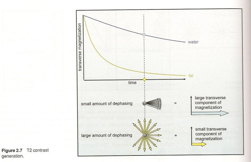
11 T2 Fat vs. Water 11
12 Transverse Relaxation Time T2 Mxy 37% T2 t Transverse Relaxation = Decay of magnetization by interaction between nuclei (Spin-Spin-Relaxation) 12
13 13
14 Relaxation Times are Tissue Specific M z S Tissue 1 Tissue 1 Tissue 2 Tissue 2 TR Short TE Medium TE Long TE Longitudinal Relaxation Transverse Relaxation 14
15 15
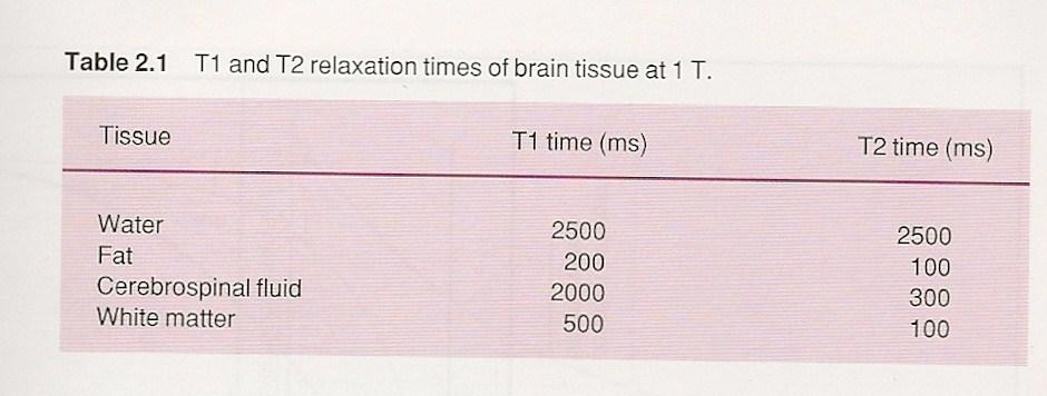
16 Relaxation Times: T1 Relaxation Time: 63 % relaxation of the tissue along the longitudinal (z) axis. T2 Relaxation Time: 63% decay of the tissue along the transverse (xy) axis. 16

17 Free Induction Decay (FID): The induction in reduced signal is called Free Induction Decay (FID) 17
18 Spin Echo Contrasts TR PD T1 T TE 18
19 K-space 19

20 K-space 20

21 K-space 21


22 K-Space 22

23 K-Space 23

24 K-Space 24
25 K-Space (Filling) TR and K-space filling 25
26 K-Space (Filling) Outer Lines = Low signal amplitude (resolution) Inner Lines = High signal amplitude (contrast) 26
27 K-Space (Filling) Low-high High-low Linear Reverse Linear Elliptical concentric 27
28 K-Space 28

29 SNR/Volume size 29

30 TE vs. SNR 30

31 SNR/NSA 31

32 Factors that effect SNR/Spatial Res./Scan Time 32

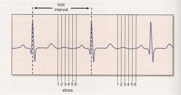
33 Maximizing SNR/Spatial Res./Scan Time 33

34 Benefits and Limitations 34

35 Types of flow 35

36 Time of Flight (TOF) 36

37 TOF vs. TE 37
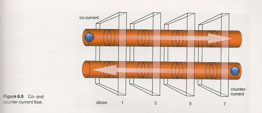
38 Co and Counter-Current flow 38

39 TOF pro s and con s 39

40 Cardiac Imaging 40

41 Cardiac Imaging 41
42 Pulse Sequence Diagrams These diagrams show the relation of the 3 gradients (slice,freq, and phase) to the signal formation. 42
43 Pulse Sequence Diagrams 43
44 Reading a Pulse Diagram: Spinecho revisited The timing diagram for a spinecho imaging sequence has entries for the RF pulses, the gradients in the magnetic field, and the signal. A slice selective 90 o RF pulse is applied in conjunction with a slice selection gradient. A period of time equal to TE/2 elapses and a 180 o slice selective 180 o pulse is applied in conjunction with the slice selection gradient.. ignored 44
45 Reading a Pulse Diagram: Spinecho revisited A phase encoding gradient is applied between the 90 o and 180 o pulses. As in the previous imaging sequences, the phase encoding gradient is varied in 128 or 256 steps between G m and -G m. The phase encoding gradient could be applied after the 180 o pulse, however if we want to minimize the TE period the pulse is applied between the 90 o and 180 o RF pulses. ignored 45
46 Reading a Pulse Diagram: Spinecho revisited The frequency encoding gradient is applied after the 180 o pulse during the time that echo is collected. The recorded signal is the echo. The FID, which is found after every 90 o pulse, is not used. One additional gradient is applied between the 90 o and 180 o pulses. This gradient is along the same direction as the frequency encoding gradient. It dephases the spins so that they will rephase by the center of the echo. This gradient in effect prepares the signal to be at the edge of k-space by the start of the acquisition of the echo. ignored 46
47 Multi Echo Sequence 47
48 Turbo Spin Echo 48
49 Inversion Recovery 49
50 Pulse Sequence Pro s & Con s 50

51 Spatial Encoding 51
52 Spatial Localization (image encoding) Magnetic Isocenter. Steepness of gradients. Slice selecting gradient. Frequency encoding (long axis of body part). Phase encoding (short axis of body part). 52
53 Spatial Localization (image encoding) Magnetic Isocenter. 53

54 Spatial Localization (image encoding) Slice selecting gradients. Axial Sagittal Coronal 54
55 Spatial Localization (image encoding) Slice selecting gradient (slice thickness). Steep slope = narrow transmit bandwidth (thin slice). Shallow slope = broad transmit bandwidth (thick slice). 55

56 Slice thickness and location 56

57 Changing slice thickness and location with the gradient. 57

58 Changing slice thickness and location with the pulse 58
59 Spatial Localization (image encoding) Slice selecting gradient (cont.) A slice can be selectively excited by transmitting RF with a and of frequencies coinciding wih the Larmor frequencies of spins in a particular slice defined by the slice select gradient. 59
60 Spatial Localization (image encoding) Frequency encoding gradient (long axis of body part). The frequency encoding gradient is switched on when the signal is received and is often called the readout gradient. The echo is centered in the middle of the frequency encoding gradient. The steepness of the slope of the frequency encoding gradient determines the size of the anatomy covered along the frequency encoding axis during the scan. This is called the field of view (FOV). 60
61 Spatial Localization (image encoding) Phase encoding gradient (short axis of body part). Small phase shift = shallow gradient. Large phase shift = steep gradient. 61
62 Gradient Amplitude vs. Rise Time 62

63 Components of an MRI system 63

64 Magnet Safety As you approach the magnet, the fringe magnetic field gets STRONGER 64 64
65 Fringe Diagram of Active Shield1.5 T* 5 Gauss Siemens Symphony 65 65

66 Room Safety MR Safe MR Conditional MR Unsafe 66
67 SAR and FDA Guidelines -Specific Absorption Rate (SAR) Tissue heating and the body s ability to dissipate excess heat. -Measured in Watts/Kg FDA: -Updated July 2004: 0.4 W/kg whole body 3.2 W/kg head 8.0 W/kg small volume 67
 and not just ferromagnetic properties.")
68 Time-Varying Magnetic Field (TVMF) Safety Consider electrical conductivity (Faraday s law of induction) and not just ferromagnetic properties. Scalp burn from a cervical halo 68
69 External Magnet Field Strength Remember safety considerations are different depending on field strength. ALWAYS double and triple check for safety! 69
70 Gd-DTPA (Gad + Chelate) Gadolinium + DiethyleneTriaminePentaAcetic acid-chelate The gadolinium chelates are 100% renally excreted, with the exception of two agents with combined renal and hepatobiliary excretion(multihance and Primovist) 70
71 Relaxivity The ability of magnetic compounds to increase the relaxation rates of the surrounding water proton spins. 71
72 Relaxivity How does it work? T1-relaxation is dependent on the molecular tumbling rate. (T1 weighted exams) The gadolinium molecule is a big tumbling magnet The slower a molecule tumbles, the shorter the T1-relaxation time of the hydrogen molecule (the greater the relaxation rate). 72
73 Relaxivity How does it work? When the gadolinium molecule gets next to the water molecule, it effects the rate at which the water molecule tumbles, slowing it down. 73
74 Efficacy The gadolinium chelates currently available for clinical use can be differentiated on the basis of: charge (ionic or nonionic) structure (linear or cyclic) stability 74
75 Charge (Ionicity) Given that the gadolinium ion carries a 3 charge, if the ligand, for example, is HPDO3A (that for ProHance, with a charge of 3), the metal chelate itself will carry a net charge of zero, and thus be nonionic. In the U.S. market, considering only the gadolinium chelates with 100% renal excretion, there are three nonionic agents (ProHance, Omniscan, and Optimark) and one ionic agent (Magnevist). 75
76 Structure (Linear or Cyclic) The structure of the chelate can be linear or macrocyclic (ring-like), with the cyclic chelates demonstrating higher in vivo stability and thus an improved safety margin. ProHance is the only macrocyclic chelate available in the United States. Internationally, two other extracellular gadolinium chelates are in widespread use, both macrocyclic: Dotarem (ionic) and Gadovist (nonionic). 76

77 MRI Contrast Agents 77
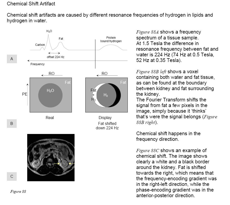
78 Stability Stability is related to structure of the chelate. Gadolinium Chelates differ in their thermodynamic stability constants and their kinetic stability. Macrocyclic chelates are more stable than linear molecules. Even among linear agents there are differences in stabilty. Fundam. Clin. Pharmacol Dec: 20 (6):
79 Osmolality Osmotic (Ion) concentration. An indicator of fluid balance in the bodies tissues. The term "osmolal" describes an ion concentration of a solution in moles per kilogram of solvent (mol/kg), while "osmolar" describes an ion concentration in moles per liter (mol/l). mosm = milliosmole 79

80 Contrast Agents 80
81 Low osmolar contrast media (LOCM) are less nephrotoxic than high osomolar contrast media (HOCM). 81
82 Nephrogenic Systemic Fibrosis (NFS) Development of the disease is due to gadolinium chelate dissociation, with deposition of the free metal, and is thus related to chelate stability, dose, and cumulative (lifetime) dose. 82
83 Pregnancy 83
84 Artifacts Motion (types) Susceptibility Chemical Shift Wrap Around Partial Volume Gibbs Zebra Zipper Cross-Excitation 84

85 Artifacts 85
86 Chemical Shift This is due to the precessional differences in fat and water (fat has a lower precessional frequency than water). This difference is 220 Hz in a 1.5T magnet. It is worse in higher field strength magnets. It creates a dark band at the interfaces of water and fat. It occurs in the frequency direction. 86
87 87


88 Partial Volume Averaging 88
89 Magnetic susceptibility artifact The ability of a substance to become magnetized. (metal and hemosiderin) The differences in these substances with the surrounding tissues results in different precessional frequencies. It occurs in the frequency direction. More obvious in gradient echo sequences. 89

90 Magnetic Susceptibility Artifacts (Understanding MRI) 90

91 Image Artifacts: Magnetic Susceptibility 91

92 Image Artifacts: Aliasing: Anatomy that is bigger than the FOV will wrap back into itself if anti-aliasing techniques are not used. 92

93 Phase Wrap Artifact 93

94 Phase Mismapping Artifact 94
, holes in the wall (nails, screws), door open while")
95 Image Artifacts: Zipper: RF leak in the room. Caused by doors (sticky fingers), holes in the wall (nails, screws), door open while scanning, and pipes from air gases or water. 95


96 Image Artifacts: Field Distortion: Scanning off of Isocenter in the z- axis direction with a large FOV. 96
97 Slice Gap 97

98 Motion Artifacts 98
99 Zebra Artifact 99
100 Bandwidth 100
101 6 Key Imaging Goals Optimize Contrast Optimize Coverage and Slice Thickness Optimize Resolution Minimize Artifacts Maximize Patient Throughput Optimize Signal to Noise (SNR) 101
102 Scenario 1 Radiologist asks for a MRCP to visualize the biliary system. 102
103 Scenario 2 Radiologist wants a sagittal T2 FSE fatsat knee with better signal and better resolution. 103
104 Scenario 3 A coronal T1 wrist is too grainy 104
105 Scenario 4 A FSE T2 fatsat brachial plexus does not get good fat saturation. 105
106 Scenario 5 FSE T2 IACs have low spatial resolution 106
107 Build a Protocol/Optimize It! Brain w/wo contrast Routine knee Routine L-spine 107
108 Advantages/Disadvantages Changing FOV Changing Slice Thickness Changing ETL Changing NSA/NEX Changing TE Changing Bandwidth Changing TR 108
109 If this..then this.. P. 137 and P.138 MRI in Practice 109
110 I have a question? 110
111 Website MRI/MRI-Sequences/Sequences-acronyms 111
112 References: Westbrook, Catherine and Kaut, Carolyn (1998). MRI In Practice, 3rd Edition, Malden, MA: Blackwell Science, Inc. 112
MRI in Review: Simple Steps to Cutting Edge Part I
 MRI in Review: Simple Steps to Cutting Edge Part I DWI is now 2 years old... Mike Moseley Radiology Stanford DWI, b = 1413 T2wt, 28/16 ASN 21 San Francisco + Disclosures: Funding NINDS, NCRR, NCI 45 minutes
MRI in Practice. Catherine Westbrook MSc, DCRR, CTC Senior Lecturer Anglia Polytechnic University Cambridge UK. John Talbot MSc, DCRR
 MRI in Practice Third edition Catherine Westbrook MSc, DCRR, CTC Senior Lecturer Anglia Polytechnic University Cambridge UK and Carolyn Kaut RothRT(R) (MR) (CT) (M) (CV) Fellow SMRT (Section for Magnetic
Introduction to Biomedical Imaging
 Alejandro Frangi, PhD Computational Imaging Lab Department of Information & Communication Technology Pompeu Fabra University www.cilab.upf.edu MRI advantages Superior soft-tissue contrast Depends on among
RADIOLOGIV TECHNOLOGY 4912 COMPREHENSEIVE REVIEW/MRI WORSHEET #1- PATIENT CARE AND SAFETY/PHYSICAL PRINCIPLES
 RADIOLOGIV TECHNOLOGY 4912 COMPREHENSEIVE REVIEW/MRI WORSHEET #1- PATIENT CARE AND SAFETY/PHYSICAL PRINCIPLES 1. What are potential consequences to patients and personnel should there be a release of gaseous
Rad Tech 4912 MRI Registry Review. Outline of the Registry Exam: Certification Fees
 Rad Tech 4912 MRI Registry Review Outline of the Registry Exam: Category: # of questions: A. Patient Care 30 B. Imaging Procedures 62 C. Data Acquisition and Processing 65 D. Physical Principles of Image
Basic p rinciples COPYRIGHTED MATERIAL. Introduction. Atomic s tructure
 1 Basic p rinciples Introduction 1 Atomic structure 1 Motion in the atom 2 MR active nuclei 2 The hydrogen nucleus 4 Alignment 4 Precession 8 The Larmor equation 9 Introduction The basic principles of
Introduction to MRI. Spin & Magnetic Moments. Relaxation (T1, T2) Spin Echoes. 2DFT Imaging. K-space & Spatial Resolution.
 Spin Echoes. 2DFT Imaging. K-space & Spatial Resolution.") Introduction to MRI Spin & Magnetic Moments Relaxation (T1, T2) Spin Echoes 2DFT Imaging Selective excitation, phase & frequency encoding K-space & Spatial Resolution Contrast (T1, T2) Acknowledgement:
Introduction to MRI Spin & Magnetic Moments Relaxation (T1, T2) Spin Echoes 2DFT Imaging Selective excitation, phase & frequency encoding K-space & Spatial Resolution Contrast (T1, T2) Acknowledgement:
Part III: Sequences and Contrast
 Part III: Sequences and Contrast Contents T1 and T2/T2* Relaxation Contrast of Imaging Sequences T1 weighting T2/T2* weighting Contrast Agents Saturation Inversion Recovery JUST WATER? (i.e., proton density
NMR and MRI : an introduction
 Intensive Programme 2011 Design, Synthesis and Validation of Imaging Probes NMR and MRI : an introduction Walter Dastrù Università di Torino walter.dastru@unito.it \ Introduction Magnetic Resonance Imaging
The Basics of Magnetic Resonance Imaging
 The Basics of Magnetic Resonance Imaging Nathalie JUST, PhD nathalie.just@epfl.ch CIBM-AIT, EPFL Course 2013-2014-Chemistry 1 Course 2013-2014-Chemistry 2 MRI: Many different contrasts Proton density T1
Contrast Mechanisms in MRI. Michael Jay Schillaci
 Contrast Mechanisms in MRI Michael Jay Schillaci Overview Image Acquisition Basic Pulse Sequences Unwrapping K-Space Image Optimization Contrast Mechanisms Static and Motion Contrasts T1 & T2 Weighting,
Nuclei, Excitation, Relaxation
 Outline 4.1 Principles of MRI uclei, Excitation, Relaxation Carolyn Kaut Roth, RT (R)(MR)(CT)(M)(CV) FSMRT CEO Imaging Education Associates www.imaginged.com candi@imaginged.com What nuclei are MR active?
FREQUENCY SELECTIVE EXCITATION
 PULSE SEQUENCES FREQUENCY SELECTIVE EXCITATION RF Grad 0 Sir Peter Mansfield A 1D IMAGE Field Strength / Frequency Position FOURIER PROJECTIONS MR Image Raw Data FFT of Raw Data BACK PROJECTION Image Domain
MRI at a Glance. Blackwell Science CATHERINE WESTBROOK. MSC DCRR CTC Director of Training and Education Lodestone Patient Care Ltd
 MRI at a Glance MRI at a Glance CATHERINE WESTBROOK MSC DCRR CTC Director of Training and Education Lodestone Patient Care Ltd Blackwell Science 2002 by Blackwell Science Ltd, a Blackwell Publishing Company
The NMR Inverse Imaging Problem
 The NMR Inverse Imaging Problem Nuclear Magnetic Resonance Protons and Neutrons have intrinsic angular momentum Atoms with an odd number of proton and/or odd number of neutrons have a net magnetic moment=>
MR Advance Techniques. Flow Phenomena. Class I
 MR Advance Techniques Flow Phenomena Class I Flow Phenomena In this class we will explore different phenomenona produced from nuclei that move during the acquisition of data. Flowing nuclei exhibit different
EL-GY 6813/BE-GY 6203 Medical Imaging, Fall 2016 Final Exam
 EL-GY 6813/BE-GY 6203 Medical Imaging, Fall 2016 Final Exam (closed book, 1 sheets of notes double sided allowed, no calculator or other electronic devices allowed) 1. Ultrasound Physics (15 pt) A) (9
Fundamental MRI Principles Module Two
 Fundamental MRI Principles Module Two 1 Nuclear Magnetic Resonance There are three main subatomic particles: protons neutrons electrons positively charged no significant charge negatively charged Protons
Magnetic Resonance Imaging. Pål Erik Goa Associate Professor in Medical Imaging Dept. of Physics
 Magnetic Resonance Imaging Pål Erik Goa Associate Professor in Medical Imaging Dept. of Physics pal.e.goa@ntnu.no 1 Why MRI? X-ray/CT: Great for bone structures and high spatial resolution Not so great
Magnetic Resonance Imaging (MRI)
") Magnetic Resonance Imaging Introduction The Components The Technology (MRI) Physics behind MR Most slides taken from http:// www.slideworld.org/ viewslides.aspx/magnetic- Resonance-Imaging- %28MRI%29-MR-Imaging-
Magnetic Resonance Imaging Introduction The Components The Technology (MRI) Physics behind MR Most slides taken from http:// www.slideworld.org/ viewslides.aspx/magnetic- Resonance-Imaging- %28MRI%29-MR-Imaging-
Exam 8N080 - Introduction to MRI
 Exam 8N080 - Introduction to MRI Friday April 10 2015, 18.00-21.00 h For this exam you may use an ordinary calculator (not a graphical one). In total there are 5 assignments and a total of 50 points can
M R I Physics Course. Jerry Allison Ph.D., Chris Wright B.S., Tom Lavin B.S., Nathan Yanasak Ph.D. Department of Radiology Medical College of Georgia
 M R I Physics Course Jerry Allison Ph.D., Chris Wright B.S., Tom Lavin B.S., Nathan Yanasak Ph.D. Department of Radiology Medical College of Georgia M R I Physics Course Spin Echo Imaging Hahn Spin Echo
Fundamental MRI Principles Module 2 N. Nuclear Magnetic Resonance. X-ray. MRI Hydrogen Protons. Page 1. Electrons
 Fundamental MRI Principles Module 2 N S 1 Nuclear Magnetic Resonance There are three main subatomic particles: protons positively charged neutrons no significant charge electrons negatively charged Protons
EE225E/BIOE265 Spring 2013 Principles of MRI. Assignment 9 Solutions. Due April 29th, 2013
 EE5E/BIOE65 Spring 013 Principles of MRI Miki Lustig This is the last homework in class. Enjoy it. Assignment 9 Solutions Due April 9th, 013 1) In class when we presented the spin-echo saturation recovery
Basis of MRI Contrast
 Basis of MRI Contrast MARK A. HORSFIELD Department of Cardiovascular Sciences University of Leicester Leicester LE1 5WW UK Tel: +44-116-2585080 Fax: +44-870-7053111 e-mail: mah5@le.ac.uk 1 1.1 The Magnetic
Field trip: Tuesday, Feb 5th
 Pulse Sequences Field trip: Tuesday, Feb 5th Hardware tour of VUIIIS Philips 3T Meet here at regular class time (11.15) Complete MRI screening form! Chuck Nockowski Philips Service Engineer Reminder: Project/Presentation
The physics of medical imaging US, CT, MRI. Prof. Peter Bogner
 The physics of medical imaging US, CT, MRI Prof. Peter Bogner Clinical radiology curriculum blocks of lectures and clinical practice (7x2) Physics of medical imaging Neuroradiology Head and neck I. Head
MRI Physics I: Spins, Excitation, Relaxation
 MRI Physics I: Spins, Excitation, Relaxation Douglas C. Noll Biomedical Engineering University of Michigan Michigan Functional MRI Laboratory Outline Introduction to Nuclear Magnetic Resonance Imaging
Physics of MR Image Acquisition
 Physics of MR Image Acquisition HST-583, Fall 2002 Review: -MRI: Overview - MRI: Spatial Encoding MRI Contrast: Basic sequences - Gradient Echo - Spin Echo - Inversion Recovery : Functional Magnetic Resonance
NMR/MRI examination (8N080 / 3F240)
") NMR/MRI examination (8N080 / 3F240) Remarks: 1. This test consists of 3 problems with at total of 26 sub-questions. 2. Questions are in English. You are allowed to answer them in English or Dutch. 3. Please
NMR/MRI examination (8N080 / 3F240) Remarks: 1. This test consists of 3 problems with at total of 26 sub-questions. 2. Questions are in English. You are allowed to answer them in English or Dutch. 3. Please
Basic MRI physics and Functional MRI
 Basic MRI physics and Functional MRI Gregory R. Lee, Ph.D Assistant Professor, Department of Radiology June 24, 2013 Pediatric Neuroimaging Research Consortium Objectives Neuroimaging Overview MR Physics
G Medical Imaging. Outline 4/13/2012. Physics of Magnetic Resonance Imaging
 G16.4426 Medical Imaging Physics of Magnetic Resonance Imaging Riccardo Lattanzi, Ph.D. Assistant Professor Department of Radiology, NYU School of Medicine Department of Electrical and Computer Engineering,
Lab 2: Magnetic Resonance Imaging
 EE225E/BIOE265 Spring 2013 Principles of MRI Miki Lustig Developed by: Galen Reed and Miki Lustig Lab 2: Magnetic Resonance Imaging Introduction In this lab, we will get some hands-on experience with an
COPYRIGHTED MATERIAL. Production of Net Magnetization. Chapter 1
 Chapter 1 Production of Net Magnetization Magnetic resonance (MR) is a measurement technique used to examine atoms and molecules. It is based on the interaction between an applied magnetic field and a
Tissue Characteristics Module Three
 Tissue Characteristics Module Three 1 Equilibrium State Equilibrium State At equilibrium, the hydrogen vector is oriented in a direction parallel to the main magnetic field. Hydrogen atoms within the vector
Nuclear Magnetic Resonance Imaging
 Nuclear Magnetic Resonance Imaging Jeffrey A. Fessler EECS Department The University of Michigan NSS-MIC: Fundamentals of Medical Imaging Oct. 20, 2003 NMR-0 Background Basic physics 4 magnetic fields
Topics. The concept of spin Precession of magnetic spin Relaxation Bloch Equation. Bioengineering 280A Principles of Biomedical Imaging
 Bioengineering 280A Principles of Biomedical Imaging Fall Quarter 2006 MRI Lecture 1 Topics The concept of spin Precession of magnetic spin Relaxation Bloch Equation 1 Spin Intrinsic angular momentum of
Fundamentals of MR Imaging
 Fundamentals of MR Imaging Shantanu Sinha. Department of Radiology UCSD School of Medicine, San Diego, CA-92103. E-mail: shsinha@ucsd.edu Background References: R.B.Lufkin, The MRI Manual (2nd Edition).
K-space. Spin-Warp Pulse Sequence. At each point in time, the received signal is the Fourier transform of the object s(t) = M( k x
 = M( k x") Bioengineering 280A Principles of Biomedical Imaging Fall Quarter 2015 MRI Lecture 4 k (t) = γ 2π k y (t) = γ 2π K-space At each point in time, the received signal is the Fourier transform of the object
Bioengineering 280A Principles of Biomedical Imaging Fall Quarter 2015 MRI Lecture 4 k (t) = γ 2π k y (t) = γ 2π K-space At each point in time, the received signal is the Fourier transform of the object
Chapter 24 MRA and Flow quantification. Yongquan Ye, Ph.D. Assist. Prof. Radiology, SOM Wayne State University
 Chapter 24 MRA and Flow quantification Yongquan Ye, Ph.D. Assist. Prof. Radiology, SOM Wayne State University Previous classes Flow and flow compensation (Chap. 23) Steady state signal (Cha. 18) Today
Relaxation times in nuclear magnetic resonance
 Relaxation times in TEP Related topics Nuclear spins, atomic nuclei with a magnetic moment, precession movement of the nuclear spins, Landau-Lifshitz equation, Bloch equation, magnetisation, resonance
MRI in Clinical Practice
 MRI in Clinical Practice MRI in Clinical Practice Gary Liney With 62 Figures Gary Liney, PhD MRI Lecturer University of Hull Centre for MR Investigations Hull Royal Infirmary Hull UK British Library Cataloguing
Biomedical Imaging Magnetic Resonance Imaging
 Biomedical Imaging Magnetic Resonance Imaging Charles A. DiMarzio & Eric Kercher EECE 4649 Northeastern University May 2018 Background and History Measurement of Nuclear Spins Widely used in physics/chemistry
Basic principles COPYRIGHTED MATERIAL. Introduction. Introduction 1 Precession and precessional
 1 Basic principles Introduction 1 Precession and precessional Atomic structure 2 (Larmor) frequency 10 Motion in the atom 2 Precessional phase 13 MR-active nuclei 4 Resonance 13 The hydrogen nucleus 5
Apodization. Gibbs Artifact. Bioengineering 280A Principles of Biomedical Imaging. Fall Quarter 2013 MRI Lecture 5. rect(k x )
") Bioengineering 280A Principles of Biomedical Imaging Fall Quarter 2013 MRI Lecture 5 GE Medical Systems 2003 Gibbs Artifact Apodization rect(k ) Hanning Window h(k )=1/2(1+cos(2πk ) 256256 image 256128
Bioengineering 280A Principles of Biomedical Imaging Fall Quarter 2013 MRI Lecture 5 GE Medical Systems 2003 Gibbs Artifact Apodization rect(k ) Hanning Window h(k )=1/2(1+cos(2πk ) 256256 image 256128
Background II. Signal-to-Noise Ratio (SNR) Pulse Sequences Sampling and Trajectories Parallel Imaging. B.Hargreaves - RAD 229.
 Pulse Sequences Sampling and Trajectories Parallel Imaging. B.Hargreaves - RAD 229.") Background II Signal-to-Noise Ratio (SNR) Pulse Sequences Sampling and Trajectories Parallel Imaging 1 SNR: Signal-to-Noise Ratio Signal: Desired voltage in coil Noise: Thermal, electronic Noise Thermal
Background II Signal-to-Noise Ratio (SNR) Pulse Sequences Sampling and Trajectories Parallel Imaging 1 SNR: Signal-to-Noise Ratio Signal: Desired voltage in coil Noise: Thermal, electronic Noise Thermal
Physical fundamentals of magnetic resonance imaging
 Physical fundamentals of magnetic resonance imaging Stepan Sereda University of Bonn 1 / 26 Why? Figure 1 : Full body MRI scan (Source: [4]) 2 / 26 Overview Spin angular momentum Rotating frame and interaction
2.1.1 A Brief History of NMR The conception of NMR sprouted after the Pauli s prediction of nuclear spin in
 CHAPTER--2 BASICS OF NMR IMAGING AND SPECTROSCOPY 2.1 Introduction 2.1.1 A Brief History of NMR The conception of NMR sprouted after the Pauli s prediction of nuclear spin in 1924. Later Gorter (1936)
Spin Echo Imaging Sequence
 1 MRI In Stereotactic Procedures Edward F. Jackson, Ph.D. The University of Texas M.D. Anderson Cancer Center Houston, Texas 2 RF G slice G phase G freq Signal k-space Spin Echo Imaging Sequence TE 1st
Magnetic Resonance Imaging
 http://www.qldxray.com.au/filelibrary/mri_cardiovascular_system_ca_0005.jpg Magnetic Resonance Imaging 1 Overview 1. The magnetic properties of nuclei, and how they behave in strong magnetic fields. 2.
BMB 601 MRI. Ari Borthakur, PhD. Assistant Professor, Department of Radiology Associate Director, Center for Magnetic Resonance & Optical Imaging
 BMB 601 MRI Ari Borthakur, PhD Assistant Professor, Department of Radiology Associate Director, Center for Magnetic Resonance & Optical Imaging University of Pennsylvania School of Medicine A brief history
Sketch of the MRI Device
 Outline for Today 1. 2. 3. Introduction to MRI Quantum NMR and MRI in 0D Magnetization, m(x,t), in a Voxel Proton T1 Spin Relaxation in a Voxel Proton Density MRI in 1D MRI Case Study, and Caveat Sketch
Nuclear Magnetic Resonance Imaging
 Nuclear Magnetic Resonance Imaging Simon Lacoste-Julien Electromagnetic Theory Project 198-562B Department of Physics McGill University April 21 2003 Abstract This paper gives an elementary introduction
Basic Pulse Sequences II - Spin Echoes. TE=12ms TE=47ms TE=106ms TE=153ms UCLA. Radiology
 TE TR 90 180 90 Basic Pulse Sequences II - Spin Echoes TE=12ms TE=47ms TE=106ms TE=153ms TE=235ms Lecture #6 Summary B1(t) RF TR RF t ~M (1) (0 )= ~ M 0 = 2 4 0 0 M 0 3 5 Initial Condition ~M (1) (0 +
Introduction to Magnetic Resonance Imaging (MRI) Pietro Gori
 Pietro Gori") Introduction to Magnetic Resonance Imaging (MRI) Pietro Gori Enseignant-chercheur Equipe IMAGES - Télécom ParisTech pietro.gori@telecom-paristech.fr September 20, 2017 P. Gori BIOMED 20/09/2017 1 / 76
Introduction to Magnetic Resonance Imaging (MRI) Pietro Gori Enseignant-chercheur Equipe IMAGES - Télécom ParisTech pietro.gori@telecom-paristech.fr September 20, 2017 P. Gori BIOMED 20/09/2017 1 / 76
The physics US and MRI. Prof. Peter Bogner
 The physics US and MRI Prof. Peter Bogner Sound waves mechanical disturbance, a pressure wave moves along longitudinal wave compression rarefaction zones c = nl, (c: velocity, n: frequency, l: wavelength
How is it different from conventional MRI? What is MR Spectroscopy? How is it different from conventional MRI? MR Active Nuclei
 What is MR Spectroscopy? MR-Spectroscopy (MRS) is a technique to measure the (relative) concentration of certain chemical or biochemical molecules in a target volume. MR-Spectroscopy is an in vivo (in
HY Ιατρική Απεικόνιση. Διδάσκων: Kώστας Μαριάς
 HY 571 - Ιατρική Απεικόνιση Διδάσκων: Kώστας Μαριάς 11. MRI Τ1,Τ2, PD and physiological parameter imaging Summary and Clarifications Resonance is referred to as the property of an atom to absorb energy
Ferdowsi University of Mashhad
 Spectroscopy in Inorganic Chemistry Nuclear Magnetic Resonance Spectroscopy spin deuterium 2 helium 3 The neutron has 2 quarks with a -e/3 charge and one quark with a +2e/3 charge resulting in a total
10.4 Continuous Wave NMR Instrumentation
 10.4 Continuous Wave NMR Instrumentation coherent detection bulk magnetization the rotating frame, and effective magnetic field generating a rotating frame, and precession in the laboratory frame spin-lattice
Chapter 14:Physics of Magnetic Resonance
 Chapter 14:Physics of Magnetic Resonance Slide set of 141 slides based on the chapter authored by Hee Kwon Song of the publication (ISBN 978-92-0-131010-1): Diagnostic Radiology Physics: A Handbook for
BNG/ECE 487 FINAL (W16)
") BNG/ECE 487 FINAL (W16) NAME: 4 Problems for 100 pts This exam is closed-everything (no notes, books, etc.). Calculators are permitted. Possibly useful formulas and tables are provided on this page. Fourier
BNG/ECE 487 FINAL (W16) NAME: 4 Problems for 100 pts This exam is closed-everything (no notes, books, etc.). Calculators are permitted. Possibly useful formulas and tables are provided on this page. Fourier
Chapter 26 Sequence Design, Artifacts and Nomenclature. Yongquan Ye, Ph.D. Assist. Prof. Radiology, SOM Wayne State University
 Chapter 26 Sequence Design, Artifacts and Nomenclature Yongquan Ye, Ph.D. Assist. Prof. Radiology, SOM Wayne State University Previous classes: RF pulse, Gradient, Signal Readout Gradient echo, spin echo,
Physical Background Of Nuclear Magnetic Resonance Spectroscopy
 Physical Background Of Nuclear Magnetic Resonance Spectroscopy Michael McClellan Spring 2009 Department of Physics and Physical Oceanography University of North Carolina Wilmington What is Spectroscopy?
Cambridge University Press MRI from A to Z: A Definitive Guide for Medical Professionals Gary Liney Excerpt More information
 Main glossary Aa AB systems Referring to molecules exhibiting multiply split MRS peaks due to spin-spin interactions. In an AB system, the chemical shift between the spins is of similar magnitude to the
MR Fundamentals. 26 October Mitglied der Helmholtz-Gemeinschaft
 MR Fundamentals 26 October 2010 Mitglied der Helmholtz-Gemeinschaft Mitglied der Helmholtz-Gemeinschaft Nuclear Spin Nuclear Spin Nuclear magnetic resonance is observed in atoms with odd number of protons
Principles of Magnetic Resonance Imaging
 Principles of Magnetic Resonance Imaging Hi Klaus Scheffler, PhD Radiological Physics University of 1 Biomedical Magnetic Resonance: 1 Introduction Magnetic Resonance Imaging Contents: Hi 1 Introduction
Introductory MRI Physics
 C HAPR 18 Introductory MRI Physics Aaron Sodickson EXRNAL MAGNETIC FIELD, PROTONS AND EQUILIBRIUM MAGNETIZATION Much of the bulk of the magnetic resonance imaging (MRI) scanner apparatus is dedicated to
Introduction to Magnetic Resonance Imaging
 Introduction to Magnetic Resonance Imaging MRI of the brain, ca. 1978. ca. 1993 ca. 2006 2014 Modality Characteristics and Comparison Radiography CT scanning Nuclear medicine MRI transmission modalities
BASIC MRI PHYSICS SPIN GYMNASTICS Don Plewes PhD, Walter Kucharczyk MD
 BASIC MRI PHYSICS SPIN GYMNASTICS Don Plewes PhD, Walter Kucharczyk MD Introduction To understand MRI, it is first necessary to understand the physics of proton Nuclear Magnetic Resonance (NMR). The most
With that first concept in mind, it is seen that a spinning nucleus creates a magnetic field, like a bar magnet
 NMR SPECTROSCOPY This section will discuss the basics of NMR (nuclear magnetic resonance) spectroscopy. Most of the section will discuss mainly 1H or proton spectroscopy but the most popular nuclei in
Chapter 13: Nuclear Magnetic Resonance (NMR) Spectroscopy direct observation of the H s and C s of a molecules
 Spectroscopy direct observation of the H s and C s of a molecules") hapter 13: Nuclear Magnetic Resonance (NMR) Spectroscopy direct observation of the s and s of a molecules Nuclei are positively charged and spin on an axis; they create a tiny magnetic field + + Not all
hapter 13: Nuclear Magnetic Resonance (NMR) Spectroscopy direct observation of the s and s of a molecules Nuclei are positively charged and spin on an axis; they create a tiny magnetic field + + Not all
Magnetic resonance imaging MRI
 Magnetic resonance imaging MRI Introduction What is MRI MRI is an imaging technique used primarily in medical settings that uses a strong magnetic field and radio waves to produce very clear and detailed
Topics. Spin. The concept of spin Precession of magnetic spin Relaxation Bloch Equation
 Bioengineering 280A Principles of Biomedical Imaging Fall Quarter 2005 MRI Lecture 1 Topics The concept of spin Precession of magnetic spin Relaation Bloch Equation Spin Intrinsic angular momentum of elementary
NUCLEAR MAGNETIC RESONANCE. The phenomenon of nuclear magnetic resonance will be used to study magnetic moments of nuclei.
 14 Sep 11 NMR.1 NUCLEAR MAGNETIC RESONANCE The phenomenon of nuclear magnetic resonance will be used to study magnetic moments of nuclei. Theory: In addition to its well-known properties of mass, charge,
Introduction to the Physics of NMR, MRI, BOLD fmri
 Pittsburgh, June 13-17, 2011 Introduction to the Physics of NMR, MRI, BOLD fmri (with an orientation toward the practical aspects of data acquisition) Pittsburgh, June 13-17, 2001 Functional MRI in Clinical
Outlines: (June 11, 1996) Instructor:
 Instructor:") Magnetic Resonance Imaging (June 11, 1996) Instructor: Tai-huang Huang Institute of Biomedical Sciences Academia Sinica Tel. (02) 2652-3036; Fax. (02) 2788-7641 E. mail: bmthh@ibms.sinica.edu.tw Reference:
Magnetic Resonance Imaging (June 11, 1996) Instructor: Tai-huang Huang Institute of Biomedical Sciences Academia Sinica Tel. (02) 2652-3036; Fax. (02) 2788-7641 E. mail: bmthh@ibms.sinica.edu.tw Reference:
MRI. made. likely simplistic. Sorry, guys! We have less than one hour! Part 2: Sources of contrast. Endogenous (tissue) contrast
 contrast") .. made simple likely simplistic Sorry, guys! We have less than one hour! U. Himmelreich K.U.Leuven.. B. Gallez U.C.Louvain.. B. Jordan U.C.Louvain Part 2: Sources of contrast Endogenous (tissue) contrast
.. made simple likely simplistic Sorry, guys! We have less than one hour! U. Himmelreich K.U.Leuven.. B. Gallez U.C.Louvain.. B. Jordan U.C.Louvain Part 2: Sources of contrast Endogenous (tissue) contrast
Chapter 15:Magnetic Resonance Imaging
 Chapter 15:Magnetic Resonance Imaging Slide set of 242 slides based on the chapter authored by Martin O. Leach of the publication (ISBN 978-92-0-131010-1): Diagnostic Radiology Physics: A Handbook for
Magnetic Resonance Imaging in a Nutshell
 Magnetic Resonance Imaging in a Nutshell Oliver Bieri, PhD Department of Radiology, Division of Radiological Physics, University Hospital Basel Department of Biomedical Engineering, University of Basel,
Chem 325 NMR Intro. The Electromagnetic Spectrum. Physical properties, chemical properties, formulas Shedding real light on molecular structure:
 Physical properties, chemical properties, formulas Shedding real light on molecular structure: Wavelength Frequency ν Wavelength λ Frequency ν Velocity c = 2.998 10 8 m s -1 The Electromagnetic Spectrum
Chemistry 431. Lecture 23
 Chemistry 431 Lecture 23 Introduction The Larmor Frequency The Bloch Equations Measuring T 1 : Inversion Recovery Measuring T 2 : the Spin Echo NC State University NMR spectroscopy The Nuclear Magnetic
The Physical Basis of Nuclear Magnetic Resonance Part I ESMRMB. Jürgen R. Reichenbach
 The Physical Basis of Nuclear agnetic Resonance Part I Jürgen R. Reichenbach odule 1 October 17, 216 Outline of odule Introduction Spin and magnetic moment Spin precession, Larmor frequency agnetic properties
} B 1 } Coil } Gradients } FFT
 Introduction to MRI Daniel B. Ennis, Ph.D. Requirements for MRI UCLA DCVI Requirements for MRI Dipoles to Images MR Active uclei e.g. 1 H in H20 Cryogen Liquid He and 2 Magnetic Field (B0) Polarizer ystem
Doppler echocardiography & Magnetic Resonance Imaging. Doppler echocardiography. History: - Langevin developed sonar.
 1 Doppler echocardiography & Magnetic Resonance Imaging History: - Langevin developed sonar. - 1940s development of pulse-echo. - 1950s development of mode A and B. - 1957 development of continuous wave
Velocity Images. Phase Contrast Technique. G. Reiter 1,2, U. Reiter 1, R. Rienmüller 1
 Velocity Images - the MR Phase Contrast Technique G. Reiter 1,2, U. Reiter 1, R. Rienmüller 1 SSIP 2004 12 th Summer School in Image Processing, Graz, Austria 1 Interdisciplinary Cardiac Imaging Center,
PETER PAZMANY CATHOLIC UNIVERSITY Consortium members SEMMELWEIS UNIVERSITY, DIALOG CAMPUS PUBLISHER
 PETER PAZMANY CATHOLIC UNIVERSITY SEMMELWEIS UNIVERSITY Development of Complex Curricula for Molecular Bionics and Infobionics Programs within a consortial* framework** Consortium leader PETER PAZMANY
Lab 1: Intro to NMR. March 10, 2014
 Lab 1: Intro to NMR March 10, 2014 Christine Leon-Swisher (GSI), Miki Lustig (Prof) 1 Preliminaries Never bring anything metal into the room with an MRI (i.e. keys, metallic jewelry, chairs) Do not enter
Principles of MRI. Vinyl Record. Last time: Today: Homework Due tonight! EE225E / BIO265. Transforms a temporal signal to a spatial signal
 What is this? ` Principles of MRI Lecture 05 EE225E / BIO265 Instructor: Miki Lustig UC Berkeley, EECS The first NMR spectrum of ethanol 1951. 1 2 Today Last time: Linear systems, Fourier Transforms, Sampling
Topics. The History of Spin. Spin. The concept of spin Precession of magnetic spin Relaxation
 Topics Bioengineering 280A Principles of Biomedical Imaging Fall Quarter 2008 MRI Lecture 1 The concept of spin Precession of magnetic spin Relaation Spin The History of Spin Intrinsic angular momentum
MR Basics: Module 2 Equipment and Instrumentation
 Module 2 Transcript For educational and institutional use. This transcript is licensed for noncommercial, educational inhouse or online educational course use only in educational and corporate institutions.
Introduction to MRI Acquisition
 Introduction to MRI Acquisition James Meakin FMRIB Physics Group FSL Course, Bristol, September 2012 1 What are we trying to achieve? 2 What are we trying to achieve? Informed decision making: Protocols
Advanced Topics and Diffusion MRI
 Advanced Topics and Diffusion MRI Slides originally by Karla Miller, FMRIB Centre Modified by Mark Chiew (mark.chiew@ndcn.ox.ac.uk) Slides available at: http://users.fmrib.ox.ac.uk/~mchiew/teaching/ MRI
Disclosures. MR Physics. Recipe to Creating Images without Radiation. MRI Physics. Vector Math. What s In an Image 12/21/2012. Siemens Medical Systems
 Disclosures MR Physics Joseph V. Fritz, PhD Dent Neurologic Institute Sunday, January 20, 2013 9:00 9:50 AM Siemens Medical Systems Research Agreement Philips Healthcare Research Agreement Toshiba Medical
Lecture 12 February 11, 2016
 MATH 262/CME 372: Applied Fourier Analysis and Winter 2016 Elements of Modern Signal Processing Lecture 12 February 11, 2016 Prof. Emmanuel Candes Scribe: Carlos A. Sing-Long, Edited by E. Bates 1 Outline
Pulse Sequences: RARE and Simulations
 Pulse Sequences: RARE and Simulations M229 Advanced Topics in MRI Holden H. Wu, Ph.D. 2018.04.19 Department of Radiological Sciences David Geffen School of Medicine at UCLA Class Business Final project
QUALITY ASSURANCE OF MAGNETIC RESONANCE IMAGING FOR ADAPTIVE RADIOTHERAPY: PRELIMINARY INVESTIGATIONS TREVOR THANG 1 Supervisors: Dr.
 QUALITY ASSURANCE OF MAGNETIC RESONANCE IMAGING FOR ADAPTIVE RADIOTHERAPY: PRELIMINARY INVESTIGATIONS TREVOR THANG 1 Supervisors: Dr. Eugene Wong 2, Dr. Rob Bartha 1 Department of Medical Biophysics 1,
Measuring Spin-Lattice Relaxation Time
 WJP, PHY381 (2009) Wabash Journal of Physics v4.0, p.1 Measuring Spin-Lattice Relaxation Time L.W. Lupinski, R. Paudel, and M.J. Madsen Department of Physics, Wabash College, Crawfordsville, IN 47933 (Dated:
影像假影與磁振安全 影像假影 本週課程內容. Hardware-related Artifacts. A Course of MRI. 影像假影 (Image Artifacts) 磁振安全
 磁振安全") 本週課程內容 影像假影 (Image Artifacts) 影像假影與磁振安全 A Course of MRI 盧家鋒助理教授國立陽明大學物理治療暨輔助科技學系 alvin4016@ym.edu.tw 磁振安全 2 Hardware-related Artifacts 影像假影 Image Artifacts Radio frequency (RF)-related artifact Zipper
本週課程內容 影像假影 (Image Artifacts) 影像假影與磁振安全 A Course of MRI 盧家鋒助理教授國立陽明大學物理治療暨輔助科技學系 alvin4016@ym.edu.tw 磁振安全 2 Hardware-related Artifacts 影像假影 Image Artifacts Radio frequency (RF)-related artifact Zipper
Rochester Institute of Technology Rochester, New York. COLLEGE of Science Department of Chemistry. NEW (or REVISED) COURSE:
 COURSE:") Rochester Institute of Technology Rochester, New York COLLEGE of Science Department of Chemistry NEW (or REVISED) COURSE: 1014-730 1.0 Title: Magnetic Resonance Imaging (MRI) Date: July 2006 Credit Hours:
Rochester Institute of Technology Rochester, New York COLLEGE of Science Department of Chemistry NEW (or REVISED) COURSE: 1014-730 1.0 Title: Magnetic Resonance Imaging (MRI) Date: July 2006 Credit Hours: