PETER PAZMANY CATHOLIC UNIVERSITY Consortium members SEMMELWEIS UNIVERSITY, DIALOG CAMPUS PUBLISHER
|
|
|
- Rosemary Francis
- 5 years ago
- Views:
Transcription


1 PETER PAZMANY CATHOLIC UNIVERSITY SEMMELWEIS UNIVERSITY Development of Complex Curricula for Molecular Bionics and Infobionics Programs within a consortial* framework** Consortium leader PETER PAZMANY CATHOLIC UNIVERSITY Consortium members SEMMELWEIS UNIVERSITY, DIALOG CAMPUS PUBLISHER The Project has been realised with the support of the European Union and has been co-financed by the European Social Fund *** **Molekuláris bionika és Infobionika Szakok tananyagának komplex fejlesztése konzorciumi keretben ***A projekt az Európai Unió támogatásával, az Európai Szociális Alap társfinanszírozásával valósul meg TÁMOP /2/A/KMR
2 Peter Pazmany Catholic University Faculty of Information Technology BIOMEDICAL IMAGING (Orvosbiológiai képalkotás ) MAGNETIC RESONANCE IMAGING (MRI) - BASICS (Mágneses Rezonancia Képalkotás (MRI) - Bevezetés) ISTVÁN KÓBOR, GYÖRGY ERŐSS TÁMOP /2/A/KMR


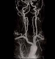

3 MR Images TÁMOP /2/A/KMR

4 Tesla and Gauss are measures of magnetic field strength Earth s magnetic field ~0.5 Gauss. 1Tesla = 10,000 Gauss. Our fmri system is 3T. ~x60,000 earth s field strength TÁMOP /2/A/KMR
5 Signal and Field Strength Outside magnetic field: Spins randomly oriented In magnetic field: Spins tend to align parallel or anti-parallel to magnetic field At room temperature, ~4 parts per million more protons per Tesla align with versus against field As field strength increases, there is a bigger energy difference between parallel and anti-parallel alignment (faster rotation = more energy) A larger proportion will align parallel to field More energy will be released as nuclei align Therefore, MR signal increases with square of field strength TÁMOP /2/A/KMR
6 Signal and Field Strength Most clinical MRI: 1.5T fmri systems: 3.0T Maximum for NbTi MRI ~11.7T Field strength influences: Faster Larmor frequency Bigger energy difference between parallel and anti-parallel alignment Larger ratio of nuclei aligned = more signal More signal as nuclei realign Reduced TR and TE: less time to take images TÁMOP /2/A/KMR
7 Signal and Field Strength In theory: Signal increases with square of field strength Noise increases linearly with field strength A 3T scanner should have twice SNR of 1.5T scanner; 7T should have ~4.7 times SNR of 1.5T Unfortunately, physiological artifacts also increase, so advantage is less in practice Benefits: speed, resolution Costs: artifacts, money, wavelength effects, auditory noise TÁMOP /2/A/KMR
8 Electromagnetic Spectrum MRI signals are in the same range as FM radio and TV (30-300MHz) MRI frequency is non-ionizing radiation, unlike X-rays Absorbed RF will cause heating Specific absorption rate (SAR): measure of the energy absorbed by tissue Increases ~ with square of field strength Higher SAR = more energy = more signal = more heating FDA limits SAR, and is a limiting factor for some protocols (3 W/kg averaged over 10 minutes) 1 MHz 1 GHz 1 THz Hz Hz Power, telephone FM radio, TV MR microwaves infrared TÁMOP /2/A/KMR UV X-rays Repetitive Visual gamma 8

9 MRI terminology Orientation: typically coronal, sagittal or axial, can be in-between these (oblique) Matrix Size: Voxels in each dimension Field of view: Spatial extent of each dimension Resolution: FOV/Matrix size Philips Achieva 3T Scanner TÁMOP /2/A/KMR
10 MRI magnetic resonance imaging images of biological tissues, structural studies static magnetic field + a series of changing magnetic fields and oscillating electromagnetic fields (pulse sequence) depending on frequency of electromagnetic fields, energy is absorbed by hydrogen nuclei (excitation) later the energy is emitted by the nuclei the amount of energy depends on numbers and types of nuclei present Advantages of MRI No ionizing radiation exposure Better spatial resolution than CT Disadvantages No ferrous metal! TÁMOP /2/A/KMR


11 History of MR/ MRI/ fmri: NMR = nuclear magnetic resonance Felix Block and Edward Purcell 1946: atomic nuclei absorb and re-emit radio frequency energy 1952: Nobel prize in physics nuclear: properties of nuclei of atoms magnetic: magnetic field required resonance: interaction between magnetic field and radio frequency Felix Bloch Edward Purcell TÁMOP /2/A/KMR
12 History of MR/ MRI/ fmri: -1971: MRI Tumor detection (Damadian) -1973: Lauterbur suggests NMR could be used to form images -1977: clinical MRI scanner patented -1977: Mansfield proposes echo-planar imaging (EPI) to acquire images faster -2003: Nobel prize was awarded to Paul Lauterbur and Sir Peter Mansfield (excluding Damadian huge controversy) fmri -1990: Ogawa observes BOLD effect with T2* blood vessels became more visible as blood oxygen decreased -1991: Belliveau observes first functional images using a contrast agent -1992: Ogawa et al. and Kwong et al. publish first functional images using BOLD signal TÁMOP /2/A/KMR
13 The First ZMR NMR Image Lauterbur, P.C. (1973). Image formation by induced local interaction: Examples employing nuclear magnetic resonance. Nature, 242, TÁMOP /2/A/KMR

14 Early Human MR Images (Damadian) TÁMOP /2/A/KMR

15 Mink5 Image Damadian (1977) TÁMOP /2/A/KMR

16 The first Philips MR, 1978 (0,15T) TÁMOP /2/A/KMR

17 The first Siemens MR, 1980 (0,2T) TÁMOP /2/A/KMR


18 Typical 1.5/3.0T MR system Special Open-MR system TÁMOP /2/A/KMR

19 Nuclear spins A nucleus of hydrogen consists of one proton carries a positive charge rotates around its axis because of thermal energy electrical current and magnetic source spin TÁMOP /2/A/KMR
20 Nuclei line up with magnetic moments either in a parallel (lower energy level) or anti-parallel configuration (higher energy level). In body tissues more line up in parallel creating a small additional magnetization M in the direction of B 0. B0 Nuclei spin axis not parallel to B 0 field direction. Nuclear magnetic moments precess about B TÁMOP /2/A/KMR
21 Absorption and Relaxation Our RF transmission is absorbed by atoms at Larmor frequency After the RF pulse, atoms will begin to realign with the magnetic field: relaxation During this period, an RF signal is emitted This signal will be at the Larmor frequency An antenna can measure this signal TÁMOP /2/A/KMR
 g ~ 43 mhz/tesla Larmor frequencies of RICs MRIs 3T ~ 130 mhz 7T ~ 300 mhz 11.7T ~ 500 mhz 2011.10.04.. TÁMOP 4.1.2-08/2/A/KMR-2009-0006 22")
22 Frequency of precession of magnetic moments given by Larmor relationship f = γ x B 0 B0 f = Larmor frequency (mhz) g = Gyromagnetic ratio (mhz/tesla) B 0 = Magnetic field strength (Tesla) g ~ 43 mhz/tesla Larmor frequencies of RICs MRIs 3T ~ 130 mhz 7T ~ 300 mhz 11.7T ~ 500 mhz TÁMOP /2/A/KMR


23 Radiofrequency Pulses A radiofrequency (RF) pulse at the Larmor frequency will be absorbed This higher energy state tips the spin, so it is no longer aligned to the field An RF pulse at any other frequency will not influence the nuclei, only resonance frequency TÁMOP /2/A/KMR
 23 Na and 31 P are relatively abundant, so can be imaged Larmor frequency varies for elements: 1 H = 42.58 Mhz/T 13 C = 10.7 Mhz/T 19 F = 40.1 Mhz/T 31 P = 17.")
24 Hydrogen is the mainstay for MRI We will focus on Hydrogen Hydrogen abundant in body (63% of atoms) Elements with even numbers of neutrons and protons have no spin, so we can not image them ( 4 He, 12 C) 23 Na and 31 P are relatively abundant, so can be imaged Larmor frequency varies for elements: 1 H = Mhz/T 13 C = 10.7 Mhz/T 19 F = 40.1 Mhz/T 31 P = 17.7 Mhz/T Therefore, by sending in a RF pulse at a specific frequency we can selectively energize hydrogen TÁMOP /2/A/KMR
 Proton density relates to the number of parallel states per unit volume Signal producing capability depends on proton density 2011.10.04.. TÁMOP 4.1.2-08/2/A/KMR-2009-0006 25")
25 M is parallel to B 0 since transverse components of magnetic moments are randomly oriented The difference between the numbers of protons in the parallel and anti-parallel states leads to the net magnetization (M) Proton density relates to the number of parallel states per unit volume Signal producing capability depends on proton density TÁMOP /2/A/KMR


26 RF Pulse Frequency of rotation of M about B 1 determined by the magnitude (strength) of B 1 strength RF Pulse RF pulse duration and strength determine flip angle duration TÁMOP /2/A/KMR


27 90 RF pulse rotates M into transverse (x-y) plane Rotation of M within transverse plane induces signal in receiver coil at Larmor frequency Magnitude signal dependent on proton density and M xy FID = Free Induction Decay FID magnitude decays in an exponential manner with a time constant T2. Decay due to spin-spin relaxation TÁMOP /2/A/KMR


28 T1-Relaxation: Recovery Recovery of longitudinal orientation of M along z-axis T1 time refers to time interval for 63% recovery of longitudinal magnetization Spin-Lattice interactions T2-Relaxation: Dephasing Loss of transverse magnetization M xy T2 time refers to time interval for 37% loss of original transverse magnetization Spin-spin interactions, and more TÁMOP /2/A/KMR
29 T1 is shorter in fat (large molecules) and longer in cerebrospinal fluid (CSF) (small molecules). T1 contrast is higher for lower TRs T2 is shorter in fat and longer in CSF. Signal contrast increased with TE TR determines T1 contrast TE determines T2 contrast TÁMOP /2/A/KMR
30 Properties of Body Tissues T1 (msec) Grey Matter White Matter Fat Blood Cerebrospinal Fluid Muscle T1 values for B 0 ~ 1Tesla. T2 ~ 1/10 th T1 for soft tissues T2 (msec) TÁMOP /2/A/KMR

31 T1/T2 weighted images: TÁMOP /2/A/KMR

32 Contrast, Imaging Parameters: Short TEs reduce T2W Long TRs reduce T1W TÁMOP /2/A/KMR

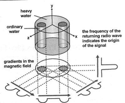
33 Making a spatial image To create spatial images, we need a way to cause different locations in the scanner to generate different signals Lauterbur To do this, we apply gradients Gradients make the magnetic field slightly stronger at one location compared to another Lauterbur: first MRI: 2003 Nobel Prize TÁMOP /2/A/KMR


34 Slice Selection Gradient Gradients make field stronger at one location compared to another Larmor frequency different along this dimension RF pulse only energizes slice where field strength matches Larmor frequency Gradual slice selection gradients will select thick slices, while steep gradients select thinner slices The strength of your scanner s gradients can limit minimum slice thickness FDA limits speed of gradient shift (db/dt) and some of our protocols can elicit slight tingling sensation or brief muscle twitches Position of gradient determines which 2D slice is selected TÁMOP /2/A/KMR


35 Phase Encoding Phase encoding gradient: Orthogonal gradient applied between RF pulse and readout This adjusts the phase along this dimension Analogy: Phase encoding is like time zones. Clocks in different zones will have different phases TÁMOP /2/A/KMR


36 Frequency Encoding Frequency encoding gradient: Apply final orthogonal gradient when we wish to acquire image Slice will emit signal at Lamour frequency, e.g. lines at higher fields will have higher frequency signals Aka Readout gradient TÁMOP /2/A/KMR

37 Raw MRI image: k-space (frequency domain) a k-space domain image is formed using frequency and phase encoding TÁMOP /2/A/KMR
38 Reconstruction Medical scanners automatically reconstruct your data You can manually reconstruct data Fourier Transforms are slow: 1021-sample data requires >2 million multiplications (2*N 2 ) Fast Fourier Transform: 1024-sample data requires 20,000 multiplications (2(N log N)) Optimal when data is power of two (64,128,256, 512), reverts to traditional Fourier for prime numbers This is why most image matrices are a power of TÁMOP /2/A/KMR

39 MRI task is to acquire k-space image then transform to a spatial-domain image. k x is sampled (read out) in real time to give N samples. k y is adjusted before each readout MR image is the magnitude of the Fourier transform of the k-space image TÁMOP /2/A/KMR
40 The k-space Trajectory Equations that govern 2D k-space trajectory if G x is constant k k y x = = t 0 t 0 G G y x ()dt t ()dt t k x = gg x t The k x, k y frequency coordinates are established by durations (t) and strength of gradients (G) TÁMOP /2/A/KMR

41 Primary types of Pulse sequences Spin Echo (SE): The most commonly used pulse sequence Uses 90 radio frequency pulses to excite the magnetization and one or more 180 pulses to refocus the spins to generate signal echoes: SE The two variables of interest in spin echo sequences is TR and TE TÁMOP /2/A/KMR


42 Spin-Echo Image TE = 20 ms TE = 50 ms TE = 75 ms TE = 100 ms TR = 250 ms TR = 500 ms TR = 1000 ms TÁMOP /2/A/KMR


43 Fast Spin Echo (FSE): Characterized by a series of rapidly applied 180 rephasing pulses and multiple echoes, changing the phase encoding gradient for each echo TE may vary from echo to echo in the echo train T2 weighted imaging profits most from this technique T2 weighted FSE images, both water and fat are hyperintense TÁMOP /2/A/KMR

44 Fast Spin Echo (FSE) TÁMOP /2/A/KMR
: a fast sequence producing")
45 Gradient Echo (GRE): generated by using a pair of bipolar gradient pulses no refocusing 180 pulse and the data are sampled during a gradient echo, which is achieved by dephasing the spins with a negatively pulsed gradient before they are rephased by an opposite gradient with opposite polarity to generate the echo short repetition time Fast Low Angle Shot (FLASH): a fast sequence producing signals called gradient echo with low flip angles uses a semi-random spoiler gradient after each echo to spoil the steady state by causing a spatially dependent phase shift extremely short TR times are possible, as a result the sequence provides a mechanism for gaining extremely high T1 contrast TÁMOP /2/A/KMR
 of a gradient echo or spin echo sequence with an acquisition time of about 20 to 100 ms 2011.10.04.. TÁMOP 4.1.2-08/2/A/KMR-2009-0006 46")
46 Echo Planar Imaging (EPI): used in applications like diffusion, perfusion, and functional magnetic resonance imaging complete image is formed from a single data sample (all k-space lines are measured in one repetition time) of a gradient echo or spin echo sequence with an acquisition time of about 20 to 100 ms TÁMOP /2/A/KMR

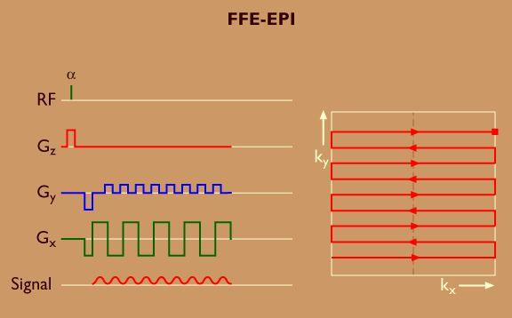

47 EPI TÁMOP /2/A/KMR
48 shielding main magnet gradient coils RF coils p a t i e n t patient support RF coils gradient coils magnet MRI equipment schematics gradient amplifier RF receiver RF amplifier A/D convert gradient pulse RF pulse forming forming controller RF source TÁMOP /2/A/KMR


49 Vacuum Biomedical Imaging: Magnetic Resonance Imaging - Basics Typical structure of an MR superconductive magnet bore liquid nitrogen ( 77 0 K) liquid helium ( 0 0 K) support superconductive coils examination field TÁMOP /2/A/KMR

50 Gradient coils TÁMOP /2/A/KMR
51 TÁMOP /2/A/KMR


52 Gradient coil system naked receiver coils without cover TÁMOP /2/A/KMR


53 SENSE Head SENSE NV SENSE Spine SENSE Cardiac SENSE XL Torso SENSE Breast SENSE Knee SENSE FootAnkle SENSE Shoulder SENSE PV SENSE Wrist SENSE Flex L SENSE Flex M SENSE Flex S Syn Pediatric TÁMOP /2/A/KMR
54 TÁMOP /2/A/KMR

55 MR safety: Projectile Effects: External Scanner visit Projectile Effects: Internal Acoustic Noise Radiofrequency Energy Gradient field changes Claustrophobia Anyone with implanted metal should see a doctor before going to the scanner Pacemaker, cochlear implant, shunt, clip, etc. Dental work and piercings are fine TÁMOP /2/A/KMR
56 Projectile Effects: Internal Motion of implanted medical devices Clips, shunts, valves, etc. Motion or rotation of debris, shrapnel, filings Primary risk: Metal fragments in eyes Swelling/irritation of skin due to motion of iron oxides in tattoo and makeup pigments Acoustic noise: Potential problem with all scans Short-term and long-term effects Sound level OSHA maximum exposure guidelines 2-4 hours per day Earplugs reduce these values by db, depending upon fit TÁMOP /2/A/KMR
57 Radiofrequency Energy Tissue Heating Specific Absorption Rate (SAR; W/kg) Pulse sequences are limited to cause less than a one-degree rise in core body temperature Scanners can be operated at up to 4 W/kg (with large safety margin) for normal subjects, 1.5 W/kg for compromised patients (infants, fetuses, cardiac) Weight of subject critical for SAR calculations Burns Looped wires can act as RF antennas and focus energy in a small area Most common problem: ECG leads Necklaces, earrings, piercings, pulse oximeters, any other cabling TÁMOP /2/A/KMR
58 Gradient field changes: Peripheral nerve stimulation May range from distracting to painful Risk greatly increased by conductive loops Arms clasped Legs crossed Theoretical risk of cardiac stimulation No evidence for effects at gradient strengths used in MRI Claustrofobia: Most common subject problem About 10% of patients Ameliorated with comfort measures Talking with subject Air flow through scanner Panic button Slow entry into scanner TÁMOP /2/A/KMR
59 FDA MRI Guidelines Adults, Children, and Infants age > 1 month 8 T B o neonates (infants age < 1 month) 4 T db/dt No discomfort, pain, or nerve stimulation SAR Specific Absorption Rate Acoustic Level whole body, average, over >15 min head, average, over >10 min head or torso, per g of tissue, in >5 min extremities, per g of tissue, in >5 min Peak unweighted A-weighted rms with hearing protection 4 W/Kg 3 W/Kg 8 W/Kg 12 W/Kg 140 db 99 dba TÁMOP /2/A/KMR
MRI Physics I: Spins, Excitation, Relaxation
 MRI Physics I: Spins, Excitation, Relaxation Douglas C. Noll Biomedical Engineering University of Michigan Michigan Functional MRI Laboratory Outline Introduction to Nuclear Magnetic Resonance Imaging
Magnetic Resonance Imaging
 Magnetic Resonance Imaging History Nuclear magnetic resonance was first described by Isidor Rabi in 1938 - Columbia University, New York City, (Nobel Prize Nobel Prize in Physics 1944) 1946 - Edward Mills
Introduction to MRI. Spin & Magnetic Moments. Relaxation (T1, T2) Spin Echoes. 2DFT Imaging. K-space & Spatial Resolution.
 Spin Echoes. 2DFT Imaging. K-space & Spatial Resolution.") Introduction to MRI Spin & Magnetic Moments Relaxation (T1, T2) Spin Echoes 2DFT Imaging Selective excitation, phase & frequency encoding K-space & Spatial Resolution Contrast (T1, T2) Acknowledgement:
Introduction to MRI Spin & Magnetic Moments Relaxation (T1, T2) Spin Echoes 2DFT Imaging Selective excitation, phase & frequency encoding K-space & Spatial Resolution Contrast (T1, T2) Acknowledgement:
Magnetic Resonance Imaging. Pål Erik Goa Associate Professor in Medical Imaging Dept. of Physics
 Magnetic Resonance Imaging Pål Erik Goa Associate Professor in Medical Imaging Dept. of Physics pal.e.goa@ntnu.no 1 Why MRI? X-ray/CT: Great for bone structures and high spatial resolution Not so great
Contrast Mechanisms in MRI. Michael Jay Schillaci
 Contrast Mechanisms in MRI Michael Jay Schillaci Overview Image Acquisition Basic Pulse Sequences Unwrapping K-Space Image Optimization Contrast Mechanisms Static and Motion Contrasts T1 & T2 Weighting,
Introduction to Biomedical Imaging
 Alejandro Frangi, PhD Computational Imaging Lab Department of Information & Communication Technology Pompeu Fabra University www.cilab.upf.edu MRI advantages Superior soft-tissue contrast Depends on among
Field trip: Tuesday, Feb 5th
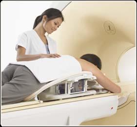
 Pulse Sequences Field trip: Tuesday, Feb 5th Hardware tour of VUIIIS Philips 3T Meet here at regular class time (11.15) Complete MRI screening form! Chuck Nockowski Philips Service Engineer Reminder: Project/Presentation
Nuclear Magnetic Resonance Imaging
 Nuclear Magnetic Resonance Imaging Jeffrey A. Fessler EECS Department The University of Michigan NSS-MIC: Fundamentals of Medical Imaging Oct. 20, 2003 NMR-0 Background Basic physics 4 magnetic fields
MRI in Review: Simple Steps to Cutting Edge Part I
 MRI in Review: Simple Steps to Cutting Edge Part I DWI is now 2 years old... Mike Moseley Radiology Stanford DWI, b = 1413 T2wt, 28/16 ASN 21 San Francisco + Disclosures: Funding NINDS, NCRR, NCI 45 minutes
Basic MRI physics and Functional MRI
 Basic MRI physics and Functional MRI Gregory R. Lee, Ph.D Assistant Professor, Department of Radiology June 24, 2013 Pediatric Neuroimaging Research Consortium Objectives Neuroimaging Overview MR Physics
RADIOLOGIV TECHNOLOGY 4912 COMPREHENSEIVE REVIEW/MRI WORSHEET #1- PATIENT CARE AND SAFETY/PHYSICAL PRINCIPLES
 RADIOLOGIV TECHNOLOGY 4912 COMPREHENSEIVE REVIEW/MRI WORSHEET #1- PATIENT CARE AND SAFETY/PHYSICAL PRINCIPLES 1. What are potential consequences to patients and personnel should there be a release of gaseous
Magnetic Resonance Imaging (MRI)
") Magnetic Resonance Imaging Introduction The Components The Technology (MRI) Physics behind MR Most slides taken from http:// www.slideworld.org/ viewslides.aspx/magnetic- Resonance-Imaging- %28MRI%29-MR-Imaging-
Magnetic Resonance Imaging Introduction The Components The Technology (MRI) Physics behind MR Most slides taken from http:// www.slideworld.org/ viewslides.aspx/magnetic- Resonance-Imaging- %28MRI%29-MR-Imaging-
MRI in Practice. Catherine Westbrook MSc, DCRR, CTC Senior Lecturer Anglia Polytechnic University Cambridge UK. John Talbot MSc, DCRR
 MRI in Practice Third edition Catherine Westbrook MSc, DCRR, CTC Senior Lecturer Anglia Polytechnic University Cambridge UK and Carolyn Kaut RothRT(R) (MR) (CT) (M) (CV) Fellow SMRT (Section for Magnetic
The Basics of Magnetic Resonance Imaging
 The Basics of Magnetic Resonance Imaging Nathalie JUST, PhD nathalie.just@epfl.ch CIBM-AIT, EPFL Course 2013-2014-Chemistry 1 Course 2013-2014-Chemistry 2 MRI: Many different contrasts Proton density T1
The physics US and MRI. Prof. Peter Bogner
 The physics US and MRI Prof. Peter Bogner Sound waves mechanical disturbance, a pressure wave moves along longitudinal wave compression rarefaction zones c = nl, (c: velocity, n: frequency, l: wavelength
BMB 601 MRI. Ari Borthakur, PhD. Assistant Professor, Department of Radiology Associate Director, Center for Magnetic Resonance & Optical Imaging
 BMB 601 MRI Ari Borthakur, PhD Assistant Professor, Department of Radiology Associate Director, Center for Magnetic Resonance & Optical Imaging University of Pennsylvania School of Medicine A brief history
EL-GY 6813/BE-GY 6203 Medical Imaging, Fall 2016 Final Exam
 EL-GY 6813/BE-GY 6203 Medical Imaging, Fall 2016 Final Exam (closed book, 1 sheets of notes double sided allowed, no calculator or other electronic devices allowed) 1. Ultrasound Physics (15 pt) A) (9
PETER PAZMANY CATHOLIC UNIVERSITY Consortium members SEMMELWEIS UNIVERSITY, DIALOG CAMPUS PUBLISHER
 SEMMELWEIS UNIVERSITY PETER PAZMANY CATHOLIC UNIVERSITY Development of Complex Curricula for Molecular Bionics and Infobionics Programs within a consortial* framework** Consortium leader PETER PAZMANY
The physics of medical imaging US, CT, MRI. Prof. Peter Bogner
 The physics of medical imaging US, CT, MRI Prof. Peter Bogner Clinical radiology curriculum blocks of lectures and clinical practice (7x2) Physics of medical imaging Neuroradiology Head and neck I. Head
The NMR Inverse Imaging Problem
 The NMR Inverse Imaging Problem Nuclear Magnetic Resonance Protons and Neutrons have intrinsic angular momentum Atoms with an odd number of proton and/or odd number of neutrons have a net magnetic moment=>
FREQUENCY SELECTIVE EXCITATION
 PULSE SEQUENCES FREQUENCY SELECTIVE EXCITATION RF Grad 0 Sir Peter Mansfield A 1D IMAGE Field Strength / Frequency Position FOURIER PROJECTIONS MR Image Raw Data FFT of Raw Data BACK PROJECTION Image Domain
Nuclear Magnetic Resonance Imaging
 Nuclear Magnetic Resonance Imaging Simon Lacoste-Julien Electromagnetic Theory Project 198-562B Department of Physics McGill University April 21 2003 Abstract This paper gives an elementary introduction
Fundamental MRI Principles Module 2 N. Nuclear Magnetic Resonance. X-ray. MRI Hydrogen Protons. Page 1. Electrons
 Fundamental MRI Principles Module 2 N S 1 Nuclear Magnetic Resonance There are three main subatomic particles: protons positively charged neutrons no significant charge electrons negatively charged Protons
Rad Tech 4912 MRI Registry Review. Outline of the Registry Exam: Certification Fees
 Rad Tech 4912 MRI Registry Review Outline of the Registry Exam: Category: # of questions: A. Patient Care 30 B. Imaging Procedures 62 C. Data Acquisition and Processing 65 D. Physical Principles of Image
Fundamental MRI Principles Module Two
 Fundamental MRI Principles Module Two 1 Nuclear Magnetic Resonance There are three main subatomic particles: protons neutrons electrons positively charged no significant charge negatively charged Protons
NMR/MRI examination (8N080 / 3F240)
") NMR/MRI examination (8N080 / 3F240) Remarks: 1. This test consists of 3 problems with at total of 26 sub-questions. 2. Questions are in English. You are allowed to answer them in English or Dutch. 3. Please
NMR/MRI examination (8N080 / 3F240) Remarks: 1. This test consists of 3 problems with at total of 26 sub-questions. 2. Questions are in English. You are allowed to answer them in English or Dutch. 3. Please
NMR and MRI : an introduction
 Intensive Programme 2011 Design, Synthesis and Validation of Imaging Probes NMR and MRI : an introduction Walter Dastrù Università di Torino walter.dastru@unito.it \ Introduction Magnetic Resonance Imaging
Basic p rinciples COPYRIGHTED MATERIAL. Introduction. Atomic s tructure
 1 Basic p rinciples Introduction 1 Atomic structure 1 Motion in the atom 2 MR active nuclei 2 The hydrogen nucleus 4 Alignment 4 Precession 8 The Larmor equation 9 Introduction The basic principles of
Biomedical Imaging Magnetic Resonance Imaging
 Biomedical Imaging Magnetic Resonance Imaging Charles A. DiMarzio & Eric Kercher EECE 4649 Northeastern University May 2018 Background and History Measurement of Nuclear Spins Widely used in physics/chemistry
Physics of MR Image Acquisition
 Physics of MR Image Acquisition HST-583, Fall 2002 Review: -MRI: Overview - MRI: Spatial Encoding MRI Contrast: Basic sequences - Gradient Echo - Spin Echo - Inversion Recovery : Functional Magnetic Resonance
Lecture 12 February 11, 2016
 MATH 262/CME 372: Applied Fourier Analysis and Winter 2016 Elements of Modern Signal Processing Lecture 12 February 11, 2016 Prof. Emmanuel Candes Scribe: Carlos A. Sing-Long, Edited by E. Bates 1 Outline
Part III: Sequences and Contrast
 Part III: Sequences and Contrast Contents T1 and T2/T2* Relaxation Contrast of Imaging Sequences T1 weighting T2/T2* weighting Contrast Agents Saturation Inversion Recovery JUST WATER? (i.e., proton density
Magnetic resonance imaging MRI
 Magnetic resonance imaging MRI Introduction What is MRI MRI is an imaging technique used primarily in medical settings that uses a strong magnetic field and radio waves to produce very clear and detailed
Physics and Brain Imaging
 Physics and Brain Imaging Nuclear Magnetic Resonance (NMR) Magnetic Resonance Imaging (MRI) Functional MRI (fmri) Talk at Quarknet FSU Summer Workshop, July 24, 2017 Per Arne Rikvold Leonardo da Vinci
Topics. The concept of spin Precession of magnetic spin Relaxation Bloch Equation. Bioengineering 280A Principles of Biomedical Imaging
 Bioengineering 280A Principles of Biomedical Imaging Fall Quarter 2006 MRI Lecture 1 Topics The concept of spin Precession of magnetic spin Relaxation Bloch Equation 1 Spin Intrinsic angular momentum of
M R I Physics Course. Jerry Allison Ph.D., Chris Wright B.S., Tom Lavin B.S., Nathan Yanasak Ph.D. Department of Radiology Medical College of Georgia
 M R I Physics Course Jerry Allison Ph.D., Chris Wright B.S., Tom Lavin B.S., Nathan Yanasak Ph.D. Department of Radiology Medical College of Georgia M R I Physics Course Spin Echo Imaging Hahn Spin Echo
Relaxation times in nuclear magnetic resonance
 Relaxation times in TEP Related topics Nuclear spins, atomic nuclei with a magnetic moment, precession movement of the nuclear spins, Landau-Lifshitz equation, Bloch equation, magnetisation, resonance
Introduction to Magnetic Resonance Imaging (MRI) Pietro Gori
 Pietro Gori") Introduction to Magnetic Resonance Imaging (MRI) Pietro Gori Enseignant-chercheur Equipe IMAGES - Télécom ParisTech pietro.gori@telecom-paristech.fr September 20, 2017 P. Gori BIOMED 20/09/2017 1 / 76
Introduction to Magnetic Resonance Imaging (MRI) Pietro Gori Enseignant-chercheur Equipe IMAGES - Télécom ParisTech pietro.gori@telecom-paristech.fr September 20, 2017 P. Gori BIOMED 20/09/2017 1 / 76
G Medical Imaging. Outline 4/13/2012. Physics of Magnetic Resonance Imaging
 G16.4426 Medical Imaging Physics of Magnetic Resonance Imaging Riccardo Lattanzi, Ph.D. Assistant Professor Department of Radiology, NYU School of Medicine Department of Electrical and Computer Engineering,
Basis of MRI Contrast
 Basis of MRI Contrast MARK A. HORSFIELD Department of Cardiovascular Sciences University of Leicester Leicester LE1 5WW UK Tel: +44-116-2585080 Fax: +44-870-7053111 e-mail: mah5@le.ac.uk 1 1.1 The Magnetic
Tissue Characteristics Module Three
 Tissue Characteristics Module Three 1 Equilibrium State Equilibrium State At equilibrium, the hydrogen vector is oriented in a direction parallel to the main magnetic field. Hydrogen atoms within the vector
PETER PAZMANY CATHOLIC UNIVERSITY Consortium members SEMMELWEIS UNIVERSITY, DIALOG CAMPUS PUBLISHER
 SEMMELWEIS UNIVERSITY PETER PAZMANY CATLIC UNIVERSITY Development of Complex Curricula for Molecular Bionics and Infobionics Programs within a consortial* framework** Consortium leader PETER PAZMANY CATLIC
PETER PAZMANY CATHOLIC UNIVERSITY Consortium members SEMMELWEIS UNIVERSITY, DIALOG CAMPUS PUBLISHER
 PETER PAZMANY CATHOLIC UNIVERSITY SEMMELWEIS UNIVERSITY Development of Complex Curricula for Molecular Bionics and Infobionics Programs within a consortial* framework** Consortium leader PETER PAZMANY
K-space. Spin-Warp Pulse Sequence. At each point in time, the received signal is the Fourier transform of the object s(t) = M( k x
 = M( k x") Bioengineering 280A Principles of Biomedical Imaging Fall Quarter 2015 MRI Lecture 4 k (t) = γ 2π k y (t) = γ 2π K-space At each point in time, the received signal is the Fourier transform of the object
Bioengineering 280A Principles of Biomedical Imaging Fall Quarter 2015 MRI Lecture 4 k (t) = γ 2π k y (t) = γ 2π K-space At each point in time, the received signal is the Fourier transform of the object
Chapter 14:Physics of Magnetic Resonance
 Chapter 14:Physics of Magnetic Resonance Slide set of 141 slides based on the chapter authored by Hee Kwon Song of the publication (ISBN 978-92-0-131010-1): Diagnostic Radiology Physics: A Handbook for
Nuclei, Excitation, Relaxation
 Outline 4.1 Principles of MRI uclei, Excitation, Relaxation Carolyn Kaut Roth, RT (R)(MR)(CT)(M)(CV) FSMRT CEO Imaging Education Associates www.imaginged.com candi@imaginged.com What nuclei are MR active?
SEMMELWEIS UNIVERSITY, DIALOG CAMPUS PUBLISHER
 PETER PAZMANY CATHOLIC UNIVERSITY SEMMELWEIS UNIVERSITY Development of Complex Curricula for Molecular Bionics and Infobionics Programs within a consortial* framework** Consortium leader PETER PAZMANY
MRI Physics II: Gradients, Imaging. Douglas C. Noll, Ph.D. Dept. of Biomedical Engineering University of Michigan, Ann Arbor
 MRI Physics II: Gradients, Imaging Douglas C., Ph.D. Dept. of Biomedical Engineering University of Michigan, Ann Arbor Magnetic Fields in MRI B 0 The main magnetic field. Always on (0.5-7 T) Magnetizes
PETER PAZMANY CATHOLIC UNIVERSITY Consortium members SEMMELWEIS UNIVERSITY, DIALOG CAMPUS PUBLISHER
 PETER PAZMANY SEMMELWEIS CATHOLIC UNIVERSITY UNIVERSITY Development of Complex Curricula for Molecular Bionics and Infobionics Programs within a consortial* framework** Consortium leader PETER PAZMANY
Physical fundamentals of magnetic resonance imaging
 Physical fundamentals of magnetic resonance imaging Stepan Sereda University of Bonn 1 / 26 Why? Figure 1 : Full body MRI scan (Source: [4]) 2 / 26 Overview Spin angular momentum Rotating frame and interaction
Advanced Topics and Diffusion MRI
 Advanced Topics and Diffusion MRI Slides originally by Karla Miller, FMRIB Centre Modified by Mark Chiew (mark.chiew@ndcn.ox.ac.uk) Slides available at: http://users.fmrib.ox.ac.uk/~mchiew/teaching/ MRI
Background II. Signal-to-Noise Ratio (SNR) Pulse Sequences Sampling and Trajectories Parallel Imaging. B.Hargreaves - RAD 229.
 Pulse Sequences Sampling and Trajectories Parallel Imaging. B.Hargreaves - RAD 229.") Background II Signal-to-Noise Ratio (SNR) Pulse Sequences Sampling and Trajectories Parallel Imaging 1 SNR: Signal-to-Noise Ratio Signal: Desired voltage in coil Noise: Thermal, electronic Noise Thermal
Background II Signal-to-Noise Ratio (SNR) Pulse Sequences Sampling and Trajectories Parallel Imaging 1 SNR: Signal-to-Noise Ratio Signal: Desired voltage in coil Noise: Thermal, electronic Noise Thermal
Magnetic Resonance Imaging in a Nutshell
 Magnetic Resonance Imaging in a Nutshell Oliver Bieri, PhD Department of Radiology, Division of Radiological Physics, University Hospital Basel Department of Biomedical Engineering, University of Basel,
Lab 2: Magnetic Resonance Imaging
 EE225E/BIOE265 Spring 2013 Principles of MRI Miki Lustig Developed by: Galen Reed and Miki Lustig Lab 2: Magnetic Resonance Imaging Introduction In this lab, we will get some hands-on experience with an
Introduction to MRI Acquisition
 Introduction to MRI Acquisition James Meakin FMRIB Physics Group FSL Course, Bristol, September 2012 1 What are we trying to achieve? 2 What are we trying to achieve? Informed decision making: Protocols
Apodization. Gibbs Artifact. Bioengineering 280A Principles of Biomedical Imaging. Fall Quarter 2013 MRI Lecture 5. rect(k x )
") Bioengineering 280A Principles of Biomedical Imaging Fall Quarter 2013 MRI Lecture 5 GE Medical Systems 2003 Gibbs Artifact Apodization rect(k ) Hanning Window h(k )=1/2(1+cos(2πk ) 256256 image 256128
Bioengineering 280A Principles of Biomedical Imaging Fall Quarter 2013 MRI Lecture 5 GE Medical Systems 2003 Gibbs Artifact Apodization rect(k ) Hanning Window h(k )=1/2(1+cos(2πk ) 256256 image 256128
Magnetic Resonance Imaging. Qun Zhao Bioimaging Research Center University of Georgia
 Magnetic Resonance Imaging Qun Zhao Bioimaging Research Center University of Georgia The Nobel Prize in Physiology or Medicine 2003 "for their discoveries concerning magnetic resonance imaging" Paul C.
Bioengineering 278" Magnetic Resonance Imaging" Winter 2010" Lecture 1! Topics:! Review of NMR basics! Hardware Overview! Quadrature Detection!
 Bioengineering 278" Magnetic Resonance Imaging" Winter 2010" Lecture 1 Topics: Review of NMR basics Hardware Overview Quadrature Detection Boltzmann Distribution B 0 " = µ z $ 0 % " = #h$ 0 % " = µ z $
Introduction to Nuclear Magnetic Resonance Spectroscopy
 Introduction to Nuclear Magnetic Resonance Spectroscopy Dr. Dean L. Olson, NMR Lab Director School of Chemical Sciences University of Illinois Called figures, equations, and tables are from Principles
AQA Physics /7408
 AQA Physics - 7407/7408 Module 10: Medical physics You should be able to demonstrate and show your understanding of: 10.1 Physics of the eye 10.1.1 Physics of vision The eye as an optical refracting system,
MRI Fundamentals. Class II (MR Principles)
") MRI Fundamentals Class II (MR Principles) 1 Requirements for MRI Human body (Patient) Strong magnetic field (Magnet) External Radio Frequency source (RF Pulse) 2 3 Body Composition The molecular composition
MRI Fundamentals Class II (MR Principles) 1 Requirements for MRI Human body (Patient) Strong magnetic field (Magnet) External Radio Frequency source (RF Pulse) 2 3 Body Composition The molecular composition
MRI Homework. i. (0.5 pt each) Consider the following arrangements of bar magnets in a strong magnetic field.
 Consider the following arrangements of bar magnets in a strong magnetic field.") MRI Homework 1. While x-rays are used to image bones, magnetic resonance imaging (MRI) is used to examine tissues within the body by detecting where hydrogen atoms (H atoms) are and their environment (e.g.
MRI Homework 1. While x-rays are used to image bones, magnetic resonance imaging (MRI) is used to examine tissues within the body by detecting where hydrogen atoms (H atoms) are and their environment (e.g.
Chapter 1 Introduction
 Chapter 1 Introduction A journey of a thousand miles must begin with a single step. LaoZi Tomography is an important area in the ever-growing field of imaging science. The term tomos (rofio
10.4 Continuous Wave NMR Instrumentation
 10.4 Continuous Wave NMR Instrumentation coherent detection bulk magnetization the rotating frame, and effective magnetic field generating a rotating frame, and precession in the laboratory frame spin-lattice
Introductory MRI Physics
 C HAPR 18 Introductory MRI Physics Aaron Sodickson EXRNAL MAGNETIC FIELD, PROTONS AND EQUILIBRIUM MAGNETIZATION Much of the bulk of the magnetic resonance imaging (MRI) scanner apparatus is dedicated to
Sketch of the MRI Device
 Outline for Today 1. 2. 3. Introduction to MRI Quantum NMR and MRI in 0D Magnetization, m(x,t), in a Voxel Proton T1 Spin Relaxation in a Voxel Proton Density MRI in 1D MRI Case Study, and Caveat Sketch
} B 1 } Coil } Gradients } FFT
 Introduction to MRI Daniel B. Ennis, Ph.D. Requirements for MRI UCLA DCVI Requirements for MRI Dipoles to Images MR Active uclei e.g. 1 H in H20 Cryogen Liquid He and 2 Magnetic Field (B0) Polarizer ystem
Technical University of Denmark
 Technical University of Denmark Page 1 of 11 pages Written test, 9 December 2010 Course name: Introduction to medical imaging Course no. 31540 Aids allowed: none. "Weighting": All problems weight equally.
Topics. Spin. The concept of spin Precession of magnetic spin Relaxation Bloch Equation
 Bioengineering 280A Principles of Biomedical Imaging Fall Quarter 2005 MRI Lecture 1 Topics The concept of spin Precession of magnetic spin Relaation Bloch Equation Spin Intrinsic angular momentum of elementary
Magnetic Resonance Imaging
 http://www.qldxray.com.au/filelibrary/mri_cardiovascular_system_ca_0005.jpg Magnetic Resonance Imaging 1 Overview 1. The magnetic properties of nuclei, and how they behave in strong magnetic fields. 2.
Doppler echocardiography & Magnetic Resonance Imaging. Doppler echocardiography. History: - Langevin developed sonar.
 1 Doppler echocardiography & Magnetic Resonance Imaging History: - Langevin developed sonar. - 1940s development of pulse-echo. - 1950s development of mode A and B. - 1957 development of continuous wave
Chem 325 NMR Intro. The Electromagnetic Spectrum. Physical properties, chemical properties, formulas Shedding real light on molecular structure:
 Physical properties, chemical properties, formulas Shedding real light on molecular structure: Wavelength Frequency ν Wavelength λ Frequency ν Velocity c = 2.998 10 8 m s -1 The Electromagnetic Spectrum
Introduction to Magnetic Resonance Imaging
 Introduction to Magnetic Resonance Imaging MRI of the brain, ca. 1978. ca. 1993 ca. 2006 2014 Modality Characteristics and Comparison Radiography CT scanning Nuclear medicine MRI transmission modalities
Medical Imaging Physics Spring Quarter Week 9-1
 Medical Imaging Physics Spring Quarter Week 9-1 NMR and MRI Davor Balzar balzar@du.edu www.du.edu/~balzar Intro MRI Outline NMR & MRI Guest lecturer fmri Thursday, May 22 Visit to CUHSC It s not mandatory
Principles of MRI EE225E / BIO265. Instructor: Miki Lustig UC Berkeley, EECS
 Principles of MRI EE225E / BIO265 Instructor: Miki Lustig UC Berkeley, EECS Today... Administration http://inst.eecs.berkeley.edu/~ee225e/sp16/ Intro to Medical Imaging and MRI Medical Imaging (Before
Master s Program in Medical Physics. Physics of Imaging Systems Basic Principles of Magnetic Resonance Imaging I. Prof. Dr. Lothar Schad.
 1 12/9/2008 Page 1 Master s Program in Medical Physics Physics of Imaging Systems Basic Principles of Magnetic Resonance Imaging I Chair in Faculty of Medicine Mannheim University of Heidelberg Theodor-Kutzer-Ufer
ELECTRON SPIN RESONANCE & MAGNETIC RESONANCE TOMOGRAPHY
 ELECTRON SPIN RESONANCE & MAGNETIC RESONANCE TOMOGRAPHY 1. AIM OF THE EXPERIMENT This is a model experiment for electron spin resonance, for clear demonstration of interaction between the magnetic moment
Basic Principles of Magnetic Resonance Imaging
 Basic Principles of Magnetic Resonance Imaging Joseph C. McGowan, PhD, PE a,b, * KEYWORDS MR imaging MR physics Magnetic resonance Spin echo Gradient echo K-space Fast spin echo Magnetic resonance (MR)
Outlines: (June 11, 1996) Instructor:
 Instructor:") Magnetic Resonance Imaging (June 11, 1996) Instructor: Tai-huang Huang Institute of Biomedical Sciences Academia Sinica Tel. (02) 2652-3036; Fax. (02) 2788-7641 E. mail: bmthh@ibms.sinica.edu.tw Reference:
Magnetic Resonance Imaging (June 11, 1996) Instructor: Tai-huang Huang Institute of Biomedical Sciences Academia Sinica Tel. (02) 2652-3036; Fax. (02) 2788-7641 E. mail: bmthh@ibms.sinica.edu.tw Reference:
Basic Principles of MRI
 MRI for Technologists Basic Principles of MRI PROGRAM INFORMATION MRI for Technologists is a training program designed to meet the needs of radiologic technologists entering or working in the field of
The Physical Basis of Nuclear Magnetic Resonance Part I ESMRMB. Jürgen R. Reichenbach
 The Physical Basis of Nuclear agnetic Resonance Part I Jürgen R. Reichenbach odule 1 October 17, 216 Outline of odule Introduction Spin and magnetic moment Spin precession, Larmor frequency agnetic properties
BNG/ECE 487 FINAL (W16)
") BNG/ECE 487 FINAL (W16) NAME: 4 Problems for 100 pts This exam is closed-everything (no notes, books, etc.). Calculators are permitted. Possibly useful formulas and tables are provided on this page. Fourier
BNG/ECE 487 FINAL (W16) NAME: 4 Problems for 100 pts This exam is closed-everything (no notes, books, etc.). Calculators are permitted. Possibly useful formulas and tables are provided on this page. Fourier
Pulse Sequences: RARE and Simulations
 Pulse Sequences: RARE and Simulations M229 Advanced Topics in MRI Holden H. Wu, Ph.D. 2018.04.19 Department of Radiological Sciences David Geffen School of Medicine at UCLA Class Business Final project
Technical University of Denmark
 Technical University of Denmark Page 1 of 10 pages Written test, 12 December 2012 Course name: Introduction to medical imaging Course no. 31540 Aids allowed: None. Pocket calculator not allowed "Weighting":
Me myself and MRI: adventures in not understanding nuclear physics.
 Me myself and MRI: adventures in not understanding nuclear physics. Thomas E. Gladwin August 28, 2007 Contents 1 Introduction 2 2 Nuclei 2 2.1 Precession............................... 2 2.2 Spin-up and
Principles of Magnetic Resonance Imaging
 Principles of Magnetic Resonance Imaging Hi Klaus Scheffler, PhD Radiological Physics University of 1 Biomedical Magnetic Resonance: 1 Introduction Magnetic Resonance Imaging Contents: Hi 1 Introduction
SEMMELWEIS UNIVERSITY, DIALOG CAMPUS PUBLISHER
 PETER PAZMANY CATHOLIC UNIVERSITY SEMMELWEIS UNIVERSITY Development of Complex Curricula for Molecular Bionics and Infobionics Programs within a consortial* framework** Consortium leader PETER PAZMANY
Cambridge University Press MRI from A to Z: A Definitive Guide for Medical Professionals Gary Liney Excerpt More information
 Main glossary Aa AB systems Referring to molecules exhibiting multiply split MRS peaks due to spin-spin interactions. In an AB system, the chemical shift between the spins is of similar magnitude to the
Introduction to the Physics of NMR, MRI, BOLD fmri
 Pittsburgh, June 13-17, 2011 Introduction to the Physics of NMR, MRI, BOLD fmri (with an orientation toward the practical aspects of data acquisition) Pittsburgh, June 13-17, 2001 Functional MRI in Clinical
BASIC MRI PHYSICS SPIN GYMNASTICS Don Plewes PhD, Walter Kucharczyk MD
 BASIC MRI PHYSICS SPIN GYMNASTICS Don Plewes PhD, Walter Kucharczyk MD Introduction To understand MRI, it is first necessary to understand the physics of proton Nuclear Magnetic Resonance (NMR). The most
ENG4BF3 Medical Image Processing
 ENG4BF3 Medical Image Processing Medical Imaging Modalities Imaging in Medical Sciences Imaging is an essential aspect of medical sciences for visualization of anatomical structures and functional or metabolic
Chemistry 431. Lecture 23
 Chemistry 431 Lecture 23 Introduction The Larmor Frequency The Bloch Equations Measuring T 1 : Inversion Recovery Measuring T 2 : the Spin Echo NC State University NMR spectroscopy The Nuclear Magnetic
ELG7173 Topics in signal Processing II Computational Techniques in Medical Imaging
 ELG7173 Topics in signal Processing II Computational Techniques in Medical Imaging Topic #1: Intro to medical imaging Medical Imaging Classifications n Measurement physics Send Energy into body Send stuff
NMR Spectroscopy Laboratory Experiment Introduction. 2. Theory
 1. Introduction 64-311 Laboratory Experiment 11 NMR Spectroscopy Nuclear Magnetic Resonance (NMR) spectroscopy is a powerful and theoretically complex analytical tool. This experiment will introduce to
The Theory of Nuclear Magnetic Resonance Behind Magnetic Resonance Imaging. Catherine Wasko Physics 304 Physics of the Human Body May 3, 2005
 The Theory of Nuclear Magnetic Resonance Behind Magnetic Resonance Imaging Catherine Wasko Physics 304 Physics of the Human Body May 3, 2005 Magnetic resonance imaging (MRI) is a tool utilized in the medical
QUALITY ASSURANCE OF MAGNETIC RESONANCE IMAGING FOR ADAPTIVE RADIOTHERAPY: PRELIMINARY INVESTIGATIONS TREVOR THANG 1 Supervisors: Dr.
 QUALITY ASSURANCE OF MAGNETIC RESONANCE IMAGING FOR ADAPTIVE RADIOTHERAPY: PRELIMINARY INVESTIGATIONS TREVOR THANG 1 Supervisors: Dr. Eugene Wong 2, Dr. Rob Bartha 1 Department of Medical Biophysics 1,
Functional Magnetic Resonance Imaging (FMRI) is an imaging technique for
 is an imaging technique for") Chapter 2 Principles of FMRI Functional Magnetic Resonance Imaging (FMRI) is an imaging technique for examining brain function. Since its first appearance in 1991 (Belliveau et al.[8]) the use of FMRI
Chapter 2 Principles of FMRI Functional Magnetic Resonance Imaging (FMRI) is an imaging technique for examining brain function. Since its first appearance in 1991 (Belliveau et al.[8]) the use of FMRI
Basic principles COPYRIGHTED MATERIAL. Introduction. Introduction 1 Precession and precessional
 1 Basic principles Introduction 1 Precession and precessional Atomic structure 2 (Larmor) frequency 10 Motion in the atom 2 Precessional phase 13 MR-active nuclei 4 Resonance 13 The hydrogen nucleus 5
An Introduction to Nuclear Magnetic Resonance Imaging
 An Introduction to Nuclear Magnetic Resonance Imaging Craig M. Dowell Department of Physics, University of Washington, Seattle, WA 98195-1560 Abstract An overview of the significant historical events in
2.1.1 A Brief History of NMR The conception of NMR sprouted after the Pauli s prediction of nuclear spin in
 CHAPTER--2 BASICS OF NMR IMAGING AND SPECTROSCOPY 2.1 Introduction 2.1.1 A Brief History of NMR The conception of NMR sprouted after the Pauli s prediction of nuclear spin in 1924. Later Gorter (1936)