Introduction to techniques in biophysics.
|
|
|
- Clare Lucas
- 6 years ago
- Views:
Transcription
1 Introduction to techniques in biophysics. Goal : resolve the structure and functions of biological objects applying physical approaches 1 At molecular level 1.1 Long-range goal: complete structure determination: location in space of each atom Problem: Mwt (protein, AA) = N = atoms 3N 6 parameters define the structure parameters (structure + chemical identity) huge! simplification: ignore H-atoms parameters still huge! primary structure: bond length and angle, internal rot. angle (not for H) Problem: N = atoms parameters many bond lengths and angles are known still huge! further simplifications: work with residues: 2 angles for relative location + 2 parameters for side chains orientation Problem: Mwt (protein) = (~ 300 residues) parameters Mwt (AA) = (~ 200 residues) 800 parameters symmetry (identical subunits, helices ) still huge! 1.2 Near-field goal: rough size and shape of molecule Example: model the molecule as an ellipsoid of revolution, a rod, a coil of uniform or no stiffness techniques: hydrodynamic and scattering techniques characterize only a part of the macromolecule, of special interest Example: use probe molecules (a part of the macromolecule or externally added) techniques: fluorescence, EPR, some aspects of NMR, chemical modification examine certain general aspects of the structure, ignoring others Example: give a general picture, show constraints on possible 3D structure techniques: CD/ORD, UV, IR, Raman spectroscopy, tritium exchange 2 At supra molecular level: structure and physical properties 2.1 direct visualization of shape and structure: optical techniques 2.2 measure mechanical properties; force/energy: physical approach 2.3 functional characterization: chemical approach Techniques: electron microscopy, hydrodynamics (RD)
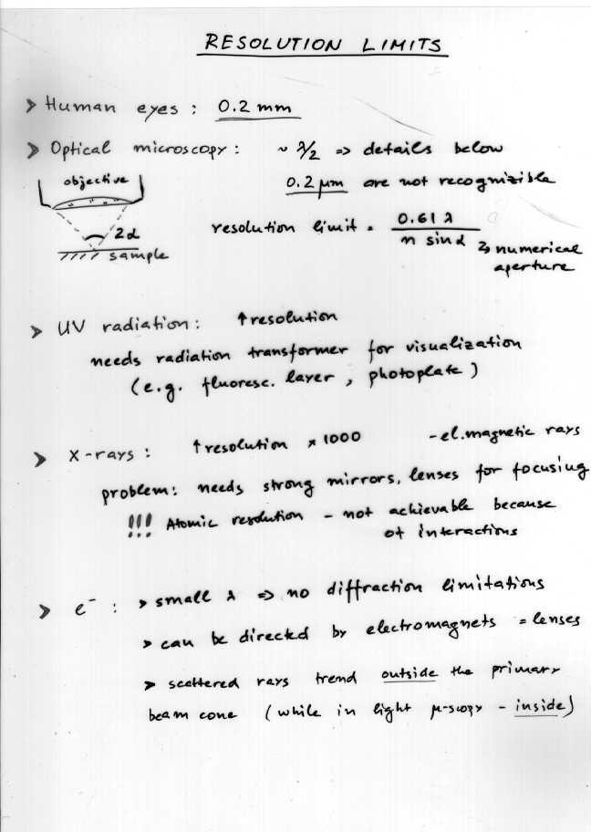
2 Structure Determination of Biomolecules by Physical Methods. Macromolecules (proteins, nucleic acids) size - up to few 10 2 nm; weight - up to 10 8 : well defined (synthetic molecules - molecular weight distributions; mean values) Human eyes: 0.2mm Method yields M-range Requirements, comments - Complete chemical analysis M no limit - Quantitative chem. identification of groups (of number M, e.g. CO) M no limit Knowledge of general chemical structure - X-ray diffraction - Neutron diffraction size, shape internal structure no limit Single crystal, heavy atom staining; - Electron microscopy Size, shape > 5000? Radiation damage - Osmotic pressure M n Vapor pressure osmometry - Viscosimetry M v (size, shape) < 10 7 Structural effects - Diffusion measurements M w, size < 10 6 Influence of structure, solvation, concentration - Sedimentation velocity M w, size < 5x10 7 Structure dependent - Sediment. velocity + diffusion M w, size < 5x10 7 Structure effects eliminated - Sedimentation equilibrium M w, M z < 5x10 6 Fast; structure independent - Electrophoresis M (poor value) Separation method for ions - Rotational diffusion, fluorescence depolarization Size, shape, M w no limit Structure dependent - Flow birefringence Size, shape, M w Empirical calibration - Kerr effect Size (shape) no limit - Dielectric dispersion Size (shape) no limit Isolating solvent; electric dipole moment of solute known - Light scattering R G, size M w 10 4 Fast, sensitive to association of molecules - X-ray small angle diffraction - Neutron small angle diffraction Size < 10 5 Dilute solutions Techniques: electron microscopy, hydrodynamics (RD)
3
4
5 Electron Microscopy 1 Features, requirements e - wavelength (de Broglie relationship): λ = h mev e - kinetic energy (if accelerated by voltage Φ): 2 m e 2v = eφ Example: for Φ = V λ = 0.04 Å! (theoretically) EM yields extremely precise structures of small molecules in gas phase work in vacuum (e - are easily stopped/absorbed by thin layers) usually on gold support possible damage of sample by e - -collision low irradiation intensity to be employed contrast sample-to-support decreases problem rule of thumb: e - -scattering (atomic number) 2 Example: U (A = 92) scatters 10 4 times more than H (A=1) 2 Principle (comparison with light microscopy) Light microscope light EM e - source anode Electrons emitted from a hot cathode are attracted to an anode; pass through a hole in the anode and create the e - beam aperture diaphragm condenser specimen objective lens scattered rays Lenses are electromagnets or magnetic coils: zoom lenses a change in the magnetic field changes their focal length objective image projection ocular intermediate lens projection lens Low numerical aperture limits the resolution: Limit of resolution = 0.61λ/numerical aperture = approx. 0.1 nm (1Å) projection image Techniques: electron microscopy (RD)

6
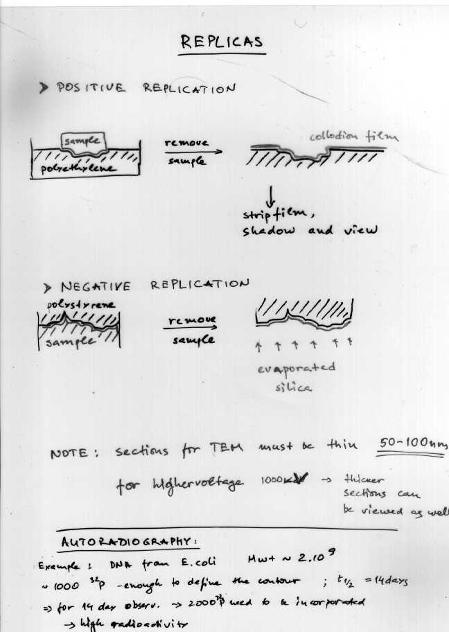
7 3 Transmission electron microscopy: more relevant for biological samples Anode Tungsten thermionic cathode overall magnification: 100x to x or 2µm to 0.4nm Resolution limit depends on: electron wavelength performance of the magnetic lenses stability of magnetic fields (1-50kV) Sample mounted on a transparent grid Electromagnetic lens ( x) Lens system ( x) Difficulties: melting in the intense electron beam damage: e - interact with the e - shell inducing energy transfer and not elastic scattering Example: bonds in aliphatic chains would break; aromatic rings would endure electrostatic charging evaporation and decomposition solvated structures are destroyed during the dry-sample preparation partially avoided when using replicas Fluorescent screen (additional 5-10 x) Possible 3D presentation: by shadow casting (firing electrons under an angle) Vacuum is required in the e - beam path usually only dead, dehydrated specimens may be viewed by EM. Exception: cryo-em; works with frozen specimen (vaporization of water is negligible) Specimen Preparation for TEM Dehydration: required except for cryo-em Embedding in epoxy resins or polyester (Vestopal) Sections: cut with an ultramicrotome, floated on water, picked up on a support grid; sections for TEM must be very thin ( nm) Exception: very high voltage (10 6 V) TEMicroscopes permit viewing thicker sections Grid (copper) may be coated with a support film of low atomic weight material; tissue sections are viewed through holes in the grid tissue slices on EM grid Techniques: electron microscopy (RD)
8

9 4 Staining techniques: heavy atom stains improve contrast, but reduce resolution Positive staining: incorporation of heavy atoms (e - -dense stain) e.g. specific complexation Example: binding uranyl ions (from uranyl acetate) to DNA; osmium (osmium tetroxide) fixation to lipid bilayers Negative staining: the sample is flooded in solution of an e - -dense stain (e.g. phosphotungstic acid, uranyl acetate); placed on a grid; dried When viewed by TEM, areas occupied by sample (not penetrated by stain) are seen to be less e - dense. sample dried solution of e - -dense stain EM grid support film Negative Staining Shadowing evaporation of heavy metal at an angle EM grid with support film Shadowing: heavy metal (e.g. tungsten) is evaporated/sprayed at an angle, under vacuum; shadows form where the metal builds up; the sample is dissolved by acid, and the metal replica viewed by TEM Autoradiography: radioactive sample placed in a fine photographic emulsion resolution - limited by the silver grains size, the traveling distance of decay particles; problem decay time (i.e. exposure time) Techniques: electron microscopy (RD)
 support liquid freon Cold knife Specimen table liquid N 2 Fracturing occurs mainly along the interface of the 2 hydrocarbon")

10 5 Freeze fracture / freeze etching: for viewing surfaces of organelles Fracturing: freeze rapidly and cut specimen Bell jar (under vacuum to prevent frosting of the knife) support liquid freon Cold knife Specimen table liquid N 2 Fracturing occurs mainly along the interface of the 2 hydrocarbon layers of the membrane Etching: sublimation of ice in vacuum (at 100 C) Inner surface Fractured face ridge surface Outer surface Normal etching Deep etching Visualizing: the exposed surface is shadowed with platinum and carbon; the deposited film is removed: replica for EM Techniques: electron microscopy (RD)
 limits the resolution beam electron current: 10-10 -10-12 A SE - emitted at low voltage easily deflected to follow curved paths of the collector 3D image")
11 6 Scanning electron microscopy: resolution about 10nm BSE - back scattered electrons SE - secondary electrons SC - specimen current EBIC - electron-beam-induced current X - X-rays CRT - cathode-ray tube the electron beam diameter (5-10 nm) limits the resolution beam electron current: A SE - emitted at low voltage easily deflected to follow curved paths of the collector 3D image depth field: times bigger than for light microscope (better contrast) Magnification can be increased by decreasing the scan-coiled current Specimen Preparation for SEM Samples are fixed and dried Non-conducting surfaces (e.g. biological objects) are coated by evaporation under vacuum with a heavy metal (to avoid disturbing electrostatic effects) Techniques: electron microscopy (RD)
12 Biomolecular Hydrodynamics 1 Hydrodynamic properties sedimentation coefficient diffusion coefficient rotational relaxation time intrinsic viscosity viscoelastic relaxation time give information about: molecular weight, size, hydration, shape, flexibility, conformation, degree of association of biological macromolecules Some classical examples: Experiments: Fick, on diffusion Svedberg, 1920s - on sedimentation Poiseuille, on viscosity Theories for simple shapes (e.g. spheres and ellipsoids): Stokes (1847) Einstein (1906) / Brownian movement Perrin (1936) / translation and diffusion of an ellipsoid Some recent developments: Experiments: dynamic laser light scattering nanosecond fluorescence depolarization measurements fluorescence recovery after photobleaching fluorescence correlation spectroscopy pulsed field gradient NMR improved analytical ultracentrifuge design Theory: new theoretical and computational tools Techniques: hydrodynamics (RD)
 concentration gradient (as in diffusion) F f R v - the frictional force opposes the particle motion rotational frictional coefficient, f r ω - angular velocity T r")
13 2 Frictional coefficients translational frictional coefficient, f t v -velocity F f - frictional force: applied force (e.g. centrifugation or electrophoresis) concentration gradient (as in diffusion) F f R v - the frictional force opposes the particle motion rotational frictional coefficient, f r ω - angular velocity T r - frictional torque: applied electric or hydrodynamic flow field diffusion of molecular axes ω T r 2.1 Viscosity of water η Def: force per unit area needed to maintain unit velocity gradient between two parallel surfaces moving relative to one another in the fluid cgs units: dyne.s/cm 2 = poise* *Poiseuille: viscous flow in tubes (1946) At 20 C η = poise or centipoise (cp) for 0 t 20 C for 20 t 100 C 2.2 Spheres Stokes law, R - Stokes radius f t - much less sensitive to molecular dimensions varies with volume Techniques: hydrodynamics (RD)
14 2.3 Ellipsoids of revolution a b oblates p < 1 a/b = p - axial ratio V = (4/3)πab 2 a b prolates (models for cylindrical rods) p > 1 Friction ratios: R e - equivalent radius (of the sphere of equal volume) r b 2.4 Random coils have on average a spherical domain behave hydrodynamically like spheres with effective hydrodynamic radius radius of gyration for a coil with N bonds of length b not swollen by excluded volume the effective hydrodynamic radius 2/3 radius of gyration 2.5 Oligomeric arrays of spheres model for proteins n number of (33) - rotation around the axis of highest symmetry (11) - for either axis perpendicular to it Techniques: hydrodynamics (RD)
15 3 Experimental determination of hydrodynamic properties 3.1 Sedimentation coefficient (centrifugation, ultracentrifugation) Types of sedimentation experiments: 1) measure the velocity of molecular motion 2) the centrifuge runs until equilibrium is reached and one measures the unchanging concentration distribution Usual components in the system: 1 water (density ρ) 2 polymer solute (molecular weight M 2 ; partial specific volume ) 3 other small molecules (salts, buffer) ω - angular velocity r - distance from axis balanced by (steady state) buoyancy angular acceleration Ultracentrifugation: ω 2 r 10 6 m/s m/s Sedimentation coefficient: range of S: s S-units: [s]; s = 1 Svedberg ω-units: [rad/s] or [rpm]; 1rpm = 60/2π rad/s determine S calculate M 2 /f t S measurable via optical techniques: r r m For nucleic acids: measure A260 as a function of r For proteins: measure around 280 nm or in the absorption band of some chromophore With an admixture of heavy salts (e.g. CsCl): molecules accumulate within a sharp layer - used for separating isotopically labeled molecules ( density gradient method ) More details: diffusional broadening, concentration dependence, interaction of charged molecules Techniques: hydrodynamics (RD)
16 3.2 Translational diffusion coefficient thermal bombardment random in magnitude and direction When a concentration gradient is set up: on macroscopic level concentration gradient or chemical potential at the molecular level the random motion of molecules translational diffusion coefficient D t 1/f t Fick s laws of diffusion First law of Fick: Second law of Fick: assumption: D t is independent on c (i.e. x) Solution: Protein with D t = 6x10⁻⁷ cm 2 /s 10s at t = 0 delta function 100s 1000s Monitoring c t : light absorption refractive index change quasi elastic light scattering Disadvantages: long observation times (1-2 days), T-drifts, mechanical vibrations Techniques: hydrodynamics (RD)
17 3.2.1 Frictional resistance and Brownian motion Einstein (1906): 1D 2D 3D Diffusion is an efficient way of traversing short distances (e.g. membrane thickness, interior of a cell) but very inefficient for long distances Diffusion across a porous barrier classical method of determining D t of small molecules e.g. drugs A c 1 c 2 and A - difficult to measure calibration with a compound of known D t monitored by radioactivity of labeled molecules or by some optical property Broadening of sedimentation boundaries - in sedimentation velocity experiment The width of the boundary (approximately the standard deviation of a Gaussian profile) = Techniques: hydrodynamics (RD)
18 3.2.5 Dynamic laser light scattering (quasielastic light scattering or photon correlation spectroscopy) The electric field and scattered intensity fluctuate with time. Three types of fluctuations: Occupation number fluctuations Phase fluctuations from translation of the molecule over a distance λ -rapid ( s); observable from molecules over a wide range in sizes - for 1% fluctuations - N 10 4 in a scattering volume of 1 mm 3 i.e. very low concentration; - slow, not important for macromolecules Amplitude fluctuations - from rotation and internal motions of molecules - motions must have a characteristic length comparable to λ; e.g DNA, large proteins 1 T autocorrelation function G (2) (t) = <I(0)I(t)> = lim It (') It (' + tdt ) ' T 2T T resolution limit G (2) Solvent Latex log 10 (t/s) β an instrumental constant q the scattering vector θ - scattering angle Fluorescence photobleaching recovery (FRP or FRAP) 2D diffusion in cell membranes or in concentrated solutions Fluorophores: green fluorescent protein (GFP) fluorescent label bound to a protein, nucleic acid, lipid spot bleached by a laser Recovered fluorescence fraction: time ω Gaussian beam waist Techniques: hydrodynamics (RD)
19 3.2.7 Pulsed field gradient NMR Apply a magnetic field gradient G in the same direction as the dominant magnetic field H: the magnetization M changes as the molecules diffuse δ duration of pulse gradient time spacing γ magnetogyric ratio 3.3 Rotation scales as R 3 sensitive to small changes in molecular size detection based on differences in optical properties of biomolecules along their principal molecular axes F -state variable jump Method: fluorescent depolarization S 1, τ F x(t) t 0 X 0 t 0 =0 τ R fixed polarizer time x(t) = x x 0 /e exp( t/ 0 τr hν e hν f UV or vis. ) D r - rotational diffusion coefficient (s -1 ) τ F 1ns - mean life time (~ fluorescence decay time) S 0 ν e sample ν f light E (z-polarization) E and E x turnable polarizer y Perrin: I P = I 1 P p p I + I 1 = 3 s s 1 P ( 1+ 6D r τ ) F Photodetector - component: I p - component: I s P 0 degree of polarization without molecular rotation (frozen solution) Techniques: hydrodynamics (RD)
20 3.4 Intrinsic viscosity h A Shear force (Newton): η depends on concentration (intermolecular interactions) size structure For spheres occupying volume fraction φ in dilute solution (Einstein) v h c 2 - volume occupied by 1g of hydrated polymer - weight concentration of polymer measure η at several concentrations and extrapolate to c 2 = 0. larger for nonspherical particles 3.5 Electrophoresis E - electric field Q - particle charge elactrophoretic mobility: F el u = = QE v E = Q f EQ v = f QDAB = kt Gives poor results due to complicated friction mechanism for ions: counterion and solvation shell (ζ-potential), shell deformation at higher velocities Techniques: hydrodynamics (RD)
21 Pressure 3.6 Electroacustics High frequency sound wave (up to 1MHz) Particle motion; faster response of the electric double layer Colloidal vibrational potential Distance Opposite approach - also possible: AC field Particle motion; faster response of the electric double layer Sound wave generation: electrokinetic sonic amplitude (ESA) Advantage: application to concentrated solutions 4 Summary on sizing methods Techniques: hydrodynamics (RD)
object objective lens eyepiece lens
 Advancing Physics G495 June 2015 SET #1 ANSWERS Field and Particle Pictures Seeing with electrons The compound optical microscope Q1. Before attempting this question it may be helpful to review ray diagram
Measuring the size and shape of macromolecules. Hydrodynamics: study of the objects in water How do the move? Translation Rotation
 Measuring the size and shape of macromolecules Hydrodynamics: study of the objects in water How do the move? Translation Rotation 1) Movement with no external forcefree diffusion 2) Movement under the
Rotational Brownian motion; Fluorescence correlation spectroscpy; Photobleaching and FRET. David A. Case Rutgers, Spring 2009
 Rotational Brownian motion; Fluorescence correlation spectroscpy; Photobleaching and FRET David A. Case Rutgers, Spring 2009 Techniques based on rotational motion What we studied last time probed translational
How DLS Works: Interference of Light
 Static light scattering vs. Dynamic light scattering Static light scattering measures time-average intensities (mean square fluctuations) molecular weight radius of gyration second virial coefficient Dynamic
Introduction to Dynamic Light Scattering with Applications. Onofrio Annunziata Department of Chemistry Texas Christian University Fort Worth, TX, USA
 Introduction to Dynamic Light Scattering with Applications Onofrio Annunziata Department of Chemistry Texas Christian University Fort Worth, TX, USA Outline Introduction to dynamic light scattering Particle
TEST BANK FOR PRESCOTTS MICROBIOLOGY 9TH EDITION BY WILLEY SHERWOOD WOOLVERTON
 TEST BANK FOR PRESCOTTS MICROBIOLOGY 9TH EDITION BY WILLEY SHERWOOD WOOLVERTON Link download full: https://testbankservice.com/download/test-bank-for-prescottsmicrobiology-9th-edition-by-willey-sherwood-woolverton/
Electron Microscopy (TEM and SEM)
") 7 Electron Microscopy (TEM and SEM) Paul Verkade Wolfson Bioimaging Facility, Physiology & Pharmacology and Biochemistry, University of Bristol, UK 7.1 Basic how-to-do and why-do section 7.1.1 Electron
7 Electron Microscopy (TEM and SEM) Paul Verkade Wolfson Bioimaging Facility, Physiology & Pharmacology and Biochemistry, University of Bristol, UK 7.1 Basic how-to-do and why-do section 7.1.1 Electron
Solution structure and dynamics of biopolymers
 Solution structure and dynamics of biopolymers Atomic-detail vs. low resolution structure Information available at different scales Mobility of macromolecules in solution Brownian motion, random walk,
EXAM I COURSE TFY4310 MOLECULAR BIOPHYSICS December Suggested resolution
 page 1 of 7 EXAM I COURSE TFY4310 MOLECULAR BIOPHYSICS December 2013 Suggested resolution Exercise 1. [total: 25 p] a) [t: 5 p] Describe the bonding [1.5 p] and the molecular orbitals [1.5 p] of the ethylene
Gaetano L Episcopo. Scanning Electron Microscopy Focus Ion Beam and. Pulsed Plasma Deposition
 Gaetano L Episcopo Scanning Electron Microscopy Focus Ion Beam and Pulsed Plasma Deposition Hystorical background Scientific discoveries 1897: J. Thomson discovers the electron. 1924: L. de Broglie propose
Electron beam scanning
 Electron beam scanning The Electron beam scanning operates through an electro-optical system which has the task of deflecting the beam Synchronously with cathode ray tube which create the image, beam moves
sin" =1.22 # D "l =1.22 f# D I: In introduction to molecular electron microscopy - Imaging macromolecular assemblies
 I: In introduction to molecular electron microscopy - Imaging macromolecular assemblies Yifan Cheng Department of Biochemistry & Biophysics office: GH-S472D; email: ycheng@ucsf.edu 2/20/2015 - Introduction
Determining Protein Structure BIBC 100
 Determining Protein Structure BIBC 100 Determining Protein Structure X-Ray Diffraction Interactions of x-rays with electrons in molecules in a crystal NMR- Nuclear Magnetic Resonance Interactions of magnetic
Chapter 1 Survey of Biomolecular Hydrodynamics
 On-Line Biophysics Textbook Volume: Separations and Hydrodynamics Todd M. Schuster, editor Chapter 1 Survey of Biomolecular Hydrodynamics Victor A. Bloomfield Department of Biochemistry, Molecular Biology,
Polymer dynamics. Course M6 Lecture 5 26/1/2004 (JAE) 5.1 Introduction. Diffusion of polymers in melts and dilute solution.
 5.1 Introduction. Diffusion of polymers in melts and dilute solution.") Course M6 Lecture 5 6//004 Polymer dynamics Diffusion of polymers in melts and dilute solution Dr James Elliott 5. Introduction So far, we have considered the static configurations and morphologies of
Course M6 Lecture 5 6//004 Polymer dynamics Diffusion of polymers in melts and dilute solution Dr James Elliott 5. Introduction So far, we have considered the static configurations and morphologies of
Hydrodynamics: Viscosity and Diffusion Hydrodynamics is the study of mechanics in a liquid, where the frictional drag of the liquid cannot be ignored
 Hydrodynamics: Viscosity and Diffusion Hydrodynamics is the study of mechanics in a liquid, where the frictional drag of the liquid cannot be ignored First let s just consider fluid flow, where the fluid
Viscometry. - neglect Brownian motion. CHEM 305
 Viscometry When a macromolecule moves in solution (e.g. of water), it induces net motions of the individual solvent molecules, i.e. the solvent molecules will feel a force. - neglect Brownian motion. To
Principles of Physical Biochemistry
 Principles of Physical Biochemistry Kensal E. van Hold e W. Curtis Johnso n P. Shing Ho Preface x i PART 1 MACROMOLECULAR STRUCTURE AND DYNAMICS 1 1 Biological Macromolecules 2 1.1 General Principles
HOW TO APPROACH SCANNING ELECTRON MICROSCOPY AND ENERGY DISPERSIVE SPECTROSCOPY ANALYSIS. SCSAM Short Course Amir Avishai
 HOW TO APPROACH SCANNING ELECTRON MICROSCOPY AND ENERGY DISPERSIVE SPECTROSCOPY ANALYSIS SCSAM Short Course Amir Avishai RESEARCH QUESTIONS Sea Shell Cast Iron EDS+SE Fe Cr C Objective Ability to ask the
Discussion Session prior to the Second Examination: Sunday evening April 13 6 to 8 pm. 161 Noyes Laboratory
 Discussion Session prior to the Second Examination: Sunday evening April 13 6 to 8 pm 161 Noyes Laboratory Determination of the Stokes Radius by measuring the Rotational Diffusion Coefficient: D rot D
Light scattering Small and large particles
 Scattering by macromolecules E B Incident light Scattered Light particle Oscillating E field from light makes electronic cloud oscillate surrounding the particle Intensity: I E Accelerating charges means
Solution set for EXAM IN TFY4265/FY8906 Biophysical microtechniques
 ENGLISH NORWEGIAN UNIVERSITY OF SCIENCE AND TECHNOLOGY DEPARTMENT OF PHYSICS Contact during exam: Magnus Borstad Lilledahl Telefon: 73591873 (office) 92851014 (mobile) Solution set for EXAM IN TFY4265/FY8906
Contents. xiii. Preface v
 Contents Preface Chapter 1 Biological Macromolecules 1.1 General PrincipIes 1.1.1 Macrornolecules 1.2 1.1.2 Configuration and Conformation Molecular lnteractions in Macromolecular Structures 1.2.1 Weak
COURSE MATERIAL: Unit 3 (Part 1) Polymer Science LT8501 (Click the link Detail to download)
 Polymer Science LT8501 (Click the link Detail to download)") COURSE MATERIAL: Unit 3 (Part 1) Polymer Science LT8501 (Click the link Detail to download) Dr. Debasis Samanta Senior Scientist & AcSIR Assistant Professor Polymer Science & Technology Department., CSIR-CLRI,
COURSE MATERIAL: Unit 3 (Part 1) Polymer Science LT8501 (Click the link Detail to download) Dr. Debasis Samanta Senior Scientist & AcSIR Assistant Professor Polymer Science & Technology Department., CSIR-CLRI,
Transmission Electron Microscopy
 L. Reimer H. Kohl Transmission Electron Microscopy Physics of Image Formation Fifth Edition el Springer Contents 1 Introduction... 1 1.1 Transmission Electron Microscopy... 1 1.1.1 Conventional Transmission
Interaction of Gold Nanoparticle with Proteins
 Chapter 7 Interaction of Gold Nanoparticle with Proteins 7.1. Introduction The interfacing of nanoparticle with biomolecules such as protein is useful for applications ranging from nano-biotechnology (molecular
Protein Dynamics, Allostery and Function
 Protein Dynamics, Allostery and Function Lecture 3. Protein Dynamics Xiaolin Cheng UT/ORNL Center for Molecular Biophysics SJTU Summer School 2017 1 Obtaining Dynamic Information Experimental Approaches
Why microscopy?
 Electron Microscopy Why microscopy? http://www.cellsalive.com/howbig.htm 2 Microscopes are used as magnifying tools (although not exclusively as will see later on). The resolution of the human eye is limited
Sem /2007. Fisika Polimer Ariadne L. Juwono
 Chapter 8. Measurement of molecular weight and size 8.. End-group analysis 8.. Colligative property measurement 8.3. Osmometry 8.4. Gel-permeation chromatography 8.5. Ultracentrifugation 8.6. Light-scattering
Skoog Chapter 6 Introduction to Spectrometric Methods
 Skoog Chapter 6 Introduction to Spectrometric Methods General Properties of Electromagnetic Radiation (EM) Wave Properties of EM Quantum Mechanical Properties of EM Quantitative Aspects of Spectrochemical
Part 8. Special Topic: Light Scattering
 Part 8. Special Topic: Light Scattering Light scattering occurs when polarizable particles in a sample are placed in the oscillating electric field of a beam of light. The varying field induces oscillating
High-Resolution. Transmission. Electron Microscopy
 Part 4 High-Resolution Transmission Electron Microscopy 186 Significance high-resolution transmission electron microscopy (HRTEM): resolve object details smaller than 1nm (10 9 m) image the interior of
Imaging Methods: Scanning Force Microscopy (SFM / AFM)
") Imaging Methods: Scanning Force Microscopy (SFM / AFM) The atomic force microscope (AFM) probes the surface of a sample with a sharp tip, a couple of microns long and often less than 100 Å in diameter.
Imaging Methods: Scanning Force Microscopy (SFM / AFM) The atomic force microscope (AFM) probes the surface of a sample with a sharp tip, a couple of microns long and often less than 100 Å in diameter.
I. Proteomics by Mass Spectrometry 1. What is an internal standard and what does it accomplish analytically?
 Name I. Proteomics by Mass Spectrometry 1. What is an internal standard and what does it accomplish analytically? Internal standards are standards added intentionally to all samples, standards and blanks.
Chapter Six: X-Rays. 6.1 Discovery of X-rays
 Chapter Six: X-Rays 6.1 Discovery of X-rays In late 1895, a German physicist, W. C. Roentgen was working with a cathode ray tube in his laboratory. He was working with tubes similar to our fluorescent
= 6 (1/ nm) So what is probability of finding electron tunneled into a barrier 3 ev high?
 So what is probability of finding electron tunneled into a barrier 3 ev high?") STM STM With a scanning tunneling microscope, images of surfaces with atomic resolution can be readily obtained. An STM uses quantum tunneling of electrons to map the density of electrons on the surface
STM STM With a scanning tunneling microscope, images of surfaces with atomic resolution can be readily obtained. An STM uses quantum tunneling of electrons to map the density of electrons on the surface
Wavelength λ Velocity v. Electric Field Strength Amplitude A. Time t or Distance x time for 1 λ to pass fixed point. # of λ passing per s ν= 1 p
 Introduction to Spectroscopy (Chapter 6) Electromagnetic radiation (wave) description: Wavelength λ Velocity v Electric Field Strength 0 Amplitude A Time t or Distance x Period p Frequency ν time for 1
Molecular electron microscopy
 Molecular electron microscopy - Imaging macromolecular assemblies Yifan Cheng Department of Biochemistry & Biophysics office: GH-S427B; email: ycheng@ucsf.edu 2/22/2013 - Introduction of Molecular Microscopy:
Final exam questions ED
 Final exam questions ED 2015-2016 1. Radiation a) Properties and types of radiation b) Physical parameters of radiation 2. Law of attenuation of radiation a) Experimental interpretation of the law b) Forms
Chapter 10: Wave Properties of Particles
 Chapter 10: Wave Properties of Particles Particles such as electrons may demonstrate wave properties under certain conditions. The electron microscope uses these properties to produce magnified images
Correlation Spectroscopy in Polymer Physics Methodenseminar im Wahlpflichtfach Basics diffusion and brownian motion correlations functions
 Correlation Spectroscopy in Polymer Physics Methodenseminar im Wahlpflichtfach 3 1. Basics diffusion and brownian motion correlations functions 2. Dynamic light scattering (DLS) DLS on cellulose solutions
QUANTUM PHYSICS. Limitation: This law holds well only for the short wavelength and not for the longer wavelength. Raleigh Jean s Law:
 Black body: A perfect black body is one which absorbs all the radiation of heat falling on it and emits all the radiation when heated in an isothermal enclosure. The heat radiation emitted by the black
Optics Definitions. The apparent movement of one object relative to another due to the motion of the observer is called parallax.
 Optics Definitions Reflection is the bouncing of light off an object Laws of Reflection of Light: 1. The incident ray, the normal at the point of incidence and the reflected ray all lie in the same plane.
Chap. 3. Elementary Quantum Physics
 Chap. 3. Elementary Quantum Physics 3.1 Photons - Light: e.m "waves" - interference, diffraction, refraction, reflection with y E y Velocity = c Direction of Propagation z B z Fig. 3.1: The classical view
Particles, drops, and bubbles. Lecture 3
 Particles, drops, and bubbles Lecture 3 Brownian Motion is diffusion The Einstein relation between particle size and its diffusion coefficient is: D = kt 6πηa However gravitational sedimentation tends
Polymers Reactions and Polymers Production (3 rd cycle)
") EQ, Q, DEQuim, DQuim nd semester 017/018, IST-UL Science and Technology of Polymers ( nd cycle) Polymers Reactions and Polymers Production (3 rd cycle) Lecture 5 Viscosity easurements of the viscosity
EQ, Q, DEQuim, DQuim nd semester 017/018, IST-UL Science and Technology of Polymers ( nd cycle) Polymers Reactions and Polymers Production (3 rd cycle) Lecture 5 Viscosity easurements of the viscosity
An Introduction to Diffraction and Scattering. School of Chemistry The University of Sydney
 An Introduction to Diffraction and Scattering Brendan J. Kennedy School of Chemistry The University of Sydney 1) Strong forces 2) Weak forces Types of Forces 3) Electromagnetic forces 4) Gravity Types
AP5301/ Name the major parts of an optical microscope and state their functions.
 Review Problems on Optical Microscopy AP5301/8301-2015 1. Name the major parts of an optical microscope and state their functions. 2. Compare the focal lengths of two glass converging lenses, one with
DEVIL PHYSICS THE BADDEST CLASS ON CAMPUS IB PHYSICS
 DEVIL PHYSICS THE BADDEST CLASS ON CAMPUS IB PHYSICS TSOKOS OPTION I-2 MEDICAL IMAGING Reading Activity Answers IB Assessment Statements Option I-2, Medical Imaging: X-Rays I.2.1. I.2.2. I.2.3. Define
Basic physics Questions
 Chapter1 Basic physics Questions S. Ilyas 1. Which of the following statements regarding protons are correct? a. They have a negative charge b. They are equal to the number of electrons in a non-ionized
Electron Microscopy. SEM = Scanning Electron Microscopy TEM = Transmission Electron Microscopy. E. coli, William E. Bentley, Maryland, USA
 Electron Microscopy Transmission Electron Microscopy SEM = Scanning Electron Microscopy TEM = Transmission Electron Microscopy Sara Henriksson, UCEM 2017-02-16 E. coli, William E. Bentley, Maryland, USA
MOCK cet paper II 2012 (PHYSICS)
") MOCK cet paper II 2012 (PHYSICS) 1. The equations of two sound waves are given by Y 1 = 3 sin 100πt and Y 2 = 4 Sin 150 πt. The ratio of the intensities of sound produced in the medium is 1)1:2 2) 1:4
MOCK cet paper II 2012 (PHYSICS) 1. The equations of two sound waves are given by Y 1 = 3 sin 100πt and Y 2 = 4 Sin 150 πt. The ratio of the intensities of sound produced in the medium is 1)1:2 2) 1:4
DEVELOPMENT OF NANO PARTICLE SIZING SYSTEM USING FLUORESCENCE POLARIZATION
 XX IMEKO World Congress Metrology for Green Growth September 9 14, 2012, Busan, Republic of Korea DEVELOPMENT OF NANO PARTICLE SIZING SYSTEM USING FLUORESCENCE POLARIZATION Terutake Hayashi, Masaki Michihata,
INDUSTRIAL RADIOGRAPHY
 1 BASICS INDUSTRIAL RADIOGRAPHY -Dr.O.Prabhakar, OP-TECH INTRODUCTION From the time Roentgen discovered X-rays and used it to radiograph his rifle, X-rays are being used in the industry to reveal internal
Spring 2009 EE 710: Nanoscience and Engineering
 Spring 009 EE 710: Nanoscience and Engineering Part 10: Surface Plasmons in Metals Images and figures supplied from Hornyak, Dutta, Tibbals, and Rao, Introduction to Nanoscience, CRC Press Boca Raton,
Surface Sensitivity & Surface Specificity
 Surface Sensitivity & Surface Specificity The problems of sensitivity and detection limits are common to all forms of spectroscopy. In its simplest form, the question of sensitivity boils down to whether
Chem 406 Biophysical Chemistry Lecture 1 Transport Processes, Sedimentation & Diffusion
 Chem 406 Biophysical Chemistry Lecture 1 Transport Processes, Sedimentation & Diusion I. Introduction A. There are a group o biophysical techniques that are based on transport processes. 1. Transport processes
Scanning Electron Microscopy
 Scanning Electron Microscopy Field emitting tip Grid 2kV 100kV Anode ZEISS SUPRA Variable Pressure FESEM Dr Heath Bagshaw CMA bagshawh@tcd.ie Why use an SEM? Fig 1. Examples of features resolvable using
EP118 Optics. Content TOPIC 1 LIGHT. Department of Engineering Physics University of Gaziantep
 EP11 Optics TOPIC 1 LIGHT Department of Engineering Physics University of Gaziantep July 2011 Sayfa 1 Content 1. History of Light 2. Wave Nature of Light 3. Quantum Theory of Light 4. Elecromagnetic Wave
Lecture 15: Optoelectronic devices: Introduction
 Lecture 15: Optoelectronic devices: Introduction Contents 1 Optical absorption 1 1.1 Absorption coefficient....................... 2 2 Optical recombination 5 3 Recombination and carrier lifetime 6 3.1
PhysicsAndMathsTutor.com 1
 PhysicsAndMathsTutor.com 1 1. Millikan determined the charge on individual oil droplets using an arrangement as represented in the diagram. The plate voltage necessary to hold a charged droplet stationary
PhysicsAndMathsTutor.com 1
 PhysicsAndMathsTutor.com 1 Q1. (a) The diagram below shows a narrow beam of electrons produced by attracting electrons emitted from a filament wire to a metal plate which has a small hole in it. (i) Why
Static and dynamic light scattering. Cy Jeffries EMBL Hamburg
 Static and dynamic light scattering. Cy Jeffries EMBL Hamburg Introduction. The electromagnetic spectrum. visible 10-16 10-10 10-8 10-4 10-2 10 4 (l m) g-rays X-rays UV IR micro wave Long radio waves 400
Gestão e Produção Sustentável. Durability Electromagnetic radiation. ENG K49 Materiais de origem vegetal aplicados na construção
 Gestão e Produção Sustentável Durability Electromagnetic radiation ENG K49 Materiais de origem vegetal aplicados na construção Ricardo Fernandes Carvalho Electromagnetic radiation spectral band Microwaves
EEE4106Z Radiation Interactions & Detection
 EEE4106Z Radiation Interactions & Detection 2. Radiation Detection Dr. Steve Peterson 5.14 RW James Department of Physics University of Cape Town steve.peterson@uct.ac.za May 06, 2015 EEE4106Z :: Radiation
MSE 321 Structural Characterization
 Auger Spectroscopy Auger Electron Spectroscopy (AES) Scanning Auger Microscopy (SAM) Incident Electron Ejected Electron Auger Electron Initial State Intermediate State Final State Physical Electronics
Lecture 5: Macromolecules, polymers and DNA
 1, polymers and DNA Introduction In this lecture, we focus on a subfield of soft matter: macromolecules and more particularly on polymers. As for the previous chapter about surfactants and electro kinetics,
INDIAN INSTITUTE OF TECHNOLOGY ROORKEE NPTEL NPTEL ONLINE CERTIFICATION COURSE. Biomedical Nanotechnology. Lec-05 Characterisation of Nanoparticles
 INDIAN INSTITUTE OF TECHNOLOGY ROORKEE NPTEL NPTEL ONLINE CERTIFICATION COURSE Biomedical Nanotechnology Lec-05 Characterisation of Nanoparticles Dr. P. Gopinath Department of Biotechnology Indian Institute
Physics Important Terms and their Definitions
 Physics Important Terms and their S.No Word Meaning 1 Acceleration The rate of change of velocity of an object with respect to time 2 Angular Momentum A measure of the momentum of a body in rotational
Fluorescence polarisation, anisotropy FRAP
 Fluorescence polarisation, anisotropy FRAP Reminder: fluorescence spectra Definitions! a. Emission sp. b. Excitation sp. Stokes-shift The difference (measured in nm) between the peak of the excitation
Basic structure of SEM
 Table of contents Basis structure of SEM SEM imaging modes Comparison of ordinary SEM and FESEM Electron behavior Electron matter interaction o Elastic interaction o Inelastic interaction o Interaction
Particle Characterization Laboratories, Inc.
 Analytical services Particle size analysis Dynamic Light Scattering Static Light Scattering Sedimentation Diffraction Zeta Potential Analysis Single Point Titration Isoelectric point determination Aqueous
Basic Laboratory. Materials Science and Engineering. Atomic Force Microscopy (AFM)
") Basic Laboratory Materials Science and Engineering Atomic Force Microscopy (AFM) M108 Stand: 20.10.2015 Aim: Presentation of an application of the AFM for studying surface morphology. Inhalt 1.Introduction...
Basic Laboratory Materials Science and Engineering Atomic Force Microscopy (AFM) M108 Stand: 20.10.2015 Aim: Presentation of an application of the AFM for studying surface morphology. Inhalt 1.Introduction...
QENS in the Energy Domain: Backscattering and Time-of
 QENS in the Energy Domain: Backscattering and Time-of of-flight Alexei Sokolov Department of Polymer Science, The University of Akron Outline Soft Matter and Neutron Spectroscopy Using elastic scattering
Nanophysics: Main trends
 Nano-opto-electronics Nanophysics: Main trends Nanomechanics Main issues Light interaction with small structures Molecules Nanoparticles (semiconductor and metallic) Microparticles Photonic crystals Nanoplasmonics
Modern Optical Spectroscopy
 Modern Optical Spectroscopy X-Ray Microanalysis Shu-Ping Lin, Ph.D. Institute of Biomedical Engineering E-mail: splin@dragon.nchu.edu.tw Website: http://web.nchu.edu.tw/pweb/users/splin/ Backscattered
Secondary Ion Mass Spectrometry (SIMS)
") CHEM53200: Lecture 10 Secondary Ion Mass Spectrometry (SIMS) Major reference: Surface Analysis Edited by J. C. Vickerman (1997). 1 Primary particles may be: Secondary particles can be e s, neutral species
CHEM53200: Lecture 10 Secondary Ion Mass Spectrometry (SIMS) Major reference: Surface Analysis Edited by J. C. Vickerman (1997). 1 Primary particles may be: Secondary particles can be e s, neutral species
CHARACTERIZATION of NANOMATERIALS KHP
 CHARACTERIZATION of NANOMATERIALS Overview of the most common nanocharacterization techniques MAIN CHARACTERIZATION TECHNIQUES: 1.Transmission Electron Microscope (TEM) 2. Scanning Electron Microscope
Prentice Hall: Conceptual Physics 2002 Correlated to: Tennessee Science Curriculum Standards: Physics (Grades 9-12)
") Tennessee Science Curriculum Standards: Physics (Grades 9-12) 1.0 Mechanics Standard: The student will investigate the laws and properties of mechanics. The student will: 1.1 investigate fundamental physical
Tennessee Science Curriculum Standards: Physics (Grades 9-12) 1.0 Mechanics Standard: The student will investigate the laws and properties of mechanics. The student will: 1.1 investigate fundamental physical
Introduction to Electron Microscopy Andres Kaech. Instrumentation
 Center for Microscopy and Image Analysis Introduction to Electron Microscopy Andres Kaech Instrumentation The types of electron microscopes Transmission electron microscope (TEM) Scanning electron microscope
DELHI PUBLIC SCHOOL, BAHADURGARH Sample Paper 1 PHYSICS CLASS-XII Date- Duration:3hr
 SET: 1 General Instructions:- DELHI PUBLIC SCHOOL, BAHADURGARH Sample Paper 1 PHYSICS CLASS-XII Date- Duration:3hr All questions are compulsory. There are 30 questions in total. Questions 1 to 8 carry
r1 (D) r 2 = 2 r 1 (E) r 2 = 4r 1 2
 r 2 = 2 r 1 (E) r 2 = 4r 1 2") April 24, 2013; Page 2 PART A FOR EACH OF THE FOLLOWING QUESTIONS IN PART A, ENTER THE MOST APPROPRIATE RESPONSE ON THE OMR SHEET. A1. A thin rod of mass M and length L is initially able to rotate through
April 24, 2013; Page 2 PART A FOR EACH OF THE FOLLOWING QUESTIONS IN PART A, ENTER THE MOST APPROPRIATE RESPONSE ON THE OMR SHEET. A1. A thin rod of mass M and length L is initially able to rotate through
Chapter 9. Electron mean free path Microscopy principles of SEM, TEM, LEEM
 Chapter 9 Electron mean free path Microscopy principles of SEM, TEM, LEEM 9.1 Electron Mean Free Path 9. Scanning Electron Microscopy (SEM) -SEM design; Secondary electron imaging; Backscattered electron
Fundamentals of Mass Spectrometry. Fundamentals of Mass Spectrometry. Learning Objective. Proteomics
 Mass spectrometry (MS) is the technique for protein identification and analysis by production of charged molecular species in vacuum, and their separation by magnetic and electric fields based on mass
End correction = 0.2cm Actual length of object X = = 1.3cm. Constant gradient (straight line) = uniform acceleration. = 98 cm/s 100 cm/s
 = uniform acceleration. = 98 cm/s 100 cm/s") # Ans Workings/Remarks C End correction = 0.2cm Actual length of object X =. + 0.2 =.3cm 2 D Force, Acceleration, Velocity and Displacement are vectors. Work, Time and Mass are scalars. 3 D Gradient of
# Ans Workings/Remarks C End correction = 0.2cm Actual length of object X =. + 0.2 =.3cm 2 D Force, Acceleration, Velocity and Displacement are vectors. Work, Time and Mass are scalars. 3 D Gradient of
History of 3D Electron Microscopy and Helical Reconstruction
 T H E U N I V E R S I T Y of T E X A S S C H O O L O F H E A L T H I N F O R M A T I O N S C I E N C E S A T H O U S T O N History of 3D Electron Microscopy and Helical Reconstruction For students of HI
M2 TP. Low-Energy Electron Diffraction (LEED)
") M2 TP Low-Energy Electron Diffraction (LEED) Guide for report preparation I. Introduction: Elastic scattering or diffraction of electrons is the standard technique in surface science for obtaining structural
M2 TP Low-Energy Electron Diffraction (LEED) Guide for report preparation I. Introduction: Elastic scattering or diffraction of electrons is the standard technique in surface science for obtaining structural
Chapter 2. Dielectric Theories
 Chapter Dielectric Theories . Dielectric Theories 1.1. Introduction Measurements of dielectric properties of materials is very important because it provide vital information regarding the material characteristics,
1. Transition dipole moment
 1. Transition dipole moment You have measured absorption spectra of aqueous (n=1.33) solutions of two different chromophores (A and B). The concentrations of the solutions were the same. The absorption
Homework Due by 5PM September 20 (next class) Does everyone have a topic that has been approved by the faculty?
 Does everyone have a topic that has been approved by the faculty?") Howdy Folks. Homework Due by 5PM September 20 (next class) 5-Problems Every Week due 1 week later. Does everyone have a topic that has been approved by the faculty? Practice your presentation as I will
Howdy Folks. Homework Due by 5PM September 20 (next class) 5-Problems Every Week due 1 week later. Does everyone have a topic that has been approved by the faculty? Practice your presentation as I will
Atomic and molecular interactions. Scanning probe microscopy.
 Atomic and molecular interactions. Scanning probe microscopy. Balázs Kiss Nanobiotechnology and Single Molecule Research Group, Department of Biophysics and Radiation Biology 27. November 2013. 2 Atomic
Republic of Somaliland. Somaliland National Examination Board. Form Four. Physics Examination. June 2009 TIME 2 HOURS
 Name..... Total Score School... Roll No... Republic of Somaliland Form Four Physics Examination June 2009 TIME 2 HOURS Plus 10 minutes for reading through the paper Total time 2 Hours and 10 Minutes INSTRUCTIONS
How does your eye form an Refraction
 Astronomical Instruments Eyes and Cameras: Everyday Light Sensors How does your eye form an image? How do we record images? How does your eye form an image? Refraction Refraction is the bending of light
Chapter 4 Scintillation Detectors
 Med Phys 4RA3, 4RB3/6R03 Radioisotopes and Radiation Methodology 4-1 4.1. Basic principle of the scintillator Chapter 4 Scintillation Detectors Scintillator Light sensor Ionizing radiation Light (visible,
CHEM Atomic and Molecular Spectroscopy
 CHEM 21112 Atomic and Molecular Spectroscopy References: 1. Fundamentals of Molecular Spectroscopy by C.N. Banwell 2. Physical Chemistry by P.W. Atkins Dr. Sujeewa De Silva Sub topics Light and matter
Lecture CIMST Winter School. 1. What can you see by TEM?
 Lecture CIMST Winter School Cryo-electron microscopy and tomography of biological macromolecules 20.1.2011 9:00-9:45 in Y03G91 Dr. Takashi Ishikawa OFLB/005 Tel: 056 310 4217 e-mail: takashi.ishikawa@psi.ch
h p λ = mν Back to de Broglie and the electron as a wave you will learn more about this Equation in CHEM* 2060
 Back to de Broglie and the electron as a wave λ = mν h = h p you will learn more about this Equation in CHEM* 2060 We will soon see that the energies (speed for now if you like) of the electrons in the
1986 s Nobel Prize in Physics
 Revised version: 2017.12.19 1986 s Nobel Prize in Physics (Electron Microscope & STM) Huiwon Ahn Seoul National University Department of Physics & Astronomy, Korea Abstract The structure of matter or organisms
Lecture 20 Optical Characterization 2
 Lecture 20 Optical Characterization 2 Schroder: Chapters 2, 7, 10 1/68 Announcements Homework 5/6: Is online now. Due Wednesday May 30th at 10:00am. I will return it the following Wednesday (6 th June).
Optical properties of spherical and anisotropic gold shell colloids
 8 Optical properties of spherical and anisotropic gold shell colloids Core/shell colloids consisting of a metal shell and a dielectric core are known for their special optical properties. The surface plasmon