THE LEAF STRUCTURE OF SOME NEPENTHES DANSER SPECIES IRINA STĂNESCU, C. TOMA. Introduction
|
|
|
- Tobias French
- 5 years ago
- Views:
Transcription
1 Analele ştiinţifice ale Universităţii Al. I. Cuza Iaşi Tomul LIV, fasc. 1, s. II a. Biologie vegetală, 2008 THE LEAF STRUCTURE OF SOME NEPENTHES DANSER SPECIES IRINA STĂNESCU, C. TOMA Abstract: The authors analyze a few aspects referring to the modified leaf of four Nepenthes species, at different levels, stress being laid on the structure of the vascular bundles, digestive and nectariferous glands. Key words: Nepenthes, digestive glands, nectariferous glands, hydathodes. Introduction The Nepenthes genus consists of more than 80 tropical species [8], spread around S- E Africa, Sri-Lanka and Madagascar. Etienne de Flancourt described it in 1658 for the first time [3] and in 1753 Linné called it Nepenthes. The plant is a climbing, weakly branched liana. It presents a basal rosette of leaves with short internodes when young; on sexual maturity, the internodes become elongated and the plant starts being climbing or prostrate, according to the species. The plant creates a special impression by its bizarre leaves, consisting of a basal assimilatory part, a tendril which rolls up around different supports and a trap [2]. This trap is like a pitcher with a lid, which covers the trap, avoiding the dilution of the liquid from inside the trap by the rainwater. Some authors believe that the assimilatory part, the tendril and the pitcher belong to the petiole of an archaic leaf, while the lid represents the limb. Others consider that the pitcher and the lid form the limb, and the assimilatory part and the tendril form the petiole. Darwin stated that a carnivorous plant attracts, captures and digests the prey; the supplementary nutritive elements brought by the prey are necessary in developing and blooming. The capturing system in Nepenthes is passive; the plant does not need to move to capture the prey, unlike those which have active traps, such as Dionaea muscipula. The plants bear flowers with shiny colours and abundant nectar to attract the pollinating insects; on the other hand, the plants use different traps with different attraction elements: shiny colours or the reflection of the UV radiation, attracting odours or nectar secreted by the nectariferous extrafloral glands; all these characteristics belong to the leaf. Some authors [5, 7] evidenced the structure of the digestive glands; others [1, 6] considered that the digestive glands are closely associated with the vascular bundles. Some histoanatomical aspects were evidenced in a previous work [9] devoted to Nepenthes maxima. Botanical Gardens of Iasi, Dumbrava Roşie Street, no. 7-9, Romania Al. I. Cuza University, Faculty of Biology, Carol I. Bd., no. 20A, , Iaşi, Romania 5
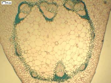
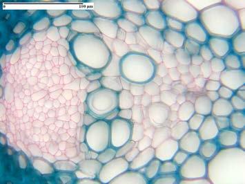
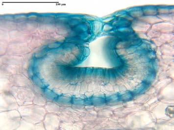
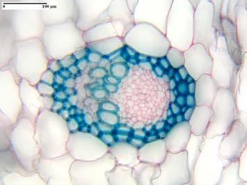
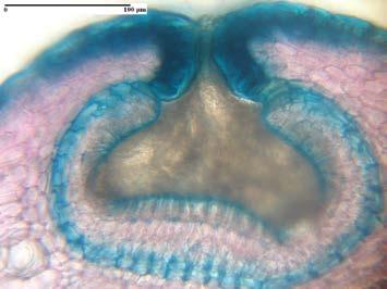
2 Materials and methods The material under study, coming from the collection of the Alexandru Borza Botanical Gardens of Cluj-Napoca, belongs to four taxa: N. x coccinea Mast, N. distillatoria L., N. maxima Reinw. ex Nees and N. northiana Hook f. The material subjected to analysis (the modified leaves of the plants) has been fixed and preserved in 70% ethylic alcohol. The sections (from the assimilatory part, tendril and pitcher) were cut with a microtome, then coloured with iodine green and alaun-carmine, mounted in gel and analyzed on a Novex (Holland) light microscope. The light micrographs were performed by means of Novex (Holland) microscope, using a Canon A95 camera. Results and discussions As already mentioned, the leaf of Nepenthes consists of three parts: a basal, assimilatory one, a tendril and a pitcher which represents the trap of the plant. In front side view, the upper epidermis of the assimilatory part appears as formed of polygonal cells (Fig. 1); here and there, a few hydathodes are present. The lower epidermis consists of small cells, bearing weakly waved walls (Fig. 2). Here and there, stomata of the anomocytic type and hydathodes are present. A hydathode bears a short pedicel formed of a few cells and a stellate part, formed of 4-10 cells. Another author [4] suggests that the hydathodes do not only secrete water, but even absorb it from time to time. In cross section, the upper epidermis evidences small cells covered by a thick cuticle. Just beneath the epidermis, a few isodiametric-celled layers are present, forming an acviferous tissue (Fig. 3); some authors [5] call it an acviferous hypodermis. Then, a 2-3 layered palisade tissue, with short cells, in which chloroplasts can be observed, is present. The lacunary tissue is multi-layered, with small aeriferous spaces between the component cells. A lot of isolated mechanical cells (idioblasts) with spiral thickenings can be observed in the mesophyll; these were evidenced by other authors [5], too. The lower epidermis consists of small, isodiametric cells, covered by a cuticle thinner than the one covering the upper epidermis. Numerous stomata are present, as well as numerous calcium oxalate crystals in the mesophyll. The midvein is very prominent at the lower side of the assimilatory part (Fig. 4). A large number of vascular bundles is present (8 big bundles, one of its being situated in the centre or 6 bundles and a central one at N. northiana); most of them are implanted in a thick sclerenchyma ring, formed of sclerenchymatic fibres with thickened and lignified walls (or unlignified at N. coccinea). The vascular bundles have different orientation in the sclerenchymatic ring. A vascular bundle (Fig. 5) consists of a phloem (sieved tubes and companion cells) and a xylem (xylem vessels separated by celulosic parenchyma). Sometimes, the sclerenchyma sheath bears very small vascular bundles, consisting of a few phloem elements or of phloem and 1-2 xylem vessels. In the fundamental, external parenchyma, idioblasts and small, isolated vascular bundles are present, often consisting of a few phloem elements, surrounded by a thin sclerenchymatic sheath. 6
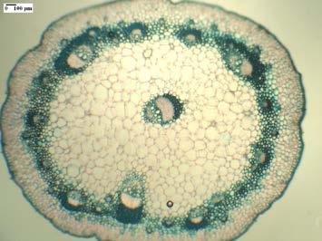
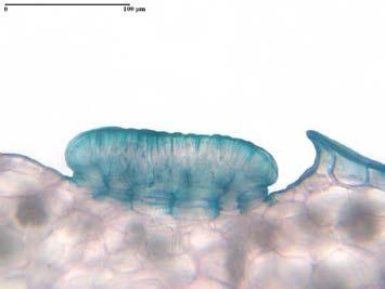
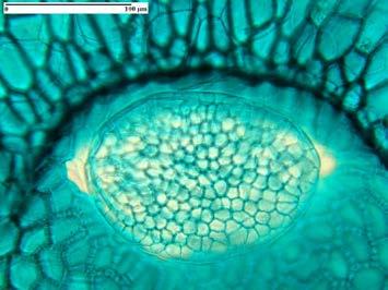
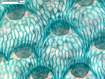
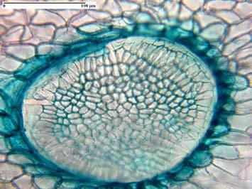
3 The tendril In cross section, the tendril shows a circular shape, with 7-8 ribs at N. maxima (Fig. 6). Small cells, covered by a thick cuticle, form the epidermis. Here an there, hydathodes, short, sometimes branched tector hairs and stomata prominig above the epidermis are present. The cortical parenchyma is formed of 5-6 layers of large cells. Most of the vascular bundles are implanted in a strong sclerenchyma ring. There is a high variability regarding the number of bundles and on their position (N. coccinea and N. maxima present a lot of vascular bundles of different size in the sclerenchyma ring and a central one in the fundamental parenchyma; N. distillatoria presents 2 big vascular bundles in the center of the parenchyma and a smaller one, close to them, while N. northiana shows the largest number of vascular bundles, implanted in the sclerenchyma ring, and also two smaller ones in the fundamental parenchyma, but close to the sclerenchyma. The tendril has an homogenous parenchyma, formed of big, turgescent cells and a few idioblasts. Near the pitcher, the cross section of the tendril is quite circular. The vascular bundles form 2-3 rings (the internal bundles are bigger than the external ones, in the fundamental parenchyma). A sclerenchymatic sheath surrounds each vascular bundle. Numerous calcium oxalates are present in the fundamental parenchyma. At the inferior level of the pitcher, a typical limb structure is present. In front side view, the internal epidermis presents polygonal elongated cells, with thick walls. Here and there, a lot of multicellular digestive glands are present (Fig. 7); a small epidermal prolongation can be observed near each gland, yet without touching it. The external epidermis (Fig. 8) consists of small polygonal cells with thin walls, anomocytic stomata, hydathodes and nectariferous glands, which appear like multicellular, massive structures, communicating with the exterior through a short channel. In cross section, the wall of the pitcher is quite thick. The internal epidermis shows elongated cells, covered by a thick cuticle. Numerous big digestive glands are present in small epidermal cavities (Fig. 9); the epidermal cells form a small fold, without touching the gland. A digestive gland shows 2-3 layers of oblate cells, 1-2 layers of isodiametrical cells and an external layer of columnar-shaped cells. Each digestive gland is associated with small vascular bundles (tracheids with ringed and spiral thickenings). In longitudinal section, the small fold can be better observed (Fig. 10). The external epidermis consists of small cells, covered by a thin cuticle. The nectariferous glands are formed of three layers of cells delimiting a cavity which opens towards the exterior through a short channel (Fig. 11). The nectariferous glands attract the insects (the prey) to the trap and make them climb the wall of the pitcher to reach the slippery peristome. The assimilatory parenchyma is thick, homogenous, formed of small cells outside and bigger inside. A lot of calcium oxalates are present all over the parenchyma. The vascular bundles have different sizes, the biggest ones occupying the external part of the parenchyma, while the smallest ones occupy the centre of it. Each vascular bundle consists in phloem facing the exterior part of the pitcher and a xylem facing the internal one, so that the internal epidermis represents the old upper epidermis of an archaic leaf and the external epidermis represents the old lower epidermis. All the studied species show mechanical sheaths surrounding the vascular bundles, consisting of fibres with 7
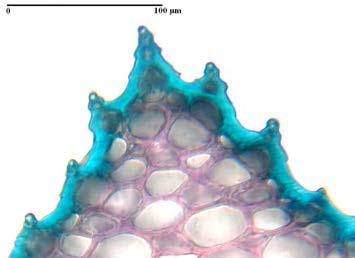
4 moderately thickened and lignified walls. The assimilatory parenchyma also presents a few idioblasts. The middle level of the pitcher presents a quite similar structure to that of the anterior level. In front side view, the internal epidermis consists of polygonal cells with thick walls and secretory glands, smaller than those occurring at the inferior level; the integumentary fold do not touch the gland (Fig. 12). The external epidermis is formed of polygonal cells, anomocytic stomata, multicellular tector hairs, nectariferous glands and hydathodes (Fig. 13). In cross section, the pitcher shows a thinner wall. The internal epidermis presents digestive glands which communicate with the tracheids (Fig. 14). In longitudinal section, the fold can be better observed (Fig. 15). The external epidermis consists of small cells covered by a thin cuticle. Anomocytic stomata, hydathodes, nectariferous glands (Fig. 16) and multicellular tector hairs, often branched, are present. In the assimilatory parenchyma, numerous calcium oxalates can be observed. Each vascular is bounded by a sclerenchymatic sheath (Fig. 17). There are vascular bundles consisting only of a few phloem elements. The assimilatory parenchyma presents idioblasts, too. The superior level of the pitcher shows the same structure as the other levels. In front side view, the internal epidermis presents large polygonal cells and digestive glands (Fig. 18) smaller than those occurring at the other levels. The epidermal fold covers more than half of the digestive glands. The external epidermis (Fig. 19) consists of small polygonal cells, with waved lateral walls, tector hairs, hydathodes, nectariferous glands and anomocytic stomata. Cross section through the superior level of the pitcher shows the digestive glands in their incipient stage of development (Fig. 20), consisting of a small number of cells. Almost half of the gland is covered by the integumentary fold (Fig. 21); the developing stages of the glands during ontogenesis were previously presented [9]. The external epidermis shows similar structures to those of the anterior levels (Figs. 22 and 23). The assimilatory parenchyma is thinner; the vascular bundles are bounded by a very thin mechanical sheath, consisting of fibres with thickened walls, but weakly lignified; some of the bundles present only a few phloem elements; calcium oxalates are not present. The lid In front side view, both the upper and the lower epidermis present polygonal cells, with waved lateral walls, anomocytic stomata, secretory glands surrounded by an integumentary fold (Fig. 24) and hydathodes (Fig. 25); tector hairs, sometimes branched, are present only in the lower epidermis. The cross section of the lid is similar to that occurring in the pitcher s wall. Numerous digestive glands (Fig. 26) are present in both epidermis. The fundamental parenchyma presents vascular bundles of different sizes (Fig. 27), the largest occupying the centre of the parenchyma. All vascular bundles are surrounded by a thin sclerenchyma sheath formed of thin-walled and weakly lignified fibres. The peristome (Fig. 28) is a common characteristic of the pitcher plants. All four investigated Nepenthes species have a ridged peristome. The epidermal cells are small, covered by a very thick cuticle. Stomata and hydathodes are present in the lower epidermis. 8
5 The peristome has an homogenous parenchyma, with large cells, a few idioblasts and small vascular bundles, most of them consisting of phloem elements surrounded by sclerenchyma fibres, with cellulosed unthickened walls. Conclusions In spite of the high variability of the pitcher size recorded from one species to another, their histo-anatomy is quite similar. There have been observed mostly quantitative differences (number of the vascular bundles, size of the digestive glands at different levels, thickness of the sclerenchymatic sheath, length of the integumentary fold which covers each digestive gland) and not qualitative differences. REFERENCES 1. ANDERSON A. N., Secretion and absorbtion in glands of the carnivorous plant Nepenthes alata. B. A. honors thesis, Connecticut College, New London, C. T. 2. DALTON M. JOS., Note sur l origin et le développement des urnes dans les plantes du genre Nepenthes. Ann. des Sci. Nat.; sér.bot., 12: LLOYD F. E., The Carnivorous Plants. Chronica Botanica, 9. Ronald Press, New York 4. MACFARLANE J. M., Observations on pitchered insectivorous plants. Part I. Ann. of Bot., 3: METCALFE C. R., CHALK L., Nepenthaceae in Anatomy of the Dicotyledons. 1: , Clarendon Press, Oxford 6. STERN K., Contribution to the knowledge of Nepenthes. Flora, 109: PARKES D. M., Adaptive mechanisms of surfaces and glands in some carnivorous plants. MSc Thesis, Monash University, Clayton, Victoria, Australia 8. STAROSTA P., LABAT J.-J., L univers des plantes carnivores. Éd. Du May, Paris 9. TOMA I., TOMA C., STĂNESCU I., Histo-anatomical aspects of the Nepenthes maxima Reinw. ex Nees metamorphosed leaf. Rev. Roum. Biol., sér. Biol. végét., 47, 1-2: 3-7 Acknowledgments The authors gratefully thank to Felician Micle PhD, Ex-Director of the Botanical Garden of Cluj-Napoca and to Elena Rânba for supplying the material for the present investigation. 9
6 Explanation of plates: Plate I 1. The upper epidermis of the assimilatory part of N. maxima, in front side view 2. The lower epidermis of the assimilatory part of N. distillatoria, in front side view 3. The mesophyll of the assimilatory part of N. distillatoria 4. Cross section through the midvein of the assimilatory part of N. maxima 5. The biggest vascular bundle of the midvein belonging to the assimilatory part of N. maxima 6. Cross section through the tendril of N. maxima Plate II 7. The internal epidermis of the inferior level of N. distillatoria pitcher, in front side view 8. The external epidermis of the inferior level of N. distillatoria pitcher, in front side view 9. Cross section through the inferior level of N. distillatoria pitcher 10. Longitudinal section of the inferior level of N. maxima pitcher 11. Cross section through the inferior level of N. distillatoria pitcher 12. The internal epidermis of the middle level of N. northiana pitcher, in front side view Plate III 13. The internal epidermis of the middle level of N. maxima pitcher, in front side view 14. Cross section through the middle level of N. northiana pitcher 15. Longitudinal section through the middle level of N. maxima pitcher 16. Cross section through the middle level of N. distillatoria pitcher 17. Cross section through the middle level of N. maxima pitcher 18. The internal epidermis of the superior level of N. coccinea pitcher, in front side view Plate IV 19. The internal epidermis of the superior level of N. distillatoria pitcher, in front side view 20. Cross section through the superior level of N. distillatoria pitcher 21. Longitudinal section through the superior level of N. maxima pitcher 22. Cross section through the superior level of N. distillatoria pitcher 23. Cross section through the superior level of N. northiana pitcher 24. The upper epidermis of the lid belonging to N. northiana pitcher, in front side view Plate V 25. The lower epidermis of the lid belonging to N. northiana pitcher, in front side view 26. Cross section through the lid of N. coccinea pitcher 27. Cross section through the lid of N. northiana pitcher 28. Cross section through the peristome of N. northiana pitcher 10


7 IRINA STĂNESCU, C.TOMA PLATE I


8 IRINA STĂNESCU, C.TOMA PLATE II


9 IRINA STĂNESCU, C.TOMA PLATE III


10 IRINA STĂNESCU, C.TOMA PLATE IV


11 IRINA STĂNESCU, C.TOMA PLATE V
Irina Berciu *, Constantin Toma Department of Biology, Al. I. Cuza University, Iasi
 HISTO-ANATOMICAL ASPECTS OF VEGETATIVE ORGANS OF THYMUS DACICUS BORB. AND THYMUS GLABBRESCENS WILLD. Irina Berciu *, Constantin Toma Department of Biology, Al. I. Cuza University, Iasi * Correspondence:
CONTRIBUTIONS REGARDING THE LEAF HISTO-ANATOMY OF SOME PELARGONIUM SPECIES
 Rev. Med. Chir. Soc. Med. Nat., Iaşi 2013 vol. 117, no. 3 PHARMACY ORIGINAL PAPERS CONTRIBUTIONS REGARDING THE LEAF HISTO-ANATOMY OF SOME PELARGONIUM SPECIES Cristina Elena Iancu, Oana Cioanca, Cornelia
Anatomy of Flowering Plants. K C Meena PGT Biology
 Anatomy of Flowering Plants K C Meena PGT Biology Tissues A group of similar cells performing same function. Types of plant tissues - Meristematic tissues and permanent tissues. Meristematic tissues Have
Visit For All NCERT solutions, CBSE sample papers, Question papers, Notes for Class 6 to 12. Chapter-6 ANATOMY OF FLOWERING PLANTS
 Chapter-6 ANATOMY OF FLOWERING PLANTS POINTS TO REMEMBER Anatomy : Anatomy is the study of internal structure of organisms. Plant anatomy includes organisation and structure of tissues. Tissue : A group
Downloaded from
 POINTS TO REMEMBER : 6. Anatomy of Flowering Plants Study of internal structure of plant is called anatomy. In plants cells are the basic unit. Cells organized into tissues and tissues organized into organs.
IRINA STANESCU', CONSTANTIN TOMA, IRINA GOSTIN
 CYTO-HISTOLOGICAL ASPECTS IN THE MODIFIED LEAF OF DIONAEA MUSCIPULA ELLIS IRINA STANESCU', CONSTANTIN TOMA, IRINA GOSTIN In the present work, the authors evidence some of the particularities observed in
COMPARATIVE HISTO-ANATOMICAL ANALYSIS OF THE VEGETATIVE ORGANS OF SEDUM TELEPHIUM L. SSP. MAXIMUM (L.) KROCK. IN VITRO AND FROM NATURE
 KROCK. IN VITRO AND FROM NATURE") ARDELEAN MIRELA, STĂNESCU IRINA, CACHIŢĂ-COSMA DORINA J. Plant Develop. 16 (2009): 3 8 COMPARATIVE HISTO-ANATOMICAL ANALYSIS OF THE VEGETATIVE ORGANS OF SEDUM TELEPHIUM L. SSP. MAXIMUM (L.) KROCK. IN VITRO
ARDELEAN MIRELA, STĂNESCU IRINA, CACHIŢĂ-COSMA DORINA J. Plant Develop. 16 (2009): 3 8 COMPARATIVE HISTO-ANATOMICAL ANALYSIS OF THE VEGETATIVE ORGANS OF SEDUM TELEPHIUM L. SSP. MAXIMUM (L.) KROCK. IN VITRO
Plant Anatomy: roots, stems and leaves
 Plant Anatomy: roots, stems and leaves The plant body has a hierarchy of organs, tissues and cells Plants, like animals, have organs composed of different tissues, which are composed of cells. Tissue is
Non Permanent Tissues - Meristematic Tissue
 PLANT TISSUES Non Permanent Tissues - Meristematic Tissue Undifferentiated plant cells that are continually dividing by mitosis Large thin walled cells No vacuole Dense cytoplasm Large nucleus Found at
Plant Anatomy: roots, stems and leaves
 Plant Anatomy: roots, stems and leaves The plant body has a hierarchy of organs, tissues and cells Plants, like animals, have organs composed of different tissues, which are composed of cells. Tissue is
Class XI Chapter 6 Anatomy of Flowering Plants Biology
 Class XI Chapter 6 Anatomy of Flowering Plants Biology Question 1: State the location and function of different types of meristem. Meristems are specialised regions of plant growth. The meristems mark
UNIT 6 - STRUCTURES OF FLOWERING PLANTS & THEIR FUNCTIONS
 6.1 Plant Tissues A tissue is a group of cells with common function, structures or both. In plants we can find 2 types of tissues: Meristem Permanent tissues Meristem is found in regions with continuous
PLANT TISSUES 12 MARCH 2014
 PLANT TISSUES 12 MARCH 2014 Lesson Description In this lesson we: Identify the different types of plant tissue Be able to relate the different structures with the different functions Plant Tissue Summary
 Question 1: State the location and function of different types of meristem. Meristems are specialised regions of plant growth. The meristems mark the regions where active cell division and rapid division
Plant Structure. Objectives At the end of this sub section students should be able to:
 Name: 3.2 Organisation and the Vascular Structures 3.2.1 Flowering plant structure and root structure Objectives At the end of this sub section students should be able to: 1. Label a diagram of the external
ANATOMY OF PLANTS Introduction: The study of gross internal structure of plant organs by the technique of section cutting is called plant anatomy.
 ANATOMY OF PLANTS Introduction: The study of gross internal structure of plant organs by the technique of section cutting is called plant anatomy. (Pandey, 2002). Various plant organ viz. root, stem, leaves,
CONTRIBUTIONS TO THE HISTO-ANATOMICAL STUDY OF THE CALENDULA OFFICINALIS L. LEAVES TREATED WITH THIOPHANATE METHYL (TOPSIN M) Introduction
 Introduction") Analele ştiinţifice ale Universităţii Al. I. Cuza Iaşi Tomul LIV, fasc. 1, s. II a. Biologie vegetală, 2008 CONTRIBUTIONS TO THE HISTO-ANATOMICAL STUDY OF THE CALENDULA OFFICINALIS L. LEAVES TREATED WITH
Analele ştiinţifice ale Universităţii Al. I. Cuza Iaşi Tomul LIV, fasc. 1, s. II a. Biologie vegetală, 2008 CONTRIBUTIONS TO THE HISTO-ANATOMICAL STUDY OF THE CALENDULA OFFICINALIS L. LEAVES TREATED WITH
Plants. Tissues, Organs, and Systems
 Plants Tissues, Organs, and Systems Meristematic cells Specialized cells that are responsible for producing specialized cells, they produce three types of tissue in the body of a plant. Meristematic Cells
Plant Anatomy and Tissue Structures
 Plant Anatomy and Tissue Structures The Two Major Plant Systems Reproductive shoot (flower) Terminal bud Node Internode Angiosperm plants have threse major organs: Roots Stems Leaves & Flowers Terminal
HISTO-ANATOMICAL OBSERVATIONS REFERRING TO SOME MELAMPYRUM SPECIES
 Analele ştiinţifice ale Universităţii Al. I. Cuza Iaşi Tomul LV, fasc. 2, s.ii a. Biologie vegetală, 2009 HISTO-ANATOMICAL OBSERVATIONS REFERRING TO SOME MELAMPYRUM SPECIES ASPAZIA BĂEŞU *, C. TOMA **,
RODICA RUGINĂ, C. TOMA
 Analele ştiinţifice ale Universităţii Al. I. Cuza Iaşi Tomul LIII, s. II a. Biologie vegetală, 2007 HISTO-ANATOMICAL ASPECTS OF SOME LONICERA L. SPECIES RODICA RUGINĂ, C. TOMA Abstract. The authors investigate
Overview of Plant Tissues
 Plant Tissue Growth Key Concepts Overview of Plant Tissues Seed-bearing vascular plants have a shoot system with stems, leaves, and reproductive parts Most also have a root system These systems consist
Plant Structure. Lab Exercise 24. Objectives. Introduction
 Lab Exercise Plant Structure Objectives - Be able to identify plant organs and give their functions. - Learn distinguishing characteristics between monocot and dicot plants. - Understand the anatomy of
Plant Tissues and Organs. Topic 13 Plant Science Subtopics , ,
 Plant Tissues and Organs Topic 13 Plant Science Subtopics 13.1.2, 13.1.3, 13.1.4 Objectives: List and describe the major plant organs their structure and function List and describe the major types of plant
II. SIMPLE TISSUES Bot 404--Fall A. Introduction to Tissues (DIAGRAM allow a full page)
") II. SIMPLE TISSUES Bot 404--Fall 2004 A. Introduction to Tissues (DIAGRAM allow a full page) B. Definitions Adaxial = facing the axil; upper surface of leaf Abaxial = facing away from the axil; lower surface
II. SIMPLE TISSUES Bot 404--Fall 2004 A. Introduction to Tissues (DIAGRAM allow a full page) B. Definitions Adaxial = facing the axil; upper surface of leaf Abaxial = facing away from the axil; lower surface
Plant Structure and Growth
 Plant Structure and Growth A. Flowering Plant Parts: The flowering plants or are the most diverse group of plants. They are divided into 2 classes and. Examples of monocots: Examples of dicots: The morphology
A COMPARATIVE STUDY REGARDING THE MORPHOLOGY AND ANATOMY OF THE VEGETATIVE APPARATUS IN TWO OCIMUM BASILICUM L. BREEDS.
 Analele ştiinţifice ale Universităţii Al. I. Cuza Iaşi Tomul LIV, fasc. 2, s.ii a. Biologie vegetală, 2008 A COMPARATIVE STUDY REGARDING THE MORPHOLOGY AND ANATOMY OF THE VEGETATIVE APPARATUS IN TWO OCIMUM
The Shoot System: Primary Stem Structure - 1
 The Shoot System: Primary Stem Structure - 1 Shoot System The shoot system comprises the leaves and stems of plants. Leaves are located at nodes on the stem; the distance along the stem between nodes is
Plant Structure and Function (Ch. 23)
") Plant Structure and Function (Ch. 23) Basic plant anatomy 1 root root tip root hairs Roots Roots anchor plant in soil, absorb minerals & water, & store food fibrous roots (1) mat of thin roots that spread
Plant Structure and Function (Ch. 23) Basic plant anatomy 1 root root tip root hairs Roots Roots anchor plant in soil, absorb minerals & water, & store food fibrous roots (1) mat of thin roots that spread
-Each asexual organs. -Anchors the plant -Absorbs water and minerals -Stores sugars and starches
 Plants are made up of: -organs, tissues, and cells The three major plant organs are: -Roots, stems, and leaves -Each asexual organs Plants have a Root System beneath the ground that us a multicellular
NOTES ON THE MORPHO-ANATOMY OF ACONITUM DEGENII GAYER. Introduction
 Analele ştiinţifice ale Universităţii Al. I. Cuza Iaşi Tomul LV, fasc. 2, s.ii a. Biologie vegetală, 2009 NOTES ON THE MORPHO-ANATOMY OF ACONITUM DEGENII GAYER IRINA STĂNESCU *, C. MARDARI *, C. TĂNASE
Chapter 35~ Plant Structure and Growth
 Chapter 35~ Plant Structure and Growth Plant Organization Plant morphology is based on plant s evolutionary history Need to draw in nutrients from the ground and the air Plant Organs Root system = roots
THE SPECIE PARIETARIA LUSITANICA L., CAULINE AND FOLIAR HISTOANATOMICAL STUDY Mariana Arcuş, Gabriela Lilios, Emanuela Gheorma, Corina Moromete
 THE SPECIE PARIETARIA LUSITANICA L., CAULINE AND FOLIAR HISTOANATOMICAL STUDY Mariana Arcuş, Gabriela Lilios, Emanuela Gheorma, Corina Moromete OVIDIUS CONSTANŢA UNIVERSITY, FACULTY OF PHARMACY Summary
A group of cells with common origin is called a tissue. The cells of a tissue usually perform a common function.
 Anatomy of Flowering Plants Tissues A group of cells with common origin is called a tissue. The cells of a tissue usually perform a common function. Types of Tissue: There are two main types of plant tissues,
Forms strands that conduct water, minerals, and organic compounds. Much of the inside of nonwoody parts of plants. Includes roots, stems, and leaves
 Biology II Vascular plants have 3 tissue systems: Dermal Protective outer layer of plant Vascular Forms strands that conduct water, minerals, and organic compounds Ground Much of the inside of nonwoody
23 4 Leaves Slide 1 of 32
 23 4 Leaves 1 of 32 Leaf Structure The structure of a leaf is optimized for absorbing light and carrying out photosynthesis. 2 of 32 Leaf Structure To collect sunlight, most leaves have thin, flattened
! Xylem - Chief conducting tissue for water and minerals absorbed by the roots.
 + Complex Tissues! Complex tissues are made up of two or more cell types.! Xylem - Chief conducting tissue for water and minerals absorbed by the roots.! Vessels - Made of vessel elements.! Long tubes
HISTO-ANATOMICAL LESS KNOW ASPECTS UPON SOME LAMIACEAE TAXA CAMELIA IFRIM *, IRINA TOMA ** Introduction. Material and method
 Analele ştiinţifice ale Universităţii Al. I. Cuza Iaşi Tomul L, s. II a. Biologie vegetală, 2004 HISTO-ANATOMICAL LESS KNOW ASPECTS UPON SOME LAMIACEAE TAXA CAMELIA IFRIM *, IRINA TOMA ** Abstract: The
Simple Leaf Compound Leaf
 Leaves Outline Overview Leaf Arrangements and Types Internal Structures of Leaves Stomata Mesophyll and Veins Specialized Leaves Autumnal Changes in Color Abscission Relevance of Leaves Overview Some of
2.1 PLANT TISSUE HALIMAHTUN SAEDIAH BT ABU BAKAR KOLEJ TEKNOLOGI TIMUR
 2.1 PLANT TISSUE HALIMAHTUN SAEDIAH BT ABU BAKAR KOLEJ TEKNOLOGI TIMUR GENERAL Plant cell are differentiated possessing structural adaptations that make specific functions possible. Modifications of cell
Chapter 29: Plant Tissues
 Chapter 29: Plant Tissues Shoots and Roots Shoots (Leaves and Stem) Produce food by photosynthesis Carry out reproductive functions Roots Anchor the plant Penetrate the soil and absorb water and dissolved
Chapter #35~ Plant Structure and Growth
 Chapter #35~ Plant Structure and Growth What part of a plant is represented by each of these: Carrot Celery Red Pepper Tomato Lettuce Garbanzo Bean Angiosperm structure Three basic organs: Roots (root
Organization of Plant Tissue. Wednesday, March 2, 16
 Organization of Plant Tissue Plant Systems Shoot System The Leaf The Stem The Flower Root System The Shoot System Has two main functions: to conduct photosynthesis and to produce flowers for sexual reproduction
Today: Plant Structure Exam II is on F March 31
 Next few lectures are on plant form and function Today: Plant Structure Exam II is on F March 31 Outline Plant structure I. Plant Cells structure & different types II. Types of meristems Apical meristems:
The three principal organs of seed plants are roots, stems, and leaves.
 23 1 Specialized Tissues in Plants Seed Plant Structure The three principal organs of seed plants are roots, stems, and leaves. 1 of 34 23 1 Specialized Tissues in Plants Seed Plant Structure Roots: absorb
BI 103: Leaves. Learning Objectives
 BI 103: Leaves An examination of leaves Chapter 43 cont. Learning Objectives What is the function of the plant leaf? How are specific cells and tissues adapted in the leaf in order to help it function?
Exercise 12. Procedure. Aim: To study anatomy of stem and root of monocots and dicots.
 Aim: To study anatomy of stem and root of monocots and dicots. Principle: The study of internal morphology, i.e., cells of various tissues in an organ of a living body is called Anatomy. Tissue, which
Biology. Slide 1 of 32. End Show. Copyright Pearson Prentice Hall
 Biology 1 of 32 23 4 Leaves 2 of 32 Leaf Structure Leaf Structure How does the structure of a leaf enable it to carry out photosynthesis? 3 of 32 Leaf Structure The structure of a leaf is optimized for
Lecture 4 Root Put line under your answer! There is only one correct answer in the multiple choice questions
 Lecture 4 Root Put line under your answer! There is only one correct answer in the multiple choice questions 1. The perception of gravity by a root is thought to take place in a) root hairs b) the region
Biology 2 Chapter 21 Review
 Biology 2 Chapter 21 Review Multiple Choice Identify the choice that best completes the statement or answers the question. 1. Which of the following is not a tissue system of vascular plants? a. vascular
Leaf. It is composed of:
 LEAF It is composed of: Leaf a leaf stalk called petiole; if it lacks leaf is sessile; the expanded part called lamina or blade; a strand of vascular tissue (veins) in the blade; a pair of leafy outgrowth
The plant body has a hierarchy of organs, tissues, and cells. Plants, like multicellular animals:
 Chapter 28 The plant body has a hierarchy of organs, tissues, and cells Plants, like multicellular animals: o Have organs composed of different tissues, which are in turn composed of cells 3 basic organs:
Plant Structure And Growth
 Plant Structure And Growth The Plant Body is Composed of Cells and Tissues Tissue systems (Like Organs) made up of tissues Made up of cells Plant Tissue Systems Ground Tissue System Ø photosynthesis Ø
Chapter 23 Notes Roots Stems Leaves
 Chapter 23 Notes Roots Stems Leaves I. Specialized tissue in plants - effective way to ensure the plant s survival A. Seed plant structure 1. Roots - a. Absorbs water and dissolves nutrients b. anchors
Recap. Waxy layer which protects the plant & conserves water. Contains chloroplasts: Specialized for light absorption.
 Recap Contains chloroplasts: Specialized for light absorption Waxy layer which protects the plant & conserves water mesophyll Layer contains air spaces: Specialized for gas exchange Vascular Tissue Exchange
Plant Anatomy AP Biology
 Plant Anatomy 2006-2007 Basic plant anatomy 1 root root tip root hairs Roots 1 Roots anchor plant in soil, absorb minerals & water, & store food fibrous roots (1) mat of thin roots that spread out monocots
Honors Biology I Ch 29 Plant Structure & Function
 3 Basic types of plant cells Honors Biology I Ch 29 Plant Structure & Function 1) Parenchyma cells- loosely packed or cells with a and thin, Involved in metabolic functions 2) Collenchyma cells- thicker
ANATOMY OF FLOWERING PLANTS
 ANATOMY OF FLOWERING PLANTS Finish Line & Beyond The Tissues The Tissue System Anatomy of Dicotyledonous and Monocotyledonous Plants Secondary Growth THE TISSUES A tissue is a group of cells having a common
Chapter 8: Plant Organs: Leaves
 Leaf Form & Function Chapter 8: Plant Organs: Leaves Leaves are the most variable Composed of a and a May have (pair of leaf like outgrowths at petiole) : having a single blade : having a blade divided
(A) Buds (B) Lateral meristem (C) Apical meristem (D) Stem (E) Trichomes
 Buds (B) Lateral meristem (C) Apical meristem (D) Stem (E) Trichomes") AP Biology - Problem Drill 17: Plant Structure Question No. 1 of 10 1. What are hair-like outgrowths that protect and absorb nutrients? Question #01 (A) Buds (B) Lateral meristem (C) Apical meristem (D)
AP Biology - Problem Drill 17: Plant Structure Question No. 1 of 10 1. What are hair-like outgrowths that protect and absorb nutrients? Question #01 (A) Buds (B) Lateral meristem (C) Apical meristem (D)
Bio Factsheet. Transport in Plants. Number 342
 Number 342 Transport in Plants This Factsheet: Explains why plants need a transport system Describes what plants transport Describes the tissues which carry out transport Outlines the position of the xylem
THE TISSUES A tissue is a group of cells having a common origin and usually performing a common function. Tissues. Parenchyma
 1 CHAPTER 6 ANATOMY OF FLOWERING PLANTS Study of internal structure of plants is called anatomy. Plants have cells as the basic unit, cells are organised into tissues and in turn the tissues are organised
TARGET STUDY MATERIAL
 TARGET STUDY MATERIAL Plus-1 Botany VOL I TARGET EDUCATIONAL INSTITUTION Target Educational institution is the one and only Entrance coaching and CBSE 10 th coaching centre at Mukkam with advanced technologies
Introduction. Most land animals, including humans, depend on plants directly or indirectly for sustenance.
 Introduction With about 250,000 known species, the angiosperms are by far the most diverse and widespread group of land plants. As primary producers, flowering plants are at the base of the food web of
CHAPTER 6 ANATOMY OF FLOWERING PLANTS
 84 BIOLOGY CHAPTER 6 ANATOMY OF FLOWERING PLANTS 6.1 The Tissues 6.2 The Tissue System 6.3 Anatomy of Dicotyledonous and Monocotyledonous Plants 6.4 Secondary Growth You can very easily see the structural
CHAPTER 6 ANATOMY OF FLOWERING PLANTS
 84 BIOLOGY CHAPTER 6 ANATOMY OF FLOWERING PLANTS 6.1 The Tissues 6.2 The Tissue System 6.3 Anatomy of Dicotyledonous and Monocotyledonous Plants 6.4 Secondary Growth You can very easily see the structural
2/25/2013. o Plants take up water and minerals from below ground o Plants take up CO2 and light from above ground THREE BASIC PLANT ORGANS ROOTS
 o Plants take up water and minerals from below ground o Plants take up CO2 and light from above ground THREE BASIC PLANT ORGANS o Roots o Stems o Leaves ROOTS o Anchor plant o Absorb water and minerals
OCR (A) Biology A-level
 Biology A-level") OCR (A) Biology A-level Topic 3.3: Transport in plants Notes Plants require a transport system to ensure that all the cells of a plant receive a sufficient amount of nutrients. This is achieved through
OCR (A) Biology A-level Topic 3.3: Transport in plants Notes Plants require a transport system to ensure that all the cells of a plant receive a sufficient amount of nutrients. This is achieved through
CONSIDERATIONS UPON THE ANATOMICAL FEATURES OF SOME TAXA OF TRADESCANTIA GENERA
 Buletinul Grădinii Botanice Iaşi Tomul 14, 2007 CONSIDERATIONS UPON THE ANATOMICAL FEATURES OF SOME TAXA OF TRADESCANTIA GENERA IFRIM CAMELIA Abstract: The structure of two taxa of the Tradescantia genre
Sept 26 - Lecture notes. Plant 1 tissues I: overview and leaves
 Plant 1 tissues I: overview and leaves The structure of a typical plant (we ll address this generalization later ) is organized at three levels: Organs: Over the next three lectures, and two labs, we consider
MORPHO-ANATOMICAL CONSIDERATIONS UPON THE SHOOT OF SOME ROSA L. CULTIVARS FROM THE BOTANIC GARDEN OF IASI (1 ST NOTE)
") DELINSCHI VIOLETA, STĂNESCU IRINA, MIHALACHE MIHAELA, ADUMITRESEI LIDIA J. Plant Develop. 16 (2009):9 16 MORPHO-ANATOMICAL CONSIDERATIONS UPON THE SHOOT OF SOME ROSA L. CULTIVARS FROM THE BOTANIC GARDEN
DELINSCHI VIOLETA, STĂNESCU IRINA, MIHALACHE MIHAELA, ADUMITRESEI LIDIA J. Plant Develop. 16 (2009):9 16 MORPHO-ANATOMICAL CONSIDERATIONS UPON THE SHOOT OF SOME ROSA L. CULTIVARS FROM THE BOTANIC GARDEN
Division Ave. High School AP Biology
 Monocots & dicots Angiosperm are divide into 2 classes dicots (eudicot) 2 cotyledons (seed leaves) leaves with network of veins woody plants, trees, shrubs, beans monocots 1 cotyledon leaves with parallel
Plants. Plant Form and Function. Tissue Systems 6/4/2012. Chapter 17. Herbaceous (nonwoody) Woody. Flowering plants can be divided into two groups:
 Woody. Flowering plants can be divided into two groups:") Monocots Dicots 6/4/2012 Plants Plant Form and Function Chapter 17 Herbaceous (nonwoody) In temperate climates, aerial parts die back Woody In temperate climates, aerial parts persist The Plant Body Functions
Monocots Dicots 6/4/2012 Plants Plant Form and Function Chapter 17 Herbaceous (nonwoody) In temperate climates, aerial parts die back Woody In temperate climates, aerial parts persist The Plant Body Functions
Organs and leaf structure
 Organs and leaf structure Different types of tissues are arranged together to form organs. Structure: 2 parts (Petiole and Leaf Blade) Thin flat blade, large surface area Leaves contain all 3 types of
Chapter 6. Biology of Flowering Plants. Anatomy Seedlings, Meristems, Stems, and Roots
 BOT 3015L (Outlaw/Sherdan/Aghoram); Page 1 of 6 Chapter 6 Biology of Flowering Plants Anatomy Seedlings, Meristems, Stems, and Roots Objectives Seedling germination and anatomy. Understand meristem structure
Chapter 29. Table of Contents. Section 1 Plant Cells and Tissues. Section 2 Roots. Section 3 Stems. Section 4 Leaves. Plant Structure and Function
 Plant Structure and Function Table of Contents Section 1 Plant Cells and Tissues Section 2 Roots Section 3 Stems Section 4 Leaves Section 1 Plant Cells and Tissues Objectives Describe the three basic types
Roots anchor plants and absorb water and minerals in solution. A germinating seed radicle becomes the first root. Four zones, or regions, of young
 Roots anchor plants and absorb water and minerals in solution. A germinating seed radicle becomes the first root. Four zones, or regions, of young roots are recognized: (1) A protective root cap that also
Tissues and organs PART 2
 Tissues and organs PART 2 The structure and function of the mesophytic leaf (a plant organ) The mesopyhtic leaf (lives in a moderately moist environment) contains 7 layers of tissue: 1. Upper epidermis
MORPHOLOGICAL AND HISTO-ANATOMICAL ASPECTS AT SOME DICOTILEDONATE SEEDLINGS RELATED TO THE VASCULAR TRANSITION
 Analele ştiinţifice ale Universităţii Al. I. Cuza Iaşi Tomul LII, s. II a. Biologie vegetală, 2006 MORPHOLOGICAL AND HISTO-ANATOMICAL ASPECTS AT SOME DICOTILEDONATE SEEDLINGS RELATED TO THE VASCULAR TRANSITION
Botanical studies of the leaf of Cordia myxa L.
 2017; 6(6): 2086-2091 E-ISSN: 2278-4136 P-ISSN: 2349-8234 JPP 2017; 6(6): 2086-2091 Received: 22-09-2017 Accepted: 24-10-2017 Enas R AbdEl-Aleem Fatma El-Zahraa F Sedik Mamdouh N Samy Samar Y Desoukey.
Histology and Anatomy of Flowering Plants
 Histology and Anatomy of Flowering Plants Very Short Answer Type Questions 1. The transverse section of a plant material shows the following anatomical features: a) The vascular bundles are conjoint, scattered
AP Biology. Basic anatomy. Chapter 35. Plant Anatomy. Shoots. Expanded anatomy. Roots. Modified shoots root shoot (stem) leaves
 leaves") Chapter 35. Basic anatomy root shoot (stem) leaves Plant Anatomy Expanded anatomy root root tip root hairs shoot (stem) nodes internodes apical buds axillary buds flowers leaves veins Shoots Shoots consist
Chapter 35. Basic anatomy root shoot (stem) leaves Plant Anatomy Expanded anatomy root root tip root hairs shoot (stem) nodes internodes apical buds axillary buds flowers leaves veins Shoots Shoots consist
Phytochemical resources on Thymus stojanovii degen. Species (labiatae)
") Phytochemical resources on Thymus stojanovii degen. Species (labiatae) M. ARCUŞ, V. SCHRÖDER Faculty of Pharmacy, Ovidius University, Romania, Constanta *Corresponding author Arcus Mariana Ovidius University
Phytochemical resources on Thymus stojanovii degen. Species (labiatae) M. ARCUŞ, V. SCHRÖDER Faculty of Pharmacy, Ovidius University, Romania, Constanta *Corresponding author Arcus Mariana Ovidius University
From smallest to largest plants
 Plant anatomy From smallest to largest plants What is plant anatomy? ANATOMY: study of the structure of organisms looking at cells, tissues How can water move from the ground all the way to the top of
SESSION 6: SUPPORT AND TRANSPORT SYSTEMS IN PLANTS PART 1
 SESSION 6: SUPPORT AND TRANSPORT SYSTEMS IN PLANTS PART 1 KEY CONCEPTS In this session we will focus on summarising what you need to know about: - Anatomy of dicotyledonous plants Root and stem: distribution
Microscopical Studies on the leaf and petiole of Vernonia amygadlina Del.
 Available online at www.pelagiaresearchlibrary.com Advances in Applied Science Research, 2011, 2 (2): 398-406 ISSN: 0976-8610 CODEN (USA): AASRFC Microscopical Studies on the leaf and petiole of Vernonia
Lab Exercise 4: Primary Growth and Tissues in Stems
 Lab Exercise 4: Primary Growth and Tissues in Stems Tissues of the plant body can be classified in a variety of ways: functionally (based on the tissue function, e.g. vascular tissue ), morphologically
Plant Structure, Growth, and Development
 Chapter 35 Plant Structure, Growth, and Development PowerPoint Lecture Presentations for Biology Eighth Edition Neil Campbell and Jane Reece Lectures by Chris Romero, updated by Erin Barley with contributions
THE ROOTS OF WILD RICE. ZIZANIA AQUATICA L.
 THE ROOTS OF WILD RICE. ZIZANIA AQUATICA L. E. L. STOVER, Eastern Illinois State Teachers College. This grass grows from Maine to Minnesota in aquatic habitats (2 and 5). It is common in marsh lands all
Two major categories. BIOLOGY 189 Fundamentals of Life Sciences. Spring 2004 Plant Structure and Function. Plant Structure and Function
 BIOLOGY 189 Fundamentals of Life Sciences Spring 2004 Plant Structure and Function 18 16 14 12 10 8 6 Examination #1 Class Average: 33/60 for 55% 4 Chapters 31-32 32 2 0 6 10 15 20 25 30 35 40 45 50 55
A group of cells performing a common function is called a tissue. Apical meristems are found in the vicinity of the tips of roots and stems; the
 A group of cells performing a common function is called a tissue. Apical meristems are found in the vicinity of the tips of roots and stems; the vascular cambium and the cork cambium occur as lengthwise
Topic 2: Plant Structure & Growth Ch. 35 Angiosperms are the most complex plants. They are composed of cells, tissues, organs and organ systems.
 Topic 2: Plant Structure & Growth Ch. 35 Angiosperms are the most complex plants. They are composed of cells, tissues, organs and organ systems. Fig. 35.8 Plant Cells pp.798-802 Types of plant cells Include:
IGCSE Double Award Extended Coordinated Science
 IGCSE Double Award Extended Coordinated Science Biology 4.2 - Plant Nutrition Photosynthesis You need to know the definition of photosynthesis as: the fundamental process by which plants manufacture carbohydrates
HISTO-ANATOMICAL ASPECTS OF THE AJUGA GENEVENSIS L. AND AJUGA REPTANS L. VEGETATIVE ORGANS. Introduction
 Analele Ştiinţifice ale Universităţii Al. I. Cuza Iaşi s. II a. Biologie vegetală, 2012, 58, 1: 11-18 http://www.bio.uaic.ro/publicatii/anale_vegetala/anale_veg_index.html ISSN: 1223-6578, E-ISSN: 2247-2711
Grade XI. Biology. (Important Questions) #GrowWithGreen
 #GrowWithGreen") Grade XI Biology (Important Questions) #GrowWithGreen Questions 1. Give answers to the following. (a) What would happen to the genome of a sexually reproducing organism in the absence of meiosis? (b) What
Grade XI Biology (Important Questions) #GrowWithGreen Questions 1. Give answers to the following. (a) What would happen to the genome of a sexually reproducing organism in the absence of meiosis? (b) What
Plant Bodies as Systems
 Plant Bodies as Systems Objectives: Explain the organization of Plants Identify and describe the different body systems in a plant Evaluate how the survival needs of plants are met by systems working together
The Shoot System of the Primary Plant Body
 BIOL 221 Concepts of Botany Topic 03: The Shoot System of the Primary Plant Body A. Introduction The shoot consists of stems and leaves. It is quite modular in its construction. A shoot is made up of repeated
Page 1. Gross Anatomy of a typical plant (Angiosperm = Flowering Plant): Gross Anatomy of a typical plant (Angiosperm = Flowering Plant):
: Gross Anatomy of a typical plant (Angiosperm = Flowering Plant):") Chapter 43: Plant Form and Function Gross Anatomy of a typical plant (Angiosperm = Flowering Plant): Root System Anchor plant Absorb water / nutrients Store surplus sugars Transport materials from / to
Chapter 43: Plant Form and Function Gross Anatomy of a typical plant (Angiosperm = Flowering Plant): Root System Anchor plant Absorb water / nutrients Store surplus sugars Transport materials from / to
Primary Internal structure & Normal Secondary growth in Sunflower stem
 Primary Internal structure & Normal Secondary growth in Sunflower stem B. Sc. II - Botany Dr. (Miss) Kalpana R. Datar Assistant Professor DEPARTMENT OF BOTANY Willingdon College, Sangli. kalpana_datar@yahoo.com.
Plant Structure and Function. Roots, Stems, and Leaves
 Plant Structure and Function Roots, Stems, and Leaves What is a Plant? Plants are living things that have: roots, stems, and leaves (some have flowers) Plants are made of cells that have cell walls, a