Structural organization of the FtsB/FtsL transmembrane. subcomplex of the bacterial divisome
|
|
|
- Maude Dixon
- 5 years ago
- Views:
Transcription
1 Structural organization of the FtsB/FtsL transmembrane subcomplex of the bacterial divisome By Loren Marie LaPointe A dissertation submitted in partial fulfillment of the requirements for the degree of Doctor of Philosophy (Biochemistry) at the UNIVERSITY OF WISCONSIN MADISON 2014 Date of final oral examination: 12/1/14 The dissertation is approved by the following members of the Final Oral Committee: Alessandro Senes, Associate Professor, Biochemistry Ivan Rayment, Professor, Biochemistry Patricia Kiley, Professor and Chair, Biomolecular Chemistry Douglas Weibel, Associate Professor, Biochemistry and Chemistry Samuel H. Gellman, Professor, Chemistry
2 To James Granlund, Chemistry, Comstock High School
3 i Table of Contents Chapter 1 Introduction: Structural review of the divisome Introduction to cell division and the divisome Identification of the essential proteins in the divisome through the use of genetic and biochemical techniques The divisome proteins localize to the septum in a hierarchical fashion The early stage of cell division is constriction of the outer cell layers and chromosome separation through action by FtsZ, FtsA, ZipA, and FtsK The intermediate proteins FtsQ, FtsL, and FtsB form a stable subcomplex that serves as a scaffold for the remaining divisome proteins The late step in cell division: synthesis of peptidoglycan and closing the division septum Summary of the major events of cell division The cell division proteins comprise a complicated web of interactions n and around the membrane New methods used to characterize our understanding of the divisome FtsZ must polymerize to initiate division septum formation and is stabilized through protein-protein interactions Actin homologue FtsA regulates formation of FtsZ filaments and co-assemble on the cell membrane...15
4 1.4.2 Domains of ZipA are separate functional domains...16 ii FtsZ is stabilized by nonessential, accessory Zap proteins FtsK is a multipass transmembrane protein that functions as a DNA translocase FtsL, FtsB, and FtsQ form a higher order oligomer subcomplex central to cell division Central cell division protein, FtsQ, interacts with numerous components of the divisome FtsB/FtsL subcomplex has a structural role in the divisome Nonessential intermediate divisome proteins The penicillin binding proteins interact with FtsW and the outer membrane and cell wall in the final step of cell division Late division protein FtsN may play a regulating role and its domains are diverse in function Many of the essential divisome proteins span the membrane with at least one transmembrane domain Membrane protein folding knowledge lags behind that of soluble proteins Oligomerization is important in the divisome Stable subcomplexes form in the divisome The divide and conquer strategy to study divisome proteins Discussion Summary of the Dissertation...32
5 Figures (see Figures List)...35 iii 1.12 References...41 Chapter 2 Structural organization of FtsB, a transmembrane protein of the bacterial divisome...55 Abstract Introduction Results and Discussion The transmembrane domains of FtsB and FtsL self-associate The transmembrane self-association of FtsB is mediated by a critical polar amino acidpp FtsB self-association is mediated by inter-helical hydrogen bonding Computational model of a FtsB left-handed homo-dimer Gln 16 and the interfacial amino acids of the transmembrane domain of FtsB are evolutionarily conserved The X-ray crystal structure of the periplasmic region of FtsB reveals a canonical coiled coil Flexibility may be important between the transmembrane and coiled coil region of FtsB Conclusions Is FtsL required to stabilize the periplasmic domain of FtsB?...68
6 2.4 Materials and Methods...70 iv Figures (see figure list) Supplementary Information Acknowledgements References Chapter 3 Functional Studies of FtsB in vivo Introduction Materials and Methods Results Mutations in the transmembrane domain disrupt cell division in vivo Mutations in the conserved flexible linker region of FtsB do not disrupt cell division but adding alanine insertions does disrupt cell division FtsL point mutations in the transmembrane domain do not affect cell division Discussion The FtsL/FtsB subcomplex is stable in vivo, but a few point mutations in FtsB cause a filamentous phenotype Point mutations in the transmembrane domain of FtsL do not disrupt cell division The model of FtsB/FtsL must be refined in vitro before we can test mutants in vivo Figures (see figure list)...118
7 3.5 Future studies v 3.6 Acknowledgements References Chapter 4 Oligomerization State of Photosynthetic Core Complexes Is Correlated with the Dimerization Affinity of a Transmembrane Helix Abstract Introduction Materials and Methods Molecular dynamics construction of monomeric and dimeric PufX Equilibrium molecular dynamics Free-Energy calculations TOXCAT Results and Discussion Equilibrium molecular dynamics TOXCAT Free-Energy Calculations Concluding Remarks Figures (see Figure List) Acknowledgements...165
8 4.6 References vi 4.7 Supporting Information Chapter 5 The Oligomeric States of the Purified Sigma 1 Receptor are Stabilized by Ligands Abstract Background Experimental Procedures Results Evidence for oligomerization Ligand binding Stability of oligomers Role of GxxxG motif in oligomerization Oligomerization of TM Discussion Figures (see Figures List) Supplementary Information Acknowledgements References...241
9 Appendix Self-association of the transmembrane domains of ZipA and FtsN vii A1 Introduction A2 Materials and Methods A3 Results and Discussion A3.1 ZipA shows weak self-association in TOXCAT with variation across different sequence lengths A3.2 FtsN shows weak self-association in TOXCAT, but the variation across different C-terminal truncations is profound Figures (see Figures List) A4 Acknowledgements A5 References...240
10 viii List of Figures 1.1 Summary of the three stages of cell division Assembly of the FtsZ proto ring The intermediate stage of cell division is regulated by FtsQ, FtsL, and FtsB Membrane topology of FtsW and FtsI (PBP3) The recruitment hierarchy of the divisome and the predicted topology of FtsB and FtsL FtsB and FtsL self-associate in TOXCAT Mutagenesis of the transmembrane helix of FtsB FtsB mutagenesis identifies a helical interface and an essential polar residue Molecular model of the FtsB transmembrane dimer Computational model of FtsB-TM (model 1) Sequence alignment of FtsB indicates the the interfaction positions are evolutionarily conserved X-ray crstal structure of the Gp7-FtsBCC fusion protein A theoretical model of a FtsB dimer that encompasses the transmembrane and coiled-coil domains A functional hypothesis for the formation of FtsB/FtsL complex and its recruitment to the divisome...88 Table S2.1 Data collection and refinement statistics...89 S2.1 Mutagenesis of the transmembrane helix of FtsL...90
11 S2.2 Sequence alignment of FtsB sequences from related bacteria...91 ix S2.3 X-ray cyrstal structure of a Gp7-FtsB cc fusion protein...97 S2.4 CD analysis of Gp7-FtsB cc fusion protein Phenotypes of positive and negative control samples Representative FtsB point mutants of FtsB dimer tested in vivo Representative point mutations in FtsB transmembrane dimer interface tested in vivo A table of mutants of FtsB and their affect on cell division and association of FtsB in TOXCAT Alanine insertions in region between coiled-coil and transmembrane domain of FtsB Timelapse experiment of FtsB depletion assay Point mutations of FtsL tested in vivo Proposed models for the protein organization of a dimeric photosynthetic core complex Simulated molecular systems with POPE lipid bilayers Stability of PufX monomeric and homodimeric helices during equilibrium MD simulations Interhelical interactions observed during the equilibrium molecular dynamics simulations (a) blasticus-dimer-pope, (b) capsulatus-dimer-pope, and (c) veldkampii-dimer-pope Networks of interhelical C α-h'''o hydrogen bond contacts in the thre PufX dimer models identified from simulations (a) blasticus-dimer-pope, (b) capsulatus-dimer-pope, and (c) veldkampii-dimer- POPE...160
12 4.6 Quantification of association of TM constructs in E. coli membranes using TOXCAT x 4.7 Spontaneous bending and straightening of Rba. Sphaeroides PufX Potential of meal n force measured in ABF simulations Free energy of association, ΔG app, for various PufX sequences as a fraction of GpA ΔG app S4.1 An example setup of a molecular dynamics simulation using a dodecane patch as a lipid mimetic S4.2 Measurement of helix tilt with respect to the membrane normal during equilibrium simulations blasticus-dimer-pope (blue), capsulatus-dimer-pope (green), and veldkampii-dimer-pope (red) S4.3 Location of the transmembrane proline residue in Rba. capsulatus PufX S4.4 Common conformations of PufX helices seen in ABF simulations with a helix-helix separation of Å SDS-PAGE of MBP-4A-S1R (A) and S1R (B) Evidence from size exclusion chromatography for oligomeric states of S1R Oligomeric molecular weight of MBP-4A-S1R determined by light scattering Analysis of oligomeric states of MBP-4A-S1R by chemical cross-linking agent disuccinimidyl suberate (DSS) Comparison of specific pentazocine binding activity of S1R oligomers before and after repeat chromatography of peaks from Fig. 2B Stabilization of oligomeric S1R by ligand binding...206
13 5.7 Comparison of the ability of various S1R ligands to prevent conversion to the inactive monomer state xi 5.8 Ligand binding stoichiometry determined by titration of BD-1047 into a 300 nm sample of peak 1 (see Figure 5.2B) Size exclustion chromatography of MBP-4A-S1R with mutations in the GxxxG motif [3H]-pentazocine specific binding for MBP-4A-S1R and the variants with mutations in the GxxxG motif TOXCAT measurements for mutations of the TM2 domain of S1R A model representing the proposed interconversions of S1R between the monomer form and ligand stabilized oligomeric forms Table S5.1 List of primers used in cloning experiments S5.1 Alignment of mammalian amino acid sequences for Sigma 1 receptor protein (variant 1 only) A1 Self-association of ZipA A2 Self-association of FtsN...238
14 xii Acknowledgements I would like to thank my funding from the graduate school; I was supported on the WARF Distinguished Fellowship for two years of my graduate career. I would also like to thank my advisor, Professor Alessandro Senes, for his guidance the past five years. He has taught me to think critically about my experiments, and to never settle for not fully understanding a concept or a result. He has truly made me a better, more confident scientist. I am excited to carry forward what I have learned as a graduate student in his lab to my postdoctoral studies. I would also like to thank the current and former members of the Senes Lab. Dr. Sabareesh Subramaniam was integral to the success of my first publication. Ambalika Khadria has been an excellent collaborator of mine and also a helpful support system. Ben Mueller has also been a supportive lab mate, providing insightful discussion, and also adding some fun to being in lab with his sense of humor. Sam Craven and Sam Condon, the newest members of the lab, have really grown into their positons well and are taking great strides toward becoming leaders in the lab. Gladys Díaz-Vázquez is the newest member to join the lab, just a week ago! She is our first biophysics student and I am very excited at the prospect of someone with molecular dynamics experience working in our lab. I would very much like to thank all of the undergraduate students I have had the pleasure to mentor over the years. Evan Lange started as a high school student with me in 2010 and is now a senior undergraduate at UW working independently in the lab. Claire Armstrong has been in the lab since her sophomore year and has been a huge help in the molecular biology portion of my projects and I am proud to see her also working on her own independent TOXCAT project in the lab. Joel Lange worked with me for his sophomore and junior years at UW and was a huge help with troubleshooting western blotting, to name just one technique that he helped with. Joel has just been accepted to Loyola Medical School where I know he will be very successful. Finally, I would like to thank my current
15 xiii undergraduate student, Claire Holesovsky, who is going to help me apply brute force to my project to complete Chapter 4 of this dissertation before I transition to my post doc position at Vanderbilt University. I am anxious to follow the careers of these bright young scientists and hope that they have learned a lot under my guidance. I would like to thank my graduate thesis committee for sticking with me through five years of committee meetings. Professor Ivan Rayment has served as the chair of my committee as well as one of my most fruitful collaborators here at UW. I would like to thank Ivan for his structural expertise, critical questions of my work, and for including me at his lab meeting for extra discussion. His students, Keenan Taylor and Becky Phillips, have assisted me in many aspects of protein purification as well as shared equipment from their lab. I would like to thank the Rayment lab for being such a big part of my graduate school experience. I would like to thank Professor Patricia Kiley for her insightful discussions and challenging questions about the physiology of E. coli when I have presented my in vivo data. Without her insight, I wouldn't have been able to get that project off the ground. I would like to thank Professor Doug Weibel for his interest and contribution to my dissertation. Our discussions have always been lively because his lab is also interested in the mechanism of bacterial cell division, so his questions always arise from a point of similar interest to mine. Finally, I would like to thank Professor Sam Gellman for joining my committee from the Chemistry department. His vast knowledge of the literature in the field of protein folding has been extremely important to the success of my dissertation as well as his challenging questions during my committee meetings. My past scientific mentors have also been a big part of my success: Dr. Dongil Lee and Dr. Susan Stapelton at Western Michigan University, Dr. Doug Williams and the analytical lab at Kalsec, Inc., and Dr. Karina Kwok and Dr. John Hoogerheide of the Bioprocess Development Group at Pfizer. I would like to thank my family for their support through the years. My parents, Don and Donna
16 xiv LaPointe, have always encouraged me to pursue any career I want. My father has been my inspiration for work ethic as he is the most hardworking person I know. My sister, Lindsey, has been a constant motivator as she is currently a medical resident at New York Medical College. Our competition over the years has driven me to this point (even though she beat me to adding doctor to her name). My brother, Lucas, has also been supportive of my career and goals. My extended family is also making the drive to my graduation and I would like to thank them for their support. I would also like to acknowledge my cats, Mookah and Mufasa, because I am a cat lady and would be miserable without them. I would like to thank my partner, Dustin Green, for his encouragement during the last two years of graduate school. He is always pushing me to work hard and always patient when I say I am going to be late because my experiment is taking longer than I thought. I would also like to thank his parents, Doug and Jackie, for being second parents to me and lending me a place to stay when I needed to get away from a bad roommate situation last summer. They have been extremely supportive throughout this last year as well. Finally, graduate school would not have been a success without the support of my friends. My IPiB class of 2009 friends have stayed close throughout this phase of our careers and I would like to specifically mention Jess Feldman (basketball game buddy), Kristin Dittenhafer-Reed (running buddy), Matt Mead (comedy buddy), Ben Mueller (lab buddy), Sean Johnston (workout buddy), Darryl Wesener (4 th floor buddy), Chris Lapointe (last name buddy), and Keenan Taylor (post doc buddy) and all of their significant others. IPiB students have meant a lot to me over the years as well especially Shruti Waghray and Chelcie Eller for their help in editing this dissertation and Becky Phillips for her help in collaboration on Chapter 4. Other IPiB students that have been close friends include Sarah Wessel, Kelly Stecker, Shruti Waghray, and Erin Ronayne (the Butler street girls); all of whom have
17 xv contributed to my scientific career as well as kept my social life in downtown Madison in tact. I also need to thank Pfizer life partner in crime, Kelly Watson, who is attending the University of North Carolina at Chapel Hill. I couldn't have made it all the way to the end without her endless support through google chat (both scientific and emotionally support). Finally, my best girl friends who I have met through ultimate frisbee leagues in Madison and stayed close with through the last four years have really meant the world to me: Cassie McKay, Peggy Boone and Caroline Chappell. There are so many people to acknowledge I could go on for a few more pages, but the take home message here should be that I have had an awesome support group to be very thankful for in Madison during my PhD and I will miss everyone so much when I move on to Nashville to do a post doc with Charles Sanders. It seems unreal that I made it this far, but I'm so thankful I pushed through to the end and look forward to the next step of this very special career. ON WISCONSIN!
18 xvi Abstract Cell division is one of the most fundamental processes in the bacterial life cycle. At its core, cell division requires a set of essential proteins to complete (at least) three major steps. These essential proteins are known as the divisome. The three major events of division include 1) DNA replication, segregation of chromosomes between two dividing cells and cell membrane constriction, 2) full assembly of the complex of division proteins and 3) cell wall reformation including synthesis of new peptidoglycan. While the bacterial divisome has been extensively studied using molecular genetic techniques and interaction studies in vivo, most of the structural details of the assembly of the complex remain mysterious. A full literature review of the proteins of the divisome is included in the introductory chapter of the dissertation. The work, herein, is to characterize two small integral membrane proteins of the divisome, FtsL and FtsB. FtsL and FtsB are central to the bacterial divisome, participating in the second step of assembly of other divisome proteins. Their presence is required to link together steps one and three. Included in this work is the first structural analysis of an integral membrane protein (FtsB) of the complex. The hypothesis is that the transmembrane dimer of FtsB forms a stable core for its association with FtsL, and that FtsL is required to stabilize the periplasmic domain of FtsB, leading to the formation of a complex that is competent for binding to FtsQ and its recruitment to the division septum. This structural analysis is the foundation for functional studies in vivo. The data show that positions in the transmembrane domain of FtsB that are essential for its self-association also disrupt cell division in vivo. Also included are two collaborations which involve some characterization of an integral membrane protein where I performed an assay to measure self association of the transmembrane domain, which appeared to be important for function of the protein. The first collaboration was on the
19 xvii PufX protein in purple bacteria. In the Rhodobacter (Rba.) species of photosynthetic purple bacteria, a single transmembrane α-helix, PufX, is found within the core complex, an essential photosynthetic macromolecular assembly that performs the absorption and the initial processing of light energy. The results of this work suggest that the different oligomerization states of core complexes in various Rba. species can be attributed, among other factors, to the different propensity of its PufX helix to homodimerize. The second collaboration was to measure the association of the second transmembrane domain of the Sigma 1 receptor. Sigma 1 receptor (S1R) is a mammalian member of the ERG2 and sigma1 receptor like protein family (pfam04622). It has been implicated in drug addiction and many human neurological disorders including Alzheimer s and Parkinson s diseases and amyotrophic lateral sclerosis. The results presented in the study support the proposal that S1R function may be regulated by its oligomeric state. The contribution made here was to analyze self association of the transmembrane domains. Structure and function analyses of integral membrane proteins, the main research topic of the laboratory, is a central focus of the dissertation here.
20 1 Chapter 1 Introduction: Structural review of the divisome This chapter is prepared for publication as: LaPointe, L.M.; Craven, S.; and Senes, A. The divisome: a complex of membrane associated proteins essential for cell division. In preparation
21 2 1.1 Introduction to cell division and the divisome Cell division is one of the most fundamental processes in the bacterial life cycle. At its core, cell division requires a set of essential proteins to complete (at least) three major steps. The three major events of division include 1) DNA replication, segregation of chromosomes between two dividing cells and cell membrane constriction, 2) full assembly of the complex of division proteins and 3) cell wall reformation including synthesis of new peptidoglycan. It will be helpful to consider cell division as occurring in these three major steps for the duration of this section through this Chapter as well as Chapters 2 and 3. Herein, I review the current literature surrounding the complex of proteins required for cell division and their role in cell division, specifically discussing the role that structural data has played in our current understanding a. Escherichia coli has served as the model organism for studying cell division in prokaryotes. E. coli is a rod shaped gram-negative bacterium, meaning that the cell wall is composed of one layer of peptidoglycan, unlike its gram-positive counterparts which contain several layers of peptidoglycan. Peptidoglycan is a complex polymer consisting of various sugars and amino acids that surrounds the inner cellular membrane of bacteria to protect the cell from the surrounding environment. The cellular membrane is a fluid structure composed of many different types of lipids with proteins embedded throughout. E. coli contains an outer cellular membrane and an inner cellular membrane. The fluid region between the two membranes is the periplasm and the central region of the cell (containing genetic material) is the cytoplasm. This complex cellular structure must be modified during cell division. The cellular membrane and peptidoglycan layers must each constrict, separate, and rebuild in order for a cycle of cell division to be complete. This process is completed by a complex of proteins a This introduction chapter will be submitted as a review article in early There is more information in this chapter than is relevant to the core dissertation chapters 2, 3, and 4.
22 3 called the divisome. At least ten essential proteins in the divisome work together to complete the major events of cell division. These proteins localize to midcell in a linear fashion (FtsZ, FtsA, ZipA, FtsK, FtsQ, FtsB, FtsL, FtsW, FtsI, FtsN, Figure 1). This hierarchy will be revisited later in this chapter when I discuss which proteins play a role in each of the aforementioned steps of cell division; they are spread out over the early, intermediate, and late steps. Additionally, several nonessential proteins play a role in cell division and likely many more left to be discovered. These nonessential proteins will also be discussed in later sections. First, I will give a general review of the essential divisome proteins, their placement and potential role in cell division and some discussion on how we have arrived to the current understanding of the field Identification of the essential proteins in the divisome through the use of genetic and biochemical techniques The genes responsible for bacterial cell division were primarily discovered through identification of a group of thermosensitive mutants which produced very long, filamentous cells at high temperature (filamentous thermosensitive, fts) 1. The first cell division gene product to be identified was FtsA, by gamma-transducing phage, when Joe Lutkenhaus (now a professor at University of Kansas Medical Center) did his postdoctoral studies in the Donachie lab 2. The ftsa and ftsz genes were differentiated from one another as well as neighboring murein genes (late division) using the same method, 3 and discovery of the rest of the cell division gene products followed 4,5. Genetic experiments to study cell division and chromosome replication were pioneered by the W.D. Donachie group in the late 1960s and 1970s at the University of Edinburgh. These experiments showed that cell division and chromosome replication were separate events. 6,7 In the beginning it was believed that FtsA
23 4 was associated with chromosome separation 8, instead of FtsK, but now it is known that both proteins are essential in the divisome and have separate functions. Pioneering genetic work identified the specific loci of each gene and later the gene products were identified by transduction. The characterization of the divisome proteins, many of which share similar topologies, but varied functions, started with a similar workflow of experiments. The initial work performed on the characterization of individual cell division proteins was pioneered by the laboratory of Professor Jon Beckwith at Harvard University. First, they used immunofluorescence microscopy to observe whether or not a temperature sensitive strain could be complemented with a modified version of the deficient protein Next, they constructed merodiploid strains which contained a wild type allele and an allele that contained a fusion to GFP For example, the cytoplasmic domain of the protein was removed from the divisome protein the ability of the protein to localize was visualized by the GFP tag. Studies using these strains, allowed for determination of what other divisome proteins needed to be present and functional in order for the GFP-tagged protein of interest to localize to the division site. An early and important finding using this method was that FtsQ required FtsZ and FtsA (but not FtsL or FtsI) to be present for localization 14. This put FtsQ in the middle of the chain of localization. The merodiploid strain method is useful for analyzing different versions of essential proteins, say lacking or modifying one of the major domains. An example is the malf transmembrane domain swapped in place of the transmembrane domain of a divisome protein to determine whether the transmembrane domain is functionally important in the localization of the protein to the septum In later sections, I will discuss the architecture of the proteins of the divisome in detail and will highlight the results from these experiments, uncovering the functional domains of each individual protein. Localization and domain swapping experiments confirmed the hierarchy of localization of the divisome proteins.
24 5 1.2 The divisome proteins localize to the septum in a hierarchical fashion The divisome hierarchy of assembly (FtsZ, FtsA, ZipA, FtsK, FtsQ, FtsB, FtsL, FtsW, FtsI, FtsN, Figure 1) has been a guiding theme in the literature and is very important to the current understanding of the divisome. One protein's function might involve an interaction with another protein already localized to the division site, which then recruits a downstream divisome protein to the septum. For the purposes of this review I will break the hierarchy into three parts: early, intermediate, and late proteins, which align with the three main steps of cell division aligned earlier. However, the simplicity of this hierarchy is challenged as some divisome proteins interact with members that do not fall in their category, as shown by biochemical methods A second complication is that many proteins can compensate for the loss of others in null mutant strains This indicates that there is likely some redundancy in the divisome proteins as well as some feedback regulation, an obvious requirement for a fundamental process like cell division. The following sections outline the essential divisome proteins in each stage of the hierarchy. This will provide a general overview to help facilitate a better understanding of the intricate details in later sections The early stage of cell division is constriction of the outer cell layers and chromosome separation through action by FtsZ, FtsA, ZipA, and FtsK The early essential divisome proteins are FtsZ, FtsA, ZipA, and FtsK. These proteins coordinate the first major event in cell division which is the formation of the FtsZ ring at the midcell and separation of chromosomes to daughter cells. FtsZ is a GTPase that is homologous to tubulin and forms a ring structure at the site of cell division 28. The polymeric form of FtsZ monomers 29 forms varied conformations, either straight or curved filaments 30 depending on certain regulatory proteins (to be discussed later). FtsA and ZipA tether FtsZ to the membrane as the FtsZ filaments assemble 31,32. Either
25 6 one or both of FtsA and ZipA need to be present for the tethering to occur, showing some possible redundancy of function between these two proteins. The assembly of FtsZ, FtsA, and ZipA is better known as the assembly of the proto-ring (Figure 2). Before the proto-ring has formed and division begins the cell must replicate and distribute their DNA to the daughter cells. This function is assisted by the DNA translocase FtsK 33, which actively separates chromosomes in order for cells to export genetic material to the new cell 34. Importantly, the soluble parts of FtsZ and FtsA are located in the cytoplasm (Figure 1 shown in depth in Figure 2). As such, the formation of the proto-ring occurs in the cytoplasm whereas the final step in cell division occurs in the periplasm. FtsZ is both the most conserved cell division protein as well as the most well studied. Recent reviews emphasize that we have learned a great deal about how bacteria determine where within the cell the division complex should form 35, which begins with the formation and localization of the FtsZring in rod shaped bacteria 36, and how that formation is regulated 37,38. Because the topic has been extensively reviewed, this chapter is going to focus on the lesser known divisome proteins; the intermediate stage proteins, which are the primary focus of the dissertation The intermediate proteins FtsQ, FtsL, and FtsB form a stable subcomplex that serves as a scaffold for the remaining divisome proteins The intermediate divisome proteins FtsQ, FtsL, and FtsB share a similar topology and are grouped together for two reasons. First, evidence suggests that they form a subcomplex 39. Another reason is that between the early proteins and the late proteins a change in the topological orientation across the inner membrane is observed. In other words, these proteins are the first to localize that contain a large globular domain in the periplasm rather than cytoplasm ( Figure 1, details shown in Figure 3). It has been hypothesized that this structural detail is important for the function of FtsL,
26 7 FtsB, and FtsQ 40,41. Perhaps this function is to serve as a structural scaffold linking early and late division proteins together. The FtsQ, FtsL, FtsB subcomplex will be discussed in greater detail in sections to follow b The late step in cell division: synthesis of peptidoglycan and closing the division septum FtsW, FtsI(PBP3), and FtsN are considered the late proteins of cell division. These proteins primarily function in rebuilding the peptidoglycan layer between two daughter cells. FtsW is an essential enzyme for transport of lipid II (part of the peptidoglycan layer) in the final process of cell division 42. FtsI is a penicillin binding protein (PBP), a class of proteins targeted by Beta-lactam antibiotics 43. Both FtsW and FtsI have already been targeted for development of antibiotics because of their diverse enzymatic activity (reviewed in 44 ). FtsW and FtsI topology is shown in Figure 4. Although it is always listed last in the hierarchy due to late recruitment to the cell division site 18,45, evidence suggests that FtsN interacts with the early cell division protein FtsA Summary of the major events of cell division In summary, the major events of cell division and the proteins involved include: (1 or Early) DNA replication 47 and cell membrane constriction 48,49 initiated by the formation of the FtsZ protofilament (association of FtsZ with FtsA and/or ZipA) 50,51 which all occurs during chromosome separation (facilitated by FtsK); (2 or Intermediate) assembly of the proteins on the division septum facilitated by the intermediate proteins FtsQ, FtsL, and FtsB; and (3 or Late) cell wall reformation (carried out largely by the penicillin binding proteins, FtsI/PBP3 and others, and its association with b Chapters 2, and 3 of this dissertation discuss FtsB and FtsL in structural and functional detail and describe future studies.
27 8 FtsQ, FtsW, and FtsN) 52,53. This process is highly dynamic and also extremely regulated: a regulatory checkpoint may be seen in the FtsB/FtsL interaction as well as the FtsN/FtsA interaction. From here, the minor events in cell division become much more intricate and complicated.
28 9 1.3 The cell division proteins comprise a complicated web of interactions, in and around the membrane The proteins of the bacterial divisome participate in a complex web of interactions, some are essential while some are accessory. These interactions include protein-protein interactions (both intermolecular and intramolecular), protein interactions with the inner membrane, and protein interactions with the outer wall and peptidoglycan layer upon reformation of a daughter cell. Many proteins also self associate into higher oligomers before associating with other components (section 1.9.3). It is well understood that the aforementioned hierarchy of divisome interactions is transient and multifaceted. Although some interactions in the divisome seem to be transient, some seem to be quite stable. There are stable interactions that occur within stable subcomplexes of divisome proteins capable of associating on their own without other players in the divisome (see section 1.9.2). The goal of this Chapter is to shed light on the structural details that are known about the divisome proteins, how structure contributes to function, and where there is still missing information. The Senes lab is interested in the structure and folding of integral membrane proteins, so the divisome serves our purpose well and is an extremely complex system with several questions that still need to be answered, complicated by the fact that this complex is so closely associated with the cell membrane. In recent years, knowledge of the structure of the divisome proteins has been greatly improved, but the data is not yet complete. This is largely because the essential divisome proteins are almost entirely membrane spanning, with a few exceptions (FtsZ, FtsA, and some nonessential proteins). Structural characterization of membrane proteins has lagged far behind their soluble counterparts, therefore studies of the divisome proteins are more complicated (see section 1.9.1). Structures of the soluble domains of the essential divisome proteins of E. coli have been elucidated (see figures in 36,54 )
29 10 but we still lack high resolution data for the membrane components. It is true that transmembrane domains often serve as an anchor domain, but in recent years the association of transmembrane domains within the membrane has been more important for protein function 55. This is especially true in oligomerization of higher order subcomplexes; a few of which occur among the divisome proteins. Oligomerization has been an important structural motif in the divisome, but it is not yet understood how oligomers of proteins or complexes behave in the divisome as a whole (see section 1.9.2). The association of one required protein with several others in the divisome has been shown for several members using bacterial two hybrid assays (BACTH) 24,56. The interactions of divisome proteins and their complexities will be revisited in the discussion of divisome proteins on an individual basis, but first I will discuss some of the diverse methods that have been used to study these proteins since their discovery New methods used to characterize our understanding of the divisome Once the proteins in the divisome were established as a hierarchy, studies probed the protein protein interactions occurring within the divisome. A useful method for understanding the interactions of the divisome is the bacterial two hybrid assay (BACTH) which showed that Fts proteins can associate to form a multiprotein complexes 24,57. To explore the contacts between individual proteins at the division septum, a new method was developed by the Beckwith lab that was termed aritficial septal targeting 58. This method works by using nonessential protein ZapA, FtsZ and the protein protein interaction of interest. ZapA will recruit to the FtsZ ring without the assistance of other division proteins, so it can be fused to protein 1 of interest, the bait protein. The prey protein is fused to GFP so if an interaction is observed at midcell, a GFP ring will be observed. This method precisely defined the hierarchy of the divisome down to the specific interactions. 59 Artificial septal targeting
30 11 further analyzed divisome proteins in other bacterial divisomes in an E. coli background to sort out any other stabilizing interactions that might be occuring in vivo 60. Artificial septal targeting is a great tool for identifying interactions in vivo, and now it is even more important to characterize these interactions using high resolution techniques to improve our understanding of the intricate details of the divisome. Recently, in vitro reconstitution has been used to visualize divisome proteins in their membrane environment, pioneered by the group of Professors Miguel Vicente and Germán Rivas Caballero of the Centro de Investigaciones Biológicas in Spain. Specific studies include reconstitution of fluorescently labeled FtsZ with FtsA into giant unilamellar inner membrane vesicles (GUIMVs) which allowed observation of the spatial distribution in a membrane environment by confocal microscopy 61. They found that when GTP was present, FtsZ assembled inside the GUIMVs, forming dense spots, but that FtsA was found attached to the inner face of the GUIMVs. This suggests that the FtsZ polymers regulate the FtsA tethering interaction with the membrane. Microscopy techniques, in general, have really evolved to enhance the way researchers can observe the behavior of proteins in live cells. Confocal fluorescence microscopy in conjunction with fluorescence recovery after photobleaching (FRAP) is another new technique used to study cell division by being able to determine if the cell septum has separated during another cell division event through the FRAP 62. FRAP has also been used in experiments where the size and shape of the bacteria has been in question as a response to inhibitors 63. Certainly, there are many more microscopy techniques on the horizon that will be at our disposal in the near future. I previously summarized the pioneering methods that have built the current picture of the divisome, from genetic assays to phenotypic analysis to biochemical experiments. These methods are extremely important for understanding the functional importance of individual proteins, and are still
31 12 widely used today. In order to build a full picture of a system as complicated and dynamic as the divisome, a combination of techniques must be used. The divisome represents an excellent system for understanding membrane protein association and dynamics through various different methods, both biophysical and biological. If we can improve our understanding of the sum of the intricate biophysical and structural components of the E. coli divisome we will be able to functionally test hypotheses using the well established in vivo techniques. We will be able to target bacterial cell division proteins in the drug development field and contribute to potentially more effective antibiotics (see Discussion). This includes characterizing the transmembrane domains and membrane associated domains and understanding how they work with other proteins to affect successful cell division. Taken together, these experiments will enhance our understanding of the divisome in unprecedented ways. The goal of this Chapter is to summarize this overwhelming amount of interactions, to start to build a map for quick reference in future studies and to emphasize the role that structural analysis has played in determining divisome protein function. This includes self association of divisome proteins and association with other members, down to pinpointing residues and interfaces involved in interactions. The structural knowledge of these protein interactions has advanced significantly in the past ten years, and this will also be summarized here. I will stress the structural and mutational information available that is attributed to protein association and function. As technology advances we will learn more about the intricate structural details of the complex membrane associated proteins of the divisome. From here, our understanding of the most fundamental process in the lifecycle of bacteria will greatly improve. Next, I will give a deep analysis of each protein in the divisome, in a sequential manner beginning with the first event, the formation of the FtsZ ring.
32 FtsZ must polymerize to initiate division septum formation and is stabilized through protein-protein interactions FtsZ polymerization is the first major step in bacterial cell division. It occurs along with DNA replication and chromosome segregations, discussed in section 1.5. FtsZ is the most conserved cell division protein in bacteria and is homologous to tubulin in eukaryotes 28. The structure of FtsZ is very important for its function the physics (constriction) of the dividing bacterial cell is regulated by the polymerization of FtsZ and its association with regulatory proteins. FtsZ is by far the most well studied protein of the divisome to date with expansive research articles and numerous reviews available, some discussed in the following paragraphs. Therefore, my focus here will be on interaction details between FtsZ and participating proteins rather than, for example, cell wall constriction or GTPase activity. FtsZ monomers assemble into protofilament sheets and smaller rings in the same way that tubulin does 50 and the cell membrane begins to constrict. This constriction mechanism is powered through GTP hydrolysis 30. In order for the Z ring to assemble, FtsZ must bind to FtsA or ZipA. Both proteins bind to FtsZ through the same C-terminal peptide of FtsZ 64,65. FtsA and ZipA can compensate for loss of one another through recovery mutants, a possible regulatory event. The FtsZ monomers assembly at the center of the bacterial cell is guided by an antagonistic mechanism produced by proteins called the bacterial Min system 35,37. In rod shaped bacteria, the Min proteins oscillate between cell poles opposing cell division at equal intensities, preventing FtsZ polymerization near the poles. This complex mechanism defines where division should occur. As the Min proteins do not assemble directly on the division septum, these proteins will not be discussed in this chapter in terms of interactions in the divisome; but in their absence cell division occurs in only some cells and multiple Z- rings form throughout the cell 66.
33 14 The structure of FtsZ was elucidated by the laboratory of Professor Jan Lӧwe in The structure and biochemical data from this study revealed that the N and C terminal domains of FtsZ function independently from one another. The two domains fold independently and are stable on their own. The C-terminal domain and its polar residues can function as a GTP-binding pocket on its own. Later, the mechanism evolved to include the N-terminal domain as part of the GTP-binding domain and the C-terminal domain as the GTPase complementing domain. Figure 1 shows an assembly of FtsZ monomers forming a Z ring and tethering to the membrane via interaction with FtsA and ZipA 68. A recent review from the Vicente group provides a summary of where the research regarding the formation of the FtsZ proto-ring currently stands 51. The proto-ring consists of FtsZ, FtsA, and ZipA, and this complex directs the downstream assembly of the divisome (intermediate cell division). The assembly of the proto-ring and its structural components is shown in Figure 2 in detail. Another recent review from Meier and Goley 48 adds in a summary of in vitro reconstitution experiments that have been performed on FtsZ and its protein binding partners in addition to high resolution imaging techniques. A combination of confocal microscopy images and fluorescence recovery after photobleaching (FRAP) provides new insight into the final Z-ring constriction event by showing that FtsZ and ZapA dissociate from the membrane before the cells compartmentalize, indicating that FtsZ is not present in this final step of constriction 69. Recent research using high resolution microscopy has revealed much about this initial division event, but we still do not have a complete, detailed map of the mechanism of the protoring. In order to define the complete molecular details of the proto ring, a combination with super high resolution imaging, in vitro reconstitution, and advanced structural and biophysical techniques will be essential. I will now discuss the binding partners of FtsZ that stabilize the proto ring at the site of cell division.
34 Actin homologue FtsA regulates formation of FtsZ filaments and co-assemble on the cell membrane FtsA was hypothesized to have a role in the formation of the cell septum in the 1980s 8. FtsA, which laterally associates with the cell membrane, is required for Z ring attachment to the inner cell membrane. FtsA localization depends on presence of FtsZ and vice versa 70. FtsA is the second most conserved protein in the divisome, likely due to its role here in the first major step of cell division. The crystal structure of FtsA from Thermotoga maritima was solved in 2000 by Jan Löwe's group showing structural homology to another eukaryotic cytoskeleton protein, actin 69. Subsequently, the same group showed that FtsA contains a conserved peptide binding region in the C-terminus linked to the rest of FtsA by an unstructred linker region. This likely allows FtsA access to binding multiple proteins, including its function of tethering FtsZ to the membrane 71. Bacteria lacking FtsA contain a protein with a similar architecture known as SepF which has also been analyzed using structural techniques to perform the same function as FtsA 72. The recent crystal structure of FtsA from Staphylococcus aureus shows twisted FtsA filaments rather than straight, stacked filaments, indicating that the organization may be different from species to species. 73 FtsA is able to self-associate based on BACTH experiments 74,75. A study from the laboratory of Professor William Margolin, showed that oligomerization of FtsA actually regulates the extent of FtsZ assembly 76. There, it was shown that a dominant negative missense mutation of FtsA inhibited homodimerization of FtsA, blocking the formation of the Z ring. However, mutants that cause inhibition of Z ring formation (M17A) could be suppressed by altering levels of other divisome proteins. A more recent study from the laboratory of Professor KC Huang used computational modeling and molecular dynamics to probe the dimer dynamics of FtsA from known crystal structures 77. In this
35 16 study key interfacial residues were identified as having an important role in oligomerization of FtsA. The interfacial residues were shown to form two major clusters, one surrounding the ATP binding site of FtsA and the other at the monomer-monomer interface. This study also found a conserved pair of negatively charged residues (D140 and D150 of FtsA) in the 1c domain of FtsA which were proposed to interact with a postively charged segment of late division protein, FtsN 22. Indeed, mutants D140 and D150 were sensitive in the molecular dynamics simulations 78 of FtsA described above. The large amount of structural data available for FtsA may be a result of the fact that FtsA is likely slightly easier protein to work with experimentally. It has an amphipathic helix associated with the membrane, unlike some of the other divisome proteins containing a membrane spanning domain. It is also interesting that the unstructured portion (in FtsZ tethering segment and in FtsA the peptide binding region with unstructured linker region at the C-terminus) is important for the function of FtsA, which would not be known without high resolution structural data. One can see the importance of flexible regions in the tethering of FtsZ to the membrane by FtsA in Figure 2. If FtsA is not present, bacteria can compensate with another protein, membrane spanning protein, ZipA Domains of ZipA are separate functional domains ZipA was discovered later than the majority of the essential divisome proteins 79. ZipA seems to be a partially redundant protein for bacterial cell division as it is not as highly conserved as FtsA and FtsA and ZipA can compensate for loss of one another. Either protein is sufficient for proper tethering of FtsZ to the membrane, but both proteins must be present for long term Z ring stability 78. The domains of ZipA are diverse in function, as is the case with many divisome proteins with single-pass membrane protein topology. The N-terminal domain of ZipA is the transmembrane domain. The C- terminal domain of ZipA is charged and contains a segment rich in Pro and Glu (P/Q domain) that has
36 17 been proposed to be unfolded and flexible through microscopy studies 80. So, like FtsA, it is the lack of structure that seems to be important for the function as this domain is that which tethers FtsZ to the membrane and is unfolded FtsZ is stabilized by nonessential, accessory Zap proteins FtsZ associating proteins, or Zap proteins, stabilize FtsZ by increasing lateral interactions found in FtsZ filament bundling. Cells are still able to divide without the presence of the Zap proteins, but this may result in oddly shaped Z-rings or bacteria cells. Known Zap proteins are ZapA 82, ZapB 83, ZapC 84, and ZapD 85. Not all of these proteins are essential for cell division and are likely redundant because of the importance of the stabilization of the FtsZ ring. In its functional form as a stabilizer of FtsZ bundling, ZapA exists as a tetramer and structural studies have identified a mutant in ZapA (I83E) where ZapA is fully folded and binds to FtsZ but is a constitutive dimer 86. Through the use of this mutant it was shown that ZapA is required for FtsZ bundling and GTPase activity. Another crystal structure of ZapA in conjunction with mutagenesis showed key residues in the charged helix of ZapA that affect FtsZ bundling. Another study using single molecule based super resolution imaging method Photoactivated Localization Microscopy (PALM) analyzed the roles of ZapA and ZapB in assembly dynamics of FtsZ 87 where it was found that in cells lacking zapa or zapb presented abnormal septa as well as unstable dynamic FtsZ structures. Before high resolution structures and imaging were available, not much was known about ZapA's specific role in the first event of cell division. The literature still states that this process is unknown. This is the case for many divisome proteins, due to the details lacking in their structural data.
37 FtsK is a multipass transmembrane protein that functions as a DNA translocase Before the proto-ring has formed and the cells start dividing, they must replicate and distribute their DNA to the daughter cell. This function is assisted by FtsK. FtsK is a DNA translocase that actively separates chromosomes in order for cells to export genetic material to the new cell 33,34. Its N- terminal domain (200 amino acids) contains four transmembrane spanning segments and is essential for cell division; its presence at the septum being required 88,89. It's C-terminal domain contains the DNA translocase and is considered a RecA-fold ATPase 90. The hexameric structure of FtsK's DNA translocase has been solved by Jan Löwe's group 91. Now, much more is known about the DNA translocase activity of FtsK, its ability to segregate chromosomes, and affects on DNA recombination, which has all been summarized in reviews 92,93. In order for FtsK to perform its work as a DNA translocase, it will occasionally encounter proteins bound to DNA that it must dislodge for translocation to continue. These proteins are a major source of stalled replication forks which lead to genome instability 94,95. A recent study used single molecule imaging to visualize FtsK interacting with these DNA bound proteins 96. They found that FtsK collides with DNA bound proteins and can push them off or bypass them. They were able to relate the outcome of the collisions to the relative affinity that the DNA binding protein has for its binding site. Specifically they show that protein-protein interactions between FtsK and XerD helps with removal of XerCD (DNA recombinases) from DNA. It has been shown previously that FtsK's C-terminal domain interacts with XerCD site-specific recombinases, but it was unclear how this mechanism actually worked 97. This use of single molecule technology is somewhat unprecedented in the divisome only a few studies currently exist, 98,99 and all have been performed in only the last year or so. The function of FtsK's C-terminal is well known, but unfortunately, the function of the N-
38 19 terminal domain, containing the four transmembrane spanning segments, is unknown even though this domain is the only portion that is essential for cell division 89. A very recent study reveals a revised topology for the N-terminal domain using site-directed fluorescence labeling 100. They found that a loop exists between transmembrane 3 and 4 that contains residues that, when mutated, result in asymmetric cell division of the cytoplasm (visualized by high resolution transmission electron microscopy). Additionally, there is a linker domain between the N and C-terminal domains of FtsK rich in Pro and Glu that varies in length in different bacterial species, but no role has been given to this linker region 101,102. The bifunctional nature of FtsK has allowed some speculation that FtsK could serve as a cell division checkpoint 103. Its N-terminal domain is important for forming the scaffold where downstream divisome proteins will be recruited, and has functional importance for proper invagination of the membrane as shown in the revised topology, described above. Meanwhile, it's C-terminal domain is able to sense whether or not chromosome segregation has been completed 102. Other divisome proteins have multiple domains, with possibly diverse functions, as is seen in the intermediate division proteins, discussed next.
39 FtsL, FtsB, and FtsQ form a higher order oligomer subcomplex central to cell division FtsL, FtsB, and FtsQ comprise the intermediate stage of cell division: they serve as a scaffold, along with FtsK, for the remaining downstream divisome proteins to localize. The discovery of FtsQ, FtsL, and FtsB occurred about ten years apart from one another, respectively 17,104,105. It was first determined that FtsL had a binding partner, FtsB, in 2002 after a computer search of the E. coli genome predicted it as a potential interacting partner for FtsL 17. It is established that FtsB and FtsL interacted with FtsQ, forming a subcomplex independent of their localization to the septum 39. Because we now know that different elements of the divisome machinery can interact independently in a lot more cases (section 1.9.3) it is possible to characterize these protein complexes further structurally and biophysically and improve our understanding of these essential proteins in vivo. The FtsQLB subcomplex serves as an example of a completely membrane spanning complex that is absolutely critical for division. These proteins are termed intermediate divisome proteins because they are recruited to the divisome after the Z ring forms and is stabilized and before the peptidoglycan binding proteins begin the division process. They are the first proteins in the divisome hierarchy that have a larger periplasmic segment than cytoplasmic segment. It is thought that this flip of topology over the inner membrane is relevant in the pinching step of the membrane in cell division, though the actual mechanism is not understood in any detail 40,41. All three proteins have a similar topology including a short cytoplasmic tail, a membrane spanning segment, and a periplasmic domain. Here I will discuss each protein in the FtsQLB subcomplex; FtsB and FtsL being the main focus of my dissertation project.
40 Central cell division protein, FtsQ, interacts with numerous components of the divisome Early experiments to measure localization of FtsQ were performed using immunofluorescence microscopy and showed that the periplasmic domain was essential for proper localization 13. The domains of FtsQ were further explored using domain swapping experiments, which was a very common technique used for divisome proteins with the bitopic membrane spanning topology 19. The domains of FtsQ were attributed separate functions in a later mutational analysis, separating the localization ability of FtsQ from its other functions (namely recruiting downstream partners, FtsL, FtsB, and FtsI) 106. In this way it was learned that the cytoplasmic as well as the periplasmic domain of FtsQ was required, but the transmembrane domain could be swapped with a nonessential transmembrane domain, such as MalF. The domains of FtsQ were further understood using the wrinkled colony phenotypic method from the Beckwith lab 107. This method was used to screen method for mutant ftsq alleles that are not able to complement a temperature sensitive strain containing ftsq at restrictive temperature. It was using this assay that it was learned that FtsQ interacts with FtsL and FtsB via the C-terminal domains. Recent studies have probed this interaction further. The extracellular beta domain of FtsQ found to interact with the unstructured C-terminal regions of FtsB and FtsL shown by computational modeling and site specific photo cross-linking 108. In this study it was shown that the C- terminal domains of FtsL and FtsB interact with FtsQ, and both domains are predicted to be unstructured. This result was also confirmed after the crystal structure of the periplasmic domain of FtsQ was solved and interactions with the periplasmic domains of FtsL and FtsB were probed using X- ray scattering and surface plasmon resonance 109. The crystal structure also showed that FtsQ's C- terminal domain contains a POTRA domain (polypeptide-transport-associated-domain), which is
41 22 typically associated with polypeptide transport over the outer membrane 110. It was also observed the mutants affecting the two main functions of FtsQ, localization to septum and recruitment of downstream protein, were found in different domains. The former were found in the POTRA domain and the later in the C-terminal domain 111. FtsQ is certainly a key protein for the complexity of the divisome because it has numerous interactions with other divisome proteins. FtsQ has been shown to self interact and interact with FtsI, FtsL, FtsN, FtsB, and FtsW through bacterial two hybrid analyses. 23 Point mutations involved in these interactions have been examined using a two-hybrid two-phage assay and random mutagenesis 112. FtsQ's diverse interactions make it an attractive candidate for antibiotic development because it could potentially inhibit many aspects of the cell division process FtsB/FtsL subcomplex has a structural role in the divisome In terms of function, FtsB and FtsL are the two proteins in the divisome that we know the least about. The interaction of FtsL and FtsB with each other and with FtsQ is essential for the divisome to function properly 39. FtsL and FtsB contain a topology that mirrors one another: a short cytoplasmic tail, a single pass transmembrane domain, and a periplasmic coiled-coil domain. Because the coiled-coil is a motif that is often involved in dimerization, it was initially hypothesized that the structure of the FtsL/B subcomplex follows that of a helical heterodimer 113. My studies have shown that FtsB forms a dimer through self-association mediated by a conserved polar residue, Gln16, in the transmembrane domain, whereas the periplasmic domain forms a canonical coiled-coil 114 (Chapter 2). Polar amino acids present in transmembrane domains have proven to be driving forces of association in other systems 115,116. FtsB contains a conserved glycine rich region between the transmembrane domain and coiled-coil, which has been computationally modeled. 114 FtsL stabilizes the FtsB dimer by forming a
42 complex with 1:1 stoichiometry, possibly a tetramer, though the precise structure of this subcomplex 23 remains unknown 117. FtsL, on its own, is also highly unstable in the cell 118 and has been shown to degrade in the absence of FtsB by proteolysis in Bacillus subtilis 119. Figure 3 summarizes the topology of FtsL and FtsB, shows the computational model of the FtsB transmembrane homodimer, and also includes the hypothesis of interaction between FtsB, FtsL, and FtsQ 114,117. The FtsL/B subcomplex is an example of where structural and biophysical techniques brought our knowledge up to speed with the other, more thoroughly studied divisome proteins. Chapter 2 discusses the structural organization of FtsB oligomer in much more detail. However, they also represent a great example of how difficult the divisome proteins are to study in isolation. Both FtsB and FtsL are unstable on their own, overexpression leads to toxicity in most cases (data not shown), but single point mutations do not upset the complex as a whole in vivo (see Chapter 3), indicating that as a complex the proteins actually are quite stable. The stability of this complex is tested in a structural experiment which fuses the coiled coil domains of FtsL and FtsB to a eukaryotic heterotetramer coiledcoil complex. Preliminary data for this experiment include a stable, co-expressed, co-purified heterotetramer complex (not included in this thesis) which is the future of this project as it stands Nonessential intermediate divisome proteins FtsE and FtsX are ABC transporter homologues that participate in the intermediate stage of cell division, especially in media lacking salts, but are not essential for cell division 120. FtsE comprises the nucleotide binding domain of the transporter and FtsX supplies the transmembrane domain 121. This complex acts as a regulator of cell wall hydrolysis at the cell division site 121. FtsX is also a participant in the late division stage, by interacting with nonessential proteins involved in the hydrolysis of peptidoglycan 122. The whole complex serves as a linker between early and late division events with
43 24 FtsE interacting with FtsZ and FtsX modulating cell wall synthesis 121, so FtsQ, FtsL, and FtsB are not alone in this process. Though it is a nonessential complex, it clearly has a profound role in proper cell division in bacteria.
44 The penicillin binding proteins interact with FtsW and the outer membrane and cell wall in the final step of cell division The last step, or late step, in cell division, reformation of the cell wall, is a highly dynamic process involving many protein and cellular components. A major component of the cell wall is peptidoglycan, and once the cells complete the division cycle, they must rebuild the cell wall. This process is catalyzed by two classes of penicillin binding proteins (the targets of β-lactams) class A and class B 123. Escherichia coli contain three class A PBPs (PBP1A, PBP1B, and PBP1C) and two class B PBPs (PBP2 and PBP3/FtsI) 124,125. These proteins work with other proteins during elongation of the cells and division of the cells to rebuild the peptidoglycan layer 41. PBP1B and FtsI (PBP3, called FtsI from here forward) are the essential PBPs working with the other essential divisome proteins. However, PBP1B is not generally listed in the hierarchy because it does not assemble on the inner membrane like FtsI does. Instead, it localizes at the lateral cell wall during elongation and then at the division site only during septation 126. It's localization is dependent on the presence of FtsI. The PBPs and synthesis of peptidoglycan have been reviewed extensively in the literature 41,124,127,128. What I want to discuss are the specific interactions that we know between proteins in this stage of cell division. A recent study showed that the transmembrane and cytoplasmic domains of two PBPs (2X and 2B) were essential for viable cell division, rather than merely membrane anchors, using reciprocal domain swapping and mutagenesis 129. The crystal structure of PBP3 from E. coli was recently solved (not including the transmembrane domain) to reveal the transpeptidase activity domain as well as a site for potential dimerization 98. FtsW is another participant in the late step in cell division. FtsW is a multi pass transmembrane protein with ten transmembrane domains 130. FtsW belongs to the SEDS (shape, elongation, division and
45 26 sporulation) family of proteins 131. FtsW has been proposed to communicate signals from divisome proteins of cytoplasm (early division proteins) and division proteins of the periplasm (intermediate division proteins) 132. The proposed function of FtsW is as a lipid II flippase that translocates lipid II (part of the peptidoglycan layer) from the cytoplasm to the periplasm 42. The enzymatic activity of FtsW has been targeted in development of antimicrobials 44. FtsW interacts with FtsI, FtsQ, FtsL, and FtsN in BACTH assays 24,56. Not only have FtsW and FtsI been shown to interact via BACTH assays, they have been shown to interact in vitro and in vivo through FRET analyses and co-immunoprecipitation experiments 133. Their topology and interaction sites are displayed in Figure 4. Specifically, the 9/10 transmembrane loop of FtsW appears to be involved in the interaction with FtsI, possibly playing an important role in the positioning of these proteins on the septum during cell division. This is another example of subcomplex forming independently of other proteins in the divisome.
46 Late division protein FtsN may play a regulating role and its domains are diverse in function FtsN is a small, bitopic, integral membrane protein that is recruited to the division site very late in the hierarchy 18,45. Localization of FtsN does not require the transmembrane domain as shown by domain swapping experiments 11. The cytoplasmic region of FtsN is sufficient for binding to early cell division protein FtsA, but only when tethered to the TM of FtsN or a leucine zipper 22. FtsN has been studied using the divide and conquer strategy, though no work has been published on the structure of the transmembrane domain. The NMR structure of part of the soluble domains of FtsN was solved, breaking up the non-transmembrane domain into 5 separate domains with possibly different functions 134. These domains include: a short cytoplasmic domain, a transmembrane domain, three partially formed helices, and a Q-rich domain linking the C-terminal globular domain. The C-terminal domain is homologous to other peptidoglycan binding domains 134. Despite its late recruitment it has been shown to interact with several proteins in the divisome, including early divisome protein FtsA 22. The previously reported nonessential N terminus of FtsN was also later determined to act cooperatively with another early cell division protein, FtsK, to stabilize the divisome 135. FtsN has also been shown to interact with early protein ZapA, as well as late proteins FtsI and FtsW through FRET analyses at endogenous protein levels 136. Given that FtsN interacts with an early division protein, yet is recruited to the hierarchy last, but can also bind peptidoglycan, it seems that this protein's role is to link all of these events together.
47 Many of the essential divisome proteins span the membrane with at least one transmembrane domain Determining the structure and thermodynamic stability of a protein is one of the most important factors in determining the function of the protein in live cells. If the transmembrane (TM) domain is thought to be a membrane anchor in the function of the protein the practice has often been to remove the TM and characterize the soluble portion of the protein separately. In the case of the proteins of the E. coli divisome, this is severely complicated by the fact that almost all of these proteins span the inner cell membrane. Folding of membrane proteins in the correct and energy favorable fashion is crucial for proper protein function at the cellular level, and this is of course true in the case of cell division. An excellent review on the progress made toward understanding the driving forces in membrane protein folding was recently published by H. Hong Membrane protein folding knowledge lags behind that of soluble proteins Over 25% of genomes across a number of organisms are predicted to code integral membrane proteins like those found in the majority of the divisome ( 138,139 ). Despite this fact, the knowledge of membrane protein structure and folding dynamics severely lags behind that of soluble proteins. Currently the comparision is ~471 solved structures of membrane proteins (Prof. Stephen White website: to over 22,000 solved structures of soluble proteins (Protein Data Bank). One reason for this lag is due to the difficulty in studying membrane proteins in the lab using traditional biochemical and structural techniques. It is difficult to understand the behavior of a protein without being able to replicate the specific membrane environment that they exist within. The first step to analyzing protein structure is to overexpress the protein to isolate in a high yield and herein lies another challenge in gaining structural information. In the case of integral membrane proteins the
48 29 overexpression often leads to cell lysis because of toxicity ( 140,141 ). Variations on the E. coli overexpression system have allowed researchers to overcome the toxicity challenge 142. Once the target protein is overexpressed, it is extremely difficult to mimic this heterogeneous membrane environment in the test tube. Detergents and lipids have been used a membrane mimic, but it is well established that plasma membranes can contain hundreds of lipid components 143. We do not currently have a complete understanding of the intricate membrane environment of a living cell and as a result the target protein often ends up in an aggregated state when isolated. Aggregated protein will not behave in a manageable way in downstream biophysical experiments. Lastly, measurements of reversible folding, while possible, are extremely challenging to make in membrane environments ( 144 ) Oligomerization is important in the divisome For a long time single-pass membrane proteins (also known as single-pass transmembrane proteins) have been considered to function as membrane anchors. It was believed that these membrane anchors purely tether catalytic and other functional domains to the membrane. Helix association in the membrane has also been shown to drive membrane protein folding 145. The two stage model of membrane protein folding proposed that membrane helices insert into the membrane (stage one) followed by a stage where the helices interact and from higher order structures (stage two) 146. This oligomerization of transmembrane helices has been shown to be important across diverse families of proteins. Oligomerization is a common motif in the divisome proteins, so it is likely that association of the transmembrane helices is occuring, but again, structural details on the transmembrane domains are lacking. Oligomerization is seen in FtsZ, FtsA, ZapA, ZipA, FtsN, FtsB, which I will now summarize. FtsZ functions as a polymeric Z-ring 29. A very recent study showed that oligomerization of the
49 30 conserved C-terminal tail of FtsZ enhanced binding to multiple ZipA proteins in vitro 147. ZipA forms homodimers prior to the association with FtsZ 148. FtsA showed self-association in BACTH 75 assays and was dimerization was confirmed to be important for the function of FtsA by molecular dynamics 77. ZapA exists as a functional tetramer, competent for lateral association with FtsZ bundles 86. Finally, FtsB self-associates via a polar residue in the transmembrane domain 114 (see Chapter 2). Not only do many proteins have self-association properties, a few subcomplexes between proteins can also form in the divisome Stable protein subcomplexes form in the divisome As I have stated, many proteins in the divisome have multiple interaction partners, but in some cases a stable subcomplex can form. The ability for a complex of proteins to form independently in the divisome makes for a clear route to high resolution structure characterization. It is possible to measure the thermodynamic stability of the complex in vitro, which can tell us a lot about a protein's function in vivo. FtsL, FtsB, and FtsQ form an independent subcomplex 39, but the oligomeric state of this complex is currently unknown. Based on their presence in the intermediate division step it is hypothesized that this subcomplex has a structural as a scaffold 49, though that is not confirmed. Within this subcomplex, FtsL and FtsB form another subcomplex and their interaction is essential for cell division 113. Because it is believed that FtsL and FtsB form a complex that is a tetramer at the smallest size 114,117, it is quite possible that the complex with FtsQ could be a large complex with multiple subunits. FtsW and FtsI form a stable, functional subcomplex that is specific for peptidoglycan synthesis 133 (Figure 4). The association between FtsN and FtsA is also quite significant, so a subcomplex could form here as well The divide and conquer strategy to study divisome proteins In order to bypass the difficulties in biophysical characterization of membrane proteins,
50 31 researchers will analyze the soluble domain(s) of the protein in isolation from the transmembrane domain. In my work, this process is dubbed the divide and conquer strategy. This can take two forms: in some cases it is easier to work with the membrane domain in isolation from the rest of the protein, and in other cases there may be separate functions for separate domains. This has been seen in the work done beyond genetic and phenotypic experiments with the divisome proteins. In these studies, most of the soluble domains have been characterized and/or crystallized separated from the rest of the protein. I have described this strategy in previous sections with FtsK, FtsB, FtsQ, FtsI, and FtsN. FtsL has not been characterized independently and could potentially be analyzed using this strategy. This divide and conquer strategy can be very useful for building a picture of how the structure contributes to the function of the protein, but requires much more hypothesis driven research regarding the unexplored domains. For example, the transmembrane domain of FtsQ is essential for the localization of this protein to the inner membrane, but it's periplasmic POTRA domain has been shown to interact with several other members of the divisome 23,109. Another example is the interaction of early division protein FtsA with late division protein FtsN, though they are not sequential in the hierarchy 22. A third example is the case of FtsK, a bifunctional protein whose N-terminal domain and C-terminal domain function independently in cell division 103.
51 Discussion The divisome is a complex and intriguing set of proteins whose dynamic interactions serve as the mechanism for bacterial cell division. Not only is this a fundamental process in the bacterial lifecycle, it is also a process that can be targeted for synthesis of more effective antimicrobials, especially since many components do not have a counterpart in eukaryotes. As we know, the current rate at which bacteria evolve resistance the antimicrobials is unprecedented. The question now is, how exactly do we target the cell division machinery to engineer these new antibiotics? Most current antibiotics targeting cell division proteins are designed to disrupt enzymatic activity in proteins like FtsI and FtsW 44. However, disruption of essential protein-protein interactions in the divisome could be a target for antibiotic engineering 149. The fact that many of these interactions appear to be conserved is also promising for targeting this machinery. This review discusses these interactions at length. Proteinprotein interactions can be difficult to target, but methods to screen small molecules for inhibiting such reactions are developing as well 150. Our lab's interest in the divisome stems from our interest in understanding the folding and function of integral membrane proteins, both computationally and biochemically. My project to characterize the divisome proteins FtsB and FtsL has employed an array of techniques in collaboration, all across this spectrum Summary of the dissertation My graduate thesis work has mainly focused on the structural characterization of the FtsL/FtsB subcomplex of the bacterial divisome. However, I have also participated in collaborations with other groups (Professor Klaus Schulten group of University of Illinois and a large collaboration with Professor Brian Fox and Professor Arnold Ruoho of University of Wisconsin-Madison) wherein I performed a detailed mutational analysis of a transmembrane domain of interest. Additionally, I will
52 33 include analyses that were unpublished collaborations with my own group. These include other proteins of the divisome with single-pass transmembrane domains (FtsN and ZipA) as well as some unpublished computational collaborations with Dr. Sabareesh Subramaniam, a former member of our group. I have received very diverse training throughout the course of these projects and will take these skills to the lab where I will do my postdoctoral research also studying membrane protein structure, but this time using NMR. In Chapter 2 I will discuss the structural organization of FtsB, which has been published in ACS Biochemistry. Prior to my work, not much was known about the function or structure of FtsB other than its interaction with FtsL is essential for cell division. It was believed they formed a single, possibly helical heterodimer. I found that FtsB actually forms an oligomer, driven by association of a polar residue (Glu 16) in the transmembrane domain. I mapped out the interaction interface for the FtsB oligomer, finding precise disruption scores for each residue involved. I solved the structure of the FtsB dimer coiled-coil and found that it was mildly stable. We hypothesize that this oligomer is stabilized through lateral interactions with FtsL, which was confirmed by FRET work from my labmate, Ambalika Khadria, and published in ACS Biochemistry. In Chapter 3 I will discuss the work I have completed to analyze the functional importance of the FtsB oligomer in vivo. From the interaction interface found in Chapter 2, I tested point mutations in vivo to see if cell division would be disrupted; in other words, cells would become filamentous. The purpose of this chapter is mainly to describe the set up of the assay and what mutations I have tested. Most point mutations did not cause a large effect on filamentation, so future work and further characterization of the structural organization in vitro, will need to be complete in order to complete the functional analysis.
53 34 I n Chapter 4, I include the collaboration with Jen Hsin and Klaus Schulten of University of Illinois. Jen performed molecular dynamics simulations of four species of Rhodobacter PufX proteins which form the photosynthetic core of these purple bacteria. I confirmed the MD simulations by analyzing the transmembrane domains of the four species of PufX using the TOXCAT assay. This work was published in the Journal of the American Chemical Society in I n Chapter 5, I include the collaboration with Professor Brian Fox, Professor Arnold Ruoho, and others. They determined that the Sigma-1 receptor oligomerizes and is stabilized by ligands through ligand binding assays and chromatography. I performed the work on the second single-pass transmembrane domain (TM2) which was found to drive oligomerization of this complex. I performed TOXCAT and mutagenesis on the TM2 domain to measure oligomerization. I also confirmed expression of constructs that I tested using western blotting. This work was published in the Journal of Biological Chemistry in In the Appendix, I will briefly summarize self-association of division proteins ZipA and FtsN via the TOXCAT assay. I will not discuss the preliminary data I have for the refinement of our model of the structural organization of the FtsB/FtsL subcomplex, but this work is ongoing and expected to be completed in The goal here is to build a higher resolution picture of this subcomplex and some exciting preliminary work has been done. To date, I have been able to develop a protocol for purification of FtsB from inclusion bodies in E. coli and I have also been able to purify a stable heterotetramer complex of the coiled coil domains of FtsB and FtsL. These two projects will be completed before I start my postdoctoral research.
.")
. Membrane topology of each protein has been shown here.")
54 35 Figure 1.1 Figure 1.1 Summary of the three stages of cell division. The early stage of cell division is the segregation of chromosomes by FtsK and assembly of the Z ring by FtsZ, FtsA, and ZipA (red). The intermediate stage of cell division is assembly of the rest of the divisome starting with the formation of the FtsQ, FtsL, FtsB subcomplex (blue). The final stage of cell division is synthesis of peptidoglycan after cells have separated assisted by FtsW and FtsI (green). Membrane topology of each protein has been shown here. All of the soluble domains have solved crystal structures which can be found in another review 54. The membrane component of each protein in the divisome is still structurally uncharacterized, except for that of FtsB, as shown in my work.

55 36 Figure 1.2 Figure 1.2 Assembly of the FtsZ proto ring. FtsZ filaments assemble from FtsZ monomers and are tethered to the inner membrane by FtsA and ZipA. FtsA and ZipA bind to the conserved C-terminal tail of FtsZ which is connected to FtsZ by a flexible linker. Known structures are shown in this image and
56 37 were discovered from the same group. Figure from Lutkenhaus, J.; Pichoff, S.; and Du, S. Bacterial Cytokinesis: From Z ring to divisome. Cytoskeleton (Hoboken) (1): (permission to resuse requested).
57 38 Figure 1.3 Figure 1.3 The intermediate stage of cell division is regulated by FtsQ, FtsL, and FtsB. FtsB and FtsL share a similar topology (a): both proteins contain a short cytoplasmic tail, a transmembrane domain, and a coiled-coil domain. FtsB self associates through a critical polar residue in the transmembrane domain (Glu16) modeled computationally, highlighting hydrogen bonding between interacting glutamine resides (b). The working hypothesis for the association of FtsB, FtsL, and FtsQ (c): FtsB self associates via Glu 16 in the transmembrane domain and is laterally stabilized by FtsL and the fully folded complex is able to bind FtsQ and localize to the division septum. Permission to reuse
58 figure granted from Khadria AS and Senes A. The transmembrane domains of the bacterial cell division proteins FtsB and FtsL form a stable high-order oligomer. Biochemistry. (2013) 52(43):

59 40 Figure 1.4 Figure 1.4 Membrane topology of FtsW and FtsI(PBP3). These are essential proteins that participate in the late step of cell division. They are shown to interact in vitro through the specific positions to form a subcomplex shown in the figure 133.
60 References 1. Hirota, Y., Ryter, A. & Jacob, F. Thermosensitive mutants of E. coli affected in the processes of DNA synthesis and cellular division. Cold Spring Harb. Symp. Quant. Biol. 33, (1968). 2. Lutkenhaus, J. F. & Donachie, W. D. Identification of the ftsa gene product. J. Bacteriol. 137, (1979). 3. Lutkenhaus, J. F., Wolf-Watz, H. & Donachie, W. D. Organization of genes in the ftsa-enva region of the Escherichia coli genetic map and identification of a new fts locus (ftsz). J. Bacteriol. 142, (1980). 4. Yi, Q. M., Rockenbach, S., Ward, J. E. & Lutkenhaus, J. Structure and expression of the cell division genes ftsq, ftsa and ftsz. J. Mol. Biol. 184, (1985). 5. Storts, D. R., Aparicio, O. M., Schoemaker, J. M. & Markovitz, A. Overproduction and identification of the ftsq gene product, an essential cell division protein in Escherichia coli K-12. J. Bacteriol. 171, (1989). 6. Donachie, W. D. Relationship between cell size and time of initiation of DNA replication. Nature 219, (1968). 7. Donachie, W. D. & Begg, K. J. Growth of the bacterial cell. Nature 227, (1970). 8. Tormo, A. & Vicente, M. The ftsa gene product participates in formation of the Escherichia coli septum structure. J. Bacteriol. 157, (1984). 9. Pogliano, J., Pogliano, K., Weiss, D. S., Losick, R. & Beckwith, J. Inactivation of FtsI inhibits constriction of the FtsZ cytokinetic ring and delays the assembly of FtsZ rings at potential division sites. Proc. Natl. Acad. Sci. U. S. A. 94, (1997). 10. Weiss, D. S. et al. Localization of the Escherichia coli cell division protein Ftsl (PBP3) to the division site and cell pole. Mol. Microbiol. 25, (1997). 11. Addinall, S. G., Cao, C. & Lutkenhaus, J. FtsN, a late recruit to the septum in Escherichia coli. Mol. Microbiol. 25, (1997).
61 Weiss, D. S., Chen, J. C., Ghigo, J. M., Boyd, D. & Beckwith, J. Localization of FtsI (PBP3) to the septal ring requires its membrane anchor, the Z ring, FtsA, FtsQ, and FtsL. J. Bacteriol. 181, (1999). 13. Buddelmeijer, N., Aarsman, M. E., Kolk, A. H., Vicente, M. & Nanninga, N. Localization of cell division protein FtsQ by immunofluorescence microscopy in dividing and nondividing cells of Escherichia coli. J. Bacteriol. 180, (1998). 14. Chen, J. C., Weiss, D. S., Ghigo, J. M. & Beckwith, J. Septal localization of FtsQ, an essential cell division protein in Escherichia coli. J. Bacteriol. 181, (1999). 15. Ghigo, J. M., Weiss, D. S., Chen, J. C., Yarrow, J. C. & Beckwith, J. Localization of FtsL to the Escherichia coli septal ring. Mol. Microbiol. 31, (1999). 16. Chen, J. C. & Beckwith, J. FtsQ, FtsL and FtsI require FtsK, but not FtsN, for co-localization with FtsZ during Escherichia coli cell division. Mol. Microbiol. 42, (2001). 17. Buddelmeijer, N., Judson, N., Boyd, D., Mekalanos, J. J. & Beckwith, J. YgbQ, a cell division protein in Escherichia coli and Vibrio cholerae, localizes in codependent fashion with FtsL to the division site. Proc. Natl. Acad. Sci. U. S. A. 99, (2002). 18. Dai, K., Xu, Y. & Lutkenhaus, J. Topological characterization of the essential Escherichia coli cell division protein FtsN. J. Bacteriol. 178, (1996). 19. Guzman, L. M., Weiss, D. S. & Beckwith, J. Domain-swapping analysis of FtsI, FtsL, and FtsQ, bitopic membrane proteins essential for cell division in Escherichia coli. J. Bacteriol. 179, (1997). 20. Ghigo, J.-M. & Beckwith, J. Cell Division in Escherichia coli: Role of FtsL Domains in Septal Localization, Function, and Oligomerization. J Bacteriol 182, (2000). 21. Shiomi, D. & Margolin, W. Compensation for the loss of the conserved membrane targeting sequence of FtsA provides new insights into its function. Mol. Microbiol. 67, (2008). 22. Busiek, K. K., Eraso, J. M., Wang, Y. & Margolin, W. The early divisome protein FtsA interacts
62 43 directly through its 1c subdomain with the cytoplasmic domain of the late divisome protein FtsN. J. Bacteriol. 194, (2012). 23. D Ulisse, V., Fagioli, M., Ghelardini, P. & Paolozzi, L. Three functional subdomains of the Escherichia coli FtsQ protein are involved in its interaction with the other division proteins. Microbiol. Read. Engl. 153, (2007). 24. Karimova, G., Dautin, N. & Ladant, D. Interaction network among Escherichia coli membrane proteins involved in cell division as revealed by bacterial two-hybrid analysis. J. Bacteriol. 187, (2005). 25. Bernard, C. S., Sadasivam, M., Shiomi, D. & Margolin, W. An altered FtsA can compensate for the loss of essential cell division protein FtsN in Escherichia coli. Mol. Microbiol. 64, (2007). 26. Geissler, B. & Margolin, W. Evidence for functional overlap among multiple bacterial cell division proteins: compensating for the loss of FtsK. Mol. Microbiol. 58, (2005). 27. Geissler, B., Shiomi, D. & Margolin, W. The ftsa* gain-of-function allele of Escherichia coli and its effects on the stability and dynamics of the Z ring. Microbiol. Read. Engl. 153, (2007). 28. Nogales, E., Downing, K. H., Amos, L. A. & Löwe, J. Tubulin and FtsZ form a distinct family of GTPases. Nat. Struct. Biol. 5, (1998). 29. Erickson, H. P., Taylor, D. W., Taylor, K. A. & Bramhill, D. Bacterial cell division protein FtsZ assembles into protofilament sheets and minirings, structural homologs of tubulin polymers. Proc. Natl. Acad. Sci. U. S. A. 93, (1996). 30. Lu, C., Reedy, M. & Erickson, H. P. Straight and curved conformations of FtsZ are regulated by GTP hydrolysis. J. Bacteriol. 182, (2000). 31. Pichoff, S. & Lutkenhaus, J. Unique and overlapping roles for ZipA and FtsA in septal ring assembly in Escherichia coli. EMBO J. 21, (2002). 32. Pichoff, S. & Lutkenhaus, J. Tethering the Z ring to the membrane through a conserved
63 44 membrane targeting sequence in FtsA. Mol. Microbiol. 55, (2005). 33. Wang, L. & Lutkenhaus, J. FtsK is an essential cell division protein that is localized to the septum and induced as part of the SOS response. Mol. Microbiol. 29, (1998). 34. Stouf, M., Meile, J.-C. & Cornet, F. FtsK actively segregates sister chromosomes in Escherichia coli. Proc. Natl. Acad. Sci. U. S. A. 110, (2013). 35. Monahan, L. G., Liew, A. T. F., Bottomley, A. L. & Harry, E. J. Division site positioning in bacteria: one size does not fit all. Front. Microbiol. 5, 19 (2014). 36. Lutkenhaus, J., Pichoff, S. & Du, S. Bacterial cytokinesis: From Z ring to divisome. Cytoskelet. Hoboken NJ 69, (2012). 37. Rowlett, V. W. & Margolin, W. The bacterial Min system. Curr. Biol. CB 23, R (2013). 38. Bramkamp, M. & van Baarle, S. Division site selection in rod-shaped bacteria. Curr. Opin. Microbiol. 12, (2009). 39. Buddelmeijer, N. & Beckwith, J. A complex of the Escherichia coli cell division proteins FtsL, FtsB and FtsQ forms independently of its localization to the septal region. Mol. Microbiol. 52, (2004). 40. Vicente, M. & Rico, A. I. The order of the ring: assembly of Escherichia coli cell division components. Mol. Microbiol. 61, 5 8 (2006). 41. Den Blaauwen, T., de Pedro, M. A., Nguyen-Distèche, M. & Ayala, J. A. Morphogenesis of rodshaped sacculi. FEMS Microbiol. Rev. 32, (2008). 42. Mohammadi, T. et al. Identification of FtsW as a transporter of lipid-linked cell wall precursors across the membrane. EMBO J. 30, (2011). 43. Wang, L., Khattar, M. K., Donachie, W. D. & Lutkenhaus, J. FtsI and FtsW are localized to the septum in Escherichia coli. J. Bacteriol. 180, (1998). 44. Den Blaauwen, T., Andreu, J. M. & Monasterio, O. Bacterial cell division proteins as antibiotic targets. Bioorganic Chem. 55, (2014).
64 Dai, K., Xu, Y. & Lutkenhaus, J. Cloning and characterization of ftsn, an essential cell division gene in Escherichia coli isolated as a multicopy suppressor of ftsa12(ts). J. Bacteriol. 175, (1993). 46. Corbin, B. D., Geissler, B., Sadasivam, M. & Margolin, W. Z-ring-independent interaction between a subdomain of FtsA and late septation proteins as revealed by a polar recruitment assay. J. Bacteriol. 186, (2004). 47. Syeda, A. H., Hawkins, M. & McGlynn, P. Recombination and Replication. Cold Spring Harb. Perspect. Biol. (2014). doi: /cshperspect.a Meier, E. L. & Goley, E. D. Form and function of the bacterial cytokinetic ring. Curr. Opin. Cell Biol. 26, (2014). 49. Rico, A. I., Krupka, M. & Vicente, M. In the beginning, Escherichia coli assembled the protoring: an initial phase of division. J. Biol. Chem. 288, (2013). 50. Erickson, H. P. The FtsZ protofilament and attachment of ZipA--structural constraints on the FtsZ power stroke. Curr. Opin. Cell Biol. 13, (2001). 51. Natale, P., Pazos, M. & Vicente, M. The Escherichia coli divisome: born to divide. Environ. Microbiol. 15, (2013). 52. Goehring, N. W. & Beckwith, J. Diverse Paths to Midcell: Assembly of the Bacterial Cell Division Machinery. Curr. Biol. 15, R514 R526 (2005). 53. Den Blaauwen, T. Prokaryotic cell division: flexible and diverse. Curr. Opin. Microbiol. 16, (2013). 54. Szwedziak, P. & Löwe, J. Do the divisome and elongasome share a common evolutionary past? Curr. Opin. Microbiol. 16, (2013). 55. Moore, D. T., Berger, B. W. & DeGrado, W. F. Protein-Protein Interactions in the Membrane: Sequence, Structural, and Biological Motifs. Structure 16, (2008). 56. Di Lallo, G., Fagioli, M., Barionovi, D., Ghelardini, P. & Paolozzi, L. Use of a two-hybrid assay to study the assembly of a complex multicomponent protein machinery: bacterial septosome
65 46 differentiation. Microbiol. Read. Engl. 149, (2003). 57. Karimova, G., Pidoux, J., Ullmann, A. & Ladant, D. A bacterial two-hybrid system based on a reconstituted signal transduction pathway. Proc. Natl. Acad. Sci. U. S. A. 95, (1998). 58. Goehring, N. W., Gueiros-Filho, F. & Beckwith, J. Premature targeting of a cell division protein to midcell allows dissection of divisome assembly in Escherichia coli. Genes Dev. 19, (2005). 59. Goehring, N. W., Gonzalez, M. D. & Beckwith, J. Premature targeting of cell division proteins to midcell reveals hierarchies of protein interactions involved in divisome assembly. Mol. Microbiol. 61, (2006). 60. Robichon, C., King, G. F., Goehring, N. W. & Beckwith, J. Artificial septal targeting of Bacillus subtilis cell division proteins in Escherichia coli: an interspecies approach to the study of proteinprotein interactions in multiprotein complexes. J. Bacteriol. 190, (2008). 61. Jiménez, M., Martos, A., Vicente, M. & Rivas, G. Reconstitution and organization of Escherichia coli proto-ring elements (FtsZ and FtsA) inside giant unilamellar vesicles obtained from bacterial inner membranes. J. Biol. Chem. 286, (2011). 62. Söderström, B. et al. Disassembly of the divisome in Escherichia coli: evidence that FtsZ dissociates before compartmentalization. Mol. Microbiol. 92, 1 9 (2014). 63. Zhou, M., Eun, Y.-J., Guzei, I. A. & Weibel, D. B. Structure-activity studies of divin: an inhibitor of bacterial cell division. ACS Med. Chem. Lett. 4, (2013). 64. Ma, X. & Margolin, W. Genetic and functional analyses of the conserved C-terminal core domain of Escherichia coli FtsZ. J. Bacteriol. 181, (1999). 65. Haney, S. A. et al. Genetic analysis of the Escherichia coli FtsZ.ZipA interaction in the yeast twohybrid system. Characterization of FtsZ residues essential for the interactions with ZipA and with FtsA. J. Biol. Chem. 276, (2001). 66. Yu, X. C. & Margolin, W. FtsZ ring clusters in min and partition mutants: role of both the Min
66 47 system and the nucleoid in regulating FtsZ ring localization. Mol. Microbiol. 32, (1999). 67. Oliva, M. A., Trambaiolo, D. & Löwe, J. Structural insights into the conformational variability of FtsZ. J. Mol. Biol. 373, (2007). 68. Lutkenhaus, J., Pichoff, S. & Du, S. Bacterial cytokinesis: From Z ring to divisome. Cytoskeleton 69, (2012). 69. Van den Ent, F. & Löwe, J. Crystal structure of the cell division protein FtsA from Thermotoga maritima. EMBO J. 19, (2000). 70. Addinall, S. G. & Lutkenhaus, J. FtsA is localized to the septum in an FtsZ-dependent manner. J. Bacteriol. 178, (1996). 71. Löwe, J. & van den Ent, F. Conserved sequence motif at the C-terminus of the bacterial celldivision protein FtsA. Biochimie 83, (2001). 72. Duman, R. et al. Structural and genetic analyses reveal the protein SepF as a new membrane anchor for the Z ring. Proc. Natl. Acad. Sci. U. S. A. 110, E (2013). 73. Fujita, J. et al. Crystal structure of FtsA from Staphylococcus aureus. FEBS Lett. 588, (2014). 74. Yan, K., Pearce, K. H. & Payne, D. J. A conserved residue at the extreme C-terminus of FtsZ is critical for the FtsA-FtsZ interaction in Staphylococcus aureus. Biochem. Biophys. Res. Commun. 270, (2000). 75. Yim, L. et al. Role of the carboxy terminus of Escherichia coli FtsA in self-interaction and cell division. J. Bacteriol. 182, (2000). 76. Shiomi, D. & Margolin, W. Dimerization or oligomerization of the actin-like FtsA protein enhances the integrity of the cytokinetic Z ring. Mol. Microbiol. 66, (2007). 77. Hsin, J., Fu, R. & Huang, K. C. Dimer dynamics and filament organization of the bacterial cell division protein FtsA. J. Mol. Biol. 425, (2013). 78. Hale, C. A. & de Boer, P. A. Recruitment of ZipA to the septal ring of Escherichia coli is dependent on FtsZ and independent of FtsA. J. Bacteriol. 181, (1999).
67 Hale, C. A. & de Boer, P. A. Direct binding of FtsZ to ZipA, an essential component of the septal ring structure that mediates cell division in E. coli. Cell 88, (1997). 80. Ohashi, T., Hale, C. A., de Boer, P. A. J. & Erickson, H. P. Structural evidence that the P/Q domain of ZipA is an unstructured, flexible tether between the membrane and the C-terminal FtsZ-binding domain. J. Bacteriol. 184, (2002). 81. López-Montero, I. et al. Intrinsic disorder of the bacterial cell division protein ZipA: coil-to-brush conformational transition. FASEB J. Off. Publ. Fed. Am. Soc. Exp. Biol. 27, (2013). 82. Gueiros-Filho, F. J. & Losick, R. A widely conserved bacterial cell division protein that promotes assembly of the tubulin-like protein FtsZ. Genes Dev. 16, (2002). 83. Ebersbach, G., Galli, E., Møller-Jensen, J., Löwe, J. & Gerdes, K. Novel coiled-coil cell division factor ZapB stimulates Z ring assembly and cell division. Mol. Microbiol. 68, (2008). 84. Durand-Heredia, J. M., Yu, H. H., De Carlo, S., Lesser, C. F. & Janakiraman, A. Identification and characterization of ZapC, a stabilizer of the FtsZ ring in Escherichia coli. J. Bacteriol. 193, (2011). 85. Durand-Heredia, J., Rivkin, E., Fan, G., Morales, J. & Janakiraman, A. Identification of ZapD as a cell division factor that promotes the assembly of FtsZ in Escherichia coli. J. Bacteriol. 194, (2012). 86. Pacheco-Gómez, R. et al. Tetramerization of ZapA is required for FtsZ bundling. Biochem. J. 449, (2013). 87. Buss, J. et al. In vivo organization of the FtsZ-ring by ZapA and ZapB revealed by quantitative super-resolution microscopy. Mol. Microbiol. 89, (2013). 88. Diez, A. A., Farewell, A., Nannmark, U. & Nyström, T. A mutation in the ftsk gene of Escherichia coli affects cell-cell separation, stationary-phase survival, stress adaptation, and expression of the gene encoding the stress protein UspA. J. Bacteriol. 179, (1997). 89. Draper, G. C., McLennan, N., Begg, K., Masters, M. & Donachie, W. D. Only the N-terminal
68 49 domain of FtsK functions in cell division. J. Bacteriol. 180, (1998). 90. Aussel, L. et al. FtsK Is a DNA motor protein that activates chromosome dimer resolution by switching the catalytic state of the XerC and XerD recombinases. Cell 108, (2002). 91. Massey, T. H., Mercogliano, C. P., Yates, J., Sherratt, D. J. & Löwe, J. Double-stranded DNA translocation: structure and mechanism of hexameric FtsK. Mol. Cell 23, (2006). 92. Sherratt, D. J., Lau, I. F. & Barre, F. X. Chromosome segregation. Curr. Opin. Microbiol. 4, (2001). 93. Sherratt, D. J., Arciszewska, L. K., Crozat, E., Graham, J. E. & Grainge, I. The Escherichia coli DNA translocase FtsK. Biochem. Soc. Trans. 38, (2010). 94. Mizuno, K., Miyabe, I., Schalbetter, S. A., Carr, A. M. & Murray, J. M. Recombination-restarted replication makes inverted chromosome fusions at inverted repeats. Nature 493, (2013). 95. Alzu, A. et al. Senataxin associates with replication forks to protect fork integrity across RNApolymerase-II-transcribed genes. Cell 151, (2012). 96. Lee, J. Y., Finkelstein, I. J., Arciszewska, L. K., Sherratt, D. J. & Greene, E. C. Single-molecule imaging of FtsK translocation reveals mechanistic features of protein-protein collisions on DNA. Mol. Cell 54, (2014). 97. Grainge, I. Simple topology: FtsK-directed recombination at the dif site. Biochem. Soc. Trans. 41, (2013). 98. Sauvage, E. et al. Crystal structure of penicillin-binding protein 3 (PBP3) from Escherichia coli. PloS One 9, e98042 (2014). 99. Bisicchia, P., Steel, B., Mariam Debela, M. H., Löwe, J. & Sherratt, D. The N-terminal membrane-spanning domain of the Escherichia coli DNA translocase FtsK hexamerizes at midcell. mbio 4, e (2013) Berezuk, A. M., Goodyear, M. & Khursigara, C. M. Site-directed fluorescence labeling reveals a revised N-terminal membrane topology and functional periplasmic residues in the Escherichia coli cell division protein FtsK. J. Biol. Chem. 289, (2014).
69 Begg, K. J., Dewar, S. J. & Donachie, W. D. A new Escherichia coli cell division gene, ftsk. J. Bacteriol. 177, (1995) Dubarry, N., Possoz, C. & Barre, F.-X. Multiple regions along the Escherichia coli FtsK protein are implicated in cell division. Mol. Microbiol. 78, (2010) Grainge, I. FtsK--a bacterial cell division checkpoint? Mol. Microbiol. 78, (2010) Begg, K. J., Hatfull, G. F. & Donachie, W. D. Identification of new genes in a cell envelope-cell division gene cluster of Escherichia coli: cell division gene ftsq. J. Bacteriol. 144, (1980) Guzman, L. M., Barondess, J. J. & Beckwith, J. FtsL, an essential cytoplasmic membrane protein involved in cell division in Escherichia coli. J. Bacteriol. 174, (1992) Chen, J. C., Minev, M. & Beckwith, J. Analysis of ftsq mutant alleles in Escherichia coli: complementation, septal localization, and recruitment of downstream cell division proteins. J. Bacteriol. 184, (2002) Goehring, N. W., Petrovska, I., Boyd, D. & Beckwith, J. Mutants, suppressors, and wrinkled colonies: mutant alleles of the cell division gene ftsq point to functional domains in FtsQ and a role for domain 1C of FtsA in divisome assembly. J. Bacteriol. 189, (2007) Van den Berg van Saparoea, H. B. et al. Fine-mapping the contact sites of the Escherichia coli cell division proteins FtsB and FtsL on the FtsQ protein. J. Biol. Chem. 288, (2013) Masson, S. et al. Central domain of DivIB caps the C-terminal regions of the FtsL/DivIC coiledcoil rod. J. Biol. Chem. 284, (2009) Sánchez-Pulido, L., Devos, D., Genevrois, S., Vicente, M. & Valencia, A. POTRA: a conserved domain in the FtsQ family and a class of beta-barrel outer membrane proteins. Trends Biochem. Sci. 28, (2003) Van den Ent, F. et al. Structural and mutational analysis of the cell division protein FtsQ. Mol. Microbiol. 68, (2008).
70 Grenga, L., Guglielmi, G., Melino, S., Ghelardini, P. & Paolozzi, L. FtsQ interaction mutants: a way to identify new antibacterial targets. New Biotechnol. (2010). doi: /j.nbt Robichon, C., Karimova, G., Beckwith, J. & Ladant, D. Role of leucine zipper motifs in association of the Escherichia coli cell division proteins FtsL and FtsB. J. Bacteriol. 193, (2011) LaPointe, L. M. et al. Structural organization of FtsB, a transmembrane protein of the bacterial divisome. Biochemistry (Mosc.) 52, (2013) Zhou, F. X., Cocco, M. J., Russ, W. P., Brunger, A. T. & Engelman, D. M. Interhelical hydrogen bonding drives strong interactions in membrane proteins. Nat. Struct. Biol. 7, (2000) Choma, C., Gratkowski, H., Lear, J. D. & DeGrado, W. F. Asparagine-mediated self-association of a model transmembrane helix. Nat. Struct. Biol. 7, (2000) Khadria, A. S. & Senes, A. The transmembrane domains of the bacterial cell division proteins FtsB and FtsL form a stable high-order oligomer. Biochemistry (Mosc.) 52, (2013) Daniel, R. A. & Errington, J. Intrinsic instability of the essential cell division protein FtsL of Bacillus subtilis and a role for DivIB protein in FtsL turnover. Mol. Microbiol. 36, (2000) Wadenpohl, I. & Bramkamp, M. DivIC stabilizes FtsL against RasP cleavage. J. Bacteriol. 192, (2010) Schmidt, K. L. et al. A predicted ABC transporter, FtsEX, is needed for cell division in Escherichia coli. J. Bacteriol. 186, (2004) Yang, D. C. et al. An ATP-binding cassette transporter-like complex governs cell-wall hydrolysis at the bacterial cytokinetic ring. Proc. Natl. Acad. Sci. U. S. A. 108, E (2011) Mavrici, D. et al. Mycobacterium tuberculosis FtsX extracellular domain activates the peptidoglycan hydrolase, RipC. Proc. Natl. Acad. Sci. U. S. A. 111, (2014) Goffin, C. & Ghuysen, J. M. Multimodular penicillin-binding proteins: an enigmatic family of orthologs and paralogs. Microbiol. Mol. Biol. Rev. MMBR 62, (1998).
71 Sauvage, E., Kerff, F., Terrak, M., Ayala, J. A. & Charlier, P. The penicillin-binding proteins: structure and role in peptidoglycan biosynthesis. FEMS Microbiol. Rev. 32, (2008) Höltje, J. V. Growth of the stress-bearing and shape-maintaining murein sacculus of Escherichia coli. Microbiol. Mol. Biol. Rev. MMBR 62, (1998) Bertsche, U. et al. Interaction between two murein (peptidoglycan) synthases, PBP3 and PBP1B, in Escherichia coli. Mol. Microbiol. 61, (2006) Matteï, P.-J., Neves, D. & Dessen, A. Bridging cell wall biosynthesis and bacterial morphogenesis. Curr. Opin. Struct. Biol. 20, (2010) Zapun, A., Noirclerc-Savoye, M., Helassa, N. & Vernet, T. Peptidoglycan assembly machines: the biochemical evidence. Microb. Drug Resist. Larchmt. N 18, (2012) Berg, K. H., Straume, D. & Håvarstein, L. S. The function of the transmembrane and cytoplasmic domains of pneumococcal penicillin-binding proteins 2x and 2b extends beyond that of simple anchoring devices. Microbiol. Read. Engl. 160, (2014) Lara, B. & Ayala, J. A. Topological characterization of the essential Escherichia coli cell division protein FtsW. FEMS Microbiol. Lett. 216, (2002) Henriques, A. O., Glaser, P., Piggot, P. J. & Moran, C. P. Control of cell shape and elongation by the roda gene in Bacillus subtilis. Mol. Microbiol. 28, (1998) Margolin, W. Themes and variations in prokaryotic cell division. FEMS Microbiol. Rev. 24, (2000) Fraipont, C. et al. The integral membrane FtsW protein and peptidoglycan synthase PBP3 form a subcomplex in Escherichia coli. Microbiol. Read. Engl. 157, (2011) Yang, J.-C., Van Den Ent, F., Neuhaus, D., Brevier, J. & Löwe, J. Solution structure and domain architecture of the divisome protein FtsN. Mol. Microbiol. 52, (2004) Goehring, N. W., Robichon, C. & Beckwith, J. Role for the nonessential N terminus of FtsN in divisome assembly. J. Bacteriol. 189, (2007).
72 Alexeeva, S., Gadella, T. W. J., Jr, Verheul, J., Verhoeven, G. S. & den Blaauwen, T. Direct interactions of early and late assembling division proteins in Escherichia coli cells resolved by FRET. Mol. Microbiol. 77, (2010) Hong, H. Toward understanding driving forces in membrane protein folding. Arch. Biochem. Biophys. (2014). doi: /j.abb Wallin, E. & von Heijne, G. Genome-wide analysis of integral membrane proteins from eubacterial, archaean, and eukaryotic organisms. Protein Sci. Publ. Protein Soc. 7, (1998) Krogh, A., Larsson, B., von Heijne, G. & Sonnhammer, E. L. Predicting transmembrane protein topology with a hidden Markov model: application to complete genomes. J. Mol. Biol. 305, (2001) Dill, K. A. Dominant forces in protein folding. Biochemistry (Mosc.) 29, (1990) Massey-Gendel, E. et al. Genetic selection system for improving recombinant membrane protein expression in E. coli. Protein Sci. Publ. Protein Soc. 18, (2009) Studier, F. W. Protein production by auto-induction in high density shaking cultures. Protein Expr. Purif. 41, (2005) Ingólfsson, H. I. et al. Lipid organization of the plasma membrane. J. Am. Chem. Soc. 136, (2014) Stanley, A. M. & Fleming, K. G. The process of folding proteins into membranes: challenges and progress. Arch. Biochem. Biophys. 469, (2008) Popot, J. L., Gerchman, S. E. & Engelman, D. M. Refolding of bacteriorhodopsin in lipid bilayers. A thermodynamically controlled two-stage process. J. Mol. Biol. 198, (1987) Popot, J. L. & Engelman, D. M. Membrane protein folding and oligomerization: the two-stage model. Biochemistry (Mosc.) 29, (1990) Du, S., Park, K.-T. & Lutkenhaus, J. Oligomerization of FtsZ converts the FtsZ tail motif (CCTP) into a multivalent ligand with high avidity for partners ZipA and SlmA. Mol. Microbiol. (2014).
73 54 doi: /mmi Skoog, K. & Daley, D. O. The Escherichia coli cell division protein ZipA forms homodimers prior to association with FtsZ. Biochemistry (Mosc.) 51, (2012) Lock, R. L. & Harry, E. J. Cell-division inhibitors: new insights for future antibiotics. Nat. Rev. Drug Discov. 7, (2008) Arkin, M. R. & Wells, J. A. Small-molecule inhibitors of protein-protein interactions: progressing towards the dream. Nat. Rev. Drug Discov. 3, (2004).
74 55 Chapter 2 Structural organization of FtsB, a transmembrane protein of the bacterial divisome This chapter was prepared for publication as: Loren M. LaPointe, Keenan C. Taylor, Sabareesh Subramaniam, Ambalika Khadria, Ivan Rayment, Alessandro Senes. Structural organization of FtsB, a transmembrane protein of the bacterial divisome. Biochemistry. (2013) 52(15): Dept of Biochemistry, University of Wisconsin-Madison 53706
75 56 Abstract Cell division in bacteria depends on the concerted effort of a membrane bound protein complex to orchestrate the coordinated remodeling of the cell envelope and the separation of the daughter cells. While the bacterial divisome has been extensively studied using molecular genetic techniques and interaction studies in vivo, most of the structural details of the assembly of the complex remain mysterious. Here we report the first structural analysis of an integral membrane protein of the complex. Using a combination of mutagenesis, computational modeling and X-ray crystallography, we have determined the structural organization of the transmembrane and periplasmic domains of an FtsB homo-dimer from Escherichia coli. We found that the transmembrane domain of FtsB has an evolutionarily conserved interaction interface where a polar residue (Gln 16) plays a critical role in promoting association through the formation of an inter-helical hydrogen bond. The crystal structure of the periplasmic domain, solved at 2.3Å as a fusion with Gp7, shows that 30 juxta-membrane amino acids of FtsB form a canonical coiled coil. Molecular modeling and the presence of conserved Gly residues suggests that flexibility in the linker region between the transmembrane and coiled coil regions is functionally important. We hypothesize that the transmembrane dimer of FtsB forms a stable core for its association with FtsL, and that FtsL is required to stabilize the periplasmic domain of FtsB, leading to the formation of a complex that is competent for binding to FtsQ and its recruitment to the division septum.
76 Introduction Cell division is one of the most fundamental processes in the life of bacteria. In gram-negative bacteria division requires a complex and coordinated remodeling of the three-layer cell envelope, and therefore mechanisms must exist to sort the duplicated chromosome, to provide constrictive force, to synthesize the septal cell wall and, finally, to induce membrane fusion. These events are enabled by a multi-protein complex called the divisome. The assembly of the divisome begins with the formation of a ring-like structure at the site of division (the Z-ring), where the polymeric FtsZ provides constrictive force (1, 2) and forms a scaffold for the recruitment of the complex (3). In Escherichia coli the recruitment of the essential proteins follows a strikingly linear hierarchy, illustrated in Fig. 1a (4). The cytoplasmic side of the ring is formed by FtsZ and the other early components. These are FtsA, a member of the actin family (5) that tethers FtsZ to the plasma membrane (6); ZipA, a single-pass membrane protein that also contribute to FtsZ tethering (7, 8); and FtsK, a DNA translocase that is essential for unlinking chromosome dimers after homologous recombination (9). In contrast, the late proteins (FtsW, FtsI and FtsN) perform functions related to the reconstruction of the cell-wall and thus their topology is biased toward the periplasm. FtsW is a transporter of cell-wall precursors across the membrane (10, 11); FtsI is important for the cross-linking of the cell wall during division (12); and FtsN is required for septal recruitment of two non-essential septal components, the murein hydrolase AmiC (13, 14), and the Tol-Pal complex required for proper invagination during constriction (15). In between the early and late proteins there is a trio of single pass transmembrane proteins FtsQ, FtsB, and FtsL whose function remains still mysterious. As highlighted in Fig. 1a, FtsB and FtsL are mutually dependent for their recruitment at the division site, and both proteins depend on the localization of FtsQ (16, 17). A similar picture has been reported in Bacillus subtilis, where the
77 58 localization of the homologues of FtsL and FtsB (FtsL B and DivIC) depends on the FtsQ homologue (DivIB) at the temperature at which DivIB is essential (18, 19). There is strong evidence that FtsQ, FtsB and FtsL form a stable sub-complex in vivo. A complex comprising FtsQ, FtsL, and FtsB was isolated from E. coli by co-immunoprecipitation (20). The physical interaction of the E. coli and B. subtilis proteins was also confirmed by two-hybrid analysis (21 23). Further evidence of a stable interaction between FtsB, FtsL and FtsQ was obtained with a series of artificial septal targeting experiments by the Beckwith group (24 29). In these experiments one of the partners was fused with the FtsZ-binding protein ZapA to force its localization (the bait ), and its ability to recruit a second GFP-labeled protein (the prey ) was determined by epifluorescence microscopy (24). These experiments demonstrated that, even when FtsQ has been depleted from the cell, FtsL and FtsB still interact with each other and can recruit the downstream proteins (25). It was also demonstrated that the B. subtilis homologues, FtsLB and DivIC, form a stable complex in E. coli in the absence of the other B. subtilis cell division proteins (26). These findings provides further confirmation that a complex can form independently of other components, considering the fact that FtsL B and DivIC are unlikely to interact with the significantly divergent E. coli division proteins. While the interactions of FtsB and FtsL have been extensively dissected in vivo, their function is still unknown. There is some indication that the FtsB-FtsL pair may be involved in a regulatory checkpoint of division, because the depletion of FtsB from E. coli cells results in the disappearance of FtsL (16). The cellular instability of FtsL was also observed in B. subtilis (18, 21, 30), where FtsLB is rapidly degraded by the intramembrane protease RasP (31) unless it is stabilized by its interaction with DivIC. These observations lead to the hypothesis that active proteolysis of FtsL may be a regulatory factor in the timing of bacterial cell division (32). The ability of FtsB and FtsL to recruit the late
78 59 divisome elements and their domain organization also suggest that they may have a structural role in the divisome. The topology of FtsB and FtsL, shown in Fig. 1b, appears to be very similar although the two proteins are not homologous. Both proteins contain a small (FtsL) or minimimal (FtsB) cytoplasimic N-terminal tail, a transmembrane domain and a juxta-membrane coiled coil. The transmembrane and coiled coil regions of FtsB are necessary and sufficient for its interaction with FtsL, while its C-terminus is necessary for the interaction of FtsB with FtsQ (28). Similarly, the transmembrane and coiled coil regions of FtsL are both essential for its interaction with FtsB (20). The data strongly suggest that a FtsB/FtsL complex mediated by the transmembrane helices and the juxtamembrane coiled coil assembles first, and it is subsequently recruited to the divisome by the interaction with FtsQ. The structure of the FtsQ/FtsB/FtsL complex is still unknown but a low resolution model of their Streptococcus pneumoniae homologues was proposed by Masson et al. (33), based on a combination of NMR, small angle neutron and X-ray scattering, and surface plasmon resonance. In the model, the coiled coil domains of FtsB and FtsL form a hetero-dimer, while their C-terminal tails interact with one side of the β-domain of FtsQ. The model does not include the transmembrane domains. These were truncated and replaced by a soluble coiled coil pair (the e5 and k5 peptides) which also induced heterodimerization (33, 34). More recently, a bioinformatic analysis suggested two alternative models of the soluble domains of FtsB, FtsL and FtsQ with 1:1:1 or 2:2:2 oligomeric stoichiometries (35). To date, however, there is still no structural information regarding the transmembrane helices of the proteins. In fact, prior to the present study the molecular architecture of the membrane region of the entire divisome was completely unexplored.
79 60 Here we report the first structural study on the transmembrane domains of a bacterial division protein, with the computational model of an FtsB homodimer based on extensive mutagenesis. The interfacial residues appear to be evolutionarily conserved, including a key polar amino acid that stabilize the interaction through the formation of an inter-helical hydrogen bond. We have also determined the X-ray crystal structure of the juxta-membrane domain of FtsB, which forms a canonical coiled coil. Our analysis suggest the hypothesis that the association of the transmembrane helix of FtsB may form a core for the assembly of the FtsB/FtsL complex. It also suggests that the linker region between the transmembrane domain and the coiled coil of FtsB is likely flexible and that the lateral association of FtsL may be required for the stabilization of the periplasmic domains.
80 Results and Discussion The transmembrane domains of FtsB and FtsL self-associate To determine if the transmembrane domains of FtsB and FtsL from E. coli self-associate, we analyzed them with TOXCAT (36). TOXCAT is a biological assay based on a chimeric constructs in which the transmembrane domain of interest is fused to the ToxR transcriptional activator domain from Vibrio cholera (Fig. 2a). Oligomerization, driven by the transmembrane helices, results in the expression of the reporter gene chloramphenicol acetyltransferase (CAT). The expression level (measured enzymatically) is compared to a stable dimer, Glycophorin A (GpA), as a standard. Our analysis shows that the TOXCAT signal of both FtsB and FtsL are significantly above background (Fig. 2b). While the association of FtsL appears to be rather weak, the activity of FtsB is approximately half of the GpA signal, indicating that the homo-oligomerization of its transmembrane domain is rather stable The transmembrane self-association of FtsB is mediated by a critical polar amino acid To investigate what amino acids are important for the self-association of FtsB and FtsL, we systematically mutated each position and monitored the effects on association. The expectation is that the changes at interfacial positions would perturb oligomerization more than the changes at positions that are solvent exposed, as commonly observed (for example (37 41)). We adopted an initial large-toala and small-to-leu mutational strategy to identify the key positions, and then rationally expanded the mutagenesis to a larger variety of hydrophobic amino acids to further refine the analysis. The analysis of FtsB revealed that its transmembrane domain oligomerizes with a distinct
81 62 interaction interface. The mutagenesis data is reported in Figs. 3 and 4. Fig. 3 shows the TOXCAT data for about half of the 57 variants tested. Fig. 4 a schematically summarizes the entire mutagenesis using a classification of the phenotypes as WT-like, slightly destabilizing, significantly destabilizing and strongly disruptive. The scheme permits a visual assessment of the overall sensitivity of each position. A calculated average disruption value is also displayed at the bottom the scheme. When the average disruption is projected on a helical wheel diagram (Fig. 4 b) it becomes evident that the sensitive mutations cluster on the helical face defined by positions T5, L6, L8, L9, L12, L15, Q16, L19 and W20. When fit to a sine function (Fig. 4c), the average disruption shows a periodicity of 3.5 amino acids per turn, which suggests that the helices of the FtsB oligomer interact with a left-handed crossing angle. As expected, the low TOXCAT signal of FtsL did not allow a similarly comprehensive analysis. While a number of significantly disruptive mutations were identified (most noticeably C41, T52, V53 and V54), the disruption pattern does not clearly map to a helical interface as in the case of FtsB (supplementary Fig. S1). Therefore the analysis of FtsL was not expanded further FtsB self-association is mediated by inter-helical hydrogen bonding Among the positions of the transmembrane domain of FtsB that are sensitive to mutation, Gln 16 is of particular interest. Polar amino acids, such as Gln, Asn, Glu, Asp, Lys, Arg and His, are not frequent in transmembrane domains, which are primarily composed by hydrophobic residues (42, 43). When present, however, polar residues can stabilize the association of transmembrane helices through the formation of hydrogen bonds, which are enhanced in an apolar environment (44, 45). While the energetic contribution of hydrogen bonding to membrane protein folding appears to be on average
82 63 rather modest (~1 kcal/mol) (45, 46), polar amino acids can be important for the association of model peptides (47, 48) and of biological systems (49 53). When present, polar amino acids are also likely to play an important structural or functional role, and it has been observed that phenotypic alterations and disease are likely to result from mutations that reverse the polarity of an amino acid in membrane proteins (54, 55). When Gln 16 is substituted by hydrophobic amino acids (Ala, Phe and Val), the oligomerization of FtsB is almost entirely disrupted (Figs. 3 and 4). Even when the Gln is replaced by a hydrophobic amino acid with similar size and flexibility (Met) the mutation is severely disruptive. Conversely, when the position is substituted by Asn, which has the same amide terminal moiety of Gln, the variant retains most of the association. This result confirms that hydrogen bond formation plays a major role in stabilizing the transmembrane oligomer. Two side chains that could potentially hydrogen bond with Gln 16 across the interface are Tyr 17 and Ser 18. However, the removal of their hydroxyl groups (Y17F and S18A variants) did not affect oligomerization. This observation suggests that Gln 16 is likely to donate to a carbonyl oxygen atom from the backbone or to hydrogen bond with itself (from the opposing helix), an hypothesis that is supported by the computational modeling presented in the next section Computational model of a FtsB left-handed homo-dimer Molecular modeling can interpret the wealth of information contained in large scale mutagenesis and synthesize it into a structural hypothesis (37 39, 56). The modeling of the transmembrane domain of FtsB was performed with a search protocol implemented with the molecular software library developed in this laboratory (MSL) (57). The program generates helices in standard
83 64 conformation and systematically varies their relative orientation to explore conformational space. In this calculation we imposed the formation of a symmetrical oligomer and also required that Gln 16 forms an inter-helical hydrogen bond in the structure. The calculation produced two well packed dimeric low-energy solutions (Fig 5). In one solution (Model 1, panel a) Q16 is hydrogen bonded nonsymmetrically with Gln 16 on the opposite side. In Model 2 (panel b) Q16 is hydrogen bonded symmetrically to the carbonyl oxygen of Val 13. The two models are closely related (1.5Å RMSD), having a similar left-handed crossing angle and inter-helical distance, and differing by a relative rotation of approximately 60 applied around the helical axis. To identify which solution was most compatible with the experimental data, we applied in silico the same set of mutations that was experimentally tested, and computed the average disruption for the two models. The theoretical and experimental disruption patterns are compared in Fig. 5 c and d. Model 2 is in reasonable agreement with the data overall, but its periodicity appears be slightly off-phase with respect to the experimental data, and the match becomes poor toward the C-terminal end of the helix (panel d). Model 1 (panel c) is in excellent agreement with the experimental data, and therefore we propose it as the most likely structural interpretation. Model 1 is illustrated in more detail in Fig. 6 where the specific orientation of the side chains at the dimer interface is shown in panel a and a full-sphere representation demonstrates the void-free complementary packing in panel b. A PDB file of the two models is included as supplementary information or can be downloaded from Gln 16 and the interfacial amino acids of the transmembrane domain of FtsB are evolutionarily conserved To investigate if the interfacial amino acids, and Gln 16 in particular, are evolutionarily
84 65 important, we performed a multi-sequence alignment of related FtsB sequences and computed a consensus. A condensed version of the alignment is shown in Fig. 7 and the complete analysis is provided as supplementary material (Fig. S2). The transmembrane region of FtsB appears to be relatively well conserved across a broad group of gamma and beta proteobacteria. Most importantly, the pattern of conservation corresponds remarkably well to the positions that have the highest sensitivity to mutagenesis. Gln 16 in particular is almost invariable. This observation supports the hypothesis that the structural organization of the transmembrane domain of FtsB is evolutionarily conserved and therefore must be of biological importance for division The X-ray crystal structure of the periplasmic region of FtsB reveals a canonical coiled coil After establishing that the organization of the transmembrane domain of FtsB, we investigated the structure of the periplasmic coiled coil region using X-ray crystallography. Unlike transmembrane helices, which are stabilized by the hydrophobic environment, the soluble coiled coils tend to be unstable in isolation (33). For this reason we adopted a fusion strategy, replacing the transmembrane region with a soluble globular protein (bacteriophange Φ29 Gp7) which nucleates the helix and stabilizes the coiled coil. This fusion strategy has been demonstrated to greatly improve the solubility and crystallization propensity of coiled coil domains (58, 59). A Gp7 fusion construct encompassing amino acids of FtsB (Gp7-FtsB CC) crystallized readily and its structure was solved at a 2.3 Å, with two dimeric molecule in the asymmetric unit (supplementary Fig. S3a). The X-ray data collection and refinement statistics are provided in supplementary Table S1. The structure of the Gp7-FtsB CC dimer is shown in Fig. 8, where the Gp7 moiety is highlighted in gray and the FtsB component in blue. FtsB adopts a canonical coiled coil
85 66 conformation. As expected, the two Asn residue that are present at a heptad positions (Asn 43 and 50) form a hydrogen bond across the interface with their corresponding residue on the other chain (Fig. 8b). The coil is straight for one of the dimers (chain A and B) but exhibits a slight kink in the second (supplementary Fig. S3b), presumably due to the effect of crystal packing. The structure demonstrates that, as for the transmembrane domain, the coiled coil region of FtsB is also compatible with the formation of a homo-dimer. The structure also determines that the coiled coil region of FtsB can extend at least to position 60. It is not clear whether the coiled coil would extend further, at least in the absence of FtsL. CD analysis of a longer constructs that encompassed 7 heptad repeats (positions 2877) revealed that it is poorly helical (supplementary Fig. S4). A Gp7-FtsB CC construct that contains the entire soluble region of FtsB is also only moderately helical Flexibility may be important between the transmembrane and coiled coil region of FtsB There is a gap of six amino acids (positions 22-27) between the computational model of the transmembrane domain and the X-ray structural model of the periplasmic coiled coil, raising the question of how these two regions are connected. The simplest hypothesis would be that the two domains form a seamless helical structure that transverses the transmembrane region and extends into the periplasm. Our geometric analysis, however, revealed that the two models cannot be connected by a simple fusion of their helices. While the crossing angle and inter-helical distance of the two domains match each other, the orientation of the helices around their main axes is not compatible. The interface of the transmembrane region is rotated by approximately 100 with respect to the interface that would result from a natural extension of the coiled coil. The analysis of the sequence alignment (Fig. 7) also supports the hypothesis that a helical break
86 67 is likely present in the linker region between the domains. The alignment reveals that two Gly residue at positions 22 and 25 are highly conserved (highlighted cyan). Moreover, a third Gly residue is also frequently present in many species at position 24. The presence of a conserved Gly-rich region suggests that the linker requires either flexibility or adopts a backbone conformation that would be inaccessible to non-gly amino acids, or perhaps both. According to this view, we mined the structural database for protein fragments that contained two Gly with the correct spacing (G xxg) to find candidate linkers for the two models. We extracted all xgxxgx fragments existing in high-resolution structures from the PDB, where x is any amino acid. We also imposed a constraint that two additional residues at each side of the fragment must assume a helical conformation (thus the pattern becomes hhxgxxgxhh, where h is any amino acid in helical conformation). These helical amino acids were geometrically aligned with the ends of the transmembrane and coiled coil structure. With this procedure we were able to identify the low-energy solution that connects the two models illustrated in Fig. 9.
87 Conclusions Is FtsL required to stabilize the periplasmic domain of FtsB? In this article we demonstrate that the transmembrane helix of the bacterial division protein FtsB self-associates in E. coli membranes. The interaction is mediated by an inter-helical hydrogen bond formed by a critical polar residue embedded in the middle of the hydrophobic region. We also report the structure of the juxta-membrane domain of FtsB which forms a canonical coiled coil. The two domains are connected by a likely flexible linker. While the model of the linker region shown in Fig. 9 is hypothetical, it raises the question of whether the Gly-rich segment could effectively nucleate and stabilize the juxta-membrane coiled coil. The question is even more compelling when it is considered that the Gp7-FtsB CC construct has low thermal stability. Although the fusion protein crystallizes readily and is helical at low temperature, it reversibly unfolds quite rapidly and it appears to be completely unfolded at 40 C (supplementary Fig. S4). Longer constructs, including one that extends to the entire soluble region of FtsB, showed lower helicity and even lower stability. The relatively low stability of the coiled coil, however, is not surprising when it is considered that the structure includes a large number of polar amino acids (Q35, N43, N50) at the buried a and d positions, which are generally hydrophobic (60, 61). These sequence features appear conserved in the sequence alignment (Fig. 7). Therefore it is possible that association with FtsL may be required for the stabilization of the periplasmic region of FtsB. The fact that the periplasmic domain of FtsB may be partially unfolded could also account for some of the cellular instability of FtsB, which is rapidly degraded in the absence of FtsL (28). On the basis of our analysis, we hypothesize that a FtsB homo-dimer forms an initial core that
88 69 laterally recruits FtsL into a higher-order oligomer, such as a trimer or tetramer (Fig. 10). Given the presence of several Thr residues in the transmembrane helix of FtsL, an interesting possibility is that its lateral association could augment the membrane-embedded polar network by forming additional hydrogen bonds with the donor or acceptor groups that are left unsatisfied on Gln 16 (Fig. 6 c). Alternatively, instead of forming a higher-order oligomer, FtsL could compete with the FtsB homodimer to form a FtsL/FtsB hetero-dimer. In either case, the formation of the heterologous complex and the folding of the periplasmic domains may be a determinant for making FtsB competent for binding to the periplasmic domain of FtsQ (28), which is required for their septal localization and the recruitment of the late proteins. Further biophysical studies on a FtsB/FtsL binary complex are required to address these important questions.
89 Materials and Methods Vectors and strains All oligonucleotides were purchased in desalted form Integrated DNA Technologies and used without purification. The expression vectors pcckan, pccgpa-wt, and pccgpa-g83i, and male deficient Escherichia coli strain MM39 were kindly provided by Dr. Donald M. Engelman (36). Genes encoding the transmembrane domain of FtsB and FtsL were cloned into the NheI-BamHI restriction sites of the pcckan vector resulting in the following protein sequences: FtsB... N R A S L A LT L L L L A I LV W L Q Y S LW F G I L I N... ; FtsL...NRASFGKLPLCLFICIILTAVTVVTTAGILIN.... All mutagenesis was done with the QuikChange kit (Stratagene). The periplasmic sequence of FtsB was obtained from the E. coli genome (K12 strain) by PCR using PfuUltra II Fusion DNA polymerases (Stratagene), and cloned into a pet31b vector containing the fusion protein Gp7, using a modified QuikChange protocol that includes the use of PfuUltra II Fusion DNA polymerase, 1 min annealing time, 2 min/kb extension time at 65 C, 50 ng of template and ng of the first round PCR product, in the presence of 4% DMSO (62). All constructs generated throughout the studies were sequence-verified over the entire ORF insert and at least 50 bp upstream and downstream of the ORF. The resulting plasmids containing Gp7-FtsB fusions were transformed into BL21-DE3 cells for overexpression and further analyses. Expression of Chimeric Proteins in MM39 cells and MalE complementation assay The TOXCAT constructs were transformed into MM39 cells. A freshly streaked colony was inoculated into 3 ml of LB broth containing 100 μg/ml ampicillin and grown overnight at 37 C.
90 71 Overnight cultures were inoculated into 3 ml of LB broth at a ratio of 1:1000 and grown to an OD 420 of approximately 1 at 37 C (OD 600 of 0.6) at 37 C. After recording the optical density, 1 ml of cells were spun down for 10 min at 17000g and resuspended in 500 ml of sonication buffer (25 mm TrisHCl, 2 mm EDTA, ph 8.0). Cells were lysed by probe sonication at medium power for 10 seconds over ice, and an aliquot of 50 μl was removed from each sample and stored in SDS-PAGE loading buffer for western blotting. The lysates were then cleared by centrifugation and the supernatant was kept on ice for chloramphenicol acetyltransferase (CAT) activity assay. To confirm for proper membrane insertion of the TOXCAT constructs, overnight cultures were plated on M9 minimal medium plates containing 0.4% maltose as the only carbon source and grown at 37 C for 48 hours (36). Chloramphenicol Acetyltransferase (CAT) spectrophotometric assay CAT activity was measured as described (53, 63). 1 ml of buffer containing 0.1 mm acetyl coa, 0.4 mg/ml 5,5'-dithiobis-(2-nitrobenzoic acid), and 0.1 M Tris-HCl ph 7.8 was mixed with 40 μl of cleared cell lysates and the absorbance at 412 nm was measured for two minutes to establish basal activity rate. After addition of 40 μl of 2.5 mm chloramphenicol in 10% ethanol were added, the absorbance was measured for an additional two minutes to determine CAT activity. The basal CAT activity was subtracted and the value was normalized by the cell density measured as OD 420. All measurement were determined at least in duplicate and the experiments were repeated at least twice. Quantification of expression by immunoblotting Protein expression was confirmed by immunoblotting. The cell lysates (10 μl) were loaded onto a NuPAGE 4-12% Bis-Tris SDS-PAGE gel (Invitrogen) and then transferred to PVDF membranes (VWR) for 1 hour at 100 millivolts. Blots were blocked using 5% Bovine serum albumin (US
91 72 Biologicals) in TBS-Tween buffer (50 mm Tris, 150 mm NaCl, 0.05% Tween 20) for two hours at 4 C, incubated with biotinylated anti-maltose Binding Protein antibodies (Vector labs), followed by peroxidase-conjugated streptavidin (Jackson ImmunoResearch). Blots were developed with the Pierce ECL Western Blotting Substrate Kit and chemiluminescence was measured using an ImageQuant LAS 4000 (GE Healthsciences). Expression of chimeric proteins in BL21-DE3 cells for E. coli overexpression and Ni-NTA purification The Gp7-FtsB chimerae, with an added C-terminal six-his tag preceded by the recognition site for TEV protease, were expressed in E. coli BL21-DE3 cells using a modified pet31b vector. A single colony was grown at 37 C in 50 ml overnight and then inoculated into 4L of LB broth. Cells were grown to an OD600 of before addition of 1 mm IPTG to induce over-expression which was carried out for 18 hours at 18 C. The cell pellets were washed and stored at -80 C until purification. All Gp7 fusion proteins were purified using an identical protocol. Typically, 8-12 g of frozen cell pellets were mixed with 10 ml lysis buffer (50 mm NaCl, 5 mm β-mercaptoethanol, 0.5 mg/ml lysozyme, 50 mm HEPES ph 8.0, 1 mm phenylmethylsulfonyl fluoride) per gram of cell pellet and lysed by sonication. Lysates were cleared using centrifugation at 45000g for 30 min (JA 25.5 rotor). Cleared lysates were loaded onto 5 ml of Ni-NTA resin washed extensively with Buffer A (300 mm NaCl, 1 mm β-mercaptoethanol, 20 mm imidazole, 25 mm HEPES, ph 8.0), and eluted with Buffer B (same as Buffer A, but with 300 mm imidazole). The eluted fractions were mixed with TEV protease at a molar ratio of ~1:40 and dialyzed at 4 C overnight against Buffer C (10 mm HEPES, 100 mm NaCl, 0.5 mm TCEP, 0.1 mm EDTA ph 8.0). The TEV protease was prepared as described (64). The dialysate was repurified on Ni-NTA, this time collecting fractions that elute during washes with lower
92 73 imidazole (Buffer A). The pure fractions were pooled and dialyzed against Buffer C again and concentrated to ~10 mg/ml using an Ultracel - 10K (Millipore), clarified by centrifugation at 5000 g and finally flash frozen as 30 μl droplets in liquid nitrogen and stored at -80 C. Circular dichroism (CD) The purified Gp7-FtsB constructs were diluted to mg/ml for CD analysis. CD measurements were carried out on an Aviv 202SF spectropolarimeter. Samples were measured in 1 mm Hepes ph 8, 10 mm NaCl, and 0.1 mm TCEP. The thermostability studies were performed under the same buffer conditions with a temperature ramp of 3 C/min, and the ellipticity was monitored at 222 nm and 208 nm. Crystallization of Gp7-FstB. Gp7-FtsB was screened for initial crystallization conditions by vapor diffusion at 20 C with a 144-condition sparse matrix screen developed in the Rayment laboratory. Crystals of Gp7-FtsB were grown by vapor diffusion at 20 C from a 1:1 mixture of protein at 10 mg/ml with 100 mm Bis-Tris, 5 mm gamma-caprolactone, 2.1 M ammonium sulfate, 0.6 M malonate, 5% glycerol, ph 6.5. After one day hexagonal crystals measuring 0.1 x 0.1 x 0.5 mm were observed. The crystals were soaked in mother liquor for 24 hours subsequent to flash freezing in liquid nitrogen. Gp7-FtsB crystallized in the space group P61 with unit cell dimensions of a = 87.6 Å, b = 87.6 Å, and c = Å where two Gp7FtsB dimers were present in the asymmetric unit. Data collection and structure determination for Gp7-FtsB. X-ray data for Gp7-FtsB were collected at 100 K on the Structural Biology Center beam line 19ID at the Advanced Photon Source in Argonne, IL. Diffraction data were integrated and scaled with
93 74 HKL3000 (65). Data collection statistics are given in supplementary Table S1. A molecular replacement solution was obtained using residues 2 48 of Gp7 (PDB entry 1NO4) (66) as a search model in the program Molrep (67). The electron density was improved with the program Parrot and the initial model was built using Buccaneer (68, 69). Final Models were generated with alternate cycles of manual model building and least-squares refinements using the programs Coot (70) and Refmac (71). Refinement statistics are presented in supplementary Table S1. Computational modeling The transmembrane oligomer of FtsB was modeled with the program predicthelixoligomer written in house using the MSL molecular modeling libraries (57). The program creates standard helices and performs a global rigid search altering the inter-helical separation, the crossing angle, the crossing point and the axial orientation of the helices. The backbone was kept rigid during the procedure while the side chains were optimized using a greedy trials method implemented in MSL (57, 72). Side chain mobility was modeled using the Energy-Based conformer library applied at the 90% level (73). The models were evaluated using a van der Waals function with CHARMM 22 parameters and the SCWRL hydrogen bond function implemented in MSL. The models were sorted by energy and all low-energy models were visually inspected to exclude any poorly packed solutions containing cavities. To impose the formation of an inter-helical hydrogen bond involving Gln 16, prior to the analysis the conformational space was pre-screened to exclude the region of space that were incompatible. This was performed on helices in which all amino acids were converted to Ala except Gln 16, Tyr 17 and Ser 18. The computational model of the transmembrane domain and the coiled coil region were
94 75 connected together using fragments from the PDB database. To do this, protein fragments of the pattern hhxgxxgxhh (where x is any amino acid, and h is any amino acid in a helical conformation) were extracted from high resolution X-ray structures deposited in the PDB database with a resolution of 2 Å or better. The MSL program connectwithfragments takes these fragments and aligns the helical end residues with the corresponding residues in the coiled coil domain and then the modeled transmembrane domain. Only the N, C, CA and O atoms were considered for the alignment and the fragments with the lowest R.M.S.D. were selected. The side chains on the fragment were replaced with the one corresponding to the FtsB sequence and their conformation was optimized using a greedy trials method.
95 76 Figure 2.1 Figure 2.1 The recruitment hierarchy of the divisome and the predicted topology of FtsB and FtsL. a) In E. coli the recruitment of the divisome to the division site follows a strict hierarchical dependency (4). A functional FtsZ is required for the recruitment of FtsA and ZipA, which in turn are required for the recruitment of FtsK, and so on. FtsB and FtsL are co-dependent for their recruitment and both depend on FtsQ. b) FtsB and FtsL are short transmembrane proteins with a single transmembrane domain domain (TM) and a predicted juxta-membrane coiled coil region (CC). FtsL has also a short cytoplasmic N-terminal tail.
96 77 Figure 2.2 Figure 2.2 FtsB and FtsL self-associate in TOXCAT. a) TOXCAT is an in vivo assay based on a construct in which the transmembrane domain under investigation is fused to the ToxR transcriptional activator of V. cholerae. Transmembrane association results in the expression of a reporter gene in E. coli cells, which can be quantified. b) TOXCAT assay of FtsB and FtsL transmembrane domains. FtsB shows significant CAT activity, half of the activity of the strong transmembrane dimer of Glycophorin A (GpA). The activity of FtsL is low but above baseline, indicating a weak propensity to homo-oligomerize. The monomeric G83I mutant of GpA is used as a negative control.
97 78 Figure 2.3 Figure 2.3 Mutagenesis of the transmembrane helix of FtsB. The figure shows a selection of 29 point mutations analyzed in TOXCAT. Each mutation has been categorized as non disruptive (0) or slightly (1), significantly (2) or strongly disruptive (3). adopted for the categorization. scoring scheme in Fig. 4. The dashed lines represent the thresholds The association of all 57 variants is summarized using the above
98 79 Figure 2.4 Figure 2.4 FtsB mutagenesis identifies a helical interface and an essential polar residue. a) The scheme summarize the effect of all mutations of FtsB-TM measured in TOXCAT. The data has been categorized as in the legend. An average disruption score is displayed at the bottom of the scheme. While Q16 is the most sensitive position, the introduction of an Asn side chain restores association almost entirely, indicating that a hydrogen bond is important for the association. b) Diagram mapping the average disruption score to a helical wheel. The disruption pattern clusters on one helical face
99 80 defined by positions T5, L6, L8, L9, L12, L15, Q16, L19 and W20. c) Fit of the average disruption index to a sine function. The estimated periodicity is approximately 3.5 amino acid per turn, which corresponds to a helical interaction with a left-handed crossing angle.
. Model 1 and 2 are closely related (Cα RMSD of 1.")
100 81 Figure 2.5 Figure 2.5 Molecular model of the FtsB transmembrane dimer. Modeling identified two well packed low-energy structures in which Gln 16 forms an inter-helical hydrogen bond (panels a and c). Model 1 and 2 are closely related (Cα RMSD of 1.5Å), with a left-handed crossing angle (25 and 20, respectively) and an inter-helical distance of 10.1Å. In Model 1 the side chains of Gln 16 interact with a non-symmetrical hydrogen bond. The helices of Model 2 are rotated axially of about 60 with respect to Model 1. The side chains of Gln 16 interact symmetically with the carbonyl oxygen of Val 13. Panels b and d compare the average disruption index for the computational mutagenesis applied to the models to the average disruption observed experimentally. Model 1 shows an excellent agreement with the experimental data, which is better than Model 2, particularly in the C-terminal side of the
101 82 transmembrane domain.

102 83 Figure 2.6 Figure 2.6 Computational model of FtsB-TM (Model 1). a) Stereo representation. Chain A is represented as a gray surface. The interacting positions on the opposite chain are shown in sticks and a dotted surface to highlight the arrangement and packing of the side chains. b) Sphere representation highlights complementary packing. The two chains are colored in green and cyan. c) Cross-section of the dimer. The black arrows highlight unsatisfied hydrogen bonding donor and acceptors of Gln 16 that are solvent accessible. These groups could be potentially available to interact with an hydroxyl group from one of the several Thr present in the transmembrane domain of FtsL (LLPLCLFICIILTAVTVVTTA).

103 84 Figure 2.7 Figure 2.7 Sequence alignment of FtsB indicates that the interfacial positions are evolutionary conserved. Partial representation of a sequence alignment of FtsB. The complete alignment is provided in supplementary Figure S2.2. FtsB is relatively well conserved among a diverse group or beta and gamma proteobacteria. The consensus sequence is shaded and printed at the bottom of the alignment. The positions that are involved at the FtsB dimer interface (Fig. 5) are marked with a full circle ( ). A remarkable match between conservation and the interfacial positions is evident. In
104 85 particular, positions Q16 (highlighted in orange), L20 and W21 are almost invariable. The heptad repeat designation (positions a to g) of the coiled coil region is also given, and the conserved amino acid at the interfacial a a n d d positions are highlighted in yellow. In cyan are highlighted three conserved Gly amino acids (positions 22, 24 and 25) that are likely to confer flexibility to the linker region between the transmembrane domain and the coiled coil region.
.")
105 86 Figure 2.8 Figure 2.8 X-ray crystal structure of a Gp7-FtsB CC fusion protein. Ribbon representation of one of the two dimeric molecules in the asymmetric unit. This molecule forms a straight canonical coiled coil. The second molecule in the asymmetric unit exhibits a slight bend, possibly as a result of crystal packing (supplementary Figure S2.3). The N-terminal Gp7 unit which replaces the transmembrane domain is highlighted in gray, and the FtsB sequence in blue. The inset highlights a number of polar amino acids that are present at the interface in d (Q39) and a (N43 and N50) positions.
106 87 Figure 2.9 Figure 2.9 A theoretical model of a FtsB dimer that encompasses the transmembrane and coiled coil domains. The crystal structure of the coiled coil region of FtsB (yellow) and the computational Model 1 of the transmembrane domain (blue) were stitched together using a fragment based approach (see Methods). The resulting theoretical model includes a hinge between the coiled coil and the transmembrane helix where the helix unfolds (highlighted in the box). This hinge corresponds to conserved a Gly rich region in the sequence alignment, suggesting that a flexible connection may be functionally important.
107 88 Figure 2.10 Figure 2.10 A functional hypothesis for the formation of FtsB/FtsL complex and its recruitment to the divisome. The transmembrane domain of FtsB self-associates in E. coli membranes, driven by an inter-helical hydrogen bond (Gln 16, represented by a yellow circle) but the coiled coil region is likely to be marginally stable or unstable (I). This finding rises the hypothesis is that the interaction with FtsL is required to stabilize the periplasmic domain. It is likely that FtsL (blue) laterally associates with a pre-existing FtsB dimer (II). Alternatively, FtsL may compete with the self-association of FtsB to form an FtsB/FtsL hetero-dimer (not represented). Once the periplasmic domain is folded, the C-terminal tails of the FtsB/FtsL complex (dotted lines) would bind to FtsQ (red) and the proteins would subsequently be recruited to the division septum (III).
108 Supplementary Information Table S2.1. Data collection and refinement statistics Wavelength (Å) Space group Cell dimensions (Å) a, b, c (Å) α β γ ( ) Resolution (Å)a Rsym (%)a <I>/<σ> (I)a Completeness (%)a Redundancya Refinement Resolution (Å) No. of reflections ( F >0σ) Rfactor / Rfree b Total no. of protein atoms Water molecules (no.) Average B Factors (Å2) Protein Water RMSD Bond Lenghts (Å) Bond Angles ( ) Ramachandran (%) Within favored Within allowed Outliers P , 87.6, , 90.0, ( ) 9.5% (63.1%) 28.7 (5.0) (100.0) 11.1 (11.1) / a Data in parentheses represent the highest resolution shell. b Rfactor = Σ Fobs - Fcalc / Σ Fobs Where Rree refers to the Rfactor for 5% of the data that were excluded for the refinement.
109 90 Figure S2.1 Figure S2.1 Mutagenesis of the transmembrane helix of FtsL. double mutations analyzed in TOXCAT. The CAT activity is reported as percentage of the transmembrane domain of Glycophorin A (GpA). mutation) is also shown. The figure shows the single and A monomeric negative control (GpA, G83I
110 91 Figure S2.2 Escherichia coli Shigella dysenteriae Shigella boydii Shigella flexneri Escherichia albertii Salmonella bongori Citrobacter koseri Escherichia fergusonii Citrobacter sp. 30_2 Salmonella enterica Citrobacter rodentium Enterobacter cloacae Enterobacter aerogenes Klebsiella oxytoca Yokenella regensburgei Klebsiella pneumoniae Enterobacter cancerogenus Escherichia hermannii Cronobacter sakazakii Enterobacter sp. 638 Klebsiella sp. 4_1_44FAA Escherichia blattae Serratia proteamaculans Serratia plymuthica Serratia odorifera Pantoea sp. ab Pantoea sp. SL1_M5 Pantoea sp. At-9b Edwardsiella ictaluri Pantoea sp. Sc1 Pantoea vagans Yersinia bercovieri Yersinia aldovae Erwinia amylovora Providencia stuartii Enterobacteriaceae bacterium Yersinia kristensenii Erwinia billingiae Dickeya dadantii Edwardsiella tarda Serratia sp. M24T3 Dickeya zeae Erwinia sp. Ejp617 Plautia stali Pantoea stewartii Erwinia tasmaniensis Erwinia pyrifoliae Rahnella sp. Y9602 Yersinia ruckeri Pantoea ananatis Yersinia mollaretii Yersinia pestis Yersinia enterocolitica Yersinia rohdei Yersinia intermedia Yersinia frederiksenii Serratia symbiotica Pectobacterium carotovorum Sodalis glossinidius Brenneria sp. EniD312 Pectobacterium wasabiae Pectobacterium atrosepticum Arsenophonus nasoniae Candidatus Regiella Photorhabdus luminescens Xenorhabdus nematophila Providencia rustigianii Providencia rettgeri Providencia alcalifaciens Xenorhabdus bovienii Photorhabdus asymbiotica Proteus mirabilis Proteus penneri Vibrio fischeri Vibrionales bacterium Candidatus Hamiltonella Vibrio angustum Vibrio vulnificus Vibrio rotiferianus Photobacterium sp. SKA34 Vibrio splendidus Aliivibrio salmonicida Photobacterium leiognathi Vibrio parahaemolyticus Vibrio harveyi Vibrio sp. AND4 Grimontia hollisae Vibrio sinaloensis Vibrio sp. EJY3 Vibrio tubiashii Vibrio caribbenthicus MGK------L-TLLLLAILV-W--LQ--YSLW--FGKNGIH-DYTRVNDDVAAQQATNA--KLKARNDQLFAEIDDLNG----GQEALEERA-RNELSMTRPGETFY MGK------L-TLLLLAILI-W--LQ--YSLW--FGKNGIH-DYTRVNDDVAAQQATNA--KLKARNDQLFAEIDDLNG----GQEALEERA-RNELSMTRPGETFY MGK------L-TLLLLAILV-W--LQ--YSLW--FGKNGIH-DYTRVNDDVAAQQATNA--KLKARNDQLFAEIDDLNG----GQEALEERA-RNELSMTKPGETFY MGK------L-TLLLLAILV-W--LQ--YSLW--FGKNGIH-DYTRVNDDVAALQATNA--KLKARNDQLFAEIDDLNG----GQEALEERA-RNELSMTRPGETFY MGK------L-TLLLLAILV-W--LQ--YSLW--FGKNGIH-DYTCVNNDVAAQQATNA--KLKARNDQLFAEIDDLNG----GQEALEERA-RNELSMTRPGETFY MGK------L-TLLLLTLLV-W--LQ--YSLW--FGKNGIH-DYSRVNDDVVAQQATNA--KLKARNDQLFAEIDDLNG----GQEAIEERA-RNELSMTKPGETFY MGK------L-TLLLLTLLV-W--LQ--YSLW--FGKNGIH-DYSRVNDDVAAQQATNA--KLKARNDQLFAEIDDLNG----GQEAIEERA-RNELSMTKPGETFY MGK------L-TLLLLAILV-W--LQ--YSLW--FGKNGIH-DYSRVNDDVAAQQATNA--KLKARNDQLFAEIDDLNG----GQEALEERA-RNELSMTKPGETFY MGK------L-TLLLLALLV-W--LQ--YSLW--FGKNGIH-DYSRVNDDVAAQQATNA--KLKARNDQLFAEIDDLNG----GQEAIEERA-RNELSMTKPGETFY MGK------L-TLLLLALLV-W--LQ--YSLW--FGKNGIH-DYSRVNDDVVAQQATNA--KLKARNDQLFAEIDDLNG----GQEAIEERA-RNELSMTKPGETFY MGK------L-TLLLLALLV-W--LQ--YSLW--FGKNGIH-DYSRVSDDVAAQQATNA--KLKARNDQLFAEIDDLNG----GQEAIEERA-RNELSMTKPGETFY MGK------L-TLLLLALLV-W--LQ--YSLW--FGKNGLH-DYSRVSDDVASQQATNA--KLKARNDQLFAEIDDLNG----GQEAIEERA-RNELSMTRPGETFY MGK------L-TLLLLALLV-W--LQ--YSLW--FGKNGLH-DYSRVNDDVSAQQATNA--KLKARNDQLFAEIDDLNG----GQEAIEERA-RNELSMTRPGETFY MGK------L-TLLLLALLV-W--LQ--YSLW--FGKNGLH-DYSRVNDDVSAQQATNA--KLKARNDQLFAEIDDLNG----GQEAIEERA-RNELSMTRPGETFY MGK------L-TLLLLALLV-W--LQ--YSLW--FGKNGLH-DYSRVNDDVAAQQATNA--KLKARNDQLFAEIDDLNG----GQEAIEERA-RNELSMTRPGETFY MGK------L-TLLLLALLV-W--LQ--YSLW--FGKNGLH-DYTRVNDDVTAQQATNA--KLKARNDQLFAEIDDLNG----GQEAIEERA-RNELSMTRPGETFY MGK------L-TLLLLALLV-W--LQ--YSLW--FGKNGLH-DYSRVSDDVAAQQATNA--KLKARNDQLFAEIDDLNG----GQEAIEERA-RNELSMTKPGETFY MGK------L-TLLLLALVV-W--LQ--YSLW--FGKNGVH-DYTRVSDDVAAQQATNA--KLKARNDQLFAEIDDLNG----GQEAIEERA-RNELSMTRPGETFF MGK------L-TLLLLALLV-W--LQ--YSLW--FGKNGIH-DYSRVADDVAVQQATNA--KLKARNDQLFAEIDDLNG----GQEAIEERA-RNELSMTKPGETFY MGK------L-TLLLLALLV-W--LQ--YSLW--FGKNGLH-DYGRVNDDVTAQQATNA--KLKARNDQLFAEIDDLNG----GQEAIEERA-RNELSMTKPDETFY MGK------L-TLLLLALLV-W--LQ--YSLW--FGKNGLH-DYTWVNDDVTAQQATNA--KLKARNDQLFAEIDDLNG----GQEAIEERA-RNELSMTRPGETFY MGK------L-TLLLLALLV-W--LQ--YSLW--FGKNGVH-DYTRVNQDVATQQATNT--KLKARNDQLFAEIDDLNG----GQEAIEERA-RNELSMTKPGETFY MGK------L-TLLLLALLG-W--LQ--YSLW--LGKNGVH-DYVRVNDDVAVQQGSNA--KLKARNDQLFAEIDDLNG----GQEAIEERA-RNELSMIKPGETFY MGK------L-TLLLLALLG-W--LQ--YSLW--LGKNGVH-DYVRVNEDVALQQGSNA--KLKARNDQLFAEIDDLNG----GQEAIEERA-RNELSMIKPGETFY MGK------L-TLLLLALLG-W--LQ--YSLW--LGKNGVH-DYVRVNEDVALQQGSNA--KLKARNDQLFAEIDDLNG----GQEAIEERA-RNELSMIKPGETFY MGK------L-TLLLLVLLG-W--LQ--YSLW--LGKNGIH-DYTRVNDDVAVQQANNA--KLKSRNDQLFAEIDDLNG----GSEAIEERA-RNELGMIKPGETFY MGK------L-TLLLLVLLG-W--LQ--YSLW--LGKNGIH-DYTRVNDDVAVQQANNA--KLKSRNDQLFAEIDDLNG----GSEAIEERA-RNELGMIKPGETFY MGK------L-TLLLLVLLG-W--LQ--YSLW--LGKNGIH-DYTRVNDDVASQQANNA--KLKARNDQLFAEIDDLNG----GSEAIEERA-RNELGMIKPGETFY MGK------L-TLLLVVLLG-W--LQ--YSLW--VGKNGVH-DYMRVKQDVATQQANNA--KLKSRNDQLFAEIDDLNG----GQEAIEERA-RNELGMIKPGETFY MGK------L-TLLLLVLLG-W--LQ--YSLW--LGKNGIH-DYTRVNDDVAVQQANNA--KLKSRNDQLFAEIDDLNG----GSEAIEERA-RNELGMIKPGETFY MGK------L-TLLLLVLLG-W--LQ--YSLW--LGKNGIH-DYTRVNDDVAVQQANNA--KLKSRNDQLFAEIDDLNG----GSEAIEERA-RNELGMIKPGETFY MLILLG-W--LQ--YSLW--LGKNGVH-DFVRVKDDVALQETNNG--KLKARNDQLFAEIDDLNG----GQEAIEERA-RNELGMIKPGESFY MLILLG-W--LQ--YSLW--LGKNGVH-DFVRVKDDVALQEGNNG--KLKARNDQLFAEIDDLNG----GQEAIEERA-RNELGMIKPGESFY MGK------L-TLLLLVLLG-W--LQ--YSLW--LGKNGIH-DYTRVNDDVASQQGTNA--RLKARNDRLFAEIDDLNG----GSEAIEERA-RNELGMIKPGETFY MGK------L-TLLLIAVLA-W--TQ--YSLW--LGKNGIH-DYVRVKDDVAAQEIINS--RLKVRNEQLFAEINDLND----GQDAIEERA-RTELGMIKPGESFY MGK------L-TLLLLILLG-W--LQ--YSLW--IGKNGIH-DYVRVKEDVAAQQANNG--KLKSRNDQLFAEIDDLNG----GQEAIEERA-RNELGMIKPGETFY MLILLG-W--LQ--YSLW--LGKNGVH-DLVRVKDDVALQEVNNG--KLKARNDQLFAEIDDLNG----GQEAIEERA-RNELGMIKPGESFY MGK------L-TLLLLVLLG-W--LQ--YSLW--LGKNGIH-DFTRVKDDVAVQQGGNA--KLKARNDQLFAEIDDLNG----GSEAIEERA-RNELGMIKPGETFY MGK------L-SLLLLIILG-W--LQ--YSLW--LGKNGIH-DYVRVKDDVAVQQANNV--KLKSRNEQLFAEIDDLNG----GQEAIEERA-RNELGMTKPGESFY MGK------L-TLLLVVLLG-W--LQ--YSLW--VGKNGVH-DYMRVKQDVATQLANNA--KLKSRNDQLFAEIDDLNG----GQEAIEERA-RNELGMIKPGETFY MGK------L-TLLLLIVLG-W--LQ--YSLW--LGKNGIH-DYVRVNDDVEVQQGSNI--KLKSRNDQLFAEIDDLNG----GQEAIEERA-RNELGMIKPGESFY MGK------L-SLLLLIILG-W--LQ--YSLW--LGKNGVH-DYVRVKDDVAVQQANNV--KLKSRNEQLFAEIDDLNG----GQEAIEERA-RNELGMTKPGESFY MGK------L-TLLLLVLLG-W--LQ--YSLW--LGKNGIH-DYTRVNDDVAAQQGANA--RLKARNDRLFAEIDDLNG----GSEAIEERA-RNELGMIKPGEAFY MGK------L-TLLLLVLLG-W--LQ--YSLW--LGKNGIH-DYTRVSEDVASQQANNA--KLKARNDQLFAEIDDLNG----GSEAIEERA-RSELGMIRPSETFY MGK------L-TLLLLVLLG-W--LQ--YSLW--LGKNGIH-DFTRVSDDVAVQKATNA--KLKMRNDQLFAEIDDLNG----GSEAIEERA-RSELGMIKPGETFY MGK------L-TLLLLVLLG-W--LQ--YSLW--LGKNGIH-DYTRVDEDVASQQGNNA--KLKARNDRLFAEIDDLNG----GSEAIEERA-RNELGMIKPGETFY MGK------L-TLLLLVLLG-W--LQ--YSLW--LGKNGIH-DYTRVNDDVASQQGANA--RLKARNDRLFAEIDDLNG----GSEAIEERA-RNELGMIKPGEAFY MGK------L-TLLLLILLG-W--LQ--YSLW--LGKNGIH-DYVRVNDDVQVQQVNNG--KLKSRNDQLFAEIDDLNG----GQEAIEERA-RNELGMIKPGETFY MGK------L-TLLLLVLLG-W--LQ--YSLW--LGKNGVH-DFVRVQDDVAAQEGNNG--KLKARNDQLFAEIDDLNG----GQEAIEERA-RNELGMIKPGESFY MGK------L-TLLLLVLLG-W--LQ--YSLW--LGKNGIH-DFTRVNDDVAVQKANNA--KLKMRNDQLFAEIDDLNG----GSEAIEERA-RSELGMIKPGETFY MGK------L-TLLLLILLG-W--LQ--YSLW--LGKNGVH-DFVRVKDDVALQETNNG--KLKARNDQLFAEIDDLNG----GQEAIEERA-RNELGMIRPGESFY MGK------L-TLLLLVLLG-W--LQ--YSLW--LGKNGIH-DFVRVKEDVAAQEANNS--TLKARNDQLFAEIDDLNG----GQEAIEERA-RNELGMIKPGESFY MGK------L-TLLLLILLG-W--LQ--YSLW--LGKNGVH-DFVRVKDDVALQEVNNG--KLKARNDQLFAEIDDLNG----GQEAIEERA-RNELGMIKPGESFY MGK------L-TLLLLVLLG-W--LQ--YSLW--LGKNGVH-DFVRVKDDVALQEVNNG--KLKARNDQLFAEIDDLNG----GQEAIEERA-RNELGMIKPGESFY MGK------L-TLLLLILLG-W--LQ--YSLW--LGKNGVH-DFVRVKDDVALQEANNG--KLKARNDQLFAEIDDLNG----GQEAIEERA-RNELGMIKPGESFY MGK------L-TLLLLILLG-W--LQ--YSLW--LGKNGVH-DFVRVKDDVTLQEVNNS--KLKARNDQLFAEIDDLNG----GQEAIEERA-RNELGMIKPGESFY MGK------L-TLLLLALLS-W--LQ--YSLW--LGKNGIH-DYVRVNEDIVAQRGNNA--KLKARNDLFFAEIDDLNE----GQEALEERA-RNELGMIKPGETFY MGK------L-TLLLLILLG-W--LQ--YSLW--LGKNGIH-DYVRVKDDVVVQQGNNA--KLKDRNEQLFAEIDDLNG----GQEAIEERA-RNELGMIKPGESFY MGK------L-TLLLLVLLG-W--LQ--YSLW--LGKNGVH-DLVRVESDVAAQQSNNA--QLKARNDQLFAEIDDLNG----GQGAIEERS-RNELGMIKPGETFY MGK------L-TLLLLILLG-W--LQ--YSLW--LGKNGVH-DHVRVNADVVVQQGNNA--KLKARNEQLFAEIDDLNG----GQEAIEERA-RNELGMIKPGESFY MGK------L-TLLLLILLG-W--LQ--YSLW--LGKNGIH-DYVRVNDDVVVQLGNNA--KLKDRNEQLFAEIDDLNG----GQEAIEERA-RNELGMIKPGESFY MGK------L-TLLLLILLG-W--LQ--YSLW--LGKNGIH-DYVRVKDDVVVQQGNNA--KLKDRNEQLFAEIDDLNG----GQEAIEERA-RNELGMIKPGESFY MRK------L-ALILLAVLC-W--LQ--YSLW--FGKNGVH-DYLRVKNEVAALETLNM--TFKVRNERLFAEIDDLNE----GREAIEERA-RTELGMIRPGESFY MKK------I-TLLLLILLG-W--LQ--YSLW--FGKNGIH-DFIRVKNDLTAQEDHNL--TLKVRNDKLFAEIDDLNV----GQEAIEERV-RNELGMIKPGETFY MGK------L-TLLLLVLLG-W--LQ--YSLW--LGKNGIH-DYVRVKNDVAMQERNNS--KLKARNDQLSAEIDDLTG----GQEAIEERS-RSELGMIKPGETFY MGK------L-TLLLLVLLG-W--LQ--YSLW--LGKNGIH-DYVRVKSEVDKQEVDNL--KLKARNDQLFAEIKDLKG----GQEAIEERA-RNELGMIKPGESFY MGK------L-TLLLIAILA-W--LQ--YSLW--LGKNGIH-DYVKVKDDVAAQEIVNS--RLKQRNEQLFAEINDLND----GQDAIEERA-RSELGMVKPGESFY MGK------L-TLLLIAILA-W--LQ--YSLW--LGKNGIH-DYVQVKDDVAAQEIVNS--RLKMRNEQLFAEINDLND----GQDAIEERA-RTELGMIKPGESFY MGK------L-TLLLIAILA-W--LQ--YSLW--LGKNGIH-DYVKVKDDVAAQEIVNS--RLKQRNEQLFAEINDLND----GQDAIEERA-RSELGMLKPGESFY MGK------L-TLLLLVLLG-W--LQ--YSLW--FGKNGIH-DYVWVKDEVVRRNSADI--TLRARNNQLFAEIKDLKK----GQEAIEERA-RNELGMIKPGESFY MGK------L-TLLLLVLLG-W--LQ--YSLW--LGKNGIH-DYAQVKNDVAVQEFKNS--KLKVRNEQLSAEINDLYG----GQEAIEERA-RSVLGMVKPGETFY MGK------L-TVLLLILLG-F--LQ--YSLW--LGKNGIH-DYNKVSQEVESALAQNA--QLKSRNDRLFAEIDDLNG----GQEALDERA-RSELGMIKPNESFY MGK------L-TVLLLILLG-W--LQ--YSLW--LGKNGIH-DYNKVTQEVESALAQNT--QLKERNDRLFAEIDDLNG----GQEALEERA-RSELGMIKPNETFF MR TFAIFLLIALG-W--LQ--YTLW--FGKNGMS-DYAQVSNDVALQEEVNQ--GLRNRNEQMFAEIDDLKK----GSEAIEERA-RHELGMIKKGETFY MR IFALVLLIVFG-W--LQ--HTLW--LGKNGIS-DYYGVNNEIQVQQQVNE--KLKIRNDEMFAEIDDLRQ----GLDAIEERA-RHELGMVKEGETFY MKI------F-ILLLLLLAI-G--LQ--YSLW--LGKNGIR-DFVRIKKDVAEQKIKNN--ELKMRNAQLFAEINDLDG----GKEALEERA-RNDLGMIKQDEKFY MR LFIIVLLVLIA-W--LQ--YDFW--YGKNGMK-EFTAVTESVSLQQAANA--ELHQRNQQMYAEIKDLHG----GKEAVEERA-RTDLGLVKPGETFI MR LFILVLTLLFG-W--LQ--YTLW--FGKNGVS-DYYTIESDIEAQQLVNT--KLQARNSEMYAEIDDLKQ----GLDAIEERA-RHELGMLKEGETFY MR IFVIALTLLFC-W--LQ--YTLW--FGKNGVS-DYYTVEDEIEVQQQVNS--KLQARNNEMFAEIDDLRQ----GLDAIEERA-RHELGLVKDGETFY MR LFIIVLLVLIA-W--LQ--YDFW--YGKNGMN-EFTAVTESVSLQQAANA--ELHQRNQQMYAEIKDLHG----GKEAVEERA-RTDLGLVKPGETFI MR IFALVLLIVFG-W--LQ--HTLW--LGKNGIS-DYYGVNNEIQVQQQVNE--KLKVRNAEMFAEIDDLRQ----GLDAIEERA-RHELGMVKEGETFY MR AFAVLLIIALG-W--LQ--YTLW--FGKNGME-DYAQVSNDVQLQEEVNQ--GLRNRNGQMFAEIDDLRK----GSAAIEERA-RHELGMIKKGETFY MR LFIIVLLGLLA-W--LQ--YDFW--YGKNGMN-EYTAVEESVALQEKANA--ELHQRNQQMYAEIKDLHG----GKEAVEERA-RTDLGLVKPGETFV MR IFVIALTLLFG-W--LQ--YTLW--FGKNGVS-DYYTVKDEIEVQQQVNS--KLQARNNEMFAEIDDLKQ----GLDAIEERA-RHELGLVKEGETFY MR IFVIALTLLFG-W--LQ--YTLW--FGKNGVS-DYYTVEDEIEVQQQVNS--KLQARNNEMFAEIDDLRQ----GLDAIEERA-RHELGLVKDGETFY MR IFVIALTLLFG-W--LQ--YTLW--FGKNGVS-DYYTVEDEIEAQQQVNS--KLQARNNEMFAEIDDLRQ----GLDAIEERA-RHELGLVKNDETFY MR MLTVLLFVLFG-W--LQ--YHLW--WGKNGVE-DYFAVTAVAESVAQANE--KLHQRNQQMYAEIADLKR----GQEAIEERA-RNELGMIKPGETFF MR IFALTLMIVLG-W--LQ--FELW--FGKNGIS-DFQLVDADIEVQHEANS--NLQARNNEMFAEIDDLRQ----GLDAIEERA-RHELGMVKEGETFY MR IFVIALTLLFG-L--LQ--YTLW--FGKNGVS-DYYAVEDEIEVQQQVNS--KLQARNNEMFAEIDDLRQ----GIDAIEERA-RHELGLVRDGETFY MR IFALTLTLVLG-W--LQ--FELW--FGKNGIS-DFQAVNAETQVQHQVNG--NLKTRNDEMFAEIDDLRQ----GLDAIEERA-RHELGMIKEGETFY MR LFVFGLLIILA-W--LQ--FELW--AGKNGIQ-DFWAVDADIEVQNLANS--NLKSRNNEMFAEIDDLRQ----GLDAIEERA-RNELGMIKEGETFY
111 92 Vibrio scophthalmi Vibrio sp. MED222 Vibrio alginolyticus Vibrio brasiliensis Vibrio sp. N418 Photobacterium profundum Pseudoalteromonas piscicida Vibrio ichthyoenteri Pseudoalteromonas sp. BSi20495 Pseudoalteromonas sp. BSi20311 Pseudoalteromonas sp. BSi20429 Vibrio shilonii Pseudoalteromonas sp. SM9913 Pseudoalteromonas haloplanktis Vibrio anguillarum Alteromonadales bacterium Pseudoalteromonas citrea Vibrio ordalii Vibrio furnissii Vibrio coralliilyticus Psychromonas ingrahamii uncultured bacterium Pseudoalteromonas tunicata Idiomarina sp. A28L Shewanella violacea Shewanella sediminis Glaciecola sp. 4H-3-7+YE-5 Vibrio orientalis Colwellia psychrerythraea Pseudoalteromonas atlantica Vibrio nigripulchritudo Tolumonas auensis Glaciecola punicea Candidatus Blochmannia Idiomarina baltica Alteromonas sp. SN2 Idiomarina loihiensis Shewanella pealeana Candidatus Ishikawaella Haemophilus somnus Alishewanella jeotgali Aeromonas hydrophila Aggregatibacter aphrophilus Shewanella benthica Shewanella piezotolerans Shewanella sp. MR-7 Aggregatibacter actinomycetemc Glaciecola nitratireducens Shewanella oneidensis Shewanella sp. MR-4 Shewanella sp. HN-41 Glaciecola sp. HTCC2999 Pseudoalteromonas rubra Shewanella baltica Aggregatibacter segnis Beggiatoa alba Psychromonas sp. CNPT3 Actinobacillus ureae parainfluenzae] CCUG Aeromonas veronii Vibrio sp. RC586 Vibrio sp. RC341 Shewanella amazonensis Actinobacillus minor Vibrio cholerae Shewanella loihica Aeromonas salmonicida Vibrio cholera Mannheimia haemolytica Pseudoalteromonas spongiae Alteromonas macleodii Oceanospirillum sp. MED92 Shewanella sp. W Actinobacillus succinogenes Shewanella putrefaciens Beggiatoa sp. PS Neisseria elongata Shewanella woodyi Haemophilus parainfluenzae Haemophilus haemolyticus Saccharophagus degradans Haemophilus parasuis Oceanimonas sp. GK1 Methylophaga sp. JAM1 Aeromonas caviae Shewanella frigidimarina Methylobacillus flagellatus gamma proteobacterium Neisseria lactamica Actinobacillus pleuropneumonia Halomonas boliviensis Halomonas sp. TD01 Shewanella halifaxensis Haemophilus sp. oral Nitrosomonas sp. AL212 Neisseria polysaccharea Methylophaga sp. JAM7 Mannheimia succiniciproducens MR AFALILILLFG-W--LQ--YTLW--LGKNGIT-DFQAVSADIEVQNQVNT--NLAVRNNEMFAEIDDLRQ----GLDAIEERA-RHELGMVKEGETFY MR IFALVLLIVFG-W--LQ--HTLW--LGKNGIS-DYYGVNNEIQVQQQVNE--KLHLRNAEMFAEIDDLRQ----GLDAIEERA-RHELGMVKEGETFF MR IFVIALTLLFG-L--LQ--YTLW--FGKNGVS-DYYTVKDEIEVQQQVNS--KLQARNNEMFAEIDDLRQ----GLDAIEERA-RHELGLVKDGETFY MR IFALTLSLILG-W--LQ--FELW--FGKNGIS-DFQAVNAETQVQHQVNN--NLQARNNEMFAEIDDLRQ----GLDAIEERA-RHELGMIKEGETFY MR AFALILILLFG-W--LQ--FTLW--LGKNGIT-DFQAVSADIEVQNQVNT--NLAVRNNEMFAEIDDLRQ----GLDAIEERA-RHELGMVKEGETFY MR LLSVLLLATLC-W--LQ--YDFW--LGKNGLM-DYLAVEANVGIQQKANA--ELVQRNQQMYAEIDDLHR----GQESVEERA-RNELGMIKPNETFF MR FFQLALLVLAL-F--IQ--YRLW--FGHNGVE-DYTRIKSVVKSHQETNA--NLSKRNKLLKADIDDLKL----GLEGVEERA-RHELGMIKPGETFI MR AFALILLLLFG-W--LQ--YTLW--LGKNGIT-DFQVVSADIEVQNQVNA--NLALRNNEMFAEIDDLRQ----GLDAIEERA-RHELGMVKEGETFY MR FFQACLLCLAL-F--VQ--YRLW--FGHNGVQ-DYTRLKGAVASHQHTNE--KLIKRNKVLTADIEDLKL----GQEGIEERA-RNELGMIKPDETFI MR FFQFGLLCLAL-F--IQ--YRLW--FGHNGVQ-DYTRLKNAVASHQQTNE--KLIKRNKVLKADIEDLKL----GHEGIEERA-RNELGMIKPDETFI MR IFQVGLLCLAL-F--VQ--YRLW--FGHNGLQ-DYTRLKGAVASHQETNE--KLIKRNKVLKADIEDLKL----GQEGIEERA-RNELGMIKPDETFI MR IFTLVLLVVFS-W--LQ--YTLW--FGKNGIV-DFNQVESEIQVQQQVNQ--NLQTRNNEMFAEIDDLRQ----GLDAIEERA-RHELGMIQDGETFY MR FFQFGLLCLAL-F--IQ--YRLW--FGHNGVQ-DYTRLKNAVASHQQTNE--KLIKRNKVLKADIEDLKL----GLEGIEERA-RNELGMIKPDETFI MR FFQVGLLCLAL-F--VQ--YRLW--FGHNGVQ-DYTRLKSAVASHLQTNE--KLIKRNKVLTADIEDLKL----GHEGIEERA-RNELGMIKAGETFI MR LFAVTLALLFG-L--LQ--YDLW--LGKNGIA-DYRTIVDEIDVQQQVNE--NLTLRNSEMFVEIDDLRQ----GLDAIEERA-RHELGMIKEGETFY MR VFQVCLLCLAL-F--VQ--YRLW--FGQNGVQ-DYSRLKGAVATHQDTNE--KLIKRNKVLKADIEDLKL----GLEGIEERA-RNELGMIKPDETFI MR LFQFALVCLAA-F--IQ--YQLW--FGHNGLK-DYIRLKDAVTAHQQVNA--KLEKRNKLLKADIEDLKL----GLEGVEERA-RHELGMIKPNETFI MR LFAVTLALLFG-L--LQ--YDLW--LGKNGIA-DYRTIVDEIDLQQQANE--NLTLRNSEMFVEIDDLRQ----GLDAIEERA-RHELGMIKEGETFY MR VFAVALTLLFA-L--LQ--YDLW--MGKNGIA-DYRAVSSEIQVQEQVNS--NLHLRNQEMFAEIDDLRQ----GLDAIEERA-RHELGMVKEGETFY MR IFALTLLSLLG-W--LQ--YTLW--LGKNGIS-EFQSVNAEIEVQHQVNG--NLQNRNNEMFAEIDDLRQ----GLDAIEERA-RHELGMVKEGETFY MR LFVFFMLCLLV-L--LQ--YHLW--FGKNGLG-DRHNLQEEVTLILENNS--ELRQRNQMMFSEIKDLKE----GTDAIEERA-RNELGLVKEGETFF MR LLKLIFIVALC-A--LQ--YRLW--FGKNSLP-DYWHLQSDVVRQAETNT--RLMQRNQVLAADIDDLRE----GQVALEERA-RNELGLIKRHETFF MR LFQLALVCLFA-F--IQ--YKLW--FGHNGVK-DYTRLKLAVHEHQLVND--NLSKRNKLLKADVEDLKI----GLDGIEERA-RNELGLIKPGETFY MR LLTVIIIGLVL-A--LQ--YRIW--FGDYGMK-DYRELQRDVARQELTNE--TLNQRNQLIAADIDDLRN----AMEAVEERA-RNELGLVRPEETFF MK RFLFVLIALLG-V--LQ--YQLW--YGVNSLP-GSVLLRDQIAVQQEGNA--KLVARNQVLREEIIDLRS----GTEALEERA-RNELGMVKKGETFF MK RLLFVLVALLG-L--LQ--YQLW--LGVNSLP-ESFHLREQIGFQQVSNA--KLVARNEVLREEISDLRS----GTEALEERA-RNELGMVKKGETFF MK VVPILLFVLLA-A--LQ--YRLW--FGKNSIP-EYVAMEKSVAEQAKQNA--GLLQRNNLLKADIKDLKI----GLEAVEERA-RNELGLIKQGETFY MR IFALTLLLVLG-W--LQ--AELW--FGKNGIS-DFQAVNAETQVQHQVNS--NLQGRNNEMFAEIDDLRQ----GLDAIEERA-RHELGMIKEGETFY MR VFTAILLILLV-L--LQ--YRLW--FGKNSVP-DYLVLKENVVRQQSANE--KLQQRNKLLFADTDDLKL----GLEAIEERA-RNELGMIKENETFF MK VVPILLFVLLA-A--LQ--YRLW--FGKNSIP-EYVAMEKSVAEQAEQNT--ELLQRNNLLKADIQDLKV----GLEAVEERA-RNELGLIKQGETFY MR IFAIVLVGLLI-A--LQ--YAVW--FSKNGEL-DLATTQRDIQIQEEVNE--KLRQRNQEMYAEIDDLRQ----GLDAIEERA-RNELGLIKDDETFY MR LFTLILMVVLA-L--VQ--RQLW--FGKNGLV-EYRQVSENLLRRQADNQ--KLQERNMLLKEDIEDLKS----GLEAIEELA-RNDLGFIKSGETFY KK WIALLLIVMLS-A--LQ--YRLW--FGKNSVY-DFQFKQDQITTISEQNA--NLRQRNQLLRADIEDLQL----GLESMEERA-RNELGLIKEGETFY MNK------L-NCILVALLA-W--LQ--YSLW--LGKNGIC-DLINIHNTIILYKNINNIDQLKVRNNQLLYEIYDLLH----GYEAIEERS-RYDLGMIKHGETFY MR IVIILLVGVLA-A--LQ--YRLW--FGKNSLP-DYWRLQQEVQHQRKAND--NLARRNEVLYADIKDLRE----GEDALEERA-RNELGMIKKEEVFF DK WLPIVLITLLG-L--LQ--YRLW--FGKNSIP-DYLSREQEVKTQAQQNA--NLAQRNALLNADITDLKV----GLEAIEERA-RNELGLIKQGETFY MR VTLVVLLALFL-A--LQ--YRLW--FGKNSLP-DYWRLQQEVSNQKNTNE--NLERRNQLIYADIEDLRE----GEDALEERA-RNELGMVKKDEVFF MK RLLFVLIALLA-M--LQ--YRLW--LGDKSLA-DSFHLQEQIKLQQQSNA--QLVARNQVLREEISDLRS----GTEALEERA-RNELGMVKEGETFF KFK------L-NLILLMIFI-W--LQ--HSLW--LGDNGIK-ESLHVSKEISRQEEKND--KIKNDIDQLQAEVDAINN----SSEIIEEIA-RSKFNMIKPCEVFY MR LFILSLFALLV-M--FQ--YDFW--FGKNGYL-DYQDIKAEIIQRKQENK--KLSQRNQTIFAEIQDLKN----GIEAIEERA-RMEHEMIKQNEVFY MR ILIGFLLLVLA-A--LQ--YRLW--FGQLSIT-DYLRQQEEIATQQASNQ--ELIKRNRMLLADVNDLRQ----GLEAIEERA-RNELGLIAADEVFF MR LLTLILLLLLG-G--LQ--YDLW--LGKNGLS-DYHELSDAITRQQQDNL--VLKDRNNLIYREIDDLTS----GLEAIEELS-RNDLGYIKQGETFY MR LFISLLIAVLL-L--FQ--YDFW--FGKNGYW-DYKNTTKEIAVHQQENE--KLSQRNQIIAAEIKDLKE----GVDAIEERA-RSQHEMVKPNETFY MK RFLFVLFGLLG-V--LQ--YQLW--YGVNSLP-GSVELREQVALQREGNA--KLVARNQVLREEIIDLRS----GTEALEERA-RNELGMVKEGETFF MK RLLFALIVLLA-M--LQ--YRLW--LGDKSLA-DSIHLQEQIKLQQESNA--QLVARNQVLREEINDLRS----GTEALEERA-RNELGMVKEGETFF MK FFVIALIVLLG-L--LQ--YRLW--SGDNSLP-EYFVLQKQIAAQQEGNA--KLNERNQVLKEEIIDLKS----GTEAIEERA-RNELGMVKEGETFY MR LFISLLIAVLL-L--FQ--YDFW--FGKNGYT-DYKNTTKEIAVHQQENE--KLSQRNQIIAAEIKDLQE----GVDAIEERA-RLQHEMVKPNETFY NR WILVILISLLG-L--LQ--ARLW--FGKNSIA-DYNMMKSEVEELQQQNA--NLKQRNALLQADISDLQL----GLESIEERA-RNELGLIKEGETFY MK FFVITLIVLLG-L--LQ--YRLW--SGDNSLP-EYFVLQKQIAAQQDGNA--KLNERNQVLKEEIIDLKS----GTEAIEERA-RNELGMVKEGETFY MK FFVIALIVLLG-L--LQ--YRLW--SGDNSLP-EYFVLQKQIAAQQDGNA--KLNERNQVLKEEIIDLKS----GTEAIEERA-RNELGMVKEGETFY MK FFVIALIVLLG-L--LQ--YRLW--SGDNSLP-EYFVLQKQIAAQQEGNA--KLNERNQVLKEEIIDLKS----GTEAIEERA-RNELGMVKEGETFY NK IILVILVSLLC-A--LQ--YRLW--FGKRSIP-EYLALQAEVEQRQQQNA--NLTQRNKLLAADINDLKI----GLDAIEERA-RNELGLIKEGETFY MR YFQLALLCLCA-Y--TQ--YNLW--FGHNGLE-DFRRIKTAVDKHQAANA--DLAKRNKLLRADIEDLKL----GLEGVEERA-RHELGMIKPNETFI MK FFVIALIVLLG-L--LQ--FRLW--SGDNSLP-EYFVLQKQIAAQQEGNA--KLNERNQVLKEEIIDLKS----GTEAIEERA-RNELGMVKEGETFY MR LFLSLLFAVLV-L--FQ--YDFW--FGKNGYW-DYKNTTKEIAVHQQENE--KLSQRNQIIAAEIKDLKE----GVDAIEERA-RLQHEMVKPNETFY LAIVVILLL-W--VQ--VHLW--FGKGSYR-EYAQLQERITAQKQENV--QLKIRNESLAAEVNDLKN----GSEAIEERA-RAELGMIKQGEVFF MR IFFMILVFVFA-L--EQ--YHLW--WGKNGMQ-ENKVLVKEVDLAIKSNA--ELMKRNQLMFAEIDDLRQ----GNEAIEERA-RNELGLIKEGETFF MR LLIIFFAVLLT-F--FQ--YSFW--FGKNGWS-DYQDAQTAVEGLTEENA--KLSARNSLIAAEINDLKN----GVNALEERA-RLEREMVKGSETFY MR LLIVFFIGLLA-F--FQ--YSFW--FGKNGWL-EYQEMKTTVATLKAENE--KLTARNNLIEAEIHDLKT----GVNALEERA-RLEREMVKQDEVFY MR LFTLILILLLG-G--LQ--YDLW--LGKNGLS-DYQNLSEAISQQQRDNQ--TLKDRNDLIYREIDDLTS----GLEAIEELS-RNDLGYIKQGETFY MR VFALALSLLLV-W--LL--YTLM--WGKNGVM-DFRAVQSEIEVQQQVNA--NLHLRNQEMFAEIDDLRQ----GLDAIEERA-RNELGMVKDGETFY MR VFALTLSLLLV-W--LL--YTLM--WGKNGVM-DFRAVQSEIEVQQQVNA--NLHLRNQEMFAEIDDLRQ----GLDAIEERA-RNELGMVKDGETFY MK PFVLVLFALLA-L--LQ--YRLW--FGENSLT-EYFTLKDRISHQQSGNA--ELLERNEVLKEEIQDLKS----GTEALEERA-RNELGLIEQGETFF MR LLIVFLIGLLS-F--FQ--YSFW--FGKNGWA-DYQEATAEVAQLKKEHE--KLTARNELIEAEIHDLKT----GVNALEERA-RLEREMVKSDEVFY MR VFALTLSLLLV-W--LL--YTLM--WGKNGVM-DFRAVQAEIEVQQQVNA--NLHLRNQEMFAEIDDLRQ----GLDAIEERA-RNELGMVKDGETFY MK RLLIAMFVLLG-L--LQ--YRLW--WGQNSLP-ESFQLQEQIKLQRESNA--KLIERNQVLKEEILDLRR----GTEALEERA-RNELGMVKQGETFF MR LLTLILLFLLG-G--LQ--YDLW--LGKNGLS-DYHELSDAISRQQQDNL--VLKDRNNLIYREIDDLTS----GLEAIEELS-RNDLGYIKQGETFY MR VFALTLSLLLV-W--LL--YTLM--WGKNGVM-DFRAVQAEIEVQQQVNA--NLHLRNQEMFAEIDDLRQ----GLDAIEERA-RNELGMVKDGETFY MR LLITFFALLLG-F--FQ--YSFW--FGKNGWS-DYQEVQEEIALLKVENT--KLTSRNKLIEAEIYDLKN----GVDALEERA-RLEREMVKENEIFY MR LFQLALLCLFA-S--LQ--YQLW--FGHHGVK-DYKKLQSAVIQHTKVNA--RLEKRNKLLKADVEDLKL----GLEGIEERA-RNELGMIKQGETFF GK WLPIVLLVLLG-L--LQ--YRLW--FGKNSIP-DYFSRKQEVQTQALQNA--NLAQRNALLKADINDLKI----GLEAIEERA-RNELGLIKKGETFY --M---YRWL-IVALIILFI-G--LQ--YRLW--FGENNLR-DIWTLNSKIELQQEINQ--ELNQRNSELEAEVQDLKQ----GLSALEERA-RSELGMIKEGETFF MK FFVIALIVLLG-L--LQ--YRLW--SGSNSLP-EYFVLQKHIAVQQEGND--KLNERNQVLKEEIIDLKS----GTEAIEERA-RNELGMVKEGETFY MR LLIIILAFVLM-L--FQ--YDLW--FGKNGYL-DYQETQQEIAVHKEENT--KLSQRNQVIAAEIKDLKD----GVNAIQERA-RLQYEMVKPNETFY MK FFVIALIVLLG-L--LQ--YRLW--SGSNSLP-EYFVLQKHIAVQQEGND--KLNERNQVLKEEIIDLKS----GTEAIEERA-RNELGMVKEGETFY MVIFSLLLI-Y--LQ--YHLW--IGKDSYQ-EYNALKKMIAQQQQENM--KLKTRNDMFKAEVNDLKN----GLESIEEHA-RLELGMIKRGEVFY KWV-TLVLVSAIA-L--FQ--YQLW--IGKGSYH-DMVVTDGKIAAQQATNA--NLQLRNNALSAEVADLTD----GSEAITEIA-RVDLGYIEQGETYY MR RILLLLVSLLG-M--LQ--YQLW--LGVNSLP-ESFQLREQISLQEASNA--KLIARNQVLREEIIDLRS----GTEALEERA-RNELGMVKKGETFF MR LFIGILVGILV-L--FQ--YDLW--FGKNGYF-DYKDVEAQIKENKAENE--KLSQRNQMISAEIQGLTK----GFESIEERA-RMSHDMVKPNEVFY MR LLILILFAVLA-L--FQ--YDLW--FGRNGFF-DYRETAAKIVENQAENE--KLSQRNQRINAEIQGLTK----GFEAIEERA-RMQHGMVKENEVFY KWL-AIILVVALL-A--LQ--YRLW--MGEGSIA-SVVSLNREIAKQKEENA--RLRERNRLLAAEVDALKQ----GKDAIEERA-RNDMGMIKEGETFF MR VLVVVLALLLG-G--FQ--YAFW--FGQNGWN-EYQQAKQEVSQLKETNQ--KLTARNQLIQAEIEDLKT----GINALEERA-RLDREMVKPDETFY MR TLTLILLALLA-T--LQ--YHLW--WGKNGLA-EYHETAANVARQLTDNQ--RLVERNALLYREIRDLNQ----GLAAVEELA-RNDLGMIKPGETFY LIVLTLIFV-M--LQ--YRLW--LSHDGLP-ALLRLHHTVEKQRIDNA--KLEERNQVLAAEVADLKS----GLDALEERA-RSELGMVKPEETFF MR LLTLILLLLLG-G--LQ--YDLW--LGKNGLS-DYRELSTAIERQQQDNL--VLKDRNSLIYREIDDLTS----GLEAIEELS-RNDLGYIKQGETFY MK QLIFLLICLLS-L--LQ--YRLW--LGDNNLS-EYVLLQTQIAGQEQSNG--KLVARNQILKEEIIDLKR----GTEAIEERA-RNELGMVKEGETFY --M----RLL-TLIFVALIA-L--LQ--YPLW--LGKGSWL-RVWDLNQKIVAQKAVNA--ELKLRNDTLDAEVRDLKQ----GNAAIEERA-RSELGMIKQDEVFY --M----KWL-WVLLVVFIL-G--LQ--YRLW--VGENSLA-ELWGVKKKLEEQQSINS--ELQARNDALKAEVSDLKQ----GLDAIEERA-RSELGMINENETFY KWV-TVVLSVALV-C--CQ--YSLW--FGKGSIG-RNNNLKDQIAVQEEKNQ--TLALRNHSLAAEVYDLEN----GQEAISEIA-RVELGYIQDGETFY MR VLIVFFAFLLA-F--FQ--YSFW--FGKNGWS-DYQEAQTAVERLKDENT--KLEARNNLIAAEINDLKT----GVNALEERA-RFEREMVKSDETFY -MM---RKWL-VMTLLAVLA-L--LQ--YELW--LGNGGWR-DLQRVEQRVAVQEAANV--PMRERNARLAAEVTDLKT----GLDAIEERA-RSDMGMVRTDEQFF --M---HKWL-TVALLAVLG-F--FQ--YQLW--LGNGGWE-DLQLVEERVAVQEAANV--PMRERNARLAAEVTDLKT----GLDAIEERA-RSDMGMVRSDEQFF MK RLLIVLIALLA-M--LE--YRLW--FGDKSLA-ESFHLQEQIKLQQQSNA--QLVARNQILREEISDLRS----GTEALEERA-RNELGMVKEGETFF MR LLILILFAVLA-L--FQ--YDLW--FGRNGFF-DYRDTAAKIVENQAENE--KLSQRNQRINAEIQGLTK----GFEAIEERA-RMQHGMVKENEVFY --M----KPL-SFILLMLVL-A--MQ--YSLW--VGKASWL-RVLQVDQEVVAARKNNL--QLQARNNKLEAEVNDLKQ----GLEAIEERA-RSDLGMIKEGEVLF KWV-TVVLSFALV-C--CQ--YSLW--FGKGSIG-RNSSLKEQIAVQEEKNQ--TLALRNHSLAAEVYDLEN----GQEAISEIA-RVELGYIQDGETFY MILLIAIMV-V--LQ--YRLW--VSHDSLP-SLLRLHHAVEKQRQDNQ--VLRERNTVLSAEVGDLKS----GLDALEERA-RSELGMIQSGETFF MR LFILILSAILL-L--FQ--YDLW--FGKNGYL-DYKETAEEIAMHKAENT--KLSQRNQVVAAEIRDLKD----GVEAIQERA-RLQYELVKPNETFY
112 93 Chromobacterium violaceum Teredinibacter turnerae Thiocystis violascens Haemophilus pittmaniae Neisseria meningitidis Neisseria sp. oral Candidatus Glomeribacter Wigglesworthia glossinidia Rheinheimera nanhaiensis Halomonas sp. HAL1 Haemophilus ducreyi Methylotenera mobilis marine gamma Pasteurella dagmatis Oxalobacter formigenes Pasteurella multocida Thiorhodococcus drewsii Stenotrophomonas maltophilia Pseudogulbenkiania sp. NH8B Pseudogulbenkiania ferrooxidan Stenotrophomonas sp. SKA14 Methylotenera versatilis Bordetella petrii Neisseria sicca Methylophaga aminisulfidivoran Haemophilus paraphrohaemolytic Haemophilus parahaemolyticus Methylovorus glucosetrophus Methylovorus sp. MP688 endosymbiont of Neisseria wadsworthii Pseudoxanthomonas spadix Xanthomonas oryzae Xanthomonas vesicatoria Xanthomonas campestris Pasteurella bettyae Xanthomonas axonopodis Coxiella burnetii Neisseria cinerea Kangiella koreensis Achromobacter xylosoxidans Nitrosospira multiformis Marinomonas mediterranea Moritella sp. PE36 Xanthomonas gardneri Xanthomonas citri Thiobacillus denitrificans Allochromatium vinosum Achromobacter piechaudii Rhodanobacter fulvus Xanthomonas fuscans Achromobacter arsenitoxydans Burkholderia gladioli Aromatoleum aromaticum Marinomonas posidonica Methylomicrobium alcaliphilum Marinobacterium stanieri Methylophilales bacterium Bordetella avium Neisseria bacilliformis Azoarcus sp. KH32C Pseudoxanthomonas suwonensis Enhydrobacter aerosaccus Burkholderia pseudomallei Succinatimonas hippei Halomonas sp. GFAJ-1 Pseudomonas sp. S9 Neisseria weaveri Laribacter hongkongensis Rubrivivax gelatinosus Bordetella parapertussis Thiocapsa marina Marichromatium purpuratum Pseudomonas mendocina Bordetella pertussis Marinomonas sp. MWYL1 Thauera sp. MZ1T Halomonas elongata Nitrosococcus halophilus Pseudomonas fulva Psychrobacter sp. PRwf-1 Neisseria flavescens Haemophilus influenzae Burkholderia oklahomensis Methylomicrobium album Haemophilus aegyptius Thioalkalivibrio thiocyanoxida Pseudomonas stutzeri Dechlorosoma suillum Legionella pneumophila Xanthomonas albilineans Nitrosomonas eutropha Gallibacterium anatis Burkholderia dolosa Janthinobacterium sp. Marseill Burkholderia thailandensis Ferrimonas balearica Nitrosomonas europaea --M----RWL-TVTLVTVIL-A--LQ--WPLW--FGKGSWL-RVWQLDKQLQEQRATTQ--KLVARNAALDAEVRDLKQ----GSDAIEERA-RNELGMIRNGEVFF KWL-VAVLVVFVA-M--FQ--YRLW--VGEGSIA-DVVRLEREIARQEADNE--RLRERNKQLAAEVDALKT----GDDAIEERA-RSDMGMIKEGETFF MRWL-IAVLIVLLG-A--LQ--YRLW--VGQGSLA-ELHSLKQEIAFQEAEIA--RLMARNQVLQAEVADLGE----GQEALEERA-RSELGMIKAGEIFI MR LLTAILVGVLL-L--FQ--YDFW--FGKNGYF-DNKEITQQIIENKAENE--KLSQRNQMISAEIQGLTK----GFDSIEERA-RMQHDMVKPNEIFY KWV-TVVLSFALV-C--CQ--YSLW--FGKGSIG-RNSSLREQIAVQEEKNQ--TLALRNHSLAAEVYDLEN----GQEAISEIA-RVELGYIQDGETFY KWV-TVVLAAAAV-W--FQ--YSLW--IGKGSLH-DMGRIEEQLATQEEKNR--SLTLRNNALQAEVTDLAT----GQEAIAEIA-RVELGYIQDGETYY --M----PPL-TFILAVLLA-L--IQ--YPLW--WGHGGWR-SVHRLEHQLTGQQRINA--RLKQRNEQYAGEVRDLEQ----GTAAIEERA-RYELGMVKEGEVFV NKK------I-EYIFLFIFF-Y--LQ--SSLW--YGKNNIM-HLIEVRKEIKIQSEKNL--QKKMRNQKLIIEINYLKN----KNDAIEERA-RNELNMIKSNEVYY MR LLTALLLIVLA-L--LQ--YRLW--FGQHSIA-DYFRQQEELRSQQASNL--ELEKRNRVLRADVKDLQQ----GLDAIEERA-RNELGLIRQDEVFF --M---RKWL-VITLLAVVA-L--LQ--YELW--LGNGGWS-DLQRVEQRVAVQEAANV--PMRERNARLAAEVTDLKN----GLDAIEERA-RSDMGMVRTDEQFF MR LMIVFFGLLLF-F--FQ--YSFW--LGKNGWQ-DYKNAKLEVQRLTAENI--KLNARNELIAAEIDDLKN----GVDALEERA-RLDREMVKSDEYFY --M----KAL-TLIFVILIA-L--LQ--YPLW--LGKGSWL-RVWDLNRQVALQQEKNT--TLKARNGTLEAEVRDLKS----GKAAIEERA-RSELGMIKQDEVFY -MR---MRIL-AVALLGLMS-F--LQ--YRLW--FGDGGIA-ESVRLQEKIAIEEARNA--ELKARNDRLAHQVMELQN----GHLAVEQHA-REELGLVKEDEAYY MR LFIFLLVAVLL-L--FQ--YDFW--FGKNGYL-DYKQTAQQIAQHKQENE--KLSQRNQVIAAEIKDLKQ----GVEAIEERA-RFQHEMVKPDETFY --M----RPI-TITLAVLLI-I--IQ--YPLW--LGKGGWM-RVWELHRQLEAVQAKNE--EQKAKNAKLASEVQNLKE----GTEAIEERA-RSELSMIKKGEIFI MR LFIFLLVAVLL-L--FQFQYDFW--FGKNGYL-DYKRTAQQIAQHKQENE--KLSQRNQVVAAEIKDLKQ----GVEAIEERA-RFQHDMVKPDEIFY MRWL-IAILVVLLV-A--LQ--YRLW--VGEGSLA-ELHALKQEIALQEDESK--RLIARNQELQAEVEDLGD----GLDAVEERA-RSELGMIKPGEIFI RWM-LLVLALLLG-W--LQ--YRFW--FGPGNSG-EVMMLEAQVANQERDNE--GLQQRNDALAAEVKDLKE----GQSAIEERA-RSELGMIKPGEKFY --M----RWL-TLVLVVLIT-T--LQ--WPLW--FGKGSWL-RVWQLDKQLQEQRAVNQ--TLIARNAALDAEVGDLKR----GTDAIEERA-RNELGMIRQGEVFF --M----RWL-TLVLVVLIT-T--LQ--WPLW--FGKGSWL-RVWQLDKQLQEQRAVNQ--TLIARNAALDAEVGDLKR----GTDAIEERA-RNELGMIRQGEVFF RWM-LLVLALLLG-W--LQ--YRFW--FGPGNSG-EVMMLEAQVANQERDNE--GLQQRNDALAAEVKDLKE----GQSAIEERA-RSELGMIKPGEKFY --M----KAL-TLIFVILIA-L--LQ--YPLW--LGKGSWL-RVWDLSRQLATQQEKNS--ALKARNETLDAEVRDLKS----GRAAIEERA-RSELGMIKQDEVFY --M----RLL-FLVLLVLLG-L--IQ--YPLW--LGKGGWF-KVWDLQRQVAAQHETND--GLRARNAALEAEVRDLAT----GVGAIEERA-RSELGMMREGEVFV KWV-TVVLSIALA-Y--CQ--YSLW--FGKGSIG-HTEELQEQLSVQEEKNQ--TLTLRNQFLAAEVDDLAN----GQEAISEIA-RVELGYVQDGETYY MILLAAILV-L--LQ--YRLW--LSHDGLP-SLLRLHQAVEKQRLDNT--ELKERNQVLAAEVQDLKS----GLDALEERA-RSELGMVKPGETFF MR VLVGFLAFLLI-F--FQ--YNFW--FGENGWN-DYRAASANLEEVKKENE--RLAMRNGLIEAEIYDLKH----GVNALEERA-RTEHEMVKSDEVFY MR VLVGFLAFLLI-F--FQ--YNFW--FGENGWN-DYRDASASLEEVKKENG--RLAMRNGLIEAEIYDLKH----GVNALEERA-RTEHEMVKSDEVFY --M----RFL-TLIFVILIA-T--LQ--YPLW--LGKGSWL-RVWDLNRQISEQKDKNA--QLKARNDTLDAEVRDLKQ----GFAAIEERA-RSELGMIKQDEVFY --M----RFL-TLIFVILIA-T--LQ--YPLW--LGKGSWL-RVWDLNRQISEQKDKNA--QLKARNDTLDAEVRDLKQ----GFAAIEERA-RSELGMIKQDEVFY MRIL-IAVLAILFL-F--LQ--FRLW--VGEGSLA-EVNNLKQEIARQEQALA--GLRERNRRLQAEVDDLRS----GKAAIEERA-RSELGMIKSGEIFY RWV-TVFLSICII-G--SQ--YDLW--LSKGGWR-DMWRLQNEVTAQETENS--MLTLRNNALAAEVDDLAN----GKEAIAEIA-RVDLGYIRDGEIYY RWL-LLVLVLLLV-F--LQ--YHLW--FGRGSSG-EVIAMRAQVASQVRENQ--GLQQRNAALAAEVEDLKS----GEAAVEERA-RSELGMIKPGEKFY RWL-LLVLAVLLA-W--LQ--YRFW--FGPGNSG-EVMMLEAQVAHQTQDNE--GLRQRNQALAAEVKDLKD----GEAAIEERA-RSELGMIKPGETFY RWL-LLVLAVLLA-W--LQ--YRFW--FGPGNSG-EVMMLEAQVAHQTQDNE--GLRQRNQALAAEVKDLKD----GEAAIEERA-RSELGMIKPGETFY RWL-LLVLAVLLA-W--LQ--YRFW--FGPGNSG-EVMMLEAQVAHQTQDNE--GLRQRNQALAAEVKDLKD----GEAAIEERA-RSELGMIKPGETFY MR LLILTLTVVLL-L--FQ--YDFW--FGKNGYL-DYKETASEIAVHKEENT--KLSQRNQVVAAEIKDLRE----GVDAIQERA-RLQYELVKPNETFY RWL-LLVLAVLLA-W--LQ--YRFW--FGPGNSG-EVMMLEAQVAHQTQDNE--GLRQRNQALAAEVKDLKD----GEAAIEERA-RSELGMIKPGETFY -RPI-I------AILIALF--I-LLQ--YQLW--FAAGGIV-SVHHLNENINHQIMENQ--KLKDRNTALLADIDDLKH----GAEAIEEHA-RNDLGMIKKNEVFY KWV-TVVLSVSLV-C--CQ--YSLW--FGKGSIG-RNNNLKDQIAVQEEKNQ--TLALRNNSLAAEVYDLEN----GQEAISEIA-RVELGYIQDGETFY KWV-ALTLLVILT-S--LQ--YRMW--FGQTSFR-EIKQQEARAELVKSENA--ELALRNQKILAEIHDLRE----GTDAIEERA-RYQLGMIKEGETFF --M----RLL-FLVLFVLVG-L--IQ--YPLW--LGKGGWF-KVWDLQRQVAAQRETNE--GLRARNAALEAEVRDLDN----GSGAIEERA-RGELGMMREGEVFV -EV----KVL-TLILVALIV-L--LQ--YPLW--LGKGSWL-KVWEVDQQLATQYETNE--KLKTRNSALDAEVRDLKQ----GYDAVEERA-RNELGMIKEGEIFF MT---QRIILITLFILVCVL-S--YR----LY--FGDQGIK-RQEELAKQVEYQERIND--RLRQRNDALRAQVSDLRQ----GEEAVEEHV-RAELQYIKEGETFY MR LLYFVLILILS-L--LS--YHFI--VGNNGVM-DYKRIEREVNIQHSNNQ--VLIERNTALKNEILDLRN----GYDAIEERT-RNELGMIKEGETFY RWL-LLVLAVLLA-W--LQ--YRFW--FGPGNSG-EVMMLEAQVAHQTQDNE--GLRQRNQALAAEVKDLKD----GEAAIEERA-RSELGMIKPGETFY RWL-LLVLAVLLA-W--LQ--YRFW--FGPGNSG-EVMMLEAQVVHQTQDNE--GLRQRNQALAAEVKDLKD----GEAAIEERA-RSELGMIKPGETFY IRR----RSL-TWGLAGAVV-L--LQ--YPLW--LGEGGWL-KVREQAHRIESQHALNL--RLQSRNAGLQAELGDLKQ----GRDAVEERA-RSELGMIAPDEWFV VLR---MHWL-ILVLILLLG-A--LQ--YRLW--VGEGSIA-ELHSLKREIAFEESELE--RLRTRNRELQAEVDDLRE----GSEAIEERA-RSELGMIKPGEIFI --M----RLL-FLVLFVLVG-L--IQ--YPLW--LGKGGWF-KVWDLQRQVAEQRETNE--GLRARNAALEAEVRDLEG----GSGAIEERA-RGELGMMREGEVFV LRWV-A------LILIVTL--V-GLQ--LKLW--SGSGGVR-EVDTLRVSLKKQTDENA--RLVQRNQALAADVLDLKH----GEQAVEARA-RNELGLIKPGETFY RWL-LLVLAVLLA-W--LQ--YRFW--FGPGNSG-EVMMLEAQVAHQTQDNE--GLRQRNQALAAEVKDLKD----GEAAIEERA-RSELGMIKPGETFY --M----RLL-FLVLFVLLG-L--IQ--YPLW--LGKGGWF-KVWDLQRQVAEQRETNE--GLRARNAALEAEVRDLEG----GSGAIEERA-RGELGMMRDGEVFV --M----RLV-TVVLIALLV-L--IQ--YPLW--WGHGGWL-RVHELRQELANQTQKNA--DEKLRNERIAGEVQDLQN----GTAAIEERA-RYEMGMVKDGEVFV --M----RWP-LIVLAVLVI-V--LQ--YPLW--LGKGGWL-RVWDVDRQLQAQRETNQ--RLEQRNAGLEAEVRDLKS----GNEAVEERA-RFELGLTKPDEIFV MA---R----LLLAFFVCAI-G--YQ-CYHLY--FGEQGVK-RQEELAKQIAYQERINQ--RLKHRNDALRAQVEDLRL----GEEAVEEHV-RTELQYIKDGEVFY --N---FKIL-VAVLIAIIV-H--LQ--YRLW--FGDGGIV-QINHYQSRLDELALQVQ--EKKERNAALYGEVLDLRK----GQEAIEERA-RYELGMIREDETFF --M---YRWL-LLILLVMLL-G--LQ--YRLW--FGEANLR-QVWQLEDKILEQREINQ--QLAERNKRLEAEVQDLKQ----GLSALEERA-RSEMGMVRKGETFF --M----RFL-NYIFWILIV-L--IQ--YPLW--FDRGGWI-NVFDLHQQYESQKAINL--QLEKENDALLAEVNDLKD----GTDAIEERA-RDELGMIKKGEIFF --M----RLL-FLVLFALVG-L--IQ--YPLW--MGKGGWL-KVWDYRKEVAAQREVNE--GLRARNNALEAEVRDLES----GTGALEERA-RGDLGMMREGEVFV KWV-TLVLLAALG-H--FQ--YKLW--AGKGSYE-DLAQIDRRVQAQLAANR--VLELRNNALAAEVADLQN----GRDAIEEIA-RTDLGYIAEGEIYY --M----RWS-LLILALIVA-A--LQ--YPLW--VGKGGWL-RVWDVDRQVQAQRETNR--ELEQRNAGLEAEVRDLKS----GNEAIEERA-RFELGLIKPNEVFV RWV-LLGLVVLLG-W--LQ--YRLW--FGIGNAG-EVTALAAQVEDQRRENS--GLEERNAALAAEVRDLKE----GVAAVEERA-RSELGMIKPGEVFY LFMIIFAVIVLV-C--LQ--YQYW--FGTNGRG-DLAALNKQISEQQSINT--DQQKANEVLLADVKDLKN----GLEAVEEHA-RSDLGLIKQGETFV --M----RLV-TVVLIALLI-V--IQ--YPLW--WGHGGWL-RVHELRQQLSDQLQKNA--DAKLRNERIAGEVQDLQG----GTSAIEERA-RYEMGMVKDGEVFV MK VISFCLLIAIG-F--LS--YDIW--AGRNGLK-QYEEISANLLKAQQQSA--KLQDRNQAVIDELNDLKQ----GNTAIEELA-RTELGLIREDETFF --M---HKWL-VVALLAVLG-L--LQ--YQLW--LGNGGWQ-DLQQVEERVAVQEAANV--PLRDRNARLAAEVTDLKT----GLDAIEERA-RSDMGMVRSDEQFF MRK---AYWL-FPVLILMLA-G--LQ--YRLW--VGDGSLA-QVTDLKHQIASQQGENE--RLLERNRVLEAEVRELKK----GMETVEERA-RHELGMVKDGETLY KWV-TLVLTFFLA-L--FQ--YNLW--LSKGGWY-DMWELQSQAKEQEEKNR--ALTLRNETLAAEIHDLTH----GQEAIAEIA-RVDLGYIQSGETYY --M----RIV-PVVLLTGIA-L--LQ--WPLW--FGKGSWV-RSLQLESQLTEQRALNE--KLLSRNMVLAADVQDLKT----GHAAVEERA-RNELGMVRQGEVFF RWL-SFVLAGLLA-A--VQ--ADLW--FGRSSVP-YTMSLRTQLAAQQAAND--RARERNERLEAEVSDLKE----GLEMVEEKA-RAELGMVKPDEILV --M----RLL-FLVLLVLLG-L--IQ--YPLW--LGKGGWF-KVWDLQRQVAEQRETND--GLRARNTALEAEVRDLAT----GVGAVEERA-RSELGMMREGEVFV TPS---MRLL-ILVLILLLA-G--LQ--FRLW--VGEGSLA-EVHGLKSEIAAQEEELI--RLRARNQELQAEVMDLRE----GVEALEERA-RRDLGMIKPGEIFI MRLL-IALLVFVLL-A--LH--YRLW--VGDGSQA-ELAALRQEIAAQRVELE--RLGARNAKLRAEVVDLGS----GLEALEERA-RAELGMIRPGETFL MRS---PYWL-FIVLILLFA-G--LQ--YRLW--VGEGSLA-QVSRLQQQIAEQQGENE--RLLERNRILEAEVMELKR----GMETVEERA-RQELGMLKEGETLY --M----RLL-FLVLLVLLG-L--IQ--YPLW--LGKGGWF-KVWDLQRQVAEQRETND--GLRARNTALEAEVRDLAT----GVGAVEERA-RSELGMMREGEVFV MA---R----LLIFFFVCAV-G--YQ-SYHLY--FGEQGVK-RQEELAKQIAYQERINL--RLKHRNQALRAQVHDLRL----GEEAVEEHV-RSELQYIKDGEVFY --M----RWP-ILILIALVV-L--LQ--YPLW--LGKGGWL-RVWEVDRQLHAQREENL--RLEQRNAALAAEVNDLKS----GNEAIEERA-RFELGLTRPGEIYV --M---LKWL-AVVLVALLA-L--LQ--YRLW--LGEGSVR-ELADVGQRVENLEAENA--PLRARNERLAAEVVDLKT----GLDAIEERA-RNEVGMVRSDEQFF VGLLLVLFL-A--LQ--YQLW--VSEDGLG-ELRRLSRSIQQQRQENA--ALVERNQVLDAEVRDLKS----GLDALEERA-RSELGMVKQGETFF MRS---PYWL-FPVLILVLA-G--LQ--YRLW--VGEGSLA-QVSELQQQIADQQGENE--RLLERNRILEAEVMELKK----GMETVEERA-RHELGMVKDGETLY FMLFLIAVAVLL-G--LQ--YQYW--LGKSGRA-ELDKLHADIQVQQQLND--QKVDENKVLLADVNDLKN----GLEAVEEHA-RLDLGLIKPGETFV KWV-TFVLTFALL-C--CQ--YSLW--FGKGSVG-HTEELQEQLVRQEEKNQ--TLTLRNNFLNAEVEDLAH----GQEAIAEIA-RVELGYVQDGEVYY MR LLILILLSVLV-L--FQ--YNFW--FGSNGFL-DYRQNAEKIKENQAENE--KLSQRNQRINAEIQGLTK----GFEAIEERA-RMQHGLVKENEVFY --M----RLV-TVVLIALLV-V--IQ--YPLW--WGHGGWL-RVHELRQQLNDQLQKNA--DAKLRNERIAGEVQDLQS----GTSAIEERA-RYEMGMVKDGEVFV MKFL-VAVIILLIL-H--FQ--YRLW--LGDGSLK-ETRAYQQQLAELKKLAE--EKRQRNEKLYAEVRDLHK----GQEAIEERA-RDELGMIREDETFF MR LLILILLSVLV-L--FQ--YNFW--FGSNGFL-DYRQNAEKIKENQAENE--KLSQRNQRINAEIQGLTK----GFEAIEERA-RMQHDLVKENEVFY --M----RWV-IGLLLLVLL-G--LQ--WPLW--FSEGGWR-DVLELREVKRLQELENE--RLRARNQALEAEVDDLRS----GTEAVEERA-RRDLGMIREDETFF MRR---PYWL-FVVLILLLG-G--LQ--YRLW--VGEGSLA-QITSLNKQIAEQQGENE--RLLERNRILEAEVLELKA----GMETVEERA-RQELGMVKEGETLY --M----RWL-TIGLIALIT-L--LQ--YPLW--LGKGGWL-KVWDVDRQLQQQKDANR--KLEVRNGGLDAEVRDLKQ----GYDAIEERA-RFELGMIKGDEVFV MRP------I-FIILIVALV-A--LQ--HKLW--LGDGNII-QWIKLEKKLEAHKSQND--KLAARNKALEADIKELKS----GDQALEEQA-RYELGMIKQNEVYY RWL-LLVLAGLLA-W--LQ--YRFW--FGPGNSG-EVLVLESQVEHQKRDNE--GLQQRNAALAAEVKDLKD----GEAAIEERA-RSELGMIKPGEKFY --M----KIT-TGLLVLMIA-L--TQ--YPLW--FGKGGWL-EIMEMHEQIIALHETNQ--SLQNRNTVLEAEVNNLKK----GLDAIEELA-RSELGMIRRNELFF MR LLTIGLFSLLL-I--SI--YNFW--FGDSGYQ-TYKQTMTEISQQKSINE--KQSQINQEAKAETQDLKQ----GFAAVEERA-RSNYEMVKPNETFY --M----RLV-TVVLIALLA-L--IQ--YPLW--WGHGGWL-RVHELRQQLDDQLRKNA--DEKLRNERIAGEVQDLQS----GTAAIEERA-RYEMGMVKDGEVFV --M----RLI-ILCLAALVV-L--IQ--FPLW--LGKGGWL-RVWDLDHQVVAAQKKND--ELKARNAKLNSEVQDLKE----GTGAVEERA-RYELGMIKENEVFV --M----RLV-TVVLIALLV-V--IQ--YPLW--WGHGGWL-RVHELRQQLNDQLQKNA--DAKLRNERIAGEVQDLQS----GTSAIEERA-RYEMGMVKDGEVFV MR ILLVVLTLLLV-A--TQ--YRLW--AGENSLR-EVVRLREQIQRQLDGNA--ALVERNQVLEQEIEDLRH----GLDAVEERA-RHELGMIREDEQFY --M----KIV-AGLLVLLIG-L--AQ--YPLW--FGKGSWL-AILEMHEQIAALQEANQ--RLQNRNTVLEAEVNNLKK----GFDAIEELA-RSELGMIRKNELFF
113 94 Thiorhodospira sibirica Rickettsiella grylli Burkholderia glumae Nitrosomonas sp. Is79A3 Burkholderia ubonensis Burkholderia sp. 383 Xanthomonas sacchari Burkholderia vietnamiensis Xylella fastidiosa Alkalilimnicola ehrlichii Burkholderia cenocepacia Thioalkalivibrio sulfidophilus Ralstonia eutropha Chromohalobacter salexigens Ectothiorhodospira sp. PHS-1 Eikenella corrodens Cupriavidus taiwanensis Burkholderia ambifaria Alcanivorax sp. DG881 Polynucleobacter necessarius Acinetobacter lwoffii Acinetobacter johnsonii Legionella longbeachae Marinomonas sp. MED121 Pseudomonas aeruginosa Buchnera aphidicola Pseudomonas entomophila Limnobacter sp. MED105 Ralstonia pickettii Variovorax paradoxus Herbaspirillum seropedicae Ralstonia sp. 5_7_47FAA Herminiimonas arsenicoxydans Simonsiella muelleri Pseudomonas putida Taylorella asinigenitalis Acidithiobacillus thiooxidans synthetic construct Ralstonia solanacearum Ramlibacter tataouinensis Taylorella equigenitalis Pseudomonas chlororaphis Cupriavidus metallidurans Psychrobacter sp. 1501(2011) Herbaspirillum sp. GW103 Acinetobacter sp. HA Advenella kashmirensis Alcanivorax borkumensis Pseudomonas sp. TJI-51 Rheinheimera sp. A13L Kingella kingae Polaromonas sp. JS666 Hydrocarboniphaga effusa Pseudomonas fluorescens Acidithiobacillus ferrooxidans Thiomonas intermedia Methyloversatilis universalis Reinekea sp. MED297 Azoarcus sp. BH72 Fluoribacter dumoffii Pseudomonas sp. M47T1 Lautropia mirabilis Cupriavidus basilensis Acinetobacter sp. NBRC Marinobacter manganoxydans Rhodoferax ferrireducens Oxalobacteraceae bacterium Dechloromonas aromatica Burkholderia rhizoxinica Rhodanobacter sp. 115 Burkholderia sp. CCGE1003 Ralstonia syzygii Cellvibrio japonicus Nitrosococcus oceani Hahella chejuensis Acinetobacter radioresistens Frateuria aurantia Leptothrix cholodnii Collimonas fungivorans Cupriavidus necator Marinobacter algicola Pseudomonas brassicacearum Acidithiobacillus ferrivorans Marinobacter adhaerens Burkholderia graminis Acinetobacter baumannii Burkholderia sp. CCGE1001 Acinetobacter oleivorans Azotobacter vinelandii Acinetobacter calcoaceticus Burkholderia xenovorans Burkholderia sp. H160 Marinobacter aquaeolei Burkholderia sp. Ch1-1 Burkholderia phytofirmans Oceanobacter sp. RED65 Hydrogenophaga sp. PBC Acinetobacter junii M-IVVLLLLLA-G--LQ--YKLW--WGEGSRA-EIRQLQQTLAAQRAEND--RLRGRNEALAAEVTDLKE----GLEALEERA-RWEMGMIRQEEKFF -KPL-I------IILTMLF--L-SLQ--YKLW--FVREGVW-QVHQLKKQIASQVKENR--QLSQRNHAMVTDISHLKS----DEAALEAHA-RHDLNMVKPNELFY --M----RLV-TVVLIALLV-L--IQ--YPLW--WGHGGWL-RVHELRQQLQDQTQRNA--DEKLRNERIAGEVQDLQN----GTAAIEERA-RYEMGMVKDGEVFV --M----KLL-SFILLLLIA-A--MQ--YPLW--YGKASWL-KVWQVDQDVVAARGNNL--ILQNRNNKLEAEVNDLKQ----GFEAIEERA-RSDLGMIKEGEILF --M----RLV-TVVLIALLA-L--IQ--YPLW--WGHGGWL-RVHELRQQLDSQLQKNA--DEKLRNERTAGEVQDLQG----GTAAIEERA-RYEMGMVKDGEVFV --M----RLV-TVVLIALLA-L--IQ--YPLW--WGHGGWL-RVHELRQQLDDQLQKNA--DEKLRNERTAGEVQDLQS----GTAAIEERA-RYEMGMVKDGEVFV RWL-LLVLAGLLA-W--LQ--YRFW--LGPGNSG-EVLVLESQVEHQKRDNE--GLQQRNAALAAEVKDLKD----GEAAIEERA-RSELGMIKPGEKFY --M----RLV-TVVLIALLA-L--IQ--YPLW--WGHGGWL-RVHELRQQLGDQLQKNA--DEKLRNERIAGEVQDLQS----GTAAIEERA-RYEMGMVKDGEVFV RWL-LLVLAALLA-W--LQ--HRFW--FGPGNSG-EVRMLQVQIVQQHQENE--RLRQRNASLAAEVKNLKD----GDAAIEERA-RSELGMIKPGEIFY LAGLTALLL-W--LQ--GLLW--FGEGGLN-DVRGLSRSVEAQREEVD--RLRQRNQALEAEVNDLKT----GLEALEERA-RSELGMIREGETFY --M----RLV-TVVLIALLA-L--IQ--YPLW--WGHGGWL-RVHELRQQLDDQLQKNA--DEKLRNERTAGEVQDLQS----GTAAIEERA-RYEMGMVKDGEVFV --M----KWV-TGLLVVLLL-G--LQ--YKLW--IGEGSVA-EVWQLRQTLEAQRAENE--ELRYRNAALDAEVTDLKT----GLDAIEERA-RRELGMIRRDETFF --M----RLI-SLLLFVLLL-A--IQ--YPLW--LGKGGWL-RVWELNHQVQEQATRNQ--MLKLRNAKLEGEVKDLQD----GTGAIEERA-RYELGMVKDGEVFV --M---LKWI-CVVLVVLIA-L--LQ--YHLW--FGEGGVR-ELNHIRSRADVLEQEND--RLQARNDRLAAEVIDLKN----GLDAIEERA-RSDLGMVRQDEQFF --M----KWL-ITLLLALFL-G--LQ--YKLW--FGEGSLV-EVWQLRQELEIQRAQNQ--ELRGRNEALEAEVIDLKT----GLEAIEERA-RRELGMIAEDEIFF KWV-TVVLAVALV-W--LQ--HDLW--LAKGGWR-DMWRLEAEAERQRQANQ--SLVLRNQALTAEVENLRT----GKDAIAEIA-RTDLGYVGPGEVYY F-M----RLI-SLLLFVLLL-A--IQ--YPLW--LGKGGWL-RVWDLNRQLTEQGARNQ--TLKLRNAKLEGEVADLQD----GTGAIEERA-RYELGMVREGEVFV --M----RLV-TVVLIALLA-L--IQ--YPLW--WGHGGWL-RVHELRQQLGDQMQKNA--DEKLRNERIAGEVQDLQS----GTAAIEERG-RYEMGMVKDGEVFV RQVA-L------ALLALLF--L-GLQ--VRLW--FGEGSLR-HVAALKKDVAVLKESNA--KLAERNRLMAADVNDLKQ----GTEAVEEIA-RKDLGMVRKGETFF --M----RIV-IYSMLVLLI-A--IQ--YPLW--LGKGGWL-KVYEMEKQVELQEAKNS--LLALRNAKLEGDVKDLKD----GTRAIEERA-RVEHGLIKEGEFFV SK VLLGLAIILIA-G--FQ--YLYW--FGEGGYQ-DHLQLTQKIQQQTEINN--DLKERNRVLAAEVYDLKN----GIEAIEEHA-RLDLGLIKPRETFI SK VLLGLAIILIA-G--FQ--YLYW--FGEGGYQ-DHLQLTQKIQQQTEINN--DLKERNRVLAAEVYDLKN----GIEAIEEHA-RLDLGLIKPRETFI MRP------L-FIILIVSLV-I--LQ--HKLW--LGDGNLI-QWRELQKKLAAHEQENN--KLATRNRSLEADIKELKN----GDQALEEQA-RYELGMIKENEVYY MMMSMQRIVSVLFAGMAIYL-A--YQ----LV--YGEQGRK-RQEELSKQVEFQELTND--KLSQRNAALRAQLSNLRN----GQDAVEEKI-RIELQYIKEGETFY MVLILALA-G--LQ--YRLW--VGDGSLA-QVRDLQKQIADQHGENE--RLLERNRILEAEVAELKK----GTETVEERA-RHELGMVKDGETLY MKI------L-KIFLLSLLF-W--LQ--YSLW--FGKNGVL-DFIKIYRRVTIEKKNNE--YLDMRNNQIILEIENFNN----HINKDKKKT MRS---PYWL-FLVLLLLLG-G--LQ--YRLW--VGNGSLA-QVTELKQQIADQHAENE--RLLERNRVLDAEVLELKK----GMETVEERA-RHELGMVKEGETLY --MN--PRLI-SLVLFALLI-L--LQ--YPLW--FGKGGML-RVSDLEDQLEQQKQVNE--ALRLRNQQLEGDVRSLTE----GVEAIEERA-RNDFGMIKKDEVFI VSM----RLI-TLFLLLLLL-A--IQ--YPLW--LGKGGWL-RVWDMQKQVTAQNQRNA--ELKQRNTKLEGEVKDLKE----GTGAIEERA-RYELGMVKDDEGFV --S----RLVPPIVLLLLLV-I--LQ--WQLW--NGRGSVR-DVAQLQSKLADQKAANA--KAVVNNERLASEVNDLKI----GLEMVEERA-RQELGMVKPNEVFV --M----RLI-ILCLSFLLV-L--IQ--YPLW--LGKGGWF-KVWELDRQVQLAHKKND--ELKERNAKLASEVDDLKQ----SKGAVEERA-RFELGMIKQNEIFV --M----RLI-TLFLLLLLL-A--IQ--YPLW--LGKGGWL-RVWDMQKQVTAQNQRNA--ELKLRNTKLEGEVKDLKE----GTGAIEERA-RYELGMVKDDEGFV --M----RLI-ILCLAALVL-L--IQ--FPLW--LGKGGWL-RVWDLDQQVIAAQKKND--ELRARNAKLNSEVQDLKE----GTGAVEERA-RYELGMIKENEIFV ISIVLTVVFL-G--LQ--YQIW--FHNGGLRNQYETMKQKAEAADAQNQ--ALVERNEALKAEVDDLKN----SYDAVTEIA-RNQLHYIQSGETFY M-FLVLLLLLG-G--LQ--YRLW--VGNGSLA-QVTELKQQIAEQHAENE--RLLERNRVLDAEVLELKK----GMETVEERA-RHELGMVKEGETLF ILLLIFACI-A--IQ--YPLY--MGEKGYK-KVEELKTQLQEEKDKTA--AMTARNNAMQAEIEDLSS----GTEALEDIA-RQEMNMISEDEVYV MRL VDFMLLAALC-A--LQ--YPLW--FGTGSWL-NVGELHHKLESKEVVLK--KLEARNDHLEAKVVSLQS----GDRAVEELA-RRHLGMVSKGEIFV LRS---PYWL-FVVLILALA-G--LQ--YRLW--VGDGSLA-QVRDLQKQIADQHGENE--RLLERNRILEAEVAELKK----GTETVEERA-RHELGMVKDGETLY --M----RLI-TLFLLLLLL-A--IQ--YPLW--LGKGGWL-RVWDMQKQVASQNLRNA--ELKQRNVKLEGEVKDLKE----GTGAIEERA-RYELGMVKDDEGFV MLIALLL-V--FH--AQLW--FGRGSVG-EVAQMQRKLEAQKAANL--LALQANERLASEVRDLKE----GLEMVEEKA-RTELGMVKPNEIFV ILVLIFACI-A--IQ--YPLY--FGSKGYK-RVEELKEQLQNEKDKSA--AMTARNNAMQAEIEDLST----GTEAIEDIA-RQEMNMISKDQIFV MRS---PNWL-FLVLLLLLA-G--LQ--YRLW--VGNGSLA-QVTDLTQQIADQRAENE--RLLERNRVLDAEVMELKK----GMETVEERA-RHELGMVKEGETLY --M----RLI-SLLLFVLLL-A--IQ--YPLW--LGKGGWL-RVWDLNRQVNEQTVHNQ--ALKLRNAKLEGEVKDLQD----GTGAIEERA-RYELGMVKDGEVFV FILFLIALAVLL-G--LQ--YQYW--LGKSGRA-GLEQLHTETEIQQQLND--QKIDENKVLRADVNDLKT----GLEAVEEHA-RLDLGLIKPGETFV --M----RLI-ILCLSFLLV-V--IQ--YPLW--LGKGGWF-KVWELDRQVQLAHKKND--ELKERNAKLASEVDDLKQ----SKGAVEERA-RFELGMIKQNEVFI SK VLLGLAILMIA-G--FQ--YLYW--FGEGGYQ-DHQALTQKIQQQVELNE--ELKERNRVLAAEVYDLKN----GDQAIEEHA-RLDLGLIKPHETFV YLALMVACV-A--IQ--YPLW--WGEEGWA-RVTVLKQQLEAQEEKNK--ALLARNNAMDAEVHDLKT----GTDALEDRA-RIEMRMIKKDETYV RQIV-L------ALLALLF--L-GLQ--ARLW--FGEGSLR-HVAMLKKDVAVLKQSNA--KLAERNQLMAADVNDLKR----GTEAVEEIA-RKDLGMVRTGETFF MRS---PYWV-FLILLLLLG-G--LQ--YRLW--VGNGSLA-QVTELKQQIAEQHAENE--RLLERNRVLDAEVLELKK----GMETVEERA-RHELGMVKEGETLF MR LLVLLLLTLLA-A--LQ--YRLW--LGQHSVM-EYWAHKTELAKMEQQNE--VLTKRNKLLKADVTDLQI----GLDAIEERA-RNELGLIRHDEVFF ITWILLAALA-M--LQ--YSLW--FHQGGLHVQYAQMEKNANNIKQQND--MLRRENEMLRAEVQDLEN----GFDTKAEIA-RSEMGYIGDGEIFY --N----RFI-PAILIALLV-L--FH--AQLW--IGRGSVP-SVREMQQRLDEQLAKNA--QAQVSNEQLAAEVRDLRE----GLEMVEEKA-RMELGMVKPNEIFV --M---RKTVAIVVLSALLT-V--LQ--WRLW--VADGGVA-HTSRLKAQAAVAAAELQ--TLRVRNASLEAEVQDLNS----GKAAIESRA-RVAMGMIRPEETFY MRS---PYWL-FLVLLLLLA-G--LQ--YRLW--VGNGSLA-QVAELNQQIADQHAENE--ALLERNRVMDAEVSELKK----GMETVEERA-RHELGMVKDGETLY LRL VDFMLLAVLC-A--LQ--YPLW--FGAGSWW-NMADLHQKLEIKQVILK--QLEARNDLLAAQVASLQT----GSRAVEELA-RRHLGMVGKGEVFV WV-TVLLVSLLL-L--LQ--WPLW--FGERGWF-AVQRLENQLSQQEQANA--LAQQDNDRLAAEVHDLKA----GLGGVQDQA-RREMGMVKPDEIFV -VR----HWP-TLLLVALIA-A--LQ--YPLW--LGKGGWL-QAWEAERALEAQRTLNT--RLEARNVALEADVKDLKT----GFDAVEERA-RIELGLVKPGEAFV LVQ IALLILIV-L--LQ--YRFW--FAENGYL-DHRRLLDSVAEEQSRLA--AQQRINANLQARVDDLKS----GNDAIEELA-RQNLGLIKPGETFV --M----RWP-VIILAVLVV-V--LQ--YPLW--LGKGGWL-RVWEVDRKLHEQREENT--RLEERNAGLDAEVRDLKS----GNEAIEERA-RLELGLTKPNEIFV MRP------L-FIILIVSLV-V--LQ--HKLW--LGDGNLI-QLNNLEKKLMAHEQENN--KLAARNRALEADISELKR----GDQALEEQA-RYELGMIKANEIYY MRS---PYWL-FLVLLLLLG-G--LQ--YRLW--VGNGSLA-QVSSLEQQIADQHAENE--RLLERNRVLDAEVAELKK----GMETVEERA-RHELGMVKDGETLY --M----RLL-FVVLVGLLV-L--IQ--YPLW--LGQGSWS-RVWRLDQSLALQREVNA--GKRLRNEGLEAEVEDLKS----GRSAVEERA-RYGLGMVKPDEIFV --M----RLI-SLLLFVLLL-S--IQ--YPLW--LGKGGWL-RVWEMDKQVQEQSAHNQ--ALKLRNAKLEGEVKDLQD----GTGAIEERA-RYELGMVKDGEVFV SK LILLLVIALVT-S--LQ--YQFW--FGEGGYL-AHQALTQQITQQSEINT--ELKERNRILAAEVFDLKN----GTEAIEEHA-RLDLGLVKPNETFV KLL-WTLMVVVIL-L--LQ--VRLW--VGEGSFA-QVWALEQSIAEQREENA--ELATRNERLYAEVRNLRN----EQGAVEERA-RMNLGLIRDDETFF --H----RVV-PAALIALLV-I--LH--AQLW--LGRGSLP-SVAHLTQQLSSQKELNQ--QAQLANDRLAAEVRDLQE----GLEMVEEKA-RMELGMVKPNEIYV --M----RLI-LTCLAALLL-L--IQ--YPLW--FGKGGWL-RVADLDQQVTAAVKKTE--DLKARNAKLGSEVQDLKT----GTGAVEERA-RYELGMVKENEVFV --M----RWL-TVGLLAAIG-L--LQ--YPLW--VGKGGWL-KVWEYDRQLQQQKEVTR--KLEIRNAGLDAEVRDLKQ----GYDAIEERA-RFELGMVKQDETFV --M----RLV-TVVLIGLIV-L--IQ--FPLW--WGHGGWL-HVHELQQQLAAQQKKNN--DLKLRNERIAGEVQDLQA----GTAAVEERA-RYEMGMIKDSEVFV LRWI-A------LLLLLLL--I-GLQ--IKLW--SPHGGMP-EVRSLRVAVQKQTDENS--KLTQRNQALEADVHDLKH----GDEAIEARA-RAELGLIKPGETFY --M----RLV-TAVLIVLLA-L--IQ--YPLW--WGHGGWL-RVHELQQQLAQQVQKNA--EAKLRNERIQGEVQDLQN----GTAAVEERA-RYEMGMVKDGEVFV --M----RLI-TLFLLLLLL-A--IQ--YPLW--LGRGGWL-RVWDMQKQVTSQHQRNA--ELKQRNLKLEGEVKDLKE----GTGAIEERA-RYELGMVKDDEGFV KWL-AAILLLLLA-V--LQ--YRLW--IGEGSLA-HAHRLETEIAQQKAEND--RMRERNRILDVEVEELKT----GLDTIEERA-RNDIGLIKKDETFF VGLLLVLLL-A--LQ--YQLW--ISKDGLG-ELRQLSRSIKQQRHENA--TLIERNQVLKAEVQDLKS----GLDALEERA-RSGLGMIKQGETFF NWL-WTVLALIIC-V--LQ--FRLW--VGEGSFA-QAWVLDRQVESQKEENK--QLVERNRRLEAEVMELKL----AQSAIEERA-RSQLGMVYPDEQFY SK VLLALAIIMIV-V--LQ--YRFW--FGEGGYF-PHQTLTQQIQQQADINQ--ELKERNRILAAEVYDLKH----GIEAIEEHA-RLDLGLIKPHETFV LRIT-F------LILLLVL--L-ALQ--FKLW--GGHGALR-QLQARRSSVAELTDQNA--QLLQRNQALGADVEDLKS----GDQAIEARA-RGELGLIKPGEVFY RGI-TLVLAVLLG-L--VH--LELW--FGHSGVP-RVIELDRQVEEQRERNI--EARMRNERLAAEVRDLRE----GQETIEEKA-RGELGMIRPDEILV --M----RLI-AIFLTALLI-L--VQ--YPLW--LGKGGWL-RVWDMDQQVHAAHDKLD--ELKARNAKLDSEVHDLKE----GTGAVEERA-RTELGMIKQDEIFI --M----RLI-SLLLFVLLL-A--IQ--YPLW--LGKGGWL-RVWDLNRQLTEQGTRTQ--TLKLRNAKLEGEVADLQD----GTGAIEERA-RYELGMVKEGEVFV KLL-WSIMIVLIL-L--LQ--VRLW--VGEGSFA-QVWGLENAIAEQREENA--ELAVRNERLYAEVRNLRG----EKGAVEERA-RMNLGLIRNDETFF MRS---PNWL-FLVLLLLLA-G--LQ--YRLW--VGNGSLA-QVADLTQQIADQHAENE--ALLERNRVMDAEVTELKK----GMETVEERA-RHELGMVKEGETLY VRL VDFMLLTVLC-A--LQ--YPLW--FASGSWW-NVVELHQKLESKQVILK--QLEDRNDLLTAQVTSLQT----GSHAVEELA-RRHLGMVSKGEVFV MVVVIL-L--LQ--VRLW--VGEGSFA-QVWALEQSIAEQREENA--ELATRNERLYAEVRNLRN----EQGAVEERA-RMNLGLIRDDETFF --M----RLV-TAVLIVLLA-L--IQ--YPLW--WGHGGWL-RVHELQQQLAQQIQKNA--DAKLRNERIQGEVQDLQN----GTAAVEERA-RYEMGMVKDGEVFV MLLVIVLVA-I--LQ--YQFW--LGEGGYF-PHQALMQQIQQQAEVND--ELKERNRILAAEVFDLKN----GTEAIEEHA-RLDLGLVKPHETFV --M----RLV-TAVLIVLLA-L--IQ--YPLW--WGHGGWL-RVHELQQQLAQQMQKNA--DAKLRNERIQGEVQDLQN----GTAAVEERA-RYEMGMVKDGEVFV MLLVIVLVA-I--LQ--YQFW--LGEGGYI-PHQALMQQIQQQAEVNE--ELKERNRILAAEVFDLKN----GTEAIEEHA-RLDLGLVKPHETFV LRS---HYWL-FVVLALLLG-G--LQ--YRLW--VGDGSLA-QVADLKRQIAEQQGENK--LLHERNRILEAEVRELKK----GMETVEERA-RHELGMLKEGETLY MLLVIVLVA-I--LQ--YQFW--LGEGGYL-PHQALMQQIQQQAEVNE--ELKERNRILAAEVFDLKN----GTEAIEEHA-RLDLGLVKPHETFV --M----RLV-TAVLIVLLA-L--IQ--YPLW--WGHGGWL-RVHELQQQLAQQLQKNA--DSKLRNERIQGEVQDLQN----GTAAVEERA-RYEMGMVKDGEVFV --M----RLV-TAVLIVLLA-L--IQ--YPLW--WGHGGWL-RVHELQGQLGQQLQKNA--DAKLRNERIEGEVQDLQN----GTAAVEERA-RYEMGMVKDGEVFV KSL-WAILVVLIL-L--LQ--VRLW--IGEGSFA-QVWALEQSIAEQREENA--ELAARNDRLYAEVRNLRN----EQGAVEERA-RMNLGLIREDETFF --M----RLV-TAVLIVLLA-L--IQ--YPLW--WGHGGWL-RVHELQQQLAQQVQKNA--DSKLRNERIQGEVQDLQN----GTAAVEERA-RYEMGMVKDGEVFV --M----RLV-TAVLIVLLA-L--IQ--YPLW--WGHGGWL-RVHELQQQLAQQVQKNA--DSKLRNERIQGEVQDLQN----GTAAVEERA-RYEMGMVKDGEVFV LTQS-LKRYGWKVIALVVFL-V--LQ--YQLW--FDQSGLL-ANWKMQSLIEQRKESNA--EFQAKNQVLTEEIIALRS----GMDSLEAKA-RKELGMIKSGETYF --N----RLI-PGLLVALLL-V--LH--AQLW--FGRGSVP-QVAQLRRDLAAQQEANE--LAKQRNAQVASELRDLQE----GLEMVEELA-RQDLGMVKPNEIFV SK LILLLAIVLIA-S--LQ--YQFW--LGEGGYF-PHQALAQQITQQAEINE--ELKERNRILAAEVFDLKN----GTEAIEEHA-RLDLGLIKPNETFV
114 95 Burkholderia sp. YI23 Neisseria shayeganii Legionella drancourtii Kingella denitrificans Cellvibrio sp. BR Burkholderia sp. CCGE1002 Burkholderiales bacterium Candidatus Accumulibacter Verminephrobacter aporrectodea Burkholderia phymatum Acidovorax citrulli Acidovorax avenae Psychrobacter cryohalolentis Thiomonas sp. 3As Rhodanobacter sp. 2APBS1 Burkholderia terrae Rhodanobacter thiooxydans Pseudomonas psychrotolerans Acinetobacter sp. ADP1 Polaromonas naphthalenivorans Congregibacter litoralis Thiorhodovibrio sp. 970 Halothiobacillus neapolitanus Methylobacter tundripaludum Burkholderia multivorans Acinetobacter sp. P8-3-8 Francisella tularensis Burkholderia sp. SJ98 Terriglobus roseus Francisella cf. Francisella novicida Kingella oralis Acinetobacter sp. ATCC Moraxella catarrhalis Rhodanobacter spathiphylli Methylococcus capsulatus Gallionella capsiferriformans Hylemonella gracilis Rubrivivax benzoatilyticus Francisella sp. TX Sutterella wadsworthensis Acinetobacter sp. SH024 Alteromonas sp. S89 Curvibacter putative Acidovorax sp. NO-1 Nitrococcus mobilis Methylomonas methanica Delftia acidovorans Acinetobacter sp. RUH2624 Methylibium petroleiphilum Sideroxydans lithotrophicus Halorhodospira halophila Psychrobacter arcticus Salinisphaera shabanensis Terriglobus saanensis Candidatus Burkholderia Acidovorax delafieldii Comamonas testosteroni Marinobacter sp. ELB17 Thioalkalivibrio sp. K90mix Granulicella tundricola Geobacter bemidjiensis Granulicella mallensis Geobacter sp. M21 delta proteobacterium Francisella noatunensis Acidovorax sp. JS42 Syntrophobacter fumaroxidans Alicycliphilus denitrificans Acidovorax radicis Anaeromyxobacter sp. Fw109-5 Endoriftia persephone Myxococcus xanthus Candidatus Nitrospira Thiothrix nivea Candidatus Koribacter Myxococcus fulvus Acidithiobacillus caldus Thioalkalimicrobium aerophilum --M----RLV-TVVLVLLLV-L--IQ--YPLW--WGHGGWL-RVHELQQQLAQQTEKNT--NLRLRNERVQGEVQDLQG----GTAAVEERA-RYEMGMVKDSEVFV KWV-TLVLLAALL-W--LQ--QDLW--LSKGGWR-DMWRLEAEVEAQGLAND--ALVVRNQALAAEVEDLRN----GQDAIAEIA-RTDLGYVQSGETFY MRP------V-FIILIIALI-V--LQ--HKLW--LGDGNLI-QWISLEKKLAEHEQENN--KLVARNKALEADIKELKS----GEQALEEQA-RHELGMIKENEVYY FSLLLLIVLC-V--FQ--FQIW--QPHSGLLQQYADIQKEAQAVQQENK--LLRHRNQMLAAEVDDLKE----GFDTTAEIA-RSELGFIEKGEILY KWL-LLVLIILLS-Y--LQ--YRLW--IGDGSLA-HAHRLENEIKLQQAEID--RMRERNRILDVEVEELKT----GLDTIEERA-RNDIGLIKKDETFF --M----RLV-TAVLIVLLA-L--IQ--YPLW--WGHGGWL-RVHELQGQLAQQLQKNA--DAKLRNERIQGEVQDLQN----GTAAVEERA-RYEMGMVKDGEVFA VKLG-VPRFLVLVLAIAVVG----IQ--YPLW--VGKGSNA-TLLDLQDQLKTQKEKNA--ALELEITRLEGEADSLRH----GSEALESRA-REKLNMIRENEYLI --M----RWL-AVTLLVLIV-L--LQ--HPLW--LGKGGWL-RVWDVDRQLRQQQDTNK--QLEMRNAGLDAEVRDLKQ----GYDAIEERA-RFELGMVRQDEVFV --P----RIV-PVILLALLV-G--LH--AQLW--LGRGSVP-SVNEMRRQIVLQNAANE--QARQVNARQASEVEDLKE----GLDMVEEKA-RSELGMVKPNEVYV --M----RLV-TVVLLVLLV-L--IQ--YPLW--WGHGGWL-RVHELQQELARQLQKNA--DAKERNERIQGEVQDLQN----GTAAVEERA-RYEMGMVKDSEVFV --P----RIV-PLVLLLLLV-S--VQ--TQLW--TGRGSIG-HVQEMKEKIAAQKQAND--RARLENERLASEVSDLRD----GLDMVEEKA-RSELGMVKPNEIYV --P----RIV-PLVLLLLLV-A--VQ--TQLW--TGRGSIG-HVQEMKERIAAQKQAND--RARQENERLASEVSDLRD----GLDMVEEKA-RSELGMVKPNEVYV FILLALAVAVLS-G--LQ--YQYW--LGENGRV-EHNKLLTEVNEQQRLND--NQVSANNLLRTDVNDLKT----GLEAVEEHA-RLDLGLIKPNETFV WV-TVLLVSLLL-L--LQ--WPLW--FGERGWF-AVQRLENQLSQQEQANA--LAQQDNDRLAAEVHDLKA----GLGGVQDQA-RREMGMVKPDEIFV LRWI-A------LVLILLL--I-GLQ--LKLW--TGSGSMH-EVDSLRVAVKKQADENA--KLLQRNQAVGADVLDLKH----GDQAVEARA-RTELGLIKPGEVFY --M----RLV-TVVLLVLLV-L--IQ--YPLW--WGHGGWL-RVHELQQELAQQLKKNA--DAKERNERIQGEVQDLQN----GTAAVEERA-RYEMGMVKDSEVFV LRWI-T------LALLLLL--I-GLQ--LKLW--TGSGSMH-EVDALRVAVKKQADENA--KLLQRNQAVGADVLDLKH----GDQAVEARA-RTELGLIKPGEVFY MRS---PYWL-FPFLLLLLG-G--LQ--YRLW--VGDGSFA-QVRELKQQIADQNGENK--RLLERNEILEAEVVELKK----GMETVEERA-RHELGMVKQGETLF MVA-G--LQ--YSFW--WGEGGYF-PHQALAQQIAQQAEINE--ELKERNRILAAEVFDLKN----GTEAIEEHA-RLDLGLIKPHETFV MLLG-L--FH--AQLW--RGRGSIP-DVHEMQQRLGEQLANNK--LRQAANDQLASEIKDLQE----GLEMVEEKA-RSELGMVKPNEMFV MRWI-LLILLLLLA-G--LQ--YRLW--WGDGGRL-ELMRLRQQAQDSQRENA--LLRERNEELARQVRDLKA----GNTVLEQRA-REELGLTGEDEIYY MTS---IRWL-VLVLLALFG-L--LQ--YRLW--VGEGSLA-QLHTLKGQIGEQQVELD--RLRARNQALIAEVESLKT----GLAAIEERA-RFDLGMIQDGELFL -----MTRTIRLSLLVGLLIILGSLQ--WRLW--FGGSSLR-ELWQKQARLSEMIQTQD--ALTERNRRLFAEVDDLKT----GLGVVEALA-RLDLGMIGPNETFY MKII-IAIIILLII-H--FQ--YRIW--VGDGSVA-QIDAYQQRLDDLKKQAE--EKRERNEALYAEVLDLRK----GQEAIEERA-RDELGMIKEDETFF --M----RLV-TVVLIVLLA-L--IQ--YPLW--WGHGGWL-RVHELRQQLDDQLQKNA--DEKLRNERIAGEVQDLQS----GTAAIEERA-RYEMGMVKDGEVFV SK VILVIAILIIA-I--LQ--YRFW--FGEGGYF-PHQALVQQIQQQAEVNA--DLKERNRILAAEVYDLKN----GAEAIEEHA-RLDLGLVKPHETFV IKSN-SFFYIFISVVLLLIA-I LQ--YNLW--FSNTGFI-KYQALKKSVISQQKEVK--HKSQTNVQLYSEVVSLRQ----NSEVLESLA-RENMGLIKQGEAFY --M----RLV-TVVLVLLLV-L--IQ--YPLW--WGHGGWL-RVHELQQQLAQQTDKNT--NLRLRNERVQGEVQDLQN----GTAAVEERA-RYEMGMVKDSEVFV EAVYKGRTKVATGAAGLLAL-M--VG--YHVV--FGQNGLT-AYQAKRHDAHDLQMQAQ--ELKRDNERLKAHVERLTNDP----DAIEHEA-REELHYTRPGEVIY IKSN-SFFYIFISVVLLLIA-I--LQ--YDLW--FSNTGFI-KYQALKKTVANQQKEVK--NKSQTNAQLYSEVVSLRQ----NSEVLESLA-RENMGLIKQGEVFY IKSN-SFFYIFISVVLLLIA-I--LQ--YDLW--FSNTGFI-KYQALKKSVISQQKEVK--YKSQTNVQLYSEVVSLRQ----NSEVLESLA-RENMGLIKQGEVFY ITWGLLITLA-G--LQ--YKIW--LHNGGLRSQYAQMQQQAESIKRENA--VIRHDNAMLRAQVDDLQN----GYEALSELA-RSELGYIEEGETYY SK LILLLAVVLIA-S--LQ--YRFW--LGEGGYV-PHQALTQQIQQQAEINT--ELKERNRILAAEVFDLKN----GSEAIEEHA-RLDLGLIKPNETFV SNQL--RLIISIIIATVVLV-L--MQ--IQYW--YGDYGHA-NLIAVKNQLHEQNRLNQ--EQINKNNILLADVKDLKS----GLSAIEEHA-RLDLGLIKPGETFI LRWI-A------VILILLL--I-GLQ--LKLW--TGNGSMR-EVDTLRVAVKKQTDDNA--KLLQRNQAVGADVQDLKH----GDQAVEARA-RTELGLIKPGEVFY MNKL-TAFLLALIA-L--LQ--YRLW--FGDGNLR-EMQRLQERIVELTEEGE--KRRQRNAALEAEIRDLRE----GTDAIEEHA-RRDLGMIKEGETLV --M----KII-TLILIVLLL-L--LQ--YPLW--LGRGGWL-RVQDLHRQVAAEQQINQ--KIQIRNGLLDAEVRDLKQ----GTEAIEERA-RSELGMIKSDEVFF --S----RLL-PLLLVALLL-I--VH--AQLW--FGRGSVP-QVAALANKLEAQTQRNI--EAKQQNERLAAEVQDLQE----GLDMVEEKA-RRELGMVKPNEIYV RWL-SFVLAGLLA-A--VQ--ADLW--FGRSSVP-YTMSLRTQLAAQQAAND--QARERNARLEAEVSDLKE----GLEMVEEKA-RAELGMVKPDEILV IKSN-SFFYIFISIVLLLIA-L--LQ--YQLW--FSNTGLL-KYEILKKSIATQTKEIK--HKSQTNAQLYSEVVSLRK----NSEVLESLA-RENMGLIKQGEIFY FICILLLGIG-A--AQ--YQLW--LGRASWS-RLSELRVILNTQREENE--SLRRTNEALQAEFNSLAN----NQDAIEERA-RRELNMVKPNEILF MQ--YQFW--LGEGGYL-PHQALMQQIQQQAEVNE--ELKERNRILAAEVFDLKN----GTEAIEEHA-RLDLGLVKPHETFV KWL-LAILTVMLL-V--TQ--YRFW--VGEGSFA-DVTRLERQLEEQQRKNA--ALERENRHLLREVRSLKE----GTDGVEAKA-RYDLGLIKEGETLF MH--GQLW--FGRGSIP-NVSKLTRQLEEQKQRNA--QASQANERLEAEIHDLKE----GLEIVEEKA-RSELGMVKANEIYV --S----RTV-TVVLLALLV-G--LH--AQLW--LGRGSVP-RVNEMQRQIDVQKAAND--QARQANERLASEVHDLKE----GLDMVEEKA-RSELGMVKPNEIYV IGLLVLLLL-A--LQ--LRLW--RGDGNIA-EVLQLRQAVAEQRQENT--ELRRRNQALAADVRDLKQ----GLQGLEERA-RRELGMIGRDETFY MKSI-IILIIALII-H--FQ--YRLW--LGDASVS-QISDYRQQLDELNKEAQ--EKKDRNDALYAEVLDLRR----GLETIEERA-RYELGMIKENETFF --N----RVV-PLVLLALLA-A--VH--AQLW--LGHGSVA-YVKELQQQIHDQNVANA--LEKAENDRLASEVNDLKD----GLAMVEEKA-RSELGMVKPNEIFV SK LILLLVIVLVA-I--LQ--YQFW--LGEGGYF-PHQALMQQIQQQAEVND--ELKERNRILAAEVFDLKN----GTEAIEEHA-RLDLGLVKPHETFV RLI-TVALVALLA-L--VH--AELW--FGKGGVG-RVVGLQAQLREQQAKND--VAQTRNDRLSAEVRDLKE----GLEMVEEKA-RSELGMLKPDEIYV --M----RVV-TYILLALLL-L--LQ--YPLW--LGKGSWL-KVWDMDRQVEAQKQLNE--QTQKRNASLDAEVRDLKN----GTEAVEERA-RSEMGMVKQGEVFF NGLLAVLLV-L--LQ--AQLW--FGQASIP-GLLELRGAVATQQQQNE--QAEARNEALAAEVENLKE----STEALEERA-RYELGMIRDDEVFY FILLALAVAVLS-G--LQ--YQYW--LGENGRV-EHNKLLTQVEEQQRLND--NQFSANNLLHIDVKDLKT----GLEAVEEHA-RLDLGLIKLNETFV --M---YRAV-LIVLLLVLA-G--LQ--YRLW--IADGGWA-EVHRLSEMKQELNAANE--RNEIRNDALQAEVDDLKS----GESATEGRA-RSDMGMIKRDEEFF EKAYQRRRRMATGAVGVLAL-M--LG--YHVV--FGRNGLT-AFQQKRMDTKSLDAQLG--DLTKENERLHAHVERLKSDP----NAIEHEA-REELHYTRPGEVIY --M----RLV-TVVLVLLLV-L--IQ--YPLW--WGHGGWL-RVHELQQQLAQQMAKNT--TLRLRNERVQGEVQDLQN----GTSAVEERA-RYEMGMVKDSEVFV --S----RLV-PVVLLALLA-A--VH--AQLW--LGRGSIP-RVQEMQRQLDAQTAAND--QARQVNERLSSEVHDLKE----GLDMVEEKA-RSELGMVKPNEVYV --N----RVV-CVTLLVLLT-A--IH--AQLW--LGNGSMA-YVHELQQQIKDQYAANA--LEKSENDRLQSEVNDLKD----GLSTVEEKA-RYELGMVKPNEIYI KLI-WAFIIVLVL-L--LQ--VRLW--IGEGSFA-QVWALEQAIAEQQQGND--TLATRNERLYAEVRNLRN----EQGAVEERA-RIDLGLIRNDETFF --M----RGL-LIGLAVVLL-L--LQ--WPLW--LGEGSWR-DVRDLREQVEEQRAANA--ELEQRNQALEAEVLDLRT----GTDAVEERA-RRDLGMIREDEVFF -SLFNQRRRVATFAAAGLAV-A--VG--YHVV--FGHNGLT-VYEQKRQETISLDRQLN--DLNRDNDRLQGHVDRLQSDP----NAIEHQA-REELHYTRPGEVII KRL---F-----FVPLAVII-FI-LY--FTV---FGDRGLL-RINHLHRDLDDTQKRLS--ELKEENDQLKREIAALQS----DRRYLESIA-RRDFGLVRSNEVVY ERVYGWRRKAATVAVGVLAL-G--MA--YGVV--FGNNGIT-VFLHKRQEARSLQQQMQ--LLQAENDRLRGHVDRLQNDP----GAIEHQA-REELHYTRAGEVIY KRL---F-----FVPLAVII-FI-LY--FTV---FGDRGLL-RINHLHRDLDDTQKRLS--ELKEENDQLKREIAALQS----DRRYLESIA-RRDFGLVRSNEVVY KKLKNIL-----KIPLICLC-FV-VP--IAVWIWYGEGGVN-HLRQTEKERQACIARIR--KLAAENQVLIEEVNRFRT----DMKYVESVA-RNELNLIRENEVIY IKSN-SFFYIFILAVLLLII-L--LQ--YQLW--FSNTGLL-KYDVLKKSIATQTKEIK--HKSQTNAQLYSEVVSLRK----NSEVLESLA-RENMGFIKQGEVFY --T----RIV-PLALLLLLV-G--IH--AQLW--TGRGSVG-HVEDMRRQIAAQQAANA--QARQANERLAAEVQDLKD----GLEMVEEKA-RSELGMVKPGEIYV FKGVQLLRFLVVILLVANLI----LL--YAIF--FSAQGIQ-GYRRHEEQVRELEAKIL--KLKRENQKLFDKIVSFKNDP-----QAQERLVRQELGWVREGELMI --T----RIV-PLVLLLLLV-A--IH--AQLW--AGRGSVG-NVQDLRQQIAAQQAANA--QARLANERLAAEVNDLKG----GLEMVEEKA-RGELGMVKQGEIYV --S----RIV-PVILLALLA-A--LH--AQLW--LGRGSVP-RVNEMQRQIDVQKVAND--QARQANERLSSEVHDLKE----GLDMVEEKA-RSELGMVKPNEVYV ----GR-GWGWRGFAGALAL-L--AA---LSA--LDPDGLR-RYLRLAEDTRRMEQENA--RLAAENARLSREVRALRTDP----SALERAA-REELRFVRPGERVY MRIL-IAVLAILFL-F--LQ--FRLW--VGEGSLA-EVNNLKQEIARQEQALA--GLRERNRRLQAEVDDLRSRQGGDRGACPQRT-GHD QVRR ----KF----LLVAVGVAAA-L--SL---VSV--VDAKGFR-RYLSLRQDVESVQARNR--SLSAQNEALRNEIAALRKDP----AALERAV-REELGFVKPGELVF QRKL--CSAGKWVGLGALFL-M--MG---TLL--FGEMGIS-RYLHLRDHAEQLDQELA--ELQRLNGELRTDLDRVQY----DPTRIEELA-RERLGYVRKGETVY MSMT---RILFLVLGVLALG-L-FVR----LW--VGSGSYS-DIQNLELKIDEQNAAND--EQAERKRKLQNDVAALGKDD----EAVEGHA-RSELGMIRKGETFY EVWEQWKRKAAIVATALLTC-A--VF--YHVV--FGANGWM-VYQKKKAEYQRLQGEFQ--KLNTENAALQKDVKSLKSDK----SAIEREA-REQLHYTRPGEVVY ----KF----LVVAVGVAAA-L--SL---VSV--VDAKGFR-RYLSLRQDVESVQARNR--SLAAQNEALRSEIAALRKDP----AALERAV-REELGYVKPGEIVF ARWRAFGFSRVDMVLFLVLL-L--LQ--YPLW--FGAGSWW-HVATLQSELHQREAHLQ--KLEQRNAKLAAQVQSLEH----SEGAIADLA-RRHLGLIGKNEIFV -----MKRAWLNFALVVALF-W--MS--FSLLS--SKGGWG-ERLFLQDRLSGLQADLA--QQESYNAVLREHLDSLYS----SQHAIETVA-RYRLGMIQQGEIFV MRM...RWLFTL.LL.LL...LQ..Y.LW..FGKGG...DV..L..QVA.QQ..NA..KLK.RN..L.AEV.DLK...G.EAIEERA.R.ELGMIKPGETFY Figure S2.2 Sequence alignment of FtsB sequences from related bacteria. The alignment was obtained by entering the sequence of E. coli FtsB as the query in BLAST ( using the blastp algorithm with default settings. The resulting 464 sequences were aligned with the multiple
115 96 alignment facility in BLAST (COBALT). The amino acids that are present in at least 30% of sequence at each position are in bold and shaded. These amino acids are also highlighted in the consensus sequence at the bottom of the alignment. The critical Gln 16 is highlighted in orange. The Gly amino acids in the linker region (positions 22, 24 and 25) are highlighted in cyan. The conserved amino acids at the interfacial a and d positions of the coiled coil region are highlighted in yellow.
116 97 Figure S2.3 Figure S2.3 X-ray crystal structure of a Gp7-FtsB CC fusion protein. Ribbon representation of the two dimeric molecules in the asymmetric unit. a) The coil is straight for one of the dimers (chain A and B, blue) but it exhibits a slight kink in the second (yellow), presumably due to the effect of crystal packing. b) The kink becomes more evident when the two dimers in the asymmetric unit are aligned according to their Gp7 moiety.
117 98 Figure S2.4 Figure S2.4 CD analysis Gp7-FtsBCC fusion proteins. a) Scans of Gp7-FtsB constructs containing 5-heptad repeats of the periplasmic coiled coil domain (positions 28-63, blue), and the entire periplasmic region (positions , green) at 4 C. b) Thermal melts of the same constructs.
118 Acknowledgements We are grateful to Dr. Darrell McCaslin of the Biophysical Instrumentation Facility of the Department of Biochemistry for assistance in the collection of the CD data. We thank Dr. Engelman and Miriam Alonso for providing the TOXCAT plasmids. We are grateful to Ben Mueller for useful discussion and assistance with TOXCAT, and to Dr. Vadim Klenchin for assistance with the QuikChange method. We thank Dr. Gevorg Gregorian for helpful discussion. IR and KCT were supported by funds from the NIH GM Use of the Structural Biology ID19 beamline Argonne National Laboratory Advanced Photon Source was supported by the U.S. Department of Energy, Office of Energy Research, under Contract No. W ENG-38.
119 References 1. Erickson HP, Anderson DE, Osawa M (2010) FtsZ in bacterial cytokinesis: cytoskeleton and force generator all in one. Microbiol Mol Biol Rev 74: Löwe J, Amos LA (2009) Evolution of cytomotive filaments: the cytoskeleton from prokaryotes to eukaryotes. Int J Biochem Cell Biol 41: Mingorance J, Rivas G, Vélez M, Gómez-Puertas P, Vicente M (2010) Strong FtsZ is with the force: mechanisms to constrict bacteria. Trends Microbiol 18: Goehring NW, Beckwith J (2005) Diverse Paths to Midcell: Assembly of the Bacterial Cell Division Machinery. Current Biology 15:R514 R Szwedziak P, Wang Q, Freund SM, Löwe J (2012) FtsA forms actin-like protofilaments. EMBO J 31: Hale CA, de Boer PAJ (2002) ZipA is required for recruitment of FtsK, FtsQ, FtsL, and FtsN to the septal ring in Escherichia coli. J Bacteriol 184: Hale CA, Rhee AC, de Boer PA (2000) ZipA-induced bundling of FtsZ polymers mediated by an interaction between C-terminal domains. J Bacteriol 182: Liu Z, Mukherjee A, Lutkenhaus J (1999) Recruitment of ZipA to the division site by interaction with FtsZ. Mol Microbiol 31: Sherratt DJ, Arciszewska LK, Crozat E, Graham JE, Grainge I (2010) The Escherichia coli DNA translocase FtsK. Biochem Soc Trans 38: Mohammadi T et al. (2011) Identification of FtsW as a transporter of lipid-linked cell wall precursors across the membrane. EMBO J 30:
120 Fraipont C et al. (2011) The integral membrane FtsW protein and peptidoglycan synthase PBP3 form a subcomplex in Escherichia coli. Microbiology (Reading, Engl) 157: Nguyen-Distèche M, Fraipont C, Buddelmeijer N, Nanninga N (1998) The structure and function of Escherichia coli penicillin-binding protein 3. Cell Mol Life Sci 54: Bernhardt TG, de Boer PAJ (2003) The Escherichia coli amidase AmiC is a periplasmic septal ring component exported via the twin-arginine transport pathway. Mol Microbiol 48: Heidrich C et al. (2001) Involvement of N-acetylmuramyl-L-alanine amidases in cell separation and antibiotic-induced autolysis of Escherichia coli. Mol Microbiol 41: Gerding MA, Ogata Y, Pecora ND, Niki H, de Boer PAJ (2007) The trans-envelope Tol-Pal complex is part of the cell division machinery and required for proper outer-membrane invagination during cell constriction in E. coli. Mol Microbiol 63: Buddelmeijer N, Judson N, Boyd D, Mekalanos JJ, Beckwith J (2002) YgbQ, a cell division protein in Escherichia coli and Vibrio cholerae, localizes in codependent fashion with FtsL to the division site. Proc Natl Acad Sci USA 99: Ghigo JM, Weiss DS, Chen JC, Yarrow JC, Beckwith J (1999) Localization of FtsL to the Escherichia coli septal ring. Mol Microbiol 31: Daniel RA, Errington J (2000) Intrinsic instability of the essential cell division protein FtsL of Bacillus subtilis and a role for DivIB protein in FtsL turnover. Mol Microbiol 36: Katis VL, Wake RG, Harry EJ (2000) Septal localization of the membrane-bound division proteins of Bacillus subtilis DivIB and DivIC is codependent only at high temperatures and requires FtsZ. J Bacteriol 182: Buddelmeijer N, Beckwith J (2004) A complex of the Escherichia coli cell division proteins FtsL, FtsB
121 102 and FtsQ forms independently of its localization to the septal region. Mol Microbiol 52: Daniel RA, Noirot-Gros M-F, Noirot P, Errington J (2006) Multiple interactions between the transmembrane division proteins of Bacillus subtilis and the role of FtsL instability in divisome assembly. J Bacteriol 188: Karimova G, Dautin N, Ladant D (2005) Interaction network among Escherichia coli membrane proteins involved in cell division as revealed by bacterial two-hybrid analysis. J Bacteriol 187: Robichon C, Karimova G, Beckwith J, Ladant D (2011) Role of leucine zipper motifs in association of the Escherichia coli cell division proteins FtsL and FtsB. J Bacteriol 193: Goehring NW, Gueiros-Filho F, Beckwith J (2005) Premature targeting of a cell division protein to midcell allows dissection of divisome assembly in Escherichia coli. Genes Dev 19: Goehring NW, Gonzalez MD, Beckwith J (2006) Premature targeting of cell division proteins to midcell reveals hierarchies of protein interactions involved in divisome assembly. Molecular Microbiology 61: Robichon C, King GF, Goehring NW, Beckwith J (2008) Artificial septal targeting of Bacillus subtilis cell division proteins in Escherichia coli: an interspecies approach to the study of protein-protein interactions in multiprotein complexes. J Bacteriol 190: Rowland SL et al. (2010) Evidence from Artificial Septal Targeting and Site-Directed Mutagenesis that Residues in the Extracytoplasmic {beta} Domain of DivIB Mediate Its Interaction with the Divisomal Transpeptidase PBP 2B. J Bacteriol 192: Gonzalez MD, Beckwith J (2009) Divisome Under Construction: Distinct Domains of the Small Membrane Protein FtsB Are Necessary for Interaction with Multiple Cell Division Proteins. J Bacteriol
122 : Gonzalez MD, Akbay EA, Boyd D, Beckwith J (2010) Multiple interaction domains in FtsL, a protein component of the widely conserved bacterial FtsLBQ cell division complex. J Bacteriol 192: Daniel RA, Harry EJ, Katis VL, Wake RG, Errington J (1998) Characterization of the essential cell division gene ftsl(yiid) of Bacillus subtilis and its role in the assembly of the division apparatus. Mol Microbiol 29: Bramkamp M, Weston L, Daniel RA, Errington J (2006) Regulated intramembrane proteolysis of FtsL protein and the control of cell division in Bacillus subtilis. Mol Microbiol 62: Wadenpohl I, Bramkamp M (2010) DivIC Stabilizes FtsL Against RasP Cleavage. J Bacteriol 192: Masson S et al. (2009) Central domain of DivIB caps the C-terminal regions of the FtsL/DivIC coiledcoil rod. J Biol Chem 284: Noirclerc-Savoye M et al. (2005) In vitro reconstitution of a trimeric complex of DivIB, DivIC and FtsL, and their transient co-localization at the division site in Streptococcus pneumoniae. Mol Microbiol 55: Villanelo F, Ordenes A, Brunet J, Lagos R, Monasterio O (2011) A model for the Escherichia coli FtsB/FtsL/FtsQ cell division complex. BMC Struct Biol 11: Russ WP, Engelman DM (1999) TOXCAT: A Measure of Transmembrane Helix Association in a Biological Membrane. PNAS 96: Adams PD, Arkin IT, Engelman DM, Brünger AT (1995) Computational searching and mutagenesis suggest a structure for the pentameric transmembrane domain of phospholamban. Nat Struct Biol
123 104 2: Fleming KG, Engelman DM (2001) Computation and mutagenesis suggest a right-handed structure for the synaptobrevin transmembrane dimer. Proteins 45: Jenei ZA, Warren GZL, Hasan M, Zammit VA, Dixon AM (2011) Packing of transmembrane domain 2 of carnitine palmitoyltransferase-1a affects oligomerization and malonyl-coa sensitivity of the mitochondrial outer membrane protein. FASEB J 25: Lemmon MA, Flanagan JM, Treutlein HR, Zhang J, Engelman DM (1992) Sequence specificity in the dimerization of transmembrane alpha-helices. Biochemistry 31: Li R et al. (2004) Dimerization of the transmembrane domain of Integrin alphaiib subunit in cell membranes. J Biol Chem 279: Senes A, Gerstein M, Engelman DM (2000) Statistical analysis of amino acid patterns in transmembrane helices: the GxxxG motif occurs frequently and in association with beta-branched residues at neighboring positions. J Mol Biol 296: Senes A et al. (2007) E(z), a depth-dependent potential for assessing the energies of insertion of amino acid side-chains into membranes: derivation and applications to determining the orientation of transmembrane and interfacial helices. J Mol Biol 366: Senes A, Engel DE, DeGrado WF (2004) Folding of helical membrane proteins: the role of polar, GxxxG-like and proline motifs. Curr Opin Struct Biol 14: Bowie JU (2011) Membrane protein folding: how important are hydrogen bonds? Curr Opin Struct Biol 21: Joh NH et al. (2008) Modest stabilization by most hydrogen-bonded side-chain interactions in membrane proteins. Nature 453:
124 Choma C, Gratkowski H, Lear JD, DeGrado WF (2000) Asparagine-mediated self-association of a model transmembrane helix. Nat Struct Biol 7: Zhou FX, Cocco MJ, Russ WP, Brunger AT, Engelman DM (2000) Interhelical hydrogen bonding drives strong interactions in membrane proteins. Nat Struct Biol 7: Fleming KG, Engelman DM (2001) Specificity in transmembrane helix-helix interactions can define a hierarchy of stability for sequence variants. Proc Natl Acad Sci USA 98: Stanley AM, Fleming KG (2007) The role of a hydrogen bonding network in the transmembrane betabarrel OMPLA. J Mol Biol 370: Li E, You M, Hristova K (2006) FGFR3 dimer stabilization due to a single amino acid pathogenic mutation. J Mol Biol 356: Lawrie CM, Sulistijo ES, MacKenzie KR (2010) Intermonomer hydrogen bonds enhance GxxxG-driven dimerization of the BNIP3 transmembrane domain: roles for sequence context in helix-helix association in membranes. J Mol Biol 396: Sulistijo ES, Mackenzie KR (2009) Structural basis for dimerization of the BNIP3 transmembrane domain. Biochemistry 48: Partridge AW, Therien AG, Deber CM (2002) Polar mutations in membrane proteins as a biophysical basis for disease. Biopolymers 66: Partridge AW, Therien AG, Deber CM (2004) Missense mutations in transmembrane domains of proteins: phenotypic propensity of polar residues for human disease. Proteins 54: Lemmon MA, Treutlein HR, Adams PD, Brünger AT, Engelman DM (1994) A dimerization motif for transmembrane alpha-helices. Nat Struct Biol 1: Kulp DW et al. (2012) Structural informatics, modeling, and design with an open-source Molecular
125 106 Software Library (MSL). J Comput Chem 33: Frye J, Klenchin VA, Rayment I (2010) Structure of the tropomyosin overlap complex from chicken smooth muscle: insight into the diversity of N-terminal recognition. Biochemistry 49: Klenchin VA, Frye JJ, Jones MH, Winey M, Rayment I (2011) Structure-function analysis of the Cterminal domain of CNM67, a core component of the Saccharomyces cerevisiae spindle pole body. J Biol Chem 286: Harbury PB, Zhang T, Kim PS, Alber T (1993) A switch between two-, three-, and four-stranded coiled coils in GCN4 leucine zipper mutants. Science 262: Grigoryan G, Keating AE (2008) Structural specificity in coiled-coil interactions. Curr Opin Struct Biol 18: Chen GJ, Qiu N, Karrer C, Caspers P, Page MG (2000) Restriction site-free insertion of PCR products directionally into vectors. BioTechniques 28: , Shaw WV (1975) Chloramphenicol acetyltransferase from chloramphenicol-resistant bacteria. Meth Enzymol 43: Blommel PG, Fox BG (2007) A combined approach to improving large-scale production of tobacco etch virus protease. Protein Expr Purif 55: Otwinowski Z, Minor W (1997) in Methods in Enzymology, ed Charles W. Carter J (Academic Press), pp Available at: [Accessed September 26, 2012]. 66. Morais MC et al. (2003) Bacteriophage phi29 scaffolding protein gp7 before and after prohead assembly. Nat Struct Biol 10: Vagin A, Teplyakov A (2000) An approach to multi-copy search in molecular replacement. Acta
126 107 Crystallogr D Biol Crystallogr 56: Cowtan K (2008) Fitting molecular fragments into electron density. Acta Crystallogr D Biol Crystallogr 64: Cowtan K (2010) Recent developments in classical density modification. Acta Crystallogr D Biol Crystallogr 66: Emsley P, Cowtan K (2004) Coot: model-building tools for molecular graphics. Acta Crystallogr D Biol Crystallogr 60: Murshudov GN, Vagin AA, Lebedev A, Wilson KS, Dodson EJ (1999) Efficient anisotropic refinement of macromolecular structures using FFT. Acta Crystallogr D Biol Crystallogr 55: Xiang Z, Honig B (2001) Extending the accuracy limits of prediction for side-chain conformations. J Mol Biol 311: Subramaniam S, Senes A (2012) An energy-based conformer library for side chain o p t i m i z a t i o n : Improved prediction and adjustable sampling. Proteins 80:
127 Chapter 3 Functional studies of FtsB in vivo 108
128 Introduction Through the literature review in Chapter 1, I have established the importance of the FtsB/FtsL subcomplex in bacterial cell divison. The presence and the interaction of both proteins is essential for cell division 1,2. To briefly summarize, FtsL and FtsB are part of the intermediate step of the assembly of the divisome; the step that links the early (Z-ring formation) and the late steps (rebuilding the cell wall). This step is much less characterized, but it is hypothesized that FtsL and FtsB form a structural scaffold for the divisome proteins 3. Research from our lab has probed some structural details of this complex as discussed in depth in Chapter 2 and in this chapter. We have shown that FtsB and FtsL form a stable subcomplex in vitro, where FtsB oligomerizes and FtsL stabilizes FtsB laterally 3,4. I have shown that a specific interaction interface exists in the FtsB oligomer and have identified key residues that are essential for this association, including a conserved gluatmine residue in the transmembrane domain. We do not yet know precisely how FtsL interacts with FtsB, nor the exact oligomeric state of the complex, but we do know that the stoichiometry of the oligomer is 1:1. It is also known that FtsQ complexes with FtsB and FtsL before localization to the midcell 5, but we have not yet characterized this interaction. In this Chapter, I ask about the functional importance of the FtsB oligomer in vivo, based on what we know from my previous work. In Chapter 2, I mapped out the interaction interface of FtsB using the TOXCAT assay 6. Here I test whether the point mutations that disrupted the FtsB oligomer in TOXCAT phenotypically affect cell division. To do this, I use a depletion strain system developed by the laboratory of Jon Beckwith at Harvard. The depletion strain contains a repressable chromosomal FtsB. The effect of FtsB point mutants (introduced on a plasmid) on cell division can be observed using microscopy. I discovered that a number of point mutations disrupt cell division both in the transmembrane domain as well as the flexible linker domain, which has not been previously
129 110 characterized in vitro. FtsL point mutations were also analyzed using the same in vivo assay, but the data was not conclusive. This work is preliminary and builds a foundation for this project to be carried out (see section 3.5 Future Studies for discussion).
130 Materials and Methods Plasmids and strains Strains and plasmids used in this study were graciously obtained from the laboratory of Jon Beckwith at Harvard University and used as previously described 1. Antibiotics and chemicals were purchased from Dot Scientific (Burton, MI). Spectinomycin was used at a final concentration of 100 μg/ml for all plasmids used. Medium was supplemented with 0.2% L-arabinose or 0.2% D-glucose to induce or repress expression of genes regulated by the P BAD promoter, respectively. Isopropyl-β-Dthiogalactoside (IPTG) was added at indicated concentration or, in some cases, not added because the plasmid expression is leaky. Mutants were constructed for this study using Quikchange mutagenesis. Oligonucleotide primers were purchased from Integrated DNA Technologies. Depletion strain experiments A single colony of cells from either FtsB or FtsL depletion strain listed in Table 1 (with or without plasmid) was grown overnight at 37 C in 3 ml of LB medium supplemented with 0.2% L- arabinose and 100 μg/ml spectinomycin then diluted 1:100 into fresh medium containing the same supplement and grown to optical density at 600 nm (OD600) of ~0.3. An aliquot of 1 ml was taken from this culture and centrifuged at 17000g for 10 min and washed twice with LB media lacking sugar. Washed aliquots were resuspended in 1 ml (or normalized) and inoculated into a fresh 3mL tube containing LB supplemented with 0.2% D-glucose and either 10μM IPTG or 0μM IPTG. Under these conditions the time required to deplete the cells of endogenous protein (FtsL or FtsB) is approximately 3 hours as reported 1. Glucose was kept in the media to repress additional expression of FtsB from the P BAD promoter. Cells were also analyzed at 42 C as long as normal cell division was observed in all control samples. This slightly restrictive temperature will cause a more visible phenotype as it adds additional stress to the cells, but does not disrupt the phenotype of the controls. Controls for analyzing
131 112 FtsB and FtsL mutations were constructed as listed in Table 1. Strain Plasmid Media Supplement Phenotype observed NB946 None 0.2% L-arabinose Normal division NB946 None 0.2% D-glucose Filamentous NB946 flag-ftsb 0.2% L-arabinose Normal division NB946 FtsB-flag 0.2% L-arabinose Normal division NB946 PNG162 empty vector 0.2% L-arabinose Filamentous MDG277 None 0.2% L-arabinose Normal division MDG277 None 0.2% D-glucose Filamentous MDG277 flag-ftsl 0.2% L-arabinose Normal division MDG277 FtsL-flag 0.2% L-arabinose Normal division Table 1 Phenotypes expected for NB946 strain (FtsB depletion strain 1 ) and MDG277 strain (FtsL depletion strain 2 ). Microscopy All microscopy experiments were performed in collaboration with the laboratory of Professor Doug Weibel. After cultures were grown for the appropriate time, samples were mounted on a slide with a 3% agarose cushion to prevent movement of the live cells. Cells were optically imaged cells using a Nikon Eclipse Ti inverted microscope equipped with crossed polarizers and a Photometics CoolSNAP HQ2 CCD camera (Tucson, AZ) using a Nikon Plan Apo λ, 100X/1.45 oil objective lens. Images were collected with the EM gain off and with a 100 ms exposure time (10 frames/sec). Images of cells were collected using Nikon NIS Elements software where bright field and crossed polar images were collected for the same field of view.
132 Results Mutations in the transmembrane domain of FtsB disrupt cell division We previously discovered that FtsB could associate into homo-oligomers in vitro. The specific interaction interface was mapped using mutagenesis and used to build a computational model of an FtsB homodimer 4 (See Chapter 1). Our hypothesis is that the oligomer of FtsB forms a stable higher oligomer with FtsL in vivo, which then is able to interact with FtsQ via the C-terminal domains (Chapter 1, Figure 10), but we do not yet know the precise oligomeric state of this complex. We do know what residues in the transmembrane domain are sensitive to mutation. To better understand the biological function of FtsB, here I investigated whether mutations that disrupt the self-association of the homo-oligomer using the TOXCAT assay 6 (Chapter 1, Figure 4) would also abolish division in living cells. A mutation that causes disruption of the association of the FtsB homo-oligomer could disrupt the overall FtsB/FtsL oligomer and inhibit cell division, causing long, filamentous cells. To probe this question, I used a system developed by the Beckwith lab. 1,2 This in vivo assay utilizes a strain of E. coli deficient in gene of interest from the chromosome; either ftsb depletion strain or ftsl depletion strain. These depletion strains contain a chromosomal copy of the wild type protein under control of an inducible promoter. A plasmid containing this gene or mutant is transformed into the cells and expressed in place of the wild type version by repressing the chromosomal copy and inducing the plasmid copy. The cells are then visualized using microscopy to determine if the phenotype changes from normal to filamentous. The results show that the majority of the point mutations that were disruptive in TOXCAT on FtsB did not show a filamentous phenotype in the cells (Figure 1). The controls used for this experiment include introduction of an unmutated FtsB (normal cell division) and strains transformed with an empty plasmid (filamentous cells) shown in Figure 1. Two mutants in FtsB caused filamentous
133 114 cells at normal cell division temperature (37ºC) that are important to discuss: Q16F and W20A. These mutants are in the transmembrane domain of FtsB showed filamentous cells at the higher temperature (Figure 2 and Figure 3). These positions were important at the interface of association of FtsB as measured previously by TOXCAT (Chapter 1, Figure 4) and are also highly conserved (Chapter 1, Figure 7). Microscopy of some of the FtsB mutations at position Q16 and W20 is shown in Figure 2. Two other FtsB mutations also showed filamentous cells at a more restrictive temperature (42ºC), which puts stress on the cells making them more sensitive to the defects of the cell division machinery. This was the case with L12F and L15A, which both occur at the interface of the FtsB dimer. An overall summary of the FtsB mutants that were sensitive to the TOXCAT assay tested and their affect on cell division phenotype at the two temperatures is presented in Figure Mutations in the conserved flexible linker region of FtsB do not disrupt cell division, but adding alanine insertions does disrupt cell division Our theoretical model of the FtsB homo-oligomer shows a conserved linker region between the transmembrane domain and coiled-coil domain (Chapter 1, Figure 7 and Figure 9). We made mutants of this region in residues G22 and G25, which may be important for flexibility, to see if this linker was important in vivo. Initially, filamentous cells were observed in the mutation G25A, but not G22A or G22A/G25A. This result is difficult to interpret. Several more mutants were cloned into the G22 and G25 positions: G22A, G22I, G22V, G25A, G25L, G25I, G25F, and G22A/G25A. All of these were tested in vivo and none of the mutants produced filamented cells at 37ºC or 42ºC. Adding an Ala linker between the transmembrane and coiled-coil domain does disrupt cell division Sequential additions of one, two, and three alanines were also tested. These alanines were inserted directly C-terminal to the transmembrane domain of FtsB in order to twist the helix around so that the interface of coiled-coil and transmembrane domains does not line up. These mutants did
134 115 produce a semi-filamentous phenotype in vivo at 37 C, with the most defective phenotype for two alanines relative to wild type (Figure 6), indicating that the linker region is important for FtsB function. Filamentation of negative control cell samples begins in the first or second division cycle after addition of glucose and IPTG To investigate when the cells began to filament in the depletion assay, images of the cells were taken for each cycle of division right after induction. The data showed that the cells began to filament immediately in the negative controls (NB946 strain with and without empty vector, Figure 6A and Figure 6C). An intermediate phenotype was also tested and this result showed filamentation immediately, but after a few cycles of cell division there were some cells that were exhibiting normal cell division (NB946 cells with Y85stop plasmid, Figure 6D). No filamentation was observed in the positive controls (NB946 strain with FtsB plasmid, Figure 6B). I am confident in the controls for this experiment and the fact that if the cells are thoroughly washed (at least three cycles) the mutants begin affecting cell division right away FtsL point mutations in the transmembrane domain do not affect cell division We also tested a series of point mutations in FtsL, hoping to be able to build a disruption map between FtsL and FtsB to help guide in vitro experiments. The assay works exactly the same as with FtsB, but with a FtsL depletion strain and plasmid with mutated copy of FtsL. These point mutations are shown in Figure 7. There appears to be a small amount of inhibition of cell division in mutant L53F, shown in Figure 7G but this has not been explored further.
135 Discussion The FtsL/FtsB subcomplex is stable in vivo, but a few mutations in FtsB cause a filamentous phenotype Our working hypothesis is that a FtsB homo-oligomer forms an initial core that then laterally recruits FtsL into a higher-order oligomer, such as a tetramer (Chapter 1, Figure 10). The mutational analysis that we previously performed was in the transmembrane domain of FtsB only, but we were still able to map a theoretical model for the FtsB oligomer (Chapter 1, Figure 9). I hypothesized that the same disruptive mutants would cause a change in cell division phenotype, so I tested almost all of these mutations in vivo (Figure 4). Because the TOXCAT assay measures association of a transmembrane domain in isolation (Chapter 1, Figure 2), our results indicate that when the full complex of FtsB/FtsL and FtsQ is present it is significantly more difficult to disrupt as a whole and in turn, affect cell division. The helical interface of FtsB is likely very important for the function of the dimer, so it is not surprising that the alanine linker mutants were more disruptive than single point mutations of FtsB. Our previous data and previous data from the Beckwith group indicate that this complex is very stable, so we were not incredibly surprised by this result. The core of our hypothesis that FtsB is stabilized by FtsL is centered around the less stable soluble domain of FtsB. This domain contains a coiled-coil domain and a glycine rich linker which links the transmembrane domain to the soluble domain. We tested mutations in the glycine rich linker region in vivo, hypothesizing that substitutions that remove this flexible would have a profound effect on cell division. The linker region is conserved and the flexibility probably allows correct association of FtsB with FtsL. None of the mutants in G22 or G25 that we tested in vivo affected cell division. Our accidental result that occurred when a stop codon was inserted prior to the coiled-coil domain of FtsB and a filamentous phenotype was observed confirms previous results from Gonzalez and Beckwith who
136 117 determined on a very small portion of the FtsB C-terminus was dispensible for full function of FtsB in vivo Point mutations in the transmembrane domain of FtsL do not disrupt cell division The association of the transmembrane domain of FtsL in TOXCAT is very small compared to FtsB (Chapter 1, Figure 2) being only slightly higher than our negative control mutant GpA G83I. I initially performed a mutational analysis of the transmembrane domain of FtsL, but I was unable to map an interaction interface (data presented in prelim). Given our current working hypothesis, this result makes sense, as we do not think FtsL self-associates. A number of mutants of the transmembrane domain of FtsL were tested in vivo without any affect on cell division. This was performed to support our initial goal that we could add dimension to our hypothetical model of the interaction of FtsB/FtsL by scanning mutants using this in vivo assay, which is not the case thus far The model of FtsB/FtsL must be refined in vitro before we can test mutants in vivo Our initial goal with this work was to build a map of mutants that disrupted cell division that would help guide in vitro experiments to refine the structural model of the FtsB/FtsL subcomplex (Chapter 3). We learned that without more knowledge of the stability of this complex in vitro, most of the mutants we observe in vivo will not have an effect on cell division. It is also possible that these two projects can continue to move forward simultaneously, giving us new information about not only the overall interaction of FtsB/FtsL and its stability but its function in vivo. A computational model docking FtsL along the FtsB dimer is currently underway using mutational information that we do have. This type of modeling could potentially guide the choice of which mutants to explore in vivo. We are also working on isolating the entire complex in vitro and measuring stability using biochemical techniques such as gel filtration, circular dichroism, X-ray crystallography, and FRET (see Future Studies).
 and")

137 118 Figure 3.1 (+) FtsB (-) FtsB Figure 3.1 Phenotypes of positive and negative control samples. Positive and negative controls of depletions strain NB946 with flag-ftsb plasmid unmutated (left) and empty plasmid (right). These results are typical of 42ºC where there are very occasional filaments in the positive control sample. For cells grown at 37ºC this does not occur. They are shown here for comparison to other figures.


138 119 Figure 3.2 A B C D 42C E 42C F G H I Figure 3.2 Representative FtsB point mutants of FtsB dimer tested in vivo. Panels D and E show result at 42ºC. All mutants were tested at both temperatures, but 42ºC did not make a difference for the rest. Specific point mutations per panel are: A Q16A, B Q16F, C Q16I, D Q16A, E Q16F, F Q16L, G Q16FW20F, H Q16V, I Q16M.

139 120 Figure 3.3 Figure 3.3 Representative point mutations in FtsB transmembrane dimer interface tested in vivo. A FtsB L12F at 37 C; B FtsB L12F at 42 C; C FtsB L15A at 37 C; D FtsB L15A at 42 C.
140 121 Figure 3.4 FtsB TM Residue Mutation TOXCAT score Phenotype 37C Phenotype 42C L12 A 3 WT WT L12 F 2 F F V13 A 1 WT WT V13 F 0 WT WT L15 A 3 F F L15 F 1 WT - Q16 A 3 WT WT Q16 I - WT WT Q16 L - WT WT Q16 F 2 F F Q16 M 3 WT WT Q16 V 2 WT WT Y17 A 0 WT WT Y17 F 0 WT WT L19 A 2 WT WT L19 F 2 WT WT W20 A - F F W20 I 0 WT WT W20 L - WT WT W20 F - WT WT W20 G 2 WT WT Q16W20 F - WT WT G22 A - WT WT G25 A - WT WT
141 122 Figure 3.4 A table of mutants of FtsB and their affect on cell division and association of FtsB in TOXCAT. An exhaustive summary of the mutants tested in TOXCAT and in vivo is given here for side by side comparison. Many of the mutants disruptive in TOXCAT did not affect cell division.


142 123 Figure 3.5 1Ala 2Ala 3Ala Figure 3.5 Alanine insertions in region between coiled-coil and transmembrane domain of FtsB. These insertions rotate the helical dimer of FtsB between the transmembrane domain and coiled-coil domain. The disruption of dimerization affects cell division in this case, especially when two and three alanines are inserted. These type of drastic changes are very likely to disrupt the folded complex of FtsL and FtsB in vivo.
143 Figure
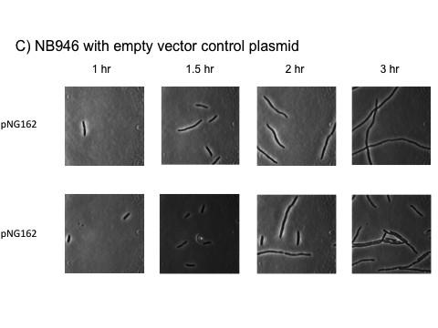
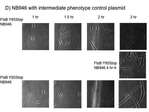
144 125
145 126 Figure 3.6 Timelapse experiment of FtsB depletion strain assay. Images were taken one hour after induction and then every half hour. It is clear that the filamentation begins in just one or two cycles of cell division and there is not a large lag, indicating that the cells are deficient of the wild type FtsB. A) Depletion strain only B) Depletion strain plus plasmids with FtsB (positive controls) C) Depletion strain plus empty vector plasmid (negative controls) D) Depletion strain plus intermediate phenotype plasmid.


146 127 Figure 3.7 A B C D E F G H I Figure 3.7 Point mutations of FtsL tested in vivo. These mutants were tested at 37ºC and 42ºC and shown here is an average image of each. Specific panels contain the following mutants: A T55F, B T56F, C L44F, D L43F, E L42F, F I47F, G A57F, H L53F, I C41F.
147 Future Studies The work on this project will be carried out by another graduate student from this point forward. I have assisted with the set up for the in vivo assay, and have discovered that we might need to use more information from our structural model before testing point mutations. I think it would also be beneficial to employ some co-immunoprecipitation experiments with these strains and the mutations that we are curious about to more directly probe the interaction. Another experiment that should be optimized is a western blot to check for expression of these plasmids in the depletion strain assays. I attempted this without being successful. Finally, once we have a clearer map of the complex and how it is disruptive, it will be interesting to figure out which positions are involved in the localization of FtsL and FtsB and which are involved in the recruitment of downstream proteins. We have received the GFP-tagged plasmids from the Beckwith lab, so we just need to clone in the mutations of interest. For recruitment experiments we can use artificial septal targeting 7. We already know that the interaction of FtsL and FtsB is essential for cell division, but the details on how this interaction is stabilized exactly, is not clear. Another aspect of the project has been to refine the structural model we currently have (Chapter 2) but this preliminary work is not included in this document.
148 Acknowledgements I would like to acknowledge Senes lab undergraduate student, Joel Lange, for his help with the molecular biology for this project. He also helped with the western blotting of the flag-tagged FtsB and FtsL proteins to check for consistent overexpression. I would also like to acknowledge Deena Al- Mahbuba who tested FtsB TM mutants during her rotation project at higher temperature, which yielded the exciting L12F and L15A results. Finally, I would like to thank graduate student from the Senes lab, Sam Craven, for his insightful discussion and for taking over this project. This project has been supported by the Senes lab grant NIH_R01_GM I was also supported by the WARF Distinguished Graduate Fellowship.
149 References 1. Gonzalez, M. D. & Beckwith, J. Divisome under construction: distinct domains of the small membrane protein FtsB are necessary for interaction with multiple cell division proteins. J. Bacteriol. 191, (2009). 2. Gonzalez, M. D., Akbay, E. A., Boyd, D. & Beckwith, J. Multiple interaction domains in FtsL, a protein component of the widely conserved bacterial FtsLBQ cell division complex. J. Bacteriol. 192, (2010). 3. Khadria, A. S. & Senes, A. The transmembrane domains of the bacterial cell division proteins FtsB and FtsL form a stable high-order oligomer. Biochemistry (Mosc.) 52, (2013). 4. LaPointe, L. M. et al. Structural organization of FtsB, a transmembrane protein of the bacterial divisome. Biochemistry (Mosc.) 52, (2013). 5. Buddelmeijer, N. & Beckwith, J. A complex of the Escherichia coli cell division proteins FtsL, FtsB and FtsQ forms independently of its localization to the septal region. Mol. Microbiol. 52, (2004). 6. Russ, W. P. & Engelman, D. M. TOXCAT: a measure of transmembrane helix association in a biological membrane. Proc. Natl. Acad. Sci. U. S. A. 96, (1999). 7. Goehring, N. W., Gonzalez, M. D. & Beckwith, J. Premature targeting of cell division proteins to midcell reveals hierarchies of protein interactions involved in divisome assembly. Mol. Microbiol. 61, (2006).
150 131 Chapter 4 Oligomerization State of Photosynthetic Core Complexes Is Correlated with the Dimerization Affinity of a Transmembrane Helix This chapter was prepared for publication as: Jen Hsin 1, Loren M. LaPointe 2, Alla Kazy 2, Christophe Chipot 1, Alessandro Senes 2, and Klaus Schulten. 1 Oligomerization state of photosynthetic core complexes is correlated with the dimerization affinity of a transmembrane helix. J. Am. Chem. Soc., (2011), 133 (35), pp Department of Physics and Beckman Institute for Advanced Science and Engineering, University of Illinois at Urbana Champaign, Urbana, Illinois 61801, United States 2 Department of Biochemistry, University of Wisconsin Madison, Madison, Wisconsin 53706, United States Contribution: This collaboration was part computational and part experimental. I performed the experimental work through TOXCAT of transmembrane domains found in PufX proteins in four species of purple bacteria.
151 132 Abstract In the Rhodobacter (Rba.) species of photosynthetic purple bacteria, a single transmembrane α- helix, PufX, is found within the core complex, an essential photosynthetic macromolecular assembly that performs the absorption and the initial processing of light energy. Despite its structural simplicity, many unresolved questions surround PufX, the most important of which is its location within the photosynthetic core complex. One proposed placement of PufX is at the center of a core complex dimer, where two PufX helices associate in the membrane and form a homodimer. Inability for PufX of certain Rba. species to form a homodimer is thought to lead to monomeric core complexes. In the present study, we employ a combination of computational and experimental techniques to test the hypothesized homodimerization of PufX. We carry out a systematic investigation to measure the dimerization affinity of PufX from four Rba. species, Rba. blasticus, Rba. capsulatus, Rba. sphaeroides, and Rba. veldkampii, using a molecular dynamics-based free-energy method, as well as experimental TOXCAT assays. We found that the four PufX helices have substantially different dimerization affinities. Both computational and experimental techniques demonstrate that species with dimeric core complexes have PufX that can potentially form a homodimer, whereas the one species with monomeric core complexes has a PufX with little to no dimerization propensity. Our analysis of the helix helix interface revealed a number of positions that may be important for PufX dimerization and the formation of a hydrogen-bond network between these GxxxG-containing helices. Our results suggest that the different oligomerization states of core complexes in various Rba. species can be attributed, among other factors, to the different propensity of its PufX helix to homodimerize.
152 Introduction Compared to algae and plants, bacterial photosynthesis, while similar in its chemical principles of energy conversion, is a lot simpler in the structure and organization of the associated protein pigment assemblies. Nonetheless, there are many unknown features regarding the macromolecular arrangement of some of the most critical photosynthetic complexes, one example being the core complex of purple photosynthetic bacteria. The photosynthetic core complex is a combination of two major transmembrane (TM) protein pigment complexes that carry out the initial steps of the photosynthetic process: light-harvesting complex 1 (LH1) and the reaction center (RC). In some species of purple bacteria, most notably the Rhodobacter (Rba.) genus, the core complex contains an additional TM protein that is largely α-helical and is named PufX for Rhodobacters. Some Rhodobacter core complexes can form dimers,(1, 2) resulting in a large assembly with a dimension of approximately 20 nm 10 nm in the membrane plane (Figure 1)(1, 3-10). The TM protein PufX is known to be crucial in the formation of dimeric photosynthetic core complexes in Rba. sphaeroides,(14) and deletion of this protein leads to monomeric core complexes.(2, 9, 15) Yet, as the location of PufX is still being debated, the molecular mechanism with which PufX determines the oligomerization state of the core complex is still an active topic of discussion.(15) Two models have been proposed for the placement and organization of PufX, each model involving a different mechanism for the PufX-assisted dimerization of the core complex (Figure 1). Figure 1a depicts a central placement of PufX, and the dimerization of the TM region of PufX fuses the two core complex monomers together.(4, 6, 16, 17) In contrast, Figure 1b shows a placement of PufX near the gap of the two open LH1 rings,(7) and in this scheme PufX is thought to induce core complex dimerization via interaction of its long N-terminal region in the cytoplasmic space.(7, 11, 12) A
153 134 crystallographic structure of a dimeric core complex is not yet available to determine unambiguously the validity of either model, although it has also been speculated that different species of Rba. bacteria might have different protein organizations in the core complex.(15, 18) Interestingly, the oligomerization states of different Rba. core complexes are not the same. Through atomic force microscopy (AFM) imaging of the Rba. blasticus photosynthetic membrane, dimeric core complexes have been identified, although monomeric core complexes were also observed at an approximately 3:1 dimer to monomer ratio.(6) The Rba. sphaeroides core complex has also been shown to form dimers,(2, 4, 7, 9) with monomeric core complexes present as well at a 1:1 dimer to monomer ratio.(18) Unlike Rba. blasticus and Rba. sphaeroides, the Rba. veldkampii core complex was observed to be monomeric in a structural and functional analysis,(19) and microscopy studies also reported no sighting of dimeric core complex in the Rba. veldkampii photosynthetic membrane,(13, 17, 20) suggesting that Rba. veldkampii core complex is unable to dimerize. While there is no structural information available for the Rba. capsulatus core complex, its PufX can replace that of Rba. sphaeroides, and the resulting Rba. sphaeroides is still photosynthetically viable,(15) prompting the idea that Rba. sphaeroides and Rba. capsulatus core complexes are likely very similar, and that the core complex of Rba. capsulatus is also capable of dimerizing. Examining the sequences of PufX in four Rba. bacteria, it can be noted that some sequence similarities exist (Figure 1c).(13, 15) In fact, it has been suggested that the GxxxG motif found in Rba. sphaeroides PufX between amino acids 31 and 35 (the N-terminal Met = 0 convention is adopted here) might serve as the dimerization region,(17, 21) similar to that in glycophorin A (GpA).(22) Computational investigations have subsequently shown that a Rba. sphaeroides PufX dimer appears to be stable in a 1-palmitoyl-2-oleoyl-sn-glycero-3-phosphoethanolamine (POPE) membrane.(23)
154 135 However, the GxxxG motif between locations 31 and 35 is only present in Rba. sphaeroides, not in Rba. blasticus or Rba. capsulatus, which also have a dimeric core complex. In addition, mutation of the glycines in this motif does not appear to abolish the ability for Rba. sphaeroides core complex to dimerize, as shown both computationally(23) and experimentally.(18) Furthermore, as shown in Figure 1c, GxxxG motifs are also present in Rba. capsulatus and Rba. veldkampii, although not between positions 31 and 35. A purely sequence-based argument for the dimerization affinity of PufX and the variability in core complex oligomerization state, thus, seems to be still inconclusive and requires further investigation. In an effort to provide new insight into the potential dimerization of the PufX TM region, a prerequisite for the validity of the core complex organization shown in Figure 1a, and to relate dimerization of PufX segments to the core complex oligomerization state, we employed both computational and experimental methods to measure the dimerization affinity of four species of PufX: Rba. blasticus, Rba. capsulatus, Rba. sphaeroides, a n d Rba. veldkampii. We first constructed monomeric and dimeric PufX models for Rba. blasticus, Rba. capsulatus, and Rba. veldkampii, and then probed the stability of these structures in a membrane environment using all-atom molecular dynamics (MD), similar to the strategy previously followed for Rba. sphaeroides PufX.(23) Subsequently, TOXCAT(24) was performed on the four PufX TM segments to quantitatively measure the strength of helix helix association. To complement the experiment, we also computed the apparent dimerization free energy for the four PufX helices using an MD-based free-energy protocol. Our data reveal a compelling trend on the strength of PufX dimerization: species capable of forming a dimeric core complex have PufX helices that show higher propensity to self-associate. Conversely, Rba. veldkampii, which is observed with only monomeric core complexes, has a PufX that exhibits very
155 136 little propensity toward homodimerization. These results strongly indicate that differences in PufX dimerization affinity is an important factor for the variability of oligomerization states in Rba. photosynthetic core complexes.
156 Materials Methods Molecular dynamics construction of monomeric and dimeric PufX As there are currently no structural data available for PufX from Rba. blasticus, Rba. capsulatus, and Rba. veldkampii, the monomeric PufX models in simulations blasticus-monomer- POPE, capsulatus-monomer-pope, and veldkampii-monomer-pope (Table 1) were constructed on the basis of that of Rba. sphaeroides PufX, for which two solution structures have been reported(11, 21) and were used in previous modeling studies.(23, 25-29) All PufX monomers were modeled with an integral TM helix with the same length as that of Rba. sphaeroides PufX.(21) Because we are only interested in the TM interaction of PufX helices, the N- and C-terminal residues that are thought to form loops(11, 21) were not included. These monomeric PufX structures were then placed in a POPE membrane patch, with addition of neutralizing Na+ and Cl ions at a total ionic strength of 300 mm, as shown in Figure 2a c. For comparison, the Rba. sphaeroides PufX monomer, constructed previously, (23) is shown in Figure 2d. Equilibrium MD simulations were carried out for the three PufX monomer systems for 15 ns each. The final conformations of the PufX helices resulting from these monomer simulations were used to construct the corresponding PufX dimer models. Each PufX helix was replicated, and the two copies of PufX were placed facing each other by mapping them onto the GpA dimer structure,(22) as was previously done for Rba. sphaeroides PufX.(23) Because Rba. blasticus, Rba. capsulatus, and Rba. veldkampii do not have a GxxxG motif at position (which Rba. sphaeroides possesses), amino acids of Rba. blasticus, Rba. capsulatus, and Rba. veldkampii PufX were mapped onto the GxxxG portion of GpA. Choice of position is based on the observation that Rba. blasticus, Rba. capsulatus, and Rba. sphaeroides all have a GxxxA or GxxxG motif at this segment, while Rba.
157 138 veldkampii does not. In fact, Gly29 is conserved in all four species as shown in Figure 1c. All PufX dimers were also placed in a POPE membrane patch, and similarly neutralized with additional ions at a total ionic strength of 300 mm, as shown in Figure 2e g. The Rba. sphaeroides PufX dimer system(23) is shown in Figure 2h for comparison. An equilibrium MD simulation was performed for each of the resulting PufX dimer systems, designated as blasticus-dimer-pope, capsulatus-dimer-pope, and veldkampii-dimer-pope in Table 1, for at least 50 ns Equilibrium molecular dynamics All simulations were performed using the MD package NAMD(30) with the CHARMM27 force field,(31, 32) including CMAP corrections.(33) Water molecules were described with the TIP3P
158 139 model.(34) Long-range electrostatic forces were evaluated by means of the particle-mesh Ewald (PME) summation approach with a grid spacing of <1 Å. An integration time step of 2 fs was used in the framework of the Verlet r-respa algorithm.(35) Bonded terms and short-range, nonbonded terms were evaluated every time step, and long-range electrostatics was evaluated every other time step. Constant temperature (T = 310 K) was maintained using Langevin dynamics,(36) with a damping coefficient of 1.0 ps 1. A constant pressure of 1 atm was enforced using the Langevin piston algorithm(37) with a decay period of 200 fs and a time constant of 50 fs Free-Energy calculations To assess computationally the dimerization affinity of the PufX helices, adaptive biasing force (ABF) calculations(38-40) were performed to determine free-energy as a function of helix helix distance.(40, 41) Prior to conducting ABF simulations, the PufX TM segments were equilibrated in a dodecane patch in a solvent environment neutralized with ions at 300 mm ionic strength. Use of dodecane as a lipid mimetic is dictated by the slow relaxation times of natural lipid molecules as compared to affordable MD time scales.(23, 40, 42) The TM segments of PufX were blocked at the N- and C-termini by Ac and NHMe groups, respectively. Two sets of PufX dimer dodecane systems were constructed (blasticus-dimer-dode-1, capsulatus-dimer-dode-1, veldkampii-dimer-dode-1, blasticus-dimer-dode-2, capsulatus-dimer-dode-2, and veldkampii-dimer-dode-2 in Table 1), using slightly different TM segments to test if inclusion of different residues would alter significantly the results of free-energy calculations. Each protein dodecane system was subject to equilibrium MD for at least 10 ns. An example setup of the dodecan PufX system is shown in Figure S1 in the Supporting Information. ABF calculations were carried out subsequently in the framework of NAMD(30) for the six
159 140 dodecane systems (blasticus-dimer-dode-abf-1, capsulatus-dimer-dode-abf-1, veldkampii- Dimer-DODE-ABF-1, blasticus-dimer-dode-abf-2, capsulatus-dimer-dode-abf-2, a n d veldkampii-dimer-dode-abf-2 in Table 1). The TM portion of a modeled Rba.sphaeroides PufX dimer was previously equilibrated in a dodecane patch,(23) and an ABF calculation was also performed for Rba.sphaeroides PufX, designated as sphaeroides-dimer-dode-abf in Table 1. For each ABF simulation, the model reaction coordinate, ξ, is defined as the distance separating the center of mass of the two helices, in the interval 4.5 Å ξ 27 Å. A small ξ indicates that the PufX helices are associated, with a large ξ indicating their separation. In the course of an ABF simulation, average forces applied on the PufX helices in an unconstrained MD simulation are projected onto ξ, and a biasing force is calculated and applied to the helices to overcome local energy barriers.(38-40) The free-energy profile along ξ is then obtained by integrating the average force, with a standard error estimated according to Rodriguez-Gomez et al.(43) TOXCAT Vectors and constructs The TOXCAT vector, pcckan, and positive controls containing the TM domain of wild type GpA (pccgpa-wt) and the G83I disruptive mutant (pccgpa-g83i) have been described previously. (24) DNA coding for the TM domains of the PufX proteins (Table 2), flanked by 5 NheI and 3 BamHI restriction sequence, was purchased as synthetic genes (IDT). The sequences were ligated in-frame to NheI and BamHI sites of the pcckan vector.

160 141 Expression of ToxR'(TM)MBP constructs Plasmids encoding ToxR (TM)MBP chimerae were transformed into Escherichia coli MM39 cells (provided by D. M. Engelman) and plated onto Luria Bertani (LB) plates (with 100 μg/ml ampicillin and 25 μg/ml streptomycin); colonies were inoculated into LB medium (with 100 μg/ml ampicillin) and stored as glycerol stocks at 80 C. LB cultures (with 100 μg/ml ampicillin) were inoculated from frozen glycerol stocks and grown overnight (approximately 18 h). Three ml LB cultures (with 100 μg/ml ampicillin) were inoculated using 50 μl overnight cultures and grown to A , and 1 ml of cells was harvested by centrifugation and resuspended in 0.5 ml of lysis buffer (25 mm Tris-HCl, 2 mm EDTA, ph 8.0). Cells were then lysed by probe sonication. The lysate was clarified by centrifugation at g, and the supernatant was stored on ice until the spectrophotometric assay was performed. Spectrophotometric CAT assay The colorimetric assay used to detect chloramphenicol acetyltransferase activity in cell lysates was described previously.(44, 45) Absorbance was measured using a PerkinElmer Lambda 25 UV/vis spectrophotometer. 40 μl of lysate was mixed with 1 ml of reaction buffer (0.1 mm acetyl-coa, 0.4
161 142 mg/ml 5,5 -dithiobis-(2-nitrobenzoic acid), 0.1 M Tris-HCl, ph 7.8), and absorbance at 412 nm was measured for a period of 2 min with intervals of 3 s to establish a basal rate of acetyl-coa hydrolysis in the absence of substrate. At 2 min, 40 μl of 2.5 mm chloramphenicol was added with mixing, and the absorbance was measured at 412 nm for another 2 min in 3 s intervals. CAT activity was calculated as the slope of 412 nm absorbance, after subtracting the basal rate prior to substrate addition. Lysates were assayed in triplicate, and the reported data are the result of three separate experiments. Maltose complementation assay To confirm correct membrane insertion, E. coli MM39 cells expressing the ToxR (TM)MBP constructs were grown overnight in LB (with 100 μg/ml ampicillin) and then streaked onto M9 minimal media plates containing 0.4% maltose as the only carbon source and incubated for 3 days at 37 C. Western Blot analysis TOXCAT protein expression levels were verified by Western blot analysis. Cell lysate was mixed with 2 SDS-PAGE sample buffer, heated to 70 C for 10 min, run on precast 12% polyacrylamide gels (Invitrogen), transferred onto an Immobilon-P polyvinylidene fluoride membrane (Millipore), and detected with rabbit anti-mpb primary antibodies (New England Biolabs) and antirabbit horseradish peroxidase conjugate secondary antibodies (Millipore).
162 Results and Discussion Below, our computational and experimental results are discussed. First, we report the stability of different species of PufX helices in monomeric and homodimeric conformations as probed by equilibrium MD simulations. Next, we present the dimerization affinity of PufX helices as measured using the TOXCAT assay. Finally, complementing TOXCAT experiments with atomic resolution and quantitative assessment, we report MD-based free-energy calculations conducted to estimate the apparent dimerization free energy, ΔG app, of PufX helices Equilibrium molecular dynamics PufX Monomers All three PufX monomers were seen to be structurally stable during their respective equilibrium MD simulations. Similar to Rba. sphaeroides, the PufX helices of Rba. blasticus, Rba. capsulatus, and Rba. veldkampii were seen to tilt with respect to the normal of the lipid bilayer during simulations. Examining the sequence content of PufX from the different species, it was observed that Rba. veldkampii is the only case without either a tyrosine or a tryptophan residue. Tyrosine and tryptophan residues are known to reside preferentially at the lipid water interface(46) and might contribute to the anchoring of a TM helix to the lipid headgroups.(47-49) As can be seen in Figure 2a d, for Rba. blasticus, Rba. capsulatus, and Rba. sphaeroides PufX that contain tyrosine and tryptophan, most of these residues appear near the membrane solvent interface. The helical structures of all PufX models persisted throughout the simulations, as shown in Figure 3a. Structural stability of each PufX monomer is consistent with the two-stage model of membrane protein folding, which postulates that TM helices act as independent stable domains and are preformed prior to their association into large protein complexes(50, 51).
163 144 PufX Dimers Each PufX dimer model was constructed using the final conformation from the equilibrium simulations of monomeric PufX, as described in the Methods. All three dimer systems were observed to be structurally robust with consistent α-helical content throughout the simulation (Figure 3b). The dimerized PufX helices also maintained a consistent crossing-angle (Figure 3c) and remained in contact during the simulation as indicated by the measurement of the buried solvent-accessible surface area (SASA) (Figure 3d). For all three species, buried SASA of the PufX dimers remained near or above 600 Å2, comparable to that of GpA (52). The most notable motion was seen in the case of Rba. veldkampii PufX, which lacks both tyrosine and tryptophan, and transformed from an originally upright orientation (Figure 3e) to one tilted relative to the membrane at 50 ns (Figure 3f), and with one of the helices submerged in the lipid phase on the C-terminus. This tilted and partially membrane-buried conformation of the Rba. veldkampii PufX dimer persisted when the simulation was extended to 100 ns. In comparison, the tyrosine and tryptophan residues in Rba. blasticus and Rba. capsulatus PufX dimers (Figure 3g and h, respectively) remained at the membrane solvent interface, preventing strong fluctuations in their helix membrane orientations. Quantitative measurement of helix tilting with respect to the membrane normal during the simulations blasticus-dimer-pope, capsulatus-dimer-pope, and veldkampii-dimer-pope is shown in Figure S2 in the Supporting Information. The instability of Rba. veldkampii PufX helices due to the lack of anchorage might be significant in the propensity of the helices to homodimerize. Additionally, it can be seen that the proline residue at position 36 in Rba. capsulatus PufX induces a moderate kink in the helix (10 30 ) that persisted throughout the simulations for both the mononeric and the dimeric conformations (Figure S3); this residue does not face the dimerization interface in the
164 145 modeled Rba. capsulatus PufX dimer. Interhelical interactions contributing to the stability of PufX dimer models are shown in Figure 4a c for Rba. blasticus, Rba. capsulatus, and Rba. veldkampii, respectively. For Rba. blasticus and Rba. capsulatus, significant molecular interactions are contributed by residues Gln25 and Met26, which interact with each other, and also with other small amino acids such as glycine and alanine. For example, Gln25 was seen to interact with Ala22, and Met26 interacted with Gly29 in Rba. blasticus PufX dimer (Figure 4a). The Met26 Gly29 interaction was also observed for Rba. capsulatus, and its Gln25 was observed to interact with Ile22 (Figure 4b). Also, helix packing is achieved through close contact between small residues Gly29 and Ala30 for both Rba. blasticus and Rba. capsulatus (Figure 4a and b). Notably, while Gly29 is conserved for all four species investigated in the present study, Rba. veldkampii is the only species that does not contain Gln25 and Ala30 (Figure 1c), two residues that contribute significantly to interhelical interactions for Rba. blasticus and Rba. capsulatus. For Rba. veldkampii PufX dimer, the helices are held together by a slightly different set of molecular interactions, although Met26, which is conserved in all four PufX sequences (Figure 1c), plays also an important role (Figure 4c). Further away from the dimerization core, bulkier amino acids such as Val37 for Rba. blasticus, and Phe37 for Rba. capsulatus and Rba. veldkampii, provide additional interhelix contact.

165 Interhelical hydrogen bonds are known to be an important factor in mediating helix helix 146
166 147 association in the membrane.(53) The close packing of the modeled PufX dimers permitted the formation of several interhelical hydrogen bonds. In particular, the side-chain amide group of Gln25 forms an interhelical hydrogen bond with the carbonyl of Ala22 in the Rba. blasticus dimer (Figure 5). The same side chain accepts a Cα H O hydrogen bond from Ile22 in the Rba. capsulatus dimer. As mentioned above, Rba. veldkampii is the only species that does not contain Gln25. Several backboneto-backbone Cα H O hydrogen bonds were also observed (illustrated in Figure 5 and Table 3), which are a hallmark of GxxxG-mediated transmembrane interactions.(53-58) The Gln25 Met26 and Gly29 Ala30 pairs were seen to be sites for the potential formation of Cα H O hydrogen bonds for the cases o f Rba. blasticus and Rba. capsulatus PufX dimers, and locations 33 and 34, which contain small amino acids such as alanine, serine, and glycine, provide additional hydrogen bonding (Figure 5 and Table 3). For the case of Rba. veldkampii PufX dimer, only two Cα H O hydrogen bonds were observed and were also formed between amino acid pairs (Ala25 Met26) and (Gly29 Met30) TOXCAT The equilibrium MD simulations of the three PufX dimers conducted here, as well as the one conducted previously for Rba. sphaeroides,(23) showed that PufX dimer models for all four species remain associated. Although it appears that the Rba. veldkampii PufX dimer has an unstable protein membrane interaction due to the lack of anchorage, no spontaneous disassociation was observed. It is possible that disassociation of PufX requires longer simulation time than is currently feasible due to the slow relaxation time of a full POPE membrane. To determine quantitatively the dimerization affinity of the PufX helices, we employed an experimental assay, the TOXCAT method,(24) which measures the association of TM helices in a
167 148 biological membrane. Three TOXCAT measurements were performed on each of the four Rba. species, with the average dimerization affinity for each species shown in Figure 6b as percent of the CAT activity of GpA. Rba. capsulatus is seen to have the highest propensity for homodimerization, with a relative CAT activity at 30%, comparable to a prior measurement reported in Aklujkar and Beatty(59). Rba. blasticus has the second highest CAT activity, albeit only at 15% GpA. Rba. sphaeroides shows even lower propensity to homodimerize, and Rba. veldkampii exhibits no significant CAT activity Free-Energy Calculations Concurrent to the experimental measurement of PufX dimerization affinity with TOXCAT, we also employed free-energy calculations to measure the apparent dimerization free energy in silico using the ABF algorithm(38-40) for PufX from four Rba. species. The computational treatment is inspired by the atomic resolution of the method, which can reveal great structural details in the dimerization and disassociation pathway. Two sets of ABF calculations were conducted corresponding to distinct choices of TM residues. In the first set, the same sequences as those used in TOXCAT (i.e., those shown in nonboldface in Table 2) were included. Because the TOXCAT experiments contain also flanking residues and are not identical to the setup of the in silico assays, to test if results from ABF are sensitive to the small difference in the sequence of amino acids, a second set of ABF simulations was conducted, using the sequences identified as the TM region from the dimer simulations carried out in the POPE environment (i.e., blasticus-monomer-pope, capsulatus-monomer-pope, and veldkampii-monomer-pope; Table 1). For Rba. sphaeroides PufX, the sequence used in TOXCAT is the same as that identified as the TM region;(23) therefore, only one ABF simulation was performed (sphaeroides-dimer-dode-abf in Table 1).
168 149 The dimerization pathway of PufX is observed to be more complex than that of GpA,(40) with an example shown in Figure 7 for the case of Rba. sphaeroides PufX. At the beginning of the ABF simulation sphaeroides-dimer-dode-abf, the two PufX helices are both straight (Figure 7a). However, spontaneous bending occurred after 13 ns (Figure 7b, left), in agreement with prior in silico observation on the inherent flexibility of the Rba. sphaeroides PufX helix(23). Furthermore, bending of the PufX helix occurs at the same location as that seen for one of the PufX solution structures,(11) and, as a result, the bent conformation seen in simulation sphaeroides-dimer-dode-abf is structurally very similar to the solution structure (Figure 7b, right). Helix bending persisted for a few tens of a nanosecond, but eventually the helix spontaneously straightened (Figure 7c). Previously, we had estimated that bending of the Rba. sphaeroides PufX only costs a few kcal/mol;(23) our present results support such a low value. We note that the tendency for Rba. sphaeroides PufX to bend might complicate self-association, but does not completely prohibit it as the helix quickly straightens back and the straight conformation can possibly be stabilized by dimerization. The results from the two sets of ABF calculations are compared in Figure 8 in the form of freeenergy profiles as a function of helix helix distance. For the first set of ABF calculations (Figure 8a), all four species of PufX are seen to have energy minima for an associated, dimerized conformation, albeit with different well depths. Rba. veldkampii has the most shallow free-energy minimum near a helix helix separation of 12 Å. Rba. sphaeroides has the second-most shallow free-energy minimum. Unlike the GpA dimer, which exhibits a well-defined free-energy well,(40) the free-energy well of Rba. sphaeroides PufX is seen to span nearly 5 Å, with the minimum occurring near 8 Å. Rba. blasticus and Rba. capsulatus have the deepest free-energy wells, and both possess multiple local minima. For Rba. capsulatus, two local free-energy minima are found at helix helix distances of 8 and 11 Å; for Rba.

169 150 blasticus, its free-energy well has several less well-defined local minima that stretch up to a helix helix distance of 15 Å. The much wider free-energy well for Rba. blasticus PufX is possibly due to additional stabilizing interhelical interactions arising from transient van der Waals contact between the bulkier Leu43, Leu44, and Thr47 residues near the C-terminal end (Figure S4 in the Supporting Information). In general, the PufX dimers exhibit more complex free-energy profiles than does the GpA dimer.(40) This extra complexity is possibly due to the usage of modeled dimer systems rather than experimentally derived structures. Alternatively, it is also possible that the complex dimerization scheme is intrinsic to PufX due to its difference to GpA. In the second set of ABF simulations (Figure 8b), Rba. veldkampii appears to have no preference for association. Rba. capsulatus PufX is seen to have a deeper free-energy well than that of Rba. blasticus and retains the two minima observed in Figure 8a, albeit at closer helix helix distances (7 and 9 Å). Rba. blasticus PufX again exhibits a wide minimum, with the global free-energy minimum occurring at a helix helix distance of 9 Å. Although the precise free-energy profiles are different in the two sets of ABF simulations, it is reassuring that distinct features in the Rba. blasticus and Rba. capsulatus free-energy profiles are preserved across the two simulations. Additionally, Rba. veldkampii PufX consistently exhibits the lowest propensity toward dimerization.
170 151 From the free-energy profiles in Figure 8, we calculated the apparent disassociation free energy, ΔG app, for the four species of PufX in two sets of ABF calculations using the expression utilized by Hénin et al.,(40) with the results shown in Table 4 and Figure 9. Calculation of ΔG app allows comparison of PufX dimerization affinity with that of GpA (Figure 9), which has a ΔG app value of 11.5 ± 0.4 kcal/mol as previously reported using also the ABF method in a dodecane environment.(40) It can be seen that the order of PufX dimerization affinity is similar to the experimental results (Figure 6), in the decreasing order of Rba. capsulatus > Rba. blasticus > Rba. sphaeroides > Rba. veldkampii. Differences in experimental and simulation setups might contribute to the consistent overestimate of in silico free energies as compared to TOXCAT measurements (Figures 9 and 6). For example, the PufX sequences used in the TOXCAT experiment and the ABF measurements are not exactly identical (Table 2 and Figure 8), and, as shown by comparing Figure 8a and b, small differences in sequence content can lead to varying dimerization affinity. Additionally, while TOXCAT was performed in a biological membrane environment, the ABF measurements were conducted with dodecane. Finally, the reaction coordinate, ξ, chosen in the ABF calculation does not consider the relative orientation of the two helices, including their intrinsic rotation about their longitudinal axis, a degree of freedom important in optimizing helix helix packing. Considering these factors limiting direct comparison between experiment and simulation results, it is significant that a consensus in the relative strength of dimerization for the four PufX sequences tested here was reached.
171 Concluding Remarks We have shown through experiments and molecular dynamics simulations that PufX from different Rba. species of purple photosynthetic bacteria exhibit distinct propensities toward homodimerization. This result can explain in part why core complexes have different oligomerization states in different species, namely, due to the different inherent affinity of the TM regions of PufX to dimerize. In particular, species with PufX shown to be least likely to dimerize, Rba. veldkampii, form only monomeric core complexes (13, 17). On the other hand, Rba. blasticus that possesses dimeric core complexes(6) is seen to have PufX with a relatively high dimerization affinity. In addition to the ability for dimerization at the TM region, the presence of aromatic amino acids with polar groups (tyrosine and tryptophan) appears to aid in stabilizing PufX helices in the membrane. Rba. veldkampii PufX contains no tyrosine or tryptophan and also shows the lowest tendency for dimerization in its TM region. These two characteristics of Rba. veldkampii PufX, the lack of anchoring residues and a TM region with low likelihood for self-association, make Rba. veldkampii PufX a poor candidate for forming homodimers. Interestingly, a tryptophan residue on the N-terminal end of the TM region is part of the recently identified PufX motif that is missing in Rba. veldkampii, but present in Rba. blasticus, Rba. capsulatus, and Rba. sphaeroides (Figure 1c) (13). While Rba. blasticus, Rba. capsulatus, and Rba. sphaeroides PufX show some preference for self-association, their dimerization affinity is significantly lower than that of GpA (52, 60). The lower dimerization affinity might be the reason why dimeric and monomeric core complexes are both present in Rba. blasticus and Rba. sphaeroides,(4, 6, 18) as a subset of PufX helices might be in monomeric forms in the photosynthetic membrane, residing in monomeric core complexes. It is also possible that dimerization of PufX requires additional molecular interactions other than those arising from the PufX
172 153 TM region, or between PufX and the rest of the core complex. For Rba. sphaeroides, there are experimental reports showing that the N-terminal segment of PufX is critical for the formation of dimeric core complexes,(12, 61) although the molecular role of these residues in PufX-assisted core complex dimerization is unclear. Dimerization of PufX might also be strengthened by interaction between PufX and LH1α helices observed previously for Rba. sphaeroides and Rba. capsulatus,(62) or by the binding of the light-absorbing pigment bacteriochlorophyll (shown in Figure 1a and b as crosses) to PufX (59, 63, 64). Speculation that different Rba. species might have different organizations for their core complexes has also been raised (15, 18). While identification of a GxxxG motif in the Rba. sphaeroides PufX sequence at the position is intriguing,(17, 21) the motif by itself does not explain the observed oligomerization states of Rba. core complexes. As alluded to above, Rba. blasticus lacks this particular sequence motif, yet it has been confirmed to contain dimeric core complexes.(6) It should be noted, however, that Rba. blasticus, Rba. capsulatus, and Rba. sphaeroides actually all feature a GxxxA/G motif at the position that is not present in Rba. veldkampii (Figure 1c), with GxxxA previously suggested as a motif for dimerization of TM helices (55, 65-69). The glycine residue at position 29 in PufX is actually conserved across the four Rba. species, and an alanine residue is found at position 30 except for Rba. veldkampii PufX (Figure 1c), providing another small amino acid that allows for potential helix helix interaction. It is conceivable that the combination of presence of protein membrane anchoring provided by tyrosine or tryptophan, and small amino acids such as glycine and alanine at the helix helix contact site, renders a PufX TM segment more prone to homodimerize.
 PufX is placed at the dimerization interface in the center of the core complex and is itself also thought to be dimerized.")
173 154 Figure 4.1 Figure 4.1 Proposed models for the protein organization of a dimeric photosynthetic core complex. (a) Model based on atomic force microscopy imaging studies of Rba. sphaeroides and Rba. blasticus photosynthetic membrane.(4, 6) PufX is placed at the dimerization interface in the center of the core complex and is itself also thought to be dimerized. (b) Model based on the highest resolution structural data to date of the dimeric Rba. sphaeroides core complex,(7) with PufX situated near the gap of the open LH1 ring, and association of PufX is facilitated through a long loop at the N-terminal region.(7, 11, 12) In (a) and (b), PufX helices are represented by black circles, while LH1 helices are shown as gray circles (outer helices, known as LH1β) and white circles (inner helices, known as LH1α), with the embedded pigments between the outer and inner helices denoted by X. RC is shown as an oval. (c) Aligned sequences of the central region of PufX from four Rhodobacter species investigated in the present study. Conserved amino acids are indicated by arrows, and amino acids conserved in Rba. blasticus, Rba. capsulatus, and Rba. sphaeroides, but not in Rba. veldkampii, are
174 shaded in gray.(13) GxxxG and GxxxA motifs are underlined. 155
 Monomeric Rba. sphaeroides system is also shown for comparison; the simulation was performed previously.(23) PufX helix is shown in blue ((a) Rba. blasticus), green ((b) Rba.")
175 156 Figure 4.2 Figure 4.2 Simulated molecular systems with POPE lipid bilayers. Protein membrane systems with monomeric PufX for (a) Rba. blasticus, (b) Rba. capsulatus, and (c) Rba. veldkampii. (d) Monomeric Rba. sphaeroides system is also shown for comparison; the simulation was performed previously.(23) PufX helix is shown in blue ((a) Rba. blasticus), green ((b) Rba. capsulatus), red ((c) Rba. veldkampii), and gray ((d) Rba. sphaeroides), lipid is shown in yellow with purple spheres representing the headgroups; polar-aromatic residues tyrosine and tryptophan of PufX are shown in orange. For clarity, water and ion molecules included in all simulations are not shown. (e g) Protein membrane systems with modeled homodimeric PufX for (e) Rba. blasticus, (f) Rba. capsulatus, and (g) Rba. veldkampii. (h) Dimeric Rba. sphaeroides system is also shown for comparison; the simulation was performed previously. 23). PufX helices in this and subsequent figures are shown with N-termini pointing upward.
, PufX retains its high α-helical content.")
176 157 Figure 4.3 Figure 4.3 Stability of PufX monomeric and homodimeric helices during equilibrium MD simulations. (a) α-helical content of the modeled PufX monomer in a full POPE membrane. For each of the three species tested (Rba. blasticus, Rba. capsulatus, and Rba. veldkampii), PufX retains its high α-helical content. Similarly, modeled PufX helices in dimeric conformation also remain largely α- helical, as shown in (b). (c) Crossing-angle between the dimerized helices. (d) Buried solventaccessible surface area (SASA) as a measure for helix helix interaction. Parts (e) and (f) show the movement of the Rba. veldkampii PufX helices. At 50 ns, one of the Rba. veldkampii helices can be seen to submerge nearly fully into the membrane on the C-terminus. For comparison, (g) and (h) show the Rba. blasticus and Rba. capsulatus PufX helices also at 50 ns; in these cases, the tyrosine and tryptophan residues aided in anchoring the helices in the membrane, and these residues remained at the
177 membrane solvent interface throughout the simulation. 158
178 159 Figure 4.4 Figure 4.4 Interhelical interactions observed during the equilibrium molecular dynamics simulations (a) blasticus-dimer-pope, (b) capsulatus-dimer-pope, and (c) veldkampii-dimer- POPE. In each interaction map, highly interacting amino acid pairs are highlighted with darker grids, and five of such pairs are shown in the insets as examples.
 blasticus-dimer-pope, (b) capsulatus-dimer-pope, and (c) veldkampii-dimer-pope.")
179 160 Figure 4.5 Figure 4.5 Networks of interhelical Cα H O hydrogen-bond contacts in the three PufX dimer models identified from simulations (a) blasticus-dimer-pope, (b) capsulatus-dimer-pope, and (c) veldkampii-dimer-pope. For the amino acids involved in formation of Cα H O contacts, carbon atoms are shown in gray, oxygen atoms in red, hydrogen atoms in white, and other backbone atoms are shown in transparent. All H O distances are shown in angstroms.
 TOXCAT(24) is an in vivo assay based on a fusion construct consisting of the TM domain under investigation, a maltose binding protein, and the ToxR transcriptional activator of V. cholerae.")
180 161 Figure 4.6 Figure 4.6 Quantification of association of TM constructs in E. coli membranes using TOXCAT. (a) TOXCAT(24) is an in vivo assay based on a fusion construct consisting of the TM domain under investigation, a maltose binding protein, and the ToxR transcriptional activator of V. cholerae. TM association results in the expression of chloramphenicol acetyl transferase (CAT) under the ctx promoter, whose enzymatic activity can be measured. (b) TOXCAT data for the PufX TM domains of Rba. capsulatus, Rba. blasticus, Rba. sphaeroides, and Rba. veldkampii. The data are reported as percent of the CAT activity of GpA, a strongly dimerizing transmembrane domain (22). The data are the average of three independent measurements, and the error bars report the standard deviation. Protein expression levels were verified by Western blot using anti-mbp antibodies.
 At 13 ns, one of the helices bent spontaneously. The bending corresponded well to the observed NMR solution structure of PufX (pdb code 2NRG(11)) and persisted for the next 40 ns.")
181 162 Figure 4.7 Figure 4.7 Spontaneous bending and straightening of Rba. sphaeroides PufX. (a) At the onset of the ABF simulation for Rba. sphaeroides PufX, both helices were straight. (b) At 13 ns, one of the helices bent spontaneously. The bending corresponded well to the observed NMR solution structure of PufX (pdb code 2NRG(11)) and persisted for the next 40 ns. (c) The bent helix was seen to straighten back at 58 ns, suggesting that bending and straightening of the helix occur spontaneously with a low energy barrier, as suggested previously (23).
.")
182 163 Figure 4.8 Figure 4.8 Potentiaof meal n force measured in ABF simulations. Highlighted sequences are those included in the ABF simulations; the sequences outlined in the red box are those included in the TOXCAT measurement (Table 2). (a) First set of ABF simulations using the same sequence for TM segments of PufX as that in TOXCAT experiments. (b) Second set of ABF simulations using the sequence identified as the TM segments in the PufX dimer POPE membrane simulations

183 164 Figure 4.9 Figure 4.9 Free energy of association, ΔG app, for various PufX sequences as a fraction of GpA ΔG app. GpA ΔG app was calculated in Hénin et al. (40).
184 Acknowledgements We thank C. Neil Hunter and Pu Qian for many helpful discussions, Simon Scheuring for illuminating communications, and Ben Mueller for assistance in performing the TOXCAT assays. This work was supported by Grants NSF MCB and NIH P41-RR Computer time was provided by NCSA and TACC via Large Resources Allocation Committee Grant MCA93S028, resources of the Argonne Leadership Computing Facility at Argonne National Laboratory, supported by the Office of Science of the U.S. Department of Energy (Contract DE-AC02-06CH11357), and resources of the Oak Ridge Leadership Computing Facility at Oak Ridge National Laboratory, which is supported by the Office of Science of the U.S. Department of Energy under Contract DE-AC05-00OR22725.
185 References 1. Jungas, C.; Ranck, J.-L.; Rigaud, J.-L.; Joliot, P.; Verméglio, A. EMBO J. 1999, 18, Siebert, C. A.; Qian, P.; Fotiadis, D.; Engel, A.; Hunter, C. N.; Bullough, P. A. EMBO J. 2004, 23, Loach, P. A. Proc. Natl. Acad. Sci. U.S.A. 2000, 97, Scheuring, S.; Francia, F.; Busselez, J.; Melandris, B. A.; Rigaud, J.-L.; Lévy, D. J. Biol. Chem. 2004, 279, Scheuring, S.; Lévy, D.; Rigaud, J.-L. Biochim. Biophys. Acta 2005, 1712, Scheuring, S.; Busselez, J.; Lévy, D. J. Biol. Chem. 2005, 280, Qian, P.; Hunter, C. N.; Bullough, P. A. J. Mol. Biol. 2005, 349, Cogdell, R. J.; Gall, A.; Köhler, J. Q. Rev. Biophys. 2006, 39, Qian, P.; Bullough, P. A.; Hunter, C. N. J. Biol. Chem. 2008, 283, Scheuring, S.; Sturgis, J. N. Photosynth. Res. 2009, 102, Tunnicliffe, R. B.; Ratcliffe, E. C.; Hunter, C. N.; Williamson, M. P. FEBS Lett. 2006, 580, Ratcliffe, E. C.; Tunnicliffe, R. B.; Ng, I. W.; Adams, P. G.; Qian, P.; Holden-Dye, K.; Jones, M. R.; Williamson, M. P.; Hunter, C. N. Biochim. Biophys. Acta 2011, 1807, Liu, L.-N.; Sturgis, J. N.; Scheuring, S. J. Struct. Biol. 2011, 173, Francia, F.; Wang, J.; Venturoli, G.; Melandri, B. A.; Barz, W. P.; Oesterhelt, D. Biochemistry 1999, 38,
186 Holden-Dye, K.; Crouch, L. I.; Jones, M. R. Biochim. Biophys. Acta 2008, 1777, Scheuring, S. Curr. Opin. Cell Biol. 2006, 10, Busselez, J.; Cottevielle, M.; Cuniasse, P.; Gubellini, F.; Boisset, N.; Lévy, D. Structure 2007, 15, Crouch, L. I.; Holden-Dye, K.; Jones, M. R. Biochim. Biophys. Acta 2010, 1797, Gubellini, F.; Francia, F.; Busselez, J.; Venturoli, G.; Lévy, D. Biochemistry 2006, 45, Milhiet, P.-E.; Gubellini, F.; Berquand, A.; Dosset, P.; Rigaud, J.-L.; Le Grimellec, C.; Lévy, D. Biophys. J. 2006, 91, Wang, Z.-Y.; Suzuki, H.; Kobayashi, M.; Nozawa, T. Biochemistry 2007, 46, MacKenzie, K. R.; Prestegard, J. H.; Engelman, D. M. Science 1997, 276, Hsin, J.; Chipot, C.; Schulten, K. J. Am. Chem. Soc. 2009, 131, Russ, W. P.; Engelman, D. M. Proc. Natl. Acad. Sci. U.S.A. 1999, 96, Chandler, D.; Hsin, J.; Harrison, C. B.; Gumbart, J.; Schulten, K. Biophys. J. 2008, 95, Hsin, J.; Gumbart, J.; Trabuco, L. G.; Villa, E.; Qian, P.; Hunter, C. N.; Schulten, K. Biophys. J. 2009, 97, Sener, M. K.; Hsin, J.; Trabuco, L. G.; Villa, E.; Qian, P.; Hunter, C. N.; Schulten, K. Chem. Phys. 2009, 357, Hsin, J.; Chandler, D. E.; Gumbart, J.; Harrison, C. B.; Sener, M.; Strumpfer, J.; Schulten, K.
187 168 ChemPhysChem 2010, 11, Trabuco, L. G.; Schreiner, E.; Gumbart, J.; Hsin, J.; Villa, E.; Schulten, K. J. Struct. Biol. 2011, 173, Phillips, J. C.; Braun, R.; Wang, W.; Gumbart, J.; Tajkhorshid, E.; Villa, E.; Chipot, C.; Skeel, R. D.; Kale, L.; Schulten, K. J. Comput. Chem. 2005, 26, MacKerell AD1, Bashford D, Bellott M, Dunbrack RL, Evanseck JD, Field MJ, Fischer S, Gao J, Guo H, Ha S, Joseph-McCarthy D, Kuchnir L, Kuczera K, Lau FT, Mattos C, Michnick S, Ngo T, Nguyen DT, Prodhom B, Reiher WE, Roux B, Schlenkrich M, Smith JC, Stote R, Straub J, Watanabe M, Wiórkiewicz-Kuczera J, Yin D, Karplus M. J. Phys. Chem. B 1998, 102, Foloppe, N.; MacKerell, A. D., Jr. J. Comput. Chem. 2000, 21, MacKerell, A. D., Jr.; Feig, M.; Brooks, C. L., III. J. Comput. Chem. 2004, 25, Jorgensen, W. L.; Chandrasekhar, J.; Madura, J. D.; Impey, R. W.; Klein, M. L. J. Chem. Phys. 1983, 79, Tuckerman, M. E.; Berne, B. J.; Martyna, G. J. J. Phys. Chem. B 1992, 97, Brünger, A. T.; Brooks, C. L., III; Karplus, M. Chem. Phys. Lett. 1984, 105, Feller, S. E.; Zhang, Y.; Pastor, R. W.; Brooks, B. R. J. Chem. Phys. 1995, 103, Darve, E.; Pohorille, A. J. Chem. Phys. 2001, 115, Hénin, J.; Chipot, C. J. Chem. Phys. 2004, 121, Hénin, J.; Pohorille, A.; Chipot, C. J. Am. Chem. Soc. 2005, 127, Kim, H.; Hsin, J.; Liu, Y.; Selvin, P. R.; Schulten, K. Structure 2010, 18,
188 Stockner, T.; Ash, W. L.; MacCallum, J. L.; Tieleman, D. P. Biophys. J. 2004, 87, Rodriguez-Gomez, D.; Darve, E.; Pohorille, A. J. Chem. Phys. 2004, 120, Shaw, W. V. Methods Enzymol. 1975, 43, Sulistijo, E. S.; Jaszewski, T. M.; MacKenzie, K. M. J. Biol. Chem. 2003, 278, Senes, A.; Chadi, D. C.; Law, P. B.; Walters, R. F. S.; Nanda, V.; DeGrado, W. F. J. Mol. Biol. 2007, 366, Killian, J. A.; von Heijne, G. Trends Biochem. Sci. 2000, 25, Yuen, C. T. K.; Davidson, A. R.; Deber, C. M. Biochemistry 2000, 39, de Planque, M. R. R.; Killian, J. A. Mol. Membr. Biol. 2003, 20, Popot, J.-L.; Engelman, D. M. Biochemistry 1990, 29, Engelman, D. M.; Chen, Y.; Chin, C.-N.; Curran, A. R.; Dixon, A. M.; Dupuy, A. D.; Lee, A. S.; Lehnert, U.; Matthews, E. E.; Reshetnyak, Y. K.; Senes, A.; Popot, J.-L. FEBS Lett. 2003, 555, Fleming, K. G.; Ackerman, A. L.; Engelman, D. M. J. Mol. Biol. 1997, 272, Bowie, J. U. Curr. Opin. Struct. Biol. 2011, 21, Senes, A.; Ubarretxena-Belandia, I.; Engelman, D. M. Proc. Natl. Acad. Sci. U.S.A. 2001, 98, Senes, A.; Engel, D. E.; DeGrado, W. F. Curr. Opin. Struct. Biol. 2004, 14, Arbely, E.; Arkin, I. T. J. Am. Chem. Soc. 2004, 126,
189 Derewenda, Z. S.; Lee, L.; Derewenda, U. J. Mol. Biol. 1995, 252, Lawrie, C. M.; Sulistijo, E. S.; MacKenzie, K. R. J. Mol. Biol. 2010, 396, Aklujkar, M.; Beatty, J. T. Photosynth. Res. 2006, 88, Duong, M. T.; Jaszewski, T. M.; Fleming, K. G.; MacKenzie, K. R. J. Mol. Biol. 2007, 371, Francia, F.; Wang, J.; Zischka, H.; Venturoli, G.; Oesterhelt, D. Eur. J. Biochem. 2002, 269, Recchia, P. A.; Davis, C. M.; Lilburn, T. G.; Beatty, J. T.; Parkes-Loach, P. S.; Hunter, C. N.; Loach, P. A. Biochemistry 1998, 37, Parkes-Loach, P. S.; Law, C. J.; Recchia, P. A.; Kehoe, J.; Nehrlich, S.; Chen, J.; Loach, P. A. Biochemistry 2001, 40, Law, C. J.; Chen, J.; Parkes-Loach, P. S.; Loach, P. A. Photosynth. Res. 2003, 75, Senes, A.; Gerstein, M.; Engelman, D. M. J. Mol. Biol. 2000, 296, Schneider, D. FEBS Lett. 2004, 577, Schneider, D.; Engelman, D. M. J. Mol. Biol. 2004, 343, Kairys, V.; Gilson, M. K.; Luy, B. Eur. J. Biochem. 2004, 271, Rath, A.; Deber, C. M. Proteins: Struct., Funct., Bioinf. 2008, 70,

190 Supporting Information Figure S4.1 Figure S4.1 An example setup of a molecular dynamics simulation using a dodecane patch as a lipid mimetic. Shown here is the configuration for simulation capsulatus-dimer-dode-1 in Table 1 in the text. Water is shown in transparent blue, dodecane molecules are shown in cyan and the two transmembrane segments of Rba. capsulatus PufX are shown in green and dark green.

191 172 Figure S4.2 Figure S4.2 Measurement of helix tilt with respect to the membrane normal during the equilibrium simulations basticus-dimer-pope (blue trace), capsulatus-dimer-pope (green trace), and veldkampii-dimer-pope (red trace).
 shwon in sphere representation. Membrane is shown in the transparent with the same color scheme as in Figure 2 of the main text.")
192 173 Figure S4.3 Figure S4.3 Location of the transmembrane proline residue in Rba. capsulatus PufX. (A) Proteinmembrane system from simulation capsulatus-monomer-pope at 15 ns. PufX is shown in green, with the proline residue at location 36 (P36) shwon in sphere representation. Membrane is shown in the transparent with the same color scheme as in Figure 2 of the main text. (B) Protein-membrane system from simulation capsulatus-dimer-pope at 35 ns. The two PufX helices are colored in green and dark green for distinction. The P36 residues can be seen to point away from the helix-helix association interface. (C) Measurement of the helix kink near the P36 residue during simulation capsulatus- Monomer-POPE (black trace) and capsulatus-dimer-pope (dark green and light green traces).
 The three most common conformations for the transmembrane segments of Rba. blasticus PufX seen in blasticus-dimer-dode-abf-1.")
193 174 Figure S4.4 Figure S4.4 Common conformations of PufX helices seen in ABF simulations with a helix-helix separation of Å. (A) The three most common conformations for the transmembrane segments of Rba. blasticus PufX seen in blasticus-dimer-dode-abf-1. (A-i) Conformation with helices associated at the N-Terminal ends via close contacts near Gly29. (A-ii) Conformation with helices interacting transiently at the C-terminal ends via van der Waals contacts between large side chains (most notably Leu43, Leu44, and Thr47). (A-iii) Conformation with the helices separated. The conformation in A-ii is only observed for the case of Rba. capsulatus. For example, for the case of the transmembrane segments of Rba. capsulatus PufX seen in capsulatus-dimer-dode-abf-1, only two common conformations are observed with a helix-helix separation of Å, one with associated N-terminal ends also mediated by the Gly29 contact (B-i) and another with completely separated helices (B-ii).
194 175 Chapter 5 The Oligomeric States of the Purified Sigma 1 Receptor are Stabilized by Ligands This chapter was prepared for publication as: Katarzyna A. Gromek, 1 Fabian P. Suchy, 1 Hannah R. Meddaugh, 1 Russell L. Wrobel, 1 Loren LaPointe, 2 Uyen B. Chu, 3 John G. Primm, 1 Arnold E. Ruoho, 3,* Alessandro Senes 2 and Brian G. Fox 1,2,* The Oligomeric States of the Purified Sigma 1 Receptor are Stabilized by Ligands. Journal of Biological Chemistry (2014) 289(29): From the 1 Transmembrane Protein Center, Departments of 2 Biochemistry and 3 Neuroscience, University of Wisconsin-Madison, Madison, Wisconsin Contribution: I performed the analysis of oligomerization of the transmembrane domain (TM2) in the sigma-1 receptor, including mutagenesis.
195 176 Abstract Sigma 1 receptor (S1R) is a mammalian member of the ERG2 and sigma1 receptor like protein family (pfam04622). It has been implicated in drug addiction and many human neurological disorders including Alzheimer s and Parkinson s diseases and amyotrophic lateral sclerosis. A broad range of synthetic small molecules including cocaine, (+)-pentazocine, haloperidol and small endogenous molecules such as N,N-dimethyltryptamine, sphingosine and steroids have been identified as regulators of S1R. However, the mechanism of activation of S1R remains obscure. Here we provide evidence in vitro that S1R has ligand binding activity only in an oligomeric state. The oligomeric state is prone to decay into an apparent monomeric form when exposed to elevated temperature, with loss of ligand binding activity. This decay is suppressed in the presence of the known S1R ligands such as haloperidol, BD-1047 and sphingosine. S1R has a GxxxG motif in its second transmembrane region, and these motifs are often involved in oligomerization of membrane proteins. Disrupting mutations within the GxxxG motif shifted the fraction of the higher oligomeric states towards smaller states and resulted in a significant decrease in specific [3H]-(+)-pentazocine binding. Results presented here support the proposal that S1R function may be regulated by its oligomeric state. Possible mechanisms of molecular regulation of interacting protein partners by S1R in the presence of small molecule ligands are discussed. Abbreviations used: The abbreviations used are: BD-1047, N-[2-(3,4-dichlorophenyl)ethyl]-N-methyl-2- (dimethylamino)ethylamine dihydrobromide; BODIPY, boron-dipyrromethene; CAT, chloramphenicol acetyltransferase; DTG, 1,3-di-(2-tolyl)guanidine; MBP, maltose binding protein; 4-PPBP, 4-phenyl-1- (4-phenylbutyl)piperidine maleate; PRE-084, 2-(4-morpholinethyl)-1-phenylcyclohexanecarboxylate
196 177 hydrochloride; S1R, sigma-1 receptor; SKF-83959, 6-chloro-7,8-dihydroxy-3-methyl-1-(3- methylphenyl)-2,3,4,5-tetrahydro-1h-3-benzazepine; TEV, tobacco etch virus; TM2, second transmembrane region; ToxR, Vibrio cholerae toxin transcriptional regulator.
197 Background The mammalian sigma 1 receptor (S1R2) is a unique 223 amino acid membrane bound protein (1-5). S1R is found in the mammalian central nervous system (CNS) and in most peripheral tissues including the immune and endocrine systems. It is primarily localized in the endoplasmic reticulum (6-8), but also in some cellular plasma membranes (9), specialized cisternae-laiden cholinergic synapses of the spinal cord ventral horn motoneuron C-terminals (10,11) and in spinal cord dorsal root ganglia (12). The amino acid sequence of S1R is approximately 95% identical between mammals including the guinea pig, mouse, rat and human. ERG2, a sterol isomerase found in yeast (13) and fungi (2), is an ortholog of the mammalian S1R with an approximately overall 30% sequence homology and 66% homology in the putative S1R ligand binding domain (1). The mammalian S1R does not possess sterol isomerase activity and has been clearly differentiated by sequence and size from the fungal sterol isomerase (1,13). S1R functions as a molecular chaperone and serves as a partner for a variety of client proteins. It stabilizes the IP3 type 3 receptor (14) in ER mitochondrial associated membranes and has been shown to interact and play an important regulatory role in many cell signaling systems including the molecular chaperone GRP78/BIP (15), several types of G-protein coupled receptors (15-18), and voltage- gated ion channels (9,19-23). S1R suppresses the production of reactive oxygen species in various mouse tissues including the retina, lung and liver, and in cultured mammalian cells possibly by activating antioxidant response element genes (6,24-26). A broad range of synthetic small molecules with widely varied structures bind with high affinity to S1R, including the (+) isomer of benzomorphan derivatives such as pentazocine and dextrallorphan, neuroleptics such as haloperidol, fluphenazine and chlorpromazine, the compounds o-ditolylguanidine,
198 179 PRE-084, BD-1047 and BD-1063, the beta-blocker propranolol and the presynaptic dopamine D2 agonist (+)-3-PPP (27-29). Several endogenous small molecules such as N, N dimethyltryptamine (30), sphingosine (31) and steroids such as progesterone (32) and dehydroepiandrosterone (33) have also been identified as regulators of S1R. Due to the broad contributions of S1R in maintaining cellular homeostasis, the receptor has been identified as a therapeutic target for the treatment of cancer (34) and neurodegenerative diseases including amyotrophic lateral sclerosis (35), Alzheimer s (36) and Parkinson s diseases (37), and for retinal neurodegeneration (38). Several studies have also connected S1R to the possible treatment of drug addiction and toxicity related to derivatives of cocaine and amphetamine (8,16,39,40). The guinea pig S1R has been purified to homogeneity following expression in E. coli as a fusion to maltose binding protein (MBP, (41)). The ligand binding region of S1R was identified by the use of specific radioiodinated photoprobes (42-45) and by mutagenesis (46,47) to be formed primarily by the juxtaposition of a short C terminal hydrophobic region (residues ) with a portion of TM2 (residues ) and perhaps a portion of TM1. Based on hydrophobicity analyses and the use of S1R-GFP constructs (9) and S1R antibody probes (14), it has been concluded that the S1R contains two putative transmembrane (TM) helices (9) with both the N and C termini occurring on the cytoplasmic side of the cellular membrane (9,14). S1R also has a GxxxG motif in TM2. This motif is often involved in helix-helix oligomerization of integral membrane proteins (48-50). High molecular weight forms (tetramer, pentamer) of the S1R were previously identified using radioiodinated photoaffinity labeling in rat liver microsomal membrane preparations (44), suggesting that S1R may oligomerize under physiological conditions. Here we report that highly purified S1R forms an oligomeric state, and also show that the
199 180 oligomeric state provides specific ligand binding, while the monomeric state does not. Stabilization of the functional oligomeric states occurs via the participation of the GxxxG oligomerization motif. These results are discussed in the context of possible mechanisms of molecular regulation of interacting protein partners by S1R in the presence of small molecule ligands.
200 Experimental Procedures Cloning Plasmid DNA containing the guinea pig sigma-1 receptor gene was used the template for all cloning work (41). All oligonucleotides were purchased from IDT (Coralville, IA). The MBP-4A- S1R plasmid used in the current work was made using PIPE mutagenesis (51) as previously reported (52) using primers listed in Table S1 of the Supplementary Material. PCR was carried out using Pfu-UltraII polymerase. When the PCR reaction was completed, a DpnI digestion was performed to remove the template. The DpnI-digested PCR product was purified using a Qiagen PCR purification kit and the eluted DNA was transformed into E. coli 10G (Lucigen, Middleton, WI). To make expression plasmids for the second transmembrane helix (TM2) for TOXCAT analysis, two partially complementary long oligonucleotides corresponding to the S1R-TM2 domain were designed to include 5 NheI and 3 BamHI overhangs (see primer list in Table 1). These single stranded oligonucleotides were allowed to anneal and the resulting dsdna was ligated into NheI- and BamHI-digested pcckan (53). Correct DNA constructs were verified by DNA sequencing of the entire MBP to ToxR fusion coding region. Mutations in TM2 were made using PIPE mutagenesis. Protein preparation Expression and purification of MBP-4A-S1R containing a stabilizing 4-Ala linker between the MBP and S1R domains, a variant with a tobacco etch virus protease site present as the interdomain linker (MBP-TEV- S1R), or with mutations in TM2 were carried out as described previously (52). MBP-TEV-S1R was purified using amylose affinity chromatography (52) and subjected to proteolysis using TEV protease in a ratio of 1 mg of protease per 1 mg of fusion protein. TEV protease was prepared as previously reported (54). The TEV protease reaction was performed at room temperature
201 182 for 96 h, and the final cleavage efficiency was greater than 95%. The sample was filtered through a 0.8 µm syringe filter and diluted with 20 mm Tris, ph 7.2, containing 0.031% Triton X-100 and 1 mm 2- mercaptoethanol to reduce the concentration of NaCl to 100 mm. A 5-mL Fast Flow HiTrap Q column (GE Healthcare Life Sciences) was prepared using 5 column volumes of 20 mm Tris, ph 7.2, containing 100 mm NaCl, 0.031% Triton X-100 and 1 mm 2-mercaptoethanol (loading buffer). The protein sample was loaded onto the Q column using the AKTA purifier sample pump at a flow rate of 1 ml/min and then washed with 5 column volumes of 20 mm Tris, ph 7.2, containing 100 mm NaCl, 0.031% Triton X-100 and 1 mm 2-mercaptoethanol (wash buffer). Elution was performed with gradient of NaCl to a final concentration of 1 M over 20 column volumes. The collected fractions were analyzed for protein content by 4-20% gradient SDS-PAGE. Appropriate fractions were combined and concentrated as described before. Protein concentrations were determined using BioRad in-gel densitometry. Samples from all purification steps were assayed for ligand binding activity. Preparative size exclusion chromatography This chromatography was conducted on a Shimadzu Prominence HPLC equipped with a DGU- 20A5 on- line degasser, LC-20AD solvent delivery module, SIL-20ACHT autosampler, CTO-20AC column oven, SPD-20A UV-vis detector, RF-10AXL spectrofluorometric detector, RID-10A differential refractometric detector, FRC-10A fraction collector module, CBM-20A system controller and LabSolution LCsolution software version 1.24 SP1. The mobile phase, 10 mm HEPES, ph 7.2, containing 150 mm NaCl, 0.3 mm TCEP and 0.018% DDM (2 CMC), was degassed for a minimum of 20 min under vacuum prior to use. The buffer was isocratically pumped at 1 ml/min through a Phenomenex 300 x 7.8 mm Yarra 3µm SEC-3000 column with SecGuard column guard. Protein elution was monitored by UV absorption at 280 and 260 nm. The column temperature was 20.0 C and the
202 183 detector flow cell temperature was 35.0 C. Columns were calibrated daily using bovine thyroglobulin, IgA, ovalbumin, myoglobin and uridine as standards (Phenomenex). High volume separation was achieved through repeated 100 µl injections while separating the protein into forty-one 125 µl fractions. Analytical size exclusion chromatography Fractions from multiple injections of MBP-4A-S1R were subjected to additional rounds of analytical sizing chromatography to assess whether changes in the distribution of oligomeric states occurred during the repeat chromatography. Elution from the repeat chromatography was monitored using the intrinsic fluorescence of tryptophan with excitation at 280 nm and emission at 340 nm. During the course of the repeated analyses, retention times varied by less than 0.25 min during the 12 min chromatographic run (2%), and were corrected for preparation of figures to correspond to the same apparent molecular weight (determined from the daily calibration) by using the earliest chromatogram as the benchmark for retention times. Similarly, S1R obtained from TEV protease proteolysis of MBP- TEV-S1R was subjected to repeat sizing chromatography. In this case, the forty-one proteolyzed fractions were stored for ~1 month at 4 C before repeat analysis. Light scattering measurements Separate fractions containing peaks 1, 2 and intermediate oligomer from Fig. 2A were subjected to size exclusion chromatography coupled to multi-angle laser light scattering, with UV absorbance and refractive index detection. Separation was performed on a Superdex S200 (GE Healthcare Life Sciences) in 150 mm NaCl, 5 mm HEPES, ph 7.2 and 0.018% DDM (2 CMC). Light scattering and refractive index were measured with a DAWN HELEOS II and OPTILab rex respectively (Wyatt Technology). Data analysis was performed using ASTRA 6.1 (Wyatt Technology).
203 184 Chemical cross-linking Chemical cross-linking with disuccinimidyl suberate (DSS) was performed on the detergentsolubilized and highly purified individual oligomeric states of MBP-4A-S1R. DSS was dissolved in DMSO and the control samples (no cross-linker) contained equivalent amount of DMSO (2%). 5 μm of each protein state was incubated with either 30- or 50- molar excess of DSS (150 µm, 250 μm, respectively) for 2 h at room temperature, in presence or absence of 10 μm BD The reactions were stopped by addition of Tris-HCl, ph 8.0, to the final concentration of 30 mm. The samples were then subject to SDS-PAGE in 7.5% Tris-HCl BioRad gel and calibrated with commercial molecular weight markers (Spectra Multicolor High Range Protein Ladder, Thermo Scientific). After staining with Brilliant Blue R, the gels were imaged and analyzed using the GelAnalyzer 2010 free software ( to calculate the approximate molecular weight of visualized bands. Oligomer stability tests Haloperidol, o-ditolylguanidine, PRE-084, BD-1047, 4-PPBP, SKF-83959, sphingosine, and sphingosine-1-phosphate were from Tocris Biosciences (Bristol, UK). Pentazocine and was from Sigma-Aldrich (St. Louis, MO). Stock solutions of PRE-084 (5 mm) and BD-1047 (10 mm) were prepared in deionized water. A pentazocine stock solution (10 mm) was prepared in 20 mm HCl in deionized water, while stock solutions of 4-PPBP (10 mm), haloperidol (10 mm), ortho-ditolylguanidine (10 mm) and SKF (10 mm) were prepared in 100% (v/v) DMSO. Sphingosine and sphingosine-1-phosphate stock solutions (5 mm) were prepared in solution containing 1.8% (w/v) DDM. The sphingosine-1-phosphate stock solution was heated to ~60 C to aid in solublization. Less than 0.5 mm of free Pi was detected in this sample, indicating the phosphoryl group was not hydrolyzed during solubilization.
204 185 Purified peak 1 (see Fig. 2B) from preparative size exclusion chromatography of MBP-4A-S1R was diluted to 15 µg/ml (0.23 µm) and a final volume of µl in 10 mm HEPES, ph 7.2, containing 0.3 mm TCEP and 0.018% DDM and incubated for up to 18 h at 4 C, 25 C and 37 C. Analytical size exclusion chromatography was run before and after incubation with various ligands. A typical HPLC injection was 10 µl and intrinsic tryptophan fluorescence was monitored. All experiments included a control sample incubated under the same conditions but with no added ligand. Ligand stock solutions were prepared as described above, and ligands were tested for stabilization at 0.45 µm and 10 µm. Ligand binding assays [3H]-(+)-pentazocine (specific activity 36 Ci/mmol) was from Perkin-Elmer (Waltham, MA). Binding assays were performed in 100 μl in a 48-well block format as described previously (41,45) with minor modifications. Protein samples at 1 ng/μl were prepared in 20 mm Tris, ph 8.0, containing 0.1% Triton X-100. The final concentration of [3H]-(+)-pentazocine in both total and non-specific binding assays was 100 nm. Haloperidol (Tocris) was used as the masking agent in the non-specific binding reaction at final concentration of 10 μm. The incubation with ligands was performed for 90 min at 32 C, followed by filtration on a glass fiber filter (Whatman GF/B) performed in Brandel cell harvester. The glass filter was then washed with 50 mm Tris, ph 8.0, and individual filters were transferred into vials containing 3 ml of scintillation cocktail (Ultima Gold, Perkin-Elmer). The level of radioactivity was measured the following day using a Packard scintillation counter. The raw count data were normalized to nmol of protein present in the assay and plotted as the percentage of specific binding activity of the original, control sample, MBP-TEV-S1R. The stoichiometry of ligand binding was determined using 300 nm of purified peak 1 (see Fig.
205 186 2B) supplemented with a range of concentrations of BD-1047 from 0 to 3000 nm in a total volume of 150 µl. The titration was assembled in a 96-well plate, and incubated for 16 h at 37 C. Aliquots (10 μl) from each well were examined with analytical size exclusion chromatography and eluted protein was detected using tryptophan fluorescence. The values for 0% and 100% oligomeric stabilization were normalized using the wells containing either no BD-1047 or the maximal amount, respectively. The binding data were analyzed using KD = [np][l]/[pl], where n is the number of protein molecules that bind one molecule of ligand, and P, L and PL are the equilibrium concentrations of free receptor, free ligand, and the ligand-bound receptor, respectively. The expression for KD was rewritten as KD = [npi x][li x]/[x], where x corresponds to the amount of [PL] formed and also the depletion in concentrations of free receptor and free ligand. Theoretical values for x at each step in the ligand binding titration were determined by solving this latter expression. Best fit values for n and KD were determined using the NonlinearModelFit routine of Mathematica v (Wolfram Research). TOXCAT A gene encoding the TM2 domain of S1R was cloned into the NheI-BamHI restriction sites of the pcckan vector resulting in the following sequence: NRASxxxGILIN. Escherichia coli MM39 cells transformed with pcckan-derived TOXCAT plasmids were inoculated into 3 ml of Luria Bertani medium containing 100 µg/ml of ampicillin and grown overnight at 37 C with shaking. To check for proper membrane insertion of the TOXCAT constructs, overnight cultures of transformed MM39 cell were plated onto M9 minimal medium agar plates containing 0.4% maltose as the only carbon source and grown at 37 C for 48 h (53). Aliquots (3 µl) of the overnight cultures were inoculated into 3 ml of Luria Bertani medium and grown to OD600 of 0.6 at 37 C with shaking. An aliquot (1 ml) of the culture medium was centrifuged for 10 min at 17,000g and the cell pellet was resuspended in 500 ml
206 187 of 25 mm Tris- HCl, ph 8.0, containing 2 mm EDTA. The resuspended cells were sonicated at medium power for 10 s and a 50-µL aliquot was removed from each sample and mixed with 4 NuPAGE SDS loading buffer, boiled for 10 min, and saved for western blotting. Lysates were clarified by centrifugation at 17,000g for 10 min. The supernatant was kept on ice and used in chloramphenicol acetyltransferase (CAT) assays. Chloramphenicol acetyltransferase assays One ml of 0.1 M Tris-HCl, ph 7.8, containing 0.1 mm acetyl-coa and 0.4 mg/ml of 5,5'- dithiobis-(2- nitrobenzoic acid) was mixed with 40 µl of supernatant from the cell lysis and the absorbance at 412 nm was measured for 2 min to establish basal activity (55). After this, 40 µl of 2.5 mm chloramphenicol dissolved in 10% ethanol was added and absorbance at 412 nm was measured for an additional 2 min to determine CAT activity. CAT activity was normalized using OD420 measurements of cell aliquots. The relative CAT activities were reported as percentages of the activity given by the strong transmembrane dimer control, glycophorin A (GpA). Quantification of TOXCAT expression Boiled cell lysates (10 µl) were loaded onto a NuPAGE 4-12% Bis-Tris SDS-PAGE gel (Invitrogen) and then transferred to polyvinylidene fluoride membranes (VWR) for 1 h at 100 mv. Blots were blocked using 5% bovine serum albumin (US Biologicals) in 50 mm Tris, ph 8.0, containing 150 mm NaCl and 0.5% Tween buffer (TBST) for 2 h at 4 C. Biotinylated anti-maltose binding protein antibody (Vector labs) was diluted 1:1500 in 1% bovine serum albumin in TBST and incubated overnight at 4 C. Blots were washed with TBST for 1 h with three buffer exchanges at room temperature before incubation with secondary antibody in 1% bovine serum albumin in TBST at 1:1500 dilution, peroxidase-conjugated streptavidin (Jackson ImmunoResearch) for 2 h at room
207 188 temperature. Blots were again washed for 1 h using three exchanges of TBST. A 1:1 mixture of buffers from the Pierce ECL Western Blotting Substrate Kit was added to the blot and chemiluminescence was measured using an ImageQuant LAS 4000 (GE Healthsciences).
208 Results For these studies, MBP-4A-S1R (Fig. 1A, lane P1) was prepared using an expression method that gives higher yield of purified protein from Escherichia coli (52). In addition, S1R without an MBP tag was prepared by treatment of a MBP-TEV-S1R fusion with TEV protease (Fig. 1B, lane P2). Denaturing SDS-PAGE showed that these protein preparations consisted of a single polypeptide with purity greater than 95%. With these preparations, we investigated the relationship between the oligomerization state of S1R and its ligand binding activity. The results show that an oligomeric form of the receptor is required for specific ligand binding Evidence for oligomerization Analytical size- exclusion chromatography of purified MBP-4A-S1R showed two major peaks, labeled 1 and 2 (solid line, Fig. 2A). After treatment with TEV protease, purified S1R also showed two major peaks, labeled 3 and 4 (dotted line, Fig. 2A). Thus both receptor preparations show evidence for formation of a predominant oligomeric state (peaks 1 and 3) along with a corresponding monomer (peaks 2 and 4). Similar behavior was observed from MBP-4A-S1R prepared in buffer containing n- octyl-beta-d-glucopyranoside or MBP-4A-S1R prepared in buffer containing Triton X-100. A variable amount of an intermediate oligomer state was also observed in the MBP-4A-S1R samples (retention time ~8 min, marked with *). Figs. 2B and 2C show that both the oligomer and monomer peaks were stable (i.e., eluted with the same retention time) when subjected to a repeat round of chromatography. Thus the major peaks shown in Fig. 2A could be obtained in a highly pure form. Fig. 2B shows that peaks 1 and 3, corresponding to the potential oligomeric states, had apparent molecular weights of 460 kda and 150 kda, respectively, for the protein-detergent micelle, while Fig. 2C shows that peaks 2 and 4, corresponding to monomeric states, had apparent molecular weights of 80 and 35 kda, respectively.
209 190 The oligomeric assemblies were only dependent on the presence of S1R, as MBP alone did not form an oligomeric state in the conditions used here. Moreover, after removal of the MBP by treatment with TEV protease, S1R remained in the oligomeric state and could be further purified by both adsorption and size exclusion chromatographies (Fig. 2B, peak 3). The estimation of oligomer stoichiometry using analytical SEC alone is complicated because of uncertainty in how the protein will be accommodated into a protein-detergent micelle and those effects on the hydrodynamic radius. Although static light scattering measurements are often used to assess oligomeric stoichiometry, the presence of detergent micelles creates high background noise and must be accounted for in the mass of the protein-detergent complex. Thus, the fusion protein was sent to the Yale Keck Biophysics Lab, which is specially equipped for these measurements, with combined SEC and light scattering instrumentation. Three peaks were analyzed. The smallest protein molecules detected (Fig. 3A) had a mass within 2% of that predicted for a monomer, while the largest molecular weight protein (Fig. 3B) was polydisperse, with molecular weights corresponding to oligomerization states of 6 to 8. Light scattering also showed that the intermediate oligomer marked with * in Fig. 2A was monodisperse with a molecular weight corresponding to a tetramer (Fig. 3C). Analysis by SDS-PAGE after cross-linking gave further insight into the light scattering results. Fig. 4A shows the monomer was unchanged in SDS-PAGE either without cross-linker (lanes 1 and 2) or with cross-linker (lanes 3 and 4), suggesting no inter-molecular interactions capable of being captured by the crosslinking reagent. In contrast, Fig. 4B shows that the polydisperse oligomer crosslinked to a size greater than 300 kda. Furthermore, Fig. 4C shows that the intermediate oligomer, assigned to be a tetramer by light scattering measurements, was cross-linked to a molecule with molecular weight again consistent with a tetramer.
210 Ligand binding Pentazocine is a well-studied ligand for S1R (56,57). Fig. 5 shows that only the oligomeric states of MBP-4A-S1R and S1R exhibited specific pentazocine binding activity. For example, peak 1 from Fig. 2 (oligomer of the MBP fusion) bound pentazocine with ~20 higher specific activity than peak 2 (monomer of the MBP fusion). Likewise, the specific binding activity for peak 2 from Fig. 3 (S1R oligomer) was ~15 higher than for peak 4 (S1R monomer). Since some S1R ligands were delivered in DMSO carrier, a sample containing a final concentration of 2% DMSO but no other ligand was tested independently and shown to have no effect on oligomeric state. With these assignments of the active form of the receptor, peak deconvolution of the original samples indicated that active MBP- 4A-S1R (solid line, Fig. 2A and B) was ~75% of the original protein sample while active S1R (dotted line, Fig. 2A and B) represented ~50%. Ligand binding was also tested using the fluorescent ligand BODIPY-sphingosine in analytical SEC experiments. Consistent with the specific pentazocine binding results (Fig. 5), the oligomeric forms of the protein migrated with haloperidol-masked BODIPY fluorescence, while the monomer did not (data not shown). The fluorescent ligand was a gift from Mary L. Kraft PhD (University of Illinois, Urbana-Champaign) Stability of oligomers The oligomeric forms of S1R were observed to decay upon incubation in buffer. Thus, after ~18 h at 37 C, the amount of protein in peak 1 decreased by ~40% and the amount in peak 2 increased by a corresponding amount (Fig. 6A). Similar instability of the S1R oligomers was also observed at 25 C and 4 C in the absence of ligand, but to a lesser extent. In all chromatograms, the total integrated area remained constant within 5%, supporting the conclusion that interconversion between oligomer and
211 192 monomer states was occurring. Disulfide bonds are apparently not involved in oligomer formation since inclusion of 100 mm 2-mercaptoethanol in the buffer used for S1R purification and repeat analytical size exclusion chromatography did not change the elution profile. Fig. 6B shows that in the presence of 0.45 µm BD-1047, a known tight-binding ligand of S1R, the relative proportions of peaks 1 and 2 were essentially unchanged after the ~18 h incubation; similar behavior was observed at each of the three temperatures tested. Several other known S1R ligands, including the natural membrane constituent sphingosine, also gave stabilizing interactions. Fig. 7 shows the percentage increase in the monomer form after incubation with these ligands relative to a control containing no ligand, where a larger increase in monomer corresponds to less effective stabilization of the active oligomer. Overall, the tightest binding synthetic ligands (e.g., BD-1047, 4- PPBP) gave the largest stabilizing effect (i.e., least conversion to the monomer). Interestingly, the natural membrane lipid sphingosine gave a stabilizing interaction that was close to that of many of the synthetic ligands. In contrast, another natural membrane lipid, sphingosine-1-phosphate, did not stabilize the receptor even when present at greater than 20 higher concentration than BD-1047 (10 µm versus 0.45 µm). Fig. 8 shows an analysis of stoichiometry of binding for BD-1047, a tight-binding ligand, as assessed by stabilization of the peak 1 oligomer. In the experiment with receptor-detergent micelles carried out here, the best-fit KD (r2 = 0.998, solid black line) was ~7 nm, which is comparable the value reported elsewhere (58). The best unconstrained fit stoichiometry of ligand bound per receptor, n, was determined to be 0.43, i.e., corresponding to ~1 mol of ligand per 2 mol of receptor. Fits with n held constant at 1 were not compatible with the binding data (dashed line), as acceptable fits could not be obtained at any KD value. Moreover, when n was held constant at 0.25, approximate fits using the
212 193 best-fit KD values gave systematic overestimation of the fraction bound at low ligand:protein ratio (dotted line). An analysis assuming n was 0.33, i.e., ligand binds to a trimer, also gave a similar overestimation; increasing the KD value gave a better fit at low ligand:protein ratios but gave under estimation of complex formation at high ligand:protein ratios. An identical experiment was conducted on the intermediate oligomer (Fig. 1A, peak labeled *) yielding the same results Role of GxxxG motif in oligomerization The GxxxG motif is often involved in helix-helix association in integral membrane proteins (49,59). In S1R, TM2 contains this motif in the primary sequence G87-G88-Trp89-Met90-G91. Mutations of the Gly residues in this motif and an adjacent His residue in MBP-4A-S1R were prepared to test their roles in oligomerization. All mutations within the motif (substitutions of either Ile or Leu at G87, G88 and G91) resulted in lower expression and significantly decreased yield for the purified fusion protein. For example, MBP-4A-S1R gave a purified yield of ~3.5 mg per L of culture medium, while G91I MBP-4A-S1R (~0.4 mg/l) and G91L MBP-4A-S1R (~0.6 mg/l) were the best yields for the proteins with mutations in the GxxxG motif. With all of the purified receptor variants, all mutations in the GxxxG motif eliminated the largest oligomer (e.g., peak 1 in Fig. 2) from size exclusion chromatographs. Fig. 9 shows representative behavior for mutation of Gly91 to either Ile (Fig. 9A) or Leu (Fig. 9B). Both mutations strongly shifted the distribution of receptor states to the monomer form (solid lines). With the G91I mutation (Fig. 9A), a smaller oligomeric state (retention time ~8.2 min, marked with ) with an apparent molecular weight of ~180 kda was observed. All G88 and G89 mutants had similar chromatograms. This peak plausibly represents a dimeric state of the S1R. Fig. 9C shows that the mutation H97A, which is not in the GxxxG motif, yielded a chromatograph almost identical to MBP-4A-S1R, suggesting this residue does not play an important role in oligomerization.
213 194 Fig. 10 shows that mutations within the GxxxG motif had a profound effect on specific ligand binding. Among the set of all GxxxG mutations, G91L MBP-4A-S1R exhibited ~20% of specific pentazocine binding activity of the non-mutated receptor, while all other mutations in the GxxxG motif yielded purified receptor variants that had less than 5% specific binding. Corresponding to the modest level of specific binding activity observed, G91L MBP-4A-S1R uniquely stabilized a significant fraction of the purified protein as the intermediate oligomer (Fig. 9B), likely tetramer (Fig. 3C). These results further implicate the role of an oligomer in ligand binding. While H97A MBP-4A- S1R had a distribution of large, intermediate and monomer states that was nearly identical with the non-mutated fusion protein (Fig. 9C), it exhibited only ~50% ligand binding activity (Fig. 10). This result suggests a role for H97 in ligand binding independent of oligomerization Oligomerization of TM2 To further assess the propensity of TM2 for self-association, TOXCAT reporter assays were performed by inserting the sequence of TM2 (WVFVNAGGWMGAMCLL- HASL) between MBP and the ToxR receptor (Fig. 11A). With this construct, oligomerization of the TM2 region can be assessed by catalytic assasy of the ToxR-mediated expression of CAT, as ToxR only functions as a transcriptional enhancer when it is present as an oligomer. Fig. 11B shows results from the TOXCAT experiment. All variants were expressed to a comparable level as assessed by western blotting, and non-mutated TM2 from S1R gave rise to a strong positive CAT assay response, corroborating the propensity of the TM2 sequence to self-associate. Interestingly, among the single mutations of the GxxxG motif, only G91I eliminated the positive response in the CAT assay, indicating this mutation strongly destabilized oligomerization of the TM2. Surprising, the G91L mutation gave a positive assay response, albeit attenuated to only 60% of that observed from the non-mutated TM2. Individually,
214 195 mutations at either G87 or G88 did not affect the assay response. While this result is apparently contradictory to the results of Fig. 10 obtained with the full length receptor, the adjacent positions of Gly87 and Gly88 in the primary sequence of the TM2 peptide presumably allowed alternative modes for association of the TM2 peptide that could not be achieved with the TM2 present in the MBP-4A- S1R fusion. The double mutation G87L/G88L eliminated the TOXCAT assay response, perhaps corresponding to more effective disruption of alternative TM2 interactions leading to oligomerization.
215 Discussion In this work, we provide biochemical evidence for the importance of an oligomeric state in the ligand binding function of guinea pig S1R, and the essential role of the GxxxG motif from TM2 in this oligomerization. Since amino acid sequences and pharmacological profiles are highly conserved among mammalian S1R (Fig. S1), these results are likely broadly relevant to many studies of this protein family (60). Mutational analysis of putative TM2 in both the full-length receptor and as a transmembrane domain in the TOXCAT studies have identified key residues involved in oligomerization (G87, G88 and G91) and in ligand binding independent of oligomerization (His97). This work adds to the list of other residues in TM2 (S99, Y103 and L105- L106) that are important for ligand binding (61), which this work suggests is intimately related to the ability to form one or more oligomeric states. Although the GxxxG motif within TM2 has an important role in oligomer formation, other studies have implicated additional residues of the S1R receptor sequence in dimerization/oligomerization (17), which could explain the appearance of the 180 kda peak (Fig. 9A peak labeled ). The S1R ligand binding site in the intact receptor, as identified by photoaffinity labeling, has been localized to a region that juxtaposes a steroid binding domain-like motif (SBDLI) in TM2 (which encompasses the oligomerization sequences identified in this work) and a C-terminal SBDLII hydrophobic sequence (42-44). Although the C- terminus alone does not bind S1R ligands, some of the chaperone functions of S1R have been localized to this region (62). It has been proposed that S1R agonists alter the structure of the receptor in such a fashion that the C-terminal chaperone region becomes accessible to its client proteins. NMR derived structures of the C-terminus have been recently reported (63). Perhaps the oligomeric states of the S1R receptor dictate the availability of the C- terminus for these interactions (64).
216 197 Several previous experiments support the conclusion that S1R functions as an oligomer in ligand binding. For example, different molecular weights have been observed by gel filtration chromatography for S1R purified from natural sources (65). Moreover, high molecular weight bands of S1R protected from photo-affinity labeling by (+)-pentazocine were detected by denaturing gel electrophoresis of rat liver microsomes (44,64). Furthermore, gel filtration of ligand-bound S1R purified from human leukemia cells showed that ligand binding activity was associated with a protein of ~100 kda (2). Many other membrane receptors adopt an oligomeric state (66-68), and recombinant expression often leads to a distribution of these states (69). For example, when human serotonin receptor 3A was overexpressed in E.coli, the protein was detected as a mixture of oligomers and the biologically active pentamer represented only 7% of the total protein (70). The percentage of active S1R protein determined in the study reported here was between 50-75%. S1R interacts with a large number of synthetic and natural ligands, and has also been documented to interact with a large number of different proteins within the cell (5,30). We found that interactions with the tightest binding synthetic ligands strongly stabilize the oligomeric state (Fig. 6, 7). The stabilizing effects of ligands on many other membrane proteins, including G-protein coupled receptors, is recognized (71,72). Our studies with purified S1R indicate a minimal binding stoichiometry of one ligand per two polypeptide chains (Fig. 8), while reconciliation of gel filtration, light scattering and denaturing gel electrophoresis results obtained with purified S1R suggest the octamer, hexamer and tetramer are the predominant ligand binding forms. A stoichiometry of one ligand bound/dimer is further supported by the work of Chu and Ruoho (64) who showed that a C12 alkyl containing photoprobe selectively and quantitatively derivatized His145 at a proposed S1R dimer interface. Further, dimerization of S1R, as assessed by SDS-PAGE, occurred via intermolecular
217 198 disulfide bond formation when a M170C mutant form of the receptor was expressed. The oligomer can decay to an intermediate tetramer and monomer in the absence of ligands (Fig. 6), while mutations of the GxxxG motif change the distribution of these species. Preliminary efforts to reassemble the monomer into functional oligomers were not successful. However, it is intriguing to consider that protein-protein interactions may be involved in reassembly of the functional receptor. In this regard, monomeric S1R has been reported to interact with ion channels such as Nav 1.5 voltage-gated Na+ channel, acid sensing channels, and dopamine D1 receptor (16,23,73). Interestingly, these interactions were disrupted in the presence of ligands such as haloperidol and/or (+)-pentazocine, so one may speculate that a ligand-gated S1R oligomer/monomer equilibrium defines the availability of monomer S1R for interaction with client ion channels or G-protein coupled receptors. An additional unusual feature of binding of the agonist, (+)-pentazocine, to S1R is the time (>90 min at 30 C) needed to reach equilibrium (41,74). It is possible that formation of stable interactions between oligomeric states of S1R in situ regulate the rate of (+)-pentazocine binding to S1R (perhaps also affected by interactions with accessory protein partners). Fig. 12 provides a schematic of these possibilities.

218 199 Figure 5.1 Figure 5.1 SDS-PAGE of MBP-4A-S1R (A) and S1R (B). The starting MBP-4A-S1R and S1R preparations prior to SEC separation are labeled P1 and P2, respectively. Peaks 1-4 from Fig. 2A are labeled respectively. Molecular weight markers (MW) are labeled in kda.
219 Figure
220 201 Figure 5.2 Evidence from size exclusion chromatography for oligomeric states of S1R. A, elution profiles for MBP- 4A-S1R (solid line) and S1R (dashed line). Peaks corresponding to different oligomerization states are marked with numbers 1-4. B, repeat chromatography of peaks 1 (solid line) and 3 (dashed line) from A. C, repeat chromatography of peaks 2 (solid line) and 4 (dashed line) from A.
221 Figure
222 203 Figure 5.3 Oligomeric molecular weight of MBP-4A-S1R determined by light scattering. Elution profiles detected by 652 nm laser light scattering are shown by dashed lines. Oligomeric stoichiometry across each peak is marked with a solid line. Peaks have the same labeling as in Fig. 1 and 2. A, The apparent monomer is confirmed to be a monomer. B, The largest molecular weight oligomer is polydisperse, with stoichiometry ranging from a hexamer to octamer. C, The intermediate oligomer is a tetramer.

223 204 Figure 5.4 Figure 5.4 Analysis of oligomeric states of MBP-4A-S1R by chemical cross-linking agent disuccinimidyl suberate (DSS). DSS-free controls are shown by lanes 1 and 2, containing 0 and 10 μm high affinity ligand BD-1047 respectively. Lanes 3 and 4 mirror the control with the addition of DSS. A, The addition of cross-linking agent to the monomer (Fig. 2A, peak 2). The bands show slight smearing with no shift in size, signifying only intramolecular cross-linking. B, The addition of crosslinking agent to the oligomer (Fig. 2A, peak 1). The bands show a mass greater than 300 kda and oligomeric stoichiometry cannot be accurately determined. However, an oligomeric state greater than tetramer is clearly visible. C, The addition of cross-linking agent to the intermediate oligomer (Fig. 2A peak labeled *). The bands show an approximate 4-fold increase in size to a mass between 250 and 300 kda, suggesting a tetrameric state.
224 205 Figure 5.5 Figure 5.5 Comparison of specific pentazocine binding activity of S1R oligomers before and after repeat chromatography of peaks from Fig. 2B and 2C. The black bars are for assays of MBP-4A- S1R; white bars are for assays of S1R. Binding assays were performed in triplicate and the error bars represent 1σ deviation.
225 Figure
226 207 Figure 5.6 Stabilization of oligomeric S1R by ligand binding. A, size exclusion chromatogram of MBP-4A-S1R before (dashed line) and after incubation for 1 day at 37 C (solid line) without added ligand. Peaks have the same labeling as in Fig. 1 and 2. B, chromatogram of MBP-4A-S1R before (dashed line) and after (solid line) incubation for 1 day at 37 C in the presence of the tight binding ligand BD-1047 (10 µm).
227 208 Figure 5.7 Figure 5.7 Comparison of the ability of various S1R ligands to prevent conversion to the inactive monomer state. The amount of MBP-4A-S1R converted to the monomer (peak 2 from Fig. 2) in the absence of ligands served as the control (white bar). Gray and black bars indicate ligand doses of 0.45 and 10 μm respectively, while the concentration of MBP-4A-S1R was always 0.23 μm. Tight-binding ligands 4-PPBP, BD-1047 and others stabilized the oligomeric states, whereas sphingosine-1-phosphate
228 allowed conversion to the monomeric state. 209
229 210 Figure 5.8 Figure 5.8 Ligand binding stoichiometry determined by titration of BD-1047 into a 300 nm sample of peak 1 (see Fig. 2B). Experimental measurements (solid circles) were made in the concentration range from 0 to 3000 nm, with results shown up to 600 nm ligand. Binding curves were calculated as described in Materials and Methods with best fit values of KD = 7 nm and n = 0.43 (solid
230 line), fixed KD = 7 nm and n = 0.25 (dotted line), or fixed KD = 7 nm and n = 1 (dashed line). 211
231 Figure
232 213 Figure 5.9 Size exclusion chromatography of MBP-4A-S1R with mutations in the GxxxG motif. The control chromatogram of MBP-4A-S1R lacking mutations is shown as a dotted line. A, G91I MBP-4A-S1R with defined peaks as in Fig. 2. A significant shift toward the monomeric state is seen, as is a new ~180 kda peak labeled with. B, G91L MBP-4A-S1R showing conversion to intermediate oligomeric (*) and monomer (peak 2) states. C, H97A MBP-4A-S1R showed little change in the oligomerization states relative to the non-mutated receptor.
233 214 Figure 5.10 Figure 5.10 [3H]-pentazocine specific binding for MBP-4A-S1R and the variants with mutations in the GxxxG motif. Binding activity is shown relative to MBP-4A-S1R (black bar); n = 3; error bars represent 1 σ deviation.

234 215 Figure 5.11 Figure 5.11 TOXCAT measurements for mutations of the TM2 domain of S1R. A, schematic of the TOXCAT experiment, where periplasmic secretion of MBP is used to place a TM domain into the cytoplasmic membrane, while ToxR resides in the cytoplasm. Dimerization of the TM promotes dimerization of ToxR, which then acts as a transcriptional activator, in this case for CAT. B) The TOXCAT response is reported as a percentage relative to the strong transmembrane oligomerization
235 control, glycophorin A (GpA). Immunoblot results obtained from anti- MBP-HRP are shown below the histogram bars, and indicate equivalent expression of all TM2 domain variants. 216

236 217 Figure 5.12 Figure 5.12 A model representing the proposed interconversions of S1R between the monomer form and ligand stabilized oligomeric forms. Protein partners of the monomer form would include voltage-gated Na+ channel, acid sensing channels, and dopamine D1 receptor (16,23,73).
237 Supplementary information Table S5.1 List of primers used in cloning experiments. Name Sequence Notes Nhe1-TM2 Pair TM2-BamH1 Pair ctagctgggtgttcgtgaacgcgggcggctggatgggcgccat GTGCCTTCTGCATGCCTCGCTGgg atccccagcgaggcatgcagaaggcacatggcgcccatccag CCGCCCGCGTTCACGAACACCCAgg TM2-ToxCat ultramere TM2-ToxCat ultramere G87I For2 GTGAACGCGatCGGCTGGATG G87I mutation G87I Rev2 CATCCAGCCGatCGCGTTCAC G87I mutation G87L For GTGAACGCGctCGGCTGGATG G87Lmutation G87L Rev CATCCAGCCGagCGCGTTCAC G87Lmutation G88I For2 GAACGCGGGCatCTGGATGG G88Imutation G88I Rev2 CCATCCAGatGCCCGCGTTC G88Imutation G88L For GAACGCGGGCctCTGGATGG G88Lmutation G88L Rev CCATCCAGagGCCCGCGTTC G88Lmutation G91I For2 GGCTGGATGatCGCCATGTGC G91Imutation G91I Rev2 GCACATGGCGatCATCCAGCC G91Imutation G91L For GGCTGGATGctCGCCATGTGC G91Lmutation G91L Rev GCACATGGCGagCATCCAGCC G91Lmutation GG87LL For GTGAACGCGctCctCTGGATGGG GG87LLmutation GG87LL Rev CCCATCCAGagGagCGCGTTCAC GG87LLmutation H97A For GTGCCTTCTGgcTGCCTCGC H97Amutation H97A Rev GCGAGGCAgcCAGAAGGCAC H97Amutation GpS1R5' For ATGCAGTGGGCCGTGGGCCGGCGATG Creation of the A4 linker GpS1R-A4 Rev GCCCACGGCCCACTGCATtgcagctgcagcAGTCTGCGCGTCTTTC AGGGCTTC Creation of the A4 linker

238 219 Figure S5.1 Figure S5.1 Alignment of mammalian amino acid sequences for Sigma 1 receptor protein (variant 1 only). Sequence is shown starting at the predicted second transmembrane region (TM2). Gray shading indicates the predicted transmembrane region 2. Absolutely conserved residues are marked with asterisk. Alignment prepared using Clustal Omega, job # clustalo-i pg (1,2).
Review. Diverse Paths to Midcell: Assembly of the Bacterial Cell Division Machinery. Nathan W. Goehring and Jon Beckwith*
 , Vol. 15, R514 R526, July 12, 2005, 2005 Elsevier Ltd. All rights reserved. DOI 10.1016/j.cub.2005.06.038 Diverse Paths to Midcell: Assembly of the Bacterial Cell Division Machinery Review Nathan W. Goehring
Review. Membrane proteins. Membrane transport
 Quiz 1 For problem set 11 Q1, you need the equation for the average lateral distance transversed (s) of a molecule in the membrane with respect to the diffusion constant (D) and time (t). s = (4 D t) 1/2
ADVANCED PLACEMENT BIOLOGY
 ADVANCED PLACEMENT BIOLOGY Description Advanced Placement Biology is designed to be the equivalent of a two-semester college introductory course for Biology majors. The course meets seven periods per week
Cytokinesis in Bacteria
 MICROBIOLOGY AND MOLECULAR BIOLOGY REVIEWS, Mar. 2003, p. 52 65 Vol. 67, No. 1 1092-2172/03/$08.00 0 DOI: 10.1128/MMBR.67.1.52 65.2003 Copyright 2003, American Society for Microbiology. All Rights Reserved.
Bio Microbiology - Spring 2014 Learning Guide 04.
 Bio 230 - Microbiology - Spring 2014 Learning Guide 04 http://pessimistcomic.blogspot.com/ Cell division is a part of a replication cycle that takes place throughout the life of the bacterium A septum
Chapter 6- An Introduction to Metabolism*
 Chapter 6- An Introduction to Metabolism* *Lecture notes are to be used as a study guide only and do not represent the comprehensive information you will need to know for the exams. The Energy of Life
Transmembrane Domains (TMDs) of ABC transporters
 of ABC transporters") Transmembrane Domains (TMDs) of ABC transporters Most ABC transporters contain heterodimeric TMDs (e.g. HisMQ, MalFG) TMDs show only limited sequence homology (high diversity) High degree of conservation
Transmembrane Domains (TMDs) of ABC transporters Most ABC transporters contain heterodimeric TMDs (e.g. HisMQ, MalFG) TMDs show only limited sequence homology (high diversity) High degree of conservation
CHEMISTRY (CHE) CHE 104 General Descriptive Chemistry II 3
 CHE 104 General Descriptive Chemistry II 3") Chemistry (CHE) 1 CHEMISTRY (CHE) CHE 101 Introductory Chemistry 3 Survey of fundamentals of measurement, molecular structure, reactivity, and organic chemistry; applications to textiles, environmental,
Chemistry (CHE) 1 CHEMISTRY (CHE) CHE 101 Introductory Chemistry 3 Survey of fundamentals of measurement, molecular structure, reactivity, and organic chemistry; applications to textiles, environmental,
Bio Microbiology - Spring 2012 Learning Guide 04.
 Bio 230 - Microbiology - Spring 2012 Learning Guide 04 http://pessimistcomic.blogspot.com/ A septum assembles at the center of the cell. This molecular "purse string" is linked to the inner surface of
AP BIOLOGY SUMMER ASSIGNMENT
 AP BIOLOGY SUMMER ASSIGNMENT Welcome to EDHS Advanced Placement Biology! The attached summer assignment is required for all AP Biology students for the 2011-2012 school year. The assignment consists of
Visual pigments. Neuroscience, Biochemistry Dr. Mamoun Ahram Third year, 2019
 Visual pigments Neuroscience, Biochemistry Dr. Mamoun Ahram Third year, 2019 References Webvision: The Organization of the Retina and Visual System (http://www.ncbi.nlm.nih.gov/books/nbk11522/#a 127) The
Cell Structure and Function
 Cell Structure and Function Cell size comparison Animal cell Bacterial cell What jobs do cells have to do for an organism to live Gas exchange CO 2 & O 2 Eat (take in & digest food) Make energy ATP Build
Major Types of Association of Proteins with Cell Membranes. From Alberts et al
 Major Types of Association of Proteins with Cell Membranes From Alberts et al Proteins Are Polymers of Amino Acids Peptide Bond Formation Amino Acid central carbon atom to which are attached amino group
Enduring understanding 1.A: Change in the genetic makeup of a population over time is evolution.
 The AP Biology course is designed to enable you to develop advanced inquiry and reasoning skills, such as designing a plan for collecting data, analyzing data, applying mathematical routines, and connecting
SAM Teacher s Guide Protein Partnering and Function
 SAM Teacher s Guide Protein Partnering and Function Overview Students explore protein molecules physical and chemical characteristics and learn that these unique characteristics enable other molecules
Model Worksheet Teacher Key
 Introduction Despite the complexity of life on Earth, the most important large molecules found in all living things (biomolecules) can be classified into only four main categories: carbohydrates, lipids,
Cell Structure and Function. The Development of Cell Theory
 Cell Structure and Function The Development of Cell Theory Hooke's original microscope, that he used at Oxford University, in 1665. Robert Hooke first examined a thin piece of cork (see below), and coined
BA, BSc, and MSc Degree Examinations
 Examination Candidate Number: Desk Number: BA, BSc, and MSc Degree Examinations 2017-8 Department : BIOLOGY Title of Exam: Molecular Biology and Biochemistry Part I Time Allowed: 1 hour and 30 minutes
Gene regulation I Biochemistry 302. Bob Kelm February 25, 2005
 Gene regulation I Biochemistry 302 Bob Kelm February 25, 2005 Principles of gene regulation (cellular versus molecular level) Extracellular signals Chemical (e.g. hormones, growth factors) Environmental
BIOLOGY STANDARDS BASED RUBRIC
 BIOLOGY STANDARDS BASED RUBRIC STUDENTS WILL UNDERSTAND THAT THE FUNDAMENTAL PROCESSES OF ALL LIVING THINGS DEPEND ON A VARIETY OF SPECIALIZED CELL STRUCTURES AND CHEMICAL PROCESSES. First Semester Benchmarks:
Computational modeling of G-Protein Coupled Receptors (GPCRs) has recently become
 has recently become") Homology Modeling and Docking of Melatonin Receptors Andrew Kohlway, UMBC Jeffry D. Madura, Duquesne University 6/18/04 INTRODUCTION Computational modeling of G-Protein Coupled Receptors (GPCRs) has recently
Homology Modeling and Docking of Melatonin Receptors Andrew Kohlway, UMBC Jeffry D. Madura, Duquesne University 6/18/04 INTRODUCTION Computational modeling of G-Protein Coupled Receptors (GPCRs) has recently
CHEMISTRY (CHEM) CHEM 5800 Principles Of Materials Chemistry. Tutorial in selected topics in materials chemistry. S/U grading only.
 CHEM 5800 Principles Of Materials Chemistry. Tutorial in selected topics in materials chemistry. S/U grading only.") Chemistry (CHEM) 1 CHEMISTRY (CHEM) CHEM 5100 Principles of Organic and Inorganic Chemistry Study of coordination compounds with a focus on ligand bonding, electron counting, molecular orbital theory,
Chemistry (CHEM) 1 CHEMISTRY (CHEM) CHEM 5100 Principles of Organic and Inorganic Chemistry Study of coordination compounds with a focus on ligand bonding, electron counting, molecular orbital theory,
An Introduction to Metabolism
 An Introduction to Metabolism I. All of an organism=s chemical reactions taken together is called metabolism. A. Metabolic pathways begin with a specific molecule, which is then altered in a series of
Protein Structure. W. M. Grogan, Ph.D. OBJECTIVES
 Protein Structure W. M. Grogan, Ph.D. OBJECTIVES 1. Describe the structure and characteristic properties of typical proteins. 2. List and describe the four levels of structure found in proteins. 3. Relate
Lassen Community College Course Outline
 Lassen Community College Course Outline BIOL-1 Principles of Molecular and Cellular Biology 4.0 Units I. Catalog Description A course in principles of biology, with special emphasis given to molecular
Course Outcomes Guide (COG)
") Course Outcomes Guide (COG) Directions: Please complete this form to document your progress toward improving student learning. For each item, indicate your progress and your anticipated next steps. Thank
Course Outcomes Guide (COG) Directions: Please complete this form to document your progress toward improving student learning. For each item, indicate your progress and your anticipated next steps. Thank
Eukaryotic Cells. Figure 1: A mitochondrion
 Eukaryotic Cells Figure 1: A mitochondrion How do cells accomplish all their functions in such a tiny, crowded package? Eukaryotic cells those that make up cattails and apple trees, mushrooms and dust
BACTERIAL PHYSIOLOGY SMALL GROUP. Monday, August 25, :00pm. Faculty: Adam Driks, Ph.D. Alan Wolfe, Ph.D.
 BACTERIAL PHYSIOLOGY SMALL GROUP Monday, August 25, 2014 1:00pm Faculty: Adam Driks, Ph.D. Alan Wolfe, Ph.D. Learning Goal To understand how bacterial physiology applies to the diagnosis and treatment
BE/APh161 Physical Biology of the Cell. Rob Phillips Applied Physics and Bioengineering California Institute of Technology
 BE/APh161 Physical Biology of the Cell Rob Phillips Applied Physics and Bioengineering California Institute of Technology Cells Decide: Where to Go The Hunters of the Immune Response (Berman et al.) There
Introduction to" Protein Structure
 Introduction to" Protein Structure Function, evolution & experimental methods Thomas Blicher, Center for Biological Sequence Analysis Learning Objectives Outline the basic levels of protein structure.
Any protein that can be labelled by both procedures must be a transmembrane protein.
 1. What kind of experimental evidence would indicate that a protein crosses from one side of the membrane to the other? Regions of polypeptide part exposed on the outside of the membrane can be probed
It s really this simple.
 Background Light harvesting complexes exist to facilitate and maximize the absorption capacity of the reaction centers (RC) as well as PSI and PSII Purple bacteria utilize these functions by having an
Ground Rules of Metabolism CHAPTER 6
 Ground Rules of Metabolism CHAPTER 6 Antioxidants You ve heard the term. What s the big deal? Found naturally in many fruits and vegetables Added to many products What do they actually do? Antioxidants
Supplementary Figure 1. Biochemical and sequence alignment analyses the
 Supplementary Figure 1. Biochemical and sequence alignment analyses the interaction of OPTN and TBK1. (a) Analytical gel filtration chromatography analysis of the interaction between TBK1 CTD and OPTN(1-119).
Advanced Certificate in Principles in Protein Structure. You will be given a start time with your exam instructions
 BIRKBECK COLLEGE (University of London) Advanced Certificate in Principles in Protein Structure MSc Structural Molecular Biology Date: Thursday, 1st September 2011 Time: 3 hours You will be given a start
The mechanisms that drive bacterial cell division have been
 Defining the rate-limiting processes of bacterial cytokinesis Carla Coltharp a, Jackson Buss a,1, Trevor M. Plumer a, and Jie Xiao a,2 a Department of Biophysics and Biophysical Chemistry, Johns Hopkins
1. (5) Draw a diagram of an isomeric molecule to demonstrate a structural, geometric, and an enantiomer organization.
 Draw a diagram of an isomeric molecule to demonstrate a structural, geometric, and an enantiomer organization.") Organic Chemistry Assignment Score. Name Sec.. Date. Working by yourself or in a group, answer the following questions about the Organic Chemistry material. This assignment is worth 35 points with the
Organic Chemistry Assignment Score. Name Sec.. Date. Working by yourself or in a group, answer the following questions about the Organic Chemistry material. This assignment is worth 35 points with the
10/4/ :31 PM Approved (Changed Course) BIO 10 Course Outline as of Summer 2017
 BIO 10 Course Outline as of Summer 2017") 10/4/2018 12:31 PM Approved (Changed Course) BIO 10 Course Outline as of Summer 2017 CATALOG INFORMATION Dept and Nbr: BIO 10 Title: INTRO PRIN BIOLOGY Full Title: Introduction to Principles of Biology
10/4/2018 12:31 PM Approved (Changed Course) BIO 10 Course Outline as of Summer 2017 CATALOG INFORMATION Dept and Nbr: BIO 10 Title: INTRO PRIN BIOLOGY Full Title: Introduction to Principles of Biology
Guided Reading Activities
 Name Period Chapter 4: A Tour of the Cell Guided Reading Activities Big Idea: Introduction to the Cell Answer the following questions as you read Modules 4.1 4.4: 1. A(n) uses a beam of light to illuminate
BIOCHEMISTRY Course Outline (Fall, 2011)
") BIOCHEMISTRY 402 - Course Outline (Fall, 2011) Number OVERVIEW OF LECTURE TOPICS: of Lectures INSTRUCTOR 1. Structural Components of Proteins G. Brayer (a) Amino Acids and the Polypeptide Chain Backbone...2
BIOCHEMISTRY 402 - Course Outline (Fall, 2011) Number OVERVIEW OF LECTURE TOPICS: of Lectures INSTRUCTOR 1. Structural Components of Proteins G. Brayer (a) Amino Acids and the Polypeptide Chain Backbone...2
AND MOLECULAR CHARACT~RIZATION. By DEBORAH MARIE LONG,
 NITRATE REDUCTA3E IN MAIZE ROOTS: LOCALIZATION AND MOLECULAR CHARACT~RIZATION By DEBORAH MARIE LONG, B.Sc. A Thesis Submitted to the School of Graduate Studies in Partial Fulfilment of the Requirements
SUPPLEMENTARY INFORMATION
 doi:10.1038/nature17991 Supplementary Discussion Structural comparison with E. coli EmrE The DMT superfamily includes a wide variety of transporters with 4-10 TM segments 1. Since the subfamilies of the
Computational Biology 1
 Computational Biology 1 Protein Function & nzyme inetics Guna Rajagopal, Bioinformatics Institute, guna@bii.a-star.edu.sg References : Molecular Biology of the Cell, 4 th d. Alberts et. al. Pg. 129 190
Mechanisms for Precise Positional Information in Bacteria: The Min system in E. coli and B. subtilis
 Mechanisms for Precise Positional Information in Bacteria: The Min system in E. coli and B. subtilis Martin Howard Imperial College London Bacterial Organization Many processes where bacterial cell needs
AP Curriculum Framework with Learning Objectives
 Big Ideas Big Idea 1: The process of evolution drives the diversity and unity of life. AP Curriculum Framework with Learning Objectives Understanding 1.A: Change in the genetic makeup of a population over
Model Worksheet Student Handout
 Introduction Despite the complexity of life on Earth, the most important large molecules found in all living things (biomolecules) can be classified into only four main categories: carbohydrates, lipids,
AP Biology Curriculum Framework
 AP Biology Curriculum Framework This chart correlates the College Board s Advanced Placement Biology Curriculum Framework to the corresponding chapters and Key Concept numbers in Campbell BIOLOGY IN FOCUS,
Ledyard Public Schools Science Curriculum. Biology. Level-2. Instructional Council Approval June 1, 2005
 Ledyard Public Schools Science Curriculum Biology Level-2 1422 Instructional Council Approval June 1, 2005 Suggested Time: Approximately 9 weeks Essential Question Cells & Cell Processes 1. What compounds
Copyright Mark Brandt, Ph.D A third method, cryogenic electron microscopy has seen increasing use over the past few years.
 Structure Determination and Sequence Analysis The vast majority of the experimentally determined three-dimensional protein structures have been solved by one of two methods: X-ray diffraction and Nuclear
Basic Structure of a Cell
 Basic Structure of a Cell Prokaryotic Cells No nucleus Archaea & Eubacteria One circular chromosome Extremely small Eukaryotic Cells Has a nucleus!!! Membrane-bound organelles Plants, Animals, Fungi, &
MODULE 2: BIOLOGICAL MOLECULES
 PEER-LED TEAM LEARNING INTRDUCTRY BILGY MDULE 2: BILGICAL MLECULES JSEP GRISWLD, + DEAN STETLER,* AND MICAEL GAINES, ( + City College of New York, *University of Kansas, Univ. of Miami;) I. Introduction
Quiz answers. Allele. BIO 5099: Molecular Biology for Computer Scientists (et al) Lecture 17: The Quiz (and back to Eukaryotic DNA)
 Lecture 17: The Quiz (and back to Eukaryotic DNA)") BIO 5099: Molecular Biology for Computer Scientists (et al) Lecture 17: The Quiz (and back to Eukaryotic DNA) http://compbio.uchsc.edu/hunter/bio5099 Larry.Hunter@uchsc.edu Quiz answers Kinase: An enzyme
BIO 5099: Molecular Biology for Computer Scientists (et al) Lecture 17: The Quiz (and back to Eukaryotic DNA) http://compbio.uchsc.edu/hunter/bio5099 Larry.Hunter@uchsc.edu Quiz answers Kinase: An enzyme
ROLE OF FtsA IN CELL DIVISION IN Neisseria gonorrhoeae
 ROLE OF FtsA IN CELL DIVISION IN Neisseria gonorrhoeae A Thesis Submitted to the College of Graduate Studies and Research In Partial Fulfillment of the Requirements For the Degree of Doctor of Philosophy
Biophysics 490M Project
 Biophysics 490M Project Dan Han Department of Biochemistry Structure Exploration of aa 3 -type Cytochrome c Oxidase from Rhodobacter sphaeroides I. Introduction: All organisms need energy to live. They
Types of biological networks. I. Intra-cellurar networks
 Types of biological networks I. Intra-cellurar networks 1 Some intra-cellular networks: 1. Metabolic networks 2. Transcriptional regulation networks 3. Cell signalling networks 4. Protein-protein interaction
Introduction. Gene expression is the combined process of :
 1 To know and explain: Regulation of Bacterial Gene Expression Constitutive ( house keeping) vs. Controllable genes OPERON structure and its role in gene regulation Regulation of Eukaryotic Gene Expression
What are Cells? How is this bacterium similar to a human? organism: a living thing. The cell is the basic unit of life.
 Have you ever wondered how people are similar to bacteria? It may seem like a silly question. After all, humans and bacteria are very different in size and complexity. Yet scientists have learned that
Lincoln County Schools Patriot Day Instructional Expectations Patriot Day 1 School: Course/Subject: Biology Teacher: Cox Brock Gilbert Carr
 Lincoln County Schools Patriot Day Instructional Expectations Patriot Day 1 School: Course/Subject: Biology Teacher: Cox Brock Gilbert Carr Learning Target: B.1.a Analyze the similarities and differences
Number of questions TEK (Learning Target) Biomolecules & Enzymes
 Biomolecules & Enzymes") Unit Biomolecules & Enzymes Number of questions TEK (Learning Target) on Exam 8 questions 9A I can compare and contrast the structure and function of biomolecules. 9C I know the role of enzymes and how
Unit Biomolecules & Enzymes Number of questions TEK (Learning Target) on Exam 8 questions 9A I can compare and contrast the structure and function of biomolecules. 9C I know the role of enzymes and how
PREREQUISITE CHECKLIST
 PREREQUISITE CHECKLIST UNIVERSITY OF CALIFORNIA, BERKELEY SCHOOL OF OPTOMETRY ADMISSIONS AND STUDENT AFFAIRS OFFICE Name: Date: Email: Status (complete, in progress, or planned) Prerequisite Course Requirements
7.06 Problem Set #4, Spring 2005
 7.06 Problem Set #4, Spring 2005 1. You re doing a mutant hunt in S. cerevisiae (budding yeast), looking for temperaturesensitive mutants that are defective in the cell cycle. You discover a mutant strain
Big Idea 1: The process of evolution drives the diversity and unity of life.
 Big Idea 1: The process of evolution drives the diversity and unity of life. understanding 1.A: Change in the genetic makeup of a population over time is evolution. 1.A.1: Natural selection is a major
Protein Structure. Hierarchy of Protein Structure. Tertiary structure. independently stable structural unit. includes disulfide bonds
 Protein Structure Hierarchy of Protein Structure 2 3 Structural element Primary structure Secondary structure Super-secondary structure Domain Tertiary structure Quaternary structure Description amino
A A A A B B1
 LEARNING OBJECTIVES FOR EACH BIG IDEA WITH ASSOCIATED SCIENCE PRACTICES AND ESSENTIAL KNOWLEDGE Learning Objectives will be the target for AP Biology exam questions Learning Objectives Sci Prac Es Knowl
Chapter 6: A Tour of the Cell
 AP Biology Reading Guide Fred and Theresa Holtzclaw Chapter 6: A Tour of the Cell Name Period Chapter 6: A Tour of the Cell Concept 6.1 To study cells, biologists use microscopes and the tools of biochemistry
Signal Transduction Phosphorylation Protein kinases. Misfolding diseases. Protein Engineering Lysozyme variants
 Signal Transduction Phosphorylation Protein kinases Misfolding diseases Protein Engineering Lysozyme variants Cells and Signals Regulation The cell must be able to respond to stimuli Cellular activities
Activation of a receptor. Assembly of the complex
 Activation of a receptor ligand inactive, monomeric active, dimeric When activated by growth factor binding, the growth factor receptor tyrosine kinase phosphorylates the neighboring receptor. Assembly
SUPPLEMENTARY INFORMATION
 SUPPLEMENTARY INFORMATION doi:10.1038/nature11524 Supplementary discussion Functional analysis of the sugar porter family (SP) signature motifs. As seen in Fig. 5c, single point mutation of the conserved
Untangling the Mechanics of Entangled Biopolymers
 Untangling the Mechanics of Entangled Biopolymers Rae M. Robertson-Anderson Physics Department University of San Diego students/postdocs: Cole Chapman, PhD Tobias Falzone, PhD Stephanie Gorczyca, USD 16
Life Sciences 1a: Section 3B. The cell division cycle Objectives Understand the challenges to producing genetically identical daughter cells
 Life Sciences 1a: Section 3B. The cell division cycle Objectives Understand the challenges to producing genetically identical daughter cells Understand how a simple biochemical oscillator can drive the
Lecture 6 - Intracellular compartments and transport I
 01.26.11 Lecture 6 - Intracellular compartments and transport I Intracellular transport and compartments 1. Protein sorting: How proteins get to their appropriate destinations within the cell 2. Vesicular
It s a Small World After All
 It s a Small World After All Engage: Cities, factories, even your own home is a network of dependent and independent parts that make the whole function properly. Think of another network that has subunits
MOLECULAR CELL BIOLOGY
 1 Lodish Berk Kaiser Krieger scott Bretscher Ploegh Matsudaira MOLECULAR CELL BIOLOGY SEVENTH EDITION CHAPTER 13 Moving Proteins into Membranes and Organelles Copyright 2013 by W. H. Freeman and Company
Biology 1 Notebook. Review Answers Pages 17 -?
 Biology 1 Notebook Review Answers Pages 17 -? The History of Cell Studies 1. Robert Hook (1665) used a microscope to examine a thin slice of cork. The little boxes he observed reminded him of the small
Learning Objectives Chapter 8
 Learning Objectives Chapter 8 Brief overview of prokaryotic cell replication The three main phases of eukaryotic cell division: Interphase, M phase, C phase Interphase is broken down into three sub-phases
Introduction to Molecular and Cell Biology
 Introduction to Molecular and Cell Biology Molecular biology seeks to understand the physical and chemical basis of life. and helps us answer the following? What is the molecular basis of disease? What
Introduction to Cells
 Life Science Introduction to Cells All life forms on our planet are made up of cells. In ALL organisms, cells have the same basic structure. The scientist Robert Hooke was the first to see cells under
Microbiology / Active Lecture Questions Chapter 10 Classification of Microorganisms 1 Chapter 10 Classification of Microorganisms
 1 2 Bergey s Manual of Systematic Bacteriology differs from Bergey s Manual of Determinative Bacteriology in that the former a. groups bacteria into species. b. groups bacteria according to phylogenetic
CELB40060 Membrane Trafficking in Animal Cells. Prof. Jeremy C. Simpson. Lecture 2 COPII and export from the ER
 CELB40060 Membrane Trafficking in Animal Cells Prof. Jeremy C. Simpson Lecture 2 COPII and export from the ER Today s lecture... The COPII coat - localisation and subunits Formation of the COPII coat at
The Chemistry department approved by the American Chemical Society offers a Chemistry degree in the following concentrations:
 Chemistry 1 Chemistry 203-C Materials Science Building Telephone: 256.824.6153 Email: chem.admin@uah.edu (chem@uah.edu) The Chemistry department approved by the American Chemical Society offers a Chemistry
Gene expression in prokaryotic and eukaryotic cells, Plasmids: types, maintenance and functions. Mitesh Shrestha
 Gene expression in prokaryotic and eukaryotic cells, Plasmids: types, maintenance and functions. Mitesh Shrestha Plasmids 1. Extrachromosomal DNA, usually circular-parasite 2. Usually encode ancillary
Map of AP-Aligned Bio-Rad Kits with Learning Objectives
 Map of AP-Aligned Bio-Rad Kits with Learning Objectives Cover more than one AP Biology Big Idea with these AP-aligned Bio-Rad kits. Big Idea 1 Big Idea 2 Big Idea 3 Big Idea 4 ThINQ! pglo Transformation
13-3. Synthesis-Secretory pathway: Sort lumenal proteins, Secrete proteins, Sort membrane proteins
 13-3. Synthesis-Secretory pathway: Sort lumenal proteins, Secrete proteins, Sort membrane proteins Molecular sorting: specific budding, vesicular transport, fusion 1. Why is this important? A. Form and
Scope and Sequence. Course / Grade Title: Biology
 Course / Grade Title: Biology Course / Grade Content: What will students be expected to know and do? Provide the core knowledge and skills (standards) that will be taught and assessed. Organize the essential
Gene Control Mechanisms at Transcription and Translation Levels
 Gene Control Mechanisms at Transcription and Translation Levels Dr. M. Vijayalakshmi School of Chemical and Biotechnology SASTRA University Joint Initiative of IITs and IISc Funded by MHRD Page 1 of 9
Chapter 1. Topic: Overview of basic principles
 Chapter 1 Topic: Overview of basic principles Four major themes of biochemistry I. What are living organism made from? II. How do organism acquire and use energy? III. How does an organism maintain its
Honors Biology Fall Final Exam Study Guide
 Honors Biology Fall Final Exam Study Guide Helpful Information: Exam has 100 multiple choice questions. Be ready with pencils and a four-function calculator on the day of the test. Review ALL vocabulary,
2012 Univ Aguilera Lecture. Introduction to Molecular and Cell Biology
 2012 Univ. 1301 Aguilera Lecture Introduction to Molecular and Cell Biology Molecular biology seeks to understand the physical and chemical basis of life. and helps us answer the following? What is the
The Intersection of Chemistry and Biology: An Interview with Professor W. E. Moerner
 The Intersection of Chemistry and Biology: An Interview with Professor W. E. Moerner Joseph Nicolls Stanford University Professor W.E Moerner earned two B.S. degrees, in Physics and Electrical Engineering,
Receptor Based Drug Design (1)
") Induced Fit Model For more than 100 years, the behaviour of enzymes had been explained by the "lock-and-key" mechanism developed by pioneering German chemist Emil Fischer. Fischer thought that the chemicals
Induced Fit Model For more than 100 years, the behaviour of enzymes had been explained by the "lock-and-key" mechanism developed by pioneering German chemist Emil Fischer. Fischer thought that the chemicals
Scale in the biological world
 Scale in the biological world 2 A cell seen by TEM 3 4 From living cells to atoms 5 Compartmentalisation in the cell: internal membranes and the cytosol 6 The Origin of mitochondria: The endosymbion hypothesis
Biology I Fall Semester Exam Review 2014
 Biology I Fall Semester Exam Review 2014 Biomolecules and Enzymes (Chapter 2) 8 questions Macromolecules, Biomolecules, Organic Compunds Elements *From the Periodic Table of Elements Subunits Monomers,
Student Learning Outcomes: Nucleus distinguishes Eukaryotes from Prokaryotes
 9 The Nucleus Student Learning Outcomes: Nucleus distinguishes Eukaryotes from Prokaryotes Explain general structures of Nuclear Envelope, Nuclear Lamina, Nuclear Pore Complex Explain movement of proteins
CAP 5510 Lecture 3 Protein Structures
 CAP 5510 Lecture 3 Protein Structures Su-Shing Chen Bioinformatics CISE 8/19/2005 Su-Shing Chen, CISE 1 Protein Conformation 8/19/2005 Su-Shing Chen, CISE 2 Protein Conformational Structures Hydrophobicity
CHAPTER 13 PROKARYOTE GENES: E. COLI LAC OPERON
 PROKARYOTE GENES: E. COLI LAC OPERON CHAPTER 13 CHAPTER 13 PROKARYOTE GENES: E. COLI LAC OPERON Figure 1. Electron micrograph of growing E. coli. Some show the constriction at the location where daughter
Supplementary Figure 1. SDS-PAGE analysis of GFP oligomer variants with different linkers. Oligomer mixtures were applied to a PAGE gel containing
 Supplementary Figure 1. SDS-PAGE analysis of GFP oligomer variants with different linkers. Oligomer mixtures were applied to a PAGE gel containing 0.1% SDS without boiling. The gel was analyzed by a fluorescent
Biology: Life on Earth
 Teresa Audesirk Gerald Audesirk Bruce E. Byers Biology: Life on Earth Eighth Edition Lecture for Chapter 4 Cell Structure and Function Copyright 2008 Pearson Prentice Hall, Inc. Chapter 4 Outline 4.1 What
Mouth animalcules (bacteria)
") Mouth animalcules (bacteria) 1684 http://en.citizendium.org/images/thumb/9/94/leeuwenhoek.jpg/300px-leeuwenhoek.jpg Prokaryotic Cell Shapes Coccus - cocci Bacillus - bacillus Spirillum - spirilli Vibrio
Mouth animalcules (bacteria) 1684 http://en.citizendium.org/images/thumb/9/94/leeuwenhoek.jpg/300px-leeuwenhoek.jpg Prokaryotic Cell Shapes Coccus - cocci Bacillus - bacillus Spirillum - spirilli Vibrio
Honors Biology 9. Dr. Donald Bowlin Ext. 1220
 Honors Biology 9 Instructor Dr. Donald Bowlin Phone 412-571-6000 Ext. 1220 Email bowlin@kosd.org Classroom Location Room 220 Mission Statement The KOSD s mission is to provide a safe learning environment
Values are straight multiplications of WT affinities, so that 25 x WT is weaker binding and ½ WT is tighter binding. IPTG ph 7.4. IPTG ph 9.
 1 of 6 10/9/2012 1:05 PM nown functional effects of mutating hypothesized charged residues. Values are straight multiplications of WT affinities, so that 25 x WT is weaker binding and ½ WT is tighter binding.