Regulation of Connexin40 Gap Junctions
|
|
|
- Junior Bishop
- 5 years ago
- Views:
Transcription

1 Georgia State University Georgia State University Biology Dissertations Department of Biology Regulation of Connexin40 Gap Junctions Thomas Vinaya Sheela Follow this and additional works at: Part of the Biology Commons Recommended Citation Sheela, Thomas Vinaya, "Regulation of Connexin40 Gap Junctions." Dissertation, Georgia State University, This Dissertation is brought to you for free and open access by the Department of Biology at Georgia State University. It has been accepted for inclusion in Biology Dissertations by an authorized administrator of Georgia State University. For more information, please contact scholarworks@gsu.edu.
2 REGULATION OF CONNEXIN40 GAP JUNCTIONS by THOMAS V. SHEELA Under the Direction of Charles F. Louis ABSTRACT Gap junctions provide direct electrical and biochemical communication between cardiomyocytes in the heart. Connexin40 (Cx40) is the major connexin in the atria of the heart and little is known regarding its regulation. Thus, the goal was to investigate the regulation of Cx40 in both physiological and pathophysiological conditions. The first objective of this thesis was to determine whether Cx40 gap junctions were regulated by β-adrenergic receptor activation. Cx40 has previously been shown to be acutely activated by camp, this camp-induced increase in Cx40-mediated cell-to-cell dye transfer has been shown to be effected through the β-adrenergic receptor-adenylyl cyclase- Protein Kinase A (PKA) pathway in Cx40-transfected HeLa cells. The second objective of this thesis was to determine whether Cx40 gap junctions were regulated by intracellular Ca 2+ concentration ([Ca 2+ ] i ). [Ca 2+ ] i was increased by addition of the ionophore ionomycin and elevating extracellular calcium [Ca 2+ ] o from 1.8 mm to 21.8 mm. This resulted in an elevation of [Ca 2+ ] i and effected an inhibition of Cx40-mediated cell-to-cell dye transfer (IC 50 of 500 ± 0.72 nm) which was Calmodulin-dependent. The third objective of this thesis was to determine whether Cx40 gap junctions were regulated by ischemia. Inducing ischemia chemically by inhibiting the electron transport chain with sodium
3 cyanide and glycolysis with iodoacetate and 2-deoxyglucose effected an inhibition of Cx40-mediated cell-to-cell dye transfer that was shown to be Calmodulin dependent. The main conclusions of this thesis were: (1) β-adrenergic receptor activation increases Cx40-mediated cell-to-cell dye transfer which requires the activation of PKA; (2) A sustained elevation in [Ca 2+ ] i causes a partial inhibition of Cx40 gap junction-mediated cell-to-cell dye transfer which was Ca 2+ -and Calmodulin dependent; (3) Chemical ischemia causes a partial inhibition of Cx40 gap junction-mediated cell-to-cell dye transfer which was shown to be Calmodulin-dependent. Index words: Connexin40, Atria, β-adrenergic receptor, Intracellular calcium, Chemical ischemia.
4 REGULATION OF CONNEXIN40 GAP JUNCTIONS by THOMAS V. SHEELA A Thesis Submitted in Partial Fulfillment of the Requirements for the Degree of Doctor of Philosophy In the College of Arts and Sciences Georgia State University 2007
5 Copyright by Thomas V. Sheela 2007
6 REGULATION OF CONNEXIN40 GAP JUNCTIONS by THOMAS V. SHEELA Major Professor: Committee: Charles F. Louis Delon Barfuss Vincent Rehder Jenny Yang Electronic Version Approved: Office of Graduate Studies College of Arts and Sciences Georgia State University August 2007
7 ACKNOWLEDGEMENT I would like to express my deepest appreciation to many people, especially my advisor, Dr. Charles Louis, who read my numerous revisions and helped make some sense of it. Also thanks to my committee members, Dr. Delon Barfuss, Dr. Vincent Rehder and Dr. Jenny Yang who offered guidance and support. Thanks to American Heart Association for awarding me a Pre-doctoral fellowship. I wish to specially thank my spouse for unwavering support and understanding during the many hours I dedicated to achieving this milestone in my life and career. And finally, thanks to my parents, and numerous friends who endured this long process with me, always offering support and love.
8 Table of Contents Chapter One: Introduction List of Tables... vi List of Figures.. vii Cell-cell junctions 2 Tight junctions 2 Adherens junctions 3 Gap junctions 4 Connexins 6 Structure 6 Formation 7 Metabolism 9 Function 10 Gap junctions in diseases. 12 Cardiac gap junctions 15 Cardiac morphology and function 15 Connexin expression and function in the heart 16 Gap junction mutations in cardiac diseases 19 Significance of Connexin40 in the heart 21 Thesis objectives 24 References 29 Figures 38 Chapter Two: Beta-adrenergic regulation of Cx40 gap junctions Introduction 44 Methods 48 Results 52 Discussion 67 References 75 Figures 80 Chapter Three: Regulation of Cx40 gap junctions by Intracellular Calcium Introduction 121 Methods 124 Results 127 Discussion 136 References 141 Figures 144 iv
9 Chapter Four: Regulation of Cx40 gap junctions during Ischemia Introduction 162 Methods 164 Results 167 Discussion 172 References 176 Figures 178 Chapter Five: Regulation of Connexin43 hemichannels Introduction 193 Methods 196 Results 198 Discussion 205 References 209 Figures 212 Chapter Six: Conclusions and Future Research Conclusions and Future Research References 242 Figure 244 v
10 List of Tables 2.1. Several putative Cx40 consensus phosphorylation sequences Several receptors, receptor ligands, activators and inhibitors of various 83 protein kinases/phosphatases.. vi
11 List of Figures Chapter A model of cell-cell junction A model of gap junction proteins with all the protein 41 Chapter Expression of Cx40 in HeLa cells transfected with rat Cx Cell-to-cell dye transfer in Cx40-transfected HeLa cells Cell-to-cell transfer of Alexa Fluor 594 dye in Cx40-transfected HeLa 90 cells Cx40 amino acid sequence with the putative transmembrane regions Schematic diagram of the Purinergic receptor pathway Effect of the purinergic receptor agonist ATP on Cx43 mediated cellto-cell 96 dye transfer 2.7. Effect of the purinergic receptor agonist ATP on Cx40 mediated cellto-cell 98 dye transfer 2.8. Schematic diagram of the β-adrenergic receptor pathway Effect of camp analogs on Cx40 mediated cell-to-cell dye transfer Effect of forskolin on Cx40 mediated cell-to-cell dye transfer Effect of the α-adrenergic agonist metaraminol on Cx40 mediated 106 cell-to-cell dye transfer Effect of the β-adrenergic agonist isoproterenol on Cx40 mediated 108 cell-to-cell dye transfer Western blots of β-adrenergic receptor subtypes in untransfected or 110 Cx40-transfected HeLa cells Effect of the the β-1 adrenergic agonist dobutamine on Cx mediated cell-to-cell dye transfer Effect of the β-2 adrenergic receptor agonist salbutamol on Cx mediated cell-to-cell dye transfer Effect of the β-3 adrenergic agonist BRL on Cx40 mediated 116 cell-to-cell dye transfer Putative consensus phosphorylation sequences in Cx Chapter Expression of Cx40 in rat Cx40 transfected-hela cells Effect of a sustained increase in [Ca 2+ ] i on Cx40-mediated cell-to-cell 147 dye transfer 3.3. Effect of Protein Kinase A inhibition on the Ca 2+ -mediated inhibition 149 of Cx40-mediated cell-to-cell dye transfer 3.4. Effect of Protein Kinase C inhibition on the Ca 2+ -dependent inhibition 151 of Cx40-mediated cell-to-cell dye transfer vii
12 3.5. Effect of CaMK II inhibition on the Ca 2+ -dependent inhibition of Cx40-mediated cell-to-cell dye transfer 3.6. Cx40 amino acid sequence with the possible Calmodulin binding region Effect of a Calmodulin inhibitor on the Ca 2+ -dependent inhibition of Cx40-mediated cell-to-cell dye transfer 3.8. Effect of a transient increase in [Ca 2+ ] i on Cx40-mediated cell-to-cell dye transfer Chapter Effect of chemical ischemia on intracellular ATP concentration in 179 Cx40-transfected HeLa cells 4.2. Effect of chemical ischemia on Cx40-mediated cell-to-cell dye transfer 181 at 37 o C 4.3. Effect of temperature on Fura-2 fluorescence Effect of chemical ischemia on [Ca 2+ ] i in Cx40-transfected HeLa cells 185 at 20 ο C 4.5. Effect of chemical ischemia on Cx40-mediated cell-to-cell dye transfer 187 at 20ºC 4.6. Effect of the Calmodulin inhibitor calmidazolium on the chemical 189 ischemia-dependent decrease in Cx40-mediated cell-to-cell dye transfer at 37 o C Chapter Expression of Cx43 in HeLa cells transfected with Cx43. Western 213 Immunoblot of Cx Effect of reduced extracellular [Ca 2+ ] on Cx43 hemichannel-mediated 215 dye uptake 5.3. Effect of dye concentration on Cx43 hemichannel-mediated dye 217 uptake 5.4. Effect of time of incubation with dye on Cx43 hemichannel-mediated 219 dye uptake 5.5. Effect of temperature on Cx43 hemichannel-mediated dye uptake Effect of ATP on Cx43 hemichannel-mediated dye uptake Effect of a PKC inhibitor on the ATP-dependent inhibition of Cx hemichannels Effect of reduced [Ca 2+ ] o on Cx43 hemichannel-mediated dye uptake Effect of reduced extracellular [Ca 2+ ] on hemichannel-mediated dye 229 uptake analyzed using the quantitative method Chapter Cx40 amino acid sequence with the possible Calmodulin binding region viii
13 1 CHAPTER 1 Introduction
14 2 Cell-cell junctions In the process of building a multicellular organism, cells are organized to form tissues that adhere to each other by specialized structures or junctions which are critical for the development and function of these tissues. Through development, and also as tissues are maintained in a mature organism, cells express informational and junctional molecules on their surface, so that cells recognize and adhere to like cells. In mammalian cells these cell-to-cell junctions are classified into three types of junctions: tight junctions, adherens junctions and gap junctions. Tight junctions Epithelial and endothelial cells have junctional complexes on their apical domains that form seals between adjacent cells called tight junctions. These are important in tissues such as the bladder, intestines and glands where they form a barrier separating two fluid compartments and allowing controlled transport of ions and solutes between these compartments through the paracellular pathway. Freeze fracture studies show that tight junctions form a network of parallel strands between the lateral faces of two opposing cells called zonula occludens (Staehelin, 1973). Tight junctions are composed of membrane proteins that include claudins, occludins, zonula occludens-1 (ZO-1), ZO-2 and ZO-3. The extracellular loops of claudins and occludins bind to claudins and occludins from the neighboring cells (Fig.1.1A). The carboxyl termini of these membrane proteins directly interact with zonula occludens (ZO-1 and ZO-2) found in the cytoplasm (Furuse et al., 1993;
15 3 Gumbiner, 1987). Occludins are integral membrane proteins with a molecular mass of 65 kda (Furuse et al., 1993). They contain four transmembrane domains with two extracellular loops and one intracellular loop, with the amino and carboxy termini in the cytoplasm. Like occludins, claudins are integral membrane proteins with four transmembrane domains, two extracellular loops, one intracellular loop, and their N- and C-termini in the cytoplasm (Furuse et al., 1998). There are 24 members in the claudin family with molecular masses ranging from kda (Gonzalez-Mariscal et al., 2003). Zonula occludens are peripheral membrane proteins in the vicinity of tight junctions in the plasma membrane. They bind to the C-termini of occludins and claudins forming an interface between these proteins and the F-actin filaments (Anderson and Itallie, 1995). There are three types of zonula occluding proteins ZO-1, ZO-2 and ZO-3 with molecular masses of 200, 160 and 100 kda respectively. Adherens junctions Adherens junctions are protein complexes found at cell-cell junctions of epithelial cells; their main function is to anchor cells by connecting the actin cytoskeleton of neighboring cells through direct interaction; they are also responsible for contact inhibition of cell growth and cell signaling. They can appear as bands encircling the apical pole of epithelial cells called zonula adherens or as spots of attachment to the extracellular matrix called adhesion plaques. Adherens junctions are composed of three major proteins, cadherins, α-catenin and β-catenin (Fig.1.1B). Cadherins are integral membrane glycoproteins that mediate intercellular adhesion in a calcium-dependent manner (Takeichi, 1990). They are composed of a long extracellular region (composed of five domains) where the calcium-
16 4 binding sites are located, a single transmembrane domain, and a relatively short cytoplasmic C-terminal tail (two domains) (Fig.1.1B). Cadherins are classified as type I and type II; Type I includes N- (Neural), E- (Epithelial), P-(placental) and R (Retinal)- cadherins to name a few depending on the tissue they are expressed in; type II comprises cadherin-5 through -12. Cadherins are anchored to the microfilament network of the cytoskeleton via a complex composed of multiple intracellular proteins including α- and β-catenin. β-catenin binds directly to the carboxy-terminal cytoplasmic domain of E- cadherin (Nagafuchi and Takeichi, 1988; Nagafuchi and Takeichi, 1989; Ozawa et al., 1990). Gap junctions In tissues, cells are connected to each other by means of intercellular channels called gap junctions. Gap junctions connect the cytoplasms of neighboring cells allowing the passage of ions (electrical coupling) and small molecules (biochemical coupling) between them. Gap junction proteins are classified into three classes: innexins, pannexins and connexins. Innexins In invertebrates, cell-to-cell communication occurs via gap junction proteins called innexins (Phelan et al., 1998a). Innexins associate to form hexameric subunits forming a hemichannel in the plasma membrane with hemichannels from adjacent cells docking to form intercellular channels. Each innexin subunit is comprised of four transmembrane domains with two extracellular loops, one intracellular loop, with the carboxy and amino termini in the cytoplasm (Fig.1.2A). 25 C.elegans and 8 Drosophila innexins have been identified to date. Innexin gap junctions mediate both electrical and
17 5 biochemical coupling. The first innexin to be expressed in Xenopus oocytes was the Shak-B from Drosophila (Phelan et al., 1998b) and Inx-3 from C.elegans (Landesman et al., 1999). Innexins are known to form homotypic junctions (both hemichannels composed of a single type of innexin), heterotypic junctions (each hemichannel is composed of a unique but different innexin) (Jacobs et al., 2000) and heteromeric junctions (both hemichannels are composed of mixtures of different innexins) (Stebbings et al., 2000). Innexins have been shown to form functional intercellular channels that are voltage (Landesman et al., 1999; Phelan et al., 1998b) and ph sensitive (Landesman et al., 1999). Pannexins Recently, the human genome database was searched for orthologs of the innexin gene family yielding three members of a new family of vertebrate gap junction proteins termed pannexins. These proteins appear to be highly conserved in worms, mollusks, insects and mammals (Panchin et al., 2000), and their sequences show significant homology to the innexins. Like the innexins, pannexins contain four transmembrane domains, two extracellular loops, and one intracellular loop with both the N- and C- termini in the cytoplasm (Fig.1.2B). This suggests that in vertebrates, gap junction proteins are classified into two families, the well studied connexins and much less studied pannexins. In mammals, three pannexin genes have been cloned called Panx1, Panx2 and Panx3 (Baranova et al., 2004). Panx1 and Panx2 mrnas were found in human adult brain and other tissues, whereas Panx3 has been found in osteoblasts and synovial fibroblasts only (Bruzzone et al., 2003). When transfected into Xenopus oocytes, Panx1 but not either Panx2 or Panx3 form functional hemichannels; Panx1 and Panx2 form both
18 6 homomeric and heteromeric gap junctions (Bruzzone et al., 2003). Panx1 protein has a molecular mass of ~ 48 kda. Panx1 hemichannels have been shown to mediate the release of ATP from erythrocytes during low oxygen conditions (Locovei et al., 2006). The function of Panx 2 and 3 are not yet understood. Connexins Structure: In mammals, gap junctions are composed of protein subunits called connexins which form intercellular channels (Fig.1.1C). Connexin gap junctions are composed of two hemichannels termed a connexon (Musil & Goodenough, 1993; Laird, 1996), with each connexon hemichannel comprised of six connexin subunits forming a channel with a central pore (Musil & Goodenough, 1993; Laird, 1996) (Fig.1.1C). Gap junctions formed from two hemichannels each containing the same connexin protein are called homotypic gap junctions, while those composed of hemichannels containing different connexins are called heterotypic gap junctions. When both hemichannels in a gap junction are composed of a single type of connexin it is called a homomeric gap junction, when composed of different connexins it is called a heteromeric gap junction. Each connexin consists of four transmembrane domains with their amino (N-) and carboxy (C-) termini in the cytoplasm (Fig.1.2C). In most connexins the amino acid sequences in the two extracellular loops are highly conserved while the sequences of the intracellular loops vary. The major difference between the different connexins lies in the length and amino acid sequence of their carboxy termini. Although innexins and pannexins do not have sequence similarity to the vertebrate gap junction connexin proteins, they share the same topology (Fig.1.2). Using atomic force microscopy and freeze fracture studies, Hoh et al
19 7 (Hoh et al., 1993) and others (Lal et al., 1995; Revel and Karnovsky, 1967) have shown that the center-to-center distance of a gap junction is ~ 9-10 nm, the central pore diameter is 2 nm, a depth of 1 nm and width of 3.8 nm (Hoh et al., 1993; Lal et al., 1995; Revel and Karnovsky, 1967). Twenty highly related human connexin genes have been identified (Willecke et al., 2002) with their nomenclature based on their expected molecular weight in kilodaltons, so that the major connexin (Cx), a protein of 43 kda, is referred to as Cx43. A problem with this system is that homologous proteins in different species may have different molecular weights or different connexin proteins in different species may have identical molecular weights. An alternative nomenclature to eliminate this problem was developed based on connexin sequence homology (Kumar and Gilula, 1992) in which connexins are divided into 3 classes α, β and γ, with different members of each class being given different number eg., α1 for rodent Cx43 and β1 for rodent Cx32 etc. Formation: Gap junctions are formed when six connexin subunits oligomerize to form a connexon, with a connexon from one cell pairing with a connexon of the neighboring cell to form a gap junction channel. Large numbers of these gap junctions aggregate in the plasma membrane to form gap junctional plaques. Like other membrane proteins, connexins are also synthesized on the endoplasmic reticulum (ER). The intracellular transport of connexins occurs via the secretory pathway, where they are co-translationally integrated into the ER membrane, transported by successive vesicle budding and fusion of the ER membrane, through the Golgi stacks to the plasma membrane. Gap junction or plaque formation is prevented when cells are treated with various inhibitors of the
20 8 secretory pathway, suggesting that gap junction formation occurrs via this pathway (Laird et al., 1995). Studies in which connexins were synthesized using translation competent cell lysates supplemented with pancreatic ER-derived microsomes have demonstrated that connexins are co-translationally integrated into the ER membrane in a signalrecognition particle-dependent manner (Falk et al., 1994; Zhang et al., 1996). The organization of the connexin topology also occurs in the ER (Falk and Gilula, 1998). A combination of protease protection assays and antibody binding studies on isolated liver and heart gap junction membranes established that Cx32 and Cx43 pass through the membrane four times forming two extracellular loops and a cytoplasmic loop with both the amino and carboxy termini exposed to the cytoplasm (Milks et al., 1988). Following their synthesis, connexins oligomerize to form connexons, with the location of connexin oligomerization known to be connexin-type specific (Diez et al., 1999; Sarma et al., 2001). Cx32 oligomerization is known to occur in the ER or the ER/golgi intermediate compartment, Cx43 oligomerization occurs in the trans-golgi network (Martin et al., 2001; Musil and Goodenough, 1993), and Cx26 oligomerization occurs in the plasma membrane (integrated directly into the plasma membranes in a posttranslational manner) (Evans, 2002). In heteromeric connexons (connexons comprised of two different connexins) an oligomeric intermediate is observed, for instance in Cx26/Cx32 heteromeric connexons where oligomerization is observed in a Golgi membrane fraction (Diez et al., 1999). After oligomerization of the connexons they are transported to the plasma membrane via the vesicular transport mechanism. Connexons can be inserted into the plasma membrane either directly into the plaque region (Guerrier et al., 1995), or
21 9 randomly in the membrane and then translocated laterally into the plaque region (Johnson et al., 1974) where they dock with a connexon from the neighboring cell to form a gap junction. Metabolism: Connexins are short lived as their half-lives are known to be in the range of ~ 1-5 hrs (Fallon and Goodenough, 1981). Pulse chase studies have demonstrated that Cx43 has a half-life of ~ 1-2 hr (Beardslee et al., 1998; Laird DW, 1991). Gap junction internalization occurs via structures called annular gap junctions (Jordan et al., 2001), double membrane vesicles containing entire gap junctions. Degradation of annular gap junctions is known to occur either in lysosomes or proteosomes. Indeed Rahman, Carlile et al. (1993) have demonstrated the presence of Cx32 in lysosomes at levels comparable to the Golgi apparatus in rat liver (Rahman et al., 1993). It has be demonstrated that inhibiting lysosomal enzymes results in the intracellular accumulation of Cx43 in BICR- M1Rk cells, suggesting that like Cx32, Cx43 gap junctions are also degraded in lysosomes. Other studies have demonstrated that specific inhibitors of proteosomal enzymes prolonged the half-life of Cx43 in BWEM cells (Laing and Beyer, 1995), suggesting that Cx43 gap junctions can be degraded in either lysosomes or proteosomes. The preference for one degradative pathway over the other may be cell type dependent and that within the same cell type the degradative pathway chosen might depend on the metabolic state of the cell.
22 10 Function: Gap junctions are regulated by various factors that include transjunctional voltage, intracellular ph, phosphorylation, intracellular Ca 2+, and more recently it has been recognized that associated proteins may also play this role. Transjunctional voltage: Gap junctions are known to be regulated by transjunctional voltage and most gap junctions close when large voltages (~ mv) are applied. Different connexins exhibit voltage gating to different degrees, for example Cx43, the major connexin ubiquitously expressed in mammals, has a half-maximal inactivation of ± 60 mv (Moreno et al., 1994); Cx40 which is expressed in the atria, conduction system and endothelium of the heart has a half maximal inactivation of ± 50 mv (Beblo et al., 1995); Cx45 which is expressed in the cardiac conduction system has a half-maximal inactivation of ± 20 mv (Moreno et al., 1995). Intracellular ph: Gap junctions are regulated by intracellular ph and in general gap junctions close in response to intracellular acidosis. Different connexins have variable degrees of sensitivity to intracellular acidification, for instance the pka for Cx43 is 6.71±0.03 (Stergiopoulos et al., 1999), while the pka for Cx50 is 7.17 ± 0.03 (more basic than the pka for Cx32) (Stergiopoulos et al., 1999). It is believed that for Cx43 the regulatory region for ph gating is within the intracellular loop and carboxy-terminus domains of the connexin described as particle-receptor or ball and chain model (Homma et al., 1998). Phosphorylation: Connexins are phospho-proteins (except Cx26) whose phosphorylation is catalyzed by various protein kinases. The C-termini and the intracellular loop of most connexins contain consensus phosphorylation sites for various
23 11 protein kinases; phosphorylation of connexins is important for gap junction assembly, trafficking, channel function, and turnover (Herve and Sarrouilhe, 2002). Connexins are differentially regulated by phosphorylation. For example phosphorylation of Cx32 (Saez et al., 1990) or Cx40 (Van-Rijen et al., 2000) by c-amp analog, 8-bromo camp resulted in an increase in gap junction permeability, whereas phosphorylation of Cx43 by PKC resulted in a decrease in gap junction permeability (Lampe et al., 2000). Lin, Warn- Cramer et al. (2001) have provided evidence for the interaction of Cx43 with the SH2 and SH3 domains of v-src, which is associated with the phosphorylation of Cx43 at Y247 and Y265 residues in the carboxyl terminal domain (Lin et al., 2001). This interaction of Cx43 with v-src is known to disrupt gap junctional communication. Intracellular Ca 2+ : Gap junctions are sensitive to Ca 2+ and are known to close at elevated intracellular [Ca 2+ ] (~ micromolar concentrations) levels. Ca 2+ does not appear to directly bind to connexins but this Ca 2+ effect on gap junction gating is mediated by the Ca 2+ -binding protein Calmodulin (CaM) (Peracchia, 2004). It has been postulated that CaM serves as a mediator for this Ca 2+ - mediated effect on Cx32 (Hertzberg and Eldik, 1987) and Cx43. Evidence for the direct interaction of CaM with connexins was provided by immunofluorescence studies, where CaM co-localized with Cx32 in HeLa cells stably transfected with Cx32 (Peracchia et al., 2000) and with Cx37 and Cx43 in transfected HeLa cells (Sotkis et al., 2001). Associated proteins: A number of proteins are known to interact with connexins to form protein complexes (Duffy et al., 2002). Such protein-protein interactions can regulate several connexin functions including connexin assembly, trafficking, turnover, and channel gating. Connexins are known to interact with various proteins including the
24 12 tight junction proteins ZO-1(Toyofuku et al., 1998), cytoskeletal proteins such as the α and β-tubulins (Giepmans et al., 2001), as well as the adherens junction proteins cadherin, β-catenin (Ai et al., 2000), and the occludins (Kojima et al., 2001). The C- terminus of Cx43 is known to interact with the second PDZ domain of ZO-1. This interaction is speculated to have a potential role in the trafficking of Cx43 to the intercalated discs of the heart, thus facilitating the formation of Cx43 gap junctions (Toyofuku et al., 1998). GST binding assays and immunolocalization studies have shown that Cx43 binds to α and β-tubulin, indicating that the connexins interact with the cytoskeletal proteins α and β-tubulin (Giepmans et al., 2001). Co-immunoprecipitation and immunofluorescence studies have provided evidence for the direct association of Cx32 with the occludins and claudin-1 in cultured hepatocytes, indicating that connexins directly associate with tight junction proteins (Kojima et al., 2001). Finally, there is evidence that connexins interact with signaling proteins such as src (Lin et al., 2001). Gap junctions in diseases Gap junction-mediated communication is important for various cellular functions such as regulation of cell growth, differentiation, and development. Gap junctions effect multiple cellular functions including the intercellular movement of ions, metabolites, transmitting signals such as second messengers between cells to regulate cell growth and development, the flow of current between cells to maintain electrical communication, maintenance of a constant internal environment (cellular homeostasis), and transport of metabolites and products in tissues. Alteration in gap junction number, distribution, function, and mutations in connexin genes are known to result in various diseases in
25 13 humans. Several studies have demonstrated the role of connexin mutations in a wide spectrum of human diseases, such as in demyelinating neuropathies/peripheral neuropathy, various skin disorders, cataracts, deafness, oculodentodigital dysplasia (ODDD) and cardiac diseases to name a few (Chang et al., 2003; Wei et al., 2004). Peripheral neuropathies: Cx32 is expressed in both the peripheral nervous system (located in the paranodal loops and Schmidt-Lantermann incisures in myelinating Schwann cells) and central nervous system (oligodendrocytes and its processes) (Scherer et al., 1995). Mutation in the Cx32 protein is associated with an X-linked form of Charcot-Marie-Tooth (CMTX) disease (Bergoffen et al., 1993). CMTX is a demyelinating syndrome associated with progressive degeneration of peripheral nerves brought on by a defect in Schwann cells. Over 200 unique Cx32 mutations have been identified in CMTX patients (Nelis et al., 1999). Skin disorders: Cx26, Cx30, Cx31 and Cx43 are the major connexins expressed in the epidermis and hair follicles where their main function is to regulate keratinocyte growth and differentiation (Choudhry et al., 1997). Cx26 mutations are associated with skin disorders such as palmoplantar keratoderma, which is a group of disorders characterized by thickening of the palms and the soles of individuals (Heathcote et al., 2000), and Vohwinkel sydrome an autosomal-dominant condition with mutilating keratoderma accompanied by deafness (Maestrini et al., 1999). Cx30.3 and Cx31 mutations are associated with erythorokeratoderma variabilis (EKV), autosomal dominant
26 14 genodermatosis characterized by persistent plaque-like or generalized hyperkeratosis and transient red patches of variable size, shape, and location (Richard et al., 2003). Cataracts: Cx43, Cx46 and Cx50 are expressed in human eye lens. Cx43 is expressed in the lens epithelium, while the fiber cells express Cx46 and Cx50. The main function of these lens connexins is to maintain homeostatis (movement of fluid, ions, nutrients and metabolites) and lens growth (White, 2002). Point mutations in Cx46 and Cx50 genes are known to cause zonular pulverulent (dust-like appearance) cataracts (Mackay et al., 1997; Shiels et al., 1998). Cx50 knockout mice developed microphthalmia (small eyes), suggesting that Cx50 is responsible for lens growth and development (Rong et al., 2002), while Cx46 knockout mice developed severe cataracts suggesting that Cx46 is responsible for the coupling of central fibers to the peripheral cells, and this coupling is essential for fiber cell homeostasis and maintaining lens clarity (Gong et al., 1998). Deafness: Mutations in at least 4 connexin genes are known to be associated with deafness namely, Cx26, Cx30, Cx30.3, and Cx31 (Kelsell et al., 2001). Cx26 mutations are known to result in autosomal-recessive (DFNB1) and dominant (DFNA3) forms of genetic hearing impairment (White, 2000). Knockout mouse studies have also revealed the role of Cx26 and Cx30 in hearing. Cx26 is responsible for cochlear function and cell survival in the sensory epithelium of the inner ear (Cohen-Salmon et al., 2002), while Cx30 is responsible for generating endocochlear potential and mediating survival of the auditory hair cells after the onset of hearing (Teubner et al., 2003).
27 15 Cardiac gap junctions Cardiac Morphology and function The heart is a muscular organ in vertebrates, responsible for pumping blood through the blood vessels to and from various tissues providing metabolic substrates and removing metabolic products from tissues in the body by repeated rhythmic contractions. The heart consists of 4 cavities/chambers; the upper 2 cavities comprise the right and left atria and the lower 2 cavities the right and left ventricles, each cavity has a valve to prevent backflow of blood. The atria and ventricles are enclosed by 4 layers, the pericardium, which is the outermost layer, the epicardium, myocardium and endocardium. The myocardium forms the bulk of the mass of the heart wall and is composed of muscular tissue. Energy, nutrients and oxygenated blood are provided to the working myocardium by blood vessels called coronary arteries. The myocardium is capable of organized contraction and relaxation propelling blood throughout the body, delivering metabolic substrates to and transferring metabolic products from each organ. Contraction occurs when the myocardium is excited by electrical impulses that are carried through the conduction system. These impulses control heart rate and rhythm. The electrical impulse originates in the sinus node (SA), which is located at the upper portion of the right atrium then travels to the atrioventricular node which is located near the junction of the four chambers of the heart. Impulses from the atrioventricular node are distributed to the ventricular myocardium via the right bundle branch and the left bundle branch. The myocardium is composed of rod-shaped or fiber-like muscle cells called cardiomyocytes. These cells are striated and contain dense bands called intercalated
28 16 discs that separate individual cells from one another at their ends. Intercalated discs contain specialized intercellular channels between cardiac muscle cells composed of gap junctions. Gap junctions allow the cell-to-cell flow of electrical current (electrical communication) and the movement of small molecules (intercellular communication) between cardiomyocytes. Propagation of electrical impulses between adjacent cardiomyocytes is accomplished via gap junctions. Any alteration in gap junction function affects electrical conduction, thereby affecting heart rate and rhythm indicating the critical role of gap junctions in cardiac function. Connexin expression and function in the heart Cell-to-cell action potential propagation in the heart is mediated through gap junction channels that are located in regions called intercalated discs found at lateral cell-to-cell borders in cardiomyocytes. The 3 major connexins in the heart are Cx40, Cx43 and Cx45, with Cx43 expressed at much higher levels in the heart compared to the other connexins (Van-Kempen et al., 1996). Cx40 is most abundant in the atria (upper chambers of the heart) and atrioventricular conduction system (conducting link between the atria and ventricles) (Delorme et al., 1995; Van-Kempen et al., 1996) whereas Cx45 is restricted to the atrioventricular conduction system. Connexin40: Cx40 is the major atrial connexin together with Cx43, it is also expressed in the ventricular conduction system along with Cx45 (Beyer et al., 1992) and in lower levels in the sinoatrial node (SAN) and atrioventricular nodes (AVN) (Kwong et al., 1998). During neonatal development Cx40 expression can be observed in the ventricles but its expression levels declines in adult developmental stages (Kwak et al., 1999). Cx40 has been shown to be regulated by voltage (Bruzzone R, 1993; Beblo DA,
29 ), intracellular ph (Peracchia et al., 2004) (Stergiopoulos et al., 1999) and campdependent protein kinase (Van-Rijen et al., 2000). Thus, in HeLa cells transfected with human Cx40, a single open channel conductance of 120 ps, and 2 substates of 30 and 80 ps were observed (Van Rijen HVM, 2000). Furthermore, in oocytes transfected with mouse Cx40, CO 2 -induced cytosolic acidification decreased the voltage gating sensitivity of these junctions (Peracchia et al., 2004), indicating Cx40 is also regulated by intracellular ph. Cx40 gap junctions are somewhat cation selective and Cx40-transfected N2A cells are only slightly permeable to 2',7'-dichlorofluorescein and also to the more polar 6-carboxyfluorescein dye (Beblo et al., 1995). Cx40 is a phospho-protein and contains potential consensus phosphorylation sites for protein kinases including casein kinase II, PKA and PKC. The effect of these protein kinases on Cx40 gap junction conductance or permeability is still unknown and therefore a focus of this study. Connexin43: Cx43 is the major connexin expressed throughout the heart including the atria, ventricles and conduction system, except in the sinoatrial and atrioventricular nodes (Davis et al., 1995). The human Cx43 protein sequence consists of 382 amino acids. Cx43 gap junctions are known to be regulated by transjunctional voltage, intracellular ph, intracellular Ca 2+ and protein kinase catalyzed phosphorylation. In Cx43-transfected SKHep1 cells, Cx43 channels are regulated by transjunctional voltage with a half-maximal inactivation at ± 60 mv (Moreno et al., 1994). These channels have also been shown to be ph sensitive; gap junctional conductance was decreased by 60 % when intracellular ph was decreased from 7.0 to 6.3 (Hermans et al., 1995). Cx43 gap junctions exhibit minimal ion selectivity and allow the cell-to-cell transfer of Lucifer Yellow dye in Cx43-transfected HeLa cells (Elfgang et al., 1995).
30 18 Cx43 is a phospho-protein and its amino acid sequence contains putative phosphorylation consensus sequences for PKC, PKA, PKG and MAP kinases in its carboxy-terminus (Kemp and Pearson, 1990). Direct activation of PKC with the phorbol ester TPA is known to catalyze the phosphorylation of Cx43 on S368, S372 (Shah et al., 2002), and S262 residues (Doble et al., 2004; Lampe et al., 2000). Activation of PKC with TPA or PKG with 8-bromo cgmp decreased both electrical (gap junctional conductance) and biochemical communication (cell-to-cell dye transfer) in Cx43-transfected SKHep1 cells (Kwak et al., 1995). Activation of PKA with 8-bromo camp did not affect Cx43- mediated gap junction conductance or dye permeability in these cells (Kwak et al., 1995). Activation of MAP kinase by epidermal growth factor in rat liver epithelial cells decreased Cx43-mediated gap junctional conductance that was associated with the phosphorylation of Cx43 at residues Ser255, 279 and 282 (Lau et al., 1992), indicating Cx43 gap junctions are regulated by MAP kinase, PKC and PKG but not PKA. Connexin45: Cx45 is expressed mainly in the ventricular conduction system (Coppen et al., 1998), in the sinoatrial (Coppen et al., 1999), and atrioventricular nodes; it is also expressed at very low levels in the atria and ventricles. Cx45 gap junctions are known to be regulated by transjunctional voltage, intracellular ph, and phosphorylation catalyzed by various protein kinases (PKA and PKC). Cx45 gap junctions are highly sensitive to transjunctional voltage with half maximal activation at ±20 mv (Moreno et al., 1995) and unitary conductances of approximately 20 and 40 ps (van-veen et al., 2000). Like Cx40, Cx45 gap junctions are cation selective and allow the cell-to-cell transfer of 2',7'-dichlorofluorescein but not the more polar dye 6-carboxyfluorescein between Cx45-transfected N2A cells (Veenstra et al., 1994). SKHep1 cells endogenously
31 19 express Cx45 and gap junctional conductance mediated by Cx45 channels in these cells was completely inhibited when intracellular ph was decreased from 7.0 to 6.3, indicating that Cx45 gap junctions like Cx40 and Cx43 are also highly sensitive to intracellular ph (Hermans et al., 1995). Cx45 is a phospho-protein that contains putative phosphorylation consensus sequences for PKA and PKC (Kemp and Pearson, 1990; Kennely and Krebs, 1991). The conductance of Cx45 gap junctions in SKHep1 cells increased upon activation of PKC with TPA and elicited a third conductance state of 16 ps. Although Cx45 electrical communication was increased by PKC activation, it did not affect Cx45- mediated biochemical communication (dye permeability) (Kwak et al., 1995). In contrast, activation of PKA with 8-bromo camp resulted in a decrease in Cx45-mediated gap junctional conductance (van-veen et al., 2000) suggesting that Cx45 gap junctions are regulated by both PKA and PKC. Gap junction mutations in Cardiac diseases Gap junctions are responsible for electrical and intercellular communication in the heart. Studies using connexin knockout mice have revealed the function of the individual cardiac connexins. Thus, Cx43 knockout mice die shortly after birth (Reaume et al., 1995). Tissue-specific Cx43 knockout mice (heart) are known to develop normal heart structure and function, but exhibit sudden cardiac death due to spontaneous ventricular arrhythmia by ~ 2 months of age (Gutstein et al., 2001). Endothelial cell-specific Cx43 knock out mice developed hypotension and bradycardia (Liao et al., 2001), suggesting that Cx43 plays an essential role in heart outflow tract morphogenesis and development of the coronary arteries. Cx43 mutations are associated with oculodentodigital dysplasia,
32 20 a rare autosomal dominant congenital disorder mainly affecting the development of the face, eyes, skeletal system, heart, dentition (Kelly et al., 2006) and congenital heart malformation (Chen et al., 2005). Connexin43 mutations are associated with abnormal heart morphogenesis that includes atrial septal defect (ASD), ventricular septal defect (VSD) and other cardiac defects (Xie et al., 2005). Cx43 mutations are thought to be associated with congenital heart malformation. Thus, when fetuses with congenital heart malformation were examined for Cx43 mutations, several amino acid substitutions were found that included Arg239Trp, Ser251Thr, Ala253Pro, Pro283Leu and Thr290Asn (Chen et al., 2005), suggesting that Cx43 mutations are associated with several cardiac diseases. Reduction in gap junction number or expression is known to be associated with a number of pathological conditions. For example there is reduction in Cx43 expression following myocardial ischemia and hypertrophy in the heart, suggesting that this reduction in Cx43 content may play an important role in the development of hypertrophy (Peters et al., 1993). During end-stage ischemic heart disease there is a remodeling of gap junctions, and Cx43 gap junction distribution is altered from the lateral cell-cell borders (intercalated discs) to the transverse cell-cell borders (Smith et al., 1991a), suggesting that alteration in gap junction distribution is a response to the development of cardiac diseases. In studies with Cx40 knock-out mice (mice lacking Cx40), there is an increased incidence of death, reduced velocity of impulse conduction in both the atria and ventricular conductive myocardium (Kirchhoff et al., 1998) (Simon et al., 1998; Verheule et al., 1999), dysrhythmias (abnormality in the rate, regularity or sequence of cardiac activation), and various morphological defects in the heart (Kirchhoff et al., 2000). Cx40
33 21 polymorphism is known to be associated with atrial standstill, a rare form of arrhythmia, where P waves are absent in electrocardiogram suggesting the absence of atrial activity (Hauer et al., 2006). Cx45 knockout mice die during embryogenesis due to abnormalities of vascular development and cardiogenesis (Kruger et al., 2000), suggesting an important role of Cx45 in the development of these organs. Significance of Connexin40 in the heart In the heart, Cx40 is most abundant in the atria and atrioventricular conduction system, with lower levels of expression in the ventricles (Vozzi et al., 1999a). Cx40 together with Cx43 is expressed in vascular smooth muscle, endothelial cells, and a subpopulation of cardiomyocytes in the vertebrate cardiovascular system (Little et al., 1995). There is evidence that Cx40, together with Cx43 and Cx45 is required for normal cardiac rhythm (Hagendorff et al., 1999),(Kumai et al., 2000), (Kruger et al., 2000), (Tamaddon et al., 2000). Thus, studies of Cx40 deficient mice show an increased incidence of death between embryonic day (ED) 11.5 and embryonic day 13.5, and fewer than 50% of the mice survive after birth. These mice that do survive show reduced velocity of impulse conduction in the atria and the ventricular conductive myocardium (Kirchhoff et al., 1998; Simon et al., 1998; Verheule et al., 1999), together with both spontaneous as well as inducible dysrhythmias (abnormal rate, regularity or sequence of cardiac activation). These mice express various morphological defects in the heart which includes an absence of the mesenchymal cup on the rim of the primary atrial septum,
34 22 myocardial hypertrophy and atrioventricular septal defects between the two bridging valvar leaflets (Kirchhoff et al., 2000). Cx40 deficiency in the heart is also associated with disturbances of sinoatrial electrophysiology, intra-atrial pulse propagation and a prolongation of the PQ interval and QRS duration. This results in spontaneous atrioventricular block (impaired electrical conduction in the AV node and His-Purkinje system) indicating a decreased AV conduction capacity (Hagendorff et al., 1999). These results point to the critical role that Cx40 plays in electrical conduction in the heart. Any alteration in Cx40 function will clearly impact normal cardiac muscle performance. In the atria, gap junctions are mainly comprised of Cx40 and Cx43 proteins. However, several studies have demonstrated that Cx40 and Cx43 do not form heterotypic gap junctions (Bruzzone et al., 1993; Elfgang et al., 1995; Haubrich et al., 1996), suggesting that gap junctions formed by these connexins are regulated independently of each other in this region of the heart. Cx43 is a well studied connexin and its regulation has been studied under both physiological and pathophysiological conditions, whereas Cx40 regulation is still not well characterized. Although Cx40 has been shown to be regulated by transjunctional voltage (Bruzzone R, 1993; Beblo DA, 1995), intracellular ph (Peracchia et al., 2004) (Stergiopoulos et al., 1999) and 8-bromo-cAMP (Van-Rijen et al., 2000), its regulation by intracellular [Ca 2+ ], purinergic and adrenergic receptor agonists and ischemia as occurs during pathological conditions in the heart is still unknown and is therefore the focus of this study. Understanding the biochemical basis and pathways involved in these conditions should provide insight into the role of Cx40 gap junctions in the progression
35 23 of arrhythmias such as atrial fibrillation, and open the possibility of developing new therapeutic approaches for their management and treatment.
36 24 Thesis objectives The goal of this thesis was to examine the regulation of Cx40 gap junctions in both physiological and pathophysiological conditions. This thesis has four main objectives: To study the regulation of 1. Cx40 gap junctions by β-adrenergic receptor activation 2. Cx40 gap junctions by intracellular [Ca 2+ ] 3. Cx40 gap junctions by ischemia 4. Cx43 hemichannels by purinergic receptor activation Cardiomyocytes express at least three different connexins, namely Cx40, Cx43 and Cx45 and therefore are not an optimal cell type for determining the functional properties of individual cardiac connexins. Thus, to study the regulation of gap junctions comprised of Cx40 alone, studies were conducted using this connexin stably transfected in communication-deficient HeLa cells. Cell-to-cell communication mediated by Cx40 gap junctions was determined by measuring the level of cell-to-cell transfer of the fluorescent dye Alexa Fluor 594. Beta-adrenergic regulation of Cx40 gap junctions Catecholamines play an important role in cardiac physiology and pathophysiology. Sympathetic nerve activation is known to release the catecholamines epinephrine and norepinephrine into the blood stream affecting both contraction and relaxation of the heart through α- and β- adrenergic receptors. The human heart expresses both α- and β- adrenergic receptors, and Cx40 is the major connexin in the atria of the heart where the action potential originates. Previous investigators have shown that cell-permeant analogs of camp caused an increase in Cx40-mediated cell-to-cell dye
37 25 transfer in Cx40-transfected SkHep cells. This study will be the first to examine whether Cx40-mediated cell-to-cell dye transfer can be regulated via β-adrenergic receptordependent activation of Protein Kinase A as would occur in the intact heart. The first objective of this thesis will be to determine whether Cx40 gap junctions are regulated by β-adrenergic receptor activation and the signaling pathway/s involved in the β-adrenergic receptor-mediated effects on Cx40 gap junctions. Cx40-transfected HeLa cells will be incubated with β- adrenergic receptor agonists for various times and Cx40 gap junction function detected by measuring the extent of cell-to-cell transfers of Alexa Fluor 594 dye. Components of the signal transduction pathway and the receptor subtypes will then be characterized through a pharmacological approach using selective agonists for the receptor subtypes and inhibitors for various protein kinases. Regulation of Connexin40 Gap junctions by Intracellular Ca 2+ During myocardial ischemia there is accumulation of intracellular Ca 2+ concentration [Ca 2+ ] i in cardiomyocytes (Watts et al., 1980) that may be due to the influx of Ca 2+ from the extracellular space via the Na + /Ca 2+ exchanger or release from the sacroplasmic reticulum (during ischemia there is increase in intracellular [H + ] along with [Ca 2+ ] (Lazdunski et al., 1985). This increased [Ca 2+ ] i is known to be associated with a decrease in gap junction-mediated electrical (gap junction conductance) and biochemical communication (gap junction permeability) in cardiomyocytes (Burt, 1987a; White et al., 1990). The major connexins present in cardiomyocytes are Cx40, Cx43 and Cx45, and Cx43, the major connexin in the heart is known be to inhibited by elevated [Ca 2+ ] i (Crow et al., 1994). Previously our laboratory has demonstrated that Cx43-mediated cell-to-cell dye transfer is inhibited by elevated [Ca 2+ ] i with an IC 50 of approximately 350 nm in
38 26 transfected HeLa cells. Although Cx40 is most abundantly expressed in the atria, its regulation by [Ca 2+ ] i is unknown. Therefore the second objective of this thesis will be to examine the possible effect of [Ca 2+ ] i on Cx40 gap junctions and the mechanisms underlying any action of [Ca 2+ ] i on Cx40 gap junction permeability. Intracellular Ca 2+ will be measured ratiometrically (340 nm /380 nm) with Fura-2. [Ca 2+ ] i will be elevated in Cx40-transfected HeLa cells by adding ionomycin (2 µm) to permeabilze the plasma membrane to Ca 2+, and then elevating extracellular Ca 2+ ([Ca 2+ ] o ) from 1.8 to 21.8 mm by adding CaCl 2 to the bathing buffer which elevates [Ca 2+ ] i from nano molar to micro molar. Cx40 gap junction function will be detected by measuring the level of cell-to-cell transfer of Alexa Fluor 594 dye. The mechanism/s responsible for the [Ca 2+ ] i -dependent changes in Cx40 gap junction permeability will be determined using inhibitors of [Ca 2+ ] i - dependent protein kinases and Ca 2+ -binding proteins. Regulation of Connexin40 Gap junctions by Ischemia Myocardial ischemia is known to cause arrhythmias which lead to sudden cardiac death (Janse and Wit, 1989). During ischemia there is a significant decrease in cellular ATP and oxygen concentration that is associated with inhibition of cardiac gap junction-mediated electrical conduction (Beardslee et al., 2000). One of the important mechanisms responsible for the myocardial ischemia-induced origin of arrhythmias is the uncoupling of gap junctions (Groot and Coronel, 2004). Other studies have also shown that gap junction-mediated electrical conduction was inhibited during ischemia in cardiomyocytes (Beardslee et al., 2000). Since cardiomyocytes express at least three different connexins, namely Cx40, Cx43 and Cx45, whether this ischemia-induced inhibition of gap junction-mediated electrical conduction is due to inhibition of all three connexins, or just one or more connexins is still not clear. Cx43, the major
39 27 connexin in the heart is known to be inhibited in ischemia (Li and Nagy, 2000). Although Cx40 is most abundantly expressed in the atria, it remains to be determined whether it is affected by ischemia. Therefore the third objective of this thesis will be to determine whether Cx40 gap junction permeability is affected by ischemia. Chemical ischemia will be induced by inhibiting the electron transport chain with sodium cyanide and glycolysis with iodoacetate and 2- deoxyglucose. Regulation of Cx43 hemichannels Hemichannels are half gap junctions, composed of 6 connexin subunits that connect the interior of the cell to its exterior space. Evidence for a physiologic role for non-junctional connexin hemichannels in vivo was first provided by electrical studies on horizontal cells of fish retina (DeVries and Schwartz, 1992; Malchow et al., 1993). Cardiomyocytes are known to express functional hemichannels in the heart (Kondo et al., 2000) and Kondo and colleagues (John et al., 1999) (Kondo et al., 2000) have examined the presence of functional hemichannels in isolated cardiomyocytes. In isolated cardiomyocytes hemichannel-mediated conductance and calcein dye uptake was shown to be increased in the presence of reduced extracellular Ca 2+ concentration or metabolic inhibition in the presence of normal extracellular Ca 2+ concentration (John et al., 1999). These results suggested that functional hemichannels existed in the heart. The presence of endogenous hemichannels in cardiac myocytes may have both physiological and pathophysiological significance, since their activation during ischemia is known to enable a non-selective current to exchange [K + ] i for [Na + ] o and [Ca 2+ ] o. The physiological function of these hemichannels is unclear, although hemichannel activity has been reported to be gated by [Ca 2+ ] o (Ebihara and Steiner, 1993; Pfahnl and Dahl, 1998;
40 28 Valiunas and Weingart, 2000), metabolic state (Contreras et al., 2002; John et al., 1999), membrane potential (Trexler et al., 1996; Valiunas and Weingart, 2000), Protein Kinase C (Li, 1996), and Ca 2+ -dependent volume regulation (Quist et al., 2000). Many of the properties of these hemichannels are similar to those of the corresponding gap junction channels (Trexler et al., 1996). ATP is released with norepinephrine from sympathetic nerve terminals (Burnstock, 1995), from endothelial cells during vascular injury (Born and Kratzer, 1984), and from cardiomyocytes during hypoxia (Forrester and Williams, 1977). Little information is known about the subcellular ionic changes which accompany purinergic receptor activation (caused by ATP) of cardiac myocytes, even under physiological conditions. It has been shown previously in our laboratory that Cx43-mediated cell-tocell dye transfer was transiently inhibited by ATP via activation of PKC in Cx43- transfected HeLa cells. Since cardiomyocytes are known to express Cx43 hemichannels (Kondo et al., 2000) it is important to understand the effect of agonists such as ATP on Cx43 hemichannels. Therefore the fourth objective of this thesis will be to determine whether like Cx43 gap junctions, Cx43 hemichannels are regulated by agonists such as ATP that are released during cardiovascular ischemia.
41 29 References Ai, Z., Fischer, A., Spray, D., Brown, A. and Fishman, G. (2000). Wnt-1 regulation of connexin43 in cardiac myocytes. J Clin Invest. 105, Anderson, J. and Itallie, C. V. (1995). Tight junctions and the molecular basis for regulation of paracellular permeability. Am J Physiol. 269, G Baranova, A., Ivanov, D., Petrash, N., Pestova, A., Skoblov, M., Kelmanson, I., Shagin, D., Nazarenko, S., Geraymovych, E., Litvin, O. et al. (2004). The mammalian pannexin family is homologous to the invertebrate innexin gap junction proteins. Genomics. 83, Beardslee, M., Laing, J., Beyer, E. and Saffitz, J. (1998). Rapid turnover of connexin43 in the adult rat heart. Circ Res. 83, Beardslee, M., Lerner, D., Tadros, P., Laing, J., Beyer, E., Yamada, K., Kleber, A., Schuessler, R. and Saffitz, J. (2000). Dephosphorylation and intracellular redistribution of ventricular connexin43 during electrical uncoupling induced by ischemia. Circ Res. 87, Beblo, D., Wang, H., Beyer, E., Westphale, E. and Veenstra, R. (1995). Unique conductance, gating, and selective permeability properties of gap junction channels formed by connexin40. Circ Res. 77, Bergoffen, J., Scherer, S., Wang, S., Scott, M., Bone, L., Paul, D., Chen, K., Lensch, M., Chance, P. and Fischbeck, K. (1993). Connexin mutations in X-linked Charcot-Marie-Tooth disease. Science. 262, Beyer, E., Reed, K., Westphale, E., Kanter, H. and Larson, D. (1992). Molecular cloning and expression of rat connexin40, a gap junction protein expressed in vascular smooth muscle. J Membr Biol. 127, Born, G. and Kratzer, M. (1984). Source and concentration of extracellular adenosine triphosphate during haemostasis in rats, rabbits and man. J Physiol. 354, Bruzzone, R., Haefliger, J., Gimlich, R. and Paul, D. (1993). Connexin40, a component of gap junctions in vascular endothelium, is restricted in its ability to interact with other connexins. Mol Biol Cell. 4, Bruzzone, R., Hormuzdi, S., Barbe, M., Herb, A. and Monyer, H. (2003). Pannexins, a family of gap junction proteins expressed in brain. Proc Natl Acad Sci U S A. 100, Burnstock, G. (1995). Noradrenaline and ATP: cotransmitters and neuromodulators. J Physiol Pharmacol. 46, Burt, J. (1987). Block of intercellular communication: interaction of intracellular H+ and Ca2+. Am J Physiol. 253, C607-C612. Chang, E., Camp, G. V. and Smith, R. (2003). The role of connexins in human disease. Ear Hear. 24, Chen, P., Xie, L., Huang, G., Zhao, X. and Chang, C. (2005). Mutations of connexin43 in fetuses with congenital heart malformations. Chin Med J (Engl). 118,
42 Choudhry, R., Pitts, J. and Hodgins, M. (1997). Changing patterns of gap junctional intercellular communication and connexin distribution in mouse epidermis and hair follicles during embryonic development. Dev Dyn. 210, Cohen-Salmon, M., Ott, T., Michel, V., Hardelin, J., Perfettini, I., Eybalin, M., Wu, T., Marcus, D., Wangemann, P., Willecke, K. et al. (2002). Targeted ablation of connexin26 in the inner ear epithelial gap junction network causes hearing impairment and cell death. Curr Biol. 12, Coppen, S., Dupont, E., Rothery, S. and Severs, N. (1998). Connexin45 expression is preferentially associated with the ventricular conduction system in mouse and rat heart. Circ Res. 82, Coppen, S., Kodama, I., Boyett, M., Dobrzynski, H., Takagishi, Y., Honjo, H., Yeh, H. and Severs, N. (1999). Connexin45, a major connexin of the rabbit sinoatrial node, is co-expressed with connexin43 in a restricted zone at the nodal-crista terminalis border. J Histochem Cytochem. 47, Crow, J., Atkinson, M. and Johnson, R. (1994). Micromolar levels of intracellular calcium reduce gap junctional permeability in lens cultures. Invest Ophthalmol Vis Sci. 35, Davis, L., Rodefeld, M., Green, K., Beyer, E. and Saffitz, J. (1995). Gap junction protein phenotypes of the human heart and conduction system. J Cardiovasc Electrophysiol. 6, DeVries, S. and Schwartz, E. (1992). Hemi-gap-junction channels in solitary horizontal cells of the catfish retina. J Physiol. 445, Diez, J., Ahmad, S. and Evans, W. (1999). Assembly of heteromeric connexons in guinea-pig liver en route to the Golgi apparatus, plasma membrane and gap junctions. Eur J Biochem. 262, Doble, B., Dang, X., Ping, P., Fandrich, R., Nickel, B., Jin, Y., Cattini, P. and Kardami, E. (2004). Phosphorylation of serine 262 in the gap junction protein connexin- 43 regulates DNA synthesis in cell-cell contact forming cardiomyocytes. J Cell Sci. 117, Duffy, H., Delmar, M. and Spray, D. (2002). Formation of the gap junction nexus: binding partners for connexins. J Physiol Paris. 96, Elfgang, C., Eckert, R., Lichtenberg-Frate, H., Butterweck, A., Traub, O., Klein, R., Hulser, D. and Willecke, K. (1995). Specific permeability and selective formation of gap junction channels in connexin-transfected HeLa cells. J Cell Biol. 129, Evans, S. A. W. (2002). Post-translational integration and oligomerization of connexin 26 in plasma membranes and evidence of formation of membrane pores: implications for the assembly of gap junctions. Biochem J. 365, Falk, M. and Gilula, N. (1998). Connexin membrane protein biosynthesis is influenced by polypeptide positioning within the translocon and signal peptidase access. J Biol Chem. 273, Falk, M., Kumar, N. and Gilula, N. (1994). Membrane insertion of gap junction connexins: polytopic channel forming membrane proteins. J Cell Biol. 127, Fallon, R. and Goodenough, D. (1981). Five-hour half-life of mouse liver gapjunction protein. J Cell Biol. 90,
43 Forrester, T. and Williams, C. (1977). Release of adenosine triphosphate from isolated adult heart cells in response to hypoxia. J Physiol. 268, Furuse, M., Fujita, K., Hiiragi, T., Fujimoto, K. and Tsukita, S. (1998). Claudin-1 and -2: novel integral membrane proteins localizing at tight junctions with no sequence similarity to occludin. J Cell Biol. 141, Furuse, M., Hirase, T., Itoh, M., Nagafuchi, A., Yonemura, S., Tsukita, S. and Tsukita, S. (1993). Occludin: a novel integral membrane protein localizing at tight junctions. J Cell Biol. 123, Giepmans, B., Verlaan, I. and Moolenaar, W. (2001). Connexin-43 interactions with ZO-1 and alpha- and beta-tubulin. Cell Commun Adhes. 8, Gong, X., Baldo, G., Kumar, N., Gilula, N. and Mathias, R. (1998). Gap junctional coupling in lenses lacking alpha3 connexin. Proc Natl Acad Sci U S A. 95, Gonzalez-Mariscal, L., Betanzos, A., Nava, P. and Jaramillo, B. (2003). Tight junction proteins. Prog Biophys Mol Biol. 81, Groot, J. D. and Coronel, R. (2004). Acute ischemia-induced gap junctional uncoupling and arrhythmogenesis. Cardiovasc Res. 62, Guerrier, A., Fonlupt, P., Morand, I., Rabilloud, R., Audebet, C., Krutovskikh, V., Gros, D., Rousset, B. and Munari-Silem, Y. (1995). Gap junctions and cell polarity: connexin32 and connexin43 expressed in polarized thyroid epithelial cells assemble into separate gap junctions, which are located in distinct regions of the lateral plasma membrane domain. J Cell Sci. 108, Gumbiner, B. (1987). Structure, biochemistry, and assembly of epithelial tight junctions. Am J Physiol. 253, C Gutstein, D., Morley, G., Tamaddon, H., Vaidya, D., Schneider, M., Chen, J., Chien, K., Stuhlmann, H. and Fishman, G. (2001). Conduction slowing and sudden arrhythmic death in mice with cardiac-restricted inactivation of connexin43. Circ Res. 88, Hagendorff, A., Schumacher, B., Kirchhoff, S., Lüderitz, B. and Willecke, K. (1999). Conduction Disturbances and Increased Atrial Vulnerability in Connexin40- Deficient Mice Analyzed by Transesophageal Stimulation. Circulation 99, Haubrich, S., Schwarz, H., Bukauskas, F., Lichtenberg-Frate, H., Traub, O., Weingart, R. and Willecke, K. (1996). Incompatibility of connexin 40 and 43 Hemichannels in gap junctions between mammalian cells is determined by intracellular domains. Mol Biol Cell. 7, Hauer, R., Groenewegen, W., Firouzi, M., Ramanna, H. and Jongsma, H. (2006). Cx40 polymorphism in human atrial fibrillation. Adv Cardiol. 42, Heathcote, K., Syrris, P., Carter, N. and Patton, M. (2000). A connexin 26 mutation causes a syndrome of sensorineural hearing loss and palmoplantar hyperkeratosis (MIM ). J Med Genet. 37, Hermans, M., Kortekaas, P., Jongsma, H. and Rook, M. (1995). ph sensitivity of the cardiac gap junction proteins, connexin 45 and 43. Pflugers Arch. 431, Hertzberg, E. and Eldik, L. V. (1987). Interaction of calmodulin and other calcium-modulated proteins with gap junctions. Methods Enzymol. 139,
44 Herve, J. and Sarrouilhe, D. (2002). Modulation of junctional communication by phosphorylation: protein phosphatases, the missing link in the chain. Biol Cell. 94, Hoh, J., Sosinsky, G., Revel, J. and Hansma, P. (1993). Structure of the extracellular surface of the gap junction by atomic force microscopy. Biophys J. 65, Homma, N., Alvarado, J., Coombs, W., Stergiopoulos, K., Taffet, S., Lau, A. and Delmar, M. (1998). A particle-receptor model for the insulin-induced closure of connexin43 channels. Circ Res. 83, Jacobs, K., Todman, M., Davies, J. and Bacon, J. (2000). Heterotypic gap junctions formed from Drosophila innexins - a model for synaptic rectification. Eur J Neurosci 12, 13. Janse, M. and Wit, A. (1989). Electrophysiological mechanisms of ventricular arrhythmias resulting from myocardial ischemia and infarction. Physiol Rev. 69, John, S., Kondo, R., Wang, S., Goldhaber, J. and Weiss, J. (1999). Connexin- 43 hemichannels opened by metabolic inhibition. J Biol Chem. 274, Johnson, R., Hammer, M., Sheridan, J. and Revel, J. (1974). Gap junction formation between reaggregated Novikoff hepatoma cells. Proc Natl Acad Sci U S A. 71, Jordan, K., Chodock, R., Hand, A. and Laird, D. (2001). The origin of annular junctions: a mechanism of gap junction internalization. J Cell Sci. 114, Kelly, S., Ratajczak, P., Keller, M., Purcell, S., Griffin, T. and Richard, G. (2006). A novel GJA 1 mutation in oculo-dento-digital dysplasia with curly hair and hyperkeratosis. Eur J Dermatol. 16, Kelsell, D., Di, W. and Houseman, M. (2001). Connexin mutations in skin disease and hearing loss. Am J Hum Genet. 68, Kemp, B. and Pearson, R. (1990). Protein kinase recognition sequence motifs. Trends Biochem Sci. 15, Kennely, P. and Krebs, E. (1991). Consensus sequences as substrate specificity determinants for protein kinases and protein phosphatases. Biol Chem. 266, Kirchhoff, S., Kim, J., Hagendorff, A., Thonnissen, E., Kruger, O., Lamers, W. and Willecke, K. (2000). Abnormal cardiac conduction and morphogenesis in connexin40 and connexin43 double-deficient mice. Circ Res. 87, Kirchhoff, S., Nelles, E., Hagendorff, A., Kruger, O., Traub, O. and Willecke, K. (1998). Reduced cardiac conduction velocity and predisposition to arrhythmias in connexin40-deficient mice. Curr Biol. 8, Kojima, T., Kokai, Y., Chiba, H., Yamamoto, M., Mochizuki, Y. and Sawada, N. (2001). Cx32 but not Cx26 is associated with tight junctions in primary cultures of rat hepatocytes. Exp Cell Res. 263, Kondo, R., Wang, S., John, S., Weiss, J. and Goldhaber, J. (2000). Metabolic inhibition activates a non-selective current through connexin hemichannels in isolated ventricular myocytes. J Mol Cell Cardiol. 32,
45 Kruger, O., Plum, A., Kim, J., Winterhager, E., Maxeiner, S., Hallas, G., S, K., Traub, O., Lamers, W. and Willecke, K. (2000). Defective vascular development in connexin 45-deficient mice. Development. 127, Kumai, M., Nishii, K., Nakamura, K., Takeda, N., Suzuki, M. and Shibata, Y. (2000). Loss of connexin45 causes a cushion defect in early cardiogenesis. Development. 127, Kumar, N. and Gilula, N. (1992). Molecular biology and genetics of gap junction channels. Semin Cell Biol. 3, Kwak, B., Hermans, M., Jonge, H. D., Lohmann, S., Jongsma, H. and Chanson, M. (1995). Differential regulation of distinct types of gap junction channels by similar phosphorylating conditions. Mol Biol Cell. 6, Kwak, B., Kempen, M. v., Theveniau-Ruissy, M., Gros, D. and Jongsma, H. (1999). Connexin expression in cultured neonatal rat myocytes reflects the pattern of the intact ventricle. Cardiovasc Res. 44, Kwong, K., Schuessler, R., Green, K., Laing, J., Beyer, E., Boineau, J. and Saffitz, J. (1998). Differential expression of gap junction proteins in the canine sinus node. Circ Res. 82, Laing, J. and Beyer, E. (1995). The gap junction protein connexin43 is degraded via the ubiquitin proteasome pathway. J Biol Chem. 270, Laird, D., Castillo, M. and Kasprzak, L. (1995). Gap junction turnover, intracellular trafficking, and phosphorylation of connexin43 in brefeldin A-treated rat mammary tumor cells. J Cell Biol. 131, Laird DW, P. K., Revel JP. (1991). Turnover and phosphorylation dynamics of connexin43 gap junction protein in cultured cardiac myocytes. Biochem J. 273, Lal, R., John, S., Laird, D. and Arnsdorf, M. (1995). Heart gap junction preparations reveal hemiplaques by atomic force microscopy. Am J Physiol. 268, C Lampe, P., TenBroek, E., Burt, J., Kurata, W., Johnson, R. and Lau, A. (2000). Phosphorylation of connexin43 on serine368 by protein kinase C regulates gap junctional communication. J Cell Biol. 149, Landesman, Y., White, T., Starich, T., Shaw, J., Goodenough, D. and Paul, D. (1999). Innexin-3 forms connexin-like intercellular channels. J Cell Sci. 112, Lau, A., Kanemitsu, M., Kurata, W., Danesh, S. and Boynton, A. (1992). Epidermal growth factor disrupts gap-junctional communication and induces phosphorylation of connexin43 on serine. Mol Biol Cell. 3, Lazdunski, M., Frelin, C. and Vigne, P. (1985). The sodium/hydrogen exchange system in cardiac cells: its biochemical and pharmacological properties and its role in regulating internal concentrations of sodium and internal ph. J Mol Cell Cardiol. 17, Li, W. and Nagy, J. (2000). Connexin43 phosphorylation state and intercellular communication in cultured astrocytes following hypoxia and protein phosphatase inhibition. Eur J Neurosci. 12, Liao, Y., Day, K., Damon, D. and Duling, B. (2001). Endothelial cell-specific knockout of connexin 43 causes hypotension and bradycardia in mice. Proc Natl Acad Sci U S A. 98,
46 Lin, R., Warn-Cramer, B., Kurata, W. and Lau, A. (2001). v-src-mediated phosphorylation of connexin43 on tyrosine disrupts gap junctional communication in mammalian cells. Cell Commun Adhes. 8, Little, T., Beyer, E. and Duling, B. (1995). Connexin 43 and connexin 40 gap junctional proteins are present in arteriolar smooth muscle and endothelium in vivo. Am J Physiol. 268, H Locovei, S., Bao, L. and Dahl, G. (2006). Pannexin 1 in erythrocytes: function without a gap. Proc Natl Acad Sci U S A. 103, Mackay, D., Ionides, A., Berry, V., Moore, A., Bhattacharya, S. and Shiels, A. (1997). A new locus for dominant "zonular pulverulent" cataract, on chromosome 13. Am J Hum Genet. 60, Maestrini, E., Korge, B., Ocana-Sierra, J., Calzolari, E., Cambiaghi, S., Scudder, P., Hovnanian, A., Monaco, A. and Munro, C. (1999). A missense mutation in connexin26, D66H, causes mutilating keratoderma with sensorineural deafness (Vohwinkel's syndrome) in three unrelated families. Hum Mol Genet. 8, Malchow, R., Qian, H. and Ripps, H. (1993). Evidence for hemi-gap junctional channels in isolated horizontal cells of the skate retina. J Neurosci Res. 35, Martin, P., Blundell, G., Ahmad, S., Errington, R. and Evans, W. (2001). Multiple pathways in the trafficking and assembly of connexin 26, 32 and 43 into gap junction intercellular communication channels. J Cell Sci. 114, Milks, L., Kumar, N., Houghten, R., Unwin, N. and Gilula, N. (1988). Topology of the 32-kd liver gap junction protein determined by site-directed antibody localizations. EMBO J. 7, Moreno, A., Laing, J., Beyer, E. and Spray, D. (1995). Properties of gap junction channels formed of connexin 45 endogenously expressed in human hepatoma (SKHep1) cells. Am J Physiol. 268, C356-C365. Moreno, A., Rook, M., Fishman, G. and Spray, D. (1994). Gap junction channels: distinct voltage-sensitive and -insensitive conductance states. Biophys J. 67, Musil, L. S. and Goodenough, D. (1993). Multisubunit assembly of an integral plasma membrane channel protein, gap junction connexin43, occurs after exit from the ER. Cell 74, Nagafuchi, A. and Takeichi, M. (1988). Cell binding function of E-cadherin is regulated by the cytoplasmic domain. EMBO J. 7, Nagafuchi, A. and Takeichi, M. (1989). Transmembrane control of cadherinmediated cell adhesion: a 94 kda protein functionally associated with a specific region of the cytoplasmic domain of E-cadherin. Cell Regul. 1, Nelis, E., Haites, N. and Broeckhoven, C. V. (1999). Mutations in the peripheral myelin genes and associated genes in inherited peripheral neuropathies. Hum Mutat. 13, Ozawa, M., Ringwald, M. and Kemler, R. (1990). Uvomorulin-catenin complex formation is regulated by a specific domain in the cytoplasmic region of the cell adhesion molecule. Proc Natl Acad Sci U S A. 87, Panchin, Y., Kelmanson, I., Matz, M., Lukyanov, K., Usman, N. and Lukyanov, S. (2000). A ubiquitous family of putative gap junction molecules. Curr Biol. 10, R473-R
47 Peracchia, C. (2004). Chemical gating of gap junction channels; roles of calcium, ph and calmodulin. Biochim Biophys Acta. 1662, Peracchia, C., Chen, J. and Peracchia, L. (2004). CO(2) sensitivity of voltage gating and gating polarity of gapjunction channels--connexin40 and its COOH-terminustruncated mutant. Journal of Membrane Biology 200, Peracchia, C., Sotkis, A., Wang, X. and Persechini, L. P. A. (2000). Calmodulin directly gates gap junction channels. J Biol Chem. 275, Peters, N., Green, C., Poole-Wilson, P. and Severs, N. (1993). Reduced content of connexin43 gap junctions in ventricular myocardium from hypertrophied and ischemic human hearts. Circulation. 88, Phelan, P., Bacon, J., Davies, J., Stebbings, L., Todman, M., Avery, L., Baines, R., Barnes, T., Ford, C., Hekimi, S. et al. (1998a). Innexins: a family of invertebrate gap-junction proteins. Trends Genet. 14, Phelan, P., Stebbings, L., Baines, R., Bacon, J., Davies, J. and Ford, C. (1998b). Drosophila Shaking-B protein forms gap junctions in paired Xenopus oocytes. Nature. 391, Rahman, S., Carlile, G. and Evans, W. (1993). Assembly of hepatic gap junctions. Topography and distribution of connexin 32 in intracellular and plasma membranes determined using sequence-specific antibodies. J Biol Chem. 268, Reaume, A., Sousa, P. d., Kulkarni, S., Langille, B., Zhu, D., Davies, T., Juneja, S., Kidder, G. and Rossant, J. (1995). Cardiac malformation in neonatal mice lacking connexin43. Science. 267, Revel, J. and Karnovsky, M. (1967). Hexagonal array of subunits in intercellular junctions of the mouse heart and liver. J Cell Biol. 33, C7-C12. Richard, G., Brown, N., Rouan, F., Schroeff, J. V. d., Bijlsma, E., Eichenfield, L., Sybert, V., Greer, K., Hogan, P., Campanelli, C. et al. (2003). Genetic heterogeneity in erythrokeratodermia variabilis: novel mutations in the connexin gene GJB4 (Cx30.3) and genotype-phenotype correlations. J Invest Dermatol. 120, Rong, P., Wang, X., Niesman, I., Wu, Y., Benedetti, L., Dunia, I., Levy, E. and Gong, X. (2002). Disruption of Gja8 (alpha8 connexin) in mice leads to microphthalmia associated with retardation of lens growth and lens fiber maturation. Development. 129, Saez, J., Nairn, A., Czernik, A., Spray, D., Hertzberg, E., Greengard, P. and Bennett, M. (1990). Phosphorylation of connexin 32, a hepatocyte gap-junction protein, by camp-dependent protein kinase, protein kinase C and Ca2+/calmodulin-dependent protein kinase II. Eur J Biochem. 192, Sarma, J. D., Meyer, R., Wang, F., Abraham, V., Lo, C. and Koval, M. (2001). Multimeric connexin interactions prior to the trans-golgi network. J Cell Sci. 114, Scherer, S., Deschenes, S., Xu, Y., Grinspan, J., Fischbeck, K. and Paul, D. (1995). Connexin32 is a myelin-related protein in the PNS and CNS. J Neurosci. 15,
48 Shah, M., Martinez, A. and Fletcher, W. (2002). The connexin43 gap junction protein is phosphorylated by protein kinase A and protein kinase C: in vivo and in vitro studies. Mol Cell Biochem. 238, Shiels, A., Mackay, D., Ionides, A., Berry, V., Moore, A. and Bhattacharya, S. (1998). A missense mutation in the human connexin50 gene (GJA8) underlies autosomal dominant "zonular pulverulent" cataract, on chromosome 1q. Am J Hum Genet. 62, Simon, A., Goodenough, D. and Paul, D. (1998). Mice lacking connexin40 have cardiac conduction abnormalities characteristic of atrioventricular block and bundle branch block. Curr Biol. 8, Smith, J., Green, C., Peters, N., Rothery, S. and Severs, N. (1991). Altered patterns of gap junction distribution in ischemic heart disease. An immunohistochemical study of human myocardium using laser scanning confocal microscopy. Am J Pathol. 139, Sotkis, A., Wang, X., Yasumura, T., Peracchia, L., Persechini, A., Rash, J. and Peracchia, C. (2001). Calmodulin colocalizes with connexins and plays a direct role in gap junction channel gating. Cell Commun Adhes. 8, Staehelin, L. (1973). Further observations on the fine structure of freeze-cleaved tight junctions. J Cell Sci. 13, Stebbings, L., Todman, M., Phelan, P., Bacon, J. and Davies, J. (2000). Two Drosophila innexins are expressed in overlapping domains and cooperate to form gapjunction channels. Mol Biol Cell. 11, Stergiopoulos, K., Alvarado, J., Mastroianni, M., Ek-Vitorin, J., Taffet, S. and Delmar, M. (1999). Hetero-domain interactions as a mechanism for the regulation of connexin channels. Circ Res. 84, Takeichi, M. (1990). Cadherins: a molecular family important in selective cellcell adhesion. Annu Rev Biochem. 59, Tamaddon, H., Vaidya, D., Simon, A., Paul, D., Jalife, J. and Morley, G. (2000). High-resolution optical mapping of the right bundle branch in connexin40 knockout mice reveals slow conduction in the specialized conduction system. Circ Res. 87, Teubner, B., Michel, V., Pesch, J., Lautermann, J., Cohen-Salmon, M., Sohl, G., Jahnke, K., Winterhager, E., Herberhold, C., Hardelin, J. et al. (2003). Connexin30 (Gjb6)-deficiency causes severe hearing impairment and lack of endocochlear potential. Hum Mol Genet. 12, Toyofuku, T., Yabuki, M., Otsu, K., Kuzuya, T., Hori, M. and Tada, M. (1998). Direct association of the gap junction protein connexin-43 with ZO-1 in cardiac myocytes. J Biol Chem. 273, Van-Rijen, H., Veen, T. v., Hermans, M. and Jongsma, H. (2000). Human connexin40 gap junction channels are modulated by camp. Cardiovasc Res. 45, van-veen, T., Rijen, H. v. and Jongsma, H. (2000). Electrical conductance of mouse connexin45 gap junction channels is modulated by phosphorylation. Cardiovasc Res. 46, Veenstra, R., Wang, H., Beyer, E. and Brink, P. (1994). Selective dye and ionic permeability of gap junction channels formed by connexin45. Circ Res. 75,
49 Verheule, S., Batenburg, C. v., Coenjaerts, F., Kirchhoff, S., Willecke, K. and Jongsma, H. (1999). Cardiac conduction abnormalities in mice lacking the gap junction protein connexin40. J Cardiovasc Electrophysiol. 10, Vozzi, C., Dupont, E., Coppen, S., Yeh, H. and Severs, N. (1999). Chamberrelated differences in connexin expression in the human heart. J Mol Cell Cardiol. 31, Watts, J., Koch, C. and LaNoue, K. (1980). Effects of Ca2+ antagonism on energy metabolism: Ca2+ and heart function after ischemia. Am J Physiol. 238, H Wei, C., Xu, X. and Lo, C. (2004). Connexins and cell signaling in development and disease. Annu Rev Cell Dev Biol. 20, White, R., Doeller, J., Verselis, V. and Wittenberg, B. (1990). Gap junctional conductance between pairs of ventricular myocytes is modulated synergistically by H+ and Ca++. J Gen Physiol. 95, White, T. (2000). Functional analysis of human Cx26 mutations associated with deafness. Brain Res Brain Res Rev. 32, White, T. (2002). Unique and redundant connexin contributions to lens development. Science. 295, Willecke, K., Eiberger, J., Degen, J., Eckardt, D., Romualdi, A., Guldenagel, M., Deutsch, U. and Sohl, G. (2002). Structural and functional diversity of connexin genes in the mouse and human genome. Biol Chem. 383, Xie, L., Huang, G., Zhao, X., Shen, Y. and Zhou, G. (2005). Histopathological analysis of neonatal mouse hearts with connexin43 gene defects. Zhonghua Yi Xue Za Zhi. 85, Zhang, J., Chen, M., Foote, C. and Nicholson, B. (1996). Membrane integration of in vitro-translated gap junctional proteins: co- and post-translational mechanisms. Mol Biol Cell. 7,
50 Figures 38
51 39 Fig.1.1: A model of cell-cell junctions. A. The protein complex (occludins, ZO-1, ZO-2 and ZO-3) that is responsible for the formation of tight junctions, B. Protein complex (cadherins and catenins) that is responsible for the formation of adherens junctions, and C. Assembly of six gap junction proteins called connexins to form a connexon. Two connexons from opposing cells dock to form an intercellular gap junction channel.
52 40 Figure 1.1 A. Tight junction B. Adherens junction ZO-2 Catenins α β Cadherin Occludin Cell membranes of adjacent cells ZO-1 ZO-3 Cell membranes of adjacent cells β α C. Gap junctions Connexin Connexon (cell A) Connexon (cell B) Cell membranes of adjacent cells
53 41 Fig.1.2: A model of gap junction proteins with all the proteins with similar topology, each protein is comprised of 4 transmembrane domains with two extracellular loops, one intracellular loop, with the carboxy (C) and amino (N) termini in the cytoplasm. A. Innexins- invertebrate gap junction proteins, B. Pannexins- possible vertebrate gap junction proteins, and C. Connexins- vertebrate gap junction proteins.
54 42 Figure 1.2 A. Innexin B. Pannexin Extracellular Extracellular Membrane bilayer Membrane bilayer N C Intracellular N C Intracellular C. Connexin Extracellular Membrane bilayer N Intracellular C
55 43 CHAPTER 2 Beta-adrenergic regulation of Cx40 gap junctions
56 44 Introduction Cardiomyocytes are tightly interconnected by gap junctions that are found in every mammalian tissue, except adult skeletal muscle. These junctions are comprised of connexin proteins that form intercellular channels and are composed of two hemichannels termed a connexon (Laird, 1996; Musil and Goodenough, 1993) with each hemichannel comprised of six connexin subunits. Each connexin consists of four transmembrane domains with the amino (N-) and carboxy (C-) termini in the cytoplasm. Twenty highly related human connexin genes have been identified with their nomenclature based on their expected molecular weight in kilodaltons, so that the major known connexin (Cx), a protein of 43 kda, is referred to as Cx43. A problem with this system is that homologous proteins in different species may have different molecular weights or different connexin proteins in different species may have identical molecular weights. An alternative nomenclature to eliminate this problem was developed based on connexin sequence homology (Kumar and Gilula, 1992), in which connexins were divided into 3 classes α, β and γ, with different members of each class being given different number eg., α1 for Cx43 and β1 for Cx32 etc. The permeability properties of gap junctions are regulated by various factors that include voltage, intracellular ph, phosphorylation and intracellular calcium and more recently it is recognized that associated proteins may play this role; gap junctions allow the passive cell-to-cell diffusion of small molecules of up to approximately 1000 Dalton. This form of cell communication is important for the intercellular movement of metabolites, transmitting signals such as second messengers between cells to regulate cell growth and development, the flow of current between cells
57 45 to maintain electrical communication, and the maintenance of a constant internal environment (cellular homeostasis) in cardiomyocytes. Three major connexins expressed in the heart include connexin 40 (Cx40), connexin 43 (Cx43) and connexin 45 (Cx45). Cx43 is expressed at much higher levels in the heart compared to other connexins (Van-Kempen et al., 1996). Cx40 is most abundant in the atria (upper chambers of the heart) and atrioventricular conduction system (conducting link between the atria and ventricles) (Delorme et al., 1995), whereas Cx45 is restricted to the atrioventricular conduction system. Impaired electrical communication due to alterations in the number, spatial distribution, or function of myocardial gap junctions have a causative role in the origin of arrhythmias which is associated with a number of different pathophysiological conditions including acute ischemia, chronic myocardial infarction, atrial fibrillation, diabetes, hypertension, valvular dysfunction, heart failure and hyperthyroidism (Cooklin et al., 1997) indicating the importance of functional gap junctions for maintaining normal functioning of the heart. In studies with Cx40 knock-out mice (mice lacking Cx40), there is an increased incidence of death, reduced velocity of impulse conduction in both the atria and ventricular conductive myocardium (Kirchhoff et al., 1998) (Simon et al., 1998; Verheule et al., 1999), dysrhythmias (abnormality in the rate, regularity or sequence of cardiac activation) and various morphological defects in the heart (Kirchhoff et al., 2000). These several studies demonstrate that Cx40 is critically important for normal cardiac rhythm (Hagendorff et al., 1999); (Kruger et al., 2000); (Tamaddon et al., 2000). Cx40, the major connexin in the atria has been shown to be regulated by voltage (Bruzzone R, 1993; Beblo DA, 1995), intracellular ph (Peracchia et al., 2004)
58 46 (Stergiopoulos et al., 1999) and camp-dependent Protein Kinase (Van-Rijen et al., 2000). Thus, in HeLa cells transfected with human Cx40, a single open channel conductance of 120 ps, and 2 substates of 30 and 80 ps respectively were observed (Van Rijen HVM, 2000). Furthermore, in oocytes transfected with mouse Cx40, CO 2 -induced cytosolic acidification decreased the voltage gating sensitivity of Cx40 gap junctions(peracchia et al., 2004) indicating Cx40 is also regulated by intracellular ph. Finally, van Rijen HVM et al have observed an increase in Cx40 permeability following addition of camp-dependent Protein Kinase (PKA) activators (Van-Rijen et al., 2000), indicating Cx40 regulation by this protein kinase. Extracellular ATP is generated from different sources in the heart, being released from cardiomyocytes during hypoxia or ischemia (Forrester and Williams, 1977), from sympathetic nerve terminals (Burnstock and Sneddon, 1985), endothelial cells (Bodin and Burnstock, 1996) during vascular injury, and activated platelets during aggregation (Mills et al., 1968). There is little information about any changes which accompany purinergic receptor activation (effected by extracellular ATP) of cardiomyocytes, even under normal physiologic conditions. In the heart, purinergic receptor activation results in the activation of Protein Kinase C, and increases intracellular Ca 2+ concentration (Kusakari et al., 2002; Scamps and Vassort, 1994). The regulation of Cx40 following purinergic receptor activation is not known; therefore this study is the first to examine whether Cx40-mediated cell-to-cell dye transfer can be regulated via purinergic receptordependent activation of Protein Kinase C as would occur in the intact heart. Catecholamines play an important role in cardiac physiology and pathophysiology. Sympathetic nerve activation is known to release the catecholamines
59 47 epinephrine and norepinephrine into the blood stream affecting both contraction and relaxation of the heart through α- and β- adrenergic receptors. Human heart expresses both α- and β- adrenergic receptors and Cx40 is the major connexin in the atria of the heart where the action potential initially originates. Previous investigators have shown that cell-permeant analogs of camp caused an increase in Cx40-mediated cell-to-cell dye transfer in Cx40-transfected SkHep cells. This study is the first to examine whether Cx40-mediated cell-to-cell dye transfer can be regulated via β-adrenergic receptordependent activation of Protein Kinase A as would occur in the intact heart. Because cardiomyocytes express at least three different connexins, namely Cx40, Cx43 and Cx45, they are not optimal for determining how the functional properties of individual cardiac connexins are regulated in this cell type. Thus, to study the regulation of gap junctions comprised of Cx40 alone, studies were conducted using this connexin that was stably transfected into communication-deficient human cervix carcinoma (HeLa) cells. These HeLa cells also contain most of the regulatory systems (second messenger systems) present in cardiomyocytes.
60 48 Methods HeLa cell culture Untransfected or stably Cx40-transfected HeLa cells generously provided by Dr. Richard Veenstra were grown on 25 mm coverslips in 35 mm culture dishes until confluent (single monolayer). Cells were grown in DMEM supplemented with 10% v/v fetal bovine serum and Pen-Strep (100 U/ml Penicillin and 0.1 mg/ml Streptomycin) with or without mg/ml (G418) geneticin, and were grown at 37ºC in a 5% CO 2 atmosphere maintained with nearly 100% relative humidity. Membrane isolation Cells were harvested from 100 mm or 150 mm culture dishes by adding 500 µl of ice-cold Buffer 1 (in mm: 25 Tris base, 100 NaCl, 10 EDTA, 50 NaF, 0.5 Na 3 VO 4, ph 8.0), supplemented with protease inhibitors: 2 mm PMSF, 1 mg/l each: aprotinin, leupeptin, and pepstatin) to each culture dish, removing the cells from the coverslip with a cell scraper (Sarstedt, Newton, NC), and transferring the suspended cells to a thickwalled 1.5 ml microfuge tube (Beckman/Coulter, Fullerton, CA). Culture dishes were washed twice with 200 µl of ice-cold Buffer 1 and the final volume in each sample brought to 1 ml. Cells were centrifuged (5 min at 1260 x g at 4ºC) in a Beckman TLA100.3 centrifuge, the supernatant was removed, and the cell pellet resuspended in 500 µl ice-cold Buffer 1 and incubated on ice 30 min. Cells were homogenized with a 1 ml glass-on-glass Potter-Elvehjem tissue homogenizer (Bellco Glass Inc., Vineland, NJ, USA). Homogenates were centrifuged at 100,000 x g at 4ºC for 30 min in a Beckman TLA centrifuge. The supernatant was removed and discarded, and the membrane pellet resuspended in 65 µl of ice-cold Buffer 2 (10 mm HEPES, ph 7.2) by drawing the
61 49 membranes through a Hamilton GASTIGHT 1700 syringe (Sigma-Aldrich, St. Louis, MO) until the membrane suspension was homogenous (~ 30 times). The protein concentration of each membrane preparation was determined using the micro BCA assay using bovine serum albumin as standard (36). Membranes were either used immediately or flash-frozen in liquid nitrogen and stored at -80ºC. Western Imunoblotting Following electrophoretic analysis of proteins on 10 % PAGE gels with a 5 % stacking gel in 0.1 % SDS (Laemmli, 1970), gels were equilibrated with transfer buffer for 15 min before electrophoretic transfer to nitrocellulose membranes (Schleicher & Schuell Inc., Keene, NH) using a Trans-Blot Cell (Bio-Rad, Richmond, CA) according to the manufacturer s instructions. Nonspecific antibody binding sites on the nitrocellulose membranes were blocked in PBS-Tween (137 mm NaCl, 2.7 mm KCl, 1.8 mm Na 2 HPO 4 and 0.1 %v/v Tween-20, ph 7.2) containing 3 % w/v dried milk for 4-5 hr at ambient temperature, or overnight at 4ºC, with gentle agitation. After blocking of non-specific antibody binding sites, the nitrocellulose membranes were washed 3 times for 15 min each in fresh PBS-Tween without dried milk, before overnight incubation (4ºC, gentle agitation) with anti Connexin40 antibody from Santa Cruz Biotechnology (1:1000 or 1:750 dilution in PBS-Tween with 3 % dried milk) as indicated in the figure legends. Following removal of the primary antibody solution, the nitrocellulose membranes were washed 3 times for 15 min in fresh PBS-Tween with 3 % dried milk before addition of the appropriate horse radish peroxidase (HRP)-conjugated secondary antibody (1:1000 dilution, in PBS-Tween with 3 % dried milk) for 3-4 hr at ambient temperature, or overnight at 4ºC, with gentle agitation. The nitrocellulose membranes
62 50 were washed 3 times for 15 min each in fresh PBS-Tween without milk, and immunoreactive components were detected on Kodak BIOMAX MS film after incubating membranes for 1 min with ECL detection reagent (Amersham Pharmacia Biotech Inc., Piscataway, NJ). To identify the β-adrenergic receptor subtypes expressed in HeLa cells, cells were analyzed by Western Immunoblotting. β-1, β-2 (Ablad B, 1974) and β -3 adrenergic receptor subtype antibodies were purchased from Santa Cruz Biotechnology (1:1000 dilution in PBS-Tween with 5 % dried milk). Goat anti-rabbit HRP conjugated secondary antibody was used for the identification of both β-1 and β-2 (Ablad B, 1974) adrenergic receptor subtypes, while donkey anti-goat HRP conjugated secondary antibody was used for the identification of the β -3 adrenergic receptor subtype (1:1000 dilution in PBS-Tween with 5 % dried milk). Immunofluorescence Untransfected or Cx40-transfected HeLa cells were grown on 25 mm coverslips in 35 mm petri-dishes until cells reached confluence. Media was removed from cells that were fixed with 4 % paraformaldehyde for 5 minutes, gently washed with 1x PBS 2x for 2 minutes on an orbital shaker. Samples were incubated with 3 % BSA, 3 % normal donkey serum and 1 % Triton X-100 diluted in 1X PBS (Blocking solution) for one hour at 20ºC to block non-specific antibody binding sites. This solution was then removed and the cells were incubated with primary antibody (1:100 Goat anti-connexin 40) in Blocking Solution overnight at 4 C on an orbital shaker. The cells were washed 3 times with 1x PBS for 5 min and then incubated with secondary antibodies (1:250 Donkey antigoat rhodamine) in Blocking Solution for 2 hours at room temperature in the dark. The
63 51 cells are then washed 4 times with 1x PBS, once with deionized water, when cells were visualized using a Nikon Epifluorescence microscope. Microinjection of dye Glass micropipettes were pulled from capillary glass using a Flaming/brown micropipette puller (Sutter Instrument Co.) and confluent monolayers of cells were then impaled with micropipette filled with 1mM Alexafluor 594 dye. Dye was iontophoretically injected into a single cell for 1 min with a pulse protocol of 5 ms every 100 ms for 3 s total using a Duo 773 electrometer with a WPI A310 Accupulser. Cells were visualized using a Nikon Epifluorescence microscope and digitized images were recorded using Universal Imaging Metafluor software from which the number of cells receiving dye (excluding the impaled cell) was counted (Fig.2.3). Analysis of consensus phosphorylation sites in Connexin40 The potential consensus phosphorylation sites for protein kinases were determined using the Expasy Prosite-Scan database from the following website: The Rat Cx40 protein sequence was inserted into the prosite tool box and a quick scan for the patterns with a high probability of occurrence for the different phosphorylation consensus sequences was performed.
64 52 Results Expression of Cx40 in Cx40-transfected HeLa cells To demonstrate that Cx40 was indeed expressed, Cx40-transfected HeLa cells were either immunolabeled or analyzed by Western Immunoblotting as shown in Fig.2.1. Punctate labeling of Cx40 at cell borders (arrows) as seen in Fig. 2.1B and some labeling in the cytoplasm (intracellular) was observed in Cx40-transfected HeLa cells (Fig. 2.1A is phase-contrast image and Fig. 2.1B is rhodamine fluorescence image). In contrast, there was no comparable immunolabeling when Cx40-transfected HeLa cells were incubated with secondary antibody alone [Fig. 2.1C (phase image) and D (rhodamine fluorescence image)]. As a negative control, when wild type untransfected HeLa cells were immunolabeled [rhodamine fluorescence image] no punctate Cx40 immunofluorescence labeling was observed in these cells. To demonstrate the expression of Cx40 by Western Immunoblotting, whole cell lysates from Cx40-transfected HeLa cells were solubilized in SDS and electrophoresed on 10 % polyacrylamide gels and prepared for Western Immunoblotting as described in Methods. Lane 1 contains whole cell lysates from untransfected HeLa cells, lane 2 and 3 contain whole cell lysates from Cx40-transfected HeLa cells. A single component of 40 kda was observed only in Cx40-transfected HeLa cells (lanes 2 and 3 in Fig. 2.1I), demonstrating the expression of this connexin only in Cx40-transfected HeLa cells. Functional analysis of Cx40 in transfected HeLa cells Functional expression of Cx40 was demonstrated as cell-to-cell transfer of injected Alexa Fluor 594 dye. Traub et al (Traub et al., 1994) have shown that Cx40- transfected HeLa cells transfer Lucifer Yellow dye from the injected cell to the
65 53 neighboring cells. In the work reported here cell-to-cell communication was determined using the fluorescent dye Alexa Fluor 594 because the fluorescence intensity is two-fold greater than that of Cy3 conjugates and Lucifer Yellow, it is photostable, and its excitation (590 nm)/emission (617 nm) wavelength allows it to be use with Fura-2 to monitor changes in [Ca 2+ ] i. To demonstrate that Cx40-transfected HeLa cells mediate cell-to-cell dye transfer, a single cell in a confluent monolayer of untransfected or Cx40- transfected HeLa cells was microinjected with 1mM Alexafluor 594 dye as described in Methods, and 5 min later the number of cells receiving dye was determined. Although Cx40 gap junctions are known to be cation selective (Beblo et al., 1995) this was not an issue in this study because significant number of cells received dye in Cx40-transfected HeLa cells even with the anionic dye Alexa Fluor 594 compared to untransfected HeLa cells (Fig 2.2). In HeLa cells stably transfected with rat Cx40, Alexa Fluor 594 dye transferred to 11.0 ± 0.7 cells (n = 8); in contrast dye transferred to 1.8 ± 0.4 cells in untransfected HeLa cells (n=11), (Fig. 2.2). This small percentage of cell-to-cell dye transfer in untransfected wild type HeLa cells could be due to membrane damage in the injected cell causing Alexa Fluor 594 to leak into neighboring cells, or the small amount of signal leakage that has been reported in untransfected HeLa cells in other laboratories (Diestel et al., 2004). Cell-to-cell dye transfer mediated by Cx40 gap junctions was inhibited by the gap junction blocker, carbenoxolone (CBX, 35 µm) (Goldberg et al., 1996) [11.0 ± 0.7 (n = 8) cells in the absence and 2.3 ± 0.2 (n = 10) cells in the presence of the inhibitor] indicating dye transfer was indeed gap junction mediated (Fig. 2.2). To determine if Alexa fluor 594 dye transfer increased with time in Cx40-transfected HeLa cells, dye was microinjected into a single cell for 1 min and then images were collected at
66 54 various times. Alexa fluor594 dye transfer reached a maximum by 2 min of dye injection as shown in Fig Identification of putative Cx40 consensus phosphorylation sequences Van Rijen HVM et al (Van-Rijen et al., 2000) have reported that Cx40 is phosphorylated by camp-dependent Protein Kinase (PKA) as indicated by a mobility shift on Western Immunoblots, indicating that Cx40 is a phospho-protein and contains consensus phosphorylation sites for camp dependent Protein Kinase. To determine which protein kinases could effect the phosphorylation of Cx40, the potential consensus phosphorylation sites for several protein kinases was determined by analysis of the Cx40 sequence using the Expasy Prosite-Scan database from the following website: as described in Methods. 11 predicted consensus substrate sites for protein kinases were identified in Cx40: five putative phosphorylation sites for casein kinase II, five for Protein Kinase C, and one for Protein Kinase A as shown in Fig The consensus sequence for casein kinase II requires acidic amino acid residues gluatamate, aspartate or serine phosphate immediately C-terminal to the phosphoacceptor serine/threonine (Kennely and Krebs, 1991). Five recognition sequences for casein kinase II in Cx40 using the Expasy prosite scan database were identified where two were in the first extracellular loop, one in the third transmembrane domain, one in the carboxyterminus region adjacent to the fourth transmembrane domain, and the last one in the carboxy-terminus. The consensus sequence for Protein Kinase C requires basic amino acids near the phosphoacceptor group; it can also be influenced by both N- and C- terminal basic residues and phosphorylates substrates containing both. Five recognition sequences were identified for Protein Kinase C in Cx40 using the Expasy prosite scan
67 55 database, with one in the intracellular loop, one in the second extracellular loop, and three in the carboxy-terminus. The consensus sequence for Protein Kinase A requires basic amino acids particularly arginine, N-terminal to the phosphoacceptor serine or threonine. A single consensus recognition sequence for Protein Kinase A that contains the basic amino acid arginine before the phosphoacceptor serine was identified in the carboxyterminus of Cx40 at residue 343. Cx40 mediated cell-to-cell communication is not affected by purinergic receptor activation Given the presence of these several Protein Kinase C consensus sequences in Cx40 it was important to determine whether agonists that activate Protein Kinase C are able to regulate Cx40 gap junctions. It has been demonstrated in this study (Fig. 2.5) and previously in our laboratory that purinergic receptor activation by ATP effects a Protein Kinase C-dependent partial inhibition of the major cardiac connexin Cx43. The heart contains P2X and P2Y purinergic receptors, that are classified based on the order of agonist potency of structural analogues of ATP (Burnstock and Kennedy, 1985). Furthermore extracellular ATP is generated from different sources in the heart, being released from cardiomyocytes during hypoxia or ischemia (Forrester and Williams, 1977), from sympathetic nerve terminals (Burnstock and Sneddon, 1985), endothelial cells (Bodin and Burnstock, 1996) during vascular injury, and activated platelets during aggregation (Mills et al., 1968). There is little information about any changes which accompany purinergic receptor activation (effected by extracellular ATP) of cardiomyocytes, even under normal physiologic conditions. In the heart, purinergic receptor activation by ATP results in the activation of Protein Kinase C, and increases
68 56 intracellular Ca 2+ concentration (Kusakari et al., 2002; Scamps and Vassort, 1994) as shown in Fig To determine whether, like Cx43, Cx40 gap junctions are regulated following purinergic receptor activation, the effect of ATP on cell-to-cell dye transfer mediated by Cx40 gap junctions was studied. As shown here and previously, extracellular addition of ATP (25 µm) resulted in a delayed, transient inhibition of Cx43 gap junctions (Fig. 2.6); it has been demonstrated previously that this concentration of ATP activates purinergic receptors increasing [Ca 2+ ] i in HeLa cells (Paemeleire et al., 2000). To determine if Cx40 is regulated in a similar manner, experiments were performed in the absence or presence of 25 µm ATP. Alexa Fluor 594 dye transferred to 9.1 ± 0.4 cells in the absence of ATP. Dye transferred to 8.5 ± 0.4 cells at 5 min, 8.5 ± 0.5 cells at 15 min and 9.1 ± 0.5 cells at 30 min following the addition of ATP (25 µm) (Fig. 2.7A). Thus, Cx40 mediated cell-to-cell dye transfer was unchanged over a 30 min period following ATP addition indicating Cx40 is not regulated by purinergic receptor activation of Protein Kinase C. It has been demonstrated previously that the Protein Kinase C activator, 12-Otetradecanoylphorbol-13-acetate (TPA) activates Protein Kinase C (Chanson et al., 1988), and that in Cx43-containing cells this results in an inhibition of cell-to-cell dye transfer that has been correlated with the phosphorylation of Cx43 (Bao et al., 2004a); (Richards et al., 2004). To further determine whether Cx40 gap junctions are regulated by direct activation of Protein Kinase C, experiments were performed in the presence or absence of the Protein Kinase C activator, TPA (81 nm); this concentration of TPA has been shown previously to inhibit gap junction communication in lens cells (Tenbroek et al., 1997) and Cx43-transfected HeLa cells via the PKC pathway. Alexa Fluor 594 dye
69 57 transferred to 9.4 ± 0.6 cells (n=7) in the absence of TPA and there was no significant change when dye transfer was determined up to 90 min in the continuous presence of TPA (In the presence of 81 nm TPA dye transferred to 9.0 ± 0.4 cells (n=5) at 5 min, 9.7 ± 0.6 cells (n=4) at 30 min, 9.2 ± 0.6 cells (n=5) at 60 min and 10.7 ± 0.7 cells (n=4) at 90 min). Thus, TPA did not appear to affect the dye permeability of Cx40 gap junctions (Fig. 2.7B), confirming that in HeLa cells Cx40 gap junctions do not appear to be regulated by Protein Kinase C. Cx40 mediated cell-to-cell communication is regulated by Protein Kinase A activation It has been demonstrated previously that activation of Protein Kinase A by membrane permeant camp analog increased the dye permeability of Cx40 gap junction channels in Cx40-transfected SkHep 1 cells. This action of camp was correlated with a mobility shift of Cx40 protein on Western Immunoblots, which was concluded to be due to Cx40 phosphorylation by these authors (Van-Rijen et al., 2000). Analysis of the Cx40 sequence with the Expasy Prosite-Scan database identifies one Protein Kinase A consensus phosphorylation sequence in the carboxy-terminus of Cx40 at amino acid residues as shown in Fig To determine whether the results obtained from the previous study in SKHep cells (Van-Rijen et al., 2000) are reproducible in Cx40- transfected HeLa cells, a monolayer of Cx40-transfected HeLa cells was incubated with the membrane permeant camp analogs dibutyryl camp (0.5 mm) or 8 bromo camp (1 mm); this concentration of dibutyryl camp is known to enhance gap junction communication in lung epithelial cells (Banoub et al., 1996) and 8 bromo camp is known to enhance gap junction communication in Cx40-transfected SKHep1 cells (Van- Rijen et al., 2000). Alexa Fluor 594 was then microinjected into one cell, and 2 min later
70 58 the number of cells receiving dye was determined. Alexa Fluor 594 dye transferred to 8.5 ± 0.3 cells (n=12) in the absence of dibutyryl camp, whereas in the presence of 0.5 mm dibutyryl camp, dye transferred to 13.1 ± 0.5 cells (n=7) at 5 min, 13.8 ± 0.9 cells (n=6) at 15 min and 12.1 ± 0.9 cells (n=6) at 30 min. Alexa Fluor 594 dye transferred to 8.7 ± 0.3 (n= 11) cells in the absence of 8 bromo camp, whereas in the presence of 1 mm 8 bromo camp dye transferred to 13.7 ± 0.7 cells (n=4) at 5 min, 12.0 ± 0.9 cells (n=4) at 15 min and 15.5 ± 0.5 cells (n=4) at 30 min (Fig. 2.9). Thus, both camp analogues, dibutyryl camp and 8 bromo camp, increased the level of Cx40-mediated cell-to-cell dye transfer as reported previously by van Rijen et al using 8-bromo camp (Van-Rijen et al., 2000). Protein Kinase A-dependent increase in Cx40 mediated cell-to-cell communication is via the β-adrenergic signaling pathway Since beta-adrenergic receptors are coupled to the adenylate cyclase/camp/pka signaling pathway as shown in Fig. 2.8, it was hypothesized that the Protein Kinase A dependent increase in Cx40-mediated cell-to-cell dye transfer (Fig. 2.9) is coupled to the β-adrenergic receptor pathway. To test this hypothesis, the effect of activating each step in this signaling pathway on Cx40-mediated cell-to-cell dye transfer was studied. The first step was to demonstrate that direct activation of adenylate cyclase resulted in an increase in Cx40-mediated cell-to-cell dye transfer which was Protein Kinase A dependent. To accomplish this, the effect of the adenylate cyclase activator forskolin (Jakobs and Watanabe, 1985) on Cx40-mediated cell-to-cell dye transfer was analyzed. Alexa Fluor 594 was microinjected into one cell after the addition of either 10 or 100 µm forskolin, concentrations of forskolin that are known to increase camp levels in oocytes
71 59 (Bornslaeger and Schultz, 1985), and 2 min later the number of cells receiving dye determined. Alexa Fluor 594 dye transferred to 8.6 ± 0.2 cells (n=6) in the absence of forskolin, whereas in the presence of 10 µm forskolin dye transferred to 10.0 ± 0.4 cells (n=4) at 5 min, 10.8 ± 0.5 cells (n=6) at 30 min, 10.0 ± 0.4 cells (n=5) at 60 min and 10.0 ± 0.5 cells (n=6) at 90 min. At higher forskolin concentrations Alexa Fluor 594 dye transferred to 8.7 ± 0.1 cells (n=16) in the absence of forskolin, whereas in the presence of 100 µm forskolin dye transferred to 11.7 ± 0.2 cells (n=8) at 5 min, 11.3 ± 0.7 cells (n=6) at 30 min, 11.7 ± 0.1 cells (n=8) at 60 min and 11.3 ± 0.4 cells (n=11) at 90 min Fig. 2.10A). Thus, the action of forskolin on Cx40-mediated cell-to-cell dye transfer appears to be concentration dependent in these HeLa cells and confirms that camp activates cell-to-cell dye transfer in Cx40-transfected HeLa cells. To confirm that this forskolin-mediated increase in cell-to-cell dye transfer in Cx40-transfected HeLa cells was mediated via the activation of camp-dependent Protein Kinase, the PKA inhibitor KT-5720 (Haddad et al., 2005) was included prior to the addition of forskolin. Thus, experiments were conducted with 0.5 µm KT-5720 given the K i for KT-5720 is 50 nm (Kase et al., 1987); adding a concentration 10 times the K i ensured the KT-5720 concentration used was sufficient to inhibit PKA in HeLa cells,. Following addition of forskolin (100 µm), Alexa Fluor 594 dye was microinjected into one cell and 2 min later the number of cells receiving dye determined. Alexa Fluor 594 dye transferred to 8.8 ± 0.1 cells (n=6) in the absence of KT-5720 and 8.0 ± 0.2 cells (n=6) in the presence of KT-5720 (0.5 µm), indicating that inhibiting Protein Kinase A alone did not affect Cx40-mediated cell-to-cell dye transfer. When forskolin was added to cells in the presence of 0.5 µm KT-5720 (5 min pre-incubation) dye transferred to 7.1
72 60 ± 0.1 cells (n=6) at 5 min, 8.6 ± 0.4 cells (n=6) at 30 min and 9.4 ± 0.2 cells (n=5) at 60 min (Fig.2. 10B). Thus, the inhibition of Protein Kinase A with KT-5720 significantly inhibited the forskolin-dependent increase in Cx40-mediated cell-to-cell dye transfer indicating that the forskolin-mediated increase in cell-to-cell dye transfer was Protein Kinase A-dependent. It has been shown previously that in frog erythrocytes TPA can effect a Protein Kinase C-mediated phosphorylation of one of the adenylate cyclase components (Sibley et al., 1986), suggesting that activation of Protein Kinase C may indirectly be responsible for activating Protein Kinase A. Therefore to confirm that the forskolin-mediated increase in cell-to-cell dye transfer in Cx40-transfected HeLa cells was not dependent on the activation of Protein Kinase C, the Protein Kinase C inhibitor, Bisindolylmaleimide I (35 µm) was added to Cx40-transfected HeLa cells prior to the addition of forskolin; this concentration of Bisindolylmaleimide I was shown previously to prevent the PKCmediated inhibition of Cx43-mediated cell-to-cell dye transfer in Cx43-transfected HeLa cells, demonstrating that 35 µm Bisindolylmaleimide I inhibits PKC in HeLa cells. A single cell in a confluent monolayer of Cx40-transfected HeLa cells was microinjected with Alexa Fluor 594 dye and the number of cells receiving dye was later determined as described in Methods. Alexa Fluor 594 dye transferred to 8.6 ± 0.2 cells (n=5) in the absence of forskolin or KT-5720, and 8.8 ± 0.3 cells (n=5) in the presence of 35 µm Bisindolylmaleimide I indicating that Bisindolylmaleimide I alone did not alter Cx40 gap junction function. When cells were pre-incubated with 35 µm Bisindolylmaleimide I and 100 µm forskolin, dye transferred to 12.8 ± 0.7 cells (n=5) at 5 min and 14.0 ± 0.3 cells (n=5) at 30 min (Fig. 2.10B). Thus, inhibition of Protein Kinase C with
73 61 Bisindolylmaleimide I did not prevent the forskolin-dependent increase in Cx40- mediated cell-to-cell dye transfer, indicating that activation of Protein Kinase C was not a required step in this forskolin-dependent increase in Cx40-mediated cell-to-cell dye transfer. Cx40 mediated cell-to-cell communication is regulated by adrenergic receptor activation Sympathetic activation is known to release the catecholamines epinephrine and norepinephrine into the blood circulating in the heart. Catecholamines are known to increase heart beat and blood pressure, dilate bronchial tubes, dilate some blood vessels while contracting others, and alter insulin secretion and sweat production. These effects of catecholamines occur via activation of adrenergic receptors. Two distinct types of adrenergic receptors have been identified in the heart; alpha (α) and beta (β), and each group has several sub-types. Catecholamines activate both α and β-adrenergic receptors, activating second messenger systems that include adenylyl cyclase and phospholipase C. α -adrenergic receptors activate the Protein Kinase C pathway, whereas β-adrenergic receptors activate the Protein Kinase A pathway. To determine which adrenergic receptor subtype effects the activation of Cx40-mediated cell-to-cell dye transfer, the effect of α and β specific agonists on Cx40-mediated cell-to-cell dye transfer was determined. To determine whether activation of α -adrenergic receptors affects Cx40-mediated cell-to-cell dye transfer, Cx40-transfected HeLa cells were incubated with the α- adrenergic agonist Metaraminol (100 µm) for various times. Alexa Fluor 594 was then microinjected into one cell and 2 min later the number of cells receiving dye was
74 62 determined. Alexa Fluor 594 dye transferred to 8.8 ± 0.2 cells (n=16) in the absence of metaraminol, whereas in the presence of metaraminol (100 µm) dye transferred to 7.6 ± 0.4 cells (n=14) at 5 min, 7.8 ± 0.5 cells (n=16) at 30 min and 8.1 ± 0.4 cells (n=14) at 60 min (Fig. 2.11). The α-1a receptor is known to be expressed in HeLa cells (Gibson and Gilman, 2006), but the expression of other α-adrenergic receptor subtypes in HeLa cells is not known. Therefore this absence of α-adrenergic receptor-mediated regulation in Cx40-transfected HeLa cells may be due either to the lack of appropriate α-adrenergic receptors in HeLa cells, or because Cx40-mediated cell-to-cell dye transfer is not regulated by α-adrenergic receptor activation. To determine whether activation of β-adrenergic receptors affects Cx40-mediated cell-to-cell dye transfer, Cx40-transfected HeLa cells were incubated with the β- adrenergic agonist Isoproterenol (10 µm); this concentration of Isoproterenol has been shown to increase camp concentration and enhance gap junction communication in cultures of rabbit gastric epithelial cells which was prevented by the β-blocker, propranolol (Ueda et al., 1994). Alexa Fluor 594 was then microinjected into one cell and 2 min later the number of cells receiving dye was determined. In the absence of Isoproterenol Alexa Fluor 594 dye transferred to 9.0 ± 0.5 cells (n=5) at 0 min, 8.8 ± 0.7 cells (n=4) at 5 min, 9.6 ± 0.2 cells (n=6) at 30 min, and 9.1 ± 0.3 cells (n=5) at 60 min. Alexa Fluor 594 dye transferred to 9.1 ± 0.2 cells (n=7) prior to the addition of Isoproterenol, whereas in the presence of Isoproterenol (10 µm) dye transferred to 12.0 ± 0.3 cells (n=6) at 5 min, 11.6 ± 0.4 cells (n=6) at 30 min, and 11.0 ± 0.8 cells (n=5) at 60 min (Fig. 2.12A), where p 0.05 when compared to the number of cells receiving dye in
75 63 the absence of Isoproterenol (Fig 2.12). Thus, β-adrenergic receptor activation affects an increase in Cx40-mediated cell-to-cell dye transfer. To determine whether the Isoproterenol-mediated increase in cell-to-cell dye transfer in Cx40-transfected HeLa cells was mediated via activation of camp-dependent Protein Kinase A, Cx40-transfected HeLa cells were pre-incubated with the PKA inhibitor, KT-5720 prior to the addition of Isoproterenol. When cells were pre-incubated with KT-5720 (0.5 µm) prior to the addition of Isoproterenol (10 µm), dye transferred to 8.9 ± 0.3 cells (n=10) at 5 min, 9.7 ± 0.5 cells (n=7) at 30 min and 9.2 ± 0.2 cells (n=10) at 60 min (Fig. 2.12A). Thus, inhibition of Protein Kinase A with KT-5720 significantly inhibited the Isoproterenol-dependent increase in Cx40-mediated cell-to-cell dye transfer, indicating that this adenylate cyclase mediated activation of Cx40 required the activation of Protein Kinase A. To determine whether the Isoproterenol-mediated increase in cell-to-cell dye transfer in Cx40-transfected HeLa cells could be further amplified by preventing the dephosphorylation of any Protein Kinase A substrate, the protein phosphatase inhibitor okadaic acid was added prior to the addition of Isoproterenol. Alexa Fluor 594 dye transferred to 8.6 ± 0.2 cells (n=9) in the absence of okadaic acid, whereas in the presence of okadaic acid (250 nm) dye transferred to 9.0 ± 0.3 cells (n=5), indicating that okadaic acid did not alter the basal level of Cx40-mediated cell-to-cell dye transfer. When cells were pre-incubated with okadaic acid (250 nm) prior to the addition of Isoproterenol (10 µm) dye transferred to 9.0 ± 0.7 cells (n=7) at 5 min, 11.8 ± 0.1 cells (n=6) at 30 min and 11.4 ± 0.6 cells (n=5) at 60 min (Fig. 2.12B). Thus, inhibition of protein phosphatases with okadaic acid did not further amplify the Isoproterenol-
76 64 dependent increase in Cx40-mediated cell-to-cell dye transfer, indicating that the increase seen in these experiments was not further enhanced by preventing the dephosphorylation of Protein Kinase A substrate. Three β-adrenergic receptor subtypes have been cloned and pharmacologically (Dhein, 1998) classified into β-1, β-2 (Ablad B, 1974) and β -3 subtypes. The β-1 receptor subtype is the most abundant subtype in the heart and is known to coexist with β-2 (del Monte F, 1993). In the human heart, the β1/ β2-subtype ratio is approximately 70:30 in the atria, and about 80:20 in the ventricles (Brodde, 1991); there is controversy regarding the expression of β-3 receptor subtype in the human heart (Chamberlain et al., 1999). Human cardiac β-1 and β-2 receptors are coupled to G s which activate adenylyl cyclase, increasing intracellular levels of camp that in turn activate camp dependent Protein Kinase. Tallman et al, 1977 have shown using radioactive ligand (1-[3H] dihydroalprenolol) binding studies that HeLa cells contain some β-adrenergic receptors and that the activity of these receptors increased with butyrate treatment. However, the β-adrenergic receptors subtypes expressed in HeLa cells are still not known. Therefore to determine which β-adrenergic receptor subtypes are expressed in HeLa cells, cells were analyzed by Western Immunoblotting as shown in Fig The molecular weight of the β receptors have been reported to be approximately 68 kda but is known to exhibit a lower molecular weight (~ 55 KDa) on SDS-PAGE gels depending on the extent of its glycolysation (Xiang and Kobilka, 2003). No components were observed in the 68 kda or 55 kda region for the β-1 subtype (Fig. 2.13A) and the β-3 subtype (Fig.2.13C) in both untransfected and Cx40-transfected HeLa cells, suggesting that β-1 and β-3 subtypes
77 65 were not expressed in HeLa cells. 55 kda and 68 kda components of the β-2 subtype were observed in both untransfected and Cx40-transfected HeLa cells as seen in Fig. 2.13B. These data demonstrate that HeLa cells express only the β-2 adrenergic receptor subtypes. To further confirm that the β-1 receptor subtype was absent in HeLa cells, Cx40- transfected HeLa cells were incubated for various times with the β-1 agonist Dobutamine (Steinle et al., 2005). Alexa Fluor 594 dye transferred to 8.8 ± 1.6 cells (n=4) in the absence of the β-1 agonist Dobutamine, whereas in the presence of Dobutamine (10 µm) dye transferred to 9.0 ± 1.0 cells (n=3) at 5 min, 8.8 ± 1.0 cells (n=4) at 30 min and 8.0 ± 0.4 cells (n=4) at 60 min (Fig. 2.14); this concentration of dobutmaine is known to activate β-1 receptor subtype in transfected HEK 293 cells and catalyses its phosphorylation (Steinle et al., 2005). Thus, incubation with a β-1 agonist did not increase cell-to-cell dye transfer in Cx40-transfected HeLa cells, likely due to the absence of β-1 receptor subtype in these cells. To further confirm that the β-2 receptor subtype was absent in HeLa cells, Cx40- transfected HeLa cells were incubated for various times with the β-2 agonist Salbutamol (Cramb, 1986). Alexa Fluor 594 dye transferred to 8.6 ± 0. 3 cells (n=10) in the absence of Salbutamol, whereas in the presence of Salbutamol (50 µm) dye transferred to 10.7 ± 0. 6 cells (n=10) at 5 min, 11.8 ± 0.6 cells (n=10) at 30 min and 12.4 ± 0.5 cells (n=8) at 60 min (Fig. 2.15); these concentrations of Salbutamol have been shown previously to inhibit propranolol uptake in BC 3 H 1 cells (Cramb, 1986). These data demonstrates that β-2 receptor activation effects an increase in Cx40-mediated cell-to-cell dye transfer in these cells that was indistinguishable from that effected by Isoproterenol (Fig. 2.12A).
78 66 To further confirm that the β-3 receptor subtype was absent in HeLa cells, Cx40- transfected HeLa cells were incubated for various times with the β-3 agonist BRL (Pott et al., 2006). Alexa Fluor 594 dye transferred to 9.2 ± 0. 3 cells (n=11) in the absence of the β-3 agonist BRL 37344, whereas in the presence of BRL (10 µm), dye transferred to 9.6 ± 0. 4 cells (n=11) at 5 min, 8.7 ± 0.3 cells (n=9) at 30 min and 9.3 ± 0.3 cells (n=11) at 60 min (Fig.2.16.); this concentration of BRL is known to activate β-3 receptor subtype in choroidal endothelial cells and is also responsible for the phosphorylation of Akt, Src, and MAPK (Pott et al., 2006). Thus, activation by β-3 receptor agonists was without effect on Cx40-transfected HeLa cells likely due to the absence of the receptor in these cells. These data demonstrate that in Cx40-transfected HeLa cells, the β-2 receptor subtype appears to be responsible for the Isoproterenoldependent increase in Cx40-mediated cell-to-cell dye transfer.
79 67 Discussion The present study has demonstrated that β-adrenergic receptor activation is tightly coupled to the regulation of gap junctions comprised of Cx40. Thus, Cx40-mediated cell-to-cell dye transfer was increased following the activation of several steps in the β- adrenergic receptor signaling pathway which in the intact heart would result in an increase in both electrical communication and an increase in cell-to-cell movement of metabolites, ions and second messengers. It has been shown previously that addition of the cell permeant camp analog, 8-bromo camp effected an increase in cell-to-cell dye transfer in Cx40-transfected SkHep cells (Van-Rijen et al., 2000), but the pathway by which this camp analog effected this activation of Cx40 gap junctions was not determined. The data reported here has identified the pathway by which this action of a camp analog on cell communication is effected. In the atria, catecholamines are released during psychological stress (Brodde and Zerkowski, 1989), exercise (Kato et al., 2006) and sex (Schouwenberg et al., 2006) and also its release is increased during a number of patho-physiological conditions associated with arrhythmias in the heart (Lameris et al., 2000; Mackins et al., 2006). Catecholamines are known to activate both α- and β- adrenergic receptors. β-adrenergic stimulation mediates an increase in heart rate that is much greater than that effected via α-adrenergic stimulation. Since electrical coupling is mediated via gap junctions in the heart, and Cx40 is abundantly expressed in the atria where the action potential originates, it was important to understand whether catecholamines regulated Cx40-mediated cell-to-cell communication. As shown in this study, the β-adrenergic-dependent increase in Cx40- mediated cell-to-cell communication may be responsible for the origin of atrial
80 68 fibrillation, because only the Cx40-mediated conduction is shown to be increased, whereas the conduction mediated by the only other major connexin in the atria Cx43 has been shown previously to not be affected by β-adrenergic agonists (Kwak et al., 1995). To demonstrate that β-adrenergic receptor activation is tightly coupled to the regulation of Cx40 gap junctions, β-adrenergic receptors were activated by Isoproterenol in Cx40-transfected HeLa cells. This in turn resulted in a Cx40-mediated increase in cell-to-cell dye transfer which was prevented by inhibiting Protein Kinase A (Fig. 2.12A). To determine whether this β- adrenergic receptor-mediated increase in Cx40- mediated cell-to-cell dye transfer was mediated via the adenylate cyclase/camp/pka pathway, each step in the pathway was activated independently, and its role in Cx40- mediated cell-to-cell dye transfer determined. Thus, activation of adenylate cyclase by forskolin was associated with an increase in Cx40-mediated cell-to-cell dye transfer (Fig.2.10B), indicating that the β- adrenergic receptor-mediated increase in Cx40- mediated cell-to-cell dye transfer was likely mediated via adenylate cyclase. Cell permeant camp analogs dibutyryl camp or 8-bromo camp resulted in an increase in Cx40-mediated cell-to-cell dye transfer (Fig. 2.9) which is consistent with previous reports (Van-Rijen et al., 2000), indicating that the β- adrenergic receptor-mediated increase in Cx40-mediated cell-to-cell dye transfer occurred via the camp pathway. The requirement for PKA was shown by using the PKA-selective inhibitor KT-5720, which prevented the β-adrenergic receptor-mediated increase in Cx40-mediated cell-to-cell dye transfer (Fig. 2.12A). The concentration of KT-5720 used in these experiments (0.5 µm) is known to specifically inhibit PKA because at higher concentrations (>2 µm) KT-5720 is also known to inhibit PKC and Protein Kinase G (PKG). Although KT-5720 did not
81 69 completely inhibit the Isoproterenol-dependent increase in Cx40-mediated cell-to-cell dye transfer, it did significantly decrease the Isoproterenol-dependent increase in Cx40- mediated cell-to-cell dye transfer. Collectively these data showed that the β- adrenergic receptor-mediated increase in Cx40-mediated cell-to-cell dye transfer occurred via the adenylate cyclase/camp/pka signaling pathway. Although, both Cx40 and Cx43 are expressed in the atria of the heart, it is unresolved as to whether or not Cx40/Cx43 form functional heterotypic gap junctions. Numerous studies have reported previously that Cx40 and Cx43 do not form functional heterotypic gap junctions (Bruzzone et al., 1993; Elfgang et al., 1995; Haubrich et al., 1996) suggesting that they are regulated independently in the atria. However a smaller number of studies have shown that Cx40/Cx43 form lower amounts of heterotypic gap junctions (Cottrell and Burt, 2001; Valiunas et al., 2000). Regulation of Cx43 by direct β-adrenergic receptor activation is still unclear; it has been shown that PKA activation with 8-bromo camp, as occurs during β-adrenergic receptor activation did not alter Cx43-mediated cell-to-cell transfer of fluorescent dyes in Cx43-transfected SKHep1 cells (Kwak et al., 1995) and neonatal rat cardiomyocytes (Kwak and Jongsma, 1996), suggesting that PKA activation via the β- adrenergic receptor pathway does not modify the permeability properties of Cx43 gap junctions. Cx45 is expressed at lower levels compared to Cx40 or Cx43 in the atria of the human heart (Davis et al., 1995). Although Cx40 is known to form heterotypic gap junctions with Cx45, there may be fewer of these channels expressed in the atria due to the low expression level of Cx45 in this region of the heart. Like Cx43, the regulation of Cx45 by direct β-adrenergic receptor activation is still unclear. It has been shown that PKA activation with 8-bromo camp, as occurs
82 70 during β-adrenergic receptor activation decreased gap junction conductance in Cx45- transfected HeLa cells (van-veen et al., 2000). Although PKA regulated Cx45 gap junction conductance in these cells, analyzing the potential consensus phosphorylation sites for several protein kinases in Cx45 sequence using the Expasy Prosite-Scan database did not yield any consensus sequences for PKA in Cx45 (Fig. 2.17). This data reported here has demonstrated for the first time that β-adrenergic receptor activation by Isoproterenol effected an increase in Cx40-mediated cell-to-cell dye transfer, suggesting that the β-adrenergic receptor activation-mediated increase in heart rate may be mediated in part via the action of this signaling pathway on Cx40- containing gap junctions in the conduction pathway in the atria. Since it has been shown previously that addition of cell permeant camp analog, 8-bromo camp effected an increase in both Cx40-mediated cell-to-cell dye transfer and Cx40-mediation electrical conductance in transfected SkHep cells (Van-Rijen et al., 2000), it can be assumed that β- adrenergic activation may increase Cx40-mediated gap junction conductance. This indicates differential modulation of all three atrial connexins following β-adrenergic activation in the heart. The three β-receptor subtypes expressed in the human heart are β-1, β-2 and β-3. Tallman et al, 1977 have shown using radioactive ligand (1-[3H] dihydroalprenolol) binding studies that HeLa cells contain β-adrenergic receptors. When cells were grown in culture in the presence of butyrate, the number of β-adrenergic receptors, cyclic AMP levels, and adenylate cyclase activation by Isoproterenol was increased (Tallman et al., 1977). This sodium butyrate-mediated increase in β-adrenergic receptors may be due to the synthesis of new receptor molecules, because this increase was prevented by the
83 71 protein synthesis inhibitor cycloheximide (Schmitt et al., 1978). The β-adrenergic receptors subtypes expressed in HeLa cells are still not known. Therefore to determine the β-adrenergic receptor subtypes expressed in HeLa cells, cells were analyzed by Western Immunoblotting as shown in Fig The molecular weight of the glycosylated β-adrenergic receptor is 68 kda, whereas the non-glycosylated receptor has a mass of 55 kda. Both the unglycosylated and glycosylated forms of β-adrenergic receptor were observed in both untransfected and Cx40-transfected HeLa cells when probed for the β-2 subtype as seen in Fig. 2.13B. These components were absent when probed for β-1 or β-3 subtypes indicating that only the β-2 adrenergic receptor subtype was expressed in the HeLa cells used in these studies. The β-2 receptor-specific agonist Salbutamol effected an increase in Cx40-mediated cell-to-cell dye transfer, indicating that β-2 receptor subtype was functionally responsible for the Isoproterenol-dependent increase in Cx40-mediated cell-to-cell dye transfer in transfected HeLa cells reported here. Although this does not directly correlate to the heart where all three β-adrenergic receptor subtypes are expressed, it does demonstrate that activation of β-2 receptors is coupled to an increase in Cx40-mediated increase in cell-to-cell communication. Both α-1 and α-2 adrenergic receptors are expressed in the human heart, while in agreement with our data reported here, the α-2 adrenergic receptor subtypes α-2a and α- 2b are the only ones shown previously to be expressed in HeLa cells (Gibson and Gilman, 2006). The α-1 adrenergic receptor agonist Metaraminol did not affect Cx40- mediated cell-to-cell dye transfer, indicating that α-2 adrenergic receptor activation does not regulate Cx40-mediated cell-to-cell dye transfer in Cx40-transfected HeLa cells. Although this does not directly correlate to the heart where all α-adrenergic receptor
84 72 subtypes are expressed, again it does demonstrate that Cx40-mediated cell-to-cell communication is not regulated by α-2 receptor activation in the heart. Potential consensus phosphorylation sites for several protein kinases have been identified following analysis of the Cx40 sequence using the Expasy Prosite-Scan database. Together with casein kinase II and Protein Kinase A, five consensus sequences were identified for Protein Kinase C (Table 2.1). Activation of Protein Kinase C by ATP (Fig. 2.7A) via the purinergic receptor pathway or direct activation of Protein Kinase C by the phorbol ester (TPA) (Fig. 2.7B) did not affect Cx40-mediated cell-to-cell dye transfer in Cx40-transfected HeLa cells indicating that activation of PKC does not appear to affect the permeability properties of Cx40 gap junctions. It has been shown in this study and in previous studies in our laboratory (Lurtz et al., 2006) that Cx43-mediated cell-to-cell dye transfer was transiently inhibited by ATP via activation of PKC (Fig. 2.6). PKC activation by the phorbol ester PMA has been shown to increase gap junctionmediated electrical conductance in Cx45-transfected HeLa cells (van-veen et al., 2000), and analysis of the potential consensus phosphorylation sites for several protein kinases in the Cx45 sequence using the Expasy Prosite-Scan database identified five consensus sequences for PKC (Fig. 2.17). This data demonstrates that unlike Cx43 or Cx45, Cx40 gap junctions do not appear to be regulated by Protein Kinase C although consensus phosphorylation sites for this protein kinase are present in the Cx40 sequence, suggesting differential modulation of these atrial connexins in the heart. In summary, our data provides evidence for the first time that β-adrenergic receptor activation is tightly coupled to the regulation of gap junctions comprised of Cx40 and that it is PKA-dependent. This β-adrenergic receptor-dependent increase in
85 73 Cx40-mediated cell-to-cell permeability may be responsible for the β-adrenergic activation-mediated increase in heart rate during physiological and pathophysiological conditions. Abnormal Ca 2+ homeostasis is known to be responsible for cardiac contractility dysfunction (Morgan, 1991). Changes in the expression or function of the Ca 2+ - regulatory molecules such as sarcoplasmic reticulum Ca 2+ -adenosine triphosphatase (SERCA), phospholamban, sarcolemmal Na + /Ca 2+ exchanger (NCX), sarcolemmal L- type Ca 2+ channels, and ryanodine receptors are known to result in abnormal Ca 2+ homeostasis in failing hearts (Hasenfuss et al., 1999; Mercadier et al., 1990; Meyer et al., 1995; Movsesian et al., 1994). β-blockers are currently thought to decrease heart rate by acting on the SERCA, and its regulatory protein phospholamban, in turn normalizing intracellular Ca 2+ -handling in failing hearts. It has been shown that in tropomodulinoverexpressing transgenic mice that suffer from dilated cardiomyopathy, there was a reduction of SERCA and an increase in Na + /Ca 2+ exchanger (NCX) protein levels (Plank et al., 2003). When these mice were treated with the β-blocker propranolol the alterations in SERCA and NCX protein levels were reversed thus restoring normalized intracellular Ca 2+ -handling (Plank et al., 2003). Others have shown that in patients with congestive heart failure there was a reduction in SERCA, phospholamban, and Na + /Ca 2+ exchanger protein levels, when these patients were treated with the β-blockers the alterations in SERCA, phospholamban, and NCX protein levels were reversed thus restoring normalized intracellular Ca 2+ -handling (Kubo et al., 2001). Collectively these studies indicate that β-blockers normalize intracellular Ca 2+ -handling in failing hearts by acting on Ca 2+ -regulatory proteins.
86 74 Although drugs such as β-blockers have had success in the treatment of atrial fibrillation by suppressing β-adrenergic receptor signaling and by intracellular Ca 2+ - handling, the downstream target of their action is still not fully understood. This study clarifies that β-adrenergic receptors are likely tightly coupled to the regulation of atrial gap junctions suggesting that the use of β-blockers may prevent the β-adrenergic receptor-dependent increase in gap junction-mediated coupling thus decreasing electrical conduction in the heart. Since atrial fibrillation is associated with alterations in atrial conduction mediated by gap junctions, gap junctions present an interesting alternative potential therapeutic target. In this case drugs that specifically inhibit or close Cx40 gap junctions might be of value when used together with β-blockers to decrease electrical conduction thus assisting in the treatment of atrial fibrillation.
87 75 References Ablad B, C. B., Carlsson E, Dahlof C, Ek L, Hultberg E. (1974). Cardiac effects of beta-adrenergic receptor antagonists. Advances in cardiology 12, Banoub, R., Fernstrom, M., Malkinson, A. and Ruch, R. (1996). Enhancement of gap junctional intercellular communication by dibutyryl cyclic AMP in lung epithelial cells. Anticancer Res. 16, Bao, X., Altenberg, G. and Reuss, L. (2004). Mechanism of regulation of the gap junction protein connexin 43 by protein kinase C-mediated phosphorylation. Am J Physiol Cell Physiol. 286, C Beblo, D., Wang, H., Beyer, E., Westphale, E. and Veenstra, R. (1995). Unique conductance, gating, and selective permeability properties of gap junction channels formed by connexin40. Circ Res. 77, Bodin, P. and Burnstock, G. (1996). ATP-stimulated release of ATP by human endothelial cells. Journal of Cardiovascular Pharmacology 27, Bornslaeger, E. and Schultz, R. (1985). Regulation of mouse oocyte maturation: effect of elevating cumulus cell camp on oocyte camp levels. Biol Reprod. 33, Brodde, O. (1991). Beta 1- and beta 2-adrenoceptors in the human heart: properties, function, and alterations in chronic heart failure. Pharmacological Reviews 43, Brodde, O. and Zerkowski, H. (1989). Do human cardiac beta-2 adrenoceptors play a (patho)physiological role in regulation of heart rate and/or contractility? Basic Res Cardiol. 84, Bruzzone, R., Haefliger, J., Gimlich, R. and Paul, D. (1993). Connexin40, a component of gap junctions in vascular endothelium, is restricted in its ability to interact with other connexins. Mol Biol Cell. 4, Burnstock, G. and Kennedy, C. (1985). Is there a basis for distinguishing two types of P2-purinoceptor? Gen Pharmacol. 16, Burnstock, G. and Sneddon, P. (1985). Evidence for ATP and noradrenaline as cotransmitters in sympathetic nerves. Clin Sci (Lond). 68, 89s - 92s. Chamberlain, P., Jennings, K., Paul, F., Cordell, J., Berry, A., Holmes, S., Park, J., Chambers, J., Sennitt, M., Stock, M. et al. (1999). The tissue distribution of the human beta3-adrenoceptor studied using a monoclonal antibody: direct evidence of the beta3-adrenoceptor in human adipose tissue, atrium and skeletal muscle. Int J Obes Relat Metab Disord. 23, Chanson, M., Bruzzone, R., Spray, D., Regazzi, R. and Meda, P. (1988). Cell uncoupling and protein kinase C: correlation in a cell line but not in a differentiated tissue. Am J Physiol. 255, C Cottrell, G. and Burt, J. (2001). Heterotypic gap junction channel formation between heteromeric and homomeric Cx40 and Cx43 connexons. Am J Physiol Cell Physiol. 281, C1559-C1567. Cramb, G. (1986). Selective lysosomal uptake and accumulation of the betaadrenergic antagonist propranolol in cultured and isolated cell systems. Biochem Pharmacol. 35,
88 Davis, L., Rodefeld, M., Green, K., Beyer, E. and Saffitz, J. (1995). Gap junction protein phenotypes of the human heart and conduction system. J Cardiovasc Electrophysiol. 6, del Monte F, K. A., Poole-Wilson PA, Wynne DG, Pepper J, Harding SE. (1993). Coexistence of functioning beta 1- and beta 2-adrenoceptors in single myocytes from human ventricle. Circulation 88, Dhein, S. (1998). Gap junction channels in the cardiovascular system: pharmacological and physiological modulation. Trends Pharmacol Sci. 19, Diestel, S., Eckert, R., Hulser, D. and Traub, O. (2004). Exchange of serine residues 263 and 266 reduces the function of mouse gap junction protein connexin31 and exhibits a dominant-negative effect on the wild-type protein in HeLa cells. Experimental Cell Research 294, Elfgang, C., Eckert, R., Lichtenberg-Frate, H., Butterweck, A., Traub, O., Klein, R., Hulser, D. and Willecke, K. (1995). Specific permeability and selective formation of gap junction channels in connexin-transfected HeLa cells. J Cell Biol. 129, Forrester, T. and Williams, C. (1977). Release of adenosine triphosphate from isolated adult heart cells in response to hypoxia. J Physiol. 268, Gibson, S. and Gilman, A. (2006). Gialpha and Gbeta subunits both define selectivity of G protein activation by alpha2-adrenergic receptors. Proc Natl Acad Sci U S A. 103, Goldberg, G., Moreno, A., Bechberger, J., Hearn, S., Shivers, R., MacPhee, D., Zhang, Y. and Naus, C. (1996). Evidence that disruption of connexon particle arrangements in gap junction plaques is associated with inhibition of gap junctional communication by a glycyrrhetinic acid derivative. Exp Cell Res. 222, Haddad, G., Coleman, B., Zhao, A. and Blackwell, K. (2005). Regulation of atrial contraction by PKA and PKC during development and regression of eccentric cardiac hypertrophy. Am J Physiol Heart Circ Physiol. 288, H Hagendorff, A., Schumacher, B., Kirchhoff, S., Lüderitz, B. and Willecke, K. (1999). Conduction Disturbances and Increased Atrial Vulnerability in Connexin40- Deficient Mice Analyzed by Transesophageal Stimulation. Circulation 99, Hasenfuss, G., Schillinger, W., Lehnart, S., Preuss, M., Pieske, B., Maier, L., Prestle, J., Minami, K. and Just, H. (1999). Relationship between Na+-Ca2+exchanger protein levels and diastolic function of failing human myocardium. Circulation. 99, Haubrich, S., Schwarz, H., Bukauskas, F., Lichtenberg-Frate, H., Traub, O., Weingart, R. and Willecke, K. (1996). Incompatibility of connexin 40 and 43 Hemichannels in gap junctions between mammalian cells is determined by intracellular domains. Mol Biol Cell. 7, Jakobs, K. and Watanabe, Y. (1985). Stimulation and inhibition of rat basophilic leukemia cell adenylate cyclase by forskolin. Biochim Biophys Acta. 846, Kase, H., Iwahashi, K., Nakanishi, S., Matsuda, Y., Yamada, K., Takahashi, M., Murakata, C., Sato, A. and Kaneko, M. (1987). K-252 compounds, novel and potent inhibitors of protein kinase C and cyclic nucleotide-dependent protein kinases. Biochem Biophys Res Commun. 142,
89 Kato, K., Fukuma, N., Kimura-Kato, Y., Aisu, N., Tuchida, T., Mabuchi, K. and Takano, T. (2006). Improvement of sympathetic response to exercise by oral administration of ascorbic acid in patients after myocardial infarction. Int J Cardiol. 111, Kennely, P. and Krebs, E. (1991). Consensus sequences as substrate specificity determinants for protein kinases and protein phosphatases. Biol Chem. 266, Kirchhoff, S., Kim, J., Hagendorff, A., Thonnissen, E., Kruger, O., Lamers, W. and Willecke, K. (2000). Abnormal cardiac conduction and morphogenesis in connexin40 and connexin43 double-deficient mice. Circ Res. 87, Kirchhoff, S., Nelles, E., Hagendorff, A., Kruger, O., Traub, O. and Willecke, K. (1998). Reduced cardiac conduction velocity and predisposition to arrhythmias in connexin40-deficient mice. Curr Biol. 8, Kruger, O., Plum, A., Kim, J., Winterhager, E., Maxeiner, S., Hallas, G., S, K., Traub, O., Lamers, W. and Willecke, K. (2000). Defective vascular development in connexin 45-deficient mice. Development. 127, Kubo, H., Margulies, K., Piacentino, V., Gaughan, J. and Houser, S. (2001). Patients with end-stage congestive heart failure treated with beta-adrenergic receptor antagonists have improved ventricular myocyte calcium regulatory protein abundance. Circulation. 104, Kumar, N. and Gilula, N. (1992). Molecular biology and genetics of gap junction channels. Semin Cell Biol. 3, Kusakari, Y., Hongo, K., Kawai, M., Konishi, M. and Kurihara, S. (2002). The mechanism of increasing Ca2+ responsiveness by alpha1-adrenoceptor stimulation in rat ventricular myocytes. Jpn J Physiol. 52, Kwak, B., Hermans, M., Jonge, H. D., Lohmann, S., Jongsma, H. and Chanson, M. (1995). Differential regulation of distinct types of gap junction channels by similar phosphorylating conditions. Mol Biol Cell. 6, Kwak, B. and Jongsma, H. (1996). Regulation of cardiac gap junction channel permeability and conductance by several phosphorylating conditions. Mol Cell Biochem. 157, Laemmli, U. (1970). Cleavage of structural proteins during the assembly of the head of bacteriophage T4. Nature. 227, Lameris, T., Zeeuw, S. d., Alberts, G., Boomsma, F., Duncker, D., Verdouw, P., Veld, A. and Meiracker, A. v. D. (2000). Time course and mechanism of myocardial catecholamine release during transient ischemia in vivo. Circulation. 101, Mackins, C., Kano, S., Seyedi, N., Schafer, U., Reid, A., Machida, T., Silver, R. and Levi, R. (2006). Cardiac mast cell-derived renin promotes local angiotensin formation, norepinephrine release, and arrhythmias in ischemia/reperfusion. J Clin Invest. 116, Mercadier, J., Lompre, A., Duc, P., Boheler, K., Fraysse, J., Wisnewsky, C., Allen, P. and Schwartz, M. K. K. (1990). Altered sarcoplasmic reticulum Ca2(+)- ATPase gene expression in the human ventricle during end-stage heart failure. J Clin Invest. 85, Meyer, M., Schillinger, W., Pieske, B., Holubarsch, C., Heilmann, C., Posival, H., Kuwajima, G., Mikoshiba, K., Just, H. and Hasenfuss, G. (1995). Alterations of 77
90 sarcoplasmic reticulum proteins in failing human dilated cardiomyopathy. Circulation. 92, Mills, D., Robb, I. and Roberts, G. (1968). The release of nucleotides, 5- hydroxytryptamine and enzymes from human blood platelets during aggregation. Journal of Physiology 195, Morgan, J. (1991). Abnormal intracellular modulation of calcium as a major cause of cardiac contractile dysfunction. N Engl J Med. 325, Movsesian, M., Karimi, M., Green, K. and Jones, L. (1994). Ca(2+)- transporting ATPase, phospholamban, and calsequestrin levels in nonfailing and failing human myocardium. Circulation. 90, Paemeleire, K., Martin, P., Coleman, S., Fogarty, K., Carrington, W., Leybaert, L., Tuft, R., Evans, W. and Sanderson, M. (2000). Intercellular calcium waves in HeLa cells expressing GFP-labeled connexin 43, 32, or 26. Mol Biol Cell. 11, Peracchia, C., Chen, J. and Peracchia, L. (2004). CO(2) sensitivity of voltage gating and gating polarity of gapjunction channels--connexin40 and its COOH-terminustruncated mutant. Journal of Membrane Biology 200, Plank, D., Yatani, A., Ritsu, H., Witt, S., Glascock, B., Lalli, M., Periasamy, M., Fiset, C., Benkusky, N., Valdivia, H. et al. (2003). Calcium dynamics in the failing heart: restoration by beta-adrenergic receptor blockade. Am J Physiol Heart Circ Physiol. 285, H305-H315. Pott, C., Steinritz, D., Bölck, B., Mehlhorn, U., Brixius, K., Schwinger, R. and Bloch, W. (2006). enos translocation but not enos phosphorylation is dependent on intracellular Ca2+ in human atrial myocardium. Am J Physiol Cell Physiol. 290, C1437- C1445. Richards, T., Dunn, C., Carter, W., Usui, M., Olerud, J. and Lampe, P. (2004). Protein kinase C spatially and temporally regulates gap junctional communication during human wound repair via phosphorylation of connexin43 on serine368. J Cell Biol. 167, Scamps, F. and Vassort, G. (1994). Pharmacological profile of the ATPmediated increase in L-type calcium current amplitude and activation of a non-specific cationic current in rat ventricular cells. Br J Pharmacol. 113, Schmitt, H., Pochet, R., Guyaux, M. and Kram, R. (1978). Characterization of beta-adrenergic receptors on Friend erythroleukemic cells [proceedings]. Arch Int Physiol Biochim. 86, Schouwenberg, B., Rietjens, S., Smits, P. and Galan, B. d. (2006). Effect of sex on the cardiovascular response to adrenaline in humans. J Cardiovasc Pharmacol. 47, Sibley, D., Jeffs, R., Daniel, K., Nambi, P. and Lefkowitz, R. (1986). Phorbol diester treatment promotes enhanced adenylate cyclase activity in frog erythrocytes. Arch Biochem Biophys. 244, Simon, A., Goodenough, D. and Paul, D. (1998). Mice lacking connexin40 have cardiac conduction abnormalities characteristic of atrioventricular block and bundle branch block. Curr Biol. 8,
91 Steinle, J., Zamora, D., Rosenbaum, J. and Granger, H. (2005). Beta 3- adrenergic receptors mediate choroidal endothelial cell invasion, proliferation, and cell elongation. Exp Eye Res. 80, Stergiopoulos, K., Alvarado, J., Mastroianni, M., Ek-Vitorin, J., Taffet, S. and Delmar, M. (1999). Hetero-domain interactions as a mechanism for the regulation of connexin channels. Circ Res. 84, Tallman, J., Smith, C. and Henneberry, R. (1977). Induction of functional beta-adrenergic receptors in HeLa cells. Proc Natl Acad Sci U S A. 74, Tamaddon, H., Vaidya, D., Simon, A., Paul, D., Jalife, J. and Morley, G. (2000). High-resolution optical mapping of the right bundle branch in connexin40 knockout mice reveals slow conduction in the specialized conduction system. Circ Res. 87, Tenbroek, E., Louis, C. and Johnson, R. (1997). The differential effects of 12- O-tetradecanoylphorbol-13-acetate on the gap junctions and connexins of the developing mammalian lens. Dev Biol. 191, Traub, O., Eckert, R., Lichtenberg-Frate, H., Elfgang, C., Bastide, B., Scheidtmann, K., Hulser, D. and Willecke, K. (1994). Immunochemical and electrophysiological characterization of murine connexin40 and -43 in mouse tissues and transfected human cells. Eur J Cell Biol. 64, Ueda, F., Kameda, Y., Yamamoto, O. and Shibata, Y. (1994). Beta-adrenergic regulation of gap-junctional intercellular communication in cultured rabbit gastric epithelial cells. J Pharmacol Exp Ther. 271, Valiunas, V., Weingart, R. and Brink, P. (2000). Formation of heterotypic gap junction channels by connexins 40 and 43. Circ Res. 86, E42-E49. Van-Rijen, H., Veen, T. v., Hermans, M. and Jongsma, H. (2000). Human connexin40 gap junction channels are modulated by camp. Cardiovasc Res. 45, van-veen, T., Rijen, H. v. and Jongsma, H. (2000). Electrical conductance of mouse connexin45 gap junction channels is modulated by phosphorylation. Cardiovasc Res. 46, Verheule, S., Batenburg, C. v., Coenjaerts, F., Kirchhoff, S., Willecke, K. and Jongsma, H. (1999). Cardiac conduction abnormalities in mice lacking the gap junction protein connexin40. J Cardiovasc Electrophysiol. 10, Xiang, Y. and Kobilka, B. (2003). The PDZ-binding motif of the beta2- adrenoceptor is essential for physiologic signaling and trafficking in cardiac myocytes. Proc Natl Acad Sci U S A. 100,
92 Figures 80
93 81 Table 2.1: Several putative Cx40 consensus phosphorylation sequences were identified for casein kinase II, Protein Kinase C and Protein Kinase A using Expasy prosite scan database.
94 82 Table 2.1 Protein Kinase Amino acid Consensus phosphorylation sites Casein kinase II taae swgd tame slae spde Protein Kinase C tvr tek srk sdk ssk Protein Kinase A rrls
95 83 Table 2.2: Several receptors, receptor ligands, activators and inhibitors of various protein kinases/phosphatases.
96 84 Table 2.2 Receptors Purinergic receptor α adrenergic receptor β adrenergic receptor β 1 adrenergic receptor β 2 adrenergic receptor β 3 adrenergic receptor Agonists ATP Metaraminol Isoproterenol Dobutamine Salbutamol BRL Protein Activators Inhibitors Kinases/ phosphatases PKC TPA Bisindolylmaleim ide I Adenylate cyclase forskolin - PKA - KT Okadaic acid Protein phosphatases 1 and 2A
97 85 Fig. 2.1: Expression of Cx40 in HeLa cells transfected with rat Cx40. Immunolabeling of Cx40 in Cx40 transfected HeLa cells. Panels A, C and E show phase images while Panels B, D and F show Rhodamine fluorescence images. (A) & (B) Cx40-transfected HeLa cells immunolabeled with both primary and secondary antibodies for Cx40. Punctate labeling of Cx40 is seen in panel B (arrows) after incubation with anti-cx40 primary antibody and rhodamine-conjugated secondary antibody. (C) & (D) Cx40- transfected HeLa cells labeled with secondary antibody only. (E) & (F) untransfected HeLa cells immunolabeled with both anti-cx40 primary antibody plus rhodamineconjugated secondary antibody. No punctuate labeling of Cx40 was observed in panels D & F. Panel I: Western blot of Cx40. Lane 1 contains cell lysate from wild type untransfected HeLa cells, lanes 2 and 3 contain cell lysates from Cx40-transfected HeLa cells. 50 µg of protein was loaded per lane. A 40 kda band was seen in Cx40-transfected HeLa cells (lanes 2 and 3) which was absent in untransfected HeLa cells (lane 1).


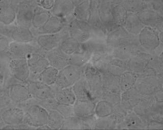

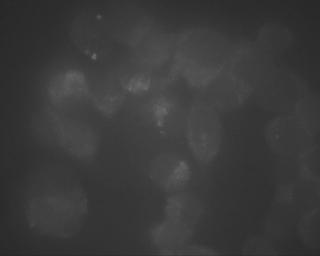
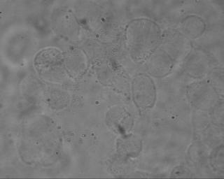
98 86 Figure 2.1 A. B. C. D. E. F.
99 87 I _ 40 kda
100 88 Fig. 2.2: Cell-to-cell dye transfer in Cx40-transfected HeLa cells. Cell-to-cell dye transfer mediated by untransfected (bar 1) or Cx40-transfected HeLa cells (bar 2). Alexa Fluor 594 (1 mm) was microinjected into one cell and 2 min later the number of cells receiving dye was determined. Pre-incubation with the gap junction blocker, carbonexolone (35 µm CBX) inhibited the Cx40-mediated cell-to-cell dye transfer (bar 3). * p 0.05 significantly different from Cx40 transfected HeLa cells.
101 89 Figure Number of cells receiving dye * * 0 WT HeLa HeLa Cx40 HeLa Cx40 35uM CBX
102 90 Fig. 2.3: Cell-to-cell transfer of Alexa Fluor 594 dye in Cx40-transfected HeLa cells. Alexa Fluor 594 (1 mm) was microinjected into one cell and the number of cells receiving dye was determined at various times after dye injection.
103 91 Figure Number of cells receiving dye control 5 min ATP 15 min ATP 30 min ATP
104 92 Fig. 2.4: Cx40 amino acid sequence with the putative transmembrane regions (TM1- TM4) and several putative consensus phosphorylation sequences for casein kinase II, Protein Kinase C and Protein Kinase A.
105 93 Figure 2.4 TM 1 mgdwsflgef leevhkhstv igkvwltvlf ifrmlvlgta aesswgdeqa dfrcdtiqpg TM 2 cqnvcydqaf pishirywvl qiifvstpsl vymghamhtv rmqekqklre aekakeaggt TM 3 gtyeylaeka elscwkevng kivlqgtlln tyvctilirt amevafmvgq yllygifldt TM 4 lhvcrrspcp hpvncyvsrp teknvfivfm mavaglslfl slaelyhlgw kkirqrlaks rqgdkhqllg pstslvqglt pppdfnqclk nspdekffsd fsnnmgsrkn pdplateevp nqeqipeegf ihtqygqkpe qpsgasaghr fpqgyhsdkr rlskasskar sddlsv CKII in pink, PKC in green, PKA in blue
106 94 Fig. 2.5: Schematic diagram of the Purinergic receptor pathway. ATP binds to the purinergic receptor and activates phospholipase C (PLC) which hydrolyzes phosphatidylinositol 4,5-bisphosphate (PIP 2 ) to inositol 1,4,5-triphosphate (IP 3 ) and diacylglycerol (DAG). DAG activates Protein Kinase C (PKC) which in turn phosphorylates various residues in connexins thus altering gap junction function, while IP 3 binds to ER receptors to effect the release of Ca 2+ from this intracellular organelle.
107 95 Figure 2.5 ATP Purinergic receptor PLC PIP 2 activates Membrane bilayer Gap junctions IP 3 DAG Ca 2+ PKC ER
108 96 Fig. 2.6: Effect of the purinergic receptor agonist ATP on Cx43 mediated cell-to-cell dye transfer. ATP (25 µm ATP ) was added to a monolayer Cx40-transfected HeLa cells when Alexa Fluor 594 (1mM) was microinjected into one cell prior to (Control) or at 5, 15 or 30 min after ATP addition; 2 min after each microinjection the number of cells receiving dye were counted. * p 0.05 significantly different from Control.
109 97 Figure Number of cells receiving dye * * 0 Control 5 min 15 min ATP ATP Time in min 30 min ATP
110 98 Fig. 2.7: Effect of the purinergic receptor agonist ATP on Cx40 mediated cell-to-cell dye transfer. Effect of (A) ATP or (B) TPA on Cx40 mediated cell-to-cell dye transfer in HeLa cells. A monolayer of Cx40-transfected HeLa cells was incubated with either ATP (25 µm) or TPA (81 nm). At various intervals following the addition or ATP or TPA, Alexa Fluor 594 was microinjected into one cell and 2 min later the number of cells receiving dye was counted.
111 99 Figure 2.7 A. B Number of cells receiving dye control 5 min ATP 15 min ATP 30 min ATP Number of cells receiving dye control 5 min TPA 30 min TPA 60 min TPA 90 min TPA
112 100 Fig. 2.8: Schematic diagram of the β-adrenergic receptor pathway. Beta-adrenergic receptor agonists bind to β -adrenergic receptors resulting in the activation of adenylate cyclase (AC) which catalyzes the conversion of ATP to camp. Subsequently, camp activates Protein Kinase A (PKA) which in turn catalyzes the phosphorylation of various residues in connexins that may or may not affect gap junction function.
113 101 Figure 2.8 Beta-adrenergic agonist Beta-adrenergic receptor AC Gap junctions Membrane bilayer activates ATP camp activates PKA
114 102 Fig. 2.9: Effect of camp analogs on Cx40 mediated cell-to-cell dye transfer. A monolayer of Cx40-transfected HeLa cells were either incubated with 0.5 mm dibutyryl camp or 1 mm 8-bromo camp. At various intervals following the addition of the camp analog, Alexa Fluor 594 was microinjected into one cell and 2 min later number of cells receiving dye was counted. * p 0.05 from control.
115 103 Figure 2.9 Number of cells receiving dye * * * * * 0.5 mm dibutyryl camp 1 mm 8-bromo-cAMP * 0 control 5 min 15 min 30 min
116 104 Fig. 2.10: Effect of forskolin on Cx40 mediated cell-to-cell dye transfer. (A) Monolayer of Cx40-transfected HeLa cells were incubated with forskolin (10 or 100 µm). (B) Cells were pre-incubated with KT-5720 (500 nm) or Bisindolylmaleimide I (35 µm) prior to the addition of forskolin. At various intervals following the addition of forskolin, Alexa Fluor 594 was microinjected into one cell and 2 min later the number of cells receiving dye was counted. * p 0.05 significantly different from cells receiving dye at zero time (i.e. prior to the addition of forskolin). ** p 0.05 significantly from cells receiving dye in the presence of forskolin.
117 105 Figure 2.10 A. B. Number of cells receiving dye µm forskolin 100 µm forskolin * * * * Number of cells receiving dye ** ** 100 µm FSK 100 µm Bis I µm FSK 500 nm KT µm FSK ** Time in min Time in min
118 106 Fig. 2.11: Effect of the α-adrenergic agonist metaraminol on Cx40 mediated cell-to-cell dye transfer. Cx40-transfected HeLa cells were incubated with metaraminol (100 µm) for various times. At various intervals following the addition of metaraminol, Alexa Fluor 594 was microinjected into one cell and 2 min later the number of cells receiving dye was counted.
119 107 Figure Number of cells receiving dye control Met 5 min Met 30 min Met 60 min
120 108 Fig. 2.12: Effect of the β-adrenergic agonist Isoproterenol on Cx40 mediated cell-to-cell dye transfer. Cx40-transfected HeLa cells were pre-incubated with the (A) PKA inhibitor KT-5720 (500 nm), or (B) phosphatase inhibitor Okadiac acid (250 nm) before addition of Isoproterenol (10 µm). At various intervals following the addition of Isoproterenol, Alexa Fluor 594 was microinjected into one cell and 2 min later the number of cells receiving dye was counted. * p 0.05 significantly different from cells receiving dye at zero time.
121 109 Figure 2.12 A. B. Number of cells receiving dye * * Control 10 µm ISO 500 nm KT µm ISO * Number of cells receiving dye * * 10 µm ISO 1 µm Okadaic acid + 10 µm ISO * Time in min Time in min
122 110 Fig. 2.13: Western blots of β-adrenergic receptor subtypes in untransfected or Cx40- transfected HeLa cells. (A) No bands at 55 or 68 kda were observed in either untransfected (lane 1) or Cx40-transfected HeLa cells (lanes 2) when incubated with β-1 adrenergic receptor antibody and goat anti-rabbit HRP conjugated secondary antibody. (B) 55 kda and 68 kda bands were observed in both untransfected (lane 1) and Cx40- transfected HeLa cells (lanes 2) when incubated with β-2 adrenergic receptor antibody and goat anti-rabbit HRP conjugated secondary antibody. (C) No bands at 55 or 68 kda were observed in either untransfected (lane 1) or Cx40-transfected HeLa cells (lanes 2) when incubated with β-3 adrenergic receptor antibody and donkey anti-goat HRP conjugated secondary antibody.


123 111 Figure 2.13 A. Beta-1 adrenergic receptor subtype B. Beta-2 adrenergic receptor subtype C. Beta-3 adrenergic receptor subtype
124 112 Fig. 2.14: Effect of the the β-1 adrenergic agonist Dobutamine on Cx40 mediated cell-tocell dye transfer. Cx40-transfected HeLa cells were incubated with the β-1 adrenergic receptor agonist Dobutamine (10 µm) for various times. At various intervals following the addition of Dobutamine, Alexa Fluor 594 was microinjected into one cell and 2 min later the number of cells receiving dye was counted.
125 113 Figure Number of cells receiving dye control DOB 5 min DOB 30 min DOB 60 min
126 114 Fig. 2.15: Effect of the β-2 adrenergic receptor agonist Salbutamol on Cx40 mediated cell-to-cell dye transfer. Cx40-transfected HeLa cells were incubated with the β-2 adrenergic receptor agonist Salbutamol (50 µm) for various times. At various intervals following the addition of Salbutamol, Alexa Fluor 594 was microinjected into one cell and 2 min later the number of cells receiving dye was counted. * p 0.05 significantly different from control (i.e. prior to the addition of Salbutamol).
127 115 Figure 2.15 Number of cells receiving dye * * * 0 control Sal 5 min Sal 30 min Sal 60 min
128 116 Fig. 2.16: Effect of the β-3 adrenergic agonist BRL on Cx40 mediated cell-to-cell dye transfer. Cx40-transfected HeLa cells were incubated with the β-3 adrenergic receptor agonist BRL (10 µm). At various intervals following the addition of BRL 37344, Alexa Fluor 594 was microinjected into one cell and 2 min later the number of cells receiving dye was counted.
129 117 Figure Number of cells receiving dye control BRL min BRL min BRL min
130 118 Fig. 2.17: Cx45 amino acid sequence with the putative transmembrane regions (TM1- TM4) and several concensus sequences for casein kinase II (highlighted in green) and Protein Kinase C are highlighted in pink while concensus sequences for both Protein Kinase C and casein kinase II are highlighted in yellow.
131 119 Figure 2.17 MSWSFLTRLL EEIHNHSTFV GKIWLTVLIV FRIVLTAVGG ESIYYDEQSK FVCNTEQPGC ENVCYDAFAP LSHVRFWVFQ IILVATPSVM YLGYAIHKIA KMEHGEADKK AARSKPYAMR WKQHRALEET EEDNEEDPMM YPEMELESDK ENKEQSQPKP KHDGRRRIRE DGLMKIYVLQ LLARTVFEVG FLIGQYFLYG FQVHPFYVCS RLPCPHKIDC FISRPTEKTI FLLIMYGVTG LCLLLNIWEM LHLGFGTIRD SLNSKRRELE DPGAYNYPFT WNTPSAPPGY NIAVKPDQIQ YTELSNAKIA YKQNKANTAQ EQQYGSHEEN LPADLEALQR EIRMAQERLD LAVQAYSHQN NPHGPREKKA KVGSKAGSNK STASSKSGDG KTSVWI PKC = pink CKII = green PKC and CKII = yellow
132 120 CHAPTER 3 Regulation of Connexin40 Gap junctions by Intracellular Calcium
133 121 Introduction Electrical and intercellular communication in the heart is mediated by gap junctions, non-specific plasma membrane channels that connect the cytoplasm of adjacent cells (Uehara and Burnstock, 1970). Alterations in gap junction function has been implicated in various diseases of the heart that are associated with altered electrical communication such as arrhythmias, myocardial infarction, atrial fibrillation, hypertension, valvular dysfunction, heart failure and hyperthyroidism (Cooklin et al, 1997; Kaprielian et al, 1998) indicating the importance of functional gap junctions for maintaining normal cardiac function. Gap junctions are comprised of proteins called connexins that form intercellular channels (Laird, 1996; Musil and Goodenough, 1993). Each connexin consists of four transmembrane domains with the amino (N-) and carboxy (C-) termini in the cytoplasm. Twenty highly related human connexin genes have been identified with their nomenclature based on their expected molecular weight in kilodaltons (kda). Gap junctions are known to be regulated by voltage, intracellular ph, phosphorylation state, intracellular calcium ([Ca 2+ ] i ) and more recently it is recognized that associated proteins may also play an important role in regulating gap junctions. The major connexins expressed in the atria of the heart include connexin 40 (Cx40) and connexin 43 (Cx43). Gap junctions are regulated by [Ca 2+ ] i, one of the most important intracellular signaling molecules (Burt, 1987a; Peracchia, 2004; Peracchia and Girsch, 1985). In the heart, maintaining Ca 2+ homeostasis (influx and efflux equilibrium) is important for excitation-contraction coupling of cardiac muscle (Zhang et al., 2001), activating various signaling pathways (El-Armouche et al., 2006), enzymes (Guo et al., 2006), genes
134 122 (Weissgerber et al., 2006), and regulating a number of important ion channels in both the plasma membrane and sacroplasmic reticulum (Kockskamper and Pieske, 2006). As in all other cell types, there is a 10 4 gradient of Ca 2+ across the plasma membrane, with extracellular [Ca 2+ ] ([Ca 2+ ] o ) in the range of ~ 1-2 mm while [Ca 2+ ] i is in the range of ~ nm. Most of the intracellular Ca 2+ is stored in the sacroplasmic reticulum (SR); release of this Ca 2+ into the cytoplasm during excitation-contraction coupling occurs via SR Ca 2+ release channels termed ryanodine receptors and to a smaller extent via IP 3 receptors (Berridge, 1993). During ischemia there is an increase in [Ca 2+ ] i in cardiomyocytes (Watts et al., 1980) which may be due to the influx of Ca 2+ from the extracellular space via the Na + /Ca 2+ exchanger [during ischemia there is increase in intracellular [H + ] and [Ca 2+ ] (Lazdunski et al., 1985)], so removal of [H + ] by the plasma membrane Na + / H + exchanger would lead to Na + influx which would in turn trigger Ca 2+ influx via the Na + /Ca 2+ exchanger). Increased [Ca 2+ ] i can also result from an influence of Ca 2+ mediated by plasma membrane store-operated Ca 2+ channels, or a decreased rate of removal of cytosolic Ca 2+ by the sarcoplasmic reticulum during ischemia (Chen et al., 1998; Moreau et al., 2005). This increased [Ca 2+ ] i is known to result in a decrease in gap junction-mediated electrical (gap junction conductance) and biochemical communication (gap junction permeability) in cardiomyocytes (Burt, 1987a; White et al., 1990). The major connexins present in cardiomyocytes are Cx40, Cx43 and Cx45. Cx43, the major connexin in the heart is known be to inhibited by elevated [Ca 2+ ] i (Crow et al., 1994). Previously our laboratory has demonstrated that Cx43-mediated cell-to-cell dye transfer is inhibited by elevated [Ca 2+ ] i with an IC 50 of approximately 350 nm in Cx43-
135 123 transfected HeLa cells. Although Cx40 is most abundantly expressed in the atria, its possible regulation by [Ca 2+ ] i is unknown. Therefore we have examined the effect of [Ca 2+ ] i on Cx40 gap junctions and the mechanisms underlying any action of [Ca 2+ ] i on Cx40 gap junction permeability. Because cardiomyocytes express at least three different connexins, namely Cx40, Cx43 and Cx45 they do not provide an optimal system for determining how the functional properties of individual cardiac connexins are regulated. Thus, to study the regulation of gap junctions comprised of Cx40 alone, studies were conducted using this connexin that was stably transfected in a communication-deficient HeLa cell line. This study, which is the first to examine the effect of [Ca 2+ ] i on Cx40 gap junction-mediated cell-to-cell dye transfer, reveals that a sustained elevation in [Ca 2+ ] i caused a partial inhibition of Cx40 gap junction-mediated cell-to-cell dye transfer with an IC 50 ~ 500 nm that appears to be Calmodulin-dependent.
136 124 Methods Materials Dulbecco s Modified Eagles Medium (DMEM), Hank s balanced salt solution without divalent cations (HBSS -- ) or with divalent cations (HBSS ++ ), ATP, CaCl 2, Calmidazolium, Ionomycin and Thapsigargin were purchased from Sigma-Aldrich Chemical Co. (St. Louis, MO). Fetal bovine serum (FBS) was purchased from Hyclone (Logan, UT). G418 (Geneticin) and Hygromycin B were obtained from Invitrogen (Carlsbad, CA). Fura-2 AM, and Alexa Fluor 594 were from Molecular Probes (Eugene, OR). Connexin40 and Donkey anti-goat IgG-HRP antibodies were from Santa Cruz Biotechnology, Inc., (Santa Cruz, CA). KT-5720 was from Tocris Bioscience (Ellisville, MI). Bisindolymaleimide I and Lavendustin C were from EMD Biosciences (San Diego, CA). HeLa cell culture Cx40 stably transfected HeLa cells generously provided by Dr. Richard Veenstra (SUNY Upstate Medical University, Syracuse, NY) were grown as a single monolayer on 25 mm coverslips in 35 mm culture dishes until confluent. Cells were grown in DMEM supplemented with 10% v/v fetal bovine serum and Pen-Strep (100 U/ml Penicillin and 0.1 mg/ml Streptomycin) with or without mg/ml (G418) geneticin, and were kept at 37ºC in a 5% CO 2 atmosphere maintained with nearly 100% relative humidity. Membrane isolation and Western Immunoblotting Membranes were isolated from untransfected of Cx40-transfected HeLa cells as described previously in Chapter 2.
137 125 Microinjection Cell-to-cell communication was detected by measuring the extent of cell-to-cell transfer of Alexa Fluor 594 dye as described previously in Chapter 2. Intracellular Ca 2+ measurements Cx40-transfected HeLa cells were loaded with Fura-2-AM (1 mm) in the dark for 45 min at room temperature. The buffer containing Fura-2 was then removed; the cells were rinsed with fresh HBSS ++ buffer, then incubated an additional 30 min in 2 ml HBSS ++ buffer to allow for complete hydrolysis of the intracellularly loaded Fura-2 AM. Intracellular Ca 2+ was measured ratiometrically (340 nm /380 nm) with Fura-2 throughout the experiment. Ca 2+ concentrations were determined as described previously by Grynkiewicz et al, The Ca 2+ concentration was calculated using the equation: [Ca 2+ ] = K d (R-R min ) (F min ) (R max -R)(F max ) Where: K d = µm (K d is the Ca 2+ dissociation constant of Fura-2) R = fluorescence intensity ratio F λ1 /F λ2, in which λ1 (340 nm) and λ2 (380 nm) are the fluorescence detection wavelengths for the ion-bound and ion-free indicator, respectively R min = fluorescence intensity ratio at 10 nm Ca 2+ ; R max = fluorescence intensity ratio at 39 µm Ca 2+ ; F min = fluorescence intensity λ nm Ca 2+ ; F max = fluorescence intensity λ 380 at 39 µm Ca 2+ ;
138 126 Raising intracellular Ca 2+ Intracellular Ca 2+ concentration ([Ca 2+ ] i ) was elevated in Cx40-transfected HeLa cells by adding ionomycin (2 µm) and then increasing extracellular Ca 2+ concentration ([Ca 2+ ] o ) from 1.8 to 21.8 mm by adding CaCl 2 to the bathing buffer (Hank s balanced salt solution with divalent cations (HBSS ++ ). Data analysis The data are expressed as Mean ± SEM. In cell-to-cell dye transfer assays, n reflects the number of times the experiments were performed. Differences between groups were analyzed by ANOVA. Student s paired t-test was used to calculate levels of significance, with p-value below 0.05 indicating significance. Statistical significance is indicated in the graphs with a * for p 0.05.
139 127 Results Expression of Cx40 in transfected HeLa cells To demonstrate the expression of Cx40 by Western Immunoblotting, membranes were isolated from Cx40-transfected HeLa cells, proteins were separated on SDS-PAGE gels and immunolabeled with Cx40 antibodies as described in Methods (Fig.3.1). A single component of 40 kda was observed only in Cx40-transfected HeLa cells as shown previously (Vozzi et al., 1999b) (Fig.3.1 lane 2 compared with lane 1), demonstrating the expression of this connexin in the Cx40-transfected HeLa cells used in this study. There was a non-specifically immunolabeled band at 52 kda in both untransfected wild type and Cx40-transfected HeLa cells. A sustained increase in intracellular Ca 2+ causes a decrease in Cx40-mediated cell-tocell dye transfer In the heart, a sustained increase in [Ca 2+ ] i occurs during myocardial ischemia where [Ca 2+ ] i increases from ~ 300 nm (resting) to ~ 900 nm (20 min ischemia) in perfused rat hearts (Marban et al., 1990). An ischemia-mediated increase in [Ca 2+ ] i is known to decrease gap junction-mediated electrical (gap junction conductance) and biochemical communication (gap junction permeability) in cardiomyocytes (Burt, 1987a; White et al., 1990). Cardiomyocytes express at least 3 different connexins, namely Cx40, Cx43 and Cx45. The question addressed here is whether inhibition of these connexins are responsible for this elevated [Ca 2+ ] i -mediated inhibition of gap junctions in cardiomyocytes during ischemia. Cx43, the major connexin in the heart is known be to inhibited by elevated [Ca 2+ ] i (Crow et al., 1994) but the possible regulation of the major connexin in the atria Cx40 by [Ca 2+ ] i is unknown, and was the focus of this study. To
140 128 determine whether like Cx43 cell-to-cell dye transfer mediated by Cx40 was inhibited by a sustained increase in [Ca 2+ ] i, Cx40-transfected HeLa cells were incubated with the Ca 2+ ionophore ionomycin and extracellular Ca 2+ was then increased from 1.8 mm (physiological [Ca 2+ ] o ) to 21.8 mm (this [Ca 2+ ] o was required to increase [Ca 2+ ] i to micromolar levels as occurs during ischemia in the heart). This high calcium gradient across the plasma membrane was likely required due to the presence of numerous plasma membrane Ca 2+ pumps which pump intracellular Ca 2+ out of cells (Negulescu and Machen, 1993). Under physiological conditions ([Ca 2+ ] o = 1.8 mm), [Ca 2+ ] i was ~ 0.1 µm and dye transferred to 9.6 ± 0.3 cells (n=13). When cells where incubated with the Ca 2+ ionophore ionomycin (2 µm) (Churchill et al., 2001) and [Ca 2+ ] o was increased to 21.8 mm, [Ca 2+ ] i increased to micromolar levels and dye transfer decreased to 4.0 ± 0.8 cells (n=10) (Fig.3.2). The number of cells receiving dye at different [Ca 2+ ] i is shown in Fig.3.2. Given the K d for Fura-2 of 224 nm (Grynkiewicz et al., 1985), intracellular Ca 2+ concentrations greater than approximately 1 µm cannot be estimated accurately in these experiments. Elevating [Ca 2+ ] i to greater than 2 µm resulted in cell shrinkage and death of the Cx40-transfected HeLa cells (data not shown). Thus, these data demonstrate a ~ 50% inhibition in Cx40-mediated cell-to-cell dye transfer when [Ca 2+ ] i 0.5 µm. A sustained elevation in [Ca 2+ ] i caused a partial inhibition of Cx40 gap junction-mediated cell-to-cell dye transfer with an IC 50 = 500 ± 0.72 nm. The [Ca 2+ ] i -dependent decrease in Cx40-mediated cell-to-cell dye transfer does not require the activation of Protein Kinase A
141 129 Elevating [Ca 2+ ] i levels to higher that 0.5 µm partially inhibited Cx40 gap junction-mediated cell-to-cell dye transfer (Fig.3.2). This [Ca 2+ ] i -mediated inhibition of Cx40 may be effected by at least three different mechanisms: 1) Ca 2+ activates a Ca 2+ - dependent Protein Kinase 2) direct binding of Ca 2+ to Cx40 protein or 3) via a Ca 2+ - binding protein such as Calmodulin. Analysis of the Cx40 sequence for putative phosphorylation sites as described in Methods (Chapter 2) using the Expasy Prosite-Scan database predicted substrate sites for Protein Kinase A and Protein Kinase C. Therefore, to determine whether the [Ca 2+ ] i - mediated decrease in Cx40 gap junction-mediated cell-to-cell dye transfer was dependent on the activation of Protein Kinase A, Cx40-transfected HeLa cells were pre-incubated with the PKA inhibitor, KT-5720 (0.5 µm) (RM Fried, 1986) before adding the Ca 2+ ionophore ionomycin, and [Ca 2+ ] i was then increased to micro molar levels by increasing [Ca 2+ ] o to 21.8 mm as described above. Alexa Fluor 594 dye transferred to 9.5 ± 0.6 cells (n=13) under physiological [Ca 2+ ] o (1.8 mm) when [Ca 2+ ] i was < 0.1 µm. In the presence of the PKA inhibitor alone dye transferred to 9.3 ± 0.9 cells (n=9), indicating that the PKA inhibitor alone did not alter [Ca 2+ ] i (0.16 ± µm) or the number of cells receiving dye from control levels [absence of PKA inhibitor]. When [Ca 2+ ] i was elevated to micromolar levels in the presence of the PKA inhibitor, dye transferred to 4.5 ± 0.4 cells (n=6) as shown in Fig.3.3 indicating that inhibition of Protein Kinase A had no effect on the [Ca 2+ ] i -dependent inhibition of Cx40-mediated cell-to-cell dye transfer. Thus, the Ca 2+ -dependent inhibition of Cx40 does not appear to require the activation of PKA.
142 130 The [Ca 2+ ] i -dependent decrease in Cx40-mediated cell-to-cell dye transfer does not require the activation of Protein Kinase C It has been demonstrated previously in our laboratory that purinergic receptor activation by ATP effects a Protein Kinase C-dependent inhibition of the major cardiac connexin Cx43. To determine whether, like Cx43, the [Ca 2+ ] i -dependent decrease in Cx40 gap junction-mediated cell-to-cell dye transfer was effected by activating the Ca 2+ - dependent enzyme Protein Kinase C (PKC), Cx40-transfected HeLa cells were preincubated with the PKC inhibitor bisindolylmaleimide I (2 µm) before the addition of the Ca 2+ ionophore ionomycin (Fried and Tashjian, 1986); [Ca 2+ ] i was then increased to micro molar levels by increasing [Ca 2+ ] o to 21.8 mm as described above (Fig 3.4). Alexa Fluor 594 dye transferred to 9.5 ± 0.2 cells (n=10) in physiological [Ca 2+ ] o (1.8 mm) when [Ca 2+ ] i was ~ 0.1 µm. In the presence of the PKC inhibitor bisindolylmaleimide I alone dye transferred to 8.9 ± 0.2 cells (n=23); [Ca 2+ ] i was unchanged from control levels in the presence of the PKC inhibitor. Thus, PKC inhibitor alone did not alter either [Ca 2+ ] i (0.21 ± µm) or the number of cells receiving dye. When [Ca 2+ ] i was elevated to micromolar levels in the absence of PKC inhibitor, dye transferred to 4.0 ± 0.2 cells (n=5) whereas in the presence of the PKC inhibitor dye transferred to 5.5 ± 0.6 cells (n=17) (Fig.3.4) indicating that inhibition of Protein Kinase C had minimal effect on the [Ca 2+ ] i -dependent inhibition of Cx40-mediated cell-to-cell dye transfer. Thus, the Ca 2+ -dependent inhibition of Cx40 does not appear to require the activation of PKC.
143 131 The [Ca 2+ ] i -dependent decrease in Cx40-mediated cell-to-cell dye transfer does not require the activation of Calmodulin Kinase II To determine whether the [Ca 2+ ] i -dependent inhibition of Cx40-mediated cell-tocell dye transfer was dependent on the activation of Calmodulin-dependent Protein Kinase II (CaMK II), experiments were performed in the presence or absence of an inhibitor of this Protein Kinase (Fig 3.5). Alexa Fluor 594 dye transferred to 9.9 ± 0.5 cells (n=12) in physiololgical [Ca 2+ ] o conditions in Cx40-transfected HeLa cells. When Cx40-transfected HeLa cells were incubated with the CaMK II inhibitor, lavendustin C (2 µm) (Watanabe et al., 1999) alone dye transferred to 9.0 ± 0.2 cells (n=31) and [Ca 2+ ] i was unchanged from control levels in the presence of this CaMK II inhibitor (Fig.3.5). This suggests that CaMK II inhibitor alone did not alter either [Ca 2+ ] i (0.18 ± µm) or the number of cells receiving dye when compared to control levels (in the absence of CaMK II inhibitor). When [Ca 2+ ] i was elevated to micromolar levels in the absence of CaMK II inhibitor dye transferred to 4.4 ± 0.8 cells (n=4) whereas in the presence of the CaMK II inhibitor dye transferred to 4.0 ± 1.0 cells (n=3) (Fig.3.5) indicating that inhibition of CaMK II had no effect on the [Ca 2+ ] i -dependent inhibition of Cx40- mediated cell-to-cell dye transfer. Thus, the Ca 2+ -dependent inhibition of Cx40 does not appear to require the activation of CaMK II. The [Ca 2+ ] i -dependent decrease in Cx40-mediated cell-to-cell dye transfer does not appear to be due to direct binding of Ca 2+ to Cx40 To determine whether the [Ca 2+ ] i -dependent inhibition of Cx40 gap junctionmediated cell-to-cell dye transfer was due to a direct binding of Ca 2+ to Cx40 protein, the
144 132 Cx40 protein sequence was scanned for EF hand Ca 2+ binding domains using the EF-hand prediction server (Zhou et al., 2006). No Ca 2+ binding domains were found in the Cx40 protein sequence suggesting that the [Ca 2+ ] i -dependent inhibition of Cx40 gap junctionmediated cell-to-cell dye transfer is unlikely due to a direct binding of Ca 2+ to Cx40 protein. Collectively these data suggest that the elevated [Ca 2+ ] i -dependent inhibition of Cx40 gap junctions was not due to the activation of Ca 2+ -activated proteins kinases or from direct binding of Ca 2+ to Cx40 protein. The [Ca 2+ ] i -dependent inhibition of Cx40-mediated cell-to-cell dye transfer appears to be Calmodulin-dependent When the Cx40 protein sequence was examined using the Calmodulin target database for identifying Calmodulin binding sites obtained from Ikura Lab, Ontario Cancer Institute (Zhou et al., 2006), one site was identified in the C-terminus just distal to the fourth transmembrane domain of Cx40 as shown in Fig.3.6. Scores range from 0-9 for protein sequences based on the probability for a Calmodulin binding site, 9 being the highest probability for the Calmodulin binding site. These normalized scores ranging from 0 to 9 are shown below the amino acid sequence indicating the highest affinity for Calmodulin within the given protein sequence. This scoring is based on a consecutive string of high values indicates the location of a putative binding site, the higher the score, the higher the predicted affinity for Calmodulin. Criteria for determining putative CaMbinding sites is based on hydropathy, alpha-helical propensity, residue weight, residue charge, hydrophobic residue content, helical class and occurrence of particular residues. Furthermore, a second scoring system also ranging from 0-9 is shown above the protein
145 133 sequence when they are scanned for Calmodulin binding affinity. This scoring system is based on the Calmodulin binding affinity of Cx40 to the Calmodulin binding affinity of other proteins in the data base. When the Cx40 protein sequence was examined using the Calmodulin target database a maximum score of 2 was obtained; although there was a probable site in Cx40 it may have a low predicted affinity for Calmodulin albeit the predictive scores do not always reflect the actual affinity of such sites for Calmodulin. Since a Calmodulin binding site was predicted in a region of Cx40 that the membrane spanning domain algorithms would indicate to be in a cytosolic region of Cx40, Calmodulin may bind directly to Cx40 thus altering the conformation of the connexin and closing the channel. To determine whether the [Ca 2+ ] i -dependent inhibition of Cx40 gap junctions was effected via the Ca 2+ -binding protein Calmodulin (CaM), Cx40-transfected HeLa cells were incubated with a Calmodulin inhibitor and [Ca 2+ ] i was elevated as described previously. Dye transferred to 9.0 ± 0.3 cells (n=12) in physiololgical [Ca 2+ ] o in Cx40- transfected HeLa cells. When Cx40-transfected HeLa cells were incubated with the CaM inhibitor Calmidazolium alone dye transferred to 9.0 ± 0.3 cells (n=19) and [Ca 2+ ] i was unchanged from control levels indicating that addition of this Calmodulin inhibitor alone did not alter either [Ca 2+ ] i (0.15 ± 0.04 µm) or the number of cells receiving dye when compared to control levels (in the absence of Calmodulin inhibitor). When [Ca 2+ ] i was elevated to micromolar levels in the absence of CaM inhibitor, dye transferred to 4.1 ± 0.3 cells (n=5) whereas in the presence of the CaM inhibitor, dye transferred to 9.0 ± 0.4 cells (n=4) indicating that the inhibition of Calmodulin prevented the [Ca 2+ ] i -dependent
146 134 inhibition of Cx40 gap junctions (Fig.3.7). Thus, the Ca 2+ -dependent inhibition of Cx40 appears to be Calmodulin-dependent. A transient increase in intracellular calcium did not affect Cx40-mediated cell-to-cell dye transfer A number of physiological agonists such as ATP (Niedergerke and Page, 1981), phenylephrine (α-1 adrenergic receptor activation) (Zhang et al., 2005), histamine (Hardwick et al., 2006), and acetylcholine (Weber et al., 2005) are also known to cause a transient increases in [Ca 2+ ] i concentration which returns to basal levels within tens of seconds, unlike the ionomycin-induced increase in [Ca 2+ ] i described here where the increase in [Ca 2+ ] i is sustained. The [Ca 2+ ] i increase during an agonist-mediated Ca 2+ release, and the ionomycin-induced increase in [Ca 2+ ] i are all in the micromolar range, the difference being that the agonist-mediated increase is transient whereas the ionomycininduced increase is sustained. The goal of these experiments was to determine whether an agonist-dependent transient elevation in [Ca 2+ ] i similar to the ionomycin-induced sustained increase in [Ca 2+ ] i, inhibited Cx40-mediated cell-to-cell dye transfer. This was accomplished by incubating Cx40-transfected HeLa cells with ATP (25 µm) as ATP has been shown to transiently increase [Ca 2+ ] i via purinergic receptor activation (Muscella et al., 2003) in HeLa cells. This same signaling system is also present in the heart, where purinergic receptor activation is coupled to an IP 3 -dependent increase in [Ca 2+ ] i (Kusakari et al., 2002; Scamps and Vassort, 1994). To confirm that extracellular ATP increases [Ca 2+ ] i in Cx40-transfected HeLa cells, these cells were incubated with ATP (25 µm) and changes in [Ca 2+ ] i was measured ratiometrically using Fura-2 as described in Methods. Addition of ATP to the media transiently increased [Ca 2+ ] i in Cx40-transfected HeLa
147 135 cells from ~ 0.1 µm to ~0.5 µm within 1 min of adding ATP and this declined to control levels by about 4 min after the addition of ATP as shown in Fig. 3.8A. To determine whether this ATP-dependent increase in [Ca 2+ ] i modulated Cx40- mediated cell-to-cell dye transfer, cells were incubated with ATP (25 µm) for various times prior to microinjection of dye. Alexa Fluor 594 dye transferred to 9.1 ± 0.5 cells (n=22) prior to addition of ATP, whereas following the addition of ATP (25 µm) during the period when there was a transient increase in [Ca 2+ ] I, dye transferred to 8.8 ± 0.4 cells at 1 min (n=5) ([Ca 2+ ] i maximum was ~ 0.5 µm), 8.6 ± 0.3 cells at 5 min (n=7) when [Ca 2+ ] i had returned back to basal levels, 8.6 ± 0.6 cells (n=7) at 15 min, and 9.1 ± 0.6 cells (n=7) at 30 min (Fig.3.8B). Thus, addition of extracellular ATP did not appear to affect Cx40-mediated cell-to-cell dye transfer suggesting that the ATP-dependent increase in [Ca 2+ ] i does not affect Cx40 gap junction permeability. This data demonstrates that an agonist-dependent, transient increase in [Ca 2+ ] i does not appear to affect cell-to-cell dye transfer in Cx40-transfected HeLa cells.
148 136 Discussion The present study demonstrates for the first time that a sustained elevation in [Ca 2+ ] i, such as occurs during myocardial ischemia, causes a partial inhibition of Cx40- mediated cell-to-cell dye transfer which appears to be Calmodulin-mediated. [Ca 2+ ] i is elevated by physiological agonists such as ATP, catecholamines, and also during pathophysiological conditions such as during arrhythmias. The absolute [Ca 2+ ] i increase following addition of agonists or during ischemia is similar, except that during ischemia there is a sustained increase in [Ca 2+ ] i whereas agonist-mediated increases in [Ca 2+ ] i are transient. [Ca 2+ ] i, one of the most important intracellular signaling molecules in the heart, is required for maintaining the contraction-relaxation function of the heart muscle (Zhang et al., 2001), activating various signaling pathways (El- Armouche et al., 2006), enzymes (Guo et al., 2006), gene expression (Weissgerber et al., 2006) and regulating a number of ion channels including gap junctions in the plasma membrane (Kockskamper and Pieske, 2006). In the heart, electrical and intercellular communication is mediated by gap junctions, and alterations in gap junction function have been implicated in various pathologic conditions in the heart (Cooklin et al., 1997; Kaprielian et al., 1998). Myocardial ischemia is known to cause arrhythmias, and during myocardial ischemia there is an elevation in [Ca 2+ ] i (Watts et al., 1980). This increase in intracellular [Ca 2+ ] is known to be associated with a decreased gap junction-mediated electrical (gap junction conductance) and biochemical communication (gap junction permeability) in cardiomyocytes (Burt, 1987a; White et al., 1990). In the atria, gap junctions are mainly comprised of Cx40 and Cx43 proteins. Thus, examining the regulation of gap junctions
149 137 comprised of individual connexins by elevated [Ca 2+ ] i would help us better understand the role of gap junctions in various pathological conditions such as arrhythmias that are associated with elevated [Ca 2+ ] i. Indeed, that the modulation of gap junction function can directly regulate electrical conduction in the heart makes gap junctions an ideal therapeutic target in the treatment of these pathologic conditions. In the atria, gap junctions are comprised mainly of Cx40 and Cx43 proteins. However, several studies have demonstrated that Cx40 and Cx43 do not form heterotypic gap junctions (Bruzzone et al., 1993; Elfgang et al., 1995; Haubrich et al., 1996), suggesting that gap junctions formed by these two connexins are regulated independently of each other in this region of the heart. It has been shown previously that elevation of [Ca 2+ ] i inhibits Cx43-mediated cell-to-cell dye transfer (Crow et al., 1994), while the role of [Ca 2+ ] i in regulating Cx40 gap junctions has not been examined previously. Therefore it was hypothesized that like Cx43, Cx40-mediated cell-to-cell dye transfer would be inhibited by increased [Ca 2+ ] i. This was accomplished by raising [Ca 2+ ] i by 2 different methods in these cells 1) a sustained elevation in [Ca 2+ ] i using the Ca 2+ ionophore ionomycin and then raising [Ca 2+ ] o and, 2) a transient increase in [Ca 2+ ] i using agonists such as ATP. During the myocardial ischemia that may cause arrhythmias, [Ca 2+ ] i has been shown to increase up to micromolar levels (Marban et al., 1990). A sustained elevation in [Ca 2+ ] i accomplished by addition of ionomycin increased [Ca 2+ ] i to micromolar levels when the [Ca 2+ ] o was elevated from 1.8 mm to 21.8 mm (Fig.3.2). This increase in [Ca 2+ ] i resulted in > 50% inhibition of Cx40-mediated cell-to-cell dye transfer which was prevented by prior addition of an inhibitor of the Ca 2+ binding protein Calmodulin.
150 138 Whether this action of Ca 2+ + Calmodulin results from the direct interaction of Calmodulin with Cx40 is not known, but it can be speculated that like other gap junction proteins [for example Calmodulin directly binds to Cx32 resulting in channel closure (Peracchia et al., 2000)], Calmodulin may directly bind to Cx40 protein. When the Cx40 protein sequence was examined using the Calmodulin target database obtained from Ikura Lab, Ontario Cancer Institute for identifying Calmodulin binding sites, there was one site identified in the C-terminus just outside the fourth transmembrane domain as shown in Fig.3.6. Since this shows that a Calmodulin binding site was found in the Cx40 C-terminus, we can speculate that Calmodulin may directly bind to Cx40 thus altering the conformation of the connexin and closing the channel. In the myocardium, Ca 2+ transients result when various agonists such as ATP, histamine, and acetylcholine are released during sympathetic nerve stimulation. The question is whether this transient increase in [Ca 2+ ] i alters electrical coupling-mediated by gap junctions in the heart. Among the three major connexins expressed in cardiomyocytes Cx43 [fully open single channel conductance of 220 ps (Contreras et al., 2003)] and Cx40 [fully open single channel conductance of 120 ps (Van-Rijen et al., 2000)] gap junctions have large conductances and are also expressed at much higher levels compared to Cx45 [fully open single channel conductance of 40 ps (van-veen et al., 2000)], thus these three major cardiac connexins play an important role in maintaining electrical conduction in the heart. However, the data reported here indicate that a transient elevation of [Ca 2+ ] i by ATP did not affect cell-to-cell dye transfer in Cx40-transfected HeLa cells, indicating that Cx40-mediated cell-to-cell communication is not affected by an agonist-dependent transient increase in [Ca 2+ ] i.
151 139 A sustained increase in [Ca 2+ ] i as occurs during myocardial ischemia, would likely result in the inhibition of Cx40-mediated cell-to-cell dye transfer, whereas a transient increase in [Ca 2+ ] i effected by ATP mediated purinergic receptor activation to ~ 0.5 µm did not result in an inhibition of Cx40-mediated cell-to-cell dye transfer. The possible explanation for this could be as described by Dakin et al (Dakin et al., 2005), when there is an influx of Ca 2+ from the extracellular space [Ca 2+ ] i is likely higher in close proximity to the gap junctions in the plasma membranes rather than in the cytoplasm. When Ca 2+ is released from intracellular stores, the cytosolic [Ca 2+ ] increases to 0.5 µm but the [Ca 2+ ] in the vicinity of gap junctions may be lower than that reported by Fura-2 for the average cytoplasmic [Ca 2+ ] because the distance when Ca 2+ enters cells from the extracellular space in the vicinity of gap junctions. Taken together with previous studies in our laboratory it is possible to conclude that under physiological conditions [Ca 2+ ] i transients induced by agonists in the heart would likely not result in a Ca 2+ -dependent inhibition of gap junction-mediated electrical conduction mediated by either Cx40 or Cx43. However, a sustained increase in [Ca 2+ ] i as occurs during myocardial ischemia might be expected to inhibit gap junction coupling mediated by either of these connexins. Such an inhibition may be both beneficial and detrimental to cardiomyocytes, this phenomenon is described by Andrade-Rozental et al (Andrade-Rozental et al., 2000) as kiss to life and kiss of death. Inhibiting gap junction-mediated biochemical communication in the heart during ischemia protects neighboring cardiomyocytes from the movement of toxic metabolites such as free radicals which causes cell injury and death described as kiss to life thus being beneficial. Reduction or inhibition of gap junction-mediated electrical coupling in the
152 140 heart during arrhythmias may slow or stop heart rate described as kiss of death thus being detrimental eventually leading to heart failure and death. Although various antiarrhythmic drugs such as β-blockers have been used with moderate success for the treatment of arrhythmias, most have significant side effects. Since arrhythmias result from an inhibition in gap junction-mediated electrical coupling in the heart leading to abnormal heart rhythms, gap junctions may be valuable therapeutic targets for the treatment of this condition. Our data reported here could suggest that this ischemiamediated inhibition of cardiac gap junction function may be due in part to the increase in [Ca 2+ ] i as occurs during myocardial ischemia (Burt, 1987b). Drugs that specifically open/enhance gap junction function may be of value in the treatment of arrhythmias, thus preventing the myocardial ischemia-mediated inhibition in gap junction-mediated electrical conduction in the heart.
153 141 Reference Andrade-Rozental, A., Rozental, R., Hopperstad, M., Wu, J., Vrionis, F. and Spray, D. (2000). Gap junctions: the "kiss of death" and the "kiss of life". Brain Res Brain Res Rev. 32, Berridge, M. (1993). Cell signalling. A tale of two messengers. Nature. 365, Bruzzone, R., Haefliger, J., Gimlich, R. and Paul, D. (1993). Connexin40, a component of gap junctions in vascular endothelium, is restricted in its ability to interact with other connexins. Mol Biol Cell. 4, Burt, J. (1987a). Block of intercellular communication: interaction of intracellular H+ and Ca2+. Am J Physiol. 253, C607-C612. Burt, J. (1987b). Block of intercellular communication: interaction of intracellular H+ and Ca2+. Am J Physiol. 253, C Chen, W., London, R., Murphy, E. and Steenbergen, C. (1998). Regulation of the Ca2+ gradient across the sarcoplasmic reticulum in perfused rabbit heart. A 19F nuclear magnetic resonance study. Circ Res. 83, Churchill, G., Lurtz, M. and Louis, C. (2001). Ca(2+) regulation of gap junctional coupling in lens epithelial cells. Am J Physiol Cell Physiol. 281, C Contreras, J., Saez, J., Bukauskas, F. and Bennett, M. (2003). Gating and regulation of connexin 43 (Cx43) hemichannels. Proc Natl Acad Sci U S A. 100, Cooklin, M., Wallis, W. R. J., Sheridan, D. J. and Fry, C. H. (1997). Changes in cell-to-cell electrical coupling associated with left ventricular hypertrophy. Circ Res. 80, Crow, J., Atkinson, M. and Johnson, R. (1994). Micromolar levels of intracellular calcium reduce gap junctional permeability in lens cultures. Invest Ophthalmol Vis Sci. 35, Dakin, K., Zhao, Y. and Li, W. (2005). LAMP, a new imaging assay of gap junctional communication unveils that Ca2+ influx inhibits cell coupling. Nat Methods. 2, El-Armouche, A., Bednorz, A., Pamminger, T., Ditz, D., Didie, M., Dobrev, D. and Eschenhagen, T. (2006). Role of calcineurin and protein phosphatase-2a in the regulation of phosphatase inhibitor-1 in cardiac myocytes. Biochem Biophys Res Commun. 346, Elfgang, C., Eckert, R., Lichtenberg-Frate, H., Butterweck, A., Traub, O., Klein, R., Hulser, D. and Willecke, K. (1995). Specific permeability and selective formation of gap junction channels in connexin-transfected HeLa cells. J Cell Biol. 129, Fried, R. and Tashjian, A. (1986). Unusual sensitivity of cytosolic free Ca2+ to changes in extracellular Ca2+ in rat C-cells. J Biol Chem. 261, Grynkiewicz, G., Poenie, M. and Tsien, R. (1985). A new generation of Ca2+ indicators with greatly improved fluorescence properties. J Biol Chem. 260, Guo, T., Mestril, T. Z. R. and Bers, D. (2006). Ca2+/Calmodulin-dependent protein kinase II phosphorylation of ryanodine receptor does affect calcium sparks in mouse ventricular myocytes. Circ Res. 99,
154 142 Hardwick, J., Kotarski, A. and Powers, M. (2006). Ionic mechanisms of histamine-induced responses in guinea pig intracardiac neurons. Am J Physiol Regul Integr Comp Physiol. 290, R241-R250. Haubrich, S., Schwarz, H., Bukauskas, F., Lichtenberg-Frate, H., Traub, O., Weingart, R. and Willecke, K. (1996). Incompatibility of connexin 40 and 43 Hemichannels in gap junctions between mammalian cells is determined by intracellular domains. Mol Biol Cell. 7, Kaprielian, R. R., Gunning, M., Dupont, E., Sheppard, M. N., Rothery, S. M., Underwood, R., Pennell, D. J., Fox, K., Pepper, J., Poole-Wilson, P. et al. (1998). Downregulation of immunodetectable connexin43 and decreased gap junction size in the pathogenesis of chronic hibernation in the human left ventricle. Circulation 97, Kockskamper, J. and Pieske, B. (2006). Phosphorylation of the cardiac ryanodine receptor by Ca2+/calmodulin-dependent protein kinase II: the dominating twin of protein kinase A? Circ Res. 99, Kusakari, Y., Hongo, K., Kawai, M., Konishi, M. and Kurihara, S. (2002). The mechanism of increasing Ca2+ responsiveness by alpha1-adrenoceptor stimulation in rat ventricular myocytes. Jpn J Physiol. 52, Laird, D. (1996). The life cycle of a connexin: gap junction formation, removal, and degradation. J Bioenerg Biomembr 28, Lazdunski, M., Frelin, C. and Vigne, P. (1985). The sodium/hydrogen exchange system in cardiac cells: its biochemical and pharmacological properties and its role in regulating internal concentrations of sodium and internal ph. J Mol Cell Cardiol. 17, Marban, E., Kitakaze, M., Koretsune, Y., Yue, D., Chacko, V. and Pike, M. (1990). Quantification of [Ca2+]i in perfused hearts. Critical evaluation of the 5F- BAPTA and nuclear magnetic resonance method as applied to the study of ischemia and reperfusion. Circ Res. 66, Moreau, B., Straube, S., Fisher, R., Putney, J. J. and Parekh, A. (2005). Ca2+-calmodulin-dependent facilitation and Ca2+ inactivation of Ca2+ release-activated Ca2+ channels. J Biol Chem. 280, Muscella, A., Elia, M., Greco, S., Storelli, C. and Marsigliante, S. (2003). Activation of P2Y2 purinoceptor inhibits the activity of the Na+/K+-ATPase in HeLa cells. Cell Signal. 15, Musil, L. S. and Goodenough, D. (1993). Multisubunit assembly of an integral plasma membrane channel protein, gap junction connexin43, occurs after exit from the ER. Cell 74, Negulescu, P. and Machen, T. (1993). Ca transport by plasma membrane and intracellular stores of gastric cells. Am J Physiol. 264, C Niedergerke, R. and Page, S. (1981). Two physiological agents that appear to facilitate calcium discharge from the sarcoplasmic reticulum in frog heart cells: adrenalin an ATP. Proc R Soc Lond B Biol Sci. 213, Peracchia, C. (2004). Chemical gating of gap junction channels; roles of calcium, ph and calmodulin. Biochim Biophys Acta. 1662, Peracchia, C. and Girsch, S. (1985). Functional modulation of cell coupling: evidence for a calmodulin-driven channel gate. Am J Physiol. 248, H
155 Peracchia, C., Sotkis, A., Wang, X. and Persechini, L. P. A. (2000). Calmodulin directly gates gap junction channels. J Biol Chem. 275, RM Fried, A. T. J. (1986). Unusual sensitivity of cytosolic free Ca2+ to changes in extracellular Ca2+ in rat C-cells. J Biol Chem. 261, Scamps, F. and Vassort, G. (1994). Pharmacological profile of the ATPmediated increase in L-type calcium current amplitude and activation of a non-specific cationic current in rat ventricular cells. Br J Pharmacol. 113, Uehara, Y. and Burnstock, G. (1970). Demonstration of "gap junctions" between smooth muscle cells. J Cell Biol. 44, Van-Rijen, H., Veen, T. v., Hermans, M. and Jongsma, H. (2000). Human connexin40 gap junction channels are modulated by camp. Cardiovasc Res. 45, van-veen, T., Rijen, H. v. and Jongsma, H. (2000). Electrical conductance of mouse connexin45 gap junction channels is modulated by phosphorylation. Cardiovasc Res. 46, Vozzi, C., Dupont, E., Coppen, S., Yeh, H. and Severs, N. (1999). Chamberrelated differences in connexin expression in the human heart. J Mol Cell Cardiol. 31, Watanabe, H., Takahashi, R., Tran, Q., Takeuchi, K., Kosuge, K., Satoh, H., Uehara, A., Terada, H., Hayashi, H., Ohno, R. et al. (1999). Increased cytosolic Ca(2+) concentration in endothelial cells by calmodulin antagonists. Biochem Biophys Res Commun. 265, Watts, J., Koch, C. and LaNoue, K. (1980). Effects of Ca2+ antagonism on energy metabolism: Ca2+ and heart function after ischemia. Am J Physiol. 238, H Weber, M., Motin, L., Gaul, S., Beker, F., Fink, R. and Adams, D. (2005). Intravenous anaesthetics inhibit nicotinic acetylcholine receptor-mediated currents and Ca2+ transients in rat intracardiac ganglion neurons. Br J Pharmacol. 144, Weissgerber, P., Held, B., Bloch, W., Kaestner, L., Chien, K., Fleischmann, B., Lipp, P., Flockerzi, V. and Freichel, M. (2006). Reduced cardiac L-type Ca2+ current in Ca(V)beta2-/- embryos impairs cardiac development and contraction with secondary defects in vascular maturation. Circ Res. 99, White, R., Doeller, J., Verselis, V. and Wittenberg, B. (1990). Gap junctional conductance between pairs of ventricular myocytes is modulated synergistically by H+ and Ca++. J Gen Physiol. 95, Zhang, S., Lin, J., Hirano, Y. and Hiraoka, M. (2005). Modulation of ICa-L by alpha1-adrenergic stimulation in rat ventricular myocytes. Canadian Journal of Physiology and Pharmacology 83. Zhang, X., Song, J., Rothblum, L., Lun, M., Wang, X., Ding, F., Dunn, J., Lytton, J., McDermott, P. and Cheung, J. (2001). Overexpression of Na+/Ca2+ exchanger alters contractility and SR Ca2+ content in adult rat myocytes. Am J Physiol Heart Circ Physiol. 281, H2079-H2088. Zhou, Y., Yang, W., Kirberger, M., Lee, H., Ayalasomayajula, G. and Yang, J. (2006). Prediction of EF-hand calcium-binding proteins and analysis of bacterial EFhand proteins. Proteins. 65,
156 Figures 144
157 145 Fig. 3.1: Expression of Cx40 in rat Cx40 transfected-hela cells. Western Immunoblot of Cx40. Lane 1 contains whole cell lysates from untransfected HeLa cells, and lane 2 contains whole cell lysates from Cx40-transfected HeLa cells (50 µg of protein per lane). A 40 kda band was seen in Cx40-transfected HeLa cells (lane 2) which was absent in untransfected HeLa cells (lane 1).

158 Figure
159 147 Fig. 3.2: Effect of a sustained increase in [Ca 2+ ] i on Cx40-mediated cell-to-cell dye transfer. Cx40-transfected HeLa cells were incubated with ionomycin (2 µm), [Ca 2+ ] o was increased from 1.8 mm to 21.8 mm and at various subsequent times Alexa Fluor 594 was microinjected into one cell and 2 min later the number of cells receiving dye was counted. Inhibition curve is shown in dashed line.
160 148 Figure Number of cells receiving dye [Ca 2+ ] i (µm) [Ca 2+ ] i (µm) Number of cells receiving dye (Mean ± SEM) ± ± ± ± ± ± n
161 149 Fig.3.3: Effect of Protein Kinase A inhibition on the Ca 2+ -mediated inhibition of Cx40- mediated cell-to-cell dye transfer. Cx40-transfected HeLa cells were pre-incubated with the PKA inhibitor KT-5720 (0.5 µm) for 15 min and then incubated with ionomycin (2 µm), [Ca 2+ ] o was increased from 1.8 mm to 21.8 mm and at various times subsequently Alexa Fluor 594 was microinjected into one cell and 2 min later the number of cells receiving dye was counted. * p 0.05 from control (1.8 mm [Ca 2+ ] o ).
162 150 Figure µm ionomycin mm [Ca 2+ ] o 500 nm KT µm ionomycin mm [Ca 2+ ] o Number of cells receiving dye * * * * * * 0 control [Ca 2+ ]i in µm [Ca 2+ ] i (µm) Number of cells receiving dye in the absence of the PKA inhibitor (Mean ± SEM) Number of experiments (n) Number of cells receiving dye in the presence of the PKA inhibitor (Mean ± SEM) Control 9.5 ± ± (1.8 mm [Ca 2+ ] o ) ± ± ± ± ± ± Number of experiments (n)
163 151 Fig. 3.4: Effect of Protein Kinase C inhibition on the Ca 2+ -dependent inhibition of Cx40- mediated cell-to-cell dye transfer. Cx40-transfected HeLa cells were pre-incubated with the PKC inhibitor, bisindolylmaleimide I (2 µm) for 15 min and then incubated with ionomycin (2 µm); [Ca 2+ ] o was increased from 1.8 mm to 21.8 mm. At various times subsequently Alexa Fluor 594 was microinjected into one cell and 2 min later the number of cells receiving dye was counted. * p 0.05 from control (1.8 mm [Ca 2+ ] o ).
164 152 Figure 3.4 Number of cells receiving dye * * 2 µm ionomycin mm [Ca 2+ ] o 100 nm bisindolylmaleimide I + 2 µm ionomycin mm [Ca 2+ ] o * * * * * * 0 control [ ] [ ] [ ] [ ] [Ca 2+ ] i µm [Ca 2+ ] i (µm) Number of cells receiving dye in the absence of the PKC inhibitor (Mean ± SEM) Number of experiments (n) Number of cells receiving dye in the presence of the PKC inhibitor (Mean ± SEM) Control 9.5 ± ± (1.8 mm [Ca 2+ ] o ) ± ± ± ± ± ± ± ± Number of experiments (n)
165 153 Fig.3.5: Effect of CaMK II inhibition on the Ca 2+ -dependent inhibition of Cx40-mediated cell-to-cell dye transfer. Cx40-transfected HeLa cells were pre-incubated with CaMK II inhibitor, Lavendustin C (2 µm) for 15 min and then incubated with 2 µm ionomycin, extracellular Ca 2+ was then raised from 1.8 mm to 21.8 mm. At various times following the elevation of [Ca 2+ ] o to 21.8 mm, Alexa Fluor 594 was microinjected into one cell and 2 min later the number of cells receiving dye was counted. * p 0.05 from control (1.8 mm [Ca 2+ ] o ).
166 154 Figure µm ionomycin mm [Ca 2+ ] o Number of cells receiving dye * * * * 2 µm Lavendustin C + 2 µm ionomycin mm [Ca 2+ ] o * * * * 0 control [ ] [ ] [ ] [ ] [Ca 2+ ] i µm [Ca 2+ ] i (µm) Number of cells receiving dye in the absence of the CaMK II inhibitor (Mean ± SEM) Number of experiments (n) Number of cells receiving dye in the presence of the CaMK II inhibitor (Mean ± SEM) Control 9.9 ± ± (1.8 mm [Ca 2+ ] o ) ± ± ± ± ± ± ± ± Number of experiments (n)
167 155 Fig.3.6: Cx40 amino acid sequence with the putative transmembrane regions (TM1- TM4 in gray) and the possible Calmodulin binding region (highlighted in pink) in the C- terminus when examined for Calmodulin binding sites using the Calmodulin target database obtained from Ikura Lab, Ontario Cancer Institute (Zhou et al., 2006).
168 156 Figure 3.6 Minimum score for sequence: 0 Maximum score: 2 TM1...1 MGDWSFLGEF LEEVHKHSTV IGKVWLTVLF IFRMLVLGTA AESSWGDEQA TM DFRCDTIQPG CQNVCYDQAF PISHIRYWVL QIIFVSTPSL VYMGHAMHTV RMQEKQKLRE AEKAKEAGGT GTYEYLAEKA ELSCWKEVNG KIVLQGTLLN TM TYVCTILIRT AMEVAFMVGQ YLLYGIFLDT LHVCRRSPCP HPVNCYVSRP TM TEKNVFIVFM MAVAGLSLFL SLAELYHLGW KKIRQRLAKS RQGDKHQLLG PSTSLVQGLT PPPDFNQCLK NSPDEKFFSD FSNNMGSRKN PDPLATEEVP NQEQIPEEGF IHTQYGQKPE QPSGASAGHR FPQGYHSDKR RLSKASSKAR SDDLSV
169 157 Fig. 3.7: Effect of a Calmodulin inhibitor on the Ca 2+ -dependent inhibition of Cx40- mediated cell-to-cell dye transfer. Cx40-transfected HeLa cells were pre-incubated with Calmodulin inhibitor, Calmidazolim (2 µm) for 15 min and then incubated with ionomycin (2 µm), [Ca 2+ ] o was then increased from 1.8 mm to 21.8 mm and at various times subsequently Alexa Fluor 594 was microinjected into one cell and 2 min later the number of cells receiving dye was counted. * p 0.05 from control (1.8 mm [Ca 2+ ] o ).
170 158 Figure µm ionomycin mm [Ca 2+ ] o 10 2 µm Calmidazolium + 2 µm ionomycin mm [Ca 2+ ] o Number of cells receiving dye * * * * 0 control [Ca 2+ ] i in µm [Ca 2+ ] i (µm) Number of cells receiving dye in the absence of the CaM inhibitor (Mean ± SEM) Number of experiments (n) Number of cells receiving dye in the presence of the CaM inhibitor (Mean ± SEM) Control 9.0 ± ± (1.8 mm [Ca 2+ ] o ) ± ± ± ± ± ± ± ± Number of experiments (n)
171 159 Fig. 3.8: Effect of a transient increase in [Ca 2+ ] i on Cx40-mediated cell-to-cell dye transfer. A. Effect of ATP on [Ca 2+ ] i in Cx40-transfected HeLa cells. B. Effect of ATPinduced increase in [Ca 2+ ] i on Cx40-mediated cell-to-cell dye transfer. Cx40-transfected HeLa cells were incubated with ATP (25 µm) for various times when Alexa Fluor 594 was microinjected into one cell and 2 min later the number of cells receiving dye was counted.
172 160 Figure 3.8 A µm ATP 0.5 [Ca 2+ ] i in µm B Time in min 12 Number of cells receiving dye control 1 min 5 min 15 min 30 min Time after the addition of ATP
173 161 CHAPTER 4 Regulation of Connexin40 Gap Junctions during Ischemia
174 162 Introduction Intercellular communication in the heart occurs through gap junctions, which mediate both electrical and biochemical cell-to-cell communication in cardiomyocytes. Gap junctions are composed of connexin proteins that form intercellular channels that are important determinants of action potential propagation in the heart. The major connexins expressed in cardiomyocytes are Cx40, Cx43, and Cx45 and alterations in gap junction function, expression and distribution have been implicated in various cardiac pathological conditions (Kirchhoff et al., 1998; Kleber et al., 1987; Simon et al., 1998) such as myocardial ischemia (Peters et al., 1993; Smith et al., 1991b), indicating the importance of functional gap junctions for maintaining normal cardiac function. Ischemia is associated with various pathological conditions including arrhythmia which leads to sudden cardiac death (Janse and Wit, 1989), as well as atrial and ventricular fibrillation in the heart. Several changes that occur in cardiomyocytes during myocardial ischemia include a reduction in [ATP] i and [Mg 2+ ] i (Kirkels et al., 1989), accumulation of various ions such as [H + ] i, [Ca 2+ ] i (Kihara et al., 1989), [Na + ] i (Malloy et al., 1990), [K + ] o (Wilde and Aksnes, 1995), accumulation of fatty acids (Vusse et al., 1994), catecholamines (Newton et al., 1997), and free radicals (Kukreja and Hess, 1992). One of the important mechanisms responsible for the myocardial ischemia-induced origin of arrhythmias is the uncoupling of gap junctions (Groot and Coronel, 2004). During ischemia there is a significant decrease in cellular ATP and oxygen concentrations that is known to be associated with inhibition in cardiac gap junction-mediated electrical conduction (Beardslee et al., 2000). Cardiomyocytes express at least three different connexins, namely Cx40, Cx43 and Cx45. Whether this ischemia-induced inhibition of gap junction-mediated electrical conduction
175 163 is due to the inhibition of all three connexins or one or more connexins is still not clear. Cx43, the major connexin in the heart is known to be inhibited in ischemia (Li and Nagy, 2000). Although Cx40 is most abundantly expressed in the atria, its permeability in ischemia is still not known. Therefore we have examined the effect of ischemia on Cx40 gap junctions and the mechanisms underlying any action of ischemia on Cx40 gap junction permeability. Because cardiomyocytes express at least three different connexins, namely Cx40, Cx43 and Cx45, they do not provide an optimal system for determining how the functional properties of individual cardiac connexins are regulated. Thus, to study the regulation of gap junctions comprised of Cx40 alone, studies were conducted using this connexin that was stably transfected in a communication-deficient HeLa cell line. In these cells ischemia was induced chemically by inhibiting the electron transport chain with sodium cyanide, and inhibiting glycolysis with iodoacetate and 2-deoxyglucose. This study, which is the first to examine the effect of chemical ischemia on Cx40 gap junction-mediated cell-to-cell dye transfer, revealed that there was a partial inhibition of Cx40 gap junction-mediated cell-to-cell dye transfer after 3 hours of chemical ischemia. This ischemia-mediated inhibition in Cx40-mediated cell-to-cell dye transfer appears to be Calmodulin-dependent.
176 164 Methods Materials Dulbecco s Modified Eagles Medium (DMEM), Hank s balanced salt solution with divalent cations (HBSS ++ ), ATP, and all other chemicals were purchased from Sigma Chemical Co. (St. Louis, MO). Fetal bovine serum (FBS) was purchased from Hyclone (Logan, UT). G418 (Geneticin) and Hygromycin B were obtained from Invitrogen (Carlsbad, CA). Fura-2 AM, and Alexa Fluor 594 were from Molecular Probes (Eugene, OR). Cell Titer-Glo Luminescent Cell Viability Assay kit was from Promega (Madison, WI). HeLa cell culture A single monolayer of untransfected or Cx40-transfected HeLa cells generously provided by Dr. Richard Veenstra were grown on 25 mm coverslips in 35 mm culture dishes until they reached confluency. Cells were grown in DMEM supplemented with 10% v/v fetal bovine serum and Pen-Strep (100 U/ml Penicillin and 0.1 mg/ml Streptomycin) with or without mg/ml (G418) geneticin, and were kept at 37ºC in a 5% CO 2 atmosphere maintained at nearly 100% relative humidity. Normoxia and Chemical ischemia Cx40-transfected HeLa cells were subjected to normoxia or chemical ischemia for various times at 37ºC (Rodriguez-Sinovas et al., 2006). Chemical ischemia was induced by inhibiting the electron transport chain with NaCN and inhibiting glycolysis with iodoacetate and 2-deoxyglucose. Cells were washed with normoxic buffer [40 NaCl, 3.6 KCl, 2 CaCl 2, 1.2 MgSO 4, 20 HEPES, and 5 glucose(in mm), ph 7.4] and then subjected
177 165 to chemical ischemia in the same buffer lacking glucose and containing NaCN (2 mm) and 2-deoxyglucose (20 mm). Intracellular Ca 2+ measurements Cx40-transfected HeLa cells were loaded with Fura-2-AM (1 mm) in the dark for 45 min at room temperature. The buffer containing Fura-2 was then removed; the cells were rinsed with fresh HBSS ++ buffer, then incubated an additional 30 min in 2 ml HBSS ++ buffer to allow for complete hydrolysis of the intracellularly loaded Fura-2 AM. Intracellular Ca 2+ was measured ratiometrically (340 nm /380 nm) with Fura-2 throughout the experiment. Intracellular Ca 2+ concentrations were determined as described previously by Grynkiewicz et al, The Ca 2+ concentration was calculated using the equation: [Ca 2+ ] = K d (R-R min ) (R max -R)(F max ) Where: K d = µm [K d is the Ca 2+ dissociation constant of the indicator (Grynkiewicz et al., 1985)] R = fluorescence intensity ratio F λ1 /F λ2, in which λ1 (340 nm) and λ2 (380 nm) are the fluorescence detection wavelengths for the ion-bound and ion-free indicator, respectively R min = fluorescence intensity ratio at 10 nm Ca 2+ ; R max = fluorescence intensity ratio at 39 µm Ca 2+ ; F min = fluorescence intensity λ 380 at 10 nm Ca 2+ ; F max = fluorescence intensity λ 380 at 39 µm Ca 2+ ;
178 166 ATP assay The amount of ATP in Cx40-transfected HeLa cells treated with normoxic or hypoxic buffer was measured using the CellTiter-Glo luminescent cell viability assay (Promega, Madison, Wis., USA). This assay quantifies the amount of ATP and is based on the luciferase-catalyzed reaction of luciferin and ATP. In particular, monooxygenation of luciferin is catalyzed by luciferase in the presence of Mg 2+, ATP, and molecular oxygen. Cx40-transfected HeLa cells were plated at the appropriate density (1.25 x 10 4 cells/well) in an opaque-walled 96-well plate. Twenty-four hours after plating, cells were exposed to either normoxic or hypoxic buffer for various times. CellTiter-Glo Reagent was then added to each well and the contents were mixed for 2 minutes on an orbital shaker to induce cell lysis. The plates were then allowed to incubate at room temperature for 10 minutes to stabilize luminescent signal. The Luminescent signal was recorded for 1 second per well using a BMG-Lumistar luminometer. At least four independent experiments were performed for each condition. Data analysis The data are expressed as Mean ± SEM. In cell-to-cell dye transfer assays, n reflects the number of times the experiments were performed. Differences between groups were analyzed by ANOVA. Student s paired t-test was used to calculate levels of significance, with p-value less than 0.05 indicating significance. Statistical significance is indicated in the graphs with a * for p 0.05.
179 167 Results Chemical ischemia causes a decrease in [ATP] i in Cx40-transfected HeLa cells One of the several changes that occur in cardiomyocytes during myocardial ischemia is a reduction in intracellularatp concentration ([ATP] i ) (Kirkels et al., 1989). To determine whether chemical ischemia indeed effected a decrease in [ATP] i, changes in [ATP] i were measured at various times during cell cultures maintained in normoxic conditions, or following chemical ischemia. Cx40-transfected HeLa cells were exposed to either maintained in normoxic or chemical ischemic buffer for various times, and total ATP concentration in the HeLa cells was determined using the CellTiter-Glo luminescent cell viability assay kit (Promega, Madison, Wis., USA) as described in Methods. ATP concentration was ~ 2 µm/ 10 4 cells (n=4) prior to the onset of normoxia and ~ 1.8 µm/ 10 4 cells (n=4) 15 min following normoxia. In contrast [ATP] was ~ 2 µm/ 10 4 cells (n=4) prior to the onset of chemical ischemia and decreased to less than ~ 0.4 µm/ 10 4 cells (n=4) within 15 min of chemical ischemia (Fig. 4.1). Thus, these data indicate that chemical ischemia resulted in a rapid decrease in ATP concentration within 15 minutes in Cx40-transfected HeLa cells confirming that chemical ischemia induced an inhibition of glycolysis and the electron transport chain in these cells, and thus caused a decrease in [ATP] i. Chemical ischemia causes a decrease in Cx40-mediated cell-to-cell dye transfer Ischemia is known to decrease gap junction-mediated electrical (gap junction conductance) and biochemical communication (gap junction permeability) in cardiomyocytes (Burt, 1987a; White et al., 1990). Cardiomyocytes express at least 3 different connexins, namely Cx40, Cx43 and Cx45. To determine whether like Cx43,
180 168 Cx40-mediated cell-to-cell dye transfer was inhibited by ischemia (Li and Nagy, 2000), Cx40-transfected HeLa cells were incubated in either normoxic or ischemic buffer at 20ºC for various times. Dye transferred initially to 8.2 ± 0.5 cells (n=7) and to 8.8 ± 0.5 cells (n=5) 3 hours in normoxic media at 20ºC. Whereas dye transferred to 8.0 ± 0.4 cells (n=5) prior to the onset of chemical ischemia, it transferred to 8.8 ± 0.6 cells (n=9) 3 hours following chemical ischemia at 20ºC (Fig. 4.5). Thus, this data suggests that chemical ischemia induced at 20ºC did not affect Cx40-mediated cell-to-cell dye transfer for up to 3 hours; therefore experiments were performed at 37 o C as this more accurately reflects the physiologic state of cells in the heart. When cells were maintained at 37 o C dye initially transferred to 8.4 ± 0.3 cells (n=9) and to 8.8 ± 0.9 cells (n=6) 3 hours in normoxic media at 37 o C. In contrast, whereas dye transferred to 8.0 ± 0.7 cells (n=5) prior to the onset of chemical ischemia, it decreased to 5.3 ± 1.5 cells (n=8) 3 hours following the onset of chemical ischemia at 37 o C (Fig.4.2). Thus, these data demonstrates that chemical ischemia 3 hours caused ~ 40% inhibition in Cx40- mediated cell-to-cell dye transfer at 37 o C. Changes in [Ca 2+ ] i following the onset of chemical ischemia in Cx40-transfected HeLa cells In the heart, a sustained increase in [Ca 2+ ] i occurs during myocardial ischemia where [Ca 2+ ] i increases from ~ 300 nm (resting) to ~ 900 nm (20 min ischemia) in perfused rat hearts (Marban et al., 1990). An ischemia-dependent increase in [Ca 2+ ] i is known to decrease gap junction-mediated electrical (gap junction conductance) and biochemical communication (gap junction permeability) in cardiomyocytes (Burt, 1987a; White et al., 1990). The question addressed here is whether inhibition of Cx40-mediated
181 169 cell-to-cell dye transfer (Fig.4.2) correlated with ischemia-mediated changes in [Ca 2+ ] i. To determine whether [Ca 2+ ] i changed in HeLa cells following the onset of chemical ischemia, Cx40-transfected HeLa cells were loaded with Fura-2 and then exposed to either normoxic or hypoxic buffer for various times at 37ºC. It was discovered that changes in [Ca 2+ ] i could not be measured during chemical ischemia at 37 o C due to the loss of Fura-2 fluorescence at this elevated temperature (37ºC) as indicated by the decrease in absolute fluorescence signals measured at either 340 nm or 380 nm (Fig. 4.3B). Therefore it could be speculated that at 37ºC, the loss of Fura-2 fluorescence may be due to its release from the cells or that this fluorescent dye is hydrolyzed to a nonfluorescent product. To determine whether inducing chemical ischemia at 20ºC affected Fura-2 fluorescence, Cx40-transfected HeLa cells were incubated with the Ca 2+ -sensitive dye Fura-2 AM and then exposed to either normoxic or ischemic buffer for various times at 20ºC. In contrast to measurements at 37 o C, this absolute fura-2 fluorescence signal did not significantly decrease at 20 o C (Fig. 4.3A). Thus, ischemia-mediated changes in [Ca 2+ ] i were measured using Fura-2 at 20 o C as this temperature was used in all previous experiments. To determine whether [Ca 2+ ] i in HeLa cells was modified following chemical ischemia at 20ºC, Cx40-transfected HeLa cells were incubated with the Ca 2+ - sensitive dye Fura-2 AM and then exposed to either normoxic or hypoxic buffer for various times at 20ºC. [Ca 2+ ] i was initially 0.03 ± µm (n=10), and 0.07 ± µm (n=12) at 15 min, 0.10 ± µm (n=11) at 30 min, 0.10 ± µm (n=10) at 45 min, 0.13 ± µm (n=10) at 1 hour, 0.12 ± µm (n=10) 2 hours and 0.11 ± µm (n=7) 3 hours incubation in normoxic buffer at 20ºC. Thus, there was a
182 170 modest increase in [Ca 2+ ] i following 3 hours incubation at 20ºC. In contrast, [Ca 2+ ] i was 0.03 ± µm (n=12) prior to the onset of chemical ischemia, 0.05 ± µm (n=15) at 15 min, 0.05 ± µm (n=11) at 30 min, 0.12 ± µm (n=10) at 45 min, 0.26 ± 0.01 µm (n=10) at 1 hour, 2.0 ± 0.17 µm (n=11) 2 hours and to 1.9 ± 0.08 µm (n=15) 3 hour following onset of chemical ischemia (Fig. 4.4), indicating that chemical ischemia induced a significant increase in [Ca 2+ ] i at 20ºC. The chemical ischemia-dependent inhibition of Cx40-mediated cell-to-cell dye transfer appears to be Calmodulin-dependent Although changes in [Ca 2+ ] i could not be measured using Fura-2 at 37 o C, based on the data at 20 o C there likely was an increase in [Ca 2+ ] i in these HeLa cells at 37 o C which could have been responsible for the ischemia-mediated decrease in Cx40-mediated cell-to-cell dye transfer. It has been shown in the previous chapter that the Ca 2+ - dependent inhibition of Cx40-mediated cell-to-cell dye transfer is Calmodulin-dependent. Therefore, to determine whether the ischemia-mediated decrease in Cx40-mediated cellto-cell dye transfer was Calmodulin-dependent, Cx40-transfected HeLa cells were preincubated with the Calmodulin inhibitor, calmidazolium prior to incubation in either normoxic or chemical ischemic media at 37 o C. Dye transferred initially to 9.6 ± 0.3 cells (n=3), and to 8.6 ± 0.3 cells (n=3) after 3 hours following incubation in normoxic media (Fig. 4.6A) in agreement with the data in Fig In contrast, whereas dye transferred to 9.5 ± 0.6 cells (n=4) prior to the onset of chemical ischemia (Fig. 4.2), it decreased to 4.8 ± 0.3 cells (n=4) 3 hours following chemical ischemia in the absence of calmidazolium (Fig. 4.6B) in agreement with the data in Fig In the presence of the Calmodulin inhibitor calmidazolium, dye transferred to 9.3 ± 0.7 cells (n=3) and to 8.5 ± 0.5 cells
183 171 (n=2) 3 hours following incubation in normoxic buffer plus calmidazolium (Fig. 4.6A), indicating no nonspecific action of this inhibitor on gap junctions in normoxic media. In contrast, whereas dye transferred to 8.6 ± 0.7 cells (n=3) prior to the onset of chemical ischemia, it transferred to 9.0± 0.4 cells (n=5) 3 hours following chemical ischemia in the presence of the Calmodulin inhibitor calmidazolium (Fig. 4.6B). Thus, pre-treating cells with the Calmodulin inhibitor calmidazolium prevented the ischemia-mediated decrease in Cx40-mediated cell-to-cell dye transfer indicating that the ischemia-dependent decrease in Cx40-mediated cell-to-cell dye transfer at 37 o C appears to be Calmodulindependent.
184 172 Discussion The present study demonstrates for the first time that chemical ischemia causes a partial inhibition of cell-to-cell dye transfer in Cx40-transfected HeLa cells that appears to be Calmodulin-dependent. During ischemia there is a significant decrease in intracellular ATP concentration that is known to be associated with the inhibition of cardiac gap junction-mediated electrical conduction (Beardslee et al., 2000). To confirm that similar changes in intracellular ATP levels occurred in HeLa cells, ATP was measured, and was shown to be significantly decreased within 15 minutes of the onset of ischemia (Fig. 4.1), indicating that the chemical ischemia described here was indeed induced by iodoacetate, cyanide and 2-deoxyglucose treatment of HeLa cells. In the heart, a sustained increase in [Ca 2+ ] i occurs during myocardial ischemia where [Ca 2+ ] i increases from ~ 300 nm (resting) to ~ 900 nm (20 min ischemia) in perfused rat hearts (Marban et al., 1990). Burt et al (1987) have demonstrated that ischemia caused an increase in [Ca 2+ ] I (Burt, 1987a; White et al., 1990), there also observed an ischemia-dependent decrease in gap junction-mediated electrical (gap junction conductance) and biochemical communication (gap junction permeability) in cardiomyocytes, whether this increase in [Ca 2+ ] i was responsible for the ischemiamediated decrease in gap junction is not known. Since there was an increase in [Ca 2+ ] i during ischemia in cardiomyocytes, an increase in [Ca 2+ ] i was expected in Cx40- transfected HeLa cells. However, the chemical ischemia-mediated changes in [Ca 2+ ] i could not be measured using Fura-2 (Fig. 4.3B) at 37 o C due to the loss of Fura-2 fluorescence at this temperature due either to the Fura-2 being released by HeLa cells, or
185 173 hydrolyzed by the cells thus making it impossible to measure [Ca 2+ ] i using Fura-2 at this temperature. When chemical ischemia-mediated changes in [Ca 2+ ] i were determined with Fura- 2 at 20ºC, an increase in [Ca 2+ ] i was observed, confirming many other studies (Churchill et al., 1996; Yashiro and Duling, 2000) that the fluorescence intensity of Fura-2 was not affected at this temperature (Fig.4.3A). To determine if this ischemia-mediated change in [Ca 2+ ] i correlated with the inhibition of Cx40-mediated cell-to-cell dye transfer, dye transfer was measured during normoxia or ischemia for various times at 20ºC. Inducing chemical ischemia at 20ºC did not affect Cx40-mediated cell-to-cell dye transfer even by 2 or 3 hours by which time [Ca 2+ ] i had increased (Fig. 4.5). Thus, although chemical ischemia effected a significant increase in [Ca 2+ ] i as early as 2 hours (Fig. 4.4), this change in [Ca 2+ ] i did not correlate with decrease in Cx40-mediated cell-to-cell dye transfer. Rodriguez-Sinovas et al. (2006) have demonstrated an increase in [Ca 2+ ] i in rat cardiomyocytes using the same ischemia protocol as used in this study (Rodriguez- Sinovas et al., 2006), so an increase in [Ca 2+ ] i was expected in Cx40-transfected HeLa cells. Although changes in [Ca 2+ ] i could not be measured using Fura-2 at 37 o C, there was an ischemia-dependent increase in [Ca 2+ ] i observed at 20ºC that did not affect Cx40- mediated cell-to-cell dye transfer. It could be speculated that if there is an increase in [Ca 2+ ] i at 37 o C which may be in part responsible for the chemical ischemia-dependent decrease in Cx40-mediated cellto-cell dye transfer at this temperature, it may be Calmodulin-dependent as shown in Chapter 3. To determine if the chemical ischemia dependent decrease in Cx40-mediated
186 174 cell-to-cell dye transfer was Calmodulin-dependent, cells were pre-incubated with the Calmodulin inhibitor calmidazolium and cell-to-cell dye transfer was subsequently measured during normoxia or ischemia for various times at 37 o C. The data reported here demonstrated that calmidazolium prevented the chemical ischemia-dependent decrease in Cx40-mediated cell-to-cell dye transfer at 37 o C (Fig. 4.6B), indicating that the ischemiamediated decrease in Cx40-mediated cell-to-cell dye transfer at 37 o C appears to be Calmodulin and therefore likely Ca 2+ -dependent. A possible explanation for this slow onset of gap junction closure at 37 o C may be that there are confounding changes in intracellular ph ([ph] i ) or changes in Cx40 phosphorylation state. Although changes in [ph] i were not measured during chemical ischemia in this study, it could be speculated that there may be a decrease in [ph] i during chemical ischemia. An increase in [H + ] i as occurs during chemical ischemia may decrease the Ca 2+ sensitivity of proteins due to H + ions binding to the Ca 2+ binding sites thus decreasing their affinity for Ca 2+ ions. Thus an increase in [Ca 2+ ] i may not now affect Cx40 gap junctions because of its inability to bind to its receptor that our data would indicate is likely Calmodulin. In Cx40-transfected HeLa cells [Ca 2+ ] i increased within 1 min after ATP addition as demonstrated in chapter 2 and within 15 min of chemical ischemia at 20ºC, yet neither of these increases in [Ca 2+ ] i were associated with a decrease in Cx40-mediated cell-to-cell dye transfer. That both ATP and chemical ischemia-mediated increase in [Ca 2+ ] I at 20ºC did not correlate with a decrease in Cx40-mediated cell-to-cell dye transfer may be due the protocol/method used to raise [Ca 2+ ] i. As described by Dakin et al (2005) and discussed in Chapter 3 (Dakin et al., 2005), a sustained elevation in [Ca 2+ ] i using
187 175 ionomycin increased [Ca 2+ ] i to micromolar levels when the [Ca 2+ ] o was elevated from 1.8 mm to 21.8 mm. This increase in [Ca 2+ ] i resulted in > 50% inhibition of Cx40-mediated cell-to-cell dye transfer, whereas a transient increase in [Ca 2+ ] i using agonists such as ATP also increased [Ca 2+ ] i to micromolar levels but did not affect Cx40-mediated cell-tocell dye transfer. The possible explanation for this is described by the same group (Dakin et al., 2005), where [Ca 2+ ] is likely higher in the close proximity of gap junctions than in the cytoplasm following an influx of Ca 2+ from the extracellular space. When Ca 2+ is released from intracellular stores, the cytosolic [Ca 2+ ] increases to 0.5 µm but the [Ca 2+ ] in the vicinity of gap junctions may be lower than that reported by Fura-2 for the average cytoplasmic [Ca 2+ ] because the distance traveled by Ca 2+ from the intracellular stores to the plasma membrane is greater than the distance Ca 2+ traveled from the extracellular space to the vicinity of gap junctions. Acute cardiac ischemia is known to be associated with ventricular arrhythmia and fibrillation. One of the major factors responsible for the impaired action potential propagation during arrhythmias and fibrillation is uncoupling of gap junctions (Dhein, 2006). This gap junction uncoupling is known to lead to predominant transverse (myocardial end-to-end) uncoupling. Complete gap junction uncoupling would initiate arrhythmia (Kojodjojo et al., 2006), while intermediate uncoupling has been shown to enhance the safety factor of propagation, limiting the current loss to non-depolarized areas. Therefore, as demonstrated in this study, an ischemia-dependent decrease in Cx40- mediated cell-to-cell dye transfer would enhance the safety factor of propagation therefore preventing the complete block of electrical conduction to the infarct areas of the heart.
188 176 Reference Beardslee, M., Lerner, D., Tadros, P., Laing, J., Beyer, E., Yamada, K., Kleber, A., Schuessler, R. and Saffitz, J. (2000). Dephosphorylation and intracellular redistribution of ventricular connexin43 during electrical uncoupling induced by ischemia. Circ Res. 87, Burt, J. (1987). Block of intercellular communication: interaction of intracellular H+ and Ca2+. Am J Physiol. 253, C607-C612. Churchill, G., Atkinson, M. and Louis, C. (1996). Mechanical stimulation initiates cell-to-cell calcium signaling in ovine lens epithelial cells. J Cell Sci. 109, Dakin, K., Zhao, Y. and Li, W. (2005). LAMP, a new imaging assay of gap junctional communication unveils that Ca2+ influx inhibits cell coupling. Nat Methods. 2, Dhein, S. (2006). Cardiac ischemia and uncoupling: gap junctions in ischemia and infarction. Adv Cardiol. 42, Groot, J. D. and Coronel, R. (2004). Acute ischemia-induced gap junctional uncoupling and arrhythmogenesis. Cardiovasc Res. 62, Grynkiewicz, G., Poenie, M. and Tsien, R. (1985). A new generation of Ca2+ indicators with greatly improved fluorescence properties. J Biol Chem. 260, Janse, M. and Wit, A. (1989). Electrophysiological mechanisms of ventricular arrhythmias resulting from myocardial ischemia and infarction. Physiol Rev. 69, Kihara, Y., Grossman, W. and Morgan, J. (1989). Direct measurement of changes in intracellular calcium transients during hypoxia, ischemia, and reperfusion of the intact mammalian heart. Circ Res. 65, Kirchhoff, S., Nelles, E., Hagendorff, A., Kruger, O., Traub, O. and Willecke, K. (1998). Reduced cardiac conduction velocity and predisposition to arrhythmias in connexin40-deficient mice. Curr Biol. 8, Kirkels, J., Echteld, C. v. and Ruigrok, T. (1989). Intracellular magnesium during myocardial ischemia and reperfusion: possible consequences for postischemic recovery. J Mol Cell Cardiol. 21, Kleber, A., Riegger, C. and Janse, M. (1987). Electrical uncoupling and increase of extracellular resistance after induction of ischemia in isolated, arterially perfused rabbit papillary muscle. Circ Res. 61, Kojodjojo, P., Kanagaratnam, P., Segal, O., Hussain, W. and Peters, N. (2006). The effects of carbenoxolone on human myocardial conduction: a tool to investigate the role of gap junctional uncoupling in human arrhythmogenesis. J Am Coll Cardiol. 48, Kukreja, R. and Hess, M. (1992). The oxygen free radical system: from equations through membrane-protein interactions to cardiovascular injury and protection. Cardiovasc Res. 26, Li, W. and Nagy, J. (2000). Connexin43 phosphorylation state and intercellular communication in cultured astrocytes following hypoxia and protein phosphatase inhibition. Eur J Neurosci. 12,
189 Malloy, C., Buster, D., Castro, M., Geraldes, C., Jeffrey, F. and Sherry, A. (1990). Influence of global ischemia on intracellular sodium in the perfused rat heart. Magn Reson Med. 15, Marban, E., Kitakaze, M., Koretsune, Y., Yue, D., Chacko, V. and Pike, M. (1990). Quantification of [Ca2+]i in perfused hearts. Critical evaluation of the 5F- BAPTA and nuclear magnetic resonance method as applied to the study of ischemia and reperfusion. Circ Res. 66, Newton, G., Adelman, A., Lima, V., Seidelin, P., Schampaert, E. and Parker, J. (1997). Cardiac sympathetic activity in response to acute myocardial ischemia. Am J Physiol. 272, H Peters, N., Green, C., Poole-Wilson, P. and Severs, N. (1993). Reduced content of connexin43 gap junctions in ventricular myocardium from hypertrophied and ischemic human hearts. Circulation. 88, Rodriguez-Sinovas, A., Garcia-Dorado, D., Ruiz-Meana, M. and Soler-Soler, J. (2006). Protective effect of gap junction uncouplers given during hypoxia against reoxygenation injury in isolated rat hearts. Am J Physiol Heart Circ Physiol. 290, H Simon, A., Goodenough, D. and Paul, D. (1998). Mice lacking connexin40 have cardiac conduction abnormalities characteristic of atrioventricular block and bundle branch block. Curr Biol. 8, Smith, J., Green, C., Peters, N., Rothery, S. and Severs, N. (1991). Altered patterns of gap junction distribution in ischemic heart disease. An immunohistochemical study of human myocardium using laser scanning confocal microscopy. Am J Pathol. 139, Vusse, G. v. d., Bilsen, M. v. and Reneman, R. (1994). Ischemia and reperfusion induced alterations in membrane phospholipids: an overview. Ann N Y Acad Sci. 723, White, R., Doeller, J., Verselis, V. and Wittenberg, B. (1990). Gap junctional conductance between pairs of ventricular myocytes is modulated synergistically by H+ and Ca++. J Gen Physiol. 95, Wilde, A. and Aksnes, G. (1995). Myocardial potassium loss and cell depolarisation in ischaemia and hypoxia. Cardiovasc Res. 29, Yashiro, Y. and Duling, B. (2000). Integrated Ca(2+) signaling between smooth muscle and endothelium of resistance vessels. Circ Res. 87,
190 Figures 178
191 179 Fig. 4.1: Effect of chemical ischemia on intracellular ATP concentration in Cx40- transfected HeLa cells. Cx40-transfected HeLa cells were incubated with either normoxic or ischemic buffer (see Methods) for various times at 37 o C and [ATP] was measured using the CellTiter-Glo luminescent cell viability assay. The Luminescent signal was recorded using a BMG-Lumistar luminometer. * p 0.05 from [ATP] prior to ischemia or maintenance in normoxic buffer.
192 180 Figure Normoxia Chemical ischemia [ATP] in µm/ 10 4 cells * * * * * * Time in hours * *
193 181 Fig. 4.2: Effect of chemical ischemia on Cx40-mediated cell-to-cell dye transfer at 37 o C. Cx40-transfected HeLa cells were incubated in either normoxic or chemical ischemic buffer for various times at 37 o C. At various times subsequently Alexa Fluor 594 was microinjected into one cell and 2 min later the number of cells receiving dye was counted. * p 0.05 from number of cells receiving dye prior to further incubation in normoxic or chemical ischemia media.
194 182 Figure Normoxia Chemical ischemia Number of cells receiving dye * * Time (hours)
195 183 Fig. 4.3: Effect of temperature on Fura-2 fluorescence. Cx40-transfected HeLa cells were loaded with Fura-2 AM as described in "Methods". Cells were then maintained in (A) 20 o C or (B) 37 o C for various times when the fluorescence at 340 nm and 380 nm was captured using the Metafluor software from which the average number of fluorescence pixels in each cell was later determined. * p 0.05 from average number of fluorescence pixels in each cell prior to further incubation in normoxic or chemical ischemia media.
196 184 Figure 4.3 A. Temperature = 20 o C nm 380 nm Number of fluorscence pixels Time in hours B. Temperature = 37 o C Number of fluorescence pixels * * * * * 340 nm 380 nm * Time in hours
197 185 Fig. 4.4: Effect of chemical ischemia on [Ca 2+ ] i in Cx40-transfected HeLa cells at 20 ο C. Cx40-transfected HeLa cells were incubated with either normoxic or ischemic buffer for various times at 20ºC and then intracellular Ca 2+ was measured with Fura-2. * p 0.05 from [Ca 2+ ] i prior to further incubation in normoxic or chemical ischemia media.
198 186 Figure 4.4 Normoxia 2.5 Chemical ischemia * * 2.0 [Ca 2+ ]i in µm Time in hours
199 187 Fig. 4.5: Effect of chemical ischemia on Cx40-mediated cell-to-cell dye transfer at 20ºC. Cx40-transfected HeLa cells were incubated with in either normoxic media or chemical ischemic buffer for various times at 20ºC. At various times subsequently Alexa Fluor 594 was microinjected into one cell and 2 min later the number of cells receiving dye was counted.
200 188 Figure Normoxia Chemical ischemia 10 [Ca 2+ ] i in µm Time in hours
201 189 Fig. 4.6: Effect of the Calmodulin inhibitor calmidazolium on the chemical ischemiadependent decrease in Cx40-mediated cell-to-cell dye transfer at 37 o C. Cx40-transfected HeLa cells were pre-incubated with calmidazolium (2 µm) before incubation in either (A) normoxic media or (B) chemical ischemia. At various times subsequently, Alexa Fluor 594 was microinjected into one cell and 2 min later the number of cells receiving dye were counted. * p 0.05 from number of cells receiving dye at prior to further incubation in normoxic or chemical ischemia media.
202 190 Figure 4.6 A. Normoxia Number of cells receiving dye Normoxia CDZ + Normoxia Time in hours
203 191 Figure 4.6 B. Chemical ischemia 11 Chemical ischemia CDZ+ Chemical ischemia Number of cells receiving dye * Time in hours
204 192 CHAPTER 5 Regulation of Connexin43 Hemichannels
205 193 Introduction Evidence for the existence of non-junctional gap junction connexon hemichannels in the plasma membrane was first provided by electrical studies on horizontal cells of fish retina (Malchow, 1993; De Vries, 1992). Since then, many different connexins have been shown to form functional hemichannels including all three of the cardiac connexins, Cx40, Cx43 and Cx45 (Beyer et al, 1990; Hagendorff and Plum, 2000; Kondo et al, 2000; John et al, 1999). Hemichannels are half gap junctions, composed of 6 protein subunits called connexins. Like any plasma membrane channel, hemichannels connect the interior of the cell to the exterior space. Each connexin has 4 transmembrane domains with the aminoand carboxyl-termini in the cytoplasm. Cardiomyocytes are known to express functional hemichannels (Kondo et al., 2000). Thus, John, Kondo et al. (1999) and Kondo, Wang et al (2000) have examined the properties of functional hemichannels in isolated cardiomyocytes demonstrating hemichannel-mediated conductance and calcein dye uptake that was increased in the presence of reduced extracellular Ca 2+ or metabolic inhibition in the presence of normal extracellular Ca 2+ (John et al., 1999) indicating the presence of functional hemichannels in the heart (Kondo et al., 2000). The presence of endogenous hemichannels in cardiac myocytes may have both physiological and pathophysiological significance, since their activation during ischemia is known to enable a non-selective current to exchange [K + ] i for [Na + ] o and [Ca 2+ ] o. The physiological function of these hemichannels is unclear, although hemichannel activity has been reported to be gated by [Ca 2+ ] o (Valiunas, 2000; Ebihara, 1993; Pfahnl, 1998), metabolic state (Contreras, 2002; John, 1999), membrane potential
206 194 (Valiunas, 2000; Trexler, 1996; Ebihara, 1993; De Vries, 1992), Protein Kinase C (Li, 1996), and Ca 2+ (Quist, 2000). Many of the properties of these hemichannels are similar to those of the corresponding gap junction channels (Francis, 1999; Trexler, 1996). Cx43 is ubiquitously expressed through out the body and is the major connexin in the heart. The existence of functional hemichannels has been demonstrated in a number of cell types such as astrocytes (Ye et al., 2003), monocytes, macrophages (Wong et al., 2006), osteocytes in the bone (Cherian et al., 2005), outer hair cell in the cochlea (Zhao and Fleming, 2005) including cardiomyocytes in the heart (Kondo et al., 2000). Hemichannels provide a direct link between the extracellular space and the cytoplasm, and have been proposed to open under both physiological and pathophysiological conditions allowing movement of molecules, ions, and second messengers (< 1000 Da) from the cytoplasm to the extracellular space and vice versa. Hemichannels have been shown to open when [Ca 2+ ] o is reduced to nano molar concentrations, when the plasma membrane is depolarized (Gomez-Hernandez et al., 2003; Pfahnl et al., 1997; White et al., 1999), by metabolic inhibition, and by mechanical stimulation (Stout et al., 2002). Hemichannels have been shown to close in the presence of millimolar [Ca 2+ ] o, by plasma membrane hyperpolarization (Trexler et al., 1996), by reduced [ph] o (Trexler et al., 1999) and by gap junction blockers (John et al., 1999). Cx43 has been shown to form functional hemichannels when exogenously expressed in HEK293 cells (John et al., 1999), decreasing [Ca 2+ ] o from 1.8 mm to nanomolar concentrations opened hemichannels and made them permeable to large fluorescent dyes such as Lucifer Yellow, and were reversibly blocked by halothane and irreversibly blocked by lanthanum, indicating that the dye uptake was through Cx43 hemichannels in Cx43-transfected
207 195 HEK293 cells. Metabolic inhibition in the presence of [Ca 2+ ] o also opened Cx43 hemichannels in these Cx43-transfected HEK293 cells (John et al., 1999). ATP is released with norepinephrine from sympathetic nerve terminals (Burnstock, 1995), from endothelial cells during vascular injury (Born and Kratzer, 1984), and from cardiomyocytes during hypoxia (Forrester and Williams, 1977). A close association between elevated extracellular ATP concentration and ischemia has also been reported (Kuzmin et al., 1998). The increased premature ventricular contraction and increased frequency of ventricular tachycardias in ischemic rat hearts corresponded to the peak levels of interstitial ATP concentration (Kuzmin et al., 1998). Little information is known about the subcellular ionic changes which accompany purinergic receptor activation (caused by ATP) of cardiac myocytes, even under physiological conditions. Since cardiomyocytes are known to express Cx43 hemichannels (Kondo et al., 2000) it is important to understand the effect of agonists such as ATP on Cx43 hemichannels. Therefore it was important to understand whether cardiac Cx43 hemichannel activity was regulated by agonists such as ATP that are released during cardiovascular ischemia. To study the regulation of Cx43 hemichannels alone, studies were conducted using Cx43- transfected HeLa cells and measuring the uptake of fluorescent dye via functional Cx43 hemichannels.
208 196 Methods Materials Dulbecco s Modified Eagles Medium (DMEM), Hank s balanced salt solution without divalent cations (HBSS -- ) or with divalent cations (HBSS ++ ), ATP, Staurosporine and Lucifer Yellow were purchased from Sigma Chemical Co. (St. Louis, MO). Fetal bovine serum (FBS) was purchased from Hyclone (Logan, UT). G418 (Geneticin) and Hygromycin B were obtained from Invitrogen (Carlsbad, CA). HeLa cell culture Untransfected or stably Cx43-transfected HeLa cells were grown overnight on 25 mm coverslips in 35 mm culture dishes. Cells were grown in DMEM supplemented with 10% v/v fetal bovine serum and Pen-Strep (100 U/ml Penicillin and 0.1 mg/ml Streptomycin) with or without 0.4 mg/ml (G418) geneticin, and were kept at 37ºC in a 5% CO atmosphere maintained with nearly 100% relative humidity. 2 Dye Uptake measurements Visual method: Untransfected wild type and Cx43-transfected HeLa cells were grown on 18 mm glass coverslips overnight for all hemichannel experiments. Glass coverslips with adherent cells were transferred to an experimental microincubation chamber and washed with bath solution (1.8mM Ca 2+ or Ca 2+ free HBSS buffer) once. Cells were then incubated in bath solution containing 1.8mM Ca 2+ or Ca 2+ free HBSS buffer and 1mM Lucifer Yellow CH dilithium salt solution for 20 min at 20ºC. The cells were then washed ten times with bath solution containing 1.8 mm Ca 2+. Cells were imaged using Nikon Epi-fluorescence microscope and images were captured using the Metafluor
209 197 software to permit visual determination of the number of Lucifer Yellow positive cells in a given field of cells. Cells were considered Lucifer Yellow positive when the fluorescence intensity in the cell was greater than that of the the background fluorescence of neighboring cells that had not taken up dye. Data is reported as the percentage of cells that took up Lucifer Yellow dye relative to the total number of cells in the image. Measuring dye uptake by this visual method was used as the initial standard assay for determining hemichannel activity. Quantitative method: Metafluor software was used to capture images from which the number of Lucifer Yellow positive cells was determined in each image. The fluorescence intensity of each cell in the field was calculated by subtracting a threshold value (background) from the mean of the fluorescence intensity of each cell [i.e., the number of fluorescence pixels in each cell = Mean of the fluorescence pixels in each cell number of fluorescence pixels in an identically sized area that was cell-free (background)]. Data Analysis and Statistical Procedures Data for each experimental set was pooled, averaged, and expressed as the Mean ± SE for identical treatments and time points. For each treatment a minimum of five observations were made. Statistical differences were tested for multiple treatments against the control using the One-Way ANOVA, and differences between two samples were conducted using a t-test. Differences were considered significant when p 0.05.
210 198 Results Expression of Cx43 in Cx43-transfected HeLa cells To demonstrate that Cx43 was indeed expressed in these cells, Cx43-transfected HeLa cells were analyzed by Western Immunoblotting. Membranes from Cx43- transfected HeLa cells were isolated, solubilized in SDS, electrophoresed on 10% PAGE and prepared for Western Immunoblotting as described in Methods (Fig. 5.1). A single component of 43 kda was observed only in Cx43-transfected HeLa cells (Fig. 5.1 lane 1) which was absent in untransfected HeLa cells (Fig. 5.1 lane 2), confirming the expression of this connexin in Cx43-transfected HeLa cells. Functional analysis of Cx43 hemichannels in transfected HeLa cells Uptake of Lucifer Yellow (LY) dye from the bath solution was used as a measure of hemichannel functional activity. To examine the existence of functional Cx43 hemichannels, transfected HeLa cells were treated with reduced extracellular calcium ([Ca 2+ ] o) (~ 10 nm) by incubation in 1 mm EGTA. It has been demonstrated previously that reduced [Ca 2+ ] o resulted in an increased open probability of Cx43 hemichannels (DeVries and Schwartz, 1992). When non-confluent Cx43-transfected HeLa cells were incubated in low Ca 2+ buffer (HBSS -- ) [[Ca 2+ ] ~ 10 nm] Lucifer Yellow (1 mm) uptake from the bathing solution occurred in a significant percentage of cells (~ 40 %) (Fig. 5.2B) compared to cells incubated in 1.8 mm [Ca 2+ ] o. A series of experiments was carried out to determine what influenced dye uptake levels. In this study the effect of Lucifer Yellow dye concentration, incubation time and temperature were examined.
211 199 To determine whether dye uptake varied with dye concentration, Cx43-transfected HeLa cells were incubated in buffer containing different concentrations of Lucifer Yellow (e.g., 0.25, 0.5, 0.75 or 1mM ) for 20 min at 20ºC. The cells were then washed with ba th solution containing 1.8 mm Ca 2+ fifteen times. Experiments were performed in both physiological Ca 2+ (1.8 mm) and reduced [Ca 2+ ] o (10 nm) (1 mm EGTA) buffer. Reducing the [Ca 2+ ] o from 1.8 mm to ~10 nm Ca 2+ resulted in an increased uptake of dye such that 6.9 ± 2.8% cells took up dye in the presence of 0.25 mm LY, 25.1 ± 8.4% cells took up dye in 0.5 mm LY, 39.1 ± 13.7% cells took up dye in 0.75 mm LY, and 44.0 ± 13.5% cells took up dye in 1.0 mm LY (Fig.5.1). In contrast, in 1.8 mm [Ca 2+ ] o only 1.23 ± 1.23 % cells took up dye in the presence of 0.25 mm LY, 1.46 ± 0.9 % cells took up dye in 0.5 mm LY, 3.0 ± 1.9 % cells took up dye in 0.75 mm LY and 1.6 ± 1.1 % cells took up dye in 1.0 mm LY (Fig.5.3). Thus, there was no significant dye uptake when cells were incubated in varying concentrations of Lucifer Yellow in 1.8 mm [Ca 2+ ] o. To determine whether Lucifer Yellow dye uptake varied with time of incubation with dye, Cx43-transfected HeLa cells were incubated in 1 mm Lucifer Yellow in low [Ca 2+ ] o ~10 nm [Ca 2+ ] o buffer for various times at 20 C. The cells were then washed fifteen times with media containing 1.8 mm [Ca 2+ ] o. The number of cells taking up dye increased with time with 8.2 ± 1.38 % cells taking up dye at 5 min, 10.9 ±1.5 % cells taking up dye at 10 min, 15.3 ± 4.2 % cells taking up dye at 15 min and 20.8 ± 2.6 % cells taking up dye at 20 min (Fig. 5.4). No significant dye uptake was observed when cells were incubated with Lucifer Yellow in media containing 1.8 mm [Ca 2+ ] o.
212 200 To determine whether Lucifer Yellow dye uptake varied with temperature, Cx43- transfected HeLa cells were incubated at different temperatures in media containing 1 mm Lucifer Yellow in either ~10 nm or 1.8 mm [Ca 2+ ] o. The cells were then washed fifteen times with media containing 1.8 mm [Ca 2+ ] o. The percentage of cells taking up dye increased with increasing temperature such that in ~10 nm [Ca 2+ ] o 7.7 ± 1.8 % cells took up dye at 15 o C, 28.9 ± 3.3 % cells at 20 o C, 48.3 ± 1.7 % cells at 25 o C, 60.7 ± 5.3 % cells at 30 o C, and 62.6 ± 2.6 % cells took up dye at 35 o C (Fig.5.5). In the presence of 1.8 mm [Ca 2+ ] o, 3.8 ± 1.5 % cells took up dye at 15 o C, 3.6 ± 2.0 % cells took up dye at 20 o C, 10.7 ± 2.2 % cells took up dye at 25 o C, 17.6 ± 4.7 % cells took up dye at 30 o C, and 37.2 ± 4.7 % cells took up dye at 35 o C indicating Lucifer Yellow dye uptake increased with increasing temperature at both ~10 nm and 1.8 mm [Ca 2+ ] o, although the percentage of cells taking up dye was very much lower when cells were incubated in 1.8 mm [Ca 2+ ] (Fig. 5.5). o Cx43 hemichannel permeability is inhibited by purinergic receptor activation Extracellular ATP levels are known to be elevated during myocardial ischemia (Zhang et al, 2001). Li et al. (1996) and others have shown that like intact gap junctions, connexin hemichannels are also regulated by 2 nd messenger-activated protein kinases. Given the presence of these several Protein Kinase C (PKC) consensus sequences in Cx43 it was important to determine whether agonists that activate PKC are also able to regulate Cx43 hemichannels. The heart contains P2X and P2Y purinergic receptors that are classified based on the order of agonist potency of structural analogues of ATP (Burnstock and Kennedy, 1985). There is little information about any changes which
213 201 accompany purinergic receptor activation of cardiomyocytes even under normal physiologic conditions. Purinergic receptor activation results in the activation of PKC, and a transient increase in intracellular [Ca 2+ ] in cardiomyocytes (Kusakari et al., 2002; Scamps and Vassort, 1994). It has been shown previously in this laboratory that extracellular addition of ATP resulted in a delayed, transient inhibition of Cx43 gap junctions that was Protein Kinase C-dependent. To determine whether like Cx43 gap junctions, Cx43 hemichannels are also regulated following purinergic receptor activation, the effect of ATP on Cx43 hemichannel-mediated Lucifer Yellow dye uptake was studied. Cx43-transfected HeLa cells were grown overnight on glass coverslips as described in Methods, then incubated with 1 mm Lucifer Yellow in the presence or absence of ATP (25 µm) in ~10 nm [Ca 2+ ] o HBSS buffer for 20 min. Cells were then washed with 1.8 mm [Ca 2+ ] HBSS buffer and digitized images were recorded from which the number of cells taking up dye was determined ± 2.0 % cells took up dye in the absence of ATP, whereas in the presence of ATP 9.1 ± 0.9 % cells took up dye in 10 nm [Ca 2+ ] o (Fig. 5.6). Thus, Cx43 hemichannels appear to be partially inhibited in the presence of ATP (25 µm) in ~ 10 nm [Ca 2+ ] o indicating that purinergic receptor activation by ATP effected an inhibition of Cx43 hemichannel permeability. In the heart, purinergic receptor activation results in the activation of Protein Kinase C (Kusakari et al., 2002; Scamps and Vassort, 1994). To determine whether the purinergic receptor activation-mediated inhibition in Cx43 hemichannel permeability was mediated by the activation of Protein Kinase C, experiments were performed in the presence or absence of Protein Kinase C inhibitor, Staurosporin. Cells were incubated with the PKC-inhibitor Staurosporin (10 nm) prior to addition of ATP. In the presence of
214 mm [Ca 2+ ] o, 8.1 ± 3.0 % cells took up dye, while in the presence of ~ 10 nm [Ca 2+ ] o 28.2 ± 2.9 % cells took up dye. When cells were incubated with ATP (25 µm) alone in ~ 10 nm [Ca 2+ ] o 11.1 ± 1.5 % cells took up dye, while 27.7 ± 3.1 % cells took up dye when incubated with Staurosporin only. When cells were incubated with the PKC-inhibitor Staurosporin (10 nm) prior to the addition of ATP 28.6 ± 3.3 % cells took up dye indicating that staurosporin appeared to prevent the ATP-mediated inhibition of Cx43 hemichannel-mediated dye uptake (Fig. 5.7). Thus, inhibition of Protein Kinase C prevented the ATP-mediated inhibition of Cx43 hemichannel-mediated Lucifer Yellow dye uptake, suggesting that purinergic receptor activation mediated inhibition of Cx43 hemichannels required the activation of Protein Kinase C. Reliability of the Hemichannel dye uptake Assay All the above data was collected using the visual method as described in Methods, since this has been the most commonly used method to analyze hemichannel functional activity along with measuring single channel currents (Alves et al., 1996; Kondo et al., 2000). Subsequent to the completion of the ATP experiments (Fig. 5. 7), the visual method for assaying hemichannel activity did not produce results that were consistent with our previous data (where Cx43 hemichannel-mediated dye uptake should have been ~ 40% in ~ 10 nm [Ca 2+ ] o and < 2% in 1.8 mm [Ca 2+ ] o ), so a quantitative method for assaying hemichannel activity was examined. In this method the fluorescence intensity of each cell (i.e., the number of fluorescent pixels in each cell) was calculated by subtracting background fluorescence (i.e., the number of fluorescent pixels in an identical sized cell-free area) from the mean of the fluorescence intensity of each cell (Fluorescence intensity of cell = Mean fluorescence of cell background fluorescence).
215 203 The data was then analyzed using different measures of variation or difference from the mean values. The goal was to determine whether it was possible to identify whether a defined level of variation could reproducibly demonstrate significant differences in ± Ca 2+, ± ATP experiments as had been observed using the visual method for assaying hemichannel activity. The variations tested statistically were: 1. Mean value ± 1 Standard deviation 2. Mean value ± 2 Standard deviations 3. Mean value ± 10% of Mean value 4. Mean value ± 20% of Mean value 5. Mean value ± 30% of Mean value Since standard deviation is the most common measure of statistical dispersion, it was used to measure the widely spread values in this data set. Therefore it was important to use this alternative threshold levels to measure variation about the mean fluorescence of each cell. Mean + 10, 20 or 30% of the mean was also used to measure variability within this distribution because of the data spread. To measure the percentage of cells taking up dye using this quantitative approach non-confluent wild type HeLa cells were incubated in either low Ca 2+ buffer ([Ca 2+ ] o was ~ 10 nm) or physiological Ca 2+ buffer ([Ca 2+ ] o was 1.8 mm) containing Lucifer Yellow (1 mm). A significant percentage of cells appeared to take up dye from the bathing solution in both low and normal Ca 2+ buffers when analyzed using the 5 different levels of variation of the mean (Fig. 5.9A).
216 204 To measure the percentage of Cx43-transfected HeLa cells taking up dye using this quantitative approach, cells were incubated in either low Ca 2+ buffer ([Ca 2+ ] o was ~ 10 nm) or physiological Ca 2+ buffer ([Ca 2+ ] o was 1.8 mm) containing Lucifer Yellow (1 mm). However, similar to wild type HeLa cells, a significant percentage of cells appeared to take up dye from the bathing solution in both low and normal Ca 2+ buffers when a nalyzed using the five different levels of variation of the mean (Fig. 5.9B). These high percentag es of cells taking up dye in both wild type and Cx43-transfected HeLa cells in the presence of normal [Ca 2+ ] o (1.8 mm) is not consistent with our visual method data or that published by other laboratories (Alves et al., 1996; Kondo et al., 2000). Data analyzed using the quantitative method for both untransfected and Cx43- transfected HeLa cells in low or normal [Ca 2+ ] o buffer demonstrated a high percentage of cells appeared to take up dye irrespective of the way threshold levels were calculated indicating that the dye uptake approach could not be used as a reliable assay for measuring Cx43 hemichannel activity.
217 205 Discussion Are Cx43 hemichannels regulated by the purinergic receptor agonist ATP? Using the visual method for analyzing hemichannel-mediated dye uptake, this data appeared to demonstrate that ATP, a purinergic receptor agonist that is released from sympathetic nerves during myocardial ischemia (Sesti et al., 2003) and injury, effected a decrease in the Lucifer Yellow permeability of Cx43 hemichannels. Furthermore, this purinergic receptor-mediated inhibition of Cx43 hemichannels appeared to be dependent on the activation of Protein Kinase C. In this study the presence of functional Cx43 hemichannels were demonstrated by determining the uptake of dye into Cx43-transfected HeLa cells from the medium. To optimize experimental conditions dye uptake assays were analyzed with varying dye concentrations, time of incubation with the dye, and temperature. As determined using the visual method, Cx43 hemichannel-mediated dye uptake increased with increasing Lucifer Yellow concentration (Fig. 5.3), time of incubation of dye (Fig. 5.4) and temperature (Fig. 5.5), indicating that hemichannel-mediated dye uptake appeared saturable as would be expected to a gap junction mediated process. Cx43 is a phospho-protein (Crow et al., 1990) and potential consensus phosphorylation sites for Protein Kinase C have been identified. Since ATP is an important potential regulating molecule in the heart, it was important to determine the effect of purinergic receptor activation by ATP on Cx43 hemichannel-mediated dye uptake. Extracellular addition of ATP (25 µm) resulted in an apparent ~ 50% decrease in dye uptake through Cx43 hemichannels using the visual method for assaying dye uptake (Fig. 5.6). Pre-inhibition with Staurosporin to inhibit PKC prevented the ATP-
218 206 dependent inhibition of dye uptake by Cx43 hemichannels in Cx43-transfected HeLa cells. This data demonstrates that Cx43 hemichannels are regulated by purinergic receptors via the activation of Protein Kinase C (Fig.5.7). This data complements the data published by Bao et al (2004) that phosphorylation of the Cx43 hemichannels by Protein Kinase C decreases dye coupling in oocytes, and that this appears to be via purinergic receptor activation as shown in this study. The data collected up to this point was accomplished using the visual method as described in Methods, since this has been used previously to analyze hemichannel permeability. After the ATP ± Staurosporin experiments, the visual method for assaying hemichannel activity did not produce consistent results as shown previously in Fig. 3, where < 2% of cells received dye in 1.8 mm [Ca 2+ ] o, whereas now a high percentage of cells took up dye in 1.8 mm [Ca 2+ ] o (~ 12.7%) in Cx43-transfected HeLa cells (Fig. 5.9), so a quantitative method was developed where the fluorescence intensity of each cell in the field was calculated by subtracting various threshold values (background) from the mean of the fluorescence intensity of each cell. Like the visual method, quantitative analysis of data with various threshold values also provided variabl e results as shown in Fig. 5.9 in both untransfected and Cx43-transfected HeLa cells in both low (~10 nm) and normal (1.8 mm) [Ca 2+ ] o buffer; this was not consistent with the previous data collected in this study and other data published in the literature (Alves et al., 1996; Kondo et al., 2000). Although dye uptake in the above experiments has been attributed to functional hemichannels, it is important to realize that there are other nonjunctional channels that have permeability properties that are also consistent with the data presented in favor of
219 207 hemichannel involvement, since there was also an apparent significant percentage of dye uptake in connexin-deficient wild type HeLa cells (Fig. 5.9A). A major limitation in the hemichannel field is the absence of selective hemichannel inhibitors, thereby compromising attempts to identify connexin hemichannels through their blockade. There are several reagents that are known to block gap junctions and hemichannels, including 18 -glycyrrhetinic acid and its derivative carbenoxolone, fenamates, certain lipophiles such as heptanol, octanol and arachidonic acid, and quinine and its derivatives. These gap junction blockers are also known to block P2X 7 -induced dye uptake at similar concentrations (Suadicani et al., 2006) and some are also known to block other potential hemichannel flux pathways, suggesting that these gap junction blockers should be used along with other channels blockers that mimic hemichannel behavior, further making it challenging to unambiguously measure hemichannel function. This ambiguity is due to the observations in this study that apparent hemichannel-mediated dye uptake was detected in the presence and absence of divalent cations, occurs in cells in the presence or absence of connexins as shown in this study, and is blocked by one or more non-specific gap-junction blockers as shown in several studies in the literature (Contreras et al., 2003; Contreras et al., 2002; Eskandari et al., 2002; Srinivas et al., 2005a). Even though hemichannel currents have been measured successfully in several studies, there is still controversy in the literature regarding hemichannel opening under low [Ca 2+ ] o, for example the lack of expression of functional Cx43 hemichannels in Xenopu s oocytes (where carboxyfluorescein uptake and enhancement by Protein Kinase C inhibitors have been reported in the absence of measurable hemichannel currents (Bao et al., 2004b), lack of opening of Cx43 hemichannels at potentials below 0 mv in HeLa
220 208 transfectants (Contreras et al., 2003), and by variable activation of Cx43 hemichannels by reduced Ca 2+ (Contreras et al., 2003). One main conclusion that can be deduced from this study is that studying hemichannels is technically challenging, due to the lack of specific hemichannel blockers and similar channel properties of hemichannels to other channels such as pannexins, P2X 7, and voltage dependent anion conductance. Furthermore, it is clear that the use of dye uptake to measure hemichannels is problematic because of its lack of consistency. In contrast to the dye uptake method to measure hemichannel function, studies using single channel current of connexin hemichannels expressed in oocytes have proved to be a reliable method to measure hemichannel function in other laboratories and could be used to determine the effect of ATP on Cx43 hemichannels in future experiments (Srinivas et al., 2006; Srinivas et al., 2005b; Tong and Ebihara, 2006; Tong et al., 2004).
221 209 Reference Alves, L., Coutinho-Silva, R., Persechini, P., Spray, D., Savino, W. and Carvalho, A. C. d. (1996). Are there functional gap junctions or junctional hemichannels in macrophages? Blood. 88, Bao, X., Reuss, L. and Altenberg, G. (2004). Regulation of purified and reconstituted connexin 43 hemichannels by protein kinase C-mediated phosphorylation of Serine 368. J Biol Chem. 279, Born, G. and Kratzer, M. (1984). Source and concentration of extracellular adenosine triphosphate during haemostasis in rats, rabbits and man. J Physiol. 354, Burnstock, G. (1995). Noradrenaline and ATP: cotransmitters and neuromodulators. J Physiol Pharmacol. 46, Burnstock, G. and Kennedy, C. (1985). Is there a basis for distinguishing two types of P2-purinoceptor? Gen Pharmacol. 16, Cherian, P., Siller-Jackson, A., Gu, S., Wang, X., Bonewald, L., Sprague, E. and Jiang, J. (2005). Mechanical strain opens connexin 43 hemichannels in osteocytes: a novel mechanism for the release of prostaglandin. Mol Biol Cell. 16, Contreras, J., Saez, J., Bukauskas, F. and Bennett, M. (2003). Gating and regulation of connexin 43 (Cx43) hemichannels. Proc Natl Acad Sci U S A. 100, Contreras, J., Sanchez, H., Eugenin, E., Speidel, D., Theis, M., Willecke, K., Bukauskas, F., Bennett, M. and Saez, J. (2002). Metabolic inhibition induces opening of unapposed connexin 43 gap junction hemichannels and reduces gap junctional communication in cortical astrocytes in culture. Proc Natl Acad Sci U S A. 99, Crow, D., Beyer, E., Paul, D., Kobe, S. and Lau, A. (1990). Phosphorylation of connexin43 gap junction protein in uninfected and Rous sarcoma virus-transformed mammalian fibroblasts. Mol Cell Biol. 10, DeVries, S. and Schwartz, E. (1992). Hemi-gap-junction channels in solitary horizontal cells of the catfish retina. J Physiol. 445, Eskandari, S., Zampighi, G., Leung, D., Wright, E. and Loo, D. (2002). Inhibition of gap junction hemichannels by chloride channel blockers. J Membr Biol. 185, Forrester, T. and Williams, C. (1977). Release of adenosine triphosphate from isolated adult heart cells in response to hypoxia. J Physiol. 268, Gomez-Hernandez, J., Miguel, M. d., Larrosa, B., Gonzalez, D. and Barrio, L. (2003). Molecular basis of calcium regulation in connexin-32 hemichannels. Proc Natl Acad Sci U S A. 100, John, S., Kondo, R., Wang, S., Goldhaber, J. and Weiss, J. (1999). Connexin- 43 hemichannels opened by metabolic inhibition. J Biol Chem. 274, Kondo, R., Wang, S., John, S., Weiss, J. and Goldhaber, J. (2000). Metabolic inhibition activates a non-selective current through connexin hemichannels in isolated ventricular myocytes. J Mol Cell Cardiol. 32, Kusakari, Y., Hongo, K., Kawai, M., Konishi, M. and Kurihara, S. (2002). The mechanism of increasing Ca2+ responsiveness by alpha1-adrenoceptor stimulation in rat ventricular myocytes. Jpn J Physiol. 52,
222 210 Kuzmin, A., Lakomkin, V., Kapelko, V. and Vassort, G. (1998). Interstitial ATP level and degradation in control and postmyocardial infarcted rats. Am J Physiol. 275, C Pfahnl, A., Zhou, X., Werner, R. and Dahl, G. (1997). A chimeric connexin forming gap junction hemichannels. 77Pflugers Arch. 433, Scamps, F. and Vassort, G. (1994). Pharmacological profile of the ATPmediated increase in L-type calcium current amplitude and activation of a non-specific cationic current in rat ventricular cells. Br J Pharmacol. 113, Sesti, C., Koyama, M., Broekman, M., Marcus, A. and Levi, R. (2003). Ectonucleotidase in sympathetic nerve endings modulates ATP and norepinephrine exocytosis in myocardial ischemia. J Pharmacol Exp Ther. 306, Srinivas, M., Calderon, D., Kronengold, J. and Verselis, V. (2006). Regulation of connexin hemichannels by monovalent cations. J Gen Physiol. 127, Srinivas, M., Kronengold, J., Bukauskas, F., Bargiello, T. and Verselis, V. (2005a). Correlative studies of gating in Cx46 and Cx50 hemichannels and gap junction channels. Biophys J. 88, Srinivas, M., Kronengold, J., Bukauskas, F., Bargiello, T. and Verselis, V. (2005b). Correlative studies of gating in Cx46 and Cx50 hemichannels and gap junction channels. Biophys J. 88, Stout, C., Costantin, J., Naus, C. and Charles, A. (2002). Intercellular calcium signaling in astrocytes via ATP release through connexin hemichannels. J Biol Chem. 277, Suadicani, S., Brosnan, C. and Scemes, E. (2006). P2X7 receptors mediate ATP release and amplification of astrocytic intercellular Ca2+ signaling. J Neurosci. 26, Tong, J. and Ebihara, L. (2006). Structural determinants for the differences in voltage gating of chicken Cx56 and Cx45.6 gap-junctional hemichannels. Biophys J. 91, Tong, J., Liu, X., Dong, L. and Ebihara, L. (2004). Exchange of gating properties between rat cx46 and chicken cx45.6. Biophys J. 87, Trexler, E., Bennett, M., Bargiello, T. and Verselis, V. (1996). Voltage gating and permeation in a gap junction hemichannel. Proc Natl Acad Sci U S A. 93, Trexler, E., Bukauskas, F., Bennett, M., Bargiello, T. and Verselis, V. (1999). Rapid and direct effects of ph on connexins revealed by the connexin46 hemichannel preparation. J Gen Physiol. 113, White, T., Deans, M., O'Brien, J., Al-Ubaidi, M., Goodenough, D., Ripps, H. and Bruzzone, R. (1999). Functional characteristics of skate connexin35, a member of the gamma subfamily of connexins expressed in the vertebrate retina. Eur J Neurosci. 11, Wong, C., Christen, T., Roth, I., Chadjichristos, C., Derouette, J., Foglia, B., Chanson, M., Goodenough, D. and Kwak, B. (2006). Connexin37 protects against atherosclerosis by regulating monocyte adhesion. Nat Med. 12, Ye, Z., Wyeth, M., Baltan-Tekkok, S. and Ransom, B. (2003). Functional hemichannels in astrocytes: a novel mechanism of glutamate release. J Neurosci. 23,
223 211 Zhao, H. and Fleming, N. Y. C. (2005). Gap junctional hemichannel-mediated ATP release and hearing controls in the inner ear. Proc Natl Acad Sci U S A. 102,
224 Figures 212
225 213 Fig. 5.1: Expression of Cx43 in HeLa cells transfected with Cx43. Western Immunoblot of Cx43. Lane 1 contains membrane isolates from Cx43-transfected HeLa cells and lane 2 contains membrane isolates from untransfected HeLa cells; 50 µg of protein per lane was loaded. A 40 kda band of Cx43 was seen in Cx43-transfected HeLa cells (lanes 1) which was absent in untransfected HeLa cells (lane 2).

226 Figure
227 215 Fig. 5.2: Effect of reduced extracellular [Ca 2+ ] on Cx43 hemichannel-mediated dye uptake. Cx43 transfected HeLa cells were incubated with Lucifer Yellow (1 mm) for 20 min in ~10 nm Ca 2+ buffer. A. Phase contrast view of a typical field of cells B. Lucifer Yellow fluorescence image showing cells which have taken up dye.

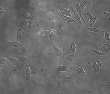
228 216 Figure 5.2 A. B.
Cardiac cell-cell Communication Part 1 Alonso P. Moreno D.Sc. CVRTI, Cardiology
 Bioengineering 6003 Cellular Electrophysiology and Biophysics Cardiac cell-cell Communication Part 1 Alonso P. Moreno D.Sc. CVRTI, Cardiology moreno@cvrti.utah.edu November 2010 poster Physiological Relevance
Amneh Auben. Abdulrahman Jabr. Diala Abu-Hassan
 21 Amneh Auben Abdulrahman Jabr Diala Abu-Hassan Matrix polysaccharides Extracellular matrix (ECM): It s a collection of components that fills the spaces outside the cell or between the cells. ---------
Lecture 3 13/11/2018
 Lecture 3 13/11/2018 1 Plasma membrane ALL cells have a cell membrane made of proteins and lipids. protein channel Cell Membrane Layer 1 Layer 2 lipid bilayer protein pump Lipid bilayer allows water, carbon
Cells to Tissues. Peter Takizawa Department of Cell Biology
 Cells to Tissues Peter Takizawa Department of Cell Biology From one cell to ensembles of cells. Multicellular organisms require individual cells to work together in functional groups. This means cells
Cell Biology Review. The key components of cells that concern us are as follows: 1. Nucleus
 Cell Biology Review Development involves the collective behavior and activities of cells, working together in a coordinated manner to construct an organism. As such, the regulation of development is intimately
Components of a functional cell. Boundary-membrane Cytoplasm: Cytosol (soluble components) & particulates DNA-information Ribosomes-protein synthesis
 & particulates DNA-information Ribosomes-protein synthesis") Cell (Outline) - Components of a functional cell - Major Events in the History of Earth: abiotic and biotic phases; anaerobic and aerobic atmosphere - Prokaryotic cells impact on the biosphere - Origin
Cell (Outline) - Components of a functional cell - Major Events in the History of Earth: abiotic and biotic phases; anaerobic and aerobic atmosphere - Prokaryotic cells impact on the biosphere - Origin
Cell (Learning Objectives)
") Cell (Learning Objectives) 1. Understand & describe the basic components necessary for a functional cell. 2. Review the order of appearance of cells on earth and explain the endosymbiotic theory. 3. Compare
Cell (Learning Objectives) 1. Understand & describe the basic components necessary for a functional cell. 2. Review the order of appearance of cells on earth and explain the endosymbiotic theory. 3. Compare
CELL BIOLOGY - CLUTCH CH. 9 - TRANSPORT ACROSS MEMBRANES.
 !! www.clutchprep.com K + K + K + K + CELL BIOLOGY - CLUTCH CONCEPT: PRINCIPLES OF TRANSMEMBRANE TRANSPORT Membranes and Gradients Cells must be able to communicate across their membrane barriers to materials
Reception The target cell s detection of a signal coming from outside the cell May Occur by: Direct connect Through signal molecules
 Why Do Cells Communicate? Regulation Cells need to control cellular processes In multicellular organism, cells signaling pathways coordinate the activities within individual cells that support the function
PHYSIOLOGY CHAPTER 9 MUSCLE TISSUE Fall 2016
 PHYSIOLOGY CHAPTER 9 MUSCLE TISSUE Fall 2016 2 Chapter 9 Muscles and Muscle Tissue Overview of Muscle Tissue types of muscle: are all prefixes for muscle Contractility all muscles cells can Smooth & skeletal
CL T123 PKA? Possible Cx26deaf mutation: T123N 2,3 EL2 T177 Intracellular kinases or ecto-kinases 2 EL2/TM4 border
 Name AA MW (Da) Cx26 =GJB2 Cx31 =GJB3 BIP Region P-site Modifying enzyme 226 26,215 9.11 NT D2, T5 or S8 1 # Functional consequences Deafness mutation at S8 1 CL T123 PKA Possible Cx26deaf mutation: T123N
Cells. Steven McLoon Department of Neuroscience University of Minnesota
 Cells Steven McLoon Department of Neuroscience University of Minnesota 1 Microscopy Methods of histology: Treat the tissue with a preservative (e.g. formaldehyde). Dissect the region of interest. Embed
Molecular Cell Biology 5068 In Class Exam 2 November 8, 2016
 Molecular Cell Biology 5068 In Class Exam 2 November 8, 2016 Exam Number: Please print your name: Instructions: Please write only on these pages, in the spaces allotted and not on the back. Write your
CHAPTER 3. Cell Structure and Genetic Control. Chapter 3 Outline
 CHAPTER 3 Cell Structure and Genetic Control Chapter 3 Outline Plasma Membrane Cytoplasm and Its Organelles Cell Nucleus and Gene Expression Protein Synthesis and Secretion DNA Synthesis and Cell Division
Cellular Neuroanatomy I The Prototypical Neuron: Soma. Reading: BCP Chapter 2
 Cellular Neuroanatomy I The Prototypical Neuron: Soma Reading: BCP Chapter 2 Functional Unit of the Nervous System The functional unit of the nervous system is the neuron. Neurons are cells specialized
Nervous System Organization
 The Nervous System Nervous System Organization Receptors respond to stimuli Sensory receptors detect the stimulus Motor effectors respond to stimulus Nervous system divisions Central nervous system Command
Intercellular Communication. Department of Physiology School of Medicine University of Sumatera Utara
 Intercellular Communication Department of Physiology School of Medicine University of Sumatera Utara Intercellular Communication and Signal Transduction The ability of cells to communicate with each other
Regulation and signaling. Overview. Control of gene expression. Cells need to regulate the amounts of different proteins they express, depending on
 Regulation and signaling Overview Cells need to regulate the amounts of different proteins they express, depending on cell development (skin vs liver cell) cell stage environmental conditions (food, temperature,
Dendrites - receives information from other neuron cells - input receivers.
 The Nerve Tissue Neuron - the nerve cell Dendrites - receives information from other neuron cells - input receivers. Cell body - includes usual parts of the organelles of a cell (nucleus, mitochondria)
Animal structure and function
 Animal structure and function The nervous system Parts of the nervous system 43C, 44B, 45D Brain structure and function Eyes Retina Neurons: How neurons communicate: Resting potential: The resting
Chapter 3: Cells. Lectures by Mark Manteuffel, St. Louis Community College
 Chapter 3: Cells Lectures by Mark Manteuffel, St. Louis Community College Learning Objectives Be able to describe: what a cell is & two main classes of cells. structure & functions of cell membranes. how
... Cardiac Gap Junctions Physiology, Regulation, Pathophysiology and Pharmacology
 ... Cardiac Gap Junctions Physiology, Regulation, Pathophysiology and Pharmacology S. Dhein, Cologne 23 figures and 3 tables, 1998 StefanDhein InstituteofPharmacology UniversityofCologne (Germany) All
Supplemental table S7.
 Supplemental table S7. GO terms significantly enriched in significantly up-regulated genes of the microarray. K: number of genes from the input cluster in the given category. F: number of total genes in
NATIONAL REVIEW COURSE. Cells, Tissues, and Membranes
 NATIONAL REVIEW COURSE Cells, Tissues, and Membranes I. Cell Types A. Prokaryote bacteria cells; a cell that does not have a nucleus in which to store its genetic material. B. Eukaryote plant or animal
Introduction Principles of Signaling and Organization p. 3 Signaling in Simple Neuronal Circuits p. 4 Organization of the Retina p.
 Introduction Principles of Signaling and Organization p. 3 Signaling in Simple Neuronal Circuits p. 4 Organization of the Retina p. 5 Signaling in Nerve Cells p. 9 Cellular and Molecular Biology of Neurons
Advanced Higher Biology. Unit 1- Cells and Proteins 2c) Membrane Proteins
 Membrane Proteins") Advanced Higher Biology Unit 1- Cells and Proteins 2c) Membrane Proteins Membrane Structure Phospholipid bilayer Transmembrane protein Integral protein Movement of Molecules Across Membranes Phospholipid
Advanced Higher Biology Unit 1- Cells and Proteins 2c) Membrane Proteins Membrane Structure Phospholipid bilayer Transmembrane protein Integral protein Movement of Molecules Across Membranes Phospholipid
CONNEXINS AND CELL SIGNALING IN DEVELOPMENT AND DISEASE 1
 Annu. Rev. Cell Dev. Biol. 2004. 20:811 38 doi: 10.1146/annurev.cellbio.19.111301.144309 First published online as a Review in Advance on July 2, 2004 CONNEXINS AND CELL SIGNALING IN DEVELOPMENT AND DISEASE
Guided Reading Activities
 Name Period Chapter 4: A Tour of the Cell Guided Reading Activities Big Idea: Introduction to the Cell Answer the following questions as you read Modules 4.1 4.4: 1. A(n) uses a beam of light to illuminate
Bio 127 Section I Introduction to Developmental Biology. Cell Cell Communication in Development. Developmental Activities Coordinated in this Way
 Bio 127 Section I Introduction to Developmental Biology Cell Cell Communication in Development Gilbert 9e Chapter 3 It has to be EXTREMELY well coordinated for the single celled fertilized ovum to develop
Domain 6: Communication
 Domain 6: Communication 6.1: Cell communication processes share common features that reflect a shared evolutionary history. (EK3.D.1) 1. Introduction to Communication Communication requires the generation,
لجنة الطب البشري رؤية تنير دروب تميزكم
 1) Hyperpolarization phase of the action potential: a. is due to the opening of voltage-gated Cl channels. b. is due to prolonged opening of voltage-gated K + channels. c. is due to closure of the Na +
Modulation of intercellular communication by differential regulation and heteromeric mixing of co-expressed connexins
 Brazilian Journal of Medical and Biological Research (2000) 33: 391-397 Co-expression of endothelial connexins ISSN 0100-879X 391 Modulation of intercellular communication by differential regulation and
Chemical aspects of the cell. Shape and structure of the cell
 Chemical aspects of the cell Shape and structure of the cell Cellular composition https://www.studyblue.com/ 2 Cellular composition Set of videos with basic information: Cell characteristics: https://www.youtube.com/watch?v=urujd5nexc8
MEMBRANE POTENTIALS AND ACTION POTENTIALS:
 University of Jordan Faculty of Medicine Department of Physiology & Biochemistry Medical students, 2017/2018 +++++++++++++++++++++++++++++++++++++++++++++++++++++++++++++++++++++++++ Review: Membrane physiology
Cells to systems. 1) Chemical Level All matter is a combination of atoms: - Oxygen - Carbon - Nitrogen - Hydrogen Make up 96% of total body chemistry
 Chemical Level All matter is a combination of atoms: - Oxygen - Carbon - Nitrogen - Hydrogen Make up 96% of total body chemistry") Cells to systems Organisation of the Human body: 1. Chemical level 2. Cellular level 3. Tissue level 4. Organ level 5. Body system level 6. Organism level 1) Chemical Level All matter is a combination
Cells to systems Organisation of the Human body: 1. Chemical level 2. Cellular level 3. Tissue level 4. Organ level 5. Body system level 6. Organism level 1) Chemical Level All matter is a combination
Nervous Tissue. Neurons Neural communication Nervous Systems
 Nervous Tissue Neurons Neural communication Nervous Systems What is the function of nervous tissue? Maintain homeostasis & respond to stimuli Sense & transmit information rapidly, to specific cells and
Signal Transduction. Dr. Chaidir, Apt
 Signal Transduction Dr. Chaidir, Apt Background Complex unicellular organisms existed on Earth for approximately 2.5 billion years before the first multicellular organisms appeared.this long period for
Zool 3200: Cell Biology Exam 5 4/27/15
 Name: Trask Zool 3200: Cell Biology Exam 5 4/27/15 Answer each of the following short answer questions in the space provided, giving explanations when asked to do so. Circle the correct answer or answers
Neurons and Nervous Systems
 34 Neurons and Nervous Systems Concept 34.1 Nervous Systems Consist of Neurons and Glia Nervous systems have two categories of cells: Neurons, or nerve cells, are excitable they generate and transmit electrical
Biology September 2015 Exam One FORM G KEY
 Biology 251 17 September 2015 Exam One FORM G KEY PRINT YOUR NAME AND ID NUMBER in the space that is provided on the answer sheet, and then blacken the letter boxes below the corresponding letters of your
Biology September 2015 Exam One FORM W KEY
 Biology 251 17 September 2015 Exam One FORM W KEY PRINT YOUR NAME AND ID NUMBER in the space that is provided on the answer sheet, and then blacken the letter boxes below the corresponding letters of your
Neurons, Synapses, and Signaling
 LECTURE PRESENTATIONS For CAMPBELL BIOLOGY, NINTH EDITION Jane B. Reece, Lisa A. Urry, Michael L. Cain, Steven A. Wasserman, Peter V. Minorsky, Robert B. Jackson Chapter 48 Neurons, Synapses, and Signaling
Physiology. Organization of the Body. Assumptions in Physiology. Chapter 1. Physiology is the study of how living organisms function
 Introduction to Physiology and Homeostasis Chapter 1 Physiology Physiology is the study of how living organisms function On the street explanations are in terms of meeting a bodily need Physiologic explanations
Gap Junctions for Engineers: A review
 Internal Report 97-01 Gap Junctions for Engineers: A review Alejandro F Frangi Division of Intrumentation and Bioengineering Department of Electronic Engineering Polytechnic University of Catalonian March
Visual pigments. Neuroscience, Biochemistry Dr. Mamoun Ahram Third year, 2019
 Visual pigments Neuroscience, Biochemistry Dr. Mamoun Ahram Third year, 2019 References Webvision: The Organization of the Retina and Visual System (http://www.ncbi.nlm.nih.gov/books/nbk11522/#a 127) The
Cardiac Cx43, Cx40 and Cx45 coassembling: epitopes in formation of hemichannels and Gap junction channels
 The Author(s) BMC Cell Biology 2017, 18(Suppl 1):3 DOI 10.1186/s12860-016-0118-4 REVIEW Cardiac Cx43, Cx40 and Cx45 coassembling: involvement of connexins epitopes in formation of hemichannels and Gap
6 Mechanotransduction
 6.1 Motivation The process of converting physical forces into biochemical signals and integrating these signals into the cellular response is referred to as mechnotransduction [11, 20]. To fully understand
According to the diagram, which of the following is NOT true?
 Instructions: Review Chapter 44 on muscular-skeletal systems and locomotion, and then complete the following Blackboard activity. This activity will introduce topics that will be covered in the next few
Cell-Cell Communication in Development
 Biology 4361 - Developmental Biology Cell-Cell Communication in Development October 2, 2007 Cell-Cell Communication - Topics Induction and competence Paracrine factors inducer molecules Signal transduction
7.013 Problem Set
 7.013 Problem Set 5-2013 Question 1 During a summer hike you suddenly spot a huge grizzly bear. This emergency situation triggers a fight or flight response through a signaling pathway as shown below.
Nervous Tissue. Neurons Electrochemical Gradient Propagation & Transduction Neurotransmitters Temporal & Spatial Summation
 Nervous Tissue Neurons Electrochemical Gradient Propagation & Transduction Neurotransmitters Temporal & Spatial Summation What is the function of nervous tissue? Maintain homeostasis & respond to stimuli
Gating Properties of Heterotypic Gap Junction Channels Formed of Connexins 40, 43, and 45
 1952 Biophysical Journal Volume 92 March 2007 1952 1965 Gating Properties of Heterotypic Gap Junction Channels Formed of Connexins 40, 43, and 45 Mindaugas Rackauskas,* Maria M. Kreuzberg, z Mindaugas
7.06 Cell Biology EXAM #3 KEY
 7.06 Cell Biology EXAM #3 KEY May 2, 2006 This is an OPEN BOOK exam, and you are allowed access to books, a calculator, and notes BUT NOT computers or any other types of electronic devices. Please write
Chapter 16. Cellular Movement: Motility and Contractility. Lectures by Kathleen Fitzpatrick Simon Fraser University Pearson Education, Inc.
 Chapter 16 Cellular Movement: Motility and Contractility Lectures by Kathleen Fitzpatrick Simon Fraser University Two eukaryotic motility systems 1. Interactions between motor proteins and microtubules
MEMBRANE STRUCTURE. Lecture 9. Biology Department Concordia University. Dr. S. Azam BIOL 266/
 MEMBRANE STRUCTURE Lecture 9 BIOL 266/4 2014-15 Dr. S. Azam Biology Department Concordia University RED BLOOD CELL MEMBRANE PROTEINS The Dynamic Nature of the Plasma Membrane SEM of human erythrocytes
The Cell. C h a p t e r. PowerPoint Lecture Slides prepared by Jason LaPres North Harris College Houston, Texas
 C h a p t e r 2 The Cell PowerPoint Lecture Slides prepared by Jason LaPres North Harris College Houston, Texas Copyright 2009 Pearson Education, Inc., publishing as Pearson Benjamin Cummings Introduction
Protein Sorting, Intracellular Trafficking, and Vesicular Transport
 Protein Sorting, Intracellular Trafficking, and Vesicular Transport Noemi Polgar, Ph.D. Department of Anatomy, Biochemistry and Physiology Email: polgar@hawaii.edu Phone: 692-1422 Outline Part 1- Trafficking
Chapter 48 Neurons, Synapses, and Signaling
 Chapter 48 Neurons, Synapses, and Signaling Concept 48.1 Neuron organization and structure reflect function in information transfer Neurons are nerve cells that transfer information within the body Neurons
Hole s Human Anatomy and Physiology
 Hole s Human Anatomy and Physiology 1 Chapter 3 Cells vary in size possess distinctive shapes measured in micrometers 2 A Composite Cell hypothetical cell major parts nucleus cytoplasm cell membrane 3
Chapter 9. Nerve Signals and Homeostasis
 Chapter 9 Nerve Signals and Homeostasis A neuron is a specialized nerve cell that is the functional unit of the nervous system. Neural signaling communication by neurons is the process by which an animal
REVIEW 2: CELLS & CELL COMMUNICATION. A. Top 10 If you learned anything from this unit, you should have learned:
 Name AP Biology REVIEW 2: CELLS & CELL COMMUNICATION A. Top 10 If you learned anything from this unit, you should have learned: 1. Prokaryotes vs. eukaryotes No internal membranes vs. membrane-bound organelles
NOTES: CH 48 Neurons, Synapses, and Signaling
 NOTES: CH 48 Neurons, Synapses, and Signaling A nervous system has three overlapping functions: 1) SENSORY INPUT: signals from sensory receptors to integration centers 2) INTEGRATION: information from
Explain how cell size and shape affect the overall rate of nutrient intake and the rate of waste elimination. [LO 2.7, SP 6.2]
![Explain how cell size and shape affect the overall rate of nutrient intake and the rate of waste elimination. [LO 2.7, SP 6.2]](/thumbs/77/76271213.jpg "Explain how cell size and shape affect the overall rate of nutrient intake and the rate of waste elimination. [LO 2.7, SP 6.2]") Cells Learning Objectives Use calculated surface area-to-volume ratios to predict which cell(s) might eliminate wastes or procure nutrients faster by diffusion. [LO 2.6, SP 2.2] Explain how cell size and
Cells Learning Objectives Use calculated surface area-to-volume ratios to predict which cell(s) might eliminate wastes or procure nutrients faster by diffusion. [LO 2.6, SP 2.2] Explain how cell size and
CURRICULUM MAP. TIME CONTENT PAGE REF. SKILLS ASSESSMENT/ACTIVITIES Day 1-3
 CURRICULUM MAP COURSE TITLE: Anatomy and Physiology DESCRIPTION TEXTBOOK Essentials of Anatomy and Physiology OF COURSE: 8 th ed: Elaine Marieb Integrated approach to human anatomy. Performance and lab
Zimmerman AP Biology CBHS South Name Chapter 7&8 Guided Reading Assignment 1) What is resolving power and why is it important in biology?
 What is resolving power and why is it important in biology?") Zimmerman AP Biology CBHS South Name Chapter 7&8 Guided Reading Assignment 1) What is resolving power and why is it important in biology? 2) How does an electron microscope work and what is the difference
Zimmerman AP Biology CBHS South Name Chapter 7&8 Guided Reading Assignment 1) What is resolving power and why is it important in biology? 2) How does an electron microscope work and what is the difference
Chapter 6: A Tour of the Cell
 Chapter 6: A Tour of the Cell 1. The study of cells has been limited by their small size, and so they were not seen and described until 1665, when Robert Hooke first looked at dead cells from an oak tree.
The Cell Notes 1 of 11
 The Cell The basic unit of structure and function in living things The smallest units in living things The smallest units in living things that show the characteristics of life Organisms can be made of
Neurons, Synapses, and Signaling
 Chapter 48 Neurons, Synapses, and Signaling PowerPoint Lecture Presentations for Biology Eighth Edition Neil Campbell and Jane Reece Lectures by Chris Romero, updated by Erin Barley with contributions
Neurons, Synapses, and Signaling
 Chapter 48 Neurons, Synapses, and Signaling PowerPoint Lecture Presentations for Biology Eighth Edition Neil Campbell and Jane Reece Lectures by Chris Romero, updated by Erin Barley with contributions
13-3. Synthesis-Secretory pathway: Sort lumenal proteins, Secrete proteins, Sort membrane proteins
 13-3. Synthesis-Secretory pathway: Sort lumenal proteins, Secrete proteins, Sort membrane proteins Molecular sorting: specific budding, vesicular transport, fusion 1. Why is this important? A. Form and
Chapter 2 Basic Cardiac Electrophysiology: Excitable Membranes
 Chapter Basic Cardiac Electrophysiology: Excitable Membranes Deborah A. Jaye, Yong-Fu Xiao, and Daniel C. Sigg Abstract Cardiomyocytes are excitable cells that have the ability to contract after excitation;
Gap junctional coupling in lenses lacking 3 connexin
 Proc. Natl. Acad. Sci. USA Vol. 95, pp. 15303 15308, December 1998 Biophysics Gap junctional coupling in lenses lacking 3 connexin XIAOHUA GONG*, GEORGE J. BALDO, NALIN M. KUMAR*, NORTON B. GILULA*, AND
Biology: Life on Earth
 Teresa Audesirk Gerald Audesirk Bruce E. Byers Biology: Life on Earth Eighth Edition Lecture for Chapter 4 Cell Structure and Function Copyright 2008 Pearson Prentice Hall, Inc. Chapter 4 Outline 4.1 What
CHAPTER 9 BODY ORGANIZATION. Copyright 2007 by Mosby, Inc., an affiliate of Elsevier Inc. 1
 CHAPTER 9 BODY ORGANIZATION Copyright 2007 by Mosby, Inc., an affiliate of Elsevier Inc. 1 Anatomy and Physiology Four basic properties of life: Reception The ability of the organism to control its actions
thebiotutor.com A2 Biology Unit 5 Responses, Nervous System & Muscles
 thebiotutor.com A2 Biology Unit 5 Responses, Nervous System & Muscles 1 Response Mechanism tropism Definition A growth movement of part of plant in response to a directional stimulus examples Positive:
Neurons, Synapses, and Signaling
 CAMPBELL BIOLOGY IN FOCUS URRY CAIN WASSERMAN MINORSKY REECE 37 Neurons, Synapses, and Signaling Lecture Presentations by Kathleen Fitzpatrick and Nicole Tunbridge, Simon Fraser University SECOND EDITION
1. The plasma membrane of eukaryotic cells is supported by a. actin filaments. b. microtubules. c. lamins. d. intermediate filaments.
 ANALYSIS AND MODELING OF CELL MECHANICS Homework #2 (due 1/30/13) This homework involves comprehension of key biomechanical concepts of the cytoskeleton, cell-matrix adhesions, and cellcell adhesions.
BIOLOGY. 1. Overview of Neurons 11/3/2014. Neurons, Synapses, and Signaling. Communication in Neurons
 CAMPBELL BIOLOGY TENTH EDITION 48 Reece Urry Cain Wasserman Minorsky Jackson Neurons, Synapses, and Signaling Lecture Presentation by Nicole Tunbridge and Kathleen Fitzpatrick 1. Overview of Neurons Communication
S1 Gene ontology (GO) analysis of the network alignment results
 analysis of the network alignment results") 1 Supplementary Material for Effective comparative analysis of protein-protein interaction networks by measuring the steady-state network flow using a Markov model Hyundoo Jeong 1, Xiaoning Qian 1 and
1 Supplementary Material for Effective comparative analysis of protein-protein interaction networks by measuring the steady-state network flow using a Markov model Hyundoo Jeong 1, Xiaoning Qian 1 and
Intro and Homeostasis
 Intro and Homeostasis Physiology - how the body works. Homeostasis - staying the same. Functional Types of Neurons Sensory (afferent - coming in) neurons: Detects the changes in the body. Informations
Introduction to electrophysiology 1. Dr. Tóth András
 Introduction to electrophysiology 1. Dr. Tóth András Topics Transmembran transport Donnan equilibrium Resting potential Ion channels Local and action potentials Intra- and extracellular propagation of
Unit 2: Cells Guided Reading Questions (60 pts total)
") Name: AP Biology Biology, Campbell and Reece, 7th Edition Adapted from chapter reading guides originally created by Lynn Miriello Chapter 6 A Tour of the Cell Unit 2: Cells Guided Reading Questions (60
Name: AP Biology Biology, Campbell and Reece, 7th Edition Adapted from chapter reading guides originally created by Lynn Miriello Chapter 6 A Tour of the Cell Unit 2: Cells Guided Reading Questions (60
Lecture 13, 05 October 2004 Chapter 10, Muscle. Vertebrate Physiology ECOL 437 University of Arizona Fall instr: Kevin Bonine t.a.
 Lecture 13, 05 October 2004 Chapter 10, Muscle Vertebrate Physiology ECOL 437 University of Arizona Fall 2004 instr: Kevin Bonine t.a.: Nate Swenson Vertebrate Physiology 437 18 1. Muscle A. Sarcomere
Dr. Ketki Assistant Professor Department of Biochemistry Heritage IMS, Varanasi
 TRANSPORT MECHANISMS Dr. Ketki Assistant Professor Department of Biochemistry Heritage IMS, Varanasi Membrane selectivity allows adjustments of cell composition and function If plasma membrane is relatively
Fundamentals of Neurosciences. Smooth Muscle. Dr. Kumar Sambamurti 613-SEI; ;
 Fundamentals of Neurosciences Smooth Muscle Dr. Kumar Sambamurti 613-SEI; 792-4315; sambak@musc.edu 1 Smooth Muscle Structure Cells much smaller than skeletal muscle (2-5µM diam, 100-400µM long) Single
Massachusetts Institute of Technology Harvard Medical School Brigham and Women s Hospital VA Boston Healthcare System 2.79J/3.96J/BE.
 Massachusetts Institute of Technology Harvard Medical School Brigham and Women s Hospital VA Boston Healthcare System 2.79J/3.96J/BE.441/HST522J INTEGRINS I.V. Yannas, Ph.D. and M. Spector, Ph.D. Regulator
Nervous System Organization
 The Nervous System Chapter 44 Nervous System Organization All animals must be able to respond to environmental stimuli -Sensory receptors = Detect stimulus -Motor effectors = Respond to it -The nervous
Study Guide 11 & 12 MULTIPLE CHOICE. Choose the one alternative that best completes the statement or answers the question.
 Study Guide 11 & 12 MULTIPLE CHOICE. Choose the one alternative that best completes the statement or answers the question. 1) The receptors for a group of signaling molecules known as growth factors are
Molecular Cell Biology 5068 In Class Exam 1 September 30, Please print your name:
 Molecular Cell Biology 5068 In Class Exam 1 September 30, 2014 Exam Number: Please print your name: Instructions: Please write only on these pages, in the spaces allotted and not on the back. Write your
Chapter 3: Structure and Function of the Cell
 Chapter 3: Structure and Function of the Cell I. Functions of the Cell A. List and describe the main functions of the cell: 1. 2. 3. 4. 5. II. How We See Cells A. Light microscopes allow us to B. Electron
HUMAN ANATOMY & PHYSIOLOGY STRUCTURE 30
 HUMAN ANATOMY & PHYSIOLOGY STRUCTURE 30 Description This second-year course in biology emphasizes the workings of the human body. The course is offered in the fall semester and meets six (6) periods per
NAME: PERIOD: DATE: A View of the Cell. Use Chapter 8 of your book to complete the chart of eukaryotic cell components.
 NAME: PERIOD: DATE: A View of the Cell Use Chapter 8 of your book to complete the chart of eukaryotic cell components. Cell Part Cell Wall Centriole Chloroplast Cilia Cytoplasm Cytoskeleton Endoplasmic
NEURONS, SENSE ORGANS, AND NERVOUS SYSTEMS CHAPTER 34
 NEURONS, SENSE ORGANS, AND NERVOUS SYSTEMS CHAPTER 34 KEY CONCEPTS 34.1 Nervous Systems Are Composed of Neurons and Glial Cells 34.2 Neurons Generate Electric Signals by Controlling Ion Distributions 34.3
Organization of the nervous system. Tortora & Grabowski Principles of Anatomy & Physiology; Page 388, Figure 12.2
 Nervous system Organization of the nervous system Tortora & Grabowski Principles of Anatomy & Physiology; Page 388, Figure 12.2 Autonomic and somatic efferent pathways Reflex arc - a neural pathway that
Chapter 1. DNA is made from the building blocks adenine, guanine, cytosine, and. Answer: d
 Chapter 1 1. Matching Questions DNA is made from the building blocks adenine, guanine, cytosine, and. Answer: d 2. Matching Questions : Unbranched polymer that, when folded into its three-dimensional shape,
Information processing. Divisions of nervous system. Neuron structure and function Synapse. Neurons, synapses, and signaling 11/3/2017
 Neurons, synapses, and signaling Chapter 48 Information processing Divisions of nervous system Central nervous system (CNS) Brain and a nerve cord Integration center Peripheral nervous system (PNS) Nerves
Chapter 4 A Tour of the Cell*
 Chapter 4 A Tour of the Cell* *Lecture notes are to be used as a study guide only and do not represent the comprehensive information you will need to know for the exams. The Fundamental Units of Life Cells
Schémata zpracovalo Servisní středisko pro e-learning na MU
 Schémata zpracovalo Servisní středisko pro e-learning na MU http://is.muni.cz/stech/ Life is a dynamic system with focused behavior, with autoreproduction, characterized by flow of substrates, energies
Chapter 6: A Tour of the Cell
 AP Biology Reading Guide Fred and Theresa Holtzclaw Chapter 6: A Tour of the Cell Name Period Chapter 6: A Tour of the Cell Concept 6.1 To study cells, biologists use microscopes and the tools of biochemistry
Cell Cell Communication in Development
 Biology 4361 Developmental Biology Cell Cell Communication in Development June 25, 2008 Cell Cell Communication Concepts Cells in developing organisms develop in the context of their environment, including