Muscles and Muscle Tissue: Part A
|
|
|
- Georgina Glenn
- 5 years ago
- Views:
Transcription

1 PowerPoint Lecture Slides prepared by Janice Meeking, Mount Royal College CHAPTER 9 Muscles and Muscle Tissue: Part A
2 Warm Up 12/12/16 Describe the major differences between cardiac, skeletal and smooth muscle in terms of appearance, control, function, and location!
3 Three Types of Muscle Tissue 1. Skeletal muscle tissue: Attached to bones and skin Striated Voluntary (i.e., conscious control) Powerful Primary topic of this chapter
4 Three Types of Muscle Tissue 2. Cardiac muscle tissue: Only in the heart Striated Involuntary More details in Chapter 18
5 Three Types of Muscle Tissue 3. Smooth muscle tissue: In the walls of hollow organs, e.g., stomach, urinary bladder, and airways Not striated Involuntary More details later in this chapter
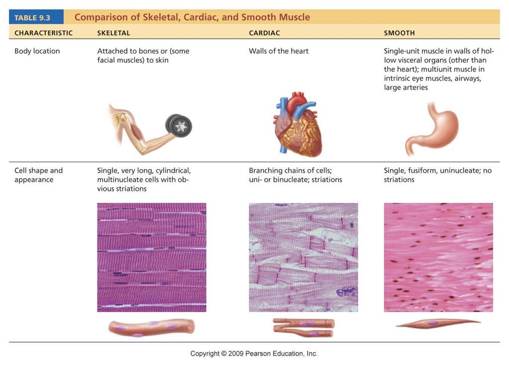
6 Table 9.3
7 Special Characteristics of Muscle Tissue Excitability (responsiveness or irritability): ability to receive and respond to stimuli Contractility: ability to shorten when stimulated Extensibility: ability to be stretched Elasticity: ability to recoil to resting length
8 Muscle Functions 1. Movement of bones or fluids (e.g., blood) 2. Maintaining posture and body position 3. Stabilizing joints 4. Heat generation (especially skeletal muscle)
9 Skeletal Muscle Each muscle is served by one artery, one nerve, and one or more veins
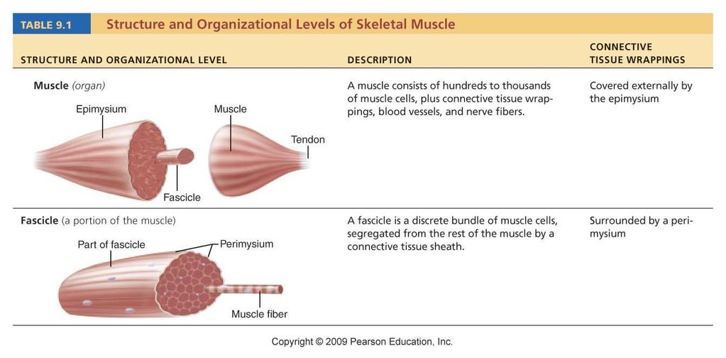
10 Skeletal Muscle Connective tissue sheaths of skeletal muscle: Epimysium: dense regular connective tissue surrounding entire muscle Perimysium: fibrous connective tissue surrounding fascicles (groups of muscle fibers) Endomysium: fine areolar connective tissue surrounding each muscle fiber
 Endomysium (between individual muscle fibers) Muscle fiber Figure")
11 Bone Epimysiu m Tendon (b ) Perimysiu (a m ) Fascicl e Epimysiu m Perimysiu m Endomysiu m Muscle fiber in middle of a fascicle Blood Fascicle vessel (wrapped by perimysium) Endomysium (between individual muscle fibers) Muscle fiber Figure 9.1
12 Skeletal Muscle: Attachments Muscles attach: Directly epimysium of muscle is fused to the periosteum of bone or perichondrium of cartilage Indirectly connective tissue wrappings extend beyond the muscle as a ropelike tendon or sheetlike aponeurosis
13 Table 9.1
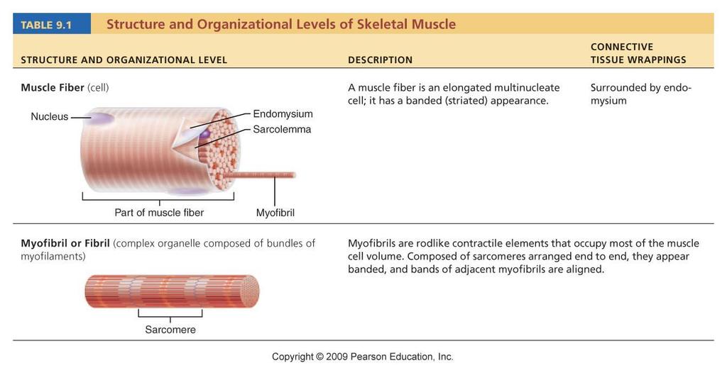
14 Microscopic Anatomy of a Skeletal Muscle Fiber Cylindrical cell 10 to 100 μm in diameter, up to 30 cm long Multiple peripheral nuclei Many mitochondria Glycosomes for glycogen storage, myoglobin for O2 storage Also contain myofibrils, sarcoplasmic reticulum, and T tubules
15 Myofibrils Densely packed, rodlike elements ~80% of cell volume Exhibit striations: perfectly aligned repeating series of dark A bands and light I bands

16 Sarcolemm a Mitochondrion Myofibri l Dark A band Light I Nucleus bandof a muscle fiber showing the myofibrils. (b) Diagram of part One myofibril is extended afrom the cut end of the fiber.
17 Sarcomere Smallest contractile unit (functional unit) of a muscle fiber The region of a myofibril between two successive Z discs Composed of thick and thin myofilaments made of contractile proteins
18 Features of a Sarcomere Thick filaments: run the entire length of an A band Thin filaments: run the length of the I band and partway into the A band Z disc: coin-shaped sheet of proteins that anchors the thin filaments and connects myofibrils to one another H zone: lighter midregion where filaments do not overlap M line: line of protein myomesin that holds adjacent thick filaments together
 responsible for the banding pattern.")
 filament Elastic (titin) filaments Thick (myosin ) filament (d")
19 Thin (actin) filament Thick (myosin) filament Z disc H zone Z disc A band I band M line Sarcomer e (c Small part of one myofibril enlarged to show the myofilaments ) responsible for the banding pattern. Each sarcomere extends from one Z disc to the next. Z disc I band Sarcomer e M line Z disc Thin (actin) filament Elastic (titin) filaments Thick (myosin ) filament (d Enlargement of one sarcomere (sectioned lengthwise). Notice the ) myosin heads on the thick filaments. Figure 9.2c, d
20 Ultrastructure of Thick Filament Composed of the protein myosin Myosin tails contain: 2 interwoven, heavy polypeptide chains Myosin heads contain: 2 smaller, light polypeptide chains that act as cross bridges during contraction Binding sites for actin of thin filaments Binding sites for ATP ATPase enzymes
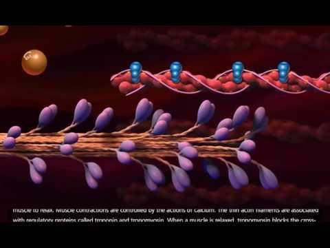
21 Ultrastructure of Thin Filament Twisted double strand of fibrous protein F actin F actin consists of G (globular) actin subunits G actin bears active sites for myosin head attachment during contraction Tropomyosin and troponin: regulatory proteins bound to actin

22 Longitudinal section of filaments within one sarcomere of a myofibril Thick filament Thin In the center of the sarcomere, the thick filament filaments lack myosin heads. Myosin heads are present only in areas of myosin-actin overlap. Thick filament Thin filament Each thick filament consists of many A thin filament consists of two strands myosin molecules whose heads protrude of actin subunits twisted into a helix at opposite ends of the filament. plus two types of regulatory proteins (troponin and tropomyosin). Portion of a thick filament Portion of a thin Myosin filament head Tropomyosi Troponi Acti n n n Actin-binding sites ATPbindin g site Head s Flexible hinge region Myosin molecule Tai l Active sites for myosin attachment Actin subunits Actin subunits Figure 9.3
23 Sarcoplasmic Reticulum (SR) Network of smooth endoplasmic reticulum surrounding each myofibril Pairs of terminal cisternae form perpendicular cross channels Functions in the regulation of intracellular Ca2+ levels
24 T Tubules Continuous with the sarcolemma Penetrate the cell s interior at each A band I band junction Associate with the paired terminal cisternae to form triads that encircle each sarcomere
 Tubules of the SR Myofibril Mitochondria s Figure")
25 Part of a skeletal muscle fiber (cell) I band Z Myofibril disc Sarcolemm a A band I band H zone M line Z disc Sarcolemm a Triad: T Terminal tubule cisternae of the SR (2) Tubules of the SR Myofibril Mitochondria s Figure 9.5
26 Triad Relationships T tubules conduct impulses deep into muscle fiber Integral proteins protrude into the intermembrane space from T tubule and SR cisternae membranes T tubule proteins: voltage sensors SR foot proteins: gated channels that regulate Ca2+ release from the SR cisternae
27
28 9A-2 Notes:
29 Warm Up! Explain the role and function of the T tubules and sarcoplasmic reticulum in relation to muscle contraction.
30 Contraction The generation of force Does not necessarily cause shortening of the fiber Shortening occurs when tension generated by cross bridges on the thin filaments exceeds forces opposing shortening
31 Sliding Filament Model of Contraction In the relaxed state, thin and thick filaments overlap only slightly During contraction, myosin heads bind to actin, detach, and bind again, to propel the thin filaments toward the M line As H zones shorten and disappear, sarcomeres shorten, muscle cells shorten, and the whole muscle shortens

32 Z Z H A I I 1 Fully relaxed sarcomere of a muscle fiber Z I Z A I 2 Fully contracted sarcomere of a muscle fiber Figure 9.6
33 Requirements for Skeletal Muscle Contraction 1. Activation: neural stimulation at a neuromuscular junction 2. Excitation-contraction coupling: Generation and propagation of an action potential along the sarcolemma Final trigger: a brief rise in intracellular Ca2+ levels
34 Events at the Neuromuscular Junction Skeletal muscles are stimulated by somatic motor neurons Axons of motor neurons travel from the central nervous system via nerves to skeletal muscles Each axon forms several branches as it enters a muscle Each axon ending forms a neuromuscular junction with a single muscle fiber
35 Action potential (AP) Myelinated axon of motor neuron Axon terminal of neuromuscular junction Nucleu s Sarcolemma of the muscle fiber 1 Action potential arrives at axon terminal of motor neuron. 2 Voltage-gated Ca2+ channels open and Ca2+ enters the axon terminal. Ca2+ Ca2 + Axon terminal of motor neuron Synaptic vesicle containing ACh Mitochondrion Synaptic cleft Fusing synaptic vesicles Figure 9.8
36 Neuromuscular Junction Situated midway along the length of a muscle fiber Axon terminal and muscle fiber are separated by a gel-filled space called the synaptic cleft Synaptic vesicles of axon terminal contain the neurotransmitter acetylcholine (ACh) Junctional folds of the sarcolemma contain ACh receptors

37 Events at the Neuromuscular Junction Nerve impulse arrives at axon terminal ACh is released and binds with receptors on the sarcolemma Electrical events lead to the generation of an action potential PL AY A&P Flix : Events at the Neuromuscular Junction
38 Myelinated axon of motor neuron Axon terminal of neuromuscular junction Sarcolemma of the muscle fiber Action potential (AP) Nucleu s 1 Action potential arrives at axon terminal of motor neuron Voltage-gated Ca channels Ca2+ Ca2+ open and Ca2+ enters the axon terminal. Axon of motor terminal neuron 3 Ca2+ entry causes some Fusing synaptic vesicle s synaptic vesicles to release their contents (acetylcholine) by exocytosis. 4 Acetylcholine, a neurotransmitter, diffuses across the synaptic cleft and binds to receptors in the sarcolemma. AC h 5 ACh binding opens ion Na+ K+ channels that allow simultaneous passage of Na+ into the muscle fiber and K+ out of the muscle fiber. 6 ACh effects are terminated by its enzymatic breakdown in the synaptic cleft by acetylcholinesterase. Ach Degraded ACh Na+ Acetylcholinesterase Synaptic vesicle containing ACh Mitochondrion Synaptic cleft Junctional folds of sarcolemm a Sarcoplasm of muscle fiber Postsynaptic membrane ion channel opens; ions pass. Postsynaptic membrane ion channel closed; ions cannot pass. K+ Figure 9.8
39 Destruction of Acetylcholine ACh effects are quickly terminated by the enzyme acetylcholinesterase Prevents continued muscle fiber contraction in the absence of additional stimulation
40 Events in Generation of an Action Potential 1. Local depolarization (end plate potential): ACh binding opens chemically (ligand) gated ion channels Simultaneous diffusion of Na+ (inward) and K+ (outward) More Na+ diffuses, so the interior of the sarcolemma becomes less negative Local depolarization end plate potential
41 Events in Generation of an Action Potential 2. Generation and propagation of an action potential: End plate potential spreads to adjacent membrane areas Voltage-gated Na+ channels open Na+ influx decreases the membrane voltage toward a critical threshold If threshold is reached, an action potential is generated
42 Events in Generation of an Action Potential Local depolarization wave continues to spread, changing the permeability of the sarcolemma Voltage-regulated Na+ channels open in the adjacent patch, causing it to depolarize to threshold
43 Events in Generation of an Action Potential 3. Repolarization: Na+ channels close and voltage-gated K+ channels open K+ efflux rapidly restores the resting polarity Fiber cannot be stimulated and is in a refractory period until repolarization is complete Ionic conditions of the resting state are restored by the Na+-K+ pump
44 Axon terminal Open Na+ Channel Na Synaptic cleft Closed K+ Channel + ACh t io n za + of de po + + l ari ACh Na K Na K + e Wav 1 Local depolarization: generation of the end plate potential on the sarcolemma K Action potential Generation and propagation of the action potential (AP) Closed Na+ Channel Na Open K+ Channel + K Sarcoplasm of muscle fiber 3 Repolarization + Figure 9.9
45 Axon terminal Open Na+ Channel Na Synapti c cleft + n + K Action potential l ar + of d ep o + K tio AC h Na K Na + iza AC h Closed K+ Channel ve Wa 1 Local depolarization: generation of the end plate potential on the sarcolemma Sarcoplasm of muscle fiber Figure 9.9, step 1

46 Axon terminal Open Na+ Channel Na Synapti c cleft + n + K l ar Action potential Generation and propagation of the action potential (AP) of d ep o + K tio AC h Na K Na + iza AC h Closed K+ Channel ve Wa 1 Local depolarization: generation of the end plate potential on the sarcolemma Sarcoplasm of muscle fiber Figure 9.9, step 2

47 Open K+ Channel Closed Na+ Channel Na + K + 3 Repolarization Figure 9.9, step 3

48 Axon terminal Open Na+ Channel Na Synapti c cleft t io n + + K Action potential Generation and propagation of the action potential (AP) of de po + Na K + za AC h Na K + l ari AC h Closed K+ Channel e Wav 1 Local depolarization: generation of the end plate potential on the sarcolemma Closed Na+ Na Channel + Open K+ Channe l K Sarcoplasm of muscle fiber 3 Repolarization + Figure 9.9

49 Depolarizatio n due to Na+ entry Na+ channels open Na+ channels close, K+ channels open Repolarizatio n due to K+ exit Threshold K+ channels close Figure 9.10
50 Excitation-Contraction (E-C) Coupling Sequence of events by which transmission of an AP along the sarcolemma leads to sliding of the myofilaments Latent period: Time when E-C coupling events occur Time between AP initiation and the beginning of contraction
51 Events of Excitation-Contraction (E-C) Coupling AP is propagated along sarcolemma to T tubules Voltage-sensitive proteins stimulate Ca2+ release from SR Ca2+ is necessary for contraction

52 Setting the stage Axon terminal of motor neuron Action Synaptic cleft potential ACh is generated Sarcolemma Terminal cisterna of SR Muscle fiber Ca2+ Triad One sarcomere Figure 9.11, step 1
53 Steps in E-C Coupling: Sarcolemma Voltage-sensitive tubule protein T tubule 1 Action potential is propagated along the sarcolemma and down the T tubules. Ca2+ release channe l 2 Calcium ions are released. Terminal cisterna of SR Ca2+ Actin Troponi n Ca2+ Tropomyosin blocking active sites Myosi n 3 Calcium binds to troponin and removes the blocking action of tropomyosin. Active sites exposed and ready for myosin binding Myosi n cross bridge 4 Contraction begins The aftermath Figure 9.11, step 2
54 1 Action potential is propagated along the sarcolemma and Steps in down E-C Coupling: the T tubules. Sarcolemma Voltage-sensitive T tubule tubule protein Ca2+ release channel Terminal cisterna of SR Ca2+ Figure 9.11, step 3

55 1 Action potential is propagated along the sarcolemma and Steps in down E-C Coupling: the T tubules. Sarcolemma Voltage-sensitive T tubule tubule protein Ca2+ release channel Terminal cisterna of SR 2 Calcium ions are released. Ca2+ Figure 9.11, step 4
56 Actin Ca2+ Troponi n Tropomyosin blocking active sites Myosin The aftermath Figure 9.11, step 5

57 Actin Ca2+ Troponi n Tropomyosin blocking active sites Myosin 3 Calcium binds to troponin and removes the blocking action of tropomyosin. Active sites exposed and ready for myosin binding The aftermath Figure 9.11, step 6
58 Actin Ca2+ Troponi n Tropomyosin blocking active sites Myosin 3 Calcium binds to Active sites exposed and ready for myosin binding troponin and removes the blocking action of tropomyosin. 4 Contraction begins Myosin cross bridge The aftermath Figure 9.11, step 7

59 Steps in E-C Coupling: Sarcolemma Voltage-sensitive tubule protein T tubule 1 Action potential is propagated along the sarcolemma and down the T tubules. Ca2+ release channe l 2 Calcium ions are released. Terminal cisterna of SR Ca2+ Actin Troponi n Ca2+ Tropomyosin blocking active sites Myosi n 3 Calcium binds to troponin and removes the blocking action of tropomyosin. Active sites exposed and ready for myosin binding Myosi n cross bridge 4 Contraction begins The aftermath Figure 9.11, step 8
60 Role of Calcium (Ca2+) in Contraction At low intracellular Ca2+ concentration: Tropomyosin blocks the active sites on actin Myosin heads cannot attach to actin Muscle fiber relaxes
61 Role of Calcium (Ca2+) in Contraction At higher intracellular Ca2+ concentrations: Ca2+ binds to troponin Troponin changes shape and moves tropomyosin away from active sites Events of the cross bridge cycle occur When nervous stimulation ceases, Ca2+ is pumped back into the SR and contraction ends
62 Cross Bridge Cycle Continues as long as the Ca2+ signal and adequate ATP are present Cross bridge formation high-energy myosin head attaches to thin filament Working (power) stroke myosin head pivots and pulls thin filament toward M line
63 Cross Bridge Cycle Cross bridge detachment ATP attaches to myosin head and the cross bridge detaches Cocking of the myosin head energy from hydrolysis of ATP cocks the myosin head into the high-energy state
64 Thin filament Actin Myosin cross bridge Ca 2+ ADP Pi Thick filamen Myosi t n 1 Cross bridge formation. ADP ADP Pi Pi ATP hydrolysi s 2 The power (working) stroke. 4 Cocking of myosin head. AT P AT P 3 Cross bridge detachment. Figure 9.12
65 Actin Ca2 Thin filament + Myosin cross bridge AD PP i Thick filament Myosi n 1 Cross bridge formation. Figure 9.12, step 1
66 AD P P i 2 The power (working) stroke. Figure 9.12, step 3

67 ATP 3 Cross bridge detachment. Figure 9.12, step 4
68 AD PP i ATP hydrolysi s 4 Cocking of myosin head. Figure 9.12, step 5
 stroke. 4 Cocking of myosin head.")
69 Thin filament Actin Myosin cross bridge Ca 2+ ADP Pi Thick filamen Myosi t n 1 Cross bridge formation. ADP ADP Pi Pi ATP hydrolysi s 2 The power (working) stroke. 4 Cocking of myosin head. AT P AT P 3 Cross bridge detachment. Figure 9.12
70 Steps in E-C Coupling - Storytelling! 1. Fill in the blanks to the steps of the E-C story that I wrote out for you! 2. Define the terms in the word bank provided. 3. Retell the story by creating your own captions on the pictures provided. Be sure to label all * structures as well!
71
72 To instigate the process at the Neuromuscular Junction. Neurotransmitters are released from the axon terminal. As they diffuse across the synaptic cleft, they attach to Acetylcholine (ACh) receptors on the sarcolemma.
73 Step 1 + Net entry of Na initiates an Action Potential which is propagated along the sarcolemma and down the T Tubules.
74 Step 2 The action potential in the T tubule activates voltage-sensitive receptors which in turn trigger Ca2+ release from the terminal cisternae of the Sarcoplasmic Reticulum in the cytosol.
75 Step 3 Calcium ions bind to troponin; this changes shape, removing the blocking action of tropomyosin therefore leave the actin active sites exposed.
76 Step 4 Contraction! (The Cross Bridge Cycle***) Myosin heads alternately attach to actin and detach, pulling the Actin filaments toward the center of the sarcomere. Powered by ATP hydrolysis.
77 Step 4 broken down 4.1 *** Myosin head attached to the actin myofilament, forming the cross bridge.
78 4.2*** Inorganic Phosphate (Pi) generated in the previous contraction cycle is released, initiating the power (working) stroke. The myosin head pivots and bends as it pulls on the actin filament, sliding it toward the M-Line. Then ADP is released.
79 4.3*** A new ATP attached to the myosin head, the link between myosin and actin weakens, and the cross bridge detaches.
80 4.4*** As ATP split into ADP and Pi, the myosin head is energized (cocked into the high-energy conformation) Ready to start over!
81 Step 5 Removal of Ca 2+ by active transport into the sarcoplasmic reticulum (SR) after the action potential ends.
82 Step 6 Tropomyosin blockage is restored, blocking the myosin binding sites on actin. Contraction ends and the muscle fiber relaxes!!
83 Warm Up 1/9/17 Through what process is ATP made?! With oxygen = Without oxygen = (Where does this process occur?)
84 What are the 6 Steps of EC Coupling?! Remember that step 4 is our dance! (the cross bridge cycle!) - broken down into 4.1, 2, 3 and 4!
85 mitochondria 1 Axon terminal Sodium rushes into the cell, sending an action potential down the length of the cell membrane (sarcolemma) and into the cell via T Tubules. Synaptic vesicle ACh Receptor Acetylcholine (ACh) Sarcolemma
86 Sarcoplasmic Reticulum (SR) Calcium T Tubule 2 Voltage gated receptor T tubules stimulate calcium release from the SR

87 3 Calcium binds to troponin, unlocking the tropomyosin gate and exposing the myosin binding sites Myosin binding site Actin Myosin head Troponin Tropomyosin Myosin

88 4 Contraction begins! Actin is pulled towards the center of the sarcomere by myosin. (Also known as the Cross Bridge Cycle) Let s pr acti our dan ce ce!
89 Sarcoplasmic Reticulum (SR) 5 Calcium Calcium is removed from the cytosol and stored back in the Sarcoplasmic Reticulum for future use!

90 6 Contraction ends! Tropomyosin gate and troponin lock are restored to their original positions, blocking the myosin sites on actin.
PHYSIOLOGY CHAPTER 9 MUSCLE TISSUE Fall 2016
 PHYSIOLOGY CHAPTER 9 MUSCLE TISSUE Fall 2016 2 Chapter 9 Muscles and Muscle Tissue Overview of Muscle Tissue types of muscle: are all prefixes for muscle Contractility all muscles cells can Smooth & skeletal
Muscle tissue. Types. Functions. Cardiac, Smooth, and Skeletal
 Types Cardiac, Smooth, and Skeletal Functions movements posture and body position Support soft tissues Guard openings body temperature nutrient reserves Muscle tissue Special Characteristics of Muscle
UNIT 6 THE MUSCULAR SYSTEM
 UNIT 6 THE MUSCULAR SYSTEM I. Functions of Muscular System A. Produces Movement Internal vs. External «locomotion & manipulation «circulate blood & maintain blood pressure «move fluids, food, baby B. Maintaining
According to the diagram, which of the following is NOT true?
 Instructions: Review Chapter 44 on muscular-skeletal systems and locomotion, and then complete the following Blackboard activity. This activity will introduce topics that will be covered in the next few
the axons of the nerve meet with the muscle cell.
 Steps to Contraction 1. A nerve impulse travels to the neuromuscular junction on a muscle cell. The neuromuscular junction is the point where the axons of the nerve meet with the muscle cell. 2. Ach is
Our patient for the day...
 Muscles Ch.12 Our patient for the day... Name: Eddy Age: Newborn Whole-body muscle contractions No relaxation Severe difficulty breathing due to inadequate relaxation of breathing muscles Diagnosed with
Lecture 13, 05 October 2004 Chapter 10, Muscle. Vertebrate Physiology ECOL 437 University of Arizona Fall instr: Kevin Bonine t.a.
 Lecture 13, 05 October 2004 Chapter 10, Muscle Vertebrate Physiology ECOL 437 University of Arizona Fall 2004 instr: Kevin Bonine t.a.: Nate Swenson Vertebrate Physiology 437 18 1. Muscle A. Sarcomere
Membrane Potential. 1. Resting membrane potential (RMP): 2. Action Potential (AP):
: 2. Action Potential (AP):") Membrane Potential 1. Resting membrane potential (RMP): 2. Action Potential (AP): Resting Membrane Potential (RMP) It is the potential difference across the cell membrane. If an electrode of a voltmeter
Membrane Potential 1. Resting membrane potential (RMP): 2. Action Potential (AP): Resting Membrane Potential (RMP) It is the potential difference across the cell membrane. If an electrode of a voltmeter
NOTE: LOOK ON MY WEBSITE FOR THE MUSCLE LABELING POWER POINT/PDF Part I. Identify the parts of the neuron that are labeled below.
 Anatomy & Physiology Nervous System Part I 2/26/16 NOTE: LOOK ON MY WEBSITE FOR THE MUSCLE LABELING POWER POINT/PDF Part I. Identify the parts of the neuron that are labeled below. 1. 2. 3. 5. 4. 6. Part
Nervous System Organization
 The Nervous System Nervous System Organization Receptors respond to stimuli Sensory receptors detect the stimulus Motor effectors respond to stimulus Nervous system divisions Central nervous system Command
CIE Biology A-level Topic 15: Control and coordination
 CIE Biology A-level Topic 15: Control and coordination Notes Neuron structure The nerve cells called neurones play an important role in coordinating communication within the nervous system. The structure
TISSUE. A) Types. (i)
 Types. (i)") MUSCLES & MUSCLE TISSUE I. OVERVIEW - Muscle ( little mouse ) - tissue designed to cause movementt thru contraction ( shortening ). A) Types - There are some SIMILARITIES between muscle types: (i) All
MUSCLES & MUSCLE TISSUE I. OVERVIEW - Muscle ( little mouse ) - tissue designed to cause movementt thru contraction ( shortening ). A) Types - There are some SIMILARITIES between muscle types: (i) All
Neurophysiology. Danil Hammoudi.MD
 Neurophysiology Danil Hammoudi.MD ACTION POTENTIAL An action potential is a wave of electrical discharge that travels along the membrane of a cell. Action potentials are an essential feature of animal
Nervous Systems: Neuron Structure and Function
 Nervous Systems: Neuron Structure and Function Integration An animal needs to function like a coherent organism, not like a loose collection of cells. Integration = refers to processes such as summation
PROPERTY OF ELSEVIER SAMPLE CONTENT - NOT FINAL. The Nervous System and Muscle
 The Nervous System and Muscle SECTION 2 2-1 Nernst Potential 2-2 Resting Membrane Potential 2-3 Axonal Action Potential 2-4 Neurons 2-5 Axonal Conduction 2-6 Morphology of Synapses 2-7 Chemical Synaptic
Fundamentals of Neurosciences. Smooth Muscle. Dr. Kumar Sambamurti 613-SEI; ;
 Fundamentals of Neurosciences Smooth Muscle Dr. Kumar Sambamurti 613-SEI; 792-4315; sambak@musc.edu 1 Smooth Muscle Structure Cells much smaller than skeletal muscle (2-5µM diam, 100-400µM long) Single
Chapter 16. Cellular Movement: Motility and Contractility. Lectures by Kathleen Fitzpatrick Simon Fraser University Pearson Education, Inc.
 Chapter 16 Cellular Movement: Motility and Contractility Lectures by Kathleen Fitzpatrick Simon Fraser University Two eukaryotic motility systems 1. Interactions between motor proteins and microtubules
Nerve Signal Conduction. Resting Potential Action Potential Conduction of Action Potentials
 Nerve Signal Conduction Resting Potential Action Potential Conduction of Action Potentials Resting Potential Resting neurons are always prepared to send a nerve signal. Neuron possesses potential energy
Modeling. EC-Coupling and Contraction
 Bioeng 6460 Electrophysiology and Bioelectricity Modeling of EC-Coupling and Contraction Frank B. Sachse fs@cvrti.utah.edu Overview Quiz Excitation-Contraction Coupling Anatomy Cross Bridge Binding Coupling
thebiotutor.com A2 Biology Unit 5 Responses, Nervous System & Muscles
 thebiotutor.com A2 Biology Unit 5 Responses, Nervous System & Muscles 1 Response Mechanism tropism Definition A growth movement of part of plant in response to a directional stimulus examples Positive:
Neurons and Nervous Systems
 34 Neurons and Nervous Systems Concept 34.1 Nervous Systems Consist of Neurons and Glia Nervous systems have two categories of cells: Neurons, or nerve cells, are excitable they generate and transmit electrical
Membrane Physiology. Dr. Hiwa Shafiq Oct-18 1
 Membrane Physiology Dr. Hiwa Shafiq 22-10-2018 29-Oct-18 1 Chemical compositions of extracellular and intracellular fluids. 29-Oct-18 2 Transport through the cell membrane occurs by one of two basic processes:
Slide 1. Slide 2. Slide 3. Muscles general information. Muscles - introduction. Microtubule Function
 Slide 1 Muscles general information Vertebrates and many invertebrates have three main classes of muscle Skeletal muscle connect bones are are used for complex coordianted activities. Smooth muscles surround
(Be sure to clearly state the principles addressed in your discussion.)
") CELL QUESTION 1992: AP BIOLOGY A laboratory assistant prepared solutions of 0.8 M, 0.6 M, 0.4 M, and 0.2 M sucrose, but forgot to label them. After realizing the error, the assistant randomly labeled the
CELL QUESTION 1992: AP BIOLOGY A laboratory assistant prepared solutions of 0.8 M, 0.6 M, 0.4 M, and 0.2 M sucrose, but forgot to label them. After realizing the error, the assistant randomly labeled the
Muscle regulation and Actin Topics: Tropomyosin and Troponin, Actin Assembly, Actin-dependent Movement
 1 Muscle regulation and Actin Topics: Tropomyosin and Troponin, Actin Assembly, Actin-dependent Movement In the last lecture, we saw that a repeating alternation between chemical (ATP hydrolysis) and vectorial
The Nervous System. Nerve Impulses. Resting Membrane Potential. Overview. Nerve Impulses. Resting Membrane Potential
 The Nervous System Overview Nerve Impulses (completed12/03/04) (completed12/03/04) How do nerve impulses start? (completed 19/03/04) (completed 19/03/04) How Fast are Nerve Impulses? Nerve Impulses Nerve
لجنة الطب البشري رؤية تنير دروب تميزكم
 1) Hyperpolarization phase of the action potential: a. is due to the opening of voltage-gated Cl channels. b. is due to prolonged opening of voltage-gated K + channels. c. is due to closure of the Na +
MEMBRANE POTENTIALS AND ACTION POTENTIALS:
 University of Jordan Faculty of Medicine Department of Physiology & Biochemistry Medical students, 2017/2018 +++++++++++++++++++++++++++++++++++++++++++++++++++++++++++++++++++++++++ Review: Membrane physiology
Nervous System Organization
 The Nervous System Chapter 44 Nervous System Organization All animals must be able to respond to environmental stimuli -Sensory receptors = Detect stimulus -Motor effectors = Respond to it -The nervous
Fundamentals of the Nervous System and Nervous Tissue
 Chapter 11 Part B Fundamentals of the Nervous System and Nervous Tissue Annie Leibovitz/Contact Press Images PowerPoint Lecture Slides prepared by Karen Dunbar Kareiva Ivy Tech Community College 11.4 Membrane
NOTES: CH 48 Neurons, Synapses, and Signaling
 NOTES: CH 48 Neurons, Synapses, and Signaling A nervous system has three overlapping functions: 1) SENSORY INPUT: signals from sensory receptors to integration centers 2) INTEGRATION: information from
MEMBRANE STRUCTURE. Lecture 9. Biology Department Concordia University. Dr. S. Azam BIOL 266/
 MEMBRANE STRUCTURE Lecture 9 BIOL 266/4 2014-15 Dr. S. Azam Biology Department Concordia University RED BLOOD CELL MEMBRANE PROTEINS The Dynamic Nature of the Plasma Membrane SEM of human erythrocytes
Modelling Muscle Contraction a multiscale approach
 Porto Ercole, M&MKT 2016 Multiscale Systems from Particles to Continuum: Modelling and Computation Modelling Muscle Contraction a multiscale approach Giovanni Naldi Dipartimento di Matematica ``F. Enriques
CELL BIOLOGY - CLUTCH CH. 9 - TRANSPORT ACROSS MEMBRANES.
 !! www.clutchprep.com K + K + K + K + CELL BIOLOGY - CLUTCH CONCEPT: PRINCIPLES OF TRANSMEMBRANE TRANSPORT Membranes and Gradients Cells must be able to communicate across their membrane barriers to materials
BIOMECHANICS 3 Origins and consequences of forces in biological systems
 BIOMECHANICS 3 Origins and consequences of forces in biological systems MOLECULAR MECHANISMS OF BIOLOGICAL MOVEMENT AT THE LEVELOF ORGANISMS MOLECULAR BASIS OF MUSCLE CONTRACTION DR. BEÁTA BUGYI - BIOPHYSICS
Nervous Tissue. Neurons Neural communication Nervous Systems
 Nervous Tissue Neurons Neural communication Nervous Systems What is the function of nervous tissue? Maintain homeostasis & respond to stimuli Sense & transmit information rapidly, to specific cells and
Structure of Biological Materials
 ELEC ENG 3BA3: Structure of Biological Materials Notes for Lecture #7 Monday, September 24, 2012 3.2 Muscle biomechanics Organization: skeletal muscle is made up of muscle fibers each fiber is a single
Electrical Signaling. Lecture Outline. Using Ions as Messengers. Potentials in Electrical Signaling
 Lecture Outline Electrical Signaling Using ions as messengers Potentials in electrical signaling Action Graded Other electrical signaling Gap junctions The neuron Using Ions as Messengers Important things
Nervous Tissue. Neurons Electrochemical Gradient Propagation & Transduction Neurotransmitters Temporal & Spatial Summation
 Nervous Tissue Neurons Electrochemical Gradient Propagation & Transduction Neurotransmitters Temporal & Spatial Summation What is the function of nervous tissue? Maintain homeostasis & respond to stimuli
Organization of the nervous system. Tortora & Grabowski Principles of Anatomy & Physiology; Page 388, Figure 12.2
 Nervous system Organization of the nervous system Tortora & Grabowski Principles of Anatomy & Physiology; Page 388, Figure 12.2 Autonomic and somatic efferent pathways Reflex arc - a neural pathway that
Physiology Unit 2. MEMBRANE POTENTIALS and SYNAPSES
 Physiology Unit 2 MEMBRANE POTENTIALS and SYNAPSES Neuron Communication Neurons are stimulated by receptors on dendrites and cell bodies (soma) Ligand gated ion channels GPCR s Neurons stimulate cells
Chapter 9. Nerve Signals and Homeostasis
 Chapter 9 Nerve Signals and Homeostasis A neuron is a specialized nerve cell that is the functional unit of the nervous system. Neural signaling communication by neurons is the process by which an animal
Purpose: Perception, Movement, Learning, Memory, Thinking, Communication Functions:
 Nervous System Purpose: Perception, Movement, Learning, Memory, Thinking, Communication Functions: Sensory Input: Obtaining stimulation from the environment (light, heat, pressure, vibration, chemical,
Overview Organization: Central Nervous System (CNS) Peripheral Nervous System (PNS) innervate Divisions: a. Afferent
 Peripheral Nervous System (PNS) innervate Divisions: a. Afferent") Overview Organization: Central Nervous System (CNS) Brain and spinal cord receives and processes information. Peripheral Nervous System (PNS) Nerve cells that link CNS with organs throughout the body.
Overview Organization: Central Nervous System (CNS) Brain and spinal cord receives and processes information. Peripheral Nervous System (PNS) Nerve cells that link CNS with organs throughout the body.
Control and Integration. Nervous System Organization: Bilateral Symmetric Animals. Nervous System Organization: Radial Symmetric Animals
 Control and Integration Neurophysiology Chapters 10-12 Nervous system composed of nervous tissue cells designed to conduct electrical impulses rapid communication to specific cells or groups of cells Endocrine
1. True or false: at this moment, some of the muscle fibers in your gluteus maximus (a whole muscle) are contracting. a. True b.
 are contracting. a. True b.") Exam III ANP 213 Spring 2008 You only need to print out the last two pages. Please do not consult classmates once you have begun this exam. Multiple Choice- 1 point each (use a ScanTron) 1. True or false:
Exam III ANP 213 Spring 2008 You only need to print out the last two pages. Please do not consult classmates once you have begun this exam. Multiple Choice- 1 point each (use a ScanTron) 1. True or false:
For more information about how to cite these materials visit
 Author(s): Matthew Velkey, 2009 License: Unless otherwise noted, this material is made available under the terms of the Creative Commons Attribution Non-Commercial Share Alike 3.0 License: http://creativecommons.org/licenses/by-nc-sa/3.0/
Dendrites - receives information from other neuron cells - input receivers.
 The Nerve Tissue Neuron - the nerve cell Dendrites - receives information from other neuron cells - input receivers. Cell body - includes usual parts of the organelles of a cell (nucleus, mitochondria)
Neurons, Synapses, and Signaling
 Chapter 48 Neurons, Synapses, and Signaling PowerPoint Lecture Presentations for Biology Eighth Edition Neil Campbell and Jane Reece Lectures by Chris Romero, updated by Erin Barley with contributions
Physiology Unit 2. MEMBRANE POTENTIALS and SYNAPSES
 Physiology Unit 2 MEMBRANE POTENTIALS and SYNAPSES In Physiology Today Ohm s Law I = V/R Ohm s law: the current through a conductor between two points is directly proportional to the voltage across the
Bioelectricity Prof. Mainak Das Department of Biological Sciences, and Bioengineering Indian Institute of Technology, Kanpur.
 Bioelectricity Prof. Mainak Das Department of Biological Sciences, and Bioengineering Indian Institute of Technology, Kanpur Lecture 17 Welcome back to the bioelectricity lecture, series. So, in the last
Membrane Protein Channels
 Membrane Protein Channels Potassium ions queuing up in the potassium channel Pumps: 1000 s -1 Channels: 1000000 s -1 Pumps & Channels The lipid bilayer of biological membranes is intrinsically impermeable
Chapter 48 Neurons, Synapses, and Signaling
 Chapter 48 Neurons, Synapses, and Signaling Concept 48.1 Neuron organization and structure reflect function in information transfer Neurons are nerve cells that transfer information within the body Neurons
Action Potentials & Nervous System. Bio 219 Napa Valley College Dr. Adam Ross
 Action Potentials & Nervous System Bio 219 Napa Valley College Dr. Adam Ross Review: Membrane potentials exist due to unequal distribution of charge across the membrane Concentration gradients drive ion
Particles with opposite charges (positives and negatives) attract each other, while particles with the same charge repel each other.
 attract each other, while particles with the same charge repel each other.") III. NEUROPHYSIOLOGY A) REVIEW - 3 basic ideas that the student must remember from chemistry and physics: (i) CONCENTRATION measure of relative amounts of solutes in a solution. * Measured in units called
III. NEUROPHYSIOLOGY A) REVIEW - 3 basic ideas that the student must remember from chemistry and physics: (i) CONCENTRATION measure of relative amounts of solutes in a solution. * Measured in units called
Biology September 2015 Exam One FORM G KEY
 Biology 251 17 September 2015 Exam One FORM G KEY PRINT YOUR NAME AND ID NUMBER in the space that is provided on the answer sheet, and then blacken the letter boxes below the corresponding letters of your
Biology September 2015 Exam One FORM W KEY
 Biology 251 17 September 2015 Exam One FORM W KEY PRINT YOUR NAME AND ID NUMBER in the space that is provided on the answer sheet, and then blacken the letter boxes below the corresponding letters of your
NEURONS, SENSE ORGANS, AND NERVOUS SYSTEMS CHAPTER 34
 NEURONS, SENSE ORGANS, AND NERVOUS SYSTEMS CHAPTER 34 KEY CONCEPTS 34.1 Nervous Systems Are Composed of Neurons and Glial Cells 34.2 Neurons Generate Electric Signals by Controlling Ion Distributions 34.3
L ia D am ayanti. D epartm ent of H istology Faculty of M edicine University of I ndonesia
 L ia D am ayanti D epartm ent of H istology Faculty of M edicine University of I ndonesia 1 Introduction M uscle tissue One of the four basic tissues Properties Contractility Converting chemical energy
The Molecules of Movement Musc 1 - Professor Michael Ferenczi
 The Molecules of Movement Musc 1 - Professor Michael Ferenczi (m.ferenczi@imperial.ac.uk) 1. Appreciate that there are a large number of molecular motors, each with its assigned role. 2. Linear molecular
Peripheral Nerve II. Amelyn Ramos Rafael, MD. Anatomical considerations
 Peripheral Nerve II Amelyn Ramos Rafael, MD Anatomical considerations 1 Physiologic properties of the nerve Irritability of the nerve A stimulus applied on the nerve causes the production of a nerve impulse,
Information processing. Divisions of nervous system. Neuron structure and function Synapse. Neurons, synapses, and signaling 11/3/2017
 Neurons, synapses, and signaling Chapter 48 Information processing Divisions of nervous system Central nervous system (CNS) Brain and a nerve cord Integration center Peripheral nervous system (PNS) Nerves
Animal structure and function
 Animal structure and function The nervous system Parts of the nervous system 43C, 44B, 45D Brain structure and function Eyes Retina Neurons: How neurons communicate: Resting potential: The resting
LESSON 2.2 WORKBOOK How do our axons transmit electrical signals?
 LESSON 2.2 WORKBOOK How do our axons transmit electrical signals? This lesson introduces you to the action potential, which is the process by which axons signal electrically. In this lesson you will learn
Movement & Muscle. 19 th Lecture Fri 27 Feb Chapter 18. Vertebrate Physiology ECOL 437 (MCB/VetSci 437) Univ. of Arizona, spring 2009
 Univ. of Arizona, spring 2009") 19 th Lecture Fri 27 Feb 2009 Vertebrate Physiology ECOL 437 (MCB/VetSci 437) Univ. of Arizona, spring 2009 Kevin Bonine & Kevin Oh Movement & Muscle Chapter 18 1 Housekeeping, Fri 27 February 2009 Readings
19 th Lecture Fri 27 Feb 2009 Vertebrate Physiology ECOL 437 (MCB/VetSci 437) Univ. of Arizona, spring 2009 Kevin Bonine & Kevin Oh Movement & Muscle Chapter 18 1 Housekeeping, Fri 27 February 2009 Readings
Movement & Muscle Chapter 18
 19 th Lecture Fri 27 Feb 2009 Vertebrate Physiology ECOL 437 (MCB/VetSci 437) Univ. of Arizona, spring 2009 Kevin Bonine & Kevin Oh Movement & Muscle Chapter 18 1 Housekeeping, Fri 27 February 2009 Readings
Ch 33. The nervous system
 Ch 33 The nervous system AP bio schedule Tuesday Wed Thursday Friday Plant test Animal behavior lab Nervous system 25 Review Day (bring computer) 27 Review Day (bring computer) 28 Practice AP bio test
Cellular Electrophysiology and Biophysics
 BIOEN 6003 Cellular Electrophysiology and Biophysics Modeling of Force Development in Myocytes II Frank B. Sachse, University of Utah Overview Experimental Studies Sliding Filament Theory Group work Excitation-Contraction
BIOLOGY 11/10/2016. Neurons, Synapses, and Signaling. Concept 48.1: Neuron organization and structure reflect function in information transfer
 48 Neurons, Synapses, and Signaling CAMPBELL BIOLOGY TENTH EDITION Reece Urry Cain Wasserman Minorsky Jackson Lecture Presentation by Nicole Tunbridge and Kathleen Fitzpatrick Concept 48.1: Neuron organization
Questions: Properties of excitable tissues Transport across cell membrane Resting potential Action potential Excitability change at excitation
 Questions: Properties of excitable tissues Transport across cell membrane Resting potential Action potential Excitability change at excitation EXCITABLE TISSUES The tissues can change the properties under
Neuron Func?on. Principles of Electricity. Defini?ons 2/6/15
 Neuron Func?on 11 Fundamentals of the Nervous System and Nervous Tissue: Part B Neurons are highly Respond to adequate s?mulus by genera?ng an ac?on poten?al (nerve impulse) Impulse is always the regardless
Ch 8: Neurons: Cellular and Network Properties, Part 1
 Developed by John Gallagher, MS, DVM Ch 8: Neurons: Cellular and Network Properties, Part 1 Objectives: Describe the Cells of the NS Explain the creation and propagation of an electrical signal in a nerve
Nervous System AP Biology
 Nervous System 2007-2008 Why do animals need a nervous system? What characteristics do animals need in a nervous system? fast accurate reset quickly Remember Poor think bunny! about the bunny signal direction
Transmission of Nerve Impulses (see Fig , p. 403)
") How a nerve impulse works Transmission of Nerve Impulses (see Fig. 12.13, p. 403) 1. At Rest (Polarization) outside of neuron is positively charged compared to inside (sodium ions outside, chloride and
How a nerve impulse works Transmission of Nerve Impulses (see Fig. 12.13, p. 403) 1. At Rest (Polarization) outside of neuron is positively charged compared to inside (sodium ions outside, chloride and
Nervous & Endocrine System
 3/19 HW Day 1 Read pages 897-900 Complete Vocab. on pg 897 Aim: What is Regulation? Do Now: What 2 organ systems are involved in regulation? Nervous & Endocrine System Regulation: The control and coordination
Neural Tissue. PowerPoint Lecture Presentations prepared by Jason LaPres. Lone Star College North Harris Pearson Education, Inc.
 12 Neural Tissue PowerPoint Lecture Presentations prepared by Jason LaPres Lone Star College North Harris An Introduction to the Nervous System The Nervous System Includes all neural tissue in the body
Chapter 37 Active Reading Guide Neurons, Synapses, and Signaling
 Name: AP Biology Mr. Croft Section 1 1. What is a neuron? Chapter 37 Active Reading Guide Neurons, Synapses, and Signaling 2. Neurons can be placed into three groups, based on their location and function.
Nervous Lecture Test Questions Set 2
 Nervous Lecture Test Questions Set 2 1. The role of chloride in a resting membrane potential: a. creates resting potential b. indirectly causes repolarization c. stabilization of sodium d. it has none,
BIO 210: Anatomy and Physiology Text: Fundamentals of Anatomy and Physiology 9ed. Chapter 12 NEURAL TISSUE
 NAME COURSE BIO 210: Anatomy and Physiology Text: Fundamentals of Anatomy and Physiology 9ed. Chapter 12 NEURAL TISSUE Like a telephone switchboard, the nervous system directs a countless number of incoming
The Neuron - F. Fig. 45.3
 excite.org(anism): Electrical Signaling The Neuron - F. Fig. 45.3 Today s lecture we ll use clickers Review today 11:30-1:00 in 2242 HJ Patterson Electrical signals Dendrites: graded post-synaptic potentials
Navigation (Chapter 16)
") Lecture 13, 04 Oct 2005 Chapter 16 & 17 Navigation & Muscle Function Vertebrate Physiology ECOL 437 (aka MCB 437, VetSci 437) University of Arizona Fall 2005 Vertebrate Physiology 437 Chapter 16 1. Navigation
Lecture 13, 04 Oct 2005 Chapter 16 & 17 Navigation & Muscle Function Vertebrate Physiology ECOL 437 (aka MCB 437, VetSci 437) University of Arizona Fall 2005 Vertebrate Physiology 437 Chapter 16 1. Navigation
Lecture 13, 04 Oct 2005 Chapter 16 & 17 Navigation & Muscle Function
 Lecture 13, 04 Oct 2005 Chapter 16 & 17 Navigation & Muscle Function Vertebrate Physiology ECOL 437 (aka MCB 437, VetSci 437) University of Arizona Fall 2005 instr: Kevin Bonine t.a.: Kristen Potter 1
Neurons, Synapses, and Signaling
 Chapter 48 Neurons, Synapses, and Signaling PowerPoint Lectures for Biology, Eighth Edition Lectures by Chris Romero, updated by Erin Barley with contributions from Joan Sharp and Janette Lewis Copyright
Lecture 2. Excitability and ionic transport
 Lecture 2 Excitability and ionic transport Selective membrane permeability: The lipid barrier of the cell membrane and cell membrane transport proteins Chemical compositions of extracellular and intracellular
Nervous system. 3 Basic functions of the nervous system !!!! !!! 1-Sensory. 2-Integration. 3-Motor
 Nervous system 3 Basic functions of the nervous system 1-Sensory 2-Integration 3-Motor I. Central Nervous System (CNS) Brain Spinal Cord I. Peripheral Nervous System (PNS) 2) Afferent towards afferent
BIOLOGY. 1. Overview of Neurons 11/3/2014. Neurons, Synapses, and Signaling. Communication in Neurons
 CAMPBELL BIOLOGY TENTH EDITION 48 Reece Urry Cain Wasserman Minorsky Jackson Neurons, Synapses, and Signaling Lecture Presentation by Nicole Tunbridge and Kathleen Fitzpatrick 1. Overview of Neurons Communication
Neurons, Synapses, and Signaling
 Chapter 48 Neurons, Synapses, and Signaling PowerPoint Lecture Presentations for Biology Eighth Edition Neil Campbell and Jane Reece Lectures by Chris Romero, updated by Erin Barley with contributions
Excitation of Skeletal Muscle: Neuromuscular Transmission and Excitation-Contraction Coupling
 C H A P T E R 7 Excitation of Skeletal Muscle: Neuromuscular Transmission and Excitation-Contraction Coupling U N I T I I TRANSMISSION OF IMPULSES FROM NERVE ENDINGS TO SKELETAL MUSCLE FIBERS: THE NEUROMUSCULAR
Membrane Potentials and Bioelectricity
 Membrane Potentials and Bioelectricity Hugh Purdy Honors University Physics II November 29, 2010 Most, if not all, cells in the human body have a net electric charge to some degree on either side of their
UNIT I INTRODUCTION TO ARTIFICIAL NEURAL NETWORK IT 0469 NEURAL NETWORKS
 UNIT I INTRODUCTION TO ARTIFICIAL NEURAL NETWORK IT 0469 NEURAL NETWORKS Elementary Neuro Physiology Neuron: A neuron nerve cell is an electricallyexcitable cell that processes and transmits information
QUESTION? Communication between neurons depends on the cell membrane. Why is this so?? Consider the structure of the membrane.
 QUESTION? Communication between neurons depends on the cell membrane Why is this so?? Consider the structure of the membrane. ECF ICF Possible ANSWERS?? Membrane Ion Channels and Receptors: neuron membranes
Overview of Physiology & Homeostasis. Biological explanations Levels of organization Homeostasis
 Overview of Physiology & Homeostasis 1 Biological explanations Levels of organization Homeostasis 2 Biological Explanations Proximate Proximate causation: an explanation of an animal's behavior based on
Nerve and Muscle. Physiology of nerve
 Nerve and Muscle Physiology of nerve The neuron The basic structural unit of the nervous system. Structure: The soma The dendrites: antenna like processes The axon: hillock, terminal buttons Types of nerve
Neurons, Synapses, and Signaling
 LECTURE PRESENTATIONS For CAMPBELL BIOLOGY, NINTH EDITION Jane B. Reece, Lisa A. Urry, Michael L. Cain, Steven A. Wasserman, Peter V. Minorsky, Robert B. Jackson Chapter 48 Neurons, Synapses, and Signaling
A Thesis. presented to. the Faculty of California Polytechnic State University, San Luis Obispo. In Partial Fulfillment
 IMPLEMENTATION OF MEDICINAL LEECH PREPARATION TO INVESTIGATE THE CONNECTION BETWEEN THE MOTOR NEURON AND MUSCLE FIBER VIA SHARP ELECTRODE ELECTROPHYSIOLOGY A Thesis presented to the Faculty of California
Curtis et al. Il nuovo Invito alla biologia.blu BIOLOGY HIGHLIGHTS KEYS
 BIOLOGY HIGHLIGHTS KEYS Watch the videos and download the transcripts of this section at: online.scuola.zanichelli.it/curtisnuovoinvitoblu/clil > THE HUMAN NERVOUS SYSTEM 2. WARM UP a) The structures that
2401 : Anatomy/Physiology
 Dr. Chris Doumen Week 6 2401 : Anatomy/Physiology Action Potentials NeuroPhysiology TextBook Readings Pages 400 through 408 Make use of the figures in your textbook ; a picture is worth a thousand words!
Use the word bank to match the appropriate letter to the definitions/descriptions on the next page.
 NATIONAL CENTER FOR CASE STUDY TEACHING IN SCIENCE All or Nothing: A Case Study in Muscle Contraction by Ryan T. Neumann*, Collin J. Quinn*, Brittany A. Whitaker*, Sean T. Woyton*, and Breanna N. Harris
BIOL Week 5. Nervous System II. The Membrane Potential. Question : Is the Equilibrium Potential a set number or can it change?
 Collin County Community College BIOL 2401 Week 5 Nervous System II 1 The Membrane Potential Question : Is the Equilibrium Potential a set number or can it change? Let s look at the Nernst Equation again.
Computational Modeling of the Cardiovascular and Neuronal System
 BIOEN 6900 Computational Modeling of the Cardiovascular and Neuronal System Modeling of Force Development in Myocytes Overview Recapitulation Modeling of Conduction Modeling of Force in Skeletal Muscle