Miiller's cells and the "middle limiting membrane" of the human retina. An electron microscopic study. Ben S. Fine* and Lorenz E.
|
|
|
- Bertram Peters
- 6 years ago
- Views:
Transcription
1 Miiller's cells and the "middle limiting membrane" of the human retina An electron microscopic study Ben S. Fine* and Lorenz E. Zimmerman Electron microscopy of 3 normal human retinas has provided evidence which generally supports the conventional ideas that have been based on light microscopy of Midler's cells. This study has revealed even greater cytoplasmic differences between the inner and outer halves of Midler's cells than has been appreciated in the past: the inner has properties of fibrous astrocytes whereas the outer resembles oligodendrocytes. Certain structural characteristics of the inner part of the outer plexiform layer give it both the appearance and properties of a limiting membrane. This structure tends to restrain the passage of hemorrhages and exudates out of the outer plexiform layer; we have proposed the name "middle limiting membrane" for this distinctive zone. I nterest in the structure and physiology of Miiller's cells (radial fibers of Miiller) has been reawakened by the recent histochemical studies of Cogan and Kuwabara, 1 " 4 Pearse, 5 and Berkow and Patz. G > 7 Long considered merely a coarse glial skeleton which constitutes the main framework of the retina, these cells are now known to possess great lactic dehydrogenase activity as well as an ability to From the Ophthalmic Pathology Branch, Armed Forces Institute of Pathology, Washington, D. C. This investigation was supported in part by Research Grants Nos. 6X and DAMEDDH from the Research and Development Command, United States Army, Washington, D. C. "Part of this work was completed while Dr. Fine was a Special Fellow of the National Institute of Neurological Diseases and Blindness, United States Public Health Service, Bethesda, Md. 304 synthesize and store glycogen. Both the lactic-diphosphopyridine nucleotide (DPN) dehydrogenase activity and the storage of glycogen may be increased in pathologic states associated with gliosis. In view of these new observations and in light of their potential importance in pathologic conditions of the retina, it seemed essential to determine by electron microscopy the cytologic characteristics of the normal Miiller cell. The conventional ideas about the structure of Miiller's cells as determined by light microscopy are sufficiently well described in standard textbooks' 5 " 10 that they need not be reviewed here. To date we are not aware of any detailed electron microscopic observations of these cells in the normal human retina. Sjostrand, in his study of the retinal rod synapses of the guinea pig eye 11 and in his subsequent study of the retinal receptors of the verte-

2 Volume 1 Number 3 Miiller's cells and "middle limiting membrane" 305 brate eye, 12 published some observations of the outer (scleral) portion of Miiller's cells. Ladman 13 briefly described the inner part of these cells in the cat retina. Cohen" included the inner part of the cells in his description of the nerve fiber layer of the retina of the rhesus monkey. One purpose of this paper, therefore, is to describe and illustrate the varying characteristics of Miiller's cells in the several layers of the human retina as determined by electron microscopy. In the course of this study it became-apparent that besides the four well-known "limiting membranes" of the retina, 1 " there is still another which might be called the "middle limiting membrane" because it lies in a plane that is approximately midway between the internal and external limiting membranes of the sensory retina. Thus, a second purpose of this presentation is to describe the "middle limiting membrane" and to indicate the role played in certain pathologic states. Materials and methods All material for the electron microscopic studies was obtained and fixed promptly at the time of surgical enucleation. Three eyes were used: one from a 57-year-old white woman who had a melanoma of the ciliary body, another from a 40-year-old white man who had a melanoma of the upper temporal choroid, and a third from a 61-year-old white man who had a melanoma of the upper nasal choroid. In each case the retina overlying the tumor was only minimally affected and the remainder of the retina was uninvolved. The eyes, except for their uveal tumors, were otherwise considered normal by clinical and histopathologic examination. Suitable portions of nasal and macular retina, as distant as possible from the tumor areas, were used for this study. The macular area was further subdivided into nasal macula, between the fovea and the optic disc, and the temporal macula, just temporal to the fovea centralis. Immediately after enucleation and within the operating room, the globes were opened through the equator and portions of retina, together with choroid and sclera, were cut out and placed in Dalton's chrome-osmium fixative 10 for 30 minutes at room temperature. The tissue was then washed thoroughly with 10 per cent ethanol, dehydrated through ascending concentrations of alcohol, and finally embedded in Epon."* *Shell Epon 812. Sections of the embedded tissue were cut in a Porter-Blum ultramicrotome with a glass knife and some of the mounted sections were then treated for 10 minutes with 1 per cent uranyl acetate in 50 per cent ethanol, washed with distilled water, drained, and allowed to dry before examination. Other sections were examined without treatment with uranyl acetate. The sections were examined in an RCA-EMU 3D or 3F electron microscope. The scale markers on the micrographs represent 1 n unless otherwise indicated. Observations Our principal observations are described in detailed legends which accompany each illustration (Figs. 1 to 15). The electron micrographs are generally arranged in such a sequence that the Miiller cell is traced from the inner to the outer limiting membrane. We will not repeat all details here, but will merely summarize. The inner ends of Miiller's cells form the inner surface of the sensory retina to which a basement membrane (the internal limiting membrane of the retina) is intimately attached. The outer ends of Miiller's cells and the adjacent receptor cells contain apposing cytoplasmic and plasma membrane densities interpreted as terminal bars which account for the structure designated external limiting membrane by light microscopists. 15 Villous projections of the Miiller cells pass outward beyond the external limiting membrane. In all retinal layers between the internal and external limiting membranes, cytoplasmic extensions of Miiller's cells surround and fill in the "spaces" between nerve cells, dendrites, and axons. Other interstitial cells (glia) are comparatively few and limited to the vascular layers of the retina. No interstitial connective tissue or ground substance is identified between the cellular elements of the retina, except in that portion outside the external limiting membrane where there is both histochemical and electron microscopic evidence of a mucoid interstitial substance between receptor cells and about the villous terminations of the Miiller cells. 1S
![306 Fine and Zimmerman Investigative Ophthnlmologtj ]une 1962 For](/docs-images/79/79319454/images/3-1.jpg "legends for electron micrographs see pages 321 to 323. ILM Fig. l.")
3 306 Fine and Zimmerman Investigative Ophthnlmologtj ]une 1962 For legends for electron micrographs see pages 321 to 323. ILM Fig. l.


4 Volume 1 Number 3 Milllers cells and "middle limiting membrane" 307


5 308 Fine and Zimmerman Investigative Ophthalmology June 1962 Fig. 3.


6 Volume 1 Number 3 Midlers cells and "middle limiting memhrane" 309 Fig. 4


7 310 Fine and Zimmerman tivi' Ophthalmology lime 1962 ft*., U5


8 Volume I Number 3 Midlers cells and "middle limiting membrane" 311 ft MC 6 Fig. 6.


9 312 Fine and Zimmerman Investigative Ophthalmology June 1962 NUC " Fig. 7.


10 Volume 1 Number 3 Midlers cells and "middle limiting membrane" 313 Fig. 8.


11 314 Fine and Zimmerman Investigative Ophthalmology June 1962


12 Volume 1 Number 3 Midler's cells and "middle limiting membrane". 315 KfC

13 316 Fine and Zimmerman Ke Ophthalmology June 1962 Fig. 11.


14 Volume 1 Number 3 Midler's cells and "middle limiting inembrane" 317 Fig. 12.


15 318 Fine and Zimmerman Incestigatioe Ophthalmology Jmie 1962 NUC 13 Fig. 13.


16 Volume 1 Number 3 Mutter's cells and "middle limiting membrane" 319 i> *


17 320 Fine and Zimmerman Inoest.igatiKe Ophthalmology June 1962 Fig. 15. jf '
 of Miiller's \"fibers\" are observed passing through the inner plexiform, ganglion cell, and nerve fiber layers to terminate at the internal limiting membrane (ILM).")
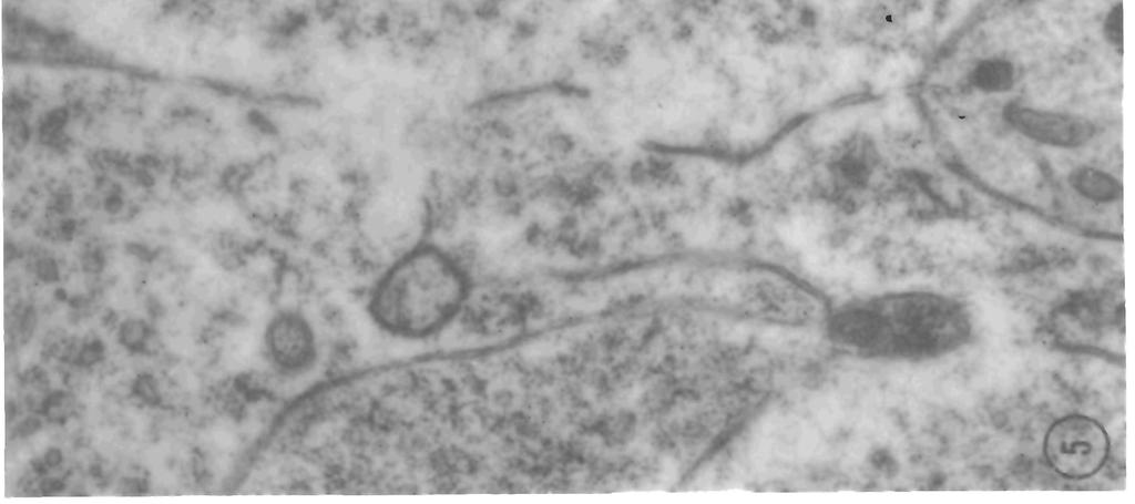
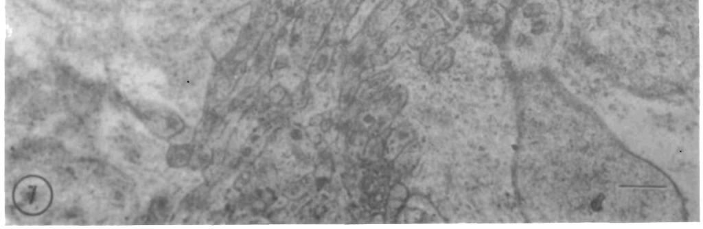
18 Volume 1 Numbers Miiller's cells and "middle limiting membrane" 321 Fig. 1. Human retina (nasal), celloidin-embedded and cut at 12 to 14 /* for orientation. The inner ends (IM) of Miiller's "fibers" are observed passing through the inner plexiform, ganglion cell, and nerve fiber layers to terminate at the internal limiting membrane (ILM). The outer portions (OM) of Miiller's cells are not visible as "fibers" but appear as clear spaces between the axons (RA) of receptor cells. (Hematoxylin and eosin stain. x440.) Fig. 2. The inner boundary of the Muller cell ends as an irregularly infolded plasma membrane (PL) to which is intimately applied a thick basement membrane (BM) measuring here 0.4 to 1.2 [i. This membrane is smooth on its innermost surface from which the filaments of the vitreous framework are presumed to have separated in these preparations. Double membranes representing the plasma membranes of adjacent Muller cells can be seen clearly at PM. Neurites (N) cut obliquely are seen in the nerve fiber layer at the bottom of the micrograph. The cytoplasm of the Muller cells contains elongated, moderately electron-dense mitochondria (M) and large aggregates of radially oriented intracellular filaments (F). Among these filaments there are electron-dense particles (P). Larger vesicles (V) of lesser electron density are found throughout the cytoplasm. (Nasal macula, uranyl acetate treated. xl2,000.) Fig. 3. Within the nerve fiber layer of the retina the Muller cells (upper right portion of field) contain large aggregates of radially oriented, delicate (100 to 120 A) intracellular filaments (F) among which are frequently found elongated mitochondria (M) that are also oriented radially. Cytoplasmic extensions (E) of the Muller cells partially enclose small groups of neurites (N) and so serve as an intercellular matrix within the nerve fiber bundles. Vesicles of moderate electron density (V) are present in many areas of the cytoplasm. (Nasal retina, untreated, x 17,000.) Fig. 4. Although the nuclei of Miiller's cells may be found at all levels of the inner nuclear layer, in this micrograph the nucleus of a Muller cell (NUC) is seen at the junction of the inner plexiform and inner nuclear layers. Interweaving neurites of the inner plexiform layer (IPL) are present in the upper right corner of the micrograph. The cytoplasm of the Muller cell here is less filamentous but more densely packed with linearly oriented granules and vesicles of moderate electron density measuring about 400 to 500 A. In the outermost part (lower left corner) of the Muller cells in this micrograph, clusters of small (150 A) dense granules interpreted as probable ribonucleoprotein particles (RNP) are seen. Several artifactitious linear densities (A) are present. The irregularly lucent area in the cytoplasm at D is interpreted as early degeneration due to extreme sensitivity of the Muller cell to the manipulations involved in obtaining and preparing the tissue. (Nasal retina, untreated. xl7,000.) Fig. 5. Muller cell cytoplasm near the nucleus (NUC) from a section of nasal retina cut obliquely. Mitochondria (M) are present but they do not have the narrow, elongated appearance of those in the innermost part of the cell. Intracellular filaments (F) are found in little aggregates, poorly oriented within the more electron-lucent cytoplasm. Very short segments of granular endoplasmic reticulum (ER) and some clusters of free ribonucleoprotein particles (RNP) are present. Tubular (T) and vesicular (V) profiles are observed in the cytoplasm. (Nasal retina, uranyl acetate treated. x24,600.) Fig. 6. Cytoplasm of Muller cell passing outward through the inner plexiform layer. Small numbers of radially oriented filaments (F) are present as well as elongated mitochondria (M) and moderately electron-dense granules, vesicles, or short segments of tubules (T). The Muller cell cytoplasm (MC) extends laterally to occupy all "space" and to serve as an interstitial matrix between the neurites. The large numbers of vesicles (V) within the neurites of the inner plexiform layer suggest the presence of synapses nearby. (Nasal macula, uranyl acetate treated. xl4,400.) Fig. 7. A Muller cell can be seen extending from its nucleus (NUC) within the inner nuclear layer through the innermost portion (IP) of the outer plexiform layer to reach past the synaptic layer into the "spaces" between the receptor cell axons of the "fiber" portions of the outer plexiform layer (lower right corner of field). The cytoplasm of the Muller cell here differs from that near the internal limiting membrane in being less fibrous and less compact in its arrangement. NI, nuclei of neurons in the inner nuclear layer. (Nasal macula, uranyl acetate treated. x9,900.)
, through the innermost portion (IP) of the outer plexiform layer to reach the synaptic layer (SL) of the outer")
19 Investigative Ophthalmology 322 Fine and Zinwiennan 7«nei962 Fig. 8. Cytoplasm of Miiller cell passing from the outer layers of the inner nuclear layer (NUC), through the innermost portion (IP) of the outer plexiform layer to reach the synaptic layer (SL) of the outer plexiform layer. Golgi apparatus in the Miiller cell is present at G. The cytoplasm of a neuron containing a grouping of granular endoplasmic reticulum and clusters of free ribonucleoprotein granules in a pattern similar to Nissl substance is seen at NS. A typical synaptic spherule (SS) of a receptor rod cell is present in the lower right corner of the field. Small numbers of disoriented intracellular filaments (F) are seen within the Miiller cell. The Muller cell cytoplasm extends laterally (MC) to occupy all "space" between the neural cells. (Nasal macula, uranyl acetate treated. xl5,500.) Fig. 9. The cytoplasm of a Muller cell in the innermost portion (IP) and synaptic layer (SL) of the outer plexiform layer. The peculiarly electron-lucent cytoplasm (MC) of the Muller cell is easily identified between the neural cells (N). Two typical rod cell terminal synaptic spherules (SS), each containing a dense synaptic lamella (SL), are present. (Nasal macula, uranyl acetate treated. x34,250.) Fig. 10. Outer plexiform layer of the retina cut obliquely to show more of Muller cell cytoplasm (MC) among the neurites (N) and about the synaptic terminals of the receptor cells (ST). A few densities (free arrows) of adjacent neurite plasma membranes and cytoplasm are seen in this plexiform layer (for more detailed demonstrations, see Figs. 11 and 12). The cytoplasm of Miiller's cell contains filaments, dense particles, fragments of granular endoplasmic reticulum, small and large vesicles of low electron density, and mitochondria similar to those found elsewhere in the cell, but, here, these elements appear to be less concentrated and less well oriented. (Nasal retina, uranyl acetate treated. xl0,200.) Fig. 11. Innermost portion of outer plexiform layer showing some dendrites (D) of a single cell leading from synaptic connection with a multisynaptic cone foot (CF). The neurites of this layer are interwoven in a complex manner but short densities (desmosomes, Ds) can be seen between a number of these neurites. Similar segmental plasma membrane densities (SD) are clearly seen in the synaptic areas. DT, is a dendrite leading away from a synaptic connection with the cone foot. (Nasal retina, untreated. xl8,600.) Fig. 12. A portion of a cone foot synaptic area (CS) (lower portion of field) filled with a large number of synaptic vesicles. A typical dense cone synaptic lamella characteristically surrounded by a grouping of synaptic vesicles is seen at SL. The upper part of the micrograph shows the interweaving neurites (N) of the innermost portion of the outer plexiform layer. Shorter densities (D) are observed periodically along adjacent plasma membranes associated with an increase in density of the adjacent neurite cytoplasm. These are interpreted as attachment plates or desmosomes. Similar short dense segments of adjacent plasma membranes are seen along the complex synaptic membranes, synaptic densities (SD). (Nasal macula, untreated. x60,000.) Fig. 13. Nuclei (NUC) and axons (A) of the visual cells within the outer nuclear layer occupy most of this field but the proximal part of the inner segments of the rods (R) and cones (C) are present at the bottom of the field. Within the outer nuclear layer the space between adjacent nerve cells is filled by the lucent disrupted cytoplasm of Miiller's cells (MC). A series of terminal bar densities (TB) make up the external limiting membrane (see Fine, 15 for more detailed description of this structure). The Muller cells terminate in a large number of delicate villous processes which project outward beyond the external limiting membrane between the inner segments of the rods and cones (Fig. 15). (Nasal macula, untreated. x6,420.) Fig. 14. A cross section through the outer nuclear layer just internal to the external limiting membrane. Nuclei (NUC) of two receptor cells are present. The Golgi apparatus (G) can be seen in the cytoplasm of one receptor cell. The cytoplasm of low density which forms the outer parts of the Muller cells (MC) occupies the spaces between receptor cells. Several cross sections of rod receptor cell axons (RA) are shown. A mitochondrion (MM) and filaments and granules of varying density are seen in the relatively electronlucent cytoplasm of Miiller's cells. (Nasal retina, uranyl acetate treated. x24,600.)
, the external limiting membrane (center of field), and the proximal portion of the rods and cones (lower part of")
20 Volume I Number 3 Midler's cells and "middle limiting membrane" 323 Fig. 15. An oblique section passing through the outer nuclear layer (upper part of field), the external limiting membrane (center of field), and the proximal portion of the rods and cones (lower part of field). The lucent cytoplasm of the outer ends of the Miiller cells (MC) fills the spaces between neural cells (NL). Densities in the apposing plasma membranes (terminal bars, TB) of adjacent neural cells and the Miiller cells give rise to the external limiting membrane. The delicate villous prolongations (V) of the Miiller cells lie free within a mucoid matrix which fills the spaces between the inner segments of the receptor cells (R). Vesicles of smooth-surfaced endoplasmic reticulum and the Golgi apparatus (G) may be seen within the receptor cell inner segments (R). (Nasal retina, uranyl acetate treated. x22,750.) The cytoplasmic constituents of Miiller's cells vary in concentration and in degree of orientation in the several retinal layers. In the inner layers there is the greatest concentration and radial orientation of delicate filaments similar to those that have been observed in fibrous astroglia. 59 " 22 Closely associated with these filaments are minute particles. Large, less dense granules and vesicles are also present in considerable numbers. Scattered, elongated, radially oriented mitochondria are present throughout. The concentration and orientation of these cytoplasmic organelles remain much the same throughout the inner portion of the cell, except in the macular area where these structures appear less compacted in the inner plexiform layer. In the vicinity of the nucleus the cytoplasmic constituents seem less concentrated and the filaments are less well oriented. The mitochondria appear less elongated. The decrease in concentration and the lack of orientation of organelles become greater from the nuclear region toward the outer limiting membrane. As a result, the cytoplasm of the Miiller cells appears less dense in the outer layers as compared with its appearance in the inner layers. Throughout the course of this study, it was apparent that the cytoplasm of Miiller's cells was more difficult to preserve and therefore interpreted as being more sensitive to artifactitious disruption during preparation as compared with the neural elements of the retina. Similarly, the outer half of the Miiller cell appeared to be much more sensitive than the inner half. In the inner portion of the outer plexiform layer numerous desmosomes were observed between adjacent neurites and similar densities were found along the synaptic membranes at the synaptic endings of the receptor cells. Discussion From our electron microscopic observations it would appear that the Miiller cell is a peculiar glial cell which has some of the characteristics of epithelial cells, astrocytes, and oligodendrocytes. In common with many epithelial cells it has a somewhat tall columnar outline, a broad base to which there is applied an adherent basement membrane (the internal limiting membrane of the retina), and an apical end containing terminal bars and villous projections into a lumen (the mucoid-filled spaces between the inner segments of receptor cells). The basal half of the cell between the nucleus and the internal limiting membrane contains a large number of radially oriented filaments similar to those of fibrous astrocytes and it also contains many other organelles including mitochondria, segments of smooth-surfaced endoplasmic reticulum, and a variety of granules and vesicles. The outer (apical) half of the cell, by contrast, seems to have properties in common with other glia that have beeninterpreted as oligodendrocytes, 19 ' not the least of which is the extreme sensitivity of the cell which permits it to undergo artifactitious alterations so promptly. This observation gives us a better understanding of the "edematous" appearance which is so characteristic of the outer plexiform layer in ordinary cell oidin or paraffin sections. The fibers observed in such sec-

21 324 Fine and Zimmerman Investigative Ophthalmology June 1962 tions may represent axons of the receptor cells, whereas the intervening clear spaces represent the disintegrated Miiller cells. It is possible, also, that similar cytologic changes occur in pathologic states. For example, in microcystoid degeneration which typically begins in the outer plexiform layer, it is possible that the "cysts" represent a group of disintegrated Miiller cells. Since these cysts contain hyaluronic acid, 23 this then would be a mucoid elaboration by the "oligodendroglial portion" of the Miiller cell. In microcystoid degeneration and retinoschisis, septa between adjacent cysts have been assumed to be Miiller cells matted together by pressure from surrounding cysts.- 8 It seems likely, however, that this assumption is incorrect and that the septa actually represent axons of the receptor cells matted together. At any rate, it has been a common observation in pathologic conditions of the retina that hyaline thickening of Miiller's fibers seems to be restricted to the inner half of the retina between the inner nuclear layer and the inner limiting membrane. It is most unusual to observe similar hyaline thickening of Miiller's fibers between the inner nuclear layer and the outer limiting membrane. Perhaps the inner half of the cell reacts in the fashion of a fibrous astrocyte whereas the outer half behaves more like an oligodendrocyte. These are, at the moment, speculations, for we have not yet studied retinas with pathologic conditions by electron microscopy. Although a variety of granules and vesicles are observed in great numbers throughout the inner portion of Miiller's cells, we have no way of determining whether any of these represent the glycogen that has been demonstrated in these cells by histochemical techniques. 4 It is of interest to note that the inner portion of the Miiller cells which shows the greatest lactic-dpn dehydrogenase activity also contains the greater number of mitochondria. Our observations of Miiller's cells of the human retina are in general agreement with the more limited descriptions of these cells in other animal eyes published by Sjostrand, 11 ' 12 Ladman, 13 and Cohen. 14 Sjostrand, 11 who made brief reference to the outer part of Miiller's cells in his study of the retinal rod synapses in the guinea pig eye, called attention to the fact that the cytoplasm of the Miiller cell can be easily recognized and distinguished from that of the other components of the retina. Cohen, 14 in his study of the inner end of these cells in the rhesus monkey retina was in agreement that the Miiller cell cytoplasm has a distinctive electron microscopic appearance. He also pointed out that near the internal limiting membrane the cells had a fibrous character similar to that of fibrous astrocytes of other parts of the central nervous system. Ladman 13 also described the inner part of Miiller's cells as having a distinctly filamentous character. Neither Cohen nor Ladman recorded any observations of the outer part of Miiller's cells and the electron microscopic similarity of the outer part of Miiller's cells to oligodendrocytes has apparently not yet been reported. A few of the observations made by Cohen and Ladman suggested the possibility of species variation. Ladman, for example, reported that mitochondria were rare in Miiller cells of the cat retina. We found them distinctly fewer in Miiller cells than in the inner segments of the receptor cells, but, on the other hand, they were certainly not rare in the inner half of the cell in human retinas. They were much less numerous in the outer part of the cell than in the inner. Cohen found that the inner (vitreal) surface of Miiller's cells of the rhesus monkey retina was more irregular with more infolding of the plasma and basement membranes than we have noted in the human eye. He observed desmosomes between adjacent glial cells which we have not yet encountered. He reported that the glial cell processes of the nerve fiber layer were not closely packed and even found appreciable extracellular spaces between adjacent cells. Our impression has been that, in the human eye, the Miiller

22 Volume 1 Number 3 Mutter's cells and "middle limiting membrane" 325 cell processes fill up all potential spaces between bundles of axons in the nerve fiber layer and that there are no appreciable extracellular spaces. Sjostrand, 24 who has made similar observations, suggested that the glial elements of the retina (the Miiller cells), like the neuroglia of the brain, represent the extracellular space. The outer plexiform layer is generally subdivided into three parts: (a) a broad outer band composed of axons of the receptor cells and the outer halves of the Miiller cells; (b) a very narrow synaptic band; and (c) another narrow band of interweaving neurites between the synaptic band and the inner nuclear layer. Together, the two narrow bands, (b) and (c), form a sort of "limiting membrane" which tends to restrict the passage of fluids and exudates. In these bands the retinal cells are tightly interwoven, there are innumerable synapses, and desmosomal structures are observed in considerable numbers. All of these structures tend to keep the retinal tissue intact in a plane just external to the inner nuclear layer. We have therefore suggested that this area be considered a "middle limiting membrane." Exudates, hemorrhages, and "cysts" developing in the outer plexiform layer typically are restricted internally by this "membrane" just as they are restricted externally by the outer limiting membrane. It would, therefore, be just as helpful to designate the inner parts of the outer plexiform layer as the "middle limiting membrane" of the retina as it has been useful to designate the series of terminal bars the "outer limiting membrane" although neither is a basement membrane comparable to the inner limiting membrane. REFERENCES 1. Cogan, D. G., and Kuwabara, T.: Retinal clehydrogenases, Tr. Am. Ophth. Soc. 57: 154, Kuwabara, T., and Cogan, D. G.: Tetrazolium studies on the retina. I. Introduction and technique, J. Histochem. & Cytochem. 7: 329, Cogan, D. G., and Kuwabara, T.: Tetrazolium studies on the retina. II. Substrate dependent patterns, J. Histochem. & Cytochem. 7: 334, Kuwabara, T., and Cogan, D. G.: Retinal glycogen, A. M. A. Arch. Ophth. 66: 680, Pearse, A. G. E.: Localization of oxidative enzymes in rat and chick retina in various physiologic conditions, in Smelser, G. K., editor: The structure of the eye, New York, 1961, Academic Press, Inc., pp Berkow, J. W., and Patz, A.: Histochemistry of the retina. I. Introduction and methods, A. M. A. Arch. Ophth. 65: 820, Berkow, J. W., and Patz, A.: Histochemistry of the retina. II. Use of phenazine methosulfate to demonstrate the succinoxidase system, A. M. A. Arch. Ophth. 65: 828, Polyak, S. L.: The retina, Chicago, 1941, University of Chicago Press. 9. Last, R. J.: Eugene Wolff's anatomy of the eye and orbit, ed. 5, London, 1961, H. K. Lewis & Co., Ltd. 10. Duke-Elder, W. S., and Wybar, K. C: The anatomy of the visual system, in Duke- Elder, W. S., editor: System of ophthalmology, London, 1961, Henry Kimpton, Vol. II. 11. Sjostrand, F. S.: Ultrastructure of retinal rod synapses of the guinea pig eye as revealed by three dimensional reconstruction from serial sections, J. Ultrastruct. Res. 2: 122, Sjostrand, F. S.: The ultrastructure of the retinal receptors of the vertebrate eye, Ergebn. Biol. 21: 128, Ladman, A. J.: Electron microscopic observations on the fine structure of Miiller cells in the retina of the cat, Anat. Rec. 139: (Abst.) 247, Cohen, A. I.: Electron microscopic observations of the internal limiting membrane and optic fiber layer of the retina of the rhesus monkey (M. Mulatta), Am. J. Anat. 108: 179, Fine, B. S.: Limiting membranes of the sensory retina and pigment epithelium: An electron microscopic study, A. M. A. Arch. Ophth. 66: 847, Dalton, A. J.: A chrome-osmium fixative for electron microscopy, Anat. Rec. 121: 281, Luft, J. H.: Improvements in epoxy resin embedding methods, J. Biophys. & Biochem. Cytol. 9: 409, Zimmerman, L. E.: Acid mucopolysaccharides in ocular histology and pathology, Proc. List. Med. Chicago 23: 267, Luse, S. A.: Electron microscopic observations of the central nervous system, J. Biophys. & Biochem. Cytol. 2: 531, 1956.

23 326 Fine and Zimmerman Inoestigatioe Ophthalmology June Luse, S. A.: Ultrastructure of reactive and neoplastic astrocytes, Lab. Invest. 7: 401, Luse, S. A.: The ultrastructure of normal and abnormal oligodendroglia, Anat. Rec. 138: 461, Luse, S. A., and Harris, B.: Brain ultrastructure in hydration and dehydration, Arch. Neurol. 4: 139, Zimmerman, L. E., and Spencer, W. H.: The pathologic anatomy of rerinoschisis with a report of two cases diagnosed clinically as malignant melanoma, A. M. A. Arch. Ophth. 63: 10, Sjostrand, F. S.: Electron microscopy of the retina, in Smelser, G. K., editor: The structure of the eye, New York, 1961, Academic Press, Inc., pp Discussion Dr. J. Reimer Wolter, Ann Arbor, Mich. The radial fibers of the retina were discovered in vertebrates by Heinrich Miiller in Kolliker found similar fibers in the human retina and called them "Miiller's fibers." Miiller, Kolliker, and most other classical anatomists considered the radial fibers the main supporting cells of the retina. Recently it has been shown that the radial fibers may also have important metabolic functions. However, it seems to be important to emphasize that Miiller's fibers must still be considered the main supporting element of the retina; other functions are additional. The Golgi silver bichromate stain has made it possible for the classical anatomist to obtain a complete demonstration of Miiller's fibers. This technique characteristically stains the outlines of only one particular cell with all its processes out of the continuous system of these cells. Cajal, Polyak, and many others showed single radial fibers after Golgi stain interconnecting the outer and inner limiting membranes and with an additional horizontal network of processes and fibrils extending between every neurone and synapse of the retinal layers. Two main difficulties prevented a final understanding of the system of the radial fibers with this technique: (1) it was impossible to stain adjacent elements, and, therefore, a demonstration of the complete system of the radial cells was impossible, and (2) artifacts caused many to doubt the results of the Golgi stain. With Hortega's silver carbonate technique for the demonstration of glia, it seems to be impossible to obtain a complete demonstration of the Miiller cells. Only the astroglia-like part in the inner retinal layers can be well demonstrated. That part which Fine and Zimmerman found to be more oligodendroglia-like does not stain with silver carbonate just as the true oligodendroglia of the retina, if there is any. This short review shows clearly how important the present study of Fine and Zimmerman is for the progress in retinal morphology. Fine and Zimmerman are finally able to state that horizontal cytoplasmic extensions from Miiller's fibers surround and support all nerve cells, dendrites, and axons of the retina. The findings of Fine and Zimmerman are supported by those of Christopher Pedler of the Institute of Ophthalmology in London, England. Dr. Pedler demonstrated his unpublished electron microscopic findings concerning the structure of Miiller's fibers to me this summer. Pedler's views are best summarized by these words from his recent letter, "I now think that the radial fiber represents negative space, that is to say, space where neuronal tissue is not. This may sound a bit strange, but I am sure it is right." Thus, it becomes clear that the Miiller fibers have a supporting function in two dimensions. Their main cell and the astroglia-like fibers support the retina as a whole. Their oligodendroglialike processes extend between all the delicate neuronal parts of the retina to support and probably also to supply nutrition. The suggestion of the authors that the inner part of the radial fibers react in the fashion of fibrous astrocytes, whereas the outer half behaves more like oligodendrocytes is a new and very exciting concept. I know that their inner part is very similar to the retinal astroglia and I am ready to believe that the outer part is oligodendroglia-like. It is good to see that the authors still use the term "inner limiting membrane" after explaining that this is the basement membrane of the retina formed by the inner end of Miiller's fibers. This prevents confusion. The structure which Fine and Zimmerman describe as "middle limiting membrane" can be seen with hematoxylin and eosin stain and after silver impregnation. This interrupted layer certainly represents a limit of pathology in many abnormal retinal conditions. Thus, this new term of the authors will help in the understanding and teaching of retinal pathology. The presence of the middle limiting membrane is very obvious in cases of horizontal folding of the retina, caused by orbital tumors. This layer as well as the inner limiting membrane is nonelastic and shows most of the folding whereas the outer limiting membrane and the retinal tissues between are more elastic and show less wrinkling.
Ultrastructural changes in moist chamber corneas. E. M. Schaeffer"
 Ultrastructural changes in moist chamber corneas E. M. Schaeffer" Crossly normal human corneas received through the Iowa Eye Bank and stored in cold moist chambers for 12, 24, 36, 72, and 96 hours were
Observations on the rod and cone layer of the human retina
 Observations on the rod and cone layer of the human retina A light and electron microscopic study Ben S. Fine and Lorenz E. Zimmerman Electron microscopy combined with certain histochemical studies on
Relation Between Superficial Capillaries and Foveal Structures in the Human Retina
 Relation Between Superficial Capillaries and Foveal Structures in the Human Retina Masayuki Iwasaki and Hajime Inomara When examining semithin Epon sections of human retinas, it became evident that superficial
R'etinal Mullerian cells have several
 Comparative study of the fine structure of retinal Miiller cells in various vertebrates Shigekazu Uga and George K. Smelser A comparative study was made of the fine structure of Midler's cells in various
Reports. Table I. Identification of specimen studied. Ultrastructural description of a "cylinder organelle" in the outer plexiform layer
 Reports Ultrastructural description of a "cylinder organelle" in the outer plexiform layer of human retinas. JOSEPH CRAT, DANIEL M. ALBERT, AND TED W. REID. A cylinder-like organelle ("cylinder organelle")
Xenopus laevis (Daudin)
") The Structure of Myelin Sheaths in the Central Nervous System of Xenopus laevis (Daudin) By A. PETERS, Ph.D. (From the Department of Anatomy, University of Edinburgh, Scotland) PLATES 43 To 47 (Received
The Structure of Myelin Sheaths in the Central Nervous System of Xenopus laevis (Daudin) By A. PETERS, Ph.D. (From the Department of Anatomy, University of Edinburgh, Scotland) PLATES 43 To 47 (Received
Fine structure of the retina of black bass, Micropterus salmoides (Centrarchidae, Teleostei)
") Histol Histopathol (1 999) 14: 1053-1 065 http://www.ehu.es/histoi-hlstopathol Histology and Histopathology Fine structure of the retina of black bass, Micropterus salmoides (Centrarchidae, Teleostei)
Histol Histopathol (1 999) 14: 1053-1 065 http://www.ehu.es/histoi-hlstopathol Histology and Histopathology Fine structure of the retina of black bass, Micropterus salmoides (Centrarchidae, Teleostei)
Cells. Steven McLoon Department of Neuroscience University of Minnesota
 Cells Steven McLoon Department of Neuroscience University of Minnesota 1 Microscopy Methods of histology: Treat the tissue with a preservative (e.g. formaldehyde). Dissect the region of interest. Embed
The formation of zymogen granules in the pancreas of the mouse By S. K. MALHOTRA
 The formation of zymogen granules in the pancreas of the mouse By S. K. MALHOTRA (From the Cytological Laboratory, Department of Zoology, Oxford) With 3 plates (figs, i to 3) Summary Electron-dense, granular
Tokuhiro JSHIHARA, Chotatsu TSUKAYAMA, Fumiya UCHINO
 (39) JOURNAL OF ELECTRON MICROSCOPY 39 Vol. 22, No. I, 39-44, 1973 Intramitochondrial Filamentous Structures in Human Reticulum Cells in the Bone Marrow Tokuhiro JSHIHARA, Chotatsu TSUKAYAMA, Fumiya UCHINO
Aberrant Mitochondria with Longitudinal Cristae Observed in the Normal Rat Hepatic Parenchymal Cell. Takuma Saito and Kazuo Ozawa
 Okajimas Fol. anat. jap., 44 : 357-363, 1968 Aberrant Mitochondria with Longitudinal Cristae Observed in the Normal Rat Hepatic Parenchymal Cell By Takuma Saito and Kazuo Ozawa Department of Anatomy, Kansai
Landolt's club in the amphibian retina: A Golgi and electron microscope study. Anita Hendrickson
 Landolt's club in the amphibian retina: A Golgi and electron microscope study Anita Hendrickson Landolt's club process has been studied in the adult newt retina. In Golgi preparations this process has
Neurochemistry 1. Nervous system is made of neurons & glia, as well as other cells. Santiago Ramon y Cajal Nobel Prize 1906
 Neurochemistry 1 Nervous system is made of neurons & glia, as well as other cells. Santiago Ramon y Cajal Nobel Prize 1906 How Many Neurons Do We Have? The human brain contains ~86 billion neurons and
Atypical Neural Sheaths Formed by Muller Cells in Chicken Retina
 Okajimas Folia Anat. Jpn., 57(2-3) : 79-88, August 1980 Atypical Neural Sheaths Formed by Muller Cells in Chicken Retina By YOSHIRO INOUE, YOSHIHIRO SUGIHARA,Yozo NISHIMURA and KAZUYO SHIMAI Department
Electron and Light Microscope Studies of Endamoeba terrapinae
 Proceedings of the Iowa Academy of Science Volume 68 Annual Issue Article 81 1961 Electron and Light Microscope Studies of Endamoeba terrapinae Marilyn Driml Cornell College Copyright Copyright 1961 by
CYTOLOGY & HISTOLOGY THE STUDY OF CELLS AND TISSUES
 NAME: DATE: PARTNER: CYTOLOGY & HISTOLOGY THE STUDY OF CELLS AND TISSUES For ease of study, multicellular animals are often examined at various levels of structural organization. Starting from the most
Chapter 6: A Tour of the Cell
 Chapter 6: A Tour of the Cell 1. The study of cells has been limited by their small size, and so they were not seen and described until 1665, when Robert Hooke first looked at dead cells from an oak tree.
Dr. Dina A. A. Hassan Associate Professor, Pharmacology
 Cytology Dr. Dina A. A. Hassan Associate Professor, Pharmacology Email: da.hassan@psau.edu.sa Cells All living things are made up of cells Basic building blocks of life It is the smallest functional and
Chapter 6: A Tour of the Cell
 AP Biology Reading Guide Fred and Theresa Holtzclaw Chapter 6: A Tour of the Cell Name Period Chapter 6: A Tour of the Cell Concept 6.1 To study cells, biologists use microscopes and the tools of biochemistry
Chapter 4 Active Reading Guide A Tour of the Cell
 Name: AP Biology Mr. Croft Chapter 4 Active Reading Guide A Tour of the Cell Section 1 1. The study of cells has been limited by their small size, and so they were not seen and described until 1665, when
UNUSUAL MITOCHONDRIAL CRISTAE IN THE VINEGAR EELWORM
 UNUSUAL MITOCHONDRIAL CRISTAE IN THE VINEGAR EELWORM BERT M. ZUCKERMAN, MARIAN KISIEL, and STANLEY HIMMELHOCH. From the Laboratory of Experimental Biology, University of Massachusetts, East Wareham, Massachusetts
Some Observations on the Fine Structure of the Giant Nerve Fibers of the Earthworm, Eisenia foetida*
 Some Observations on the Fine Structure of the Giant Nerve Fibers of the Earthworm, Eisenia foetida* By KIYOSHI HAMA,~ M.D. (From the Department of Anatomy, School of Medicine, University of Washington,
Prelab Exercise 1. Cell structure
 Prelab Exercise 1 CELLS, EPITHELIA, GLANDS AND CONNECTIVE TISSUE Prelab guide: this will function as a review of material that will be important to your performance of the lab. The first lab will examine
Cellular Neuroanatomy II The Prototypical Neuron: Neurites. Reading: BCP Chapter 2
 Cellular Neuroanatomy II The Prototypical Neuron: Neurites Reading: BCP Chapter 2 Major Internal Features of a Neuron The neuron is the functional unit of the nervous system. A typical neuron has a soma
Some observations on the fine structure of the. epithelium in the intestine of lamprey. (Lampetra japonica)
") Okajimas Fol. anat. jap., 40: 691-713, 1965 Some observations on the fine structure of the epithelium in the intestine of lamprey (Lampetra japonica) By Torao Yamamoto From the Department of Anatomy, Niigata
Okajimas Fol. anat. jap., 40: 691-713, 1965 Some observations on the fine structure of the epithelium in the intestine of lamprey (Lampetra japonica) By Torao Yamamoto From the Department of Anatomy, Niigata
(From the Department of Anatomy and the Beaumont-May Institute of Neurology, Washington University School of Medicine, St. Louis)
") ELECTRON MICROSCOPIC OBSERVATIONS OF THE OLFACTORY MUCOSA AND OLFACTORY NERVE* BY A. J. DE LORENZO, PH.D. (From the Department of Anatomy and the Beaumont-May Institute of Neurology, Washington University
ELECTRON MICROSCOPIC OBSERVATIONS OF THE OLFACTORY MUCOSA AND OLFACTORY NERVE* BY A. J. DE LORENZO, PH.D. (From the Department of Anatomy and the Beaumont-May Institute of Neurology, Washington University
Atypical mitochondria in the ellipsoid of the photoreceptor cells of vertebrate retinas
 Atypical mitochondria in the ellipsoid of the photoreceptor cells of vertebrate retinas Toyoko Ishikawa and Eichi Yamada The fine structure of ellipsoids in a number of vertebrate retinas was studied in
Concept 6.1 To study cells, biologists use microscopes and the tools of biochemistry
 Name Period Chapter 6: A Tour of the Cell Concept 6.1 To study cells, biologists use microscopes and the tools of biochemistry 1. The study of cells has been limited by their small size, and so they were
Tissues: - A group of cells similar in structure and performing a particular function forms a tissue.
 Plant Tissues Class- IX Tissues: - A group of cells similar in structure and performing a particular function forms a tissue. PLANT TISSUES ANIMAL TISSUES 1. Most of the plant tissues are Most of the tissues
* ra. VOL. 53, 1965 ANATOMY: D. BODIAN 419. FIG. 1.-Synaptic contact in spinal motoneuron of a chimpanzee. Large dendrite (D) shows
 shows") A SUGGESTIVE RELATIONSHIP OF NERVE CELL RNA WITh1 SPECIFIC SYNAPTIC SITES* BY DAVID BODIAN DEPARTMENT OF ANATOMY, JOHNS HOPKINS UNIVERSITY SCHOOL OF MEDICINE Communicated December 29, 1964 In a previous
A SUGGESTIVE RELATIONSHIP OF NERVE CELL RNA WITh1 SPECIFIC SYNAPTIC SITES* BY DAVID BODIAN DEPARTMENT OF ANATOMY, JOHNS HOPKINS UNIVERSITY SCHOOL OF MEDICINE Communicated December 29, 1964 In a previous
NATIONAL REVIEW COURSE. Cells, Tissues, and Membranes
 NATIONAL REVIEW COURSE Cells, Tissues, and Membranes I. Cell Types A. Prokaryote bacteria cells; a cell that does not have a nucleus in which to store its genetic material. B. Eukaryote plant or animal
Cells of the nervous system
 Cells of the nervous system There are approximately 100 billion neurons in the human brain There are about 100 times as many glial cells in the human brain Similar origin, different functions Other cells
7-2 Eukaryotic Cell Structure
 1 of 49 Comparing the Cell to a Factory Eukaryotic Cell Structures Structures within a eukaryotic cell that perform important cellular functions are known as organelles. Cell biologists divide the eukaryotic
infrastructure of Remnant Photoreceptors in Advanced Hereditary Retinal Degeneration
 Articles infrastructure of Remnant Photoreceptors in Advanced Hereditary Retinal Degeneration John R. Cotter* and Werner K. Noellf The outer layers of the retinas of pigmented rats affected with hereditary
Dendrites - receives information from other neuron cells - input receivers.
 The Nerve Tissue Neuron - the nerve cell Dendrites - receives information from other neuron cells - input receivers. Cell body - includes usual parts of the organelles of a cell (nucleus, mitochondria)
Class IX: Biology Chapter 5: The fundamental unit of life. Chapter Notes. 1) In 1665, Robert Hooke first discovered and named the cells.
 In 1665, Robert Hooke first discovered and named the cells.") Class IX: Biology Chapter 5: The fundamental unit of life. Key learnings: Chapter Notes 1) In 1665, Robert Hooke first discovered and named the cells. 2) Cell is the structural and functional unit of all
Class IX: Biology Chapter 5: The fundamental unit of life. Key learnings: Chapter Notes 1) In 1665, Robert Hooke first discovered and named the cells. 2) Cell is the structural and functional unit of all
Intro and Homeostasis
 Intro and Homeostasis Physiology - how the body works. Homeostasis - staying the same. Functional Types of Neurons Sensory (afferent - coming in) neurons: Detects the changes in the body. Informations
(From Departamento de Ultraestructura Celular, Instituto de Investigaci6n de Ciencias Biol6gicas, Montevideo, Uruguay)
") Published Online: 25 May, 1956 Supp nfo: http://doi.org/10.1083/jcb.2.3.319 Downloaded from jcb.rupress.org on July 5, 2018 ELECTRON MCROSCOPE OBSERVATONS ON THE SUBMCROSCOPC ORGANZATON OF THE RETNAL RODS*
Published Online: 25 May, 1956 Supp nfo: http://doi.org/10.1083/jcb.2.3.319 Downloaded from jcb.rupress.org on July 5, 2018 ELECTRON MCROSCOPE OBSERVATONS ON THE SUBMCROSCOPC ORGANZATON OF THE RETNAL RODS*
ANUMBER of electron microscope studies have been made on Amoeba
 An Electron Microscope Study of a Small Free-living Amoeba (Hartmanella astronyxis) By K. DEUTSCH and M. M. SWANN (From the Department of Zoology, University of Edinburgh) With two plates (figs. I and
For more information about how to cite these materials visit
 Author(s): Peter Hitchcock, PH.D., 2009 License: Unless otherwise noted, this material is made available under the terms of the Creative Commons Attribution Non-commercial Share Alike 3.0 License: http://creativecommons.org/licenses/by-nc-sa/3.0/
From the Daniel Baugh Institute of Anatomy, Jefferson Medical College of Philadelphia
 ELECTRON MICROSCOPY OF THE OXYNTIC CELL IN THE GASTRIC GLANDS OF THE BULLFROG, RANA CATESBIANA II. The Acid-Secreting Gastric Mucosa ALBERT W. SEDAR, Ph.D. From the Daniel Baugh Institute of Anatomy, Jefferson
Biology. 7-2 Eukaryotic Cell Structure 10/29/2013. Eukaryotic Cell Structures
 Biology Biology 1of 49 2of 49 Eukaryotic Cell Structures Eukaryotic Cell Structures Structures within a eukaryotic cell that perform important cellular functions are known as organelles. Cell biologists
CELLS STRUCTURE AND FUNCTION
 CELLS STRUCTURE AND FUNCTION Jhia Anjela D. Rivera Department of Biological Sciences School of Science and Technology Centro Escolar University DISCOVERY OF CELLS Robert Hooke (1665): Observed a thin slice
PHYSIOLOGY CHAPTER 9 MUSCLE TISSUE Fall 2016
 PHYSIOLOGY CHAPTER 9 MUSCLE TISSUE Fall 2016 2 Chapter 9 Muscles and Muscle Tissue Overview of Muscle Tissue types of muscle: are all prefixes for muscle Contractility all muscles cells can Smooth & skeletal
Cells and Their Organelles
 Mr. Ulrich Regents Biology Name:.. Cells and Their Organelles The cell is the basic unit of life. The following is a glossary of animal cell terms. All cells are surrounded by a cell membrane. The cell
Cells and Their Organelles
 Cells and Their Organelles The cell is the basic unit of life. The following is a glossary of animal cell terms. All cells are surrounded by a cell membrane. The cell membrane is semipermeable, allowing
Cell Structure and Function Practice
 Cell Structure and Function Practice 1. The National Aeronautics and Space Agency (NASA) has a command center in Houston, Texas, that directs space missions. Which part of a cell functions like this command
(From the Department of Anatomy, University of Toronto, Toronto, Canada)
") THE STRUCTURE OF PIGEON BREAST MUSCLE MITOCHONDRIA* B~t ALLAN F. HOWATSON, PH.D. (From the Department of Anatomy, University of Toronto, Toronto, Canada) PLAT~.S 124 AND 125 The fine structure of mitochondfia
THE STRUCTURE OF PIGEON BREAST MUSCLE MITOCHONDRIA* B~t ALLAN F. HOWATSON, PH.D. (From the Department of Anatomy, University of Toronto, Toronto, Canada) PLAT~.S 124 AND 125 The fine structure of mitochondfia
Basic Structure of a Cell
 Basic Structure of a Cell Introduction to Cells Cells are the basic units of organisms Cells can only be observed under microscope Basic types of cells: Animal Cell Plant Cell Bacterial Cell 1 2 Number
Parenchyma Cell. Magnification 2375X
 Parenchyma Cell The large size of parenchyma cells is due in part to their relatively large vacuole (V) and in part also to the large number of chloroplasts (Cp) they contain. From a crimson clover, Trifolium
* Work supported by a grant from The Rockefeller Foundation. Received for publication, May 20, 1958.
 Published Online: 25 September, 1958 Supp Info: http://doi.org/10.1083/jcb.4.5.667 Downloaded from jcb.rupress.org on December 16, 2018 Mitochondrial Changes in the Adrenocortex of Normal Hamsters.* BY
LI ow-resistance intercellular pathways
 Interreceptoral junctions in the teleost retina P. Witkovsky, M. Shakib, and H. Ripps Junctions between photoreceptors of carp and catfish were examined to determine the potential pathways for interaction
Cellular Neuroanatomy I The Prototypical Neuron: Soma. Reading: BCP Chapter 2
 Cellular Neuroanatomy I The Prototypical Neuron: Soma Reading: BCP Chapter 2 Functional Unit of the Nervous System The functional unit of the nervous system is the neuron. Neurons are cells specialized
Guided Reading Activities
 Name Period Chapter 4: A Tour of the Cell Guided Reading Activities Big Idea: Introduction to the Cell Answer the following questions as you read Modules 4.1 4.4: 1. A(n) uses a beam of light to illuminate
ELECTRON MNIICROSCOPY OF CELLULAR DIVISION IN ESCHERICHIA COLI
 ELECTRON MNIICROSCOPY OF CELLULAR DIVISION IN ESCHERICHIA COLI S. F. CONTII AND M. E. GETTNER' Biology Department, Brookhaven National Laboratory, Upton, New York Received for publication September 18,
Name: Date: Hour:
 Name: Date: Hour: 1 2 3 4 5 6 Comprehension Questions 1. At what level of organization does life begin? 2. What surrounds all cells? 3. What is meant by semipermeable? 4. What 2 things make up the cell
Class XI Chapter 8 Cell The Unit of Life Biology
 Question 1: Which of the following is not correct? (a) Robert Brown discovered the cell. (b) Schleiden and Schwann formulated the cell theory. (c) Virchow explained that cells are formed from pre-existing
Biology: Life on Earth
 Teresa Audesirk Gerald Audesirk Bruce E. Byers Biology: Life on Earth Eighth Edition Lecture for Chapter 4 Cell Structure and Function Copyright 2008 Pearson Prentice Hall, Inc. Chapter 4 Outline 4.1 What
THE BEHAVIOUR OF CHLOROPLASTS DURING CELL DIVISION OF ISOETES LACUSTRIS L.
 New Phytol (1974) 73, 139-142. THE BEHAVIOUR OF CHLOROPLASTS DURING CELL DIVISION OF ISOETES LACUSTRIS L. BY JEAN M. WHATLEY Botany School, University of Oxford (Received 2 July 1973) SUMMARY Cells in
Introduction Principles of Signaling and Organization p. 3 Signaling in Simple Neuronal Circuits p. 4 Organization of the Retina p.
 Introduction Principles of Signaling and Organization p. 3 Signaling in Simple Neuronal Circuits p. 4 Organization of the Retina p. 5 Signaling in Nerve Cells p. 9 Cellular and Molecular Biology of Neurons
Cell Theory. Cell Structure. Chapter 4. Cell is basic unit of life. Cells discovered in 1665 by Robert Hooke
 Cell Structure Chapter 4 Cell is basic unit of life Cell Theory Cells discovered in 1665 by Robert Hooke Early cell studies conducted by - Mathias Schleiden (1838) - Theodor Schwann (1839) Schleiden &
Chapter 4: Cells: The Working Units of Life
 Name Period Chapter 4: Cells: The Working Units of Life 1. What are the three critical components of the cell theory? 2. What are the two important conceptual implications of the cell theory? 3. Which
amphibia and the lower vertebrate retinae with their development. Society of the United Kingdom in 1932.
 RE rinal VISUAL CELLS THE RETINAL VISUAL CELLS IN MAN AND FRESH-WATER FISH BY M. S. MAYOU LONDON WHILST trying to discover some differential stain for the rods and cones I found that Mallory's connective
The Discovery of Cells
 The Discovery of Cells Microscope observations! General Cell & Organelle Discovery 1600s Observations made by scientists using more powerful microscopes in the 1800s led to the formation of the cell theory.
Cell Is the basic structural, functional, and biological unit of all known living organisms. Cells are the smallest unit of life and are often called
 The Cell Cell Is the basic structural, functional, and biological unit of all known living organisms. Cells are the smallest unit of life and are often called the "building blocks of life". The study of
8/25/ Opening Questions: Are all living things made of cells? What are at least five things you know about cells?
 Chapter 3 The Cell: Module Hyperlinks 3.1 Cells are the fundamental units of life 3.2 Plant vs. animal cells 3.3 Membranes: structure 3.4 Membranes: function 3.5 The nucleus 3.6 Organelles in protein production
University of Jordan School of Medicine MD Program Curriculum
 University of Jordan School of Medicine MD Program Curriculum Course Title: General Histology Course number: 0502111 Credit Hours: 2 credits Academic Year Level: 1st year, 2nd semester 2016/2017 Course
Outline. Cell Structure and Function. Cell Theory Cell Size Prokaryotic Cells Eukaryotic Cells Organelles. Chapter 4
 Cell Structure and Function Chapter 4 Cell Theory Cell Size Prokaryotic Cells Eukaryotic Cells Organelles! Nucleus Outline! Endomembrane System! Cytoskeleton! Centrioles, Cilia, and Flagella 1 2 Cell Theory
Vertebrate Physiology 437 EXAM I NAME, Section (circle): am pm 23 September Exam is worth 100 points. You have 75 minutes.
: am pm 23 September Exam is worth 100 points. You have 75 minutes.") 1 Vertebrate Physiology 437 EXAM I NAME, Section (circle): am pm 23 September 2004. Exam is worth 100 points. You have 75 minutes. True or False (write true or false ; 10 points total; 1 point each) 1.
1 Vertebrate Physiology 437 EXAM I NAME, Section (circle): am pm 23 September 2004. Exam is worth 100 points. You have 75 minutes. True or False (write true or false ; 10 points total; 1 point each) 1.
 Question 1: Which of the following is not correct? (a) Robert Brown discovered the cell. (b) Schleiden and Schwann formulated the cell theory. (c) Virchow explained that cells are formed from pre-existing
Histogenesis of the Proventricular Submucosal Gland of the Chick as Revealed by Light and Electron Microscopy
 The Ohio State University Knowledge Bank kb.osu.edu Ohio Journal of Science (Ohio Academy of Science) Ohio Journal of Science: Volume 69, Issue 2 (March, 1969) 1969-03 Histogenesis of the Proventricular
Note on an Amoeba-like Parasite from Clavellina.
 AN AMOEBA-LIKE PARASITE FROM CLAVEM/INA. 413 Note on an Amoeba-like Parasite from Clavellina. By.... Julian S. Huxley, Fellow of New College and Senior Demonstrator in the Department of Comparative Anatomy,
FINE STRUCTURE OF THE RETINULAE IN THE COMPOUND EYE OF THE HONEY-BEE. TIMOTHY H. GOLDSMITH, Ph.D.
 Published Online: 1 September, 1962 Supp Info: http://doi.org/10.1083/jcb.14.3.489 Downloaded from jcb.rupress.org on June 7, 2018 FINE STRUCTURE OF THE RETINULAE IN THE COMPOUND EYE OF THE HONEY-BEE TIMOTHY
Chapter 6: A Tour of the Cell
 Chapter 6: A Tour of the Cell Concept 6.2 Eukaryotic cells have internal membranes that compartmentalize their functions 1. Which two domains consist of prokaryotic cells? 2. A major difference between
World of The Cell. How big is a cell?
 World of The Cell Chapter 4 How big is a cell? The smallest cell is a Mycoplasmas (very small bacteria are barely bigger) Bacteria are just bigger than a single organelle of a animal cell Plant and animal
Electron microscopy of glial cells of the central nervous system in the crab Ucides cordatus
 Brazilian Glial cells Journal in crustaceans of Medical and Biological Research (1999) 32: 327-331 ISSN 0100-879X Short Communication 327 Electron microscopy of glial cells of the central nervous system
Reports 677 REFERENCES. Origin of ghost cell in Coats' disease.
 Reports 677 which are unique to /?n is highly variable among the primates, as it is among other species. A further point which may be significant to the area of human cataractogenesis is the presence in
Today s materials: Cell Structure and Function. 1. Prokaryote and Eukaryote 2. DNA as a blue print of life Prokaryote and Eukaryote. What is a cell?
 Today s materials: 1. Prokaryote and Eukaryote 2. DNA as a blue print of life Prokaryote and Eukaryote Achadiah Rachmawati What is a cell? Cell Structure and Function All living things are made of cells
Association of Tobacco Rattle Virus with Mitochondria
 J. gen. ViroL (I968), 3, I2I-I24 With 3 plates Printed in Great Britain I2I Association of Tobacco Rattle Virus with Mitochondria (Accepted 8 February I968) As part of a study of the way in which tobacco
Topic 3: Cells Ch. 6. Microscopes pp Microscopes. Microscopes. Microscopes. Microscopes
 Topic 3: Cells Ch. 6 -All life is composed of cells and all cells have a plasma membrane, cytoplasm, and DNA. pp.105-107 - The development of the microscope was the key to understanding that all living
Microscope History Robert Hooke
 1 Microscope History Robert Hooke First described cells in 1665. He viewed thin slices of cork and compared the boxy partitions he observed to the cells (small rooms) in a monastery. (1635 1702) 2 Microscope
Cell Structure. Chapter 4. Cell Theory. Cells were discovered in 1665 by Robert Hooke.
 Cell Structure Chapter 4 Cell Theory Cells were discovered in 1665 by Robert Hooke. Early studies of cells were conducted by - Mathias Schleiden (1838) - Theodor Schwann (1839) Schleiden and Schwann proposed
Neurons, Synapses, and Signaling
 Chapter 48 Neurons, Synapses, and Signaling PowerPoint Lecture Presentations for Biology Eighth Edition Neil Campbell and Jane Reece Lectures by Chris Romero, updated by Erin Barley with contributions
Nervous System Organization
 The Nervous System Nervous System Organization Receptors respond to stimuli Sensory receptors detect the stimulus Motor effectors respond to stimulus Nervous system divisions Central nervous system Command
Neurons and Nervous Systems
 34 Neurons and Nervous Systems Concept 34.1 Nervous Systems Consist of Neurons and Glia Nervous systems have two categories of cells: Neurons, or nerve cells, are excitable they generate and transmit electrical
Module 2: Foundations in biology
 alevelbiology.co.uk Module 2: Foundations in biology SPECIFICATION 2.1.1 Cell structure Learners should be able to demonstrate and apply their knowledge and understanding of: (a) The use of microscopy
Acta Medica Okayama. An electron microscopic study on the red, white and inter-mediate muscle fibers of mouse. Takuro Ogata OCTOBER 1964
 Acta Medica Okayama Volume 18, Issue 5 1964 Article 3 OCTOBER 1964 An electron microscopic study on the red, white and inter-mediate muscle fibers of mouse Takuro Ogata Okayama University, Copyright c
Eukaryotic Cells. Figure 1: A mitochondrion
 Eukaryotic Cells Figure 1: A mitochondrion How do cells accomplish all their functions in such a tiny, crowded package? Eukaryotic cells those that make up cattails and apple trees, mushrooms and dust
Basic Structure of a Cell
 Basic Structure of a Cell Prokaryotic Cells No nucleus Archaea & Eubacteria One circular chromosome Extremely small Eukaryotic Cells Has a nucleus!!! Membrane-bound organelles Plants, Animals, Fungi, &
Biology Exam #1 Study Guide. True/False Indicate whether the statement is true or false. F 1. All living things are composed of many cells.
 Biology Exam #1 Study Guide True/False Indicate whether the statement is true or false. F 1. All living things are composed of many cells. T 2. Membranes are selectively permeable if they allow only certain
LOW-POWER ELECTRON MICROSCOPY OF THE ROOT CAP REGION OF EUCALYPT MYCORRHIZAS
 New Phytol. (1968) 67, 663-665. LOW-POWER ELECTRON MICROSCOPY OF THE ROOT CAP REGION OF EUCALYPT MYCORRHIZAS BY G. A. CHILVERS Botany Department, School of General Studies, Australian National University,
678 Reports Invest. Ophthalmol. Visual Sci. REFERENCES
 678 Reports Invest. Ophthalmol. Visual Sci. July 1977 REFERENCES 1. Schmidt, S. Y., Berson, E. L., and Hayes, K. C: Retinal degeneration in cats fed casein. I. Taurine deficiency, INVEST. OPHTHALMOL. 15:47,
NAME: PERIOD: DATE: A View of the Cell. Use Chapter 8 of your book to complete the chart of eukaryotic cell components.
 NAME: PERIOD: DATE: A View of the Cell Use Chapter 8 of your book to complete the chart of eukaryotic cell components. Cell Part Cell Wall Centriole Chloroplast Cilia Cytoplasm Cytoskeleton Endoplasmic
Physiology 2 nd year. Neuroscience Optional Lecture
 Academic year 2018/2019 Physiology 2 nd year Semester 1 Curricula Nervous system physiology Blood physiology Acid-base equilibrium Bibliography: Boron & Boulpaep Medical Physiology, 3 rd edition Physiology
Extracellular Electrical Stimulation of Retinal Ganglion Cells
 Extracellular Electrical Stimulation of Retinal Ganglion Cells by Andrew Eli Grumet Submitted to the Department of Electrical Engineering and Computer Science in partial fulfillment of the requirements
The diagram below represents levels of organization within a cell of a multicellular organism.
 STATION 1 1. Unlike prokaryotic cells, eukaryotic cells have the capacity to a. assemble into multicellular organisms b. establish symbiotic relationships with other organisms c. obtain energy from the
Preparation of Material BRIEF NOTES 531
 BRIEF NOTES 531 Internal Structure of Apoferritin as Revealed by the "Negative Staining Technique."* BY GOETZ W. RICHTER. (From the Department of Pathology, The New York tlospital-cornell Medical Center,
Cell Structure. Chapter 4
 Cell Structure Chapter 4 Cell Theory Cells were discovered in 1665 by Robert Hooke. Early studies of cells were conducted by - Mathias Schleiden (1838) - Theodor Schwann (1839) Schleiden and Schwann proposed
Chapter Life Is Cellular
 Chapter 7 7-1 Life Is Cellular The Discovery of the Cell Anton van Leeuwenhoek used a single-lens microscope to observe tiny little organisms in pond water. The Discovery of the Cell In 1665, Robert Hooke
AS Biology Summer Work 2015
 AS Biology Summer Work 2015 You will be following the OCR Biology A course and in preparation for this you are required to do the following for September 2015: Activity to complete Date done Purchased