Xenopus laevis (Daudin)
|
|
|
- Amy Barker
- 6 years ago
- Views:
Transcription
1 The Structure of Myelin Sheaths in the Central Nervous System of Xenopus laevis (Daudin) By A. PETERS, Ph.D. (From the Department of Anatomy, University of Edinburgh, Scotland) PLATES 43 To 47 (Received for publication, August 17, 1959) ABSTRACT The structure of myelinated nerve fibres has been studied in the spinal cord and optic nerve of the tadpoles of Xenopus laevis. Potassium permanganate-fixed material was examined with the electron microscope. The myelin sheath itself is made up of spirally arranged lamellae in which the intraperiod and dense lines alternate. Inside the myelin sheath an inner cytoplasmic process surrounds the axon and where the external surfaces of its bounding membrane come together an internal mesaxon is formed. The intraperiod line begins within the mesaxon and the dense line usually begins in the same region by apposition of the cytoplasmic surfaces of the membrane. The width of each lamella is 140 A. The outer line in the sheath is the dense line and this terminates in a tongue where the cytoplasmic surfaces of the myelin-forming glial cell separate. Thus, central myelin in Xenopus tadpoles is arranged in the same way as peripheral myelin, the only difference being that in central fibres, cytoplasm on the outside of the sheath is confined to that present in the tongue. For this reason adjacent central sheaths come into apposition without any intervening material being present. When this occurs an intraperiod line is formed between them. Studies on the structure of peripheral and central myelin using x-ray diffraction (1, 2), polarised light (3), and electron microscopy (2, 4-6) show that in both cases the myelin has the same basic structure; it is made up of regular concentric lamellae. In the peripheral nervous system Geren (7) showed that these lamellae, formed by the Schwann cells, are disposed in a spiral around the axon and this helical structure is now generally accepted (4, 6). Much less information is available about the formation and arrangement of the lamellae of central myelin and the two electron microscope studies which have been carried out by Luse (8) on young rats and mice and by De Robertis, Gerschenfeld, and Wald (9) on young cats and rats appear to be inconclusive and have produced conflicting results. Luse (8) suggests that in the central nervous system a single cell does not form the lamellae, as does a Schwann cell within an internode in the peripheral system, but rather that the lamellae are formed by a plication of the membranes of many glial processes, in particular, those of the oligodendrocytes. The theory of De Robertis, J. BIoPHYS1C. AND B1OCHEM. CY~rox., 1960, Vo]. 7, No. 1 ]21 Gerschenfeld, and Wald (9) is somewhat contrary to that of Luse, for while they also state that the myelin is formed by the oligodendrocytes, they consider that it arises within the cytoplasm of these cells, where large numbers of membranes and vesicles are present, membranes being laid down in concentric layers around the axon. The results of these two electron microscope studies, which are based on osmium-fixed material, are so much at variance that it was decided to reinvestigate the problem using potassium permanganate as the fixative (10). This latter fixative produces much better staining of membranes than osmium and has been used to great advantage in the study of peripheral myelin (6, 11). Although potassium permanganate stains membranes effectively, it is a poor fixative for other cellular components, with the result that it is difficult to differentiate between axonal and glial processes. The results of the present investigation are therefore confined to a consideration of the arrangement of the lamellae within the myelin sheaths of the central nervous system.
2 122 MYELIN SHEATHS IN CENTRAL NERVOUS SYSTEM Materials and Methods The material used was the optic nerve and spinal cord of tadpoles of the toad Xenopus laevis (Daudin). The tadpoles examined were at stages 55 to 57 of Nieukoop and Faber (12). Small pieces of tissue were removed and fixed at 0 C. in 1 per cent potassium permanganate in 0.9 per cent saline for 2 hours. They were then washed in 10 per cent alcohol, dehydrated, and embedded in araldite (13). Sections were cut with glass knives on a Porter-Blum "Servall" microtome and examined, without the use of a supporting film, in a Metropolitan Vickers, EM 6, electron microscope. DESCRIPTION In the stages of Xenopus tadpoles examined, both the peripheral and central myelin are at an early phase of development. This can be seen in Fig. 1, which shows the typical arrangement of myelinated fibres in part of the spinal cord and peripheral nerve root in a stage 56 tadpole. The most striking differences between the two types of tissue are the close proximity of adjacent central fibres in comparison with the well separated peripheral fibres, and the absence between the central fibres of nuclei corresponding to those of Schwann cells in the peripheral nerve (Fig. 1 N). Processes of cells are present between the central fibres, but as previously pointed out, since potassium permanganate is a poor fixative for cellular components other than membranes, it is difficult to determine whether these processes belong to either glial cells or neurons. Some of the processes do, however, contain vesicles (Fig. 1 V) which are characteristic of adult neuronal cytoplasm in the region of a synapse (14), but in the present state of our knowledge it would be inappropriate to use these vesicles as a means of certain identification at this early stage of development. The above remarks on the structure of the spinal cord apply equally well to the optic nerve, although no vesiclecontaining processes have been seen within this nerve. The best way to understand the structure of central myelin is to compare it with that of the peripheral nervous system which has been studied extensively. It will therefore be advantageous to consider briefly how peripheral myelin is formed. At an early stage, the Schwann cell forms a spiral around the axon and by obliteration of the cytoplasm between the turns, the membranes of the Schwann cell come together to form compact myelin. The inner and outer ends of the spiral persist as the inner and outer mesaxons (Text-fig. 1 A, M1 and Mo). Schwann cell cytoplasm remains in the first turn of the spiral between the compact myelin and the axon, the inner cytoplasmic process (Text-fig. 1 A, CO, and also in the last turn of the spiral, the outer cytoplasmic process (Text-fig. 1 A, Co). It is in this latter that the nucleus of the Schwann cell is found (Fig. 1, N). As the membranes of the Schwann cell come together to form compact myelin, it has been shown by high resolution electron microscopy (2, 6, 11) that alternating thick and thin lines are formed. These will be referred to as dense lines and intraperiod lines respectively. The dense lines arise as the cytoplasmic surfaces of the Schwann cell membrane become apposed and the intraperiod lines as the external surfaces of the membranes come into contact (Text-fig. 1 A). Throughout this description the term "mesaxon" is retained to describe the double membrane formed by the apposition of the external surfaces of the myelin-forming cell where it is not producing compact myelin. Although this term is not necessarily correct, it is now generally accepted even though Robertson (6) has used the terms "surface-connecting membrane" and "meso" to describe this structure, defining "meso" as the double membrane leading from some included structure to the outside. These latter terms, though more appropriate, have not been widely employed by other workers. Inside the compact myelin, central fibres show similar structures to those present in peripheral fibres. The axon is bounded by an internal cytoplasmic process (Figs. 2 to 5, and 8, C), which surrounds the axon between it and the internal lamellae of compact myelin. As in peripheral nerves, the process forms the first turn of a spiral and at the point where the turn is completed an internal mesaxon arises by the apposition of the external surfaces of the membrane bounding the process (Figs. 2 to 5, and 8, M). Within the internal mesaxon, the thin, intraperiod line arises (Figs. 3 and 4, I). It is usual for the dense line (Figs. 3 and 4, D) to be formed as the mesaxon approaches the innermost layer of compact myelin, for at this point the cytoplasmic surfaces of the membrane bounding the internal cytoplasmic process come together. Although an internal mesaxon is always present, it is not so readily visible when the cross-sectional area of the axon approaches that of the innermost lamella of compact myelin, for then the limiting membranes of the
3 A. PETERS 123 TEXT-FIG. 1. Diagrams to show the structure of peripheral myelin, Text-fig. 1 A, and central myelin, Textfig. 1 B. Cytoplasm of the axon, Schwann cell (Text-fig. 1 A, C1 and Co) and glial cell (Text-fig. 1 B, C1 and Co) is stippled. In both nerves the inner cytoplasmic process, C1, surrounds the axon and its membrane comes together to form the inner mesaxon, MI, in which the first intraperiod line, I, arises. The first dense line, D, is formed by the apposition of the cytoplasmic surfaces of the same membrane. These lines continue in a spiral and terminate at the outside of the sheath. In the peripheral nerve the intraperiod line terminates in the outer mesaxon, Mo, while the denseline terminates by a separation of the cytoplasmic surfaces to enclose the cytoplasm of the outer process, Co, of the Schwann cell. In the central sheath, the dense line terminates as the membranes separate to enclose the cytoplasm, Co, of the tongue, and the intraperiod line ends as the tongue membrane turns away from the outside of the sheath.

4 124 MYELIN SHEATHS IN CENTRAL NERVOUS SYSTEM inner cytoplasmic process approach each other, sometimes with the result that the dense line begins before the intraperiod line is formed. Although the myelin sheaths of peripheral and central fibres have the same internal structure, very obvious differences occur at the outsides of the sheaths. Thus, in the central nervous system processes of adjacent cells may be as close as 100 to 150 A to the external surface of the myelin sheath (Figs. 1, 3, 5, and 8). Such a situation never occurs in peripheral nerves, where the myelin sheaths are well separated by the presence of the outer cytoplasmic process of the Schwann cell, external to the myelin lamellae (Fig. 1 and Text-fig. 1 A, Co). Examination of the outer region of central myelin shows that the external, or last, line in the sheath is a dense line (Figs. 2, 3, 5, and 8) which is formed by the apposition of the cytoplasmic surfaces of the cell membrane. That such is the case, is seen in Figs. 5 to 8, where the outer dense line ends in a tongue applied to the surface of the myelin sheath. In Fig. 5, such a tongue is seen in association with each of the three myelin sheaths and the structure of the tongue on the surface of the lower sheath is shown in Figs. 6 and 7. No breaks in the myelin lamellae have been observed in the present study and the outer dense line always terminates in the same manner, only one tongue being present on the external surface of a myelin sheath (see also Figs. 8 and 10). In this material, the distance from centre to centre of two adjacent dense lines, i.e. the width of each lamella, is ~140 A. These observations on the structure of myelin ~heaths of central fibres have been brought together in Text-fig. 1 B, which shows the suggested ~rrangement of the myelin sheath. For comparison, the generally accepted arrangement of peripheral myelin is shown in Text-fig. 1 A. The only part of this arrangement for which no evidence has been presented is that of the existence of a spiral in central myelin. Owing to compression in the direction in which the sections have been cut, it is not possible to trace the myelin lamellae throughout the entire sheath, but in some sheaths a few lamellae can be followed, and in these there is no doubt that the lamellae follow a spiral course (see Fig. 10). It is also significant that the direction in which the outer dense line terminates in the external tongue is that which would be expected on the basis of a spiral commencing at the internal mesaxon (Figs. 5 and 8). Indirect evidence for a spiral is obtained by counting the number of dense lines in different parts of the sheath. The number is constant except within the region between the beginning of the first dense line at the internal mesaxon and the termination of the last dense line in the tongue (see Text-fig. 1 B and Fig. 8). Assuming the myelin lamellae to be continuous, the only arrangement which can account for this result is a spiral. More direct evidence has been presented by Fernlndez-Morln and Finean (2) who, in an electron micrograph of an early myelinated fibre from the thalamus of a mouse, show the presence of a well defined spiral. Thus, peripheral and central myelin is arranged in a similar manner around the axis cylinder, the main difference being in the termination of the spiral at the outside of the sheath. While in peripheral myelin the spiral terminates where the external mesaxon leaves the sheath (Text-fig. 1 A), in central myelin the sheath terminates by the separation of the cytoplasmic surfaces of the membrane bounding the tongue. However, the two methods of termination, though superficially different, are very similar, for it can be seen that should the external cytoplasmic process of a Schwann cell incompletely surround the myelin sheath the external mesaxon will disappear and an external tongue will result. The cytoplasmic surfaces of the outer membrane of the Schwann cell will thus become applied to the surface of the compact myelin to form an outer dense line, exactly as in central myelin. In confirmation of the essential similarity of the two arrangements is the infrequent finding of outer mesaxons in the central nervous system, where cytoplasm is still present in the outer turn of the spiral. As pointed out previously, when two adjacent myelin sheaths come together intimate contact is made between them (Figs. 4, 5, and 8). The result is that an intraperiod line is formed at the point of contact (arrow, Fig. 8). Thus, the outer surfaces of the myelin sheaths show exactly the same property that is associated with the formation of lamellae of compact myelin. Furthermore, the external surfaces of the tongues can become apposed to the outsides of adjacent myelin sheaths. These situations all occur in Fig. 8 in which the tongue belonging to the small upper fibre is applied to the external surface of the lower fibre on the right of junction of the two sheaths. If this junction is analysed, it can be seen that the two sheaths come together at the right; 8 dense lines are present in the upper sheath and 9 in the lower. At the point
5 A. PETERS 125 where the cytoplasmic surfaces of the tongue membrane become apposed, an extra dense line (D) is formed, together with the intraperiod line (arrow) produced by contact between the sheaths. On the left where the sheaths separate, the lower one still has 9 dense lines, while the upper sheath now has an extra dense line making a total of 9. Such contacts, which produce an extra intraperiod line, occur between the external surfaces of sheaths and sheaths, as well as tongues and sheaths, but have not been found to exist between these structures and any other processes. Although the early stages in central myelinogenesis have not been studied extensively, those fibres which have been observed are similar in many respects to peripheral nerve fibres in the same stage of myelination. Two such examples are shown in Figs. 9 and 10. Fig. 9 is an early stage in which the process (C) of the myelin-forming cell encloses the axon (Ax) with the resulting formation of a short mesaxon (M). Fig. 10 is a later stage in which 2 or 3 myelin lamellae have been formed. The internal mesaxon (M) leaves the axon (Ax) and spirals between it and the formed myelin. An external tongue is present (T). DISCUSSION These observations on the myelin sheaths in the spinal cord and optic nerve of Xenopus tadpoles suggest that both central and peripheral myelin sheaths have the same basic structure. In each an internal mesaxon is present and there is evidence, both here and in the work of Fern~.ndez-Mor{m and Finean (2), that the myelin lamellae are arranged in a spiral. The basic difference between the structure of the two types of sheath resides in the mode of termination of the spiral on the outside of the sheath. Since an external cytoplasmic process does not surround the myelin sheath in central fibres, no external mesaxon is present, in the sense used in peripheral myelin structure. This interpretation is contrary to those of either Luse (8) or De Robertis, Gerschenfeld, and Wald (9). Luse does not mention the existence of internal mesaxons in the central nervous system and suggests that myelin is formed by the plication of the membranes of a number of glial cell processes on the surface of the axon, so that at any one point on the fibre "many glial cell processes and their plasma membranes may be involved rather than a single cell and its processes." Further, Luse suggests that the sheath about a central axon need not have the same number of lamellae in all regions. De Robertis, Gerschenfeld, and Wald (9) agree with Luse that the oligodendrocytes play the major part in the formation of central myelin, but put forward the hypothesis that the "process of myelination consists in the laying down, within the cytoplasm of the oligodendrocyte and around the axon, of concentric membranous myelin layers." The existence of mesaxons is not refuted by these workers for they state that although in some cases membranes which could be described as mesaxons are present, relative to their frequency of occurrence in the peripheral nervous system, they are scarce. The above authors relied on osmium-fixed material for their evidence, and it is well known that the resulting picture of the relations of membranes is not so well defined as after fixation with potassium permanganate. From the present observations there is no doubt of the existence of internal mesaxons in the central nervous system and they have been found to occur as frequently as in the peripheral system of Xenopus (15). The continuity of the internal mesaxons of central fibres with the internalmyelin lamellae of the sheath has also been demonstrated, a situation which would not be expected if either of the above theories applied to Xenopus. Furthermore, there is no evidence of discontinuity in the myelin lamellae, as would be expected from the work of Luse (8) and De Robertis, Gerschenfeld, and Wald (9). The pictures of De Robertis, Gerschenfeld, and Wald indicate that their discontinuities in the myelin lamel]ae on the outside of the sheath are in fact points of approximation between the sheath and surrounding cell processes and between such cell processes themselves. As to the existence of a variable number of lamellae in different regions of the sheath, this has never been found in the present study; the number in any part of a given sheath is constant, except in the region between the internal mesaxon and the external tongue, where one extra lamellae is present. It is certainly true, however, particularly in the early stages of myelin formation, that cytoplasm is often present between the lamellae in some parts of the sheath, and in osmium-fixed material, such a separation of the lamellae will give the appearance of a varying number in different regions. Separations also occur in more mature sheaths (Fig. 2), but they are less frequent than in the early stages. So far neither a tongue containing a nucleus,
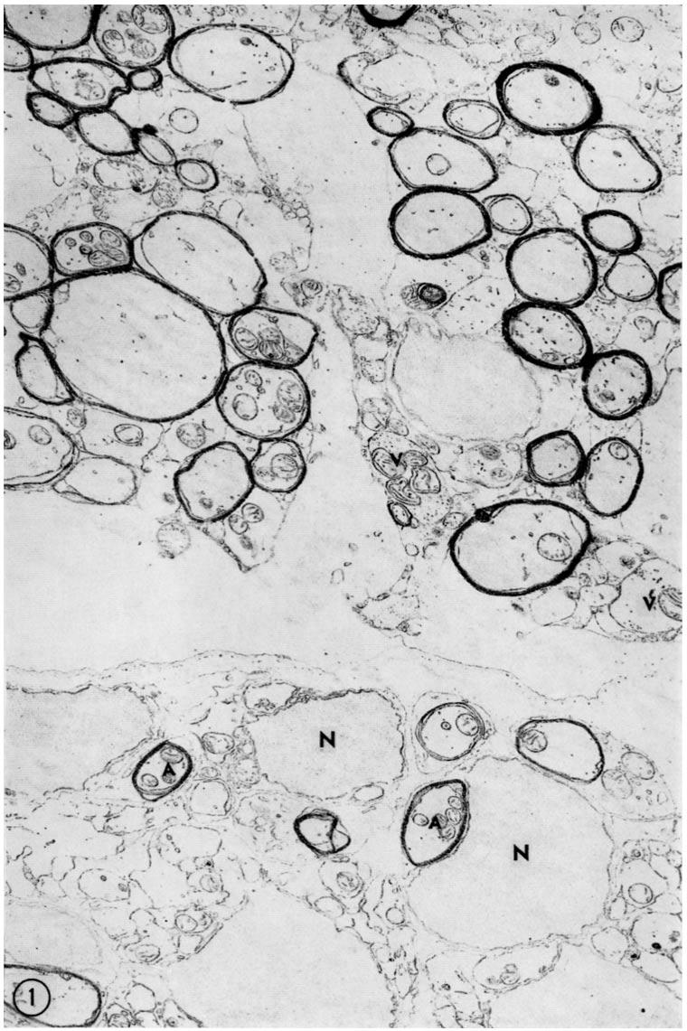
6 126 MYELIN SHEATHS IN CENTRAL NERVOUS SYSTEM nor a connection between a tongue and a glial cell have been observed. Whether one glial cell is related to a length of one axon, as is the Schwann cell to an internode in the peripheral nervous system, or whether, as De Robertis, Gerschenfeld, and Wald (9) suggest, each myelin-forming glial cell forms myelin around a number of axons, is not known. However, should the latter prove to be true, so that a number of processes from each glial cell form myelin around different axons, the geometrical relations will be such that the myelin is unlikely to be formed by a spiralling of the whole process of the glial cell around the axon, though it is conceivable that the free edge of the process could do so. I wish to thank Professor G. J. Romanes for his interest during the course of this work, Mr. G. Wilson for his skillful maintenance of the electron microscope, which is on loan from the Wellcome Trust, and Mr. H. Tully for his able technical assistance. The Xenopus tadpoles were kindly provided by Dr. B. Hobson. BIBLIOGRAPHY 1. Schmitt, F. O., and Bear, R. S., Biol. Rev., 1939, 14, Fern(mdez-Mor~m, H., and Finean, J. B., J. Biophysic, and Biochem. Cytol., 1957, 3, Schmidt, W. J., Z. wissensch. Mikr., 1957, 54, Ferngmdez-Mor~n, H., Exp. Cell Research, 1950, 1, Fern~ndez-Mor~n, H., Exp. Cell Research, 1952, 3, Robertson, J. D., J. Biophysic. and Biochem. Cytol., 1958, 4, Geren, B. B., Exp. Cell Research, 1954, 7, Luse, S. A., J. Biophysic. and Biochem. Cytol., 1956, 2, De Robertis, E., Gerschenfeld, H. M., and Wald, F., J. Biophysic. and Biochem. Cytol., 1958, 4, Luft, J. H., J. Biophysic. and Biochem. Cytol., 1956, 2, l. Robertson, J. D., Ultrastructure and cellular chemistry of neural tissue, in Progress in Neurobiology, (H. Waelsch, editor), New York, Hoeber-Harper, 1957, 2, Nieukoop, P. D., and Faber, J.,.Normal Tables of Xenopus laevis (Daudin), Amsterdam, North Holland Publishing Co., Barer, J., 1958, personal communication. 14. Fern~ndez-Mor~in, H., in Metabolism of the Nervous Tissue, (D. Richter, editor), London, Pergamon Press, 1957, Peters, A., 1958, data to be published. EXPLANATION OF PLATES PLATE 43 FIG. 1. Micrograph of part of the spinal cord, top of picture, and peripheral nerve root, bottom of picture, from a stage 56 Xenopus tadpole. The sheaths of peripheral axons (A) are well separated and are related to Schwann cell nuclei (N). This arrangement is in contrast to that of the myelinated axons of the spinal cord, which are frequently in contact with each other and are not associated with nuclei equivalent to those of the Schwann cells. Some of the structures in the spinal cord contain vesicles (V). >( 7,000.
7
 is surrounded by a cytoplasmic process (C) whose membranes come together to form the internal mesaxon (M). The probable position of the external tongue is indicated (T).")
8 PLATE 44 FIG. 2. Micrograph of a myelinated fibre from the optic nerve of a stage 57 tadpole. The axon (Ax) is surrounded by a cytoplasmic process (C) whose membranes come together to form the internal mesaxon (M). The probable position of the external tongue is indicated (T). Note the presence of cytoplasm between some myelin lamellae. X 38,000. FIG. 3. Enlargement of part of Fig. 2 showing the origin of the internal mesaxon (M). The intraperiod line (I) is formed at the point where the mesaxon comes into contact with the inside of the myelin sheath. In the same region the dense line (D) arises by the apposition of the cytoplasmic surfaces of the membrane surrounding the inner cytoplasmic process (C). Three processes (U, V, W) approach very close to the outside of the myelin sheath, the outer line of which is a dense line, (Do). X 103,000.

9 THE JOURNAL OF BIOPtfYSICAL AND BIOCHEMICAL CYTOLOGY PLATE 44 VOL. 7 (Peters: Myelin sheaths in central nervous system)
10 PLATE 45 FtG. 4. Micrograph of the point of contact between two myelin sheaths from the optic nerve of a stage 57 tadpole. No space separates the sheaths, so that a regular pattern of alternating dense and intraperiod lines extends from the inner surface of one sheath to the next. An internal mesaxon (M) is present in the right hand fibre. The axolemma (Al) is separated from the membrane bounding the inner cytoplasmic process (C), the external surfaces of which come into contact to form the mesaxon. The intraperiod line (I) arises on the inner side of the sheath within the mesaxon and the dense nine (D) is formed by contact between the cytoplasmic surfaces of the membrane bounding the inner cytoplasmic process. X ll0,000.

11 THE JOURNAL OF BIOPHYSICAL AND BIOCHEMICAL CYTOLOGY PLATE 45 VOL. 7 (Peters: Myelin sheaths in central nervous system)
12 PLATE 46 FtGs. 5 to 7. Micrograph of myelinated fibres from the optic nerve of a stage 58 Xenopus tadpole. Two myelinated fibres are shown completely, one of which (the right) has 10 lamellae and the other 4 lamellae. In each, the axon (Ax) is surrounded by a cytoplasmic process (C), the membranes of which come together to form the internal mesaxon (M). The outer surfaces of the sheaths are in contact. External tongues are present (T). In the lower right hand corner of the micrograph, part of a myelin sheath has an external tongue (T1). The structure of this tongue is shown in the enlargement, Fig. 6. Fig. 7 is taken from an adjacent section aml since it is underfocused only the dense lines are visible. The cytoplasmic surfaces of the tongue (7') come together to form the outer dense line (Do). Fig. 5, X 50,000; Figs. 6 and 7, X 85,000.

13 THE JOURNAL OF BIOPHYSICAL AND BIOCHEMICAL CYTOLOGY PLATE 46 VOL. 7 (Peters: Myelin sheaths in central nervous system)
14 PLATE 47 FIG. 8. Micrograph of a myelinated fibre from the optic nerve of a stage 58 Xenopus tadpole. The inner cytoplasmic process (C) circumscribes the axon (Ax) and forms the internal mesaxon (M). The external tongue (T) is associated with the upper fibre, but the outer surface of its bounding membrane is also applied to the myelin sheath in the lower part of the micrograph. In the region of the tongue the upper sheath has 8 dense lines and the lower one, 9. Where the sheaths meet at the right hand side, the cytoplasmic surfaces of the tongue membrane come together to form a dense line (D), which continues on the outside of the upper sheath so that when the sheaths separate on the left side both have 9 dense lines. An intraperiod line is formed where the two sheaths come into contact (arrow). Counts of the number of dense lines in different parts of the myelin sheath of the small fibre (see numbers) are consistent with the lamellae being arranged in a spiral. X 84,000. FIG. 9. Micrograph of an early stage in myelinogenesis present in the spinal cord of a stage 56 tadpole. The axon (Ax) is embedded in a cytoplasmic process (C). A short mesaxon (M) occurs where the lips of the process (C) meet. )< 42,000. FIG. 10. Micrograph of an early stage in myelination from the optic nerve of a stage 58 tadpole. Two or three lamellae are present and a loosely spiralled internal mesaxon (M) leads from the axon (Ax) to the inside of the formed myelin. On the outside of the myelin is a tongue (T). >( 35,000.

15 THE JOURNAL OF BIOPHYSICAL AND BIOCHEMICAL CYTOLOGY PLATE 47 VOL. 7 IPeters: Myelin sheaths in central nervous system)
From the Department of Anatomy, University of Edinburgh, Scotland
 Published Online: 1 February, 1964 Supp Info: http://doi.org/10.1083/jcb.20.2.281 Downloaded from jcb.rupress.org on June 14, 2018 FURTHER OBSERVATIONS ON THE STRUCTURE OF MYELIN THE CENTRAL NERVOUS SYSTEM
THE FORMATION AND STRUCTURE OF MYELIN
 THE FORMATION AND STRUCTURE OF MYELIN SHEATHS IN THE CENTRAL NERVOUS SYSTEM A. PETERS, Ph.D. From tile Department of Anatomy, University of Edinburgh, Scotland ABSTRACT The development and structure of
Some Observations on the Fine Structure of the Giant Nerve Fibers of the Earthworm, Eisenia foetida*
 Some Observations on the Fine Structure of the Giant Nerve Fibers of the Earthworm, Eisenia foetida* By KIYOSHI HAMA,~ M.D. (From the Department of Anatomy, School of Medicine, University of Washington,
Atypical Neural Sheaths Formed by Muller Cells in Chicken Retina
 Okajimas Folia Anat. Jpn., 57(2-3) : 79-88, August 1980 Atypical Neural Sheaths Formed by Muller Cells in Chicken Retina By YOSHIRO INOUE, YOSHIHIRO SUGIHARA,Yozo NISHIMURA and KAZUYO SHIMAI Department
The Use of Teased Preparations and Frozen Sections in Quantitative Studies of Mammalian Peripheral Nerve. By C. P. WENDELL-SMITH AND P. L.
 499 The Use of Teased Preparations and Frozen Sections in Quantitative Studies of Mammalian Peripheral Nerve By C. P. WENDELL-SMITH AND P. L. WILLIAMS (From the Department of Anatomy, Guy's Hospital Medical
The formation of zymogen granules in the pancreas of the mouse By S. K. MALHOTRA
 The formation of zymogen granules in the pancreas of the mouse By S. K. MALHOTRA (From the Cytological Laboratory, Department of Zoology, Oxford) With 3 plates (figs, i to 3) Summary Electron-dense, granular
Sheaths of the motor axons of the crab Carcinus
 175 Sheaths of the motor axons of the crab Carcinus By G. A. HORRIDGE and R. A. CHAPMAN (From the Gatty Marine Laboratory and Department of Natural History, the University, St. Andrews, Fife) With 3 plates
LOW-POWER ELECTRON MICROSCOPY OF THE ROOT CAP REGION OF EUCALYPT MYCORRHIZAS
 New Phytol. (1968) 67, 663-665. LOW-POWER ELECTRON MICROSCOPY OF THE ROOT CAP REGION OF EUCALYPT MYCORRHIZAS BY G. A. CHILVERS Botany Department, School of General Studies, Australian National University,
Electron and Light Microscope Studies of Endamoeba terrapinae
 Proceedings of the Iowa Academy of Science Volume 68 Annual Issue Article 81 1961 Electron and Light Microscope Studies of Endamoeba terrapinae Marilyn Driml Cornell College Copyright Copyright 1961 by
ANUMBER of electron microscope studies have been made on Amoeba
 An Electron Microscope Study of a Small Free-living Amoeba (Hartmanella astronyxis) By K. DEUTSCH and M. M. SWANN (From the Department of Zoology, University of Edinburgh) With two plates (figs. I and
* ra. VOL. 53, 1965 ANATOMY: D. BODIAN 419. FIG. 1.-Synaptic contact in spinal motoneuron of a chimpanzee. Large dendrite (D) shows
 shows") A SUGGESTIVE RELATIONSHIP OF NERVE CELL RNA WITh1 SPECIFIC SYNAPTIC SITES* BY DAVID BODIAN DEPARTMENT OF ANATOMY, JOHNS HOPKINS UNIVERSITY SCHOOL OF MEDICINE Communicated December 29, 1964 In a previous
A SUGGESTIVE RELATIONSHIP OF NERVE CELL RNA WITh1 SPECIFIC SYNAPTIC SITES* BY DAVID BODIAN DEPARTMENT OF ANATOMY, JOHNS HOPKINS UNIVERSITY SCHOOL OF MEDICINE Communicated December 29, 1964 In a previous
THE FINE STRUCTURAL ORGANIZATION OF NERVE FIBERS, SHEATHS, AND GLIAL CELLS IN
 THE FINE STRUCTURAL ORGANIZATION OF NERVE FIBERS, SHEATHS, AND GLIAL CELLS IN THE PRAWN, PALAEMONETES VULGARIS JOHN E. HEUSER and CARLOS F. DOGGENWEILER From the Harvard Medical School, Departments of
Chapter 37 Active Reading Guide Neurons, Synapses, and Signaling
 Name: AP Biology Mr. Croft Section 1 1. What is a neuron? Chapter 37 Active Reading Guide Neurons, Synapses, and Signaling 2. Neurons can be placed into three groups, based on their location and function.
* Work supported by a grant from The Rockefeller Foundation. Received for publication, May 20, 1958.
 Published Online: 25 September, 1958 Supp Info: http://doi.org/10.1083/jcb.4.5.667 Downloaded from jcb.rupress.org on December 16, 2018 Mitochondrial Changes in the Adrenocortex of Normal Hamsters.* BY
THE BEHAVIOUR OF CHLOROPLASTS DURING CELL DIVISION OF ISOETES LACUSTRIS L.
 New Phytol (1974) 73, 139-142. THE BEHAVIOUR OF CHLOROPLASTS DURING CELL DIVISION OF ISOETES LACUSTRIS L. BY JEAN M. WHATLEY Botany School, University of Oxford (Received 2 July 1973) SUMMARY Cells in
Aberrant Mitochondria with Longitudinal Cristae Observed in the Normal Rat Hepatic Parenchymal Cell. Takuma Saito and Kazuo Ozawa
 Okajimas Fol. anat. jap., 44 : 357-363, 1968 Aberrant Mitochondria with Longitudinal Cristae Observed in the Normal Rat Hepatic Parenchymal Cell By Takuma Saito and Kazuo Ozawa Department of Anatomy, Kansai
Nervous System Organization
 The Nervous System Chapter 44 Nervous System Organization All animals must be able to respond to environmental stimuli -Sensory receptors = Detect stimulus -Motor effectors = Respond to it -The nervous
(From the Department of Anatomy, University of Toronto, Toronto, Canada)
") THE STRUCTURE OF PIGEON BREAST MUSCLE MITOCHONDRIA* B~t ALLAN F. HOWATSON, PH.D. (From the Department of Anatomy, University of Toronto, Toronto, Canada) PLAT~.S 124 AND 125 The fine structure of mitochondfia
THE STRUCTURE OF PIGEON BREAST MUSCLE MITOCHONDRIA* B~t ALLAN F. HOWATSON, PH.D. (From the Department of Anatomy, University of Toronto, Toronto, Canada) PLAT~.S 124 AND 125 The fine structure of mitochondfia
Nervous System Organization
 The Nervous System Nervous System Organization Receptors respond to stimuli Sensory receptors detect the stimulus Motor effectors respond to stimulus Nervous system divisions Central nervous system Command
(From the Department of Anatomy and the Beaumont-May Institute of Neurology, Washington University School of Medicine, St. Louis)
") ELECTRON MICROSCOPIC OBSERVATIONS OF THE OLFACTORY MUCOSA AND OLFACTORY NERVE* BY A. J. DE LORENZO, PH.D. (From the Department of Anatomy and the Beaumont-May Institute of Neurology, Washington University
ELECTRON MICROSCOPIC OBSERVATIONS OF THE OLFACTORY MUCOSA AND OLFACTORY NERVE* BY A. J. DE LORENZO, PH.D. (From the Department of Anatomy and the Beaumont-May Institute of Neurology, Washington University
THE FREQUENCY OF HETEROCYSTS IN THE NOSTOC PHYCOBIONT OF THE LICHEN PELTIGERA CANINA WILLD.
 New Phytol. (1972) 71, 11-13. THE FREQUENCY OF HETEROCYSTS IN THE NOSTOC PHYCOBIONT OF THE LICHEN PELTIGERA CANINA WILLD. BY H. BRONWEN GRIFFITHS, A. D. GREENWOOD AND J. W. MILLBANK Department of Botany,
Dendrites - receives information from other neuron cells - input receivers.
 The Nerve Tissue Neuron - the nerve cell Dendrites - receives information from other neuron cells - input receivers. Cell body - includes usual parts of the organelles of a cell (nucleus, mitochondria)
PARTICLES AND MICROTUBULES IN VASCULAR CELLS OF PINUS STROBUS L. DURING CELL WALL FORMATION
 Neu'Phytol (1971) 70, 1089-1093. PARTICLES AND MICROTUBULES IN VASCULAR CELLS OF PINUS STROBUS L. DURING CELL WALL FORMATION BY LIDIJA MURMANIS Forest Products Laboratory, * Forest Service, U.S. Department
Neurons and Nervous Systems
 34 Neurons and Nervous Systems Concept 34.1 Nervous Systems Consist of Neurons and Glia Nervous systems have two categories of cells: Neurons, or nerve cells, are excitable they generate and transmit electrical
Cells. Steven McLoon Department of Neuroscience University of Minnesota
 Cells Steven McLoon Department of Neuroscience University of Minnesota 1 Microscopy Methods of histology: Treat the tissue with a preservative (e.g. formaldehyde). Dissect the region of interest. Embed
ELECTRON MNIICROSCOPY OF CELLULAR DIVISION IN ESCHERICHIA COLI
 ELECTRON MNIICROSCOPY OF CELLULAR DIVISION IN ESCHERICHIA COLI S. F. CONTII AND M. E. GETTNER' Biology Department, Brookhaven National Laboratory, Upton, New York Received for publication September 18,
Reconstruction of the Nuclear Sites of Salmonella typhimurium from Electron Micrographs of Serial Sections
 327 BIRCH-ANDERSEN, A. (1955). J. gen. Microbial. 13, 327429 Reconstruction of the Nuclear Sites of Salmonella typhimurium from Electron Micrographs of Serial Sections BY A. BIRCH-ANDERSEN Statens Seruminstitut,
Information processing. Divisions of nervous system. Neuron structure and function Synapse. Neurons, synapses, and signaling 11/3/2017
 Neurons, synapses, and signaling Chapter 48 Information processing Divisions of nervous system Central nervous system (CNS) Brain and a nerve cord Integration center Peripheral nervous system (PNS) Nerves
Chapter 48 Neurons, Synapses, and Signaling
 Chapter 48 Neurons, Synapses, and Signaling Concept 48.1 Neuron organization and structure reflect function in information transfer Neurons are nerve cells that transfer information within the body Neurons
Ch 7. The Nervous System 7.1 & 7.2
 Ch 7 The Nervous System 7.1 & 7.2 SLOs Describe the different types of neurons and supporting cells, and identify their functions. Identify the myelin sheath and describe how it is formed in the CNS and
THIN SECTIONS OF DIVIDING NEISSERIA GONORRHOEAE
 JOURNAL OF BACTERIOLOGY Vol. 87, No. 6, pp. 1477-1482 June, 1964 Copyright 1964 by the American Society for Microbiology Printed in U.S.A. THIN SECTIONS OF DIVIDING NEISSERIA GONORRHOEAE PHILIP FITZ-JAMES
CYTOLOGY & HISTOLOGY THE STUDY OF CELLS AND TISSUES
 NAME: DATE: PARTNER: CYTOLOGY & HISTOLOGY THE STUDY OF CELLS AND TISSUES For ease of study, multicellular animals are often examined at various levels of structural organization. Starting from the most
Neurochemistry 1. Nervous system is made of neurons & glia, as well as other cells. Santiago Ramon y Cajal Nobel Prize 1906
 Neurochemistry 1 Nervous system is made of neurons & glia, as well as other cells. Santiago Ramon y Cajal Nobel Prize 1906 How Many Neurons Do We Have? The human brain contains ~86 billion neurons and
NOTES: CH 48 Neurons, Synapses, and Signaling
 NOTES: CH 48 Neurons, Synapses, and Signaling A nervous system has three overlapping functions: 1) SENSORY INPUT: signals from sensory receptors to integration centers 2) INTEGRATION: information from
Intro and Homeostasis
 Intro and Homeostasis Physiology - how the body works. Homeostasis - staying the same. Functional Types of Neurons Sensory (afferent - coming in) neurons: Detects the changes in the body. Informations
Tokuhiro JSHIHARA, Chotatsu TSUKAYAMA, Fumiya UCHINO
 (39) JOURNAL OF ELECTRON MICROSCOPY 39 Vol. 22, No. I, 39-44, 1973 Intramitochondrial Filamentous Structures in Human Reticulum Cells in the Bone Marrow Tokuhiro JSHIHARA, Chotatsu TSUKAYAMA, Fumiya UCHINO
Nervous Systems: Neuron Structure and Function
 Nervous Systems: Neuron Structure and Function Integration An animal needs to function like a coherent organism, not like a loose collection of cells. Integration = refers to processes such as summation
NEURONS, SENSE ORGANS, AND NERVOUS SYSTEMS CHAPTER 34
 NEURONS, SENSE ORGANS, AND NERVOUS SYSTEMS CHAPTER 34 KEY CONCEPTS 34.1 Nervous Systems Are Composed of Neurons and Glial Cells 34.2 Neurons Generate Electric Signals by Controlling Ion Distributions 34.3
Neural Tissue. PowerPoint Lecture Presentations prepared by Jason LaPres. Lone Star College North Harris Pearson Education, Inc.
 12 Neural Tissue PowerPoint Lecture Presentations prepared by Jason LaPres Lone Star College North Harris An Introduction to the Nervous System The Nervous System Includes all neural tissue in the body
Introduction Principles of Signaling and Organization p. 3 Signaling in Simple Neuronal Circuits p. 4 Organization of the Retina p.
 Introduction Principles of Signaling and Organization p. 3 Signaling in Simple Neuronal Circuits p. 4 Organization of the Retina p. 5 Signaling in Nerve Cells p. 9 Cellular and Molecular Biology of Neurons
Ultrastructural changes in moist chamber corneas. E. M. Schaeffer"
 Ultrastructural changes in moist chamber corneas E. M. Schaeffer" Crossly normal human corneas received through the Iowa Eye Bank and stored in cold moist chambers for 12, 24, 36, 72, and 96 hours were
CONTRACTION BANDS AT SHORT SARCOMERE LENGTH IN CHICK MUSCLE
 CONTRACTION BANDS AT SHORT SARCOMERE LENGTH IN CHICK MUSCLE MARTIN HAGOPIAN. From the Department of Pathology, New York Medical College, New York 10029 INTRODUCTION The sliding filament model for contraction
NOTE: LOOK ON MY WEBSITE FOR THE MUSCLE LABELING POWER POINT/PDF Part I. Identify the parts of the neuron that are labeled below.
 Anatomy & Physiology Nervous System Part I 2/26/16 NOTE: LOOK ON MY WEBSITE FOR THE MUSCLE LABELING POWER POINT/PDF Part I. Identify the parts of the neuron that are labeled below. 1. 2. 3. 5. 4. 6. Part
Neurons. General neuron anatomy. BIOL 164 Human Biology Ch 7 Neurons. Nervous system:
 BIOL 164 Human Biology Ch 7 Neurons Nervous system: Neurons Integrates and coordinates the body s ac3vi3es Provides rapid and brief responses to s3muli Major divisions: Central nervous system (CNS) brain
Preparation of Material BRIEF NOTES 531
 BRIEF NOTES 531 Internal Structure of Apoferritin as Revealed by the "Negative Staining Technique."* BY GOETZ W. RICHTER. (From the Department of Pathology, The New York tlospital-cornell Medical Center,
DOWNLOAD OR READ : THE NEURONAL CYTOSKELETON MOTOR PROTEINS AND ORGANELLE TRAFFICKING IN THE AXON PDF EBOOK EPUB MOBI
 DOWNLOAD OR READ : THE NEURONAL CYTOSKELETON MOTOR PROTEINS AND ORGANELLE TRAFFICKING IN THE AXON PDF EBOOK EPUB MOBI Page 1 Page 2 the neuronal cytoskeleton motor proteins and organelle trafficking in
BIOLOGY 11/10/2016. Neurons, Synapses, and Signaling. Concept 48.1: Neuron organization and structure reflect function in information transfer
 48 Neurons, Synapses, and Signaling CAMPBELL BIOLOGY TENTH EDITION Reece Urry Cain Wasserman Minorsky Jackson Lecture Presentation by Nicole Tunbridge and Kathleen Fitzpatrick Concept 48.1: Neuron organization
Nervous System: Part II How A Neuron Works
 Nervous System: Part II How A Neuron Works Essential Knowledge Statement 3.E.2 Continued Animals have nervous systems that detect external and internal signals, transmit and integrate information, and
BIOLOGY. 1. Overview of Neurons 11/3/2014. Neurons, Synapses, and Signaling. Communication in Neurons
 CAMPBELL BIOLOGY TENTH EDITION 48 Reece Urry Cain Wasserman Minorsky Jackson Neurons, Synapses, and Signaling Lecture Presentation by Nicole Tunbridge and Kathleen Fitzpatrick 1. Overview of Neurons Communication
Neurons, Synapses, and Signaling
 LECTURE PRESENTATIONS For CAMPBELL BIOLOGY, NINTH EDITION Jane B. Reece, Lisa A. Urry, Michael L. Cain, Steven A. Wasserman, Peter V. Minorsky, Robert B. Jackson Chapter 48 Neurons, Synapses, and Signaling
Curtis et al. Il nuovo Invito alla biologia.blu BIOLOGY HIGHLIGHTS KEYS
 BIOLOGY HIGHLIGHTS KEYS Watch the videos and download the transcripts of this section at: online.scuola.zanichelli.it/curtisnuovoinvitoblu/clil > THE HUMAN NERVOUS SYSTEM 2. WARM UP a) The structures that
Studies upon the Gram Reaction of the Basiphil Cells of the Anterior Pituitary. Part II. Observations upon the Effects of Various Methods of Fixation
 247 Studies upon the Gram Reaction of the Basiphil Cells of the Anterior Pituitary Part II. Observations upon the Effects of Various Methods of Fixation By R. R. WILSON AND C. L. FOSTER (From the Departments
The Nervous System. Nervous System Organization. Nerve Tissue. Two parts to the nervous system 11/27/2016
 The Nervous System Nervous System Organization Animals must be able to respond to environmental stimuli. Three functions of the nervous system: Sensory input conduction of signals from sensory receptors.
FINE STRUCTURE OF THE RETINULAE IN THE COMPOUND EYE OF THE HONEY-BEE. TIMOTHY H. GOLDSMITH, Ph.D.
 Published Online: 1 September, 1962 Supp Info: http://doi.org/10.1083/jcb.14.3.489 Downloaded from jcb.rupress.org on June 7, 2018 FINE STRUCTURE OF THE RETINULAE IN THE COMPOUND EYE OF THE HONEY-BEE TIMOTHY
Cellular Neuroanatomy II The Prototypical Neuron: Neurites. Reading: BCP Chapter 2
 Cellular Neuroanatomy II The Prototypical Neuron: Neurites Reading: BCP Chapter 2 Major Internal Features of a Neuron The neuron is the functional unit of the nervous system. A typical neuron has a soma
Association of Tobacco Rattle Virus with Mitochondria
 J. gen. ViroL (I968), 3, I2I-I24 With 3 plates Printed in Great Britain I2I Association of Tobacco Rattle Virus with Mitochondria (Accepted 8 February I968) As part of a study of the way in which tobacco
(From the Department of Anatomy, Yale University School of Medicine, New'Haven)
") Published Online: 25 July, 1956 Supp Info: http://doi.org/10.1083/jcb.2.4.193 Downloaded from jcb.rupress.org on September 17, 2018 SYNAPSES IN THE CENTRAL NERVOUS SYSTEM* BY SANFORD L. PALAY,$ M.D. (From
Published Online: 25 July, 1956 Supp Info: http://doi.org/10.1083/jcb.2.4.193 Downloaded from jcb.rupress.org on September 17, 2018 SYNAPSES IN THE CENTRAL NERVOUS SYSTEM* BY SANFORD L. PALAY,$ M.D. (From
PROPERTY OF ELSEVIER SAMPLE CONTENT - NOT FINAL. The Nervous System and Muscle
 The Nervous System and Muscle SECTION 2 2-1 Nernst Potential 2-2 Resting Membrane Potential 2-3 Axonal Action Potential 2-4 Neurons 2-5 Axonal Conduction 2-6 Morphology of Synapses 2-7 Chemical Synaptic
Nervous system. 3 Basic functions of the nervous system !!!! !!! 1-Sensory. 2-Integration. 3-Motor
 Nervous system 3 Basic functions of the nervous system 1-Sensory 2-Integration 3-Motor I. Central Nervous System (CNS) Brain Spinal Cord I. Peripheral Nervous System (PNS) 2) Afferent towards afferent
CELL DIVISION IN THE FORMATION OF THE STOMATAL COMPLEX OF THE YOUNG LEAVES OF WHEAT
 J. Cell Sci. I, 121-128 (1966) 121 Printed in Great Britain CELL DIVISION IN THE FORMATION OF THE STOMATAL COMPLEX OF THE YOUNG LEAVES OF WHEAT J. D. PICKETT-HEAPS AND D. H. NORTHCOTE Department of Biochemistry,
Neurons. The Molecular Basis of their Electrical Excitability
 Neurons The Molecular Basis of their Electrical Excitability Viva La Complexity! Consider, The human brain contains >10 11 neurons! Each neuron makes 10 3 (average) synaptic contacts on up to 10 3 other
Action Potentials & Nervous System. Bio 219 Napa Valley College Dr. Adam Ross
 Action Potentials & Nervous System Bio 219 Napa Valley College Dr. Adam Ross Review: Membrane potentials exist due to unequal distribution of charge across the membrane Concentration gradients drive ion
Chapter 9. Nerve Signals and Homeostasis
 Chapter 9 Nerve Signals and Homeostasis A neuron is a specialized nerve cell that is the functional unit of the nervous system. Neural signaling communication by neurons is the process by which an animal
Neurons, Synapses, and Signaling
 Chapter 48 Neurons, Synapses, and Signaling PowerPoint Lecture Presentations for Biology Eighth Edition Neil Campbell and Jane Reece Lectures by Chris Romero, updated by Erin Barley with contributions
Assignment 7 Due February 26
 Assignment 7 Due February 26 Cells of Multicellular organisms 1. File upload (3 points) View this electron micrograph of spinach leaf cells. The central cell has a thin cell wall; it is difficult to distinguish
SHEET. Name: Class: Date: the sentence: write highest, lowest, higher, lower. level of organization is the organism; the
 1 1 Label the levels of organization Then complete the sentence: write highest, lowest, higher, lower The level of organization is the organism; the level of organization is the cell Tissues are a organs,
junction in the frog from which the greater part of the physiological evidence
 134 J Phy8iol. (1960), 150, pp. 134-144 With 6 ple8 and 3 text-ftgure8 Printed in Great Britain THE FINE STRUCTURE OF THE NEUROMUSCULAR JUNCTION OF THE FROG By R. BIRKS, H. E. HUXLEY AND B. KATZ From the
Nervous Tissue. Neurons Neural communication Nervous Systems
 Nervous Tissue Neurons Neural communication Nervous Systems What is the function of nervous tissue? Maintain homeostasis & respond to stimuli Sense & transmit information rapidly, to specific cells and
Supplementary Information
 Supplementary Information Tuning Ranvier node and internode properties in myelinated axons to adjust action potential timing Marc C. Ford, Olga Alexandrova, Lee Cossell, Annette Stange Marten, James Sinclair,
Brunswick School Department: Grades Essential Understandings
 Essential Understandings Essential Questions Essential Knowledge Vocabulary Essential Skills The Nervous system controls all voluntary and involuntary actions of the body. Neurons are the main functional
BIOLOGY. Neurons, Synapses, and Signaling CAMPBELL. Reece Urry Cain Wasserman Minorsky Jackson
 CAMPBELL BIOLOGY TENTH EDITION Reece Urry Cain Wasserman Minorsky Jackson 48 Neurons, Synapses, and Signaling Lecture Presentation by Nicole Tunbridge and Kathleen Fitzpatrick Lines of Communication The
The Nervous System. What did you learn at school today? Neurophysiology!
 The Nervous System What did you learn at school today? Neurophysiology! The Nervous System Controls heart rate, emotions, memories, consciousness, and much more. The most intricate and beautifully complex
Neurons, Synapses, and Signaling
 Chapter 48 Neurons, Synapses, and Signaling PowerPoint Lecture Presentations for Biology Eighth Edition Neil Campbell and Jane Reece Lectures by Chris Romero, updated by Erin Barley with contributions
Physiology 2 nd year. Neuroscience Optional Lecture
 Academic year 2018/2019 Physiology 2 nd year Semester 1 Curricula Nervous system physiology Blood physiology Acid-base equilibrium Bibliography: Boron & Boulpaep Medical Physiology, 3 rd edition Physiology
Neurons, Synapses, and Signaling
 Chapter 48 Neurons, Synapses, and Signaling PowerPoint Lectures for Biology, Eighth Edition Lectures by Chris Romero, updated by Erin Barley with contributions from Joan Sharp and Janette Lewis Copyright
(From Departamento de Ultraestructura Celular, Instituto de Investigaci6n de Ciencias Biol6gicas, Montevideo, Uruguay)
") Published Online: 25 May, 1956 Supp nfo: http://doi.org/10.1083/jcb.2.3.319 Downloaded from jcb.rupress.org on July 5, 2018 ELECTRON MCROSCOPE OBSERVATONS ON THE SUBMCROSCOPC ORGANZATON OF THE RETNAL RODS*
Published Online: 25 May, 1956 Supp nfo: http://doi.org/10.1083/jcb.2.3.319 Downloaded from jcb.rupress.org on July 5, 2018 ELECTRON MCROSCOPE OBSERVATONS ON THE SUBMCROSCOPC ORGANZATON OF THE RETNAL RODS*
Landolt's club in the amphibian retina: A Golgi and electron microscope study. Anita Hendrickson
 Landolt's club in the amphibian retina: A Golgi and electron microscope study Anita Hendrickson Landolt's club process has been studied in the adult newt retina. In Golgi preparations this process has
Overview Organization: Central Nervous System (CNS) Peripheral Nervous System (PNS) innervate Divisions: a. Afferent
 Peripheral Nervous System (PNS) innervate Divisions: a. Afferent") Overview Organization: Central Nervous System (CNS) Brain and spinal cord receives and processes information. Peripheral Nervous System (PNS) Nerve cells that link CNS with organs throughout the body.
Overview Organization: Central Nervous System (CNS) Brain and spinal cord receives and processes information. Peripheral Nervous System (PNS) Nerve cells that link CNS with organs throughout the body.
Miiller's cells and the "middle limiting membrane" of the human retina. An electron microscopic study. Ben S. Fine* and Lorenz E.
 Miiller's cells and the "middle limiting membrane" of the human retina An electron microscopic study Ben S. Fine* and Lorenz E. Zimmerman Electron microscopy of 3 normal human retinas has provided evidence
Electron Microscopy of Peripheral Nerves and Neuromuscular Junctions in the Wasp Leg
 Published Online: 25 January, 1958 Supp Info: http://doi.org/10.1083/jcb.4.1.107 Downloaded from jcb.rupress.org on January 13, 2019 Electron Microscopy of Peripheral Nerves and Neuromuscular Junctions
Computational Neuroscience
 Computational Neuroscience Zoltán Somogyvári senior research fellow Wigner Research Institute for Physics, Theoretical Department Supporting materials: http://www.kfki.hu/~soma/bscs/ BSCS 2012 Lengyel
! Depolarization continued. AP Biology. " The final phase of a local action
 ! Resting State Resting potential is maintained mainly by non-gated K channels which allow K to diffuse out! Voltage-gated ion K and channels along axon are closed! Depolarization A stimulus causes channels
ORDINARY section techniques have certain disadvantages when a threedimensional
 6 7 A Simultaneous Coupling Azo Dye Technique Suitable for Whole Mounts By P. R. LEWIS (From the Anatomy School, Cambridge) With one plate (fig. 1) SUMMARY A method based on the familiar coupling azo dye
Chapter 6: A Tour of the Cell
 Chapter 6: A Tour of the Cell 1. The study of cells has been limited by their small size, and so they were not seen and described until 1665, when Robert Hooke first looked at dead cells from an oak tree.
(From The Rockefeller Institute for Medical Research)
") MORPHOLOGY OF THE OMMATIDIA OF THE COMPOUND EYE OF LIMULUS* BY WILLIAM H. MILLER, M.D. (From The Rockefeller Institute for Medical Research) PLATES 126 TO 129 (Received for publication, November 13, 1956)
MORPHOLOGY OF THE OMMATIDIA OF THE COMPOUND EYE OF LIMULUS* BY WILLIAM H. MILLER, M.D. (From The Rockefeller Institute for Medical Research) PLATES 126 TO 129 (Received for publication, November 13, 1956)
Abraham Darby Academy KS3 Biology Cells, tissue, organs
 Abraham Darby Academy KS3 Biology Cells, tissue, organs Knowledge series Study Booklet 2017 Key terms Adaptation: A feature of an organism's body which helps it to survive. Bacteria: Single-celled micro-organisms.
706 [Vol. 34, 162. Somatic Syn.desis in Daphne odora.11*' The Chromosome Behavior in Meiosis
 706 [Vol. 34, 162. Somatic Syn.desis in Daphne odora.11*' The Chromosome Behavior in Meiosis By Tosisuke HIRAOKA Botanical Institute, College of Science, Kyoto University (Comm. by Y. KUWADA, M.J.A., Dec.
Acta Medica Okayama. An electron microscopic study on the red, white and inter-mediate muscle fibers of mouse. Takuro Ogata OCTOBER 1964
 Acta Medica Okayama Volume 18, Issue 5 1964 Article 3 OCTOBER 1964 An electron microscopic study on the red, white and inter-mediate muscle fibers of mouse Takuro Ogata Okayama University, Copyright c
Purpose: Perception, Movement, Learning, Memory, Thinking, Communication Functions:
 Nervous System Purpose: Perception, Movement, Learning, Memory, Thinking, Communication Functions: Sensory Input: Obtaining stimulation from the environment (light, heat, pressure, vibration, chemical,
Control and Integration. Nervous System Organization: Bilateral Symmetric Animals. Nervous System Organization: Radial Symmetric Animals
 Control and Integration Neurophysiology Chapters 10-12 Nervous system composed of nervous tissue cells designed to conduct electrical impulses rapid communication to specific cells or groups of cells Endocrine
Neurons, Synapses, and Signaling
 CAMPBELL BIOLOGY IN FOCUS URRY CAIN WASSERMAN MINORSKY REECE 37 Neurons, Synapses, and Signaling Lecture Presentations by Kathleen Fitzpatrick and Nicole Tunbridge, Simon Fraser University SECOND EDITION
Chapter 6: A Tour of the Cell
 AP Biology Reading Guide Fred and Theresa Holtzclaw Chapter 6: A Tour of the Cell Name Period Chapter 6: A Tour of the Cell Concept 6.1 To study cells, biologists use microscopes and the tools of biochemistry
AN ATYPICAL CRISTA RESEMBLING A "TIGHT JUNCTION" IN BEAN ROOT MITOCHONDRIA
 Published Online: 1 October, 1968 Supp Info: http://doi.org/10.1083/jcb.39.1.35 Downloaded from jcb.rupress.org on December 24, 2018 AN ATYPICAL CRISTA RESEMBLING A "TIGHT JUNCTION" IN BEAN ROOT MITOCHONDRIA
Chapter 4 Active Reading Guide A Tour of the Cell
 Name: AP Biology Mr. Croft Chapter 4 Active Reading Guide A Tour of the Cell Section 1 1. The study of cells has been limited by their small size, and so they were not seen and described until 1665, when
THE MECHANISM OF DENUCLEATION IN CIRCULATING ERYTHROBLASTS
 Published Online: 1 October, 1967 Supp Info: http://doi.org/10.1083/jcb.35.1.237 Downloaded from jcb.rupress.org on October 12, 2018 THE MECHANISM OF DENUCLEATION IN CIRCULATING ERYTHROBLASTS CHARLES F.
Novel Organization and Development of Copepod Myelin. II. Nonglial Origin
 RESEARCH ARTICLE Novel Organization and Development of Copepod Myelin. II. Nonglial Origin Caroline H. Wilson, 1,2 and Daniel K. Hartline 1 * 1 Békésy Laboratory of Neurobiology, Pacific Biosciences Research
The Nervous System. Nerve Impulses. Resting Membrane Potential. Overview. Nerve Impulses. Resting Membrane Potential
 The Nervous System Overview Nerve Impulses (completed12/03/04) (completed12/03/04) How do nerve impulses start? (completed 19/03/04) (completed 19/03/04) How Fast are Nerve Impulses? Nerve Impulses Nerve
37 Neurons, Synapses, and Signaling
 CAMPBELL BIOLOGY IN FOCUS Urry Cain Wasserman Minorsky Jackson Reece 37 Neurons, Synapses, and Signaling Lecture Presentations by Kathleen Fitzpatrick and Nicole Tunbridge Overview: Lines of Communication
Electron microscopy of glial cells of the central nervous system in the crab Ucides cordatus
 Brazilian Glial cells Journal in crustaceans of Medical and Biological Research (1999) 32: 327-331 ISSN 0100-879X Short Communication 327 Electron microscopy of glial cells of the central nervous system
RICHARD P. BUNGE, MARY BARTLETT BUNGE, and
 Published Online: 1 February, 1965 Supp Info: http://doi.org/10.1083/jcb.24.2.163 Downloaded from jcb.rupress.org on November 7, 2018 AN ELECTRON MICROSCOPE STUDY OF CULTURED RAT SPINAL CORD RICHARD P.