Interactions of the Drosophila gap gene giant with maternal and zygotic pattern-forming genes
|
|
|
- Phebe Clark
- 6 years ago
- Views:
Transcription

1 Development 111, (1991) Printed in Great Britain The Company of Biologists Limited Interactions of the Drosophila gap gene giant with maternal and zygotic pattern-forming genes ELIZABETH D. ELDON* and VINCENZO PIRROTTA Department of Cell Biology, Baylor College of Medicine, Texas Medical Center, Houston, TX 77030, USA 'Present address: Howard Hughes Medical Institute and Institute of Molecular Genetics, Baylor College of Medicine, Texas Medical Center, Houston, TX 77030, USA Summary The Drosophila gene giant (gt) is a segmentation gene that affects anterior head structures and abdominal segments A5-A7. Immunolocalization of the gt product shows that it is a nuclear protein whose expression is initially activated in an anterior and a posterior domain. Activation of the anterior domain is dependent on the maternal bicoid gradient while activation of the posterior domain requires maternal nanos gene product. Initial expression is not abolished by mutations in any of the zygotic gap genes. By cellular blastoderm, the initial pattern of expression has evolved into one posterior and three anterior stripes of expression. The evolution, position and width of these stripes are dependent on interactions between gt and the other gap genes. In turn, gt activity in these domains affects the expression of the other gap genes. These interactions, typical of the crossregulation previously observed among gap genes, confirm that gt is a member of the gap gene class whose function is necessary to establish the overall pattern of gap gene expression. After cellular blastoderm, gt protein continues to be expressed in the head region in parts of the maxillary and mandibular segments as well as in the labrum. Expression is never detected in the labial or thoracic segment primordia but persists in certain head structures, including the ring gland, until the end of embryonic development. Key words: Drosophila segmentation/morphogenetic gradients/gap genes/head development. Introduction The development of the anteroposterior pattern of the Drosophila embryo takes place in stages, progressing from simpler, block-like domains to units spanning double segments and then to individual segments. This strategy is revealed by the existence of zygotic genes whose localized expression is required for the differentiation of the corresponding pattern elements (Niisslein-Volhard and Wieschaus, 1980). The first stage in the formation of the anteroposterior pattern of the embryo is controlled by the zygotic gap genes. These are expressed in broad, partially overlapping domains and, in combination, are responsible for activating the expression of the next genes in the segmentation hierarchy, the pair-rule genes. Lack of gap gene activity causes the loss of several contiguous segments from that part of the body plan that corresponds roughly to the domain of expression of the gene. The initial domains of expression of the segmentation gap genes are specified by maternal cues laid down in the egg. The maternal cues are produced by three organizing systems responsible for the anterior, posterior and terminal pattern elements, respectively (Niisslein-Volhard et al. 1987). The anterior system, acting through a gradient of the bicoid (bed) gene product, has been shown to activate at least one zygotic gap gene, hunchback (hb), in the part of the embryo in which bed concentration is above a certain threshold (Driever et al. 1989). Genetic and molecular evidence (Hulskamp et al. 1989; Irish et al. 1989; Struhl, 1989) indicates that the posterior system, through the nanos (nos) product, acts negatively by preventing the accumulation of maternal hb product in a graded way from the posterior of the embryo. This activates posterior development because hb acts negatively on other gap genes. The terminal system, acting through the torso and l(l)polehole genes, activates the zygotic gap genes tailless (tit) and huckebein (hkb) in the terminal regions (Jurgens et al. 1984; Strecker et al. 1986; Pignoni etal. 1990; Weigel etal. 1990). By cellular blastoderm the initial domains of expression of the gap genes become sharper and their pattern is refined. Mutual interactions among the gap genes account for much of this elaboration of the pattern (Jackie et al. 1986). The giant (p) gene is required for normal develop-

2 368 E. D. Eldon and V. Pirrotta ment of the head and of the abdominal segments. Strong gt loss-of-function alleles or deficiencies of the gene were classified as segmentation gap mutations because they cause defects in the segmental pattern of two regions. One, in the posterior half of the larva, is a loss of contiguous abdominal segments A5, 6, 7 and sometimes 8, a type of pattern deletion characteristic of segmentation gap mutations. The other region is the head where defects have been interpreted as the loss of labral and labial structures resulting in failure to complete head involution and loss of parts of the cephalopharyngeal skeleton (Petschek et al. 1987; Mohler et al. 1989) and fusion of the labial and prothoracic segments (Petschek and Mahowald, 1990). In contrast to a classical gap mutation, lack of gt function causes less extensive gaps in the segmentation pattern and does not completely delete the abdominal, segments that it affects but only their anterior parts, leaving a considerable expanse of naked cuticle (Petschek et al. 1987; Mohler et al. 1989; Petschek and Mahowald, 1990). Earlier studies also showed that gt mutations had only minor effects on the pattern of expression of the pair-rule gene fushi tarazu (Carroll and Scott, 1985). More recent reports show that gt mutations cause alterations in the expression of even skipped (Frasch and Levine, 1987), a drastic weakening of stripes 5 and 6 of ftz expression and the concomitant fusion of engrailed stripes (Petschek and Mahowald, 1990). These results together with the pattern of defects, the early requirement for gt activity and the early onset of expression all indicate that gt functions very much like a gap gene. In this work we have studied the interactions of gt with the three maternal systems that organize the anteroposterior pattern and with the gap genes. We find that the initial expression of gt is dependent only on maternal factors but that gt affects the expression of other gap genes and is in turn influenced by them. Our results show that gt acts like a typical gap gene and is required for the establishment of the anteroposterior pattern of the embryo. Materials and methods Preparation of giant antibody We inserted a 522 bp Smal-PvuU fragment that encoded amino acids of the cdna open reading frame with no opa sequences (M. Capovilla, E. Eldon and V. Pirrotta, in preparation) into the pex-1 expression vector of Stanley and Luzio (1984). High level expression of the gio«r-/3-galactosidase fusion protein was induced in NF-1 cells by growth at 42 C. Pelleted cells were lysed in 7 M guanidinium HC1 and diluted with 6 volumes of Tris-EDTA, ph8. Insoluble material was pelleted and resuspended in urea sample buffer (USB) containing 8M urea, 100mM Tris (ph7.6), 2% SDS and 5% beta mercaptoethanol, sonicated and applied to 2 mm preparative 7.5% polyacrylamide gels. The region of the gel containing the hybrid protein was excised, homogenized and injected into rabbits as described by Benson and Pirrotta (1987). Serum obtained from the rabbits was purified first through CM Affi-Gel Blue (BioRad), to isolate an enriched immunoglobulin fraction. This was then precipitated with ammonium sulfate, resuspended and applied to affinity columns essentially as described by Pirrotta et al. (1988). The final product was greatly enriched for anti-giant activity and retained only very low levels of anti-beta galactosidase activity. Antibodies were used at a concentration of 1-2/xgml" 1 when used with horseradish peroxidase (HRP)- conjugated secondary antibodies (Cappel) or /jgmf 1 when used with the Vectastain Elite kit (Vector Laboratories). Other antisera Antisera directed against other segmentation gene products were kindly provided by other laboratories. In all cases they were preabsorbed by incubation with an excess of wild-type embryos prior to being used at the recommended dilutions. Rabbit anx\-ftz antibodies (from H. Krause; Krause et al. 1988) were used at a 1/1000 dilution. Mouse anti-hb and rabbit anti-a> antibodies (from R. Kraut and M. Levine) were used at a 1/300 and 1/100 dilution, respectively. Rabbit antien antibodies (from T. Kornberg; DiNardo et al. 1985) were used at a 1/400-1/1000 dilution. Rat anti-kni antibodies (obtained from K. Howard) were used at a 1/500-1/1000 dilution. Biotinylated secondary antibodies were obtained from Vector Laboratories (anti-mouse IgG and anti-rabbit IgG) or Jackson ImmunoResearch Laboratories (anti-rat IgG). These antibodies were also preabsorbed at a 1/10 dilution with an excess of Canton S embryos and used at a final dilution of 1/500. Embryo fixation and staining Flies of the appropriate genotype were allowed to lay eggs on grape juice agar plates for specified periods of time. The embryos were aged, if necessary, at room temperature (approx. 22 C), then collected, dechorionated, fixed and devitellinized essentially as described by Mitchison and Sedat (1983) as modified by Karr and Alberts (1986) and DiNardo and O'Farrell (1987). Embryos to be stained with peroxidaseconjugated antibodies were pretreated with 0.3 % H 2 O 2 prior to rehydration in phosphate buffered saline (PBS, ph7.4) containing 0.1 % Tween-20 and 0.1 % bovine serum albumin (BBT). Blocking was done in BBT containing 2% serum (BBS). Primary antibody incubations were carried out in BBS at 4 C overnight. The embryos were washed extensively in BBT, reblocked in BBS and incubated with the appropriate preabsorbed secondary antibody for two hours at room temperature. They were then washed extensively in PBS containing 0.1 % Tween-20 (PBT) and incubated for one hour at room temperature with ABC reagent (Elite Kit, Vector Laboratories) at concentrations recommended by the manufacturer. Following extensive washes in PBT, the embryos were treated with the appropriate substrate. For horseradish peroxidase staining, we used diaminobenzidine (0.5mgml -1 ) in the presence of % hydrogen peroxide. In the exu embryo shown in Fig. 4B, 2/il of a solution of 2 % nickel ammonium sulfate, 2% cobalt chloride was added to lml of substrate solution (DeBlas and Cherwinski, 1983). For alkaline phosphatase staining, we used 0.34 mg ml" 1 nitroblue tetrazolium salt and O.nSmgml" 1 of 5-bromo-4-chloro-3- indolyl phosphate, toluidinium salt, in buffer containing 100mM NaCl, 50mM MgCl 2, 100mM Tris, ph9.5, 0.1% Tween-20, 1 mm Levamisol. After staining, the embryos were washed in PBT, dehydrated, cleared briefly in methyl salicylate and mounted in GMM (Lawrence et al. 1986). Mutant strains Wild type embryos were obtained from Canton S parents. The giant alleles gf*, gt YAXL and Df(l)62gl8 were balanced over an FM7 chromosome carrying a P-element insertion express-
; knirps, kni lle72 (Tearle and Niisslein-Volhard, 1987); hunchback, hb 6ml (Lehmann and Nusslein-Volhard,")
3 Interactions of giant with maternal and zygotic genes 369 ing /S-galactosidase under the control of the ftz promoter (Kania et al. 1990). We used embryos from stocks carrying the following mutations: Kriippel, Kr 1 (Gloor, 1950); knirps, kni lle72 (Tearle and Niisslein-Volhard, 1987); hunchback, hb 6ml (Lehmann and Nusslein-Volhard, 1987); bicoid, bcd E1 (FrohnhSfer and Nusslein-Volhard, 1986); exuperantia, exu QR (Schiipbach and Wieschaus, 1986); nanos, nos 1-1 (Niisslein- Volhard et al. 1987); oskar, osk (Lehmann and Niisslein- Volhard, 1986); torso, tor 11 (Klingler et al. 1988) and tor (Schupback and Wieschaus, 1986); tailless, III s (Pignoni et al. 1990); empty spiracles, Df(3R)red3L (Dalton et al. 1989); buttonhead, btd ko (Jiirgens et al. 1984); orthodenticle, Df(l)KA14 (Wieschaus et al. 1984); tailless-huckebein, tlp hkb 2 (Weigelef a/. 1990). Data analysis Embryos were examined on a Zeiss Axiophot with differential interference contrast illumination. Measurements of the domains of protein expression were made midlaterally using a Zeiss E7 eyepiece micrometer and converted to egg length percentages. Ages of blastoderm embryos were estimated by measuring the length of the nuclei and invaginating cell membranes at the cell periphery as a percent of egg diameter at the broadest part of the embryo (at approximately 50 % EL). After the onset of gastrulation, morphogenic criteria were used to stage embryos according to Campos-Ortega and Hartenstein (1985). Because the gt pattern of expression changes quite rapidly, care was taken to compare only those embryos at the same developmental stage. For the mutant studies, this was the brief period between completion of cellularization (when the cellular layer was at least 10 % of the diameter of the embryo) and the earliest signs of gastrulation. At least 10 embryos of the appropriate stage and orientation were measured for embryos from maternal effect collections. At least 25 embryos of the appropriate stage and orientation were measured from zygotic lethal collections. Mean and standard deviations were calculated for the position of each stripe boundary. In most cases, determining which embryos were mutant was straightforward: the pattern of protein expression was obviously altered in the appropriate percentage of embryos. In a few cases of zygotic mutants, however, changes in the pattern were subtle. In these cases generally a standard deviation at one or more domain boundaries was greater than 2%, suggesting a large degree of scatter in the data points. The values were then plotted to see if two peaks could be resolved, consistent with a reproducible shift in approximately 25 % of the embryos. As a wild-type standard, we took the pattern obtained with Canton-S embryos but, in measuring the zygotic mutants, the giant domains of wild-type and heterozygous embryos were measured as well and compared to the domains in the 25 % of embryos that were mutant. Shifts of less than 2% EL were considered insignificant. To confirm the identification of mutant embryos, whenever possible we used double-stained embryos with a second antibody against ftz or en protein to reveal changes in the embryonic fate map corresponding to the mutant phenotype. Results The gt RNA is first detected by in situ hybridization during nuclear cycle (NC) 12 (Mohler et al. 1989). Our antiserum begins to detect gt protein at the end of NC 12, localized in nuclei in two distinct domains that parallel the pattern observed by in situ hybridization: a broad stripe at % EL and a posterior domain at 2-33% EL, excluding the pole cells (Fig. 1A). Expression is weak initially but gradually increases in strength through NC 13 and the pattern begins to sharpen. Embryos carrying strong giant mutations such as gt* n or gt YA82 still express the protein and show a very similar evolution of the pattern as the wild-type but the intensity of the antibody staining does not increase, suggesting that functional gt protein may stimulate its own expression. Embryos hemizygous for Df(l)62gl8, a deletion that removes the gt gene entirely, show no staining at all under our conditions. By the middle of NC 13, the posterior domain has retracted and sharpened forming a stripe whose posterior boundary is at 15-20% EL (Fig. IB). During NC 14 the pattern changes rapidly. As membranes progress inward from the cortex to form cells, the anterior domain begins to resolve into two stripes, a stronger one at % and a somewhat weaker one at 73-84% EL (Fig. 1C). By the time cellularization is complete, a new band of expression appears anteriorly at position % EL (Fig. ID). This domain, stripe 1, extends only laterally and dorsolaterally. Meanwhile, the separation of stripes 2 and 3 has progressed so that they now occupy positions % and % EL, respectively. The posterior stripe gradually shifts forward, reaching afinalposition at % by the end of cellularization. The complete pattern of expression at cellular blastoderm consists then of four stripes. The pattern of expression in post-blastoderm development is complex, with some regions becoming strong and others fading throughout germ band extension. We cannot determine at this point whether all the cells expressing giant at later stages are descendants of the cells that constitute stripes 1-3 at blastoderm. When we speak of elongation or changes in the three head stripes of expression, we refer only to patterns that show some local continuity and apparent evolution through time. The rapid disappearance of giant expression from certain regions indicates that the protein as well as the mrna must be rapidly turning over. In fact the pattern is continuously evolving throughout the process of cellularization. The posterior band, stripe 4, begins to fade towards the end of NC14. The ventral part of stripe 2 also fades at this time and is replaced by a distinct domain of expression that develops as a short ventral stripe slightly more anterior and more intense than the part of stripe 2 that faded away. As gastrulation begins, the anterior end of the ventral furrow reaches the ventral stripe and the g/arct-expressing cells line the interior and the edges of the resulting anterior midgut invagination (Fig. 1E,F). These cells become rapidly internalized forming a mass that stains most intensely bilaterally (Fig. 2A). These cells are not part of the anterior midgut primordia, which also differentiate at this time in the same region and are displaced posteriorly by the stomodeal invagination. Instead, the gl-expressing cells appear to move laterally and dorsally during germ band extension, forming an arch that meets anteriorly and medially with a group of strongly expressing cells inside the clypeolabrum (Fig. 2B,C).

4 370 E. D. Eldon and V. Pirrotta Initially, stripe 3 straddles the cephalic furrow, staining cells on both sides of it and with its posterior border overlapping with the thin maxillary stripe of engrailed-expressing cells (Fig. 2A). At no time from now on is there appreciable giant expression more posterior than this engrailed stripe, hence no expression in the primordia of the labial lobe. As the germ band begins to extend, stripe 3 decreases in intensity dorsally and laterally while it expands ventrally as cells enter the deepening cephalic furrow. Expression is seen internally anterior of the furrow in a region between the maxillary stripe of engrailed and the anterior midgut invagination (Fig. 2A). On the ventral side, the cells of stripe 3 fold into the ventral furrow and give rise to a thin layer of strongly expressing cells that extends to the posterior border of the maxillary segment, directly under the ectodermal layer (Fig. 2D). Most of these cells will gradually move forward as the stomodeal invagination deepens and will cease expressing gt as they fold into the stomodeum (Fig. 2D,E,F). Intriguing, but difficult to explain, is the appearance of staining in the germ cell precursors that are found in the amnio-proctodeal invagination (arrowhead in Fig. 2B). This staining, observed in many embryos during germ band elongation and again during germ band retraction (Fig. 3A), is not detectable in the pole cells at earlier stages. Whether it is a transient stage of expression or an odd artefact, we cannot tell. The staining region that corresponds to stripe 2 continues fading on the sides of the embryo but remains strong on the dorsal side where it moves posteriorly towards and then into the cephalic furrow (Fig. 2B,C). Strong expression persists in a group of dorsolateral cells while the rest fade away as the germ band reaches its maximal extension. The anteriormost stripe in the labral region, stripe 1, broadens laterally and advances anteriorly and then ventrally as the clypeolabrum begins to form and fold ventrally into the stomodeal invagination. A group of cells at the tip and inside the infolding clypeum express strongly and continue to do so until the beginning of germ band retraction. The intensity and extent of gt expression in the head peaks shortly before the germ band reaches its maximum extent. The number of cells expressing gt continues to decrease during germ band retraction (Fig. 3A) but some staining continues to be detectable even after the cuticle is secreted, nearly up to the end of embryonic development. It is interesting to note that among the last cells in which expression persists is a cluster located dorsally just behind the dorsal sac and the pharyngeal musculature but above the brain and corresponding in shape and position to the ring gland (Fig. 3B). Petschek et al. (1987) have observed that in strong gt mutants the ring gland appears to be missing or defective and have speculated that very mild defects in the ring gland of the viable gt 1 allele might be responsible for the insufficiency of ecdysteroid that is responsible for the eponymous giant larva phenotype in this mutant. Maternal effects Three classes of maternally acting genes have been Fig. 1. Early embryonic expression of giant protein in Canton S embryos. Embryos B to D are oriented with anterior to the left and dorsal side up. (A) Horizontal view of the initial pattern of gt expression in a precellular NC 13 embryo showing pale staining in two broad domains. (B) Refinement and intensification of the expression pattern in a slightly older embryo. (C) Anteriorly, stripes 2 and 3 begin to resolve and stripe 4 continues to be refined as cellularization begins during NC 14. (D) As cellularization is completed, during NC14, stripe 1 is detected anteriorly, stripe 2 ventral is seen ventrally and slightly anteriorly to stripe 2, and stripe 4 achieves its final position. Embryos E and F are seen from the ventral surface and oriented with the anterior to the left; giant expression is seen in brown, engrailed expression in blue. (E) During gastrulation, the ventral furrow extends to stripe 2 ventral, seen here as the anterior-most giant stripe in the plane of focus. (F) The anterior midgut begins to invaginate carrying the cells of stripe 2 ventral with it (arrowhead on the left). Stripe 3 flanks the cephalic furrow (arrowhead on the right) obscuring engrailed stripe 2. Fig. 2. Post-blastoderm expression of giant protein. In all embryos, giant protein is stained brown and engrailed protein in blue. Embryos A to C are oriented with anterior to the left and dorsal up. (A) Early stage 7 embryo just beginning germ band extension. Posterior expression of giant is barely detectable. From the anterior tip, arrowheads indicate the site of the anterior midgut invagination and engrailed stripe 2, respectively. (B) Stage 9 embryo nearing the end of germ band extension. The arrow indicates the pole cells which are transiently stained. Anteriorly, gt stripe 1 is at the anterior tip of the embryo, the cells from gt stripe 2 ventral that invagjnated with the anterior midgut primordia form a cluster midlaterally, cells from gt stripe 2 form the patch seen dorsally and cells from gt stripe 3 are found ventrally anterior to engrailed stripe 2. (C) Stage 10 embryo, fully germ band extended. Arrowhead indicates the stomodeal invagination. Pole cells no longer stain, and gt expression is restricted to regions of the embryo anterior to engrailed stripe 2, which marks the posterior border of the maxillary segment. Cells formerly associated with stripe 3 are now internalized. Internalized cells from stripe 2 ventral and stripe 1 surround the region of the invaginating stomodeum, and a few cells from stripe 2 are still seen dorsally. D to F are ventral views of the anterior end of progressively older embryos, beginning at stage 10 (fully germ band extended) and progressing to stage 12 (midway through germ band retraction). The arrowheads indicate engrailed stripe 2. During this time the process of head involution begins and the ventrolateral lobes of the gnathal segments (mandibular, maxillary and labial) begin to be drawn ventrally and anteriorly into the stomodeum. This movement is reflected in the pattern of gt staining. As head involution proceeds, cells that move into the region of the stomodeum extinguish gt expression. identified, whose function during oogenesis is to lay down the cues that result in the establishment of the anteroposterior axis of the embryo. Their domains of activity are independently established, largely nonoverlapping and affect a) the anterior segments, b) the posterior segments and c) the terminal structures at both ends of the embryo (Niisslein-Volhard et al. 1987). giant expression is found in all three of these domains. It is not surprising therefore, to find that loss of activity
5 B
 Late stage 12 embryo (germ band retraction) in which transient staining is again observed in the germ cells which are accumulating in the developing gonad.")
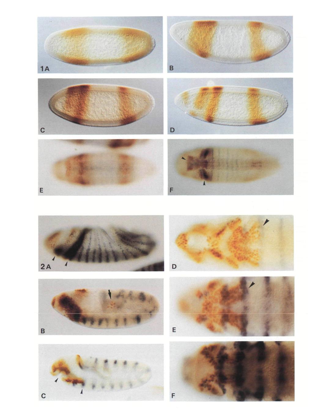
6 7 B Fig. 3. Later embryonic expression of giant in Canton S embryos. Both embryos are oriented with anterior to the left and dorsal up. (A) Late stage 12 embryo (germ band retraction) in which transient staining is again observed in the germ cells which are accumulating in the developing gonad. Anteriorly the number of cells expressing gt is much reduced and restricted primarily to a group of cells surrounding the pharynx and a smaller group of cells dorsally. (B) Stage 16 embryo (germ band fully retracted, prior to the secretion of cuticle), showing high levels of gt expression in very few cells. The arrow indicates site of the ring gland. A second cluster of cells still stains ventral to the pharynx. Fig. 4. Expression of giant in maternal mutants. In all cases embryos are at cellular blastoderm (late stage 5), anterior end to the left and dorsal side up. (A) bicoid embryos. Anterior gt expression is entirely absent while the posterior domain is broadened and shifted anteriorly. (B) exuperantia embryos. The anterior domain consists of a single broad band and stripe 4 is shifted posteriorly. (C) oskar embryos. Anterior expression is normal but posterior expression is lacking entirely. (D) torso embryos. Stripe 1 is absent leaving only stripe 2 and 3 anteriorly. Posterior expression persists to the posterior tip but not including the pole cells.
, is distributed in the early embryo in a concentration gradient with a high point near the anterior tip (Driever and")
7 Interactions of giant with maternal and zygotic genes 371 of any of these three pattern-forming systems has profound effects on the distribution of giant protein in the early embryo. The anterior domain The key maternal anterior gene, bicoid (bed), is distributed in the early embryo in a concentration gradient with a high point near the anterior tip (Driever and Niisslein-Volhard, 1988a,b). The bed gradient can activate the hb gene only in the anterior part of the embryo, where bed concentration is high enough to bind to its response elements in the hb regulatory region (Driever et al. 1989; Struhl, 1989). By manipulating the number and affinity of the bicoid binding sites, the domain of activation of the hb gene can be shifted more anteriorly or more posteriorly, demonstrating how this maternal morphogen can generate a pattern of zygotic gene expression. The activation of hb by bed is insufficient to account for the complexity of the anterior gt pattern since the specification of head structures requires bed levels higher than those required to activate hb expression and mutations in hb do not account for the totality of the phenotypes produced by lack of maternal bed. Driever et al. (1989) hypothesized a gene X that would be activated by higher concentrations of bicoid and might therefore serve to specify the more anterior head segments. Our results show that giant is such a gene X, whose anterior domain of expression is most likely directly controlled by bed. In embryos produced by bed mothers, the anterior pattern of giant expression is never observed. Stripes 1-3 fail to appear and stripe 4 develops as a broader domain shifted to a more anterior position (28-47 % EL instead of % EL; Fig. 4A). This indicates that the anterior expression of giant requires bed activity either directly or indirectly but that stripe 4 is established independently of bed. The anterior activation of giant might be mediated by some other zygotic gene dependent on bicoid, either hb or some other beddependent gene, but several lines of reasoning suggest that the bed controls gt directly: (1) gt anterior expression begins very early, before appreciable expression of other zygotic genes; (2) it is independent of hb, the major known mediator of bed; (3) it is not abolished by mutations in the other maternal factors or in the other known zygotic genes. Furthermore, the anterior pattern of gt expression depends on the gradient of bed concentration. This is shown by the distribution of gt expression in embryos derived from mothers mutant for exuperantia (exu), a gene required for the proper localization of bed mrna in the oocyte. Phenotypically such mutant embryos have greatly reduced anterior structures and expanded gnathal and thoracic structures (Schiipbach and Wieschaus, 1986). In exu eggs, the bed RNA fails to be preferentially localized anteriorly and as Driever and Niisslein-Volhard (1988ft) have shown, the bed protein gradient in these embryos is much shallower than normal, with the highest concentration at the anterior end corresponding to that normally found at about 65%. In exu embryos, gt expression at cellular blastoderm is found in a broad domain from % EL anteriorly and 15-30% EL posteriorly (Fig. 4B). We interpret this as a broadened and anteriorly shifted stripe 3, in agreement with the general anterior shift of thoracic and gnathal structures. We suppose that bed concentrations are insufficient to specify stripes 1 and 2 but that levels adequate to activate stripe 3 are now found over a good part of the head region. The fact that the domain of giant expression does not extend to the anterior tip of the embryo suggests that factors that prevent giant expression are present in the terminal region. The posterior shift of stripe 4 is probably an indirect effect mediated through other gap genes (see below). These results argue in favor of a direct dependence on the bed concentration at least for the initial gt anterior domain. The separation of stripe 2 from stripe 3 and the appearance of stripe 1 are probably due to interactions with zygotic gene products. The posterior domain Embryos derived from mothers mutant for genes of the posterior group fail to differentiate abdominal structures (Niisslein-Volhard et al. 1987). Unlike the anterior and terminal systems, the posterior system controls the embryonic pattern by negative regulation. It has been clearly established that nanos (nos), the key gene of the posterior class, acts solely by preventing the expression of the maternal hb protein in the posterior half of the embryo and is not required in embryos lacking maternal hb product (Hiilskamp et al. 1989; Irish et al. 1989; Struhl, 1989). The maternal hb protein is uniformly distributed in a nos mutant but forms a decreasing gradient in the posterior half of nos + embryos. Similarly, the distribution of maternal hb remains uniformly high along the entire anteroposterior axis of embryos derived from mothers mutant for oskar (osk) (Tautz, 1988), another member of the posterior group of genes which is required for correct positioning of nos product in the embryo. These results show conclusively that it is the abnormal persistence of the maternal hb product that suppresses abdominal development in these mutants. In embryos derived from mothers mutant for nos or osk, the posterior stripe of gt fails to appear while the anterior pattern appears to be normal (Fig. AC). This suggests that maternal hb product inhibits the expression of gt in the posterior domain but has no effect on the anterior expression. However, we have not generated hb~ germ line clones to demonstrate this directly. This distinction between the anterior gt expression, which is bed dependent and not repressed by maternal hb, and posterior expression, which is independent of bed and repressed by hb, strongly suggests that they are controlled by two separate regulatory elements in the gt gene that act independently and respond to different regulatory proteins. The terminal domains The nonsegmental termini, anteriorly the acron and posteriorly the telson, require the action of the
polehole ser/thr kinase (Sprenger et al. 1989; Ambrosio et al.")
8 372 E. D. Eldon and V. Pirrotta maternal genes of the terminal class. These are thought to act through a signalling pathway that includes genes such as torsolike, trunk etc. and results in the activation of a membrane-bound tyrosine kinase, the product of the torso gene, and of the D-raf l(l)polehole ser/thr kinase (Sprenger et al. 1989; Ambrosio et al. 1989; Stevens et al. 1990). In contrast to bicoid, these gene products are not localized in the embryo but are instead activated only at the poles by a localized signal and in turn activate zygotic genes such as tailless and huckebein in the terminal domains (Klingler et al. 1988; Strecker et al. 1989a; Weigel et al. 1990). We studied gt expression in embryos produced by mothers mutant for two members of the terminal class, torso (tor) and torsolike (tsl). As expected from the signal transduction model, their effects on gt expression are virtually identical. The posterior domain of expression appears normally at early syncytial blastoderm but fails to mature and to withdraw from the posterior region. At cellular blastoderm it still extends up to, but not including, the pole cells. The anterior domain of expression is also affected. Stripe 1 never forms. Stripe 2 is shifted anteriorly by 4-7 % EL but stripe 3 remains in its normal position (Fig. 4D). This indicates that the terminal genes have contrasting effects on giant: a repressive effect posteriorly and an inductive effect anteriorly. The fact that both of these effects take place after the initial stages of giant expression suggests that they are mediated by one or more zygotic effectors. A confirmation of these conclusions was given by the pattern of gt expression in embryos produced by mothers carrying a dominant allele of torso, tor 4021 (not shown). This mutation is thought to result in a constitutively active form of the torso product which would therefore activate terminal-specific zygotic gene functions throughout the embryo (Klingler et al. 1988; Strecker et al. 1989a). The phenotypic result is the expansion of the terminal, nonsegmental domains of the embryo at the expense of the segmental primordia. The distribution of giant product in these embryos is revealing: the posterior domain is completely absent and only two stripes are seen anteriorly. We interpret these results as showing that stripes 3 and 4, which normally correspond to segmented regions of the blastoderm fate map, are suppressed by the ectopic expression of terminal genes. Two anterior stripes remain. We suppose that these are stripes 1 and 2, which normally correspond to the anterior nonsegmented portion of the map and are shifted more posteriorly than normal by the expansion of the terminal domains. In approximately 10% of the embryos, the second stripe (63-73 % EL) succeeds in resolving into two stripes (69-77% and 59-64% EL). This incomplete suppression may reflect the fact that segmental primordia, though gteatly reduced, are not altogether absent in these embryos. The results with the three maternal classes of patternforming genes do not tell us conclusively whether giant is directly affected by maternal cues. However, the fact that the anterior (bed) and posterior (nos) systems affect the earliest expression of gt argues for a direct activating effect of bed on stripe 3 and of a repressing activity of maternal hb on stripe 4 expression. The effects of the terminal system on the later-appearing stripe 1 and on the retraction of stripe 4 are instead most likely mediated by zygotic gene products. Gap gene interactions with giant The earliest zygotic genes that affect the anteroposterior pattern belong to the segmentation gap class. The gap genes begin to be expressed very early at the syncytial blastoderm stage and are thought to respond directly to the maternal cues. It has been shown in addition that the gap genes interact both positively and negatively with one another so that the final pattern of expression is a complex resultant of the maternal and zygotic influences (reviewed by Gaul and Jackie, 1990). To determine how other gap genes influence gt expression, we examined the gt protein distribution in embryos homozygous for Kriippel (Kr), knirps (kni), hb or til mutations. In addition we looked for the effects of gt mutations on the expression pattern of hb, Kr and kni. To aid in identifying mutant embryos, we frequently double-stained using antibodies to ftz or en. As a rule, however, we measured the positions of the staining domains in a large number of unselected embryos and plotted each in a histogram. A statistically significant separate peak accounting for approximately 25 % of the embryos was taken to represent a domain of expression affected by the mutation (see the methods section). The results obtained with each mutation are summarized diagrammatically in Fig. 5. hb-gt interactions As expected from the analysis of nos mutations, zygotic hb, like maternal hb, has a negative effect on gt expression in the posterior domain. In hb mutant embryos, stripe 4 expands posteriorly into the region normally occupied by a late-appearing band of zygotic hb expression (10-20% EL; Fig. 6A). In addition, the anterior border is shifted 4% EL anteriorly. This confirms the negative effect of hb on the expression of stripe 4 and indicates that the withdrawal of this domain from the posterior end of the embryo is at least in part caused by the synthesis of zygotic hb in this region. However, in hb mutants giant expression does not continue all the way to the posterior pole, indicating that other negative interactions are involved, most likely with the zygotic interpreters of maternal terminal information. The shift of the anterior border is harder to account for. Loss of zygotic hb function is necessarily accompanied by the reduction of maternal hb to only one dose of the gene. This might lower the maternal hb concentration in the posterior half of the embryo and permit the activation of stripe 4 up to a more anterior position. Hiilskamp et al. (1990) invoke a similar explanation for the anterior expansion of the abdominal band of knirps expression. This broadening of the kni band in response to a reduction in maternal and zygotic hb may have additional direct or indirect effects on the domain of gt expression. We do not know what causes stripe 4 to fade at the end of cellular blastoderm but it

9 Interactions of giant with maternal and zygotic genes \ 3 bed emi 373 three anterior gt stripes. If this is the case, it would suggest a negative effect of gt on the anterior expression of hb. However, the anterior modulation of hb is difficult to ascertain under our staining conditions and we cannot be certain that it fails in gt-deficient embryos. Kr-gt interactions Kr has a distinct negative effect on gt expression. In Kr nos mutant embryos stripe 4 is greatly expanded anteriorly, reaching 48 % EL and invading therefore both the kni tor and Kr domains of expression (Fig. 6B). This effect could be direct or mediated through kni. Pankratz etal. tsl (1989) have shown that the full expression of kni D tor requires Kr activity. In Kr mutants, the posterior expression of kni never reaches its normal intensity, hb though its position is not shifted. The effect of Kr on gt stripe 4 could therefore be a direct negative effect of Kr Kr on gt or an indirect effect caused by Kr enhancement of /tm'rps-dependent repression of gt. We conclude that it kni is most likely a direct effect because kni mutations have no broadening effect on gt stripe 4 (see below) while, in til Kr mutants, stripe 4 expands nearly to 50 % EL. What tll+hkb prevents this gt domain from expanding further is probably the steeply rising concentration of hb whose ems anterior domain extends to 48% EL. In the anterior region, Kr mutations have only slight effects on gt, btd consisting of a slight broadening of stripe 3 and stripe 2. This could also be explained as a repressive effect otd caused by Kr expression in the central domain and in the Kr anterior domain (a stripe around 82% EL). The effects of gt mutations on Kr expression are very Fig. 5. Summary of giant expression patterns in wild-type mild. In agreement with Gaul and Jackie (1987), we and mutant backgrounds. The diagrams display the could not observe a significant broadening of the Kr averaged positions of the giant domains of expression, central domain. However, several lines of evidence converted to percent egg length (EL). Only changes of greater than 2 % EL from Canton S and internal wild-type suggest that gt has a negative effect on Kr. (1) bed (where possible) values were judged significant (see embryos, in which gt stripe 4 expands forward, show a Materials and methods). The stippled boxes shown contraction of the Kr posterior border; (2) conversely, underneath the tor0 and tll+hkb diagrams indicate in cases in which gt stripe 4 is suppressed, Kr posterior domains of expression seen in a minority of the embryos. border expands posteriorly, e.g. in nos embryos; (3) more directly, we have shown that ectopic expression of gt under control of the heat shock promoter causes cannot be simply inhibition by increasing levels of suppression of Kr expression in the central domain and hunchback posterior expression since the fading occurs generates cuticular phenotypes similar to those of Kr also in hb~ embryos. mutant embryos while gt protein binds in vitro to The only significant effect of the absence of zygotic specific sites in the Kr gene in the regulatory region hb on the anterior pattern of gt is a slight anterior shift responsible for expression in the central domain (M. of stripe 3. This indicates that hb does not mediate the Capovilla, E. Eldon and V. Pirrotta, unpublished activating effect of bicoid on gt anterior expression. data). Similar observations with ectopic expression of gt Given the complete overlap between the anterior have been made by Kraut and Levine (1991). domains of gt and of hb, this minor effect is not likely to be directly caused by hb. A more plausible explanation kni-gt interactions is that it is mediated through Kr. It has been shown that high levels of hb repress Kr and set the anterior edge of Mutations in the knirps gene affect the development of the Kr central domain. In the absence of zygotic hb, the the first seven abdominal segments, thus including most Kr domain expands anteriorly by 10 % EL (Hulskamp of the abdominal region affected by giant. In kni et al. 1989) and could affect gt anterior expression. mutants, stripe 4 of gt expression is affected only very slightly, if at all (Fig. 6F). It is possible that the There are no obvious changes in the domains of posterior border becomes less well denned and that the zygotic hb expression in embryos deficient for gt. A very intensity of posterior expression, which normally begins subtle effect may be observed in the anterior of the to fade at the end of cellular blastoderm, fades embryo where, at cellular blastoderm, the previously somewhat more rapidly in kni embryos. However, it is continuous expression of hb becomes modulated into clearly still detectable at the beginning of germ band three stripes that appear to be complementary to the

10 374 E. D. Eldon and V. Pirrotta extension and we hesitate to attach much significance to these slight effects. The lack of significant changes in the gt stripe 4 domain in these mutants indicates that neither the hb nor the Kr effect on gt are to be explained by a mediation through knirps and are therefore best interpreted as direct interactions. In the head region, it is interesting to note that the knirps anterior domain of expression is roughly complementary with that of giant (Fig. 6E). At syncytial blastoderm, knirps forms a cap at the anterior pole that extends ventrally until about 75% EL. At cellular blastoderm, a thin ring of expression appears at this position that corresponds to the interval between stripe 2 and 3. In spite of this pleasing complementarity, knirps mutants show the normal separation between gt stripe 2 and stripe 3 which cannot therefore be accounted for by a repressive action of kni on gt anterior expression. Of the gap genes, knirps is the one whose pattern of expression is most strongly altered in embryos deficient for gt. In these embryos, the posterior domain of kni, which is normally at % EL, expands posteriorly to 28 % EL. The strong negative effect of gt on kni that is implied by this result may in fact help to explain the previously reported interactions of kni with Kr. Pankratz et al. (1989) found that in Kr mutant embryos the abdominal kni stripe is present at the normal position but its intensity is greatly decreased. They concluded from this that Kr enhances kni expression. This apparent enhancement may be better explained as an indirect effect: in Kr mutants, gt stripe 4 expands anteriorly up to the middle of the embryo, fully overlapping and inhibiting expression of kni in its posterior domain. til gt interactions Lack of til function affects the structures derived from both anterior and posterior terminal domains. In til mutants abdominal segments 8-10 are missing and a decrease in the extent of the procephalon is accompanied by an expansion of the remaining body segments (Strecker et al. 1986). While til is not the only zygotic target of maternal terminal class genes, it is clearly one of the mediators of these maternal signals (Klingler et al. 1988; Strecker et al ). In the absence of til activity, the posterior domain of gt expression never matures properly. At the end of cellularization, it does not complete its withdrawal from the posterior terminal region and forms an expanded band whose posterior border is shifted 10% EL posteriorly (Fig. 6C). This position is in good agreement with the shift in the fate map seen in til embryos by Mahoney and Lengyel (1987) and with the distribution of til rarna (0-15% EL at cellular blastoderm, Pignoni et al. 1990). Surprisingly, lack of til has little effect on the anterior domains of gt expression. The apparent posterior shift of stripe three was not considered significant because the internal wild-type controls in this set of embryos showed a similar range of positions for this stripe. More significant shifts are seen with taillesshuckebein double mutant embryos (Fig. 6D). According to Weigel et al. (1990), the zygotic gene huckebein (hkb) is required to specify parts of the fate map corresponding to the posterior midgut and to anterior terminal structures. They suggest that hkb acts like a gap gene and, together with til, mediates maternal terminal cues from the tor gene product. In tll-hkb double mutants, the posterior border of gt stripe 4 retracts even less than in til mutants but at cellular blastoderm it does not extend completely to the tip of the embryo as in tor or tsl embryos. This might suggest the existence of additional components that mediate the effect of tor but a more likely explanation is that the hkb mutation used does not cause complete loss of function (Weigel et al. 1990). While til alone has little effect on gt in the head region, the sum of the two mutations results in a gt pattern approaching the greatly shifted pattern seen in the absence of tor function. Stripe 1 is shifted to the anterior pole and is present at barely detectable levels while stripes 2 and 3 are displaced forward as in tor embryos. This indicates that, at the anterior end, torso effects on gt are mediated primarily through hkb and not through til. gt expression in the head primordia Three genes have been recently described as having a gap-like role in mediating the development of head segments. These are orthodenticle (ptd), empty spiracles (ems) and buttonhead (btd) (Cohen and Jiirgens, 1990). The genes for otd and ems have been cloned by Finkelstein and Perrimon (1990) and Dalton et al. (1989), respectively, who found that they both encode homeodomain proteins. Since, after blastoderm, gt protein is found almost exclusively in the head primordia, we looked at the gt expression pattern in embryos mutant for each of these three genes. Both ems and otd affect gt expression in the anterior domain. In ems embryos, stripe 1 is normal but stripes 2 and 3 fail to sharpen, and separate at cellular blastoderm (Fig. 6H). In otd mutants, the posterior border of stripe 1 and all of stripe 2 are shifted posteriorly (not shown). In both cases, the part of the gt pattern most affected lies in the region of expression of the corresponding gene and involves the part of the fate map affected by the corresponding mutations. Both otd and ems are positively regulated by bed and it has been proposed that they are direct targets of the bed protein (Cohen and Jiirgens, 1990; Finkelstein and Perrimon, 1990; Dalton et al. 1989). However, they cannot be the mediators of bed effect on gt since their effect on gt anterior expression is negative rather than positive. Instead, the shifts in the gt stripes of expression that result from their loss of function are similar to those caused by other gap gene mutations. This is consistent with the interpretation that otd, ems and btd act as segmentation gap genes in the head region, where a greater complexity may require a greater number of components to specify the pattern. However, not all of these genes interact with giant. In btd embryos the changes in the gt anterior pattern are so slight as to be most likely insignificant. Although molecular probes for btd product are not available, the structures affected by
 Krilppel. Anterior expression is nearly normal but stripe 4 expands anteriorly to mid-embryo. (C) tailless.")
11 *> ' / 6A B H Fig. 6. Expression of giant in gap mutants. In all cases, embryos are at cellular blastoderm (late stage 5), anterior end to the left and dorsal side up. (A) hunchback. Stripe 3 is shifted anteriorly and stripe 4 expands posteriorly. (B) Krilppel. Anterior expression is nearly normal but stripe 4 expands anteriorly to mid-embryo. (C) tailless. Anterior expression is normal but stripe 4 retracts only partially from the posterior tip. (D) tailless-huckebein double mutants. Stripe 1 fails to appear and stripe 4 extends closer to the posterior tip. Blue staining indicates the stripes oifushi tarazu expression. (E) Wild type embryo showing knirps expression in brown and gt expression in blue. Compare with patterns seen in F and G. (F) knirps mutant. Note the altered pattern oifushi tarazu expression seen in blue, gt expression (brown) is nearly normal, though faint in this batch of embryos. Expression of stripe 4 continues to be detectable up to early germ band extension. (G) giant mutant embryo showing altered knirps expression. The posterior domain is expanded posteriorly, although anterior expression is normal. (H) empty spiracles embryo, giant is stained brown and fushi tarazu blue, gt stripes 2 and 3 fail to separate but posterior expression is normal.
12

13 Interactions of giant with maternal and zygotic genes 375 btd mutations imply that its domain of expression should include the mandibular, intercalary and antennal segments. Of these, at least the mandibular region should overlap with sites of gt expression at cellular blastoderm. Discussion Our results show that the giant gene behaves like a typical segmentation gap gene. Like other gap genes, gt produces a nuclear factor very likely involved in regulating the expression of other genes. The defects exhibited by giant loss-of-function mutants fall into two regions. One, in the posterior half of the larva, is a loss of contiguous abdominal segments A5, 6, 7 and sometimes 8, a type of pattern deletion characteristic of segmentation gap mutations. The posterior domain of giant expression corresponds well to this phenotype and the alterations of the gt pattern in various mutant backgrounds are indicative of interactions typical of segmentation gap genes. While the posterior domain of gt expression fades off soon after cellular blastoderm, the in situ hybridization results (Mohler et al. 1989) and the localization of the protein show that the head is the site of most intense, complex and prolonged gt expression. This later expression in the head region represents a second phase of giant activity that takes place after its function as a gap gene is completed. The anterior defects caused by gt mutations are more difficult to characterize because of the complex events and the structural rearrangements that normally result from head involution but that are perturbed or fail to take place in gt mutants. The reported lack of labral structures, epi- and hypostomal sclerites, H-piece and dorsal bridge (Mohler et al. 1989) are difficult to interpret as segmentation gaps. Rather, the defects within the head appear restricted to individual structures or elements rather than whole segments. The segmental attribution of these defects does not always correspond well with the pattern of expression that we observe in the head region. The anteriormost stripe and the later strong expression in the clypeolabrum fit well with the labral defects and the failure of head involution but there is a discrepancy between the position of stripes 2 and 3 and the sites of the other head defects. Petschek et al. (1987) and Petschek and Mahowald (1990) noted the disappearance of the labial lobe and the transient fusion of Tl and T2 in gt embryos at the germ band retraction stage, accompanied by defects in the expression of Antennapedia and Scr genes. Carroll and Scott (1985) and Petschek and Mahowald (1990) have also shown that stripes 1 and 2 of ftz are significantly broadened in gt mutants. Our results show that the posterior boundary of gt expression is within the posterior compartment of the maxillary segment and coincides with stripe 2 (maxillary) of engrailed, just in front of the labial lobe. This discrepancy can be accounted for in part by supposing that some of the mature head defects might have been incorrectly assigned to labial primordia and in part by assuming that the effects on the labial and thoracic segments are indirect, not primarily due to the lack of gt product but to the effect that it produces on other gap genes, most likely Kr. The spread of pattern defects to regions broader than the domains of strong expression is a phenomenon common to the other gap genes {hb, Kr, kni, til) and is most likely explained through combinatorial and indirect effects produced by the crossinteractions between these genes. The fact that in gt mutants no specific defects have been attributed to maxillary and mandibular derivatives may also be due in part to the early internalization of most of the cells that express gt during the complex infolding of the initial blastoderm sheet of cells in the cephalic furrow. It is possible that, as a result, the gf-expressing cells contribute principally to internal structures, most of which are not preserved in cuticle preparations and whose ontogeny is more difficult to follow. Two different mechanisms of gt activation An account of the gt pattern of expression must begin with the realization that the anterior expression is controlled by a different mechanism from that which activates the posterior. Anterior expression is feeddependent, the posterior is not. Posterior expression is /zfr-sensitive, the anterior is not. Molecular studies of several other segmentation genes have shown that their regulatory regions are complex and constituted by multiple elements that can act independently of one another (Harding etal. 1989; Picket al. 1990; Hoch etal. 1990). A preliminary analysis of the gt regulatory region shows in fact that the control elements responsible for expression in the posterior domain are distinct, independent and widely spaced from those that control anterior expression (P. Bagnaresi and V. Pirrotta, unpublished results). The results presented in this paper show that bed is responsible, most likely directly, for activating the initial anterior expression of gt. We may suppose that this occurs through a mechanism similar to that demonstrated for hb and anticipate that molecular analysis will demonstrate the presence of bed binding sites in the regulatory domain responsible for anterior expression of gt. The failure of this feeddependent activation to extend as far as the anterior tip even at the earliest stages suggests that maternal factors inhibit gt expression in the terminal region. If these were part of the terminal system, we would expect that in tor or tsl embryos this repression would be lifted and expression would extend fully to 100 % EL. Since this is not the case, we suppose that the highest bed concentrations have a repressive effect on gt expression. Although such repressive activities of bed have not yet been demonstrated at the molecular level they have been postulated by Pignoni et al. (1990) to explain anterior inhibition of til by bed and by Hiilskamp et al. (1990) to set the anterior border of Kr expression. The posterior initial pattern, like much of the rest of abdominal development, appears to be negatively rather than positively controlled. Several groups have shown that the removal of maternal hb suffices for

14 376 E. D. Eldon and V. Pirrotta normal abdominal development even in the absence of nos function (Hulskamp et al. 1989; Irish et al. 1989; Struhl, 1989). The activation of gt in the posterior domain must therefore be constitutive, dependent on ubiquitous transcription factors and severely inhibited by even relatively low levels of hb. Maternal hb RNA is initially uniformly distributed but, under the influence of nos product, the accumulation of hb protein tapers off in the posterior half of the embryo (Tautz, 1988). We can explain the activation of gt in the posterior third of the embryo as the derepression that occurs when hb concentrations drop below a threshold. Initial expression of gt would then take place from the position at which this threshold is reached to the posterior tip. The posterior expression of kni could be similarly derepressed but at a higher threshold of hb concentration. Maternal hb product, in this interpretation, acts as a graded morphogen, exerting negative control on genes that differ in their sensitivity to hb inhibition. Additional control may be exerted by bed on the posterior expression of gt. This is likely because if maternal hb is removed entirely, normal development can still ensue (Lehmann and Niisslein-Volhard, 1987). Though it has not been shown directly, this implies that gt expression in these embryos is normal and not ubiquitous. Some other maternal factor, in addition to maternal hb, must therefore inhibit gt expression in the anterior two thirds of the embryo. A likely candidate for this inhibitor is the bed product which would then have a positive effect on the anterior regulatory element of gt and a negative effect on the posterior regulatory element. That this may be the case is also suggested by the fact that a consequence of the lack of maternal bed is the broadening and anterior shift of the entire posterior stripe of gt expression (Fig. 4A) as well as of kni and Kr expression (Hulskamp et al. 1990). The result of this contrasting effect on anterior and posterior gap gene expression is to cause both the loss of anterior structures and the anterior shift of the rest of the pattern. The gradual withdrawal of gt stripe 4 from the posterior, which occurs at the end of nuclear cycle 13, is clearly dependent on the terminal system: it is abolished in tor or tsl embryos. The effect of tor is mediated in part through the zygotic gap genes til and hkb, as soon as their products begin to accumulate. In part, however, the withdrawal of stripe 4 is caused by the appearance of the posterior stripe of zygotic hb, which exerts a repressive effect on posterior gt expression. Interactions with other gap genes While maternal mutations can abolish gt expression in the anterior (bed) or posterior (nos) domains, none of the known gap genes is required for initiating gt expression. This strongly suggests that gt is a direct interpreter of maternal cues and confirms the status of gt as a gap gene. In typical gap gene fashion, giant enters into a network of cross-regulatory interactions with other members of the gap gene class. To understand the interactions of gt with other gap genes, it is once again important to consider the different stripes of gt expression as independently controlled. It is also important to bear in mind that, because of the complex cross-regulatory interactions between genes of this class, it has been difficult to distinguish direct effects of one gene product on the expression of another and indirect effects mediated by other members of the group. In many cases, molecular experiments will be necessary to establish the direct regulatory relationships. As already discussed, hb, both maternal and zygotic, plays a major role in inhibiting or sharpening the expression of stripe 4 but our results show that it has little effect on the anterior gt stripes. At the same time, gt has little effect on hb expression. Kr clearly has a strong negative effect on the gt posterior stripe and gt in turn has a negative effect on Kr. This negative effect of gt on Kr is not very visible under normal conditions, as found also by Gaul and Jackie (1987), not because it is not strong but probably because Kr is also repressed by kni at its posterior border and by hb at its anterior border. Hence, in gt mutants, the expansion of the Kr domain is immediately checked by the presence of kni and hb immediately flanking it. However, the strong repressing effect of gt on Kr is evident when gt is expressed ectopically under the control of the heat shock promoter (Kraut and Levine, 1991; M. Capovilla, E. Eldon and V. Pirrotta, unpublished data). The anterior border of Kr is therefore set by hb repression and also by gt anterior expression, as hypothesized by Hulskamp et al. (1990). This is yet another case in which expression boundaries are multiply determined. We found very little effect of kni on gt expression in the posterior domain but strong repressive effects of gt on the kni posterior stripe. The interactions of gt and Kr and of gt and kni may help to explain the apparent activating effect of Kr on kni expression as caused indirectly by Kr repression of gt. This negative role of Kr is more consistent with the demonstration that Kr protein acts as a transcriptional repressor (Licht et al. 1990). However, Kr may also interact directly with kni as suggested by Pankratz et al. (1989) who have identified a binding site for Kr protein in the kni regulatory region. The terminal gap genes til and hkb appear to have negative effects on the other gap genes, both at the anterior and at the posterior end, with the exception of hb whose late-appearing posterior stripe actually requires terminal gene function (Tautz, 1988). Although no direct data are yet available, it is possible that gt in turn represses til since the initial expression of til over a broader posterior domain recedes to a posterior cap by NC14 (Pignoni et al. 1990). Failure to reduce til posterior expression in a gt mutant might perhaps account for the appearance of secondary filzkorper in the abdominal region, noted by Petschek et al. (1987) and Mohler et al. (1989). The domains of expression of the gap genes are set by a network of mutual interactions and their boundaries are often multiply determined by the maternal morphogens, the neighboring gap genes as well as the nonadjacent gap genes. These interactions are essential to account for the stability and robustness of the patterns

15 Interactions of giant with maternal and zygotic genes 311 of expression and probably for the broad and graded morphogenetic effects that result from gap mutations. Accordingly we can interpret the extensive defects seen in Kr mutations as due not only to absence of Kr but also to the resulting expansion of gt stripe 4, which invades and overruns the kni domain and most of the Kr domain. In contrast, the more restricted effects of gt mutations in the abdomen could be attributed to the fact that the posterior expansion of kni is limited by the hb and til domains. We are very grateful to Peter Gergen, Thorn Kaufman, Henry Krause, Steve Cohen, Tom Kornberg, Ken Howard, Rachel Kraut and Mike Levine for gifts of mutant stocks and antibodies. We thank Sharon Bickel for help in antibody purification, Steve Cohen and Maria Capovilla for helpful suggestions and Rachel Kraut and Mike Levine for communicating their results to us prior to publication. E.D.E. was the recipient of an NIH postdoctoral training grant stipend and the research was supported by NIH grant GM to V.P. References AMBROSIO, L., MAHOWALD, A. P. AND PERRIMON, N. (1989). 1(1) pole hole is required maternally for pattern formation in the terminal regions of the embryo. Development 106, BENSON, M. AND PIRROTTA, V. (1987). The product of the Drosophila zeste gene binds to specific DNA sequences in white and Ubx. EMBO J. 6, CAMPOS-ORTEGA, J. A. AND HARTENSTEIN, V. (1985). The Embryonic Development of Drosophila melanogaster. Berlin: Springer-Verlag. CARROLL, S. B. AND SCOTT, M. P. (1985). Localization of the fushi tarazu protein during Drosophila embryogenesis. Cell 43, COHEN, S. M. AND JORGENS, G. (1990). Mediation of Drosophila head development by gap-like segmentation genes. Nature 346, DALTON, D., CHADWICK, R. AND MCGINNIS, W. (1989). Expression and embryonic function of empty spiracles: a Drosophila homeobox gene with two patterning functions on the anterior-posterior axis of the embryo. Genes Dev. 3, DEBLAS, A. L. AND CHERWINSKI, H. M. (1983). Detection of antigens on nitrocellulose paper immunoblots with monoclonal antibodies. Anal Biochem. 133, DINARDO, S., KUNER, J. M., THEIS, J. AND O'FARRELL, P. H. (1985). Development of embryonic pattern in Drosophila melanogaster as revealed by accumulation of the nuclear engrailed protein. Cell 43, DINARDO, S. AND O'FARRELL, P. H. (1987). Establishment and refinement of segmental pattern in the Drosophila embryo: spatial control of engrailed expression by pair-rule genes. Genes Dev. 1, DRIEVER, W. AND NOSSLEIN-VOLHARD, C. (1988a). A gradient of bicoid protein in Drosophila embryos. Cell 54, DRIEVER, W. AND NOSSLEIN-VOLHARD, C. (19886). The bicoid protein determines position in the Drosophila embryo in a concentration-dependent manner. Cell 54, DRIEVER, W. AND NOSSLEIN-VOLHARD, C. (1989). The bicoid protein is a positive regulator of hunchback transcription in the early Drosophila embryo. Nature 337, DRIEVER, W., THOMA, G. AND NOSSLEIN-VOLHARD, C. (1989). Determination of spatial domains of zygotic gene expression in the Drosophila embryo by the affinity of binding sites for the bicoid morphogen. Nature 340, FINKELSTEIN, R. AND PERRIMON, N. (1990). The orthodenticle gene is regulated by bicoid and torso and specifies Drosophila head development. Nature 346, FRASCH, M. AND LEVINE, M. (1987). Complementary patterns of even-skipped and fushi-tarazu expression involve their differential regulation by a common set of segmentation genes in Drosophila. Genes Dev. 1, FROHNHOFER, H. G. AND NOSSLEIN-VOLHARD, C. (1986). Organisation of anterior pattern in the Drosophila embryo by the maternal gene bicoid. Nature 32A, GAUL, U. AND JACKLE, H. (1987). Pole region-dependent repression of the Drosophila gap gene Krilppel by maternal gene products. Cell 51, GAUL, U. AND JACKLE, H. (1990). Role of gap genes in early Drosophila development. Adv. Gen. 27, GLOOR, H. (1950). PhSnotypen der Heterozygoten bei der unvollstsndig dominanten homozygot lethalen Mutante Kr (=Kriippe[) von Drosophila melanogaster. Arch. Julius Klaus- Stift. 29, HARDING, K., HOEY, T., WARRIOR, R. AND LEVINE, M. (1989). Autoregulatory and gap gene response elements of the evenskipped promoter of Drosophila. EMBO J. 8, HOCH, M., SCHRODER, C, SEIFERT, E. AND JACKLE, H. (1990). Cis- - acting control elements for Krilppel expression in the Drosophila embryo. EMBO J. 9, HOLSKAMP, M., PFEIFLE, C. AND TAUTZ, D. (1990). A morphogenetic gradient of hunchback protein organizes the expression of the gap genes Knlppel and knirps in the early Drosophila embryo. Nature 346, HOLSKAMP, M., SCHRODER, C, PFEIFLE, C, JACKLE, H. AND TAUTZ, D. (1989). Posterior segmentation of the Drosophila embryo in the absence of a maternal posterior organizer gene. Nature 338, IRISH, V., LEHMANN, R. AND AKAM, M. (1989). The Drosophila posterior-group gene nanos functions by repressing hunchback activity. Nature 338, JACKLE, H., TAUTZ, D., SCHUH, R., SEIFERT, E. AND LEHMANN, R. (1986). Cross-regulatory interactions among the gap genes of Drosophila. Nature 324, JORGENS, G., WIESCHAUS, E., NOSSLEIN-VOLHARD, C. AND KLODING, H. (1984). Mutations affecting the larval cuticle in Drosophila melanogaster. II. Zygotic loci on the third chromosome. Wilhelm Roux' Arch. Devi Biol. 193, KANIA, M. A., BONNER, A. S., DUFFY, J. B. AND GERGEN, J. P. (1990). The Drosophila segmentation gene, runt, encodes a novel nuclear regulatory protein that is also expressed in the nervous system. Genes Dev. (in press). KARR, T. L. AND ALBERTS, B. M. (1986). Organization of the cytoskeleton in Drosophila embryos. J. Cell Biol 102, KLINGLER, M., ERDELYI, M., SZABAD, J. AND NOSSLEIN-VOLHARD, C. (1988). Function of torso in determining the terminal anlagen of the Drosophila embryo. Nature 335, KRAUSE, H. M., KLEMENZ, R. AND GEHRING, W. J. (1988). Expression, modification and localization of the fushi tarazu protein in Drosophila embryos. Genes Dev. 2, KRAUT, R. AND LEVINE, M. (1991). Mutually repressive interactions between the gap genes guint and Krilppel define middle body regions of the Drosophila embryo. Development 111, LAWRENCE, P. A., JOHNSTON, P. AND MORATA, G. (1986). Methods of marking cells. In Drosophila a Practical Approach (ed. D. B. Roberts), pp IRL Press: Oxford. LEHMANN. R. AND NOSSLEIN-VOLHARD, C. (1986). Abdominal segmentation, pole cell formation and embryonic polarity require the localized activity of oskar, a maternal gene in Drosophila. Cell 47, LEHMANN, R. AND NOSSLEIN-VOLHARD, C. (1987). hunchback, a gene required for segmentation of an anterior and posterior region of the Drosophila embryo. Devi Biol. 119, LICHT, J. D., GROSSEL, M. J., FIGGE, J. AND HANSEN, U. M. (1990). Drosophila Krilppel protein is a transcriptional repressor. Nature 346, MAHONEY, P. A. AND LENGYEL, J. A. (1987). The zygotic mutation tailless alters the blastoderm fate map of the Drosophila embryo. Devi Biol. 122, MITCHISON, T. J. AND SEDAT, J. (1983).-Localization of antigenic determinants in whole Drosophila embryos. Devi Biol. 99,
Axis determination in flies. Sem 9.3.B.5 Animal Science
 Axis determination in flies Sem 9.3.B.5 Animal Science All embryos are in lateral view (anterior to the left). Endoderm, midgut; mesoderm; central nervous system; foregut, hindgut and pole cells in yellow.
Axis Specification in Drosophila
 Developmental Biology Biology 4361 Axis Specification in Drosophila November 2, 2006 Axis Specification in Drosophila Fertilization Superficial cleavage Gastrulation Drosophila body plan Oocyte formation
Axis Specification in Drosophila
 Developmental Biology Biology 4361 Axis Specification in Drosophila November 6, 2007 Axis Specification in Drosophila Fertilization Superficial cleavage Gastrulation Drosophila body plan Oocyte formation
Drosophila Life Cycle
 Drosophila Life Cycle 1 Early Drosophila Cleavage Nuclei migrate to periphery after 10 nuclear divisions. Cellularization occurs when plasma membrane folds in to divide nuclei into cells. Drosophila Superficial
ULRIKE GAUL 1 * and HERBERT JACKLE 2
 Development 107, 651-662 (1989) Printed in Great Britain The Company of Biologists Limited 1989 651 Analysis of maternal effect mutant combinations elucidates regulation and function of the overlap of
MOLECULAR CONTROL OF EMBRYONIC PATTERN FORMATION
 MOLECULAR CONTROL OF EMBRYONIC PATTERN FORMATION Drosophila is the best understood of all developmental systems, especially at the genetic level, and although it is an invertebrate it has had an enormous
Axis Specification in Drosophila
 Developmental Biology Biology 4361 Axis Specification in Drosophila July 9, 2008 Drosophila Development Overview Fertilization Cleavage Gastrulation Drosophila body plan Oocyte formation Genetic control
Autonomous concentration-dependent activation and repression of Krüppel by hunchback in the Drosophila embryo
 Development 120, 3043-3049 (1994) Printed in Great Britain The Company of Biologists Limited 1994 3043 Autonomous concentration-dependent activation and repression of Krüppel by hunchback in the Drosophila
Unicellular: Cells change function in response to a temporal plan, such as the cell cycle.
 Spatial organization is a key difference between unicellular organisms and metazoans Unicellular: Cells change function in response to a temporal plan, such as the cell cycle. Cells differentiate as a
Two distinct mechanisms for differential positioning of gene expression borders involving the Drosophila gap protein giant
 Development 125, 3765-3774 (1998) Printed in Great Britain The Company of Biologists Limited 1998 DEV5218 3765 Two distinct mechanisms for differential positioning of gene expression borders involving
Development of Drosophila
 Development of Drosophila Hand-out CBT Chapter 2 Wolpert, 5 th edition March 2018 Introduction 6. Introduction Drosophila melanogaster, the fruit fly, is found in all warm countries. In cooler regions,
Midterm 1. Average score: 74.4 Median score: 77
 Midterm 1 Average score: 74.4 Median score: 77 NAME: TA (circle one) Jody Westbrook or Jessica Piel Section (circle one) Tue Wed Thur MCB 141 First Midterm Feb. 21, 2008 Only answer 4 of these 5 problems.
Thoracic Patterning by the Drosophila Gap Gene hunchback
 Developmental Biology 237, 79 92 (2001) doi:10.1006/dbio.2001.0355, available online at http://www.idealibrary.com on Thoracic Patterning by the Drosophila Gap Gene hunchback Xuelin Wu,* Vikram Vasisht,*
Developmental Biology
 Developmental Biology 376 (2013) 99 112 Contents lists available at SciVerse ScienceDirect Developmental Biology journal homepage: www.elsevier.com/locate/developmentalbiology Genomes and Developmental
Multiple steps in the localization of bicoid RNA to the anterior pole of the Drosophila oocyte
 Development 1989 Supplement, 13-19 Printed in Great Britain The Company of Biologists Limited 1989 13 Multiple steps in the localization of bicoid RNA to the anterior pole of the Drosophila oocyte DANIEL
1(1)pole hole is required maternally for pattern formation in the terminal regions of the embryo
pole hole is required maternally for pattern formation in the terminal regions of the embryo") Development 106, 145-158 (1989) Printed in Great Britain The Company of Biologists Limited 1989 145 1(1)pole hole is required maternally for pattern formation in the terminal regions of the embryo LINDA
Development 106, 145-158 (1989) Printed in Great Britain The Company of Biologists Limited 1989 145 1(1)pole hole is required maternally for pattern formation in the terminal regions of the embryo LINDA
Why Flies? stages of embryogenesis. The Fly in History
 The Fly in History 1859 Darwin 1866 Mendel c. 1890 Driesch, Roux (experimental embryology) 1900 rediscovery of Mendel (birth of genetics) 1910 first mutant (white) (Morgan) 1913 first genetic map (Sturtevant
Lecture 7. Development of the Fruit Fly Drosophila
 BIOLOGY 205/SECTION 7 DEVELOPMENT- LILJEGREN Lecture 7 Development of the Fruit Fly Drosophila 1. The fruit fly- a highly successful, specialized organism a. Quick life cycle includes three larval stages
Localized surface activity of torso, a receptor tyrosine kinase, specifies terminal body pattern m Drosophila
 Localized surface activity of torso, a receptor tyrosine kinase, specifies terminal body pattern m Drosophila Jordi Casanova and Gary Struhl Howard Hughes Medical Institute, Center for Neurobiology and
Developmental genetics: finding the genes that regulate development
 Developmental Biology BY1101 P. Murphy Lecture 9 Developmental genetics: finding the genes that regulate development Introduction The application of genetic analysis and DNA technology to the study of
Development Team. Developmental Biology Axis Specification in Drosophila. Head, Department of Zoology, University of Delhi
 Paper No. : 11 Module : 6 Development Team Principal Investigator: Prof. Neeta Sehgal Head, Department of Zoology, University of Delhi Paper Coordinator: Prof. Namita Agrawal Department of Zoology, University
Cellular automata for exploring gene regulation in Drosophila segmentation
 Cellular automata for exploring gene regulation in Drosophila segmentation Matthew J. Berryman a, Andrew Allison a, and Derek Abbott a a Centre for Biomedical Engineering and School of Electrical and Electronic
BIS &003 Answers to Assigned Problems May 23, Week /18.6 How would you distinguish between an enhancer and a promoter?
 Week 9 Study Questions from the textbook: 6 th Edition: Chapter 19-19.6, 19.7, 19.15, 19.17 OR 7 th Edition: Chapter 18-18.6 18.7, 18.15, 18.17 19.6/18.6 How would you distinguish between an enhancer and
MCB 141 Midterm I Feb. 19, 2009
 Write your name and student ID# on EVERY PAGE of your exam MCB 141 Midterm I Feb. 19, 2009 Circle the name of your TA Jessica Lyons Alberto Stolfi Question #1 Question #2 Question #3 Question #4 TOTAL
Tissue- and stage-specific control of homeotic and segmentation gene expression in Drosophila embryos by the polyhomeotic gene
 Development 103, 733-741 (1988) Printed in Great Britain The Company of Biologists Limited 1988 733 Tissue- and stage-specific control of homeotic and segmentation gene expression in Drosophila embryos
Gradients and insect segmentation
 Development 104 Supplement, 3-1,6 (1988) Printed in Great Britain @ The Company of Biologists Limited 1988 Gradients and insect segmentation VERNON FRENCH Department of Zoology, University of Edinburgh,
Chapter 18 Lecture. Concepts of Genetics. Tenth Edition. Developmental Genetics
 Chapter 18 Lecture Concepts of Genetics Tenth Edition Developmental Genetics Chapter Contents 18.1 Differentiated States Develop from Coordinated Programs of Gene Expression 18.2 Evolutionary Conservation
Biology 4361 Developmental Biology The Genetics of Axis Specification in Drosophila November 2, 2006
 Biology 4361 Developmental Biology The Genetics of Axis Specification in Drosophila November 2, 2006 EARLY DROSOPHILA DEVELOPMENT Fertilization 1) Drosophila egg activation occurs at ovulation - eggs are
High Bicoid levels render the terminal system dispensable for Drosophila head development
 Development 127, 3993-3999 (2000) Printed in Great Britain The Company of Biologists Limited 2000 DEV2563 3993 High Bicoid levels render the terminal system dispensable for Drosophila head development
Gaining New Insights into Primitive Strategies for Embryonic Axis Specification Using the Wasp Nasonia
 Functional Development and Embryology 2007 Global Science Books Gaining New Insights into Primitive Strategies for Embryonic Axis Specification Using the Wasp Nasonia Eugenia C. Olesnicky Claude Desplan
Determination of the embryonic axes of Drosophila*
 Development Supplement 1, 1991, 1-10 Printed in Great Britain The Company of Biologists Limited 199J Determination of the embryonic axes of Drosophila* CHRISTIANE NUSSLEIN-VOLHARD Max-Planck-lnslitul filr
Supplementary Materials for
 www.sciencesignaling.org/cgi/content/full/6/301/ra98/dc1 Supplementary Materials for Regulation of Epithelial Morphogenesis by the G Protein Coupled Receptor Mist and Its Ligand Fog Alyssa J. Manning,
MCB 141 Midterm I Feb. 14, 2012
 Write your name and student ID# on EVERY PAGE of your exam MCB 141 Midterm I Feb. 14, 2012 Question #1 Question #2 Question #3 Question #4 BONUS / 28 pts / 27 pts / 25 pts / 20 pts / 1 pt TOTAL / 100 pts
Gonadal mesoderm and fat body initially follow a common developmental path in Drosophila
 Development 125, 837-844 (1998) Printed in Great Britain The Company of Biologists Limited 1998 DEV7627 837 Gonadal and fat body initially follow a common developmental path in Drosophila Lisa A. Moore
Development of Developmental System System(Mathematical Topics in Biolo. Citation 数理解析研究所講究録 (1993), 827:
, 827:") Title Development of Developmental System System(Mathematical Topics in Biolo Author(s) Takeda, Yasuhiko Citation 数理解析研究所講究録 (1993), 827: 57-74 Issue Date 1993-03 URL http://hdl.handle.net/2433/83294 Right
Title Development of Developmental System System(Mathematical Topics in Biolo Author(s) Takeda, Yasuhiko Citation 数理解析研究所講究録 (1993), 827: 57-74 Issue Date 1993-03 URL http://hdl.handle.net/2433/83294 Right
The pattern of cell death in fushi tarazu, a segmentation gene of Drosophila
 Development 104, 447-451 (1988) Printed in Great Britain The Company of Biologists Limited 1988 447 The pattern of cell death in fushi tarazu, a segmentation gene of Drosophila LORENZO MAGRASSI and PETER
Chapter 11. Development: Differentiation and Determination
 KAP Biology Dept Kenyon College Differential gene expression and development Mechanisms of cellular determination Induction Pattern formation Chapter 11. Development: Differentiation and Determination
Homeotic genes in flies. Sem 9.3.B.6 Animal Science
 Homeotic genes in flies Sem 9.3.B.6 Animal Science So far We have seen that identities of each segment is determined by various regulators of segment polarity genes In arthopods, and in flies, each segment
SPATIAL PROGRAMMING OF GENE EXPRESSION IN EARLY DROSOPHILA EMBRYOGENESIS
 Ann. Rev. Cell Biol. 1986.2 : 49-80 Copyright 0 1986 by Annual Reviews Inc. All rights reserved SPATIAL PROGRAMMING OF GENE EXPRESSION IN EARLY DROSOPHILA EMBRYOGENESIS Matthew P. Scott Department of Molecular,
Segment boundary formation in Drosophila embryos
 Segment boundary formation in Drosophila embryos Development 130, August 2003 Camilla W. Larsen, Elizabeth Hirst, Cyrille Alexandre and Jean Paul Vincent 1. Introduction: - Segment boundary formation:
178 Part 3.2 SUMMARY INTRODUCTION
 178 Part 3.2 Chapter # DYNAMIC FILTRATION OF VARIABILITY WITHIN EXPRESSION PATTERNS OF ZYGOTIC SEGMENTATION GENES IN DROSOPHILA Surkova S.Yu. *, Samsonova M.G. St. Petersburg State Polytechnical University,
Effects of ectopic expression of caudal during Drosophila development
 Development 109, 271-277 (1990) Printed in Great Britain The Company of Biologists Limited 1990 271 Effects of ectopic expression of caudal during Drosophila development MAREK MLODZIK*, GREG GIBSONt and
orthodenticle homeobox gene
 Development 121, 3561-3572 (1995) Printed in Great Britain The Company of Biologists Limited 1995 DEV8275 3561 Pattern formation in Drosophila head development: the role of the orthodenticle homeobox gene
Supplementary Fig. 1. Expression profiles of gap genes in large and small embryos. Shown are the mean mrna expression profiles (as a function of
 Supplementary Fig. 1. Expression profiles of gap genes in large and small embryos. Shown are the mean mrna expression profiles (as a function of fractional embryo length ξ = x/l) of the indicated six gap
Temporal regulation of gene expression in the blastoderm Drosophila embryo
 Temporal regulation of gene expression in the blastoderm Drosophila embryo Glenn K. Yasuda, 1,3 Jayne Baker, 2 and Gerold Schubiger 2 1Department of Genetics, SK-50, and 2Department of Zoology, NJ-15,
Developmental Biology 3230 Midterm Exam 1 March 2006
 Name Developmental Biology 3230 Midterm Exam 1 March 2006 1. (20pts) Regeneration occurs to some degree to most metazoans. When you remove the head of a hydra a new one regenerates. Graph the inhibitor
Morphogens in biological development: Drosophila example
 LSM5194 Morphogens in biological development: Drosophila example Lecture 29 The concept of morphogen gradients The concept of morphogens was proposed by L. Wolpert as a part of the positional information
Drosophila Somatic Anterior-Posterior Axis (A-P Axis) Formation
 Formation") Home Biol 4241 Luria-Delbruck 1943 Hershey-Chase 1952 Meselson-Stahl 1958 Garapin et al. 1978 McClintock 1953 King-Wilson 1975 Sanger et al. 1977 Rothberg et al. 2011 Jeffreys et al. 1985 Bacterial Genetics
Home Biol 4241 Luria-Delbruck 1943 Hershey-Chase 1952 Meselson-Stahl 1958 Garapin et al. 1978 McClintock 1953 King-Wilson 1975 Sanger et al. 1977 Rothberg et al. 2011 Jeffreys et al. 1985 Bacterial Genetics
Roles of wingless in patterning the larval epidermis of Drosophila
 Development 113, 471-485 (1991) Printed in Great Britain (E) The Company of Biologists Limited 1991 471 Roles of wingless in patterning the larval epidermis of Drosophila AMY BEJSOVEC* and ALFONSO MARTINEZ
Drosophila germline stem cells
 Development 125, 679-690 (1998) Printed in Great Britain The Company of Biologists Limited 1998 DEV3776 679 Nanos and Pumilio have critical roles in the development and function of Drosophila germline
Chapter 4 Evaluating a potential interaction between deltex and git in Drosophila: genetic interaction, gene overexpression and cell biology assays.
 Evaluating a potential interaction between deltex and git in Drosophila: genetic interaction, gene overexpression and cell biology assays. The data described in chapter 3 presented evidence that endogenous
BILD7: Problem Set. 2. What did Chargaff discover and why was this important?
 BILD7: Problem Set 1. What is the general structure of DNA? 2. What did Chargaff discover and why was this important? 3. What was the major contribution of Rosalind Franklin? 4. How did solving the structure
Principles of Experimental Embryology
 Biology 4361 Developmental Biology Principles of Experimental Embryology June 16, 2008 Overview What forces affect embryonic development? The embryonic environment: external and internal How do forces
A pair-rule gene circuit defines segments sequentially in the short-germ insect Tribolium castaneum
 A pair-rule gene circuit defines segments sequentially in the short-germ insect Tribolium castaneum Chong Pyo Choe, Sherry C. Miller, and Susan J. Brown* Division of Biology, Kansas State University, Manhattan,
!!!!!!!! DB3230 Midterm 2 12/13/2013 Name:
 1. (10 pts) Draw or describe the fate map of a late blastula stage sea urchin embryo. Draw or describe the corresponding fate map of the pluteus stage larva. Describe the sequence of gastrulation events
Chapter 18 Regulation of Gene Expression
 Chapter 18 Regulation of Gene Expression Differential gene expression Every somatic cell in an individual organism contains the same genetic information and replicated from the same original fertilized
How to make stripes: deciphering the transition from nonperiodic to periodic patterns in Drosophila segmentation
 RESEARCH ARTICLE 3067 Development 138, 3067-3078 (2011) doi:10.1242/dev.062141 2011. Published by The Company of Biologists Ltd How to make stripes: deciphering the transition from nonperiodic to periodic
Delimiting the conserved features of hunchback function for the trunk organization of insects
 RESEARCH ARTICLE 881 Development 135, 881-888 (2008) doi:10.1242/dev.018317 Delimiting the conserved features of hunchback function for the trunk organization of insects Henrique Marques-Souza 1, *, Manuel
DEVELOPMENT. Kay Kotkamp*, Martin Klingler and Michael Schoppmeier
 Access the Development most First recent posted version epress online at http://dev.biologists.org/lookup/doi/10.1242/dev.047043 on online 28 April publication 2010 as 10.1242/dev.047043 date 28 April
AP Biology Gene Regulation and Development Review
 AP Biology Gene Regulation and Development Review 1. What does the regulatory gene code for? 2. Is the repressor by default active/inactive? 3. What changes the repressor activity? 4. What does repressor
Identification of genes controlling germ cell migration and embryonic gonad formation in Drosophila
 Development 125, 667-678 (1998) Printed in Great Britain The Company of Biologists Limited 1998 DEV3768 667 Identification of genes controlling germ cell migration and embryonic gonad formation in Drosophila
SUPPLEMENTARY INFORMATION
 Supplementary Discussion Rationale for using maternal ythdf2 -/- mutants as study subject To study the genetic basis of the embryonic developmental delay that we observed, we crossed fish with different
Evolutionary Developmental Biology
 Evolutionary Developmental Biology a.k.a. EVO-DEVO Paedomorphosis is common among salamanders. Note how this hellbender (top) and mudpuppy (bottom) both have gills, paddle tails, and weaker limbs... Top:
Role of Organizer Chages in Late Frog Embryos
 Ectoderm Germ Layer Frog Fate Map Frog Fate Map Role of Organizer Chages in Late Frog Embryos Organizer forms three distinct regions Notochord formation in chick Beta-catenin localization How does beta-catenin
Developmental Biology Lecture Outlines
 Developmental Biology Lecture Outlines Lecture 01: Introduction Course content Developmental Biology Obsolete hypotheses Current theory Lecture 02: Gametogenesis Spermatozoa Spermatozoon function Spermatozoon
Trans-regulatory functions in the Abdominal-B gene of the bithorax complex
 Development 101, 117-122 (1987) Printed in Great Britain The Company of Biologists Limited 1987 117 Trans-regulatory functions in the Abdominal-B gene of the bithorax complex JORDI CASANOVA 1 and ROBERT
The effect of lethal mutations and deletions within the bithorax complex upon the identity of caudal metameres in the Drosophila embryo
 /. Embryol. exp. Morph. 93, 153-166 (1986) 153 Printed in Great Britain The Company of Biologists Limited 1986 The effect of lethal mutations and deletions within the bithorax complex upon the identity
EDWIN L. FERGUSON and KATHRYN V. ANDERSON
 Development 11, 58-597 (199) Printed in Great Britain The Company of Biologists Limited 199 58 Localized enhancement and repression of the activity of the TGF-fi family member, decapentaplegic, is necessary
10/03/2014. Eukaryotic Development. + Differentiation vs. Development. Differentiation. Development
 Differentiation vs. Development What comes to mind when you think of differentiation? Eukaryotic Development What about development? Presented by: Sean, Daria, Emily, and Maggie Example: Human Development
Analysis of neural elements in head-mutant Drosophila embryos suggests segmental origin of the optic lobes
 Roux's Arch Dev Biol (1995) 205:31-44 Springer-Verlag 1995 Urs Schmidt-Ott Marcos Gonzfilez-Gait~in Gerhard M. Technau Analysis of neural elements in head-mutant Drosophila embryos suggests segmental origin
pattern during early Drosophila
 Development 104 Supplement, 35-50 (1988) Printed in Great Britain @ The Company of Biologists Limited 1988 35 The generation of embryogenesis periodic pattern during early Drosophila KEN HOWARD Howard
Reverse Engineering the Gap Gene Network of Drosophila melanogaster
 of Drosophila melanogaster Theodore J. Perkins 1*, Johannes Jaeger 2,3, John Reinitz 2,3, Leon Glass 4 1 McGill Centre for Bioinformatics, McGill University, Montreal, Quebec, Canada, 2 Department of Applied
Drosophila melanogaster- Morphogen Gradient
 NPTEL Biotechnology - Systems Biology Drosophila melanogaster- Morphogen Gradient Dr. M. Vijayalakshmi School of Chemical and Biotechnology SASTRA University Joint Initiative of IITs and IISc Funded by
SUPPLEMENTARY INFORMATION
 med!1,2 Wild-type (N2) end!3 elt!2 5 1 15 Time (minutes) 5 1 15 Time (minutes) med!1,2 end!3 5 1 15 Time (minutes) elt!2 5 1 15 Time (minutes) Supplementary Figure 1: Number of med-1,2, end-3, end-1 and
Establishment of cell fate during early Drosophila embryogenesis requires transcriptional Mediator subunit dmed31
 Available online at www.sciencedirect.com Developmental Biology 313 (2008) 802 813 www.elsevier.com/developmentalbiology Establishment of cell fate during early Drosophila embryogenesis requires transcriptional
Role for mrna localization in translational activation but not spatial restriction of nanos RNA
 Development 126, 659-669 (1999) Printed in Great Britain The Company of Biologists Limited 1999 DEV7673 659 Role for mrna localization in translational activation but not spatial restriction of nanos RNA
NIH Public Access Author Manuscript Curr Opin Genet Dev. Author manuscript; available in PMC 2009 August 1.
 NIH Public Access Author Manuscript Published in final edited form as: Curr Opin Genet Dev. 2008 August ; 18(4): 342 347. doi:10.1016/j.gde.2008.06.002. Dynamics of maternal morphogen gradients in the
Segmental patterning of heart precursors in Drosophila
 Development 121, 4303-4308 (1995) Printed in Great Britain The Company of Biologists Limited 1995 DEV5025 4303 Segmental patterning of heart precursors in Drosophila Peter A. Lawrence 1, *, Rolf Bodmer
Capicua integrates input from two maternal systems in Drosophila terminal patterning
 The EMBO Journal (2004) 23, 4571 4582 & 2004 European Molecular Biology Organization All Rights Reserved 0261-4189/04 www.embojournal.org Capicua integrates input from two maternal systems in Drosophila
Developmental processes Differential gene expression Introduction to determination The model organisms used to study developmental processes
 Date Title Topic(s) Learning Outcomes: Sept 28 Oct 3 1. What is developmental biology and why should we care? 2. What is so special about stem cells and gametes? Developmental processes Differential gene
The Drosophila protein Leonardo enhances Torso signaling through D- Raf in a Ras1-dependent manner
 Development 124, 4163-4171 (1997) Printed in Great Britain The Company of Biologists Limited 1997 DEV5138 4163 The Drosophila 14-3-3 protein Leonardo enhances Torso signaling through D- Raf in a Ras1-dependent
CHAPTER 13 PROKARYOTE GENES: E. COLI LAC OPERON
 PROKARYOTE GENES: E. COLI LAC OPERON CHAPTER 13 CHAPTER 13 PROKARYOTE GENES: E. COLI LAC OPERON Figure 1. Electron micrograph of growing E. coli. Some show the constriction at the location where daughter
Mesoderm Induction CBT, 2018 Hand-out CBT March 2018
 Mesoderm Induction CBT, 2018 Hand-out CBT March 2018 Introduction 3. Books This module is based on the following books: - 'Principles of Developement', Lewis Wolpert, et al., fifth edition, 2015 - 'Developmental
Analysis of the gooseberry locus in Drosophila embryos: gooseberry determines the cuticular pattern and activates gooseberry neuro
 Development 118, 21-31 (1993) Printed in Great Britain The Company of Biologists Limited 1993 21 Analysis of the gooseberry locus in Drosophila embryos: gooseberry determines the cuticular pattern and
Spatio-temporal Registration of the Expression Patterns of Drosophila Segmentation Genes
 From: ISMB-99 Proceedings. Copyright 1999, AAAI (www.aaai.org). All rights reserved. Spatio-temporal Registration of the Expression Patterns of Drosophila Segmentation Genes Ekaterina M. Myasnikova David
Mechanisms of dorsal-ventral axis determination in Drosophila embryos revealed by cytoplasmic transplantations
 Development 117, 1385-1396 (1993) Printed in Great Britain The Company of Biologists Limited 1993 1385 Mechanisms of dorsal-ventral axis determination in Drosophila embryos revealed by cytoplasmic transplantations
Principles of Experimental Embryology
 Biology 4361 Developmental Biology Principles of Experimental Embryology September 19, 2006 Major Research Questions How do forces outside the embryo affect its development? (Environmental Developmental
Caenorhabditis elegans
 Caenorhabditis elegans Why C. elegans? Sea urchins have told us much about embryogenesis. They are suited well for study in the lab; however, they do not tell us much about the genetics involved in embryogenesis.
purpose of this Chapter is to highlight some problems that will likely provide new
 119 Chapter 6 Future Directions Besides our contributions discussed in previous chapters to the problem of developmental pattern formation, this work has also brought new questions that remain unanswered.
Gratuitous mrna localization in the Drosophila oocyte
 Development 121, 3013-3021(1995) Printed in Great Britain The Company of Biologists Limited 1995 3013 Gratuitous mrna localization in the Drosophila oocyte Thomas L. Serano 1,2, and Robert S. Cohen 1,
Big Idea 3: Living systems store, retrieve, transmit and respond to information essential to life processes. Tuesday, December 27, 16
 Big Idea 3: Living systems store, retrieve, transmit and respond to information essential to life processes. Enduring understanding 3.B: Expression of genetic information involves cellular and molecular
Exam 2 ID#: November 9, 2006
 Biology 4361 Name: KEY Exam 2 ID#: November 9, 2006 Multiple choice (one point each) Circle the best answer. 1. Inducers of Xenopus lens and optic vesicle include a. pharyngeal endoderm and anterior neural
Evolution of Transcription factor function: Homeotic (Hox) proteins
 proteins") Evolution of Transcription factor function: Homeotic (Hox) proteins Hox proteins regulate morphology in cellular zones on the anterior-posterior axis of embryos via the activation/repression of unknown
Evolution of Transcription factor function: Homeotic (Hox) proteins Hox proteins regulate morphology in cellular zones on the anterior-posterior axis of embryos via the activation/repression of unknown
Rui Dilão NonLinear Dynamics Group, IST
 1st Conference on Computational Interdisciplinary Sciences (CCIS 2010) 23-27 August 2010, INPE, São José dos Campos, Brasil Modeling, Simulating and Calibrating Genetic Regulatory Networks: An Application
16 The Cell Cycle. Chapter Outline The Eukaryotic Cell Cycle Regulators of Cell Cycle Progression The Events of M Phase Meiosis and Fertilization
 The Cell Cycle 16 The Cell Cycle Chapter Outline The Eukaryotic Cell Cycle Regulators of Cell Cycle Progression The Events of M Phase Meiosis and Fertilization Introduction Self-reproduction is perhaps
Introduction. Gene expression is the combined process of :
 1 To know and explain: Regulation of Bacterial Gene Expression Constitutive ( house keeping) vs. Controllable genes OPERON structure and its role in gene regulation Regulation of Eukaryotic Gene Expression
Model for the robust establishment of precise proportions in the early Drosophila embryo
 Journal of Theoretical Biology 234 (2005) 13 19 www.elsevier.com/locate/yjtbi Model for the robust establishment of precise proportions in the early Drosophila embryo Tinri Aegerter-Wilmsen a, Christof
Chapter 10 Development and Differentiation
 Part III Organization of Cell Populations Chapter Since ancient times, people have wondered how organisms are formed during the developmental process, and many researchers have worked tirelessly in search
Rat Prolactin ELISA Kit
 Rat Prolactin ELISA Kit Catalog No: IRPRLKT Lot No: SAMPLE INTENDED USE This rat prolactin antigen assay is intended for the quantitative determination of prolactin antigen in rat plasma. For research
PRACTICE EXAM. 20 pts: 1. With the aid of a diagram, indicate how initial dorsal-ventral polarity is created in fruit fly and frog embryos.
 PRACTICE EXAM 20 pts: 1. With the aid of a diagram, indicate how initial dorsal-ventral polarity is created in fruit fly and frog embryos. No Low [] Fly Embryo Embryo Non-neural Genes Neuroectoderm Genes