The DamX cell division protein of Escherichia coli: identification of amino acid residues critical for septal localization and peptidoglycan binding
|
|
|
- Alan Cunningham
- 5 years ago
- Views:
Transcription


1 University of Iowa Iowa Research Online Theses and Dissertations Spring 2010 The DamX cell division protein of Escherichia coli: identification of amino acid residues critical for septal localization and peptidoglycan binding Kyle Brandon Williams University of Iowa Copyright 2010 Kyle Brandon Williams This dissertation is available at Iowa Research Online: Recommended Citation Williams, Kyle Brandon. "The DamX cell division protein of Escherichia coli: identification of amino acid residues critical for septal localization and peptidoglycan binding." PhD (Doctor of Philosophy) thesis, University of Iowa, Follow this and additional works at: Part of the Microbiology Commons
2 THE DAMX CELL DIVISION PROTEIN OF ESCHERICHIA COLI: IDENTIFICATION OF AMINO ACID RESIDUES CRITICAL FOR SEPTAL LOCALIZATION AND PEPTIDOGLYCAN BINDING by Kyle Brandon Williams An Abstract Of a thesis submitted in partial fulfillment of the requirements for the Doctor of Philosophy degree in Microbiology in the Graduate College of The University of Iowa May 2010 Thesis Supervisor: Associate Professor David Weiss
3 1 ABSTRACT In the bacterium Escherichia coli, cell division involves the concerted inward growth of all three layers of the cell envelope: the cytoplasmic membrane, the peptidoglycan (PG) cell wall, and the outer membrane. This is a complex, highly regulated process that involves over 20 proteins. Four of these proteins contain a domain of ~70 amino acids known as a SPOR domain (Pfam no ). One of these SPOR domains (from a protein named FtsN) has been shown previously to bind PG. In this thesis we show that six additional SPOR domains, three from E. coli and three from other bacterial species, also bind PG. Thus, PG binding is a general activity of SPOR domains. We then examine the SPOR domain from DamX of E. coli in detail. In collaboration with Dr. Andrew Fowler of the NMR Core Facility, we determined the solution structure of the domain. The domain adopts an RNP fold, characterized by a four-stranded antiparallel β-sheet that is buttressed on one side by α-helixes. Several mutant forms of the DamX SPOR domain were constructed and studied both in vivo and in vitro. These studies support the following inferences: 1) The β-sheet is the PG-binding site; 2) The β- sheet contains critical information for targeting the SPOR domain to the midcell; 3) The SPOR domain probably localizes to the midcell by binding preferentially to septal PG; and 4) It follows, then, that septal PG must differ from PG elsewhere around the cell. We suggest that further studies of the SPOR:PG interaction will yield novel insights into PG biogenesis during septation. This thesis also presents an in vivo characterization of several mutant forms of a cytoplasmic membrane protein named FtsW, homologs of which are found in all bacteria that contain a PG cell wall. FtsW recruits a PG synthase named FtsI to the division site and might also transport PG precursors across the cytoplasmic membrane. We systematically mutagenized each of FtsW s ten transmembrane (TM) helixes and
4 2 investigated the ability of the mutant proteins to support division, localize to the division site, and recruit FtsI. This characterization leads us to propose that TM1 is involved in targeting FtsW to the division site, TM4 is involved in the putative transport activity, and TM10 is involved in recruitment of FtsI. Abstract Approved: Thesis Supervisor Title and Department Date
5 THE DAMX CELL DIVISION PROTEIN OF ESCHERICHIA COLI: IDENTIFICATION OF AMINO ACID RESIDUES CRITICAL FOR SEPTAL LOCALIZATION AND PEPTIDOGLYCAN BINDING by Kyle Brandon Williams A thesis submitted in partial fulfillment of the requirements for the Doctor of Philosophy degree in Microbiology in the Graduate College of The University of Iowa May 2010 Thesis Supervisor: Associate Professor David Weiss
6 Graduate College The University of Iowa Iowa City, Iowa CERTIFICATE OF APPROVAL PH.D. THESIS This is to certify that the Ph.D. thesis of Kyle Brandon Williams has been approved by the Examining Committee for the thesis requirement for the Doctor of Philosophy degree in Microbiology at the May 2010 graduation. Thesis Committee: David Weiss, Thesis Supervisor Steve Clegg Michael Feiss Peter Rubenstein George Stauffer
7 To my wonderful Robin. ii
8 ACKNOWLEDGEMENTS I would like to acknowledge my mentor, David Weiss. David has been an excellent person to learn from and has a wealth of scientific knowledge. He was shown me the importance of being thorough and thoughtful in my research. I cannot thank him enough for all of the advice and support he has given me during the years I ve spent in the laboratory. I have to thank the past and current members of the Weiss lab. Everyone I ve had the chance to work with has made coming to work each day fun and entertaining. Past lab members Ryan Arends and Ryan Kustusch became much more than coworkers, they became great friends who have helped make my time in Iowa so enjoyable. I would like to thank the members of my committee as well. Their comments and suggestions throughout the years have been very helpful. When working on a few highly specialized projects, it s easy to focus in too much at times. They have been extremely helpful in reminding me the importance of relating my research into the bigger picture. Two professors at Indiana University also should be noted. David White and Clay Fuqua were the first people who made me truly excited about science and provided me my first opportunity to work in a lab and conduct research. Their influence on my education was immense and I thank them both for helping me find my way. I thank my wonderful family for all of their support throughout my life. My Mom and Dad have always been there for me and have given me every opportunity possible to pursue my interests. Over the last few years I have unfortunately lost several grandparents and a great grandmother, all of whom were very important in my life. I want to thank all of them for helping make me the person I am today. Our family is very close and it was a joy growing up with them in my life. I love them all and miss them greatly. iii
9 To my wife Robin, I want to thank you for all of your love and patience. There is no way I could have accomplished this, or made it through the last few months of writing this thesis, without you. No matter how rough of a day I had, I could always come home and go on a hard bike ride with you and all that stress would disappear. I can t imagine going through life without you with me and am excited for our future together. iv
10 TABLE OF CONTENTS LIST OF TABLES... vii LIST OF FIGURES... viii CHAPTER 1 INTRODUCTION... 1 Overview of cell division... Fts proteins and assembly of the septal ring... Peptidoglycan... The inner membrane proteins FtsW and FtsI... FtsW... FtsI..... FtsN and other SPOR domain proteins... An overview of this thesis CHAPTER 2 GENETIC ANALYSIS OF THE CELL DIVISION PROTEIN FTSW AND ITS INTERACTION WITH FTSI (PBP3) Introduction... Materials and Methods... Media... Strains.... Plasmids. General microscopy methods.. Localization of 3-alanine lesion of FtsW... Recruitment to the septum of GFPFtsI by FtsW... Bacterial two-hybrid analysis... Western blotting... Results... Phenotypes of FtsW derivatives with mutant TMs. Two-hybrid analysis... Discussion CHAPTER 3 CHARACTERIZATION OF A PEPTIDOGLYCAN BINDING ASSAY USING PURIFIED SPOR DOMAINS.. 52 Introduction... Materials and Methods... Strains... Plasmids... Purified Proteins. Purification and quantification of PG PG binding assays.. Results Comparisons of published assay conditions.. Development and characterization of our assay v
11 Binding of DamX SPOR to different PG preparations.. PG binding is a general activity of SPOR domains... Discussion CHAPTER 4 IDENTIFICATION OF THE PG BINDING SITE IN THE SPOR DOMAIN FROM DAMX: A BACTERIAL CELL DIVISION PROTEIN.. 74 Introduction... Materials and Methods... Media... Strains. Plasmds.. Purified Proteins. Purification and quantification of PG PG binding assays.. Protein localization... General microscopy methods. Western blotting. NMR spectroscopy. Results... Structure of the DamX SPOR domain... Site-directed mutagenesis.. Septal Localization. PG binding N HSQC experiments of mutant DamX SPOR domains Testing the role of DamX s SPOR domain in vivo... Discussion.. The SPOR Domain of DamX Exhibits an RNP Fold The PG binding site in DamX s SPOR domain is the ß-sheet... Binding substrate for the SPOR domain of DamX Physiological importance of the SPOR domain APPENDIX A: NMR OF DAMX APPENDIX B: TWO-HYBRID ANALYSIS OF SPOR DOMAIN PROTEINS REFERENCES vi
12 LIST OF TABLES Table 2.1 Strains used in this study Plasmids used in the study Primers used in the study Strains used in this study Plasmids used in the study Development of PG binding assay Strains used in this study Plasmids used in the study Primers used in the study Summary of GFP-DamX localization and PG binding A.1 Alignment of SPOR domains from Pfam Database vii
13 LIST OF FIGURES Figure 1.1 The septal ring Assembly of the septal ring Peptidoglycan in E. coli The SPOR domain Localization of SPOR domain proteins Overview of FtsW Steady state protein levels of FtsW Localization of selected GFP-FtsW proteins with TMH lesions Recruitment of GFP-FtsI by FtsW TM mutant proteins Bacterial two-hybrid analysis of FtsW TMH lesions FtsI PG binding reactions PG binding reaction titration PG binding assay with His 6 -DamX SPOR and PG from different organisms Many SPOR domains bind PG Assigned 1 H- 15 N HSQC spectrum of the SPOR domain from DamX Ribbon cartoon of DamX SPOR Structural comparison with CwlC Structural comparison with FtsN Topology of the SPOR domain Amino acids in the DamX SPOR domain targeted for mutagenesis viii
14 4.7 Localization of GFP-DamX SPOR domains carrying amino acid substitutions PG binding assay with DamX SPOR carrying amino acid substitutions Comparison 1 H- 15 N HSQC spectra Full length DamX with a lesion in its SPOR domain Location of residues important for septal localization and PG binding Multiple sequence alignment of DamX SPOR domains identified in other organisms. A.1 Conservation mapping of the SPOR domain from DamX A.2 Space filling models of the DamX SPOR domain 142 A.3 Titration of DamX SPOR with PG fragments B.1 Two-hybrid analysis of SPOR domain proteins ix
15 1 CHAPTER 1: INTRODUCTION In the Weiss lab, we use the Gram-negative bacterium Escherichia coli as a model organism to study cell division. For a number of years, I have studied proteins from E. coli that are involved in various aspects of bacterial cell division. A common theme among these studies has been the peptidoglycan (PG) cell wall. My first work in the Weiss laboratory dealt with the cell division proteins FtsW and FtsI, two proteins involved in synthesizing new PG at the division site. Later, I focused on a group of newly discovered division proteins named DamX, DedD, and RlpA. These proteins contain a PG binding domain known as a SPOR domain. This introduction begins with a brief overview of some key aspects of cell division and progresses to specific background related to my research projects. Overview of cell division Genetic analysis of the cell division process in E. coli began in the 1960 s and resulted in the isolation of filamentation temperature sensitive mutants (fts) (Hirota et al., 1968). These mutants exhibited normal morphology when grown at 30 C but formed long filaments upon a temperature shift to 42 C. These filaments still had regularly spaced nucleoids, indicating their primary defect was with septation rather than DNA metabolism. Eventually, upon extended growth at the higher temperature, the cells would lyse and die. Now, some four decades later, cell division in E. coli is known to be mediated by over 20 proteins that localize to a ring like structure at the midcell, referred to as the septal ring or divisome (Figure 1.1, For recent reviews see: Goehring and Beckwith, 2005; Vicente and Rico, 2006; Arends et al., 2007). The septal ring is a large, complex, multi-protein structure that is responsible for many steps of the division process. About
16 2 half of the septal ring proteins are essential for viability. Mutants that lack these proteins form filaments and die. The remaining septal ring proteins are not essential, although mutants are typically elongated. In E. coli, the septum is assembled at the midcell and involves coordinated inward growth of all layers of the cell envelope. This entails remodeling of the cytoplasmic membrane, the PG cell wall, and the outer membrane. As one can imagine, this is a highly regulated and complex process that is not completely understood. Among the major questions are: How do the proteins in the septal ring work together? Many interactions among these proteins have been reported (Di Lallo et al., 2003; Karimova et al., 2005; D'Ulisse et al., 2007; Muller et al., 2007; Maggi et al., 2008; Karimova et al., 2009; Arends et al., 2010). It remains unclear which interactions are authentic and what is their relevance. Also, does the current set of cell division proteins represent a complete list of the players involved? This seems unlikely, given that new cell division proteins continue to be identified (Gerding et al., 2009; Moll & Thanbichler, 2009; Tarry et al., 2009; Arends et al., 2010). Finally, what are the specific biochemical functions of the septal ring proteins? Some of the division proteins, like FtsZ, are relatively well understood (Graumann, 2007; Osawa et al., 2008), but others are enigmatic. A prime example would be FtsEX. These proteins are predicted to constitute an ABC transporter, and, in fact, lesions in FtsE s ATP binding site inhibit division (Arends et al., 2009). Fts proteins and assembly of the septal ring The first established event in bacterial cell division is assembly of a protein named FtsZ into a contractile ring at the division site (Aarsman et al., 2005). FtsZ is a tubulin-like protein and utilizes GTP hydrolysis to drive constriction of the septal ring (Osawa et al., 2008; Monahan et al., 2009; Shlomovitz & Gov, 2009). The FtsZ ring also
17 3 serves as a landing pad for recruitment of other division proteins to the division site (Den Blaauwen et al., 1999; Hale & de Boer, 1999). The remaining proteins composing the septal ring can be divided into a number of functional groups. (i) FtsZ binding proteins have roles promoting FtsZ-ring assembly and recruitment of downstream division proteins. Examples of this class would be FtsA and ZipA. (ii) FtsK is a DNA translocase and facilitates chromosome segregation. (iii) Some proteins are involved in synthesis of new PG at the septum, such as FtsI and probably FtsW. (iv) There are several PG hydrolases, such as AmiC, which help to separate daughter cells and remodel the cell wall. (v) The Tol-Pal complex aids constriction of the outer membrane and has components that span all three layers of the cell envelope. (vi) As noted above, many division proteins have essentially no known functions. Several different approaches have been used to define the assembly pathway of the septal ring. One of the methods used has been determining which proteins still localize to the septum when another division component is removed through inactivation or depletion (reviewed in: Buddelmeijer & Beckwith, 2002; Vicente & Rico, 2006; Arends et al., 2010). What has emerged from these studies is a largely linear pathway of ordered recruitment (Figure 1.2). These findings are compatible with models where septal ring assembly is driven by a cascade of pairwise protein protein interactions. Another approach has examined the timing of arrival of division proteins at the septal ring. This has been analyzed in both E. coli and Bacillus subtilis and the findings show there are distinct early and late recruitment events, as indicated in Figure 1.2A (Aarsman et al., 2005; Gamba et al., 2009). It appears the early proteins localize simultaneously to the septum. Following a several minute break where no new protein joins the late proteins are then recruited simultaneously. The significance of the break in the temporal recruitment remains unclear. Possibly the division proteins recruited early have to remodel the cell envelope, and that action takes time to complete. Another
18 4 possibility is that the septal ring itself must undergo a conformational change, which cannot occur until all early recruits are present, before the late proteins can join the ring. Other work has focused more directly on which cell division proteins interact with each other, using two-hybrid systems and immunoprecipitation. These studies have revealed a network of interactions that is much more complex than might be predicted from the recruitment pathway work (Buddelmeijer & Beckwith, 2002; Di Lallo et al., 2003; Karimova et al., 2005; Goehring et al., 2006; Karimova et al., 2009). Figure 1.2B summarizes these interactions. Elucidating which of these interactions are authentic and how all of the division proteins work with each other has to be a major focus of the cell division field in the future. Peptidoglycan Peptidoglycan (PG), also referred to as murein, forms a large bag-like structure known as a sacculus. It encapsulates the entire surface of the cytoplasmic membrane, where it defines the cell s shape and protects it from internal turgor pressure (Weidel & Pelzer, 1964). In a Gram-negative bacteria, such as E. coli, the PG is located in the periplasmic space. PG is composed of repeating monosaccharide subunits of alternating β1,4-linked N-acetylglucosamine (GlcNAc) and N-acetylmuramic acid (MurNAc). These subunits are crosslinked via short peptide sidechains, which extend from the MurNAc sugar (Schleifer & Kandler, 1972). The PG sacculus of E. coli is comprised largely of a single layer of PG, but there is evidence that some parts of the sacculus are multilayered (Prats & de Pedro, 1989; Labischinski et al., 1991). Figure 1.3A shows the layout of a PG disaccharide, with peptide side chain, from E. coli. This unit is referred to as a PG monomer, since it is the basic building block for the cell s sacculus. The peptide sequence for E. coli is initially a pentapeptide carrying several rare D-amino acids, with a sequence of L-Ala D-iGlu m-a 2 pm D-Ala D-Ala.
19 5 The terminal D-Ala is lost to enzymatic degradation or when the peptide sidechains are crosslinked, resulting in a tetrapeptide form (Vollmer & Bertsche, 2008). The peptide side chains are typically crosslinked together between the m-a 2 pm on one chain and the D-Ala of another (Figure 1.3A), although m-a 2 pm - m-a 2 pm can also occur. Between 40-60% of peptide sidechains are crosslinked in an E. coli sacculus (Glauner et al., 1988). This crosslinking forms the net-like PG structure that contributes to the rigidity and strength of the sacculus. Also important for linking the PG to the outer membrane is a lipoprotein called Lpp, which becomes covalently attached to 5-9% of the m-a 2 pm residues in the sacculus of E. coli (Hantke & Braun, 1973). These features combine to give the E. coli sacculus its strength and architecture. PG biogenesis occurs in two distinct parts of the cell. Early steps occur in the cytoplasm and are involved in producing PG precursor molecules. This process is outlined in Figure 1.3B and reviewed in Vollmer (2007). Cytoplasmic PG precursor synthesis results in creation of undecaprenyl pyrophosphoryl-(glcnac)murnacpentapeptide, known as lipid II (this is essentially the PG monomer described above, covalently linked to a specialized C55 inner membrane lipid). The lipid II molecule is then translocated across the inner membrane, and enlargement of the PG sacculus takes place in the periplasm (van Heijenoort, 2001). The protein(s) responsible for moving lipid II across the inner membrane have not been identified, although a family of proteins named SEDS proteins is considered a likely candidate (Ikeda et al., 1989; Lara et al., 2005). Once the lipid II precursor has been translocated, the final steps of PG synthesis occur in the periplasm. Here the PG monomers are polymerized into glycan strands (with an average length of disaccharides in E. coli) by transglycosylases and their peptide sidechains are crosslinked by transpeptidases (Vollmer & Bertsche, 2008; Vollmer & Seligman, 2010).
20 6 In E. coli a few classes of proteins carry out these final steps of PG synthesis (reviewed in Vollmer, 2007). Class B High Molecular Weight Penicillin-Binding Proteins (PBPs), such as PBP2 and FtsI are monofunctional transpeptidases and can only catalyze the creation of peptide crosslinks. Class A High Molecular Weight PBPs, such as PBP1A, are dual function enzymes capable of transpeptidation and transglycosylation. There are also monofunctional transglycosylases in E. coli, such as MgtA, that contribute to PG synthesis (Di Berardino et al., 1996; Derouaux et al., 2008). Regarding PG synthesis at the septum, a protein essential to this process is FtsI. FtsI is unique, in that it is required for septal PG synthesis but not for elongation of the lateral cell wall (Spratt & Pardee, 1975). FtsI is also known as penicillin-binding protein 3 (PBP3) and is a monofunctional transpeptidase (that will be discussed in more detail below). FtsI localizes to the septal ring, where it crosslinks septal PG and recruits FtsN to the division site (Adam et al., 1997; Addinall et al., 1997; Weiss et al., 1999; Wissel & Weiss, 2004). E. coli also has another monofunctional transpeptidase, PBP2. In contrast to FtsI, PBP2 is specifically required for crosslinking PG during elongation and not division (Wientjes & Nanninga, 1991). However, PBP2 has been reported to weakly localize to the septum and might be active there even though it is not required for septal PG synthesis (Den Blaauwen et al., 2003). It is not clear which proteins are responsible for transglycosylation of septal PG. Some likely candidates in E. coli include the Class A High-Molecular-Weight PBPs, such as PBP1a, PBP1b, and PBP1c. These proteins are bifunctional transglycosylase / transpeptidase enzymes and are responsible for the majority of PG synthesis in the cell (Vollmer & Bertsche, 2008). Hints that PBP1b is involved include a report that overexpression of dominant negative forms of this protein caused E. coli cells to lyse at the division site (Meisel et al., 2003). Also, B. subtilis has several Class A HMW PBPs that localize to the septal ring, including a PBP1 homolog that shows very strong localization (Scheffers & Errington, 2004; Scheffers et al., 2004).
21 7 Besides the enzymes involved in PG synthesis, E. coli has ~ 20 PG hydrolayses (Vollmer & Bertsche, 2008). The PG hydrolases fall into three primary categories lytic transglycolases (LT), N-acetylmuramyl-L-alanine amidases (amidases), and peptidases. During lateral growth of the cell wall, openings must be made in the net-like PG structure in order to insert new subunits. Likewise, at the septum, much remodeling of the PG must occur during division. E. coli has several LTs, such as MltA and Slt70. These are muramidases that cleave the glycosidic linkage between the sugar PG subunits with concomitant formation of a 1,6-anhydro bond at the MurNAc residue (Holtje et al., 1975). There are examples, like EmtA, of endo specific LT activity (Kraft et al., 1998). Others, such as Slt70 and MltA, are exoenzymes that remove the sugar units from the end of the glycan strand (Vollmer, 2007). E. coli has three amidases (AmiA, B, and C) that cleave the bond between the glycan backbone and the peptide sidechain. These enzymes play an important role during cell division. Some amidases localize to the septum, and mutants lacking all three amidases are incapable of separating after division, giving rise to long chains of cells (Heidrich et al., 2001; Bernhardt & de Boer, 2003; Uehara T, 2010). The last class, the peptidases, has a similar function to the amidases. But, instead of cleaving the bond between the peptide and MurNAc, they cleave the bonds within peptides (either among the sidechain residues or at the linkage between two crosslinked sidechains). Examples of this category would be MepA and PBP4 (reviewed in Vollmer, 2007). Remarkably, in one generation of growth, E. coli will turnover nearly half of its total PG (Goodell, 1985). The PG synthesis and degradation machinery must function in a concerted fashion in order to maintain the integrity of the sacculus throughout growth and division. An interesting observation about the spatial location of PG metabolism during the bacterial cell cycle has been reported. With the onset of division, the majority
22 8 of PG creation moves from being diffuse throughout the cell to predominantly localized at the septum (Wientjes & Nanninga, 1989). This switch to intense synthesis at the septum raises the possibility of accumulation of transient structures in the PG at the septum, an idea that will become important in later discussions. The inner membrane proteins FtsW and FtsI FtsW. FtsW is an essential inner membrane protein that belongs to an extensive family of proteins found in all bacteria possessing a PG cell wall (Ikeda et al., 1989; Margolin, 2000). This family of proteins is known as SEDS for shape, elongation, division, and sporulation and typically works together with a class B high molecular weight penicillin-binding protein (PBP) (Henriques et al., 1998). In fact, mutants with an inactivated SEDS gene have the same phenotypes as those lacking their corresponding PBP. In the E. coli genome there are genes coding for two independent pairs of such proteins, ftsw ftsi and roda pbpa (Blattner et al., 1997). The FtsW/I pair is involved in cell division, while RodA/PBP2 are needed for proper elongation. There are over 3300 examples of the SEDS family in the Pfam database (Finn et al., 2008) and typically they occur in the same operon as their cognate PBP. FtsW from E. coli is comprised of 414 amino acids and has a predicted topology with 10 transmembrane helices (TMHs) and a large periplasmic loop between TMH 7 and 8 (Lara & Ayala, 2002). Despite the fact this protein is widely conserved and distributed throughout bacterial species, there is only one established function for FtsW. Several years ago our laboratory showed that FtsW is required for recruitment of the septal specific transpeptidase FtsI to the division site (Mercer & Weiss, 2002). The role of FtsW in FtsI recruitment was further explored through a mutagenesis approach that suggested the loop between TMH 9 and 10 is important (Pastoret et al., 2004).
23 9 Moreover, FtsW and FtsI have been shown to interact in two-hybrid systems (Di Lallo et al., 2003; Karimova et al., 2005; Maggi et al., 2008). It is unknown if FtsW has additional functions in vivo. There has been speculation that FtsW plays a role in translocation of the PG precursor lipid II to the periplasm (Ehlert & Holtje, 1996; Lara & Ayala, 2002). This hypothesis has been tested by looking at the effect of depleting FtsW on accumulation of nucleotide-linked PG precursors (Lara et al., 2005). The authors did not observe the expected buildup, arguing against the transport function. However, it is possible the deficiency was masked by the presence of RodA in the cells, which would likely be shuttling the same substrate. FtsI. FtsI is a transpeptidase required for synthesis of septal PG and is also known a penicillin binding protein 3 (PBP3). FtsI has a relatively simple architecture consisting of a short N-terminal cytoplasmic tail, a single TMH, and a large periplasmic region containing a transpeptidase domain and a large domain of unknown function (Bowler & Spratt, 1989). FtsI belong to the class B high molecular weight PBPs. These proteins are monofunctional transpeptidases and are different than class A PBPs, which have both transpeptidase and transglycosylase activities (Holtje, 1998). Also, in contrast to class A PBPs, which perform catalysis on the PG precursor molecule lipid II (Adam et al., 1997), the authentic substrate in the cell is not yet known. As mentioned previously, the single TMH of FtsI is essential for targeting the protein to the septum (Wissel & Weiss, 2004; Wissel et al., 2005). Moreover, alanine scanning mutagenesis implicated one face of this helix in driving septal localization (Wissel et al., 2005). We believe this result argues strongly for a protein - protein interaction with another division protein, most likely FtsW (which has 10 TMHs). The domain of unknown function has been proposed to interact with other division proteins, especially FtsN, which localizes directly following FtsI in the
24 10 recruitment pathway (Figure 1.2) (Marrec-Fairley et al., 2000; Wissel & Weiss, 2004). Also, there are data from two hybrid analysis that this domain interacts with FtsL (Karimova et al., 2005). FtsN and other SPOR domain proteins FtsN is a bitopic membrane protein that is essential for division (Figure 1.4) (Dai et al., 1993; Dai et al., 1996). How FtsN facilitates cell division is not clear. Because overproduction of FtsN rescues a variety of mutants with lesions in genes for other cell division proteins [ftsa(ts), ftsi(ts) ftsq(ts), ftsex null, ftsk null, and ftsp (sufi) null strains], it seems likely that one function of FtsN is to improve the assembly and/or stability of the septal ring (Glauner, 1988; Dai et al., 1993; Draper et al., 1998; Geissler & Margolin, 2005; Goehring et al., 2007; Tarry et al., 2009). Also, a very recent report suggests that FtsN plays an important role in triggering constriction, probably by allosteric activation of some other component of the septal ring (Gerding et al., 2009). A notable feature of FtsN is that it contains at its C terminus a peptidoglycan (PG) binding domain known as a SPOR domain (Pfam accession no ) (Ursinus et al., 2004; Yang et al., 2004; Finn et al., 2008). SPOR domains are a domain of ~70 amino acids and are both common and widespread in bacteria. Currently there are over 2,000 proteins that contain a SPOR domain listed in the Pfam database. These proteins come from over 500 bacterial species. The domain is named after the founding member of the protein family, a Bacillus subtilis protein named CwlC that is produced late in the process of sporulation (Kuroda et al., 1993). CwlC, which consists of an N-terminal amidase domain and a C-terminal SPOR domain, facilitates release of the mature spore by degrading PG in the mother cell (Smith & Foster, 1995; Mishima et al., 2005). Solution structures, solved using nuclear magnetic resonance spectroscopy (NMR), have been reported in the literature for the SPOR domains from both FtsN and CwlC (Yang et
25 11 al., 2004; Mishima et al., 2005). A ribbon diagram of the CwlC structure is shown in Figure 1.4B. From this it can be seen the domain has a repeating βαβ topology, which forms an antiparallel β-sheet on one face flanked by a pair of α-helices on the other. Chapter 4 in this thesis will comment more specifically on the structure of SPOR domains. E. coli has four proteins containing a SPOR domain: FtsN, DamX, DedD, and RlpA. The first three are inner membrane proteins, however RlpA is a lipid linked outer membrane protein. Recent reports from several laboratories, including ours, have shown that these and other SPOR domain proteins are likely involved in division (Gerding et al., 2009; Moll & Thanbichler, 2009; Arends et al., 2010). In particular, it was shown that DamX and DedD (fused to GFP) and RlpA (fused to mcherry) were able to localize to the midcell (Figure 1.5A). A dedd mutant, while viable, has mild division defects, yielding slightly filamentous cells. A damx mutant is normal in length but has increased sensitivity to the ionic detergent deoxycholate (Arends et al., 2010). Combing those two mutations results in a synergistic effect, producing cells longer than either of the single mutants alone (Gerding et al., 2009; Arends et al., 2010). No detectable defects were found when looking at an rlpa mutant, either by itself or in combination with damx and dedd. Interestingly, numerous isolated SPOR domains, when fused to GFP, could still localize well to the division site. This was true for many examples, including the four SPOR domains found in E. coli, CwlC from B. subtilis, and even two SPOR domains from distantly related bacteria, Aquifex aeolicus and Cytophaga hutchinsonii (Figure 1.5B) (Deckert et al., 1998; Xie et al., 2007; Gerding et al., 2009; Arends et al., 2010). So, it would seem all of the septal targeting information needed for these proteins is provided in this small domain. This finding begs the question of how the SPOR domains are targeting the septum. The canonical model in which septal localization is driven by protein-protein
26 12 interactions seems unlikely because the heterologous SPOR domains that localize in E. coli are too divergent, having less than 20% identity to any E. coli SPOR domain in pairwise alignments (Arends et al., 2010). By way of comparison, heterologous FtsZ proteins that are ~50% identical to E. coli FtsZ fail to localize properly when produced in E. coli (Osawa & Erickson, 2006). We hypothesize that SPOR domains bind preferentially to septal PG. In support of this idea, numerous SPOR domains bind PG when incubated with isolated sacculi (Ursinus et al., 2004; Gerding et al., 2009; Moll & Thanbichler, 2009; Arends et al., 2010). Moreover, according to one report, the sacculi must contain septal PG to see good binding (Ursinus et al., 2004), although we have not been able to reproduce that result (see Chapter 3). Nevertheless, one problem with the notion that SPOR domains bind septal PG is that there are no convincing reports showing septal PG is different from PG elsewhere in the cell (Glauner, 1988; Romeis et al., 1991; Obermann & Holtje, 1994; de Pedro et al., 1997; Ishidate et al., 1998). A likely reason is that septal PG might differ only in being enriched in a transient structure that arises during biogenesis of PG anywhere in the cell. Examples include multi-layered PG, glycan strands that have been extended but not yet crosslinked, PG that lacks Lpp, or degradation intermediates. Two reports indicate that SPOR domains might target naked glycan strands that lack peptide sidechains. The evidence is as follows. First, the SPOR domain from FtsN binds to such structures provided they are at least 25 disaccharides in length (Ursinus et al., 2004). Second, septal localization of SPOR domains is not observed in an E. coli triple amidase mutant, which should be unable to generate naked glycan strands (Gerding et al., 2009). Third, in E. coli the amidase activity is concentrated at the septum (Kuroda et al., 1993; Bernhardt & de Boer, 2003; Priyadarshini et al., 2007). But there are some contradictory observations in the literature as well, including that nobody has ever demonstrated the existence of naked glycan strands in PG sacculi, let alone that such structures are enriched at the septum or absent in an amidase mutant. Moreover, the
27 13 SPOR domain of FtsN has also been reported to bind short muropeptides (Muller et al., 2007) and many potential binding substrates have never been tested, such as long glycan strands that still carry peptide side chains. An overview of this thesis Chapter two of this thesis describes a systematic mutagenesis of the 10 TMHs of FtsW for the purpose of identifying a TMH that interacts with FtsI. The data suggest the last TMH of FtsW (TMH-10) is specifically required for efficient localization of FtsI to the septal ring, but we were unable to show a direct interaction between TMH-10 of FtsW and the TMH of FtsI, despite some effort. Chapter three presents a characterization of the co-sedimentation assay used to study binding of SPOR domains to PG sacculi. The results imply co-sedimentation measures a general affinity of SPOR domains for PG rather than specific binding to septal PG. Chapter 3 also documents that many SPOR domains (not just the one from FtsN) bind PG. The fourth chapter is a structure-function study of the SPOR domain from DamX. The solution structure, as determined by NMR, reveals an RNP fold, while follow-up studies using site-directed mutants implicate a cleft formed by a curved β-sheet as the PG binding site. Notably, there is extensive overlap in residues important for septal localization and PG-binding, which supports the idea that SPOR domains localize to the septal ring by binding to septal PG. Finally, appendixes describe (i) features of the SPOR domain from DamX that did not fit conveniently into chapter four and (ii) results from two-hybrid assays of DamX and DedD against other known division proteins. Some of the work presented in this thesis has been published (Arends et al., 2010). (Chen & Beckwith, 2001; Goehring & Beckwith, 2005)
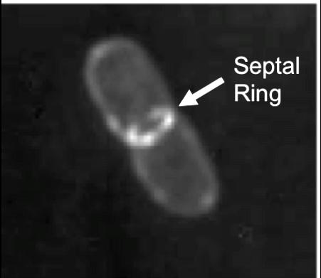
28 Figure 1.1. The septal ring. A. Overview of the proteins involved in cell division in E. coli. Proteins are shown from left to right in their order of recruitment to the septal ring, with FtsZ being the first component to arrive at the site. Proteins given a single letter designation are Fts proteins. OM, outer membrane; CM, cytoplasmic membrane; PG, peptidoglycan cell wall. B. GFP-FtsL fusion protein in E. coli. The image was taken using deconvolution microscopy and shows a pattern of fluorescence localization to a ring structure at the midcell. This ring structure is referred to as the septal ring (Image modified from (Ghigo et al., 1999)). 14

29 15 A B
30 Figure 1.2. Assembly of the septal ring. A. Recruitment hierarchy of E. coli cell division proteins as determined by localization dependencies. B. Reported interactions among Fts proteins through two-hybrid analysis (see text for source references). Lines connect proteins reported to interact in at least one assay, while circular arrows indicate selfinteraction (e.g., homodimerization). (Buddelmeijer & Beckwith, 2002; Vicente & Rico, 2006; Arends et al., 2010) 16
31 17 A B
32 Figure 1.3. Peptidoglycan in E. coli. A. Structure of peptidoglycan. Left: Layout of a PG monomer. This unit consists of alternating N-acetylglucosamine (GlcNAc) and N- acetylmuramic acid (MurNAc). MurNAc residues can have the indicated peptide side chain attached. Right: Peptide sidechains can be crosslinked by joining the D-Alanine position of one sidechain to the meso-diaminopimelic acid position of another chain. B. Mechanism of PG synthesis. Cartoon of how Lipid II is synthesized and translocated to the periplasm during PG synthesis in the E. coli cell. The enzyme responsible for the flippase activity is not yet known, although SEDS family proteins such as FtsW might accomplish this function. UDP-G, UDP-N-acetylglucosamine. 18

33 19 A B
34 Figure 1.4. The SPOR domain. A. The four SPOR domain proteins in E. coli. Membrane topology and number of amino acids in each domain as retrieved from UniProt release 15.7 ( or the GTOP update of 15 December 2008 ( N, amino terminus; CM, cytoplasmic membrane; OM, outer membrane. RlpA has a covalently attached lipid at its amino terminus. B. Ribbon diagram of the SPOR domain from the B. subtilis protein CwlC, based on the solution structure determined from NMR (Mishima et al., 2005). Cartoon was generated with Pymol from PDB ID 1x60 (DeLano, 2002), α-helices and β-strands are numbered from N to C-terminus as indicated. 20


35 21 A B
36 22 Figure 1.5. Localization of SPOR domain proteins. A. Full-length SPOR domain proteins localize to the septal ring. Fluorescence micrographs of live cells producing the indicated GFP or RFP fusion protein. B. Isolated SPOR domains localize to the septal ring. Fluorescence micrographs of live cells producing the indicated GFP fusion protein from a plasmid. These GFP fusions were targeted to the periplasm via the TAT system. Figure adapted form Arends et al, Bar, 5µm.


37 23
38 24 CHAPTER 2: GENETIC ANALYSIS OF THE CELL DIVISION PROTEIN FTSW AND ITS INTERACTION WITH FTSI (PBP3) Introduction As discussed in Chapter 1, cell division in bacteria is a complex process involving over 20 known proteins in E. coli. These proteins must function in a concerted fashion to synthesize all three layers of the cell envelope in order to gives rise to two new daughter cells. Two of the proteins involved in this process are FtsW and FtsI. FtsI is a class B high molecular weight PBP (see Chapter 1 for more information on FtsI) that localizes to the division site, where it is essential for crosslinking newly synthesized PG at the septum (Adam et al., 1997; Weiss et al., 1999; Arends, 2007). In terms of structure, FtsI is a bitopic membrane protein with a small cytoplasmic domain, a single transmembrane (TM) helix, and a large periplasmic region that contains both a domain of unknown function and a transpeptidase catalytic domain (Bowler & Spratt, 1989; Goffin et al., 1996; Goffin & Ghuysen, 1998). Several lines of evidence indicate that FtsI is targeted to the septal ring by its TM. First, random mutagenesis followed by a screen for localization-defective mutants returned only lesions in the TM (Wissel & Weiss, 2004). Second, a 26 amino acid fragment of FtsI that includes the TM and little if any flanking sequence localized to the septal ring, albeit poorly (Wissel et al., 2005). Third, alanine-scanning mutagenesis of the TM revealed that residues critical for septal localization cluster on one face of the helix, suggestive of a protein-protein interaction (Wissel et al., 2005). Because FtsI does not interact with itself in one two-hybrid system (Di Lallo et al., 2003; Karimova et al., 2005), but does interact modestly in another (Di Lallo et al., 2003), it is unclear at this point if the TM could mediate dimerization of FtsI. However, we suspect it (or a
39 25 complex consisting of two FtsI TM dimers) interacts with a TM from another division protein; the leading candidate is FtsW. FtsW belongs to a large family of polytopic membrane proteins found in all bacteria that contain a PG cell wall. This family is named SEDS, for shape, elongation division and sporulation (Henriques et al., 1998). The founding members of the SEDS family are FtsW and RodA of E. coli, and SpoVE of Bacillus subtilis (Ikeda et al., 1989; Boyle et al., 1997; Henriques et al., 1998). These proteins are required for PG synthesis during elongation (RodA), division (FtsW) and sporulation (SpoVE). Each SEDS protein appears to work together with a cognate class B penicillin-binding protein (PBP) that catalyzes the formation of cross links in PG. Examples of such pairs include RodA- PBP2, FtsW-FtsI (also called PBP3) and SpoVE-SpoVD. Typically a SEDS-PBP pair is encoded in the same operon and inactivation of either results in the same phenotype. Most SEDS proteins contain 10 transmembrane helixes (TMs) (Gerard et al., 2002; Lara & Ayala, 2002). Because of their complex transmembrane structure and involvement in PG synthesis, it has been suggested that SEDS proteins might transport Lipid II (a precursor for PG synthesis) across the cytoplasmic membrane (Ishino et al., 1986; Ikeda et al., 1989; Ehlert & Holtje, 1996). Efforts to prove this have yet to succeed (Lara et al., 2005) and more recently it has been proposed that MurJ family proteins are the missing Lipid II flippases (Ruiz, 2008; Ruiz, 2009), although this too has been challenged (Fay & Dworkin, 2009; Vasudevan et al., 2009). The one reasonably well-established function for SEDS proteins is in recruitment of their cognate transpeptidases to the correct place in the cell. In particular, our lab demonstrated that FtsW is needed for recruitment of FtsI to the septal ring, while Dworkin and co-workers showed that SpoVE recruits SpoVD to the outer forespore membrane (Mercer & Weiss, 2002; Real et al., 2008). Moreover, several studies have reported that FtsW and FtsI homologs from a variety of organisms interact either in two hybrid systems (Di Lallo et al., 2003; Karimova et al., 2005) or by immunoprecipitation
40 26 (Datta et al., 2006; Maggi et al., 2008). These considerations led us to undertake a systematic mutagenesis of the TMs of FtsW as an approach for characterizing the FtsW- FtsI interaction in molecular detail. Materials and Methods Media. E. coli strains were grown in Luria-Bertani (LB) medium containing 10 g tryptone, 5 g yeast extract, and 10 g NaCl per liter. Plates contained 15 g agar per liter. Ampicillin was used at 200 µg/ml and spectinomycin at 100 or 35 µg/ml for plasmids or chromosomal alleles, respectively. Kanamycin was used at 40 µg/ml and chloramphenicol at 30 µg/ml. Strains. Strains used in this study are listed in Table 2.1. All two-hybrid analyses were carried out in E. coli stain DMH1 (Karimova et al., 2000). Plasmids. Plasmids used in this study are listed in Table 2.2. Plasmids were constructed by PCR using VENT DNA polymerase (New England Biolabs) and sequenced to verify their integrity. All primers are listed in Table 2.3 and were obtained from Integrated DNA Technologies (Coralville, IA). Generation of triple alanine substitutions in FtsW TMs. Amino acid substitutions in three consecutive residues in each of the 10 transmembrane helices (TMs) were done using site directed mutagenesis and megapriming (Sarkar & Sommer, 1990). Megaprimers for creating the 3 alanine substitutions were generated using the primer pairs listed below and pdsw311 as template: TMH1; P845 and GFP666F, TMH2; P846 and GFP666F, TMH3; P847 and GFP666F, TMH4a; P869 and GFP666F, TMH4b; P870 and GFP666F, TMH5; P871 and P874, TMH6; P872 and P874, TMH7; P873 and P875, TMH8; P848 and P415, TMH9; P849 and P415, TMH10; P850 and P415. These
41 27 megaprimers were then purified with a PCR cleanup kit and used with either primer P415 or GFP666F in another round of PCR with pdsw311 as template. This created full length ftsw PCR product with the desired mutations and restriction sites for EcoRI (5 ) and HindIII (3 ). Plasmids for localization of GFP-FtsW. The ftsw 3-ala PCR products were digested with EcoRI and HindIII and ligated into the vector pdsw311 that had been cut with EcoRI and HindIII. The resulting plasmids contained gfp fused to the 5 end of the mutated ftsw. Plasmids for studying recruitment of GFP-FtsI. Three tandem copies of the hemagglutinin tag (3xHA) were amplified by PCR using pmpy-3xha as template and primers P902 plus P903. The 115 bp product was purified using a PCR cleanup kit (Qiagen) and used as a megaprimer, with P415, for amplifying ftsw from pdsw311. The ~1,350 bp product was then digested with MfeI and HindIII and ligated into the pam238 vector, which had been cut with EcoRI and HindIII. This leaves an EcoRI site between the HA tag sequence and the start of ftsw. This restriction site was used with the HindIII site to clone the collection of 3 alanine substitutions (described above) into this vector. Plasmids for testing FtsW s interaction with FtsI. pkt25-ftsw was cut with BamHI and KpnI. The vector was gel-purified (Qiagen). The various mutated forms of ftsw (described above) were then amplified using PCR and the primers P414 and P415. The resulting ~1,250 bp product was then cut with the same enzymes and ligated into the purified vector. put18c-ftsi was obtained from the Ladant laboratory (Karimova et al., 2005). Plasmids for testing mutant FtsI proteins in the two-hybrid system. Primers P1100 and P1096 were used in a PCR reaction, with pdsw521 (P 206 -gfp-ftsi) as template, to amplify a fragment of ftsi that encodes the first 50 amino acids of the protein. This fragment was purified using a PCR cleanup kit (Qiagen) and cut with BamHI and
42 28 KpnI and ligated into put18c that had been cut with the same enzymes. The resulting plasmid was named pdsw1160. pdsw1161 and pdsw1162 (which encode the first 80 and 239 amino acids of FtsI, respectively) were created in a similar manner. For pdsw1161 the primers used were P1100 and P1097. For pdsw1162, primers P1100 and P1098 were used. In order to generate a full length ftsi that encodes an L39P amino acid substitution, primers P1100 and P1099 were used to amplify the mutant allele from pdsw566. The fragment was purified using a PCR cleanup kit and digested with BamHI and KpnI and ligated into put18c that had been cut with the same enzymes. The resulting plasmid was named pdsw1163. pdsw1164 encodes for a full-length version of FtsI that has an extra leucine residue inserted at residue 41. This plasmid was generated as described for pdsw1163. However, pld75 was used as template for amplifying the mutant allele. General microscopy methods. Our microscope, camera, and software have been described previously, as has the fixation of cells with paraformaldehyde (Mercer & Weiss, 2002; Arends et al., 2009; Tarry et al., 2009; Arends et al., 2010). Localization of 3-alanine lesions of FtsW. The gfp-ftsw plasmids were transformed into wild type E. coli strain DHB4. Transformants were grown overnight at 30 C in LB supplemented with ampicillin to maintain the plasmid. The following morning, cells were subcultured 1:200 into fresh media and allowed to grow to an OD 600 of ~0.5. At that time cells were harvested and fixed in paraformaldehyde/glutaraldehyde and attached to glass slides for imaging GFP. Samples were also taken for Western blotting. Localization of the GFP-FtsW fusions was also tested in the FtsW depletion strain EC850, which has a wild type copy of ftsw under the control of an arabinose inducible
43 29 promoter. EC850 was transformed with the gfp-ftsw constructs or an empty gfp vector (negative control). Transformants were grown overnight at 30 C with 0.2% arabinose (to express wild type FtsW) and ampicillin and chloramphenicol for plasmid maintenance. The overnight cultures were then diluted 1:50 and grown with 0.02% arabinose until their OD 600 reached ~0.5. At this time they were shifted to growth with 0.2% glucose and no arabinose and expression of the gfp-ftsw allele to be tested was induced with the addition of 0.1 mm IPTG. After about 2 hours, the derivative carrying the empty gfp vector had become filamentous (~20 µm), which implied that cells were successfully depleted of wild type FtsW. At this time, all cultures were fixed for microscopy. Recruitment to the septum of GFP-FtsI by FtsW. The 3xHA tagged ftsw 3-ala constructs and an empty vector control were transformed into EC1655, an FtsW depletion strain that has gfp-ftsi integrated onto the chromosome in single copy. Transformants were grown as described above for localizing GFP-FtsW in EC850, except that spectinomycin and chloramphenicol were used for plasmid maintenance. In this case, addition of IPTG induced expression of both 3xHA-ftsW from a plasmid and the gfp-ftsi from the chromosome. Cells were fixed for microscopy, and samples were taken for Western blot analysis to verify production the 3xHA-FtsW protein. Bacterial two-hybrid analysis. Transformants of DHM1 carrying appropriate plasmid pairs (derivatives of put18c and pkt25) were streaked onto plates of LB medium containing ampicillin (200 µg/ml) and kanamycin (40 µg/ml), and the plates were incubated for 2 or 3 days at 30 C. Three to five colonies were used to inoculate 5 ml of LB medium containing ampicillin and kanamycin plus 0.5 mm IPTG, and the cultures were grown on a roller at 30 C for 16 h to an OD 600 of 0.6 to 0.8. Then duplicate 15-µl culture samples were assayed for β-galactosidase activity by standard procedures
44 30 (Miller, 1972). All assays were repeated at least three times (on different days). Western blotting. For Western blotting, typically 1 ml of culture at an OD 600 of 0.5 was centrifuged, and the resulting cell pellet was taken up into 0.5 ml of sodium dodecyl sulfate-polyacrylamide gel electrophoresis (SDS-PAGE) loading buffer. Samples were boiled, and 10-µl aliquots were subjected to SDS-10% PAGE. Proteins were then transferred onto nitrocellulose and detected by standard methods. Rabbit anti- GFP antibodies were obtained from W. Margolin and used at a dilution of 1:2,500. Mouse anti-ha serum (Covance) was used at a 1:10,000 dilution. The secondary antibody was horseradish peroxidase-conjugated goat anti-rabbit antibody (1:8,000; Pierce) or goat anti-mouse antibody (1:5000; Molecular Probes), which in turn was detected with SuperSignal Pico West chemiluminescent substrate (Pierce). Blots were visualized with an LAS-1000 luminescent imager from Fuji (Stamford, CT). Results Phenotypes of FtsW derivatives with mutant TMs. We mutagenized each of FtsW s 10 transmembrane helices by substituting three consecutive residues in the middle of each TM for alanine, except in the case of TM4, where two 3-ala constructs were made (black circles in Figure 2.1A). TM4 is predicted to contain highly conserved charged residues, so we were concerned that the 3-ala substitutions might disrupt assembly of the protein into the membrane. The full set of mutant proteins was cloned into two different vectors, creating fusions to GFP or three tandem copies of the hemagglutinin epitope tag (3xHA), respectively. Western blotting with anti-gfp and anti-ha antibodies verified wild type and all mutant proteins were produced at similar levels (Figure 2.2).
45 31 The FtsW derivatives were assayed for septal localization, ability to recruit FtsI to the septal ring, and complementation of an FtsW depletion strain. Only three of the mutant proteins had noteworthy defects (Figures 2.1B, 2.3, and 2.4). The TM1 mutant complemented weakly, did not localize very efficiently and failed to recruit FtsI (presumably as a consequence of its poor localization). We infer that TM1 plays a critical role in assembly of FtsW into the membrane or targeting FtsW to the septal ring. The TM4a mutant protein failed to support colony formation despite the fact that it localized to the septal ring and recruited FtsI. Perhaps TM4 is involved in the putative Lipid II transport activity of FtsW. The final interesting protein was the TM10 mutant, which localized well but did not support efficient recruitment of FtsI to the septal ring. The TM10 protein barely supported division in a complementation assay and was a mild dominant negative when co-expressed with wild type (note the elongated cell in the DHB4 background in Figure 2.3). This phenotype is what we would expect of an FtsW protein that is specifically defective in interaction with FtsI. Pastoret et al. (2004) reported a similar phenotype for a mutant FtsW with two amino acid substitutions in the periplasmic loop connecting TM9 to TM10, which led these authors to suggest the TM9- TM10 loop interacts with FtsI (which they called PBP3). Two-hybrid analysis. To try to get further evidence that TM10 of FtsW interacts with the lone TM of FtsI, we turned to a bacterial two-hybrid system that had been used previously to show FtsW interacts with FtsI (Karimova et al., 2005). First, we confirmed the previously reported FtsW-FtsI interaction (Figure 2.5), which was stronger even than the positive control (two leucine zipper domains) (Karimova et al., 2000). Next, we surveyed our collection of FtsW 3-ala proteins. Lesions in TM1, 5, 6 and 10 all reduced the apparent interaction with FtsI by 2- to 4-fold, with the TM10 mutant protein exhibiting the weakest interaction (Figure 2.5). While generally consistent with our hypothesis, the complexity of these results makes it difficult to draw firm conclusions.
46 32 We also used the two-hybrid system to test the ability of several FtsI derivatives to interact with FtsW. Lesions in the TM of FtsI that prevent septal localization (Wissel & Weiss, 2004) also abrogated the interaction (Figure 2.6; see Leu 39 to Pro, or lengthening the TM by insertion of a Leu at position 41). Thus, the TM of FtsI appears to be necessary for the interaction with FtsW. An FtsI protein truncated at residue 50, designated 50-stop, failed to interact with FtsW in the two-hybrid system (Figure 2.6). This fragment encodes the entire cytoplasmic domain and TM and localizes to the septal ring, albeit weakly (Wissel et al., 2005). To observe an interaction with FtsW, it was necessary to include more of the periplasmic domain (Figure 2.6; e.g., 80-stop and 239- stop). In toto, these findings argue that FtsI must have an intact TM to interact with FtsW but the TM itself is not sufficient portions of the periplasmic domain immediately following the TM are needed too. These data are also consistent with a report by Karimova et al (2005) in which they showed a fragment of FtsI consisting of residues 1-70 could interact with FtsW, but fragments made up of residues 1-42 or (both of which maintain the TM) were unable to interact with FtsW. Discussion Systematic mutagenesis of the TMs of FtsW has revealed three TMs that may play specific roles in FtsW function. (i) TM1 is important for septal localization. One interesting possibility is that TM1 of FtsW interacts with a septal ring component like FtsQ, FtsL or FtsB these proteins form a complex that has been implicated in recruitment of FtsW to the septal ring (Goehring et al., 2006; Gonzalez et al., 2010). At this point, we do not know if the lesions in TM1 of FtsW are blocking the protein s ability to interact with other cell division proteins or simply preventing it from properly inserting into the membrane. We do know that when expressed from a plasmid,
47 33 the mutant protein appears as stable as wild-type (Figure 2.2). But, we would need to probe the membrane fraction from cells expressing the mutant protein to address the abundance at that site. One interesting way of following up on the TM1 localization phenotype would be testing this mutant protein for its ability to interact with the cell division proteins directly upstream in the recruitment pathway (FtsQLB) using the two-hybrid assay. However, in the two-hybrid system we have chosen to use, FtsW does not interact with FtsB and only weakly interacts with FtsQ and FtsL (Karimova et al., 2005), despite the in vivo interactions reported above. The modest levels of interaction FtsW shows with FtsQ and FtsL might prove problematic in interpreting results from an FtsW mutant. However, in another two-hybrid system (based on phage repressors) FtsW has been shown to interact with FtsQ, FtsL, and FtsB (Di Lallo et al., 2003). Perhaps follow-up work could be done using that system to further explore the role of TM1 in FtsW midcell targeting. (ii) TM4 is essential for FtsW function. The TM4a 3-ala protein failed to rescue an FtsW-depletion strain, despite being present in normal amounts. Moreover, the protein localized to the septal ring and recruited FtsI. A noteworthy feature of TM4 is that it contains highly conserved charged residues. The properties of the TM4 mutant are consistent with this being a site of Lipid II interaction, although other interpretations cannot be excluded. Working with most SEDS proteins is often complicated by their essential nature for the organism. However, as noted earlier, the sporulation related SEDS proteins SpoVE is non-essential and has been studied in B. subtilis (Real et al., 2008). Perhaps the information on the 4 th TM s role in activity from our study could be applied to the SpoVE system. Analogous lesions could be engineered into SpoVE s corresponding TM and spore formation phenotypes studied. This approach would bypass the difficulties of working with depletion strains when studying the essential SEDS proteins, such as FtsW and RodA.
48 34 (iii) TM10 is involved in recruitment of FtsI to the septal ring. The TM10 3-ala protein localized to the septal ring but did not to recruit FtsI efficiently, resulting in a mild dominant negative phenotype. In a two-hybrid assay, the TM10 3-ala protein interacted only weakly with FtsI. Efforts to demonstrate a direct interaction using cysteines engineered into TM10 of FtsW and the lone TM of FtsI have been unsuccessful (not shown). While some of those difficulties were likely related directly to FtsW s problematic behavior in biochemical analysis (problems transferring protein from gels to membranes, aggregation in gel loading wells, inability to boil samples, among others), the possibility remains that the TM of FtsW doesn t make direct contact with the TM of FtsI. (iv) Several observations argue that the region of FtsI s periplasmic domain closest to the TM also contributes to septal localization and interaction with FtsW. The inability of the P50-stop truncation of FtsI to interact with FtsW in the two hybrid analysis and the report from Nguyen-Disteche s laboratory that mutations in the loop region between FtsW s TM9 and 10 were important for FtsI recruitment (Pastoret et al., 2004) support this possibility. However, it is clear that the TM of FtsI is still critical for the FtsW interaction. Either of the mutant forms of FtsI with an altered TM failed to interact with FtsW in the two-hybrid system (Figure 2.6). These observations are consistent with results from Mark Wissel in which those same mutant FtsIs failed to localize to the division site (Wissel et al., 2005).
49 35 Table 2.1. Strains used in this study Strain Relevant features Source or reference DHB4 F laci q pro/λ ΔlacX74 gale galk thi rpsl phor ΔphoA(PvuII) ΔmalF3 Boyd et al., 1987 EC850 ftsw::kan / pbad33-ftsw Mercer et al., 2002 EC1655 W3110 lacu169 gal490 ftsw φ80::gfp-ftsi / pbad33-ftsw Lab Collection DHM1 glnv44(as) reca1 enda gyra96 thi-1 hsdr17 Karimova spot1 rfbd1 cya-854 et al, 2000 (Boyd et al., 1987)
50 36 Plasmid Table 2.2. Plasmids used in the study Relevant features Source or reference pdsw311 P 206 -gfp-ftsw Amp r Weiss, 2002 Mercer and pam238 pacyc184 derivative, lac UV5 p Spc r 2004 Kadokura et al., pmpy-3xha 3x-HA tag vector Schneider, 1995 put18c BACTH vector for fusion of target proteins to B. pertussis cya gene T18 fragment; P lac ::cya MCS puc ori Ap r Karimova et al, 2000 BACTH vector for fusion of target proteins to Bordetella pertussis cya gene T25 fragment; P lac ::cya p15 ori Km r Karimova et al, 2000 pkt25 pdsw824 pdsw311(3-ala 1) This work pdsw825 pdsw311(3-ala 3) This work pdsw826 pdsw311(3-ala 8) This work pdsw827 pdsw311(3-ala 9) This work pdsw828 pdsw311(3-ala 10) This work pdsw829 pdsw311(3-ala 4a) This work pdsw830 pdsw311(3-ala 4b) This work pdsw832 pdsw311(3-ala 2) This work pdsw833 pdsw311(3-ala 5) This work pdsw834 pdsw311(3-ala 6) This work pdsw835 pdsw311(3-ala 7) This work pdsw836 pam238-3xha-ftsw This work pdsw920 pkt25-ftsw This work pdsw921 pdsw920(3-ala 8) This work pdsw922 pdsw920(3-ala 10) This work pdsw970 pdsw920(3-ala 1) This work pdsw971 pdsw920(3-ala 2) This work pdsw972 pdsw920(3-ala 3) This work pdsw973 pdsw920(3-ala 4a) This work pdsw974 pdsw920(3-ala 4b) This work pdsw975 pdsw920(3-ala 5) This work pdsw976 pdsw920(3-ala 6) This work pdsw977 pdsw920(3-ala 7) This work pdsw978 pdsw920(3-ala 1) This work (Kadokura et al., 2004) (Schneider, 1995)
51 37 Plasmid Table 2.2. continued Relevant features Source or reference pdsw1149 pam238-3xha-ftsw(3-ala 1) This work pdsw1150 pam238-3xha-ftsw(3-ala 2) This work pdsw1151 pam238-3xha-ftsw(3-ala 3) This work pdsw1152 pam238-3xha-ftsw(3-ala 4a) This work pdsw1153 pam238-3xha-ftsw(3-ala 4b) This work pdsw1154 pam238-3xha-ftsw(3-ala 5) This work pdsw1155 pam238-3xha-ftsw(3-ala 6) This work pdsw1156 pam238-3xha-ftsw(3-ala 7) This work pdsw1157 pam238-3xha-ftsw(3-ala 8) This work pdsw1158 pam238-3xha-ftsw(3-ala 9) This work pdsw1159 pam238-3xha-ftsw(3-ala 10) This work pdsw1160 put18c-ftsi(p50 truncation) This work pdsw1161 put18c-ftsi(r80 truncation) This work pdsw1162 put18c-ftsi(r239 truncation) This work pdsw1163 put18c-ftsi(l39p) This work pdsw1164 put18c-ftsi(+l41) This work
52 38 Table 2.3. Primers used in the study Primer Sequence a P415 GCGAAGCTTCTAGATCATCGTGAACCTCGTACAAACGC GFP666F GAGACCACATGGTCCTTCTTGAG P845 AAAGCCAATCGCCGCGGCCGCGGTCAGCCACAGTAA P846 CAGCGTAATGATCGCAGCGGCCGCCGCCAAAATCAGATA P847 GACGATCATCAGCAGAGCGGCCGCTCCGAGCAGCATCGT P848 GTCGGTGTGGTGCTGGCGGCCGCTATGGTATTCTTCGTC P849 TCTATTGGCATCTGGGCGGCCGCTCAGGCGCTGGTTAAC P850 TACGGTGGTTCGAGCGCGGCCGCTATGTCGACAGCCATC P869 AAACAGCGACAGTTTAGCGGCCGCCGCAGGCTGGATACG P870 GGCGATATAGCAAAAAGCGGCCGCTTTTGTCAGCTCCGC P871 TAACACTGCCAACACAGCGGCCGCGCCCATCGGTTTCAG P872 CAGGAACAACATCGCAGCGGCCGCCACAAACAACACCAC P873 CAGCAACACAACCGCTGAAATGCCCATACCGATAATGGC P874 CTTCTTCGCGAAGCGTGA P875 CGAAGTACGTAATAACCT P880 CGTGAATTCGCGGCGGCCGCTAAACTGTCGCTGTTTTGC P881 CGTGAATTCAAAGCGGCCGCTTTTTGCTATATCGCCAAC P882 CGTGAATTCGGCGCGGCCGCTGTGTTGGCAGTGTTACTG p902 AGTCAATTGTTACCCATACGATGTT P903 AACGCATGTTGTTGTTGTTGAATTCAGCGTAATCTGGAACGTC P912 GCATGAATTCTTACGATCTGCC P913 TCGACTCTAGAGAAAGCAGCGCGC P914 GCTCGGTACCTTCATCGTGAAC P915 TCGACTCTAGAGATGCGTTTATCT P993 ACTAAGCTTTCATCGTGAACCTCGTACAA P994 AGTGGTACCTTACCCATACGATGTT P1021 TACGGTGGTTCGAGCTTACTGATTTGCTCGACAGCCATC P1022 TACGGTGGTTCGAGCTTACTGTGCATGTCGACAGCCATC P1023 TACGGTGGTTCGAGCTTATGCATTATGTCGACAGCCATC P1024 TACGGTGGTTCGAGCTGCCTGATTATGTCGACAGCCATC P1025 TACGGTGGTTCGTGCTTACTGATTATGTCGACAGCCATC P1033 CTTAGGTACCGTTCATCGTGAACCTCGTACAA P1034 CTAGAGGATCCCATGCGTTTATCTCTC P1057 AGCGCCAGGCAAATACAGCC P1058 GGAGAATACAGCAGCATAACAACG P1059 GCCGCATAAGCACGCAAAACG
53 39 Table 2.3. continued Primer Sequence a P1060 CAGGAGAATAGCGCCGCATAA P1061 GAATACAGCCGGCTAACAACGC P1096 CTTAGGTACCCGGGGAGATAACTTGTA P1097 CTTAGGTACCGCGACCAGAACGGTCAGTA P1098 CTTAGGTACCCAGGCGTTCATCAATACTC P1099 CTTAGGTACCTTACGATCTGCCACCTG P1100 CTAGAGGATCCCAAAGCAGCGGCGAAA a DNA sequence is given in the 5 to 3 direction for all primers.
54 Figure 2.1. Overview of FtsW. A. Model of FtsW. Residues conserved among different γ-proteobacteria are shown in green. Residues targeted for alanine substitution are shown in bold black outline. B. Table summarizing phenotypes observed in this study. 40

55 41 A B Summary of FtsW 3 alanine lesion phenotypes Lesion Localization Localization FtsI DHB4 Depletion Recruitment a Complementation WT 45% 38% TMH1 <1% 10% - + b + TMH2 45% 15% TMH3 51% 15% TMH4a 25% 17% c + TMH4b 30% 6% TMH5 24% 30% TMH6 30% 30% TMH7 36% 17% TMH8 36% 28% TMH9 32% 12% Protein Stability TMH10 89% 90% + + b + a FtsI recruitment measured as spacing of bands of GFP-FtsI fluorescence in cells. +++ was assigned to cells expressing wild-type FtsW and represents ~11 microns per GFP band. + assigned to TMH10 mutant represents ~23 microns per band. The TMH1 mutant showed extremely low levels of GFP-FtsI recruitment. b Cells appeared filamentous when examined with phase contrast microscopy. C No colony formation on plates, but only modest filamentation in liquid culture.
56 Figure 2.2. Steady state protein levels of FtsW. A. Levels of the GFP-FtsW fusion proteins (blot probed with anti GFP primary antibody). B. Levels of the 3xHA tagged FtsW fusion proteins (blot probed with anti HA monoclonal antibody). 42


57 43 A. B.
58 Figure 2.3. Localization of selected GFP-FtsW proteins with TMH lesions. Constructs were expressed either in a strain background (DHB4) with native FtsW present or after depletion of native FtsW (by growing EC850 transformants in LB glucose). 44

59 WT FtsW FtsW Present FtsW Depleted 45
60 Figure 2.4. Recruitment of GFP-FtsI by FtsW TM mutant proteins. Cultures were grown in LB glucose to deplete wild-type FtsW. IPTG was added to induce gfp-ftsi and the plasmid-borne ftsw allele indicated. None = no addition of IPTG; this was done to monitor formation of filamentous cells. 46

61 47
62 Figure 2.5. Bacterial two-hybrid analysis of FtsW TMH lesions. A set of 10 FtsWs with 3 alanine substitutions in each TMH was tested against FtsI. ftsw derivatives were located on the pkt25 plasmid and ftsi on the put18c vector. Negative control = empty vectors. Positive control = two leucine zipper proteins known to interact (Karimova et al., 2000). 48
63 Miller Units Negative Control Positive Control FtsW (WT) / FtsI FtsW (TMH1) / FtsI FtsW (TMH2) / FtsI FtsW (TMH3) / FtsI FtsW (TMH4) / FtsI FtsW (TMH5) / FtsI FtsW (TMH6) / FtsI FtsW (TMH7) / FtsI FtsW (TMH8) / FtsI FtsW (TMH9) / FtsI FtsW (TMH10) / FtsI
64 Figure 2.6. FtsI. A. Domain structure of FtsI (modified from Wissel et al., 2005). B. Bacterial two-hybrid analysis of FtsI mutant/truncation collection against FtsW. Assays conducted as per analysis shown in Figure 2.5. ftsi variants are located on the put18c vector. Positive and negative controls as in Figure
65 51 A. B.
66 52 CHAPTER 3: CHARACTERIZATION OF A PEPTIDOGLYCAN BINDING ASSAY USING PURIFIED SPOR DOMAINS Introduction Although SPOR domains are described in databases such as Pfam and Interpro as being involved in binding peptidoglycan, this activity has only been demonstrated biochemically for the SPOR domain from FtsN from two species, E. coli and Caulobacter crescentus (Ursinus et al., 2004; Moll & Thanbichler, 2009). In both cases binding was demonstrated by showing that a purified His 6 -tagged SPOR domain cosedimented with whole PG sacculi during ultracentrifugation. Essentially the same assay has also been used to show that a PASTA domain from a Mycobacterium tuberculosis kinase also binds to PG (Shah et al., 2008). Unfortunately, none of these publications did a very thorough job of characterizing the co-sedimentation assay, so it is not entirely clear how robust the assay is or what exactly it measures. Both our lab and Piet de Boer s recently demonstrated that numerous SPOR domains localize to the division site when produced in growing E. coli cells (Gerding et al., 2009; Arends et al., 2010). Both labs also hypothesized that SPOR domains are recruited to the division site by binding preferentially to septal PG. One problem with this hypothesis, however, is that PG in the division septum is generally considered to be identical to PG elsewhere around the cell (Hungerer & Tipper, 1969; Glauner et al., 1988; Obermann & Holtje, 1994; Signoretto et al., 1996; Ishidate et al., 1998; Lara et al., 2005). We think there must be differences, and hope that studying the SPOR:PG interaction in detail will reveal what these differences are. In this chapter we describe our efforts to characterize the co-sedimentation assay that we and others have used to document binding of proteins to PG. We then use this assay to explore some of the structural features of PG that might be recognized by SPOR
67 53 domains. We also demonstrate that a wide range of SPOR domains bind PG. In this context, it is important to note that the ideal assay would be quantitative and minimize the amount of PG used per reaction, because purification of PG from E. coli is very time consuming and the yield is low. Other desirable characteristics included robustness with respect to ph and salt concentrations, linearity with protein, and specificity. By specificity we mean two things. First, only authentic PG binding proteins should cosediment with the sacculi under the assay conditions employed. Second, SPOR domains should bind to purified PG sacculi the same way they bind to PG in live cells. The results presented below show that our assay is reasonably well behaved in all respects, except one binding of SPOR domains to isolated sacculi does not appear to reflect specific binding to only the septal PG in those sacculi. Materials and Methods Media. E. coli strains were grown in Luria-Bertani (LB) medium containing 10 g tryptone, 5 g yeast extract, and 10 g NaCl per liter. Plates contained 15 g agar per liter. Ampicillin was used at 200 µg /ml. Strains. Strains used in this study are listed in Table 3.1. Aquifex aeolicus DNA was obtained from R. Huber, Cytophaga hutchinsonii ATCC was obtained from the American Type Culture Collection, Vibrio parahaemolyticus LM5674 was obtained from L. McCarter, Staphylococcus aureus SH1000 was obtained from A. Horswill, and Bacillus subtilis PY79 was obtained from C. Ellermeier. Plasmids. Plasmids used in this study are listed in Table 3.2. Plasmids were constructed by PCR using VENT DNA polymerase (New England Biolabs) and sequenced to verify their integrity. All primers were obtained from Integrated DNA
68 54 Technologies (Coralville, IA). In the following descriptions, restriction sites incorporated into primers are underlined. pdsw1000 [P T5 ::His 6 -damx( )] was constructed using primers P1144 (GCCGGATCCAACAACAACGGTTCGTTGAAATCGGCA) and P1137 (CTGAAGCTTACTTCAGATCGGCCTGTACCT) to amplify the last 90 codons of damx with pdsw918 as the template. The 301-bp product was cut with BamHI and HindIII and ligated into the corresponding sites of pqe80l. pdsw1002 [P T5 ::His 6 - dedd( )] was constructed by amplifying the last 80 codons of dedd from plasmid pdsw937 with primers P1125 (GCCGGATCCAACAACAACGGTAAAGCCTATGTTGTG) and P1126 (GCCAAGCTTTAATTCGGCGTATAGCCCATT). The 269-bp product was cut with BamHI and HindIII and ligated into the corresponding sites of pqe80l. pdsw1003 [P T5 ::His 6 -rlpa( )] was constructed by amplifying the last 80 codons of rlpa from plasmid pdsw931 with primers P1127 (GCCAGATCTAACAACAACGTCTCGCAAAGCGCCAGC) and P1128 (GCCAAGCTTTACTGCGCGGTAGTAATAAAT). The 269-bp product was cut with BglII and HindIII and ligated into the BamHI and HindIII sites of pqe80l. pdsw1114 [P T5 ::His 6 -aq1896( )] was constructed by amplifying the last 89 codons of aq1896 from A. aeolicus chromosomal DNA with primers P1223 (GCCGGATCCAACAACAACATCCCAAGAGTGCATAAA) and P1224 (CTGAAGCTTATCACTTGATTTCAACGACGAA). The 298-bp product was cut with BamHI and HindIII and ligated into the corresponding sites of pqe80l. pdsw1113 [P T5 ::His 6 -chu2221( )] was constructed by amplifying the last 89 codons of chu2221 from chromosomal DNA of C. hutchinsonii ATCC with primers P1229 (GCCGGATCCAACAACAACTTCTATTCTACTAAATTGGAG) and P1230 (CTGAAGCTTATTTTGGAGATAGGATCACACT). The 295-bp product was cut with BamHI and HindIII and ligated into the corresponding sites of pqe80l. pdsw1115 [P T5 ::His 6 -vpa1294(37-134)] was constructed by amplifying the last 97 codons of
69 55 vpa1294 from chromosomal DNA of V. parahaemolyticus strain LM5674 using primers P1317 (GCCGGATCCAACAACAACGCATACGGCTACCTGAATCCC) and P1318 (CTGAAGCTTAGCGCATTCTCACGACACTAG) and ligated into the corresponding sites of pqe80l. Purified proteins. MBP2* was purchased from New England Biolabs (Beverly, MA). FtsZ was overproduced and purified as described previously (RayChaudhuri & Park, 1992), except that Q-Sepharose Fast-Flow was substituted for DEAE-Sephacel. His 6 -tagged SPOR domains were overproduced in and purified from E. coli BL21 transformants by cobalt affinity chromatography on Talon affinity resin according to instructions from the manufacturer (Clontech, Mountain View, CA). The purified proteins were dialyzed into storage buffer (50 mm Tris-HCl, 200 mm NaCl, 5% glycerol, ph 7.5), and aliquots were stored at 80 C until needed. A 0.5-liter culture yielded 4 mg purified protein as determined by ultra violet absorption at 280 nm with a Nanodrop Spectrophotometer (Thermo). Extinction coefficients for each His 6 -SPOR domain were calculated from their sequences using ProtPram (Wilkins et al., 1999). Protein purity was >95% as judged by SDS-PAGE. Purification and quantification of PG. Cells of wild type E. coli (EC251) for isolation of PG sacculi were grown at 30 C and 210 rpm in 2 L flasks containing 0.5 L of LB. Typically two cultures at an OD 600 ~ 0.8 were harvested per preparation. Purification of PG sacculi involved boiling cells in SDS and multiple enzymatic treatments (Pronase, Amylase, DNase, RNase) essentially as described (Glauner, 1988). PG was quantified for us by David Popham (Virginia Tech) by quantifying muramic acid. Briefly, samples of purified PG were suspended in 6 N HCl and hydrolyzed at 95 C for 4 h. Hydrolysates were subjected to amino sugar analysis (González-Castro, 1997) and quantified relative to purified standards processed in parallel. The values furnished to us by Dr. Popham were nmol of NAG per µl of sample. To convert nmol of NAG to a
70 56 weight basis, we took advantage of the fact that, to a good approximation, PG is a repeating structure composed of NAG-NAM(tetrapeptides). Thus, there are as many nmols of NAG as there are nmols of NAG-NAM(tetrapeptide). The mass of a NAG- NAM(tetrapeptide) is 985 g/mol, so a PG sample that has 1 nmol of NAG per µl has 985 ng (or µg) of NAG-NAM(tetrapeptide) per µl. For generation of PG from filamentous E. coli cells that lack division septa, strain EC309 (ftsz(ts)), was used. Cells were grown at 30 C until an OD 600 of 0.4 was reached. At that time the temperature was shifted to 42 C. After one hour of growth at 42 C, cells appeared filamentous under light microscopy, with an average length of ~17 µm. PG was then prepared from these cells as described above. PG binding assays. Our standard binding assay was conducted in 25 mm potassium phosphate, ph 7.5, and 200 mm NaCl. A standard assay mixture contained 12 µg protein and 75 µg PG in a total volume of 100 µl. This is equivalent to ~1 nmol of His 6 -DamX SPOR and ~75 nmol of NAM-NAG disaccharide units. As a control, assay mixtures that lacked PG were also prepared. Mixtures were incubated for 1 h on ice and then centrifuged at 4 C in a Beckman TLA-55 rotor at 50,000 rpm (average g force, 112,000 x g) for 45 min. The supernatant was recovered and saved for analysis. The PG pellet was washed by suspending it in 100 µl of cold assay buffer and centrifuging as described above. The wash supernatant was also saved. The PG pellet was again suspended in 100 µl of binding buffer. Aliquots of all three fractions (supernatant, wash, pellet) were analyzed by SDS-15% PAGE. Gels were stained with Coomassie Blue (GelCode Blue from Pierce, Rockford, IL) and scanned using a Typhoon 8610 Imager (GE Healthcare) with the following instrument settings: excitation laser, 532 nm; emission filter, 560 nm, long pass; photomultiplier, 600 V; and pixel size, 100 µm. ImageQuant software was used to quantify fluorescence signals. Because there was not
71 57 much protein in the wash, in later versions of the assay we pooled the initial supernatant and wash fractions before loading the gels. Results Comparison of published assay conditions. Our assay was based on published versions of the procedure, which we now summarize. Ursinus et al (2004) incubated 3 µg of protein with 100 µg PG in a buffer containing 10 mm Tris-maleate (ph 6.8), 10 mm MgCl 2, 50 mm NaCl, and 0.02% NaN 3. Protein was detected by Western blot or by staining gels with zinc. Photos of representative gels were published, but there was no quantification. Möll and Thanbichler (2009) used this same buffer/salt but roughly four fold more PG. Protein was detected by Western blotting, and a picture of the blot was published. Shah et al. (2008) used 50 µg protein and 5 mg PG (Shah et al., 2008) but did not specify volumes, buffer or salt conditions. Gels were stained with GelCode Blue (Thermo) and scanned, with results presented as a (quantitative) bar graph. In summary, the most striking differences among these assays related to the amounts of protein and PG used. Of the three published studies, only Ursinus et al. (2004) included specificity controls. To demonstrate specificity with respect to protein, they used a mixture of protein molecular weight standards purchase from BioRad. They did not show the result, but said that some of these control proteins co-sedimented with PG, which could be worrisome, depending on the extent of the binding. Ursinus et al. also presented evidence that co-sedimentation of FtsN s SPOR domain reflects binding to septal PG. This was accomplished by showing that only about half as much SPOR domain bound when sacculi were prepared from non-dividing cells. The non-dividing cells were obtained by treating a culture with furazlocillin, a ß-lactam that inactivates FtsI (also known as penicillin-binding protein 3).
72 58 Development and characterization of our assay. Since PG from E. coli is a limiting resource, we chose to use a lower amount (75 µg) than the above reports for our assays. For instance, the 5 mg used in one experiment by Shah et al. (2008) would consume nearly all the PG purified from 2 liters of E. coli. (Note that Shah et al. were working with B. subtilis, which has more than 10-fold more PG per cell than does E. coli.) We also decided to switch to a phosphate buffer, since Tris-based buffers do not function that well at ph 6.8 and we wished to assay a variety of ph levels. Tris is also more affected by temperature changes. An overview of our assay is presented in Figure 3.1. Our findings when His 6 - DamX SPOR was tested at various ph and salt conditions are summarized in Table 3.3. Briefly, about 60% of the input protein bound to PG at ph 6.5, 7.0 and 7.5, but only ~30% bound at ph 8.0. Likewise, when the ph was 7.5, ~60% of the protein cosedimented with PG at 50 and 200 mm NaCl, but this dropped to ~30% at 500 mm NaCl. Inclusion of 20 mm imidazole had little or no effect on binding to PG, arguing that binding is not driven by a non-specific interaction of the His 6 tag with the sacculus. The effect of protein concentration on binding was assayed in mixtures containing 75 µg of PG, 25 mm NaPO 4 buffer (ph 7.5) and 200 mm NaCl. Binding increased rather linearly with protein over the entire range assayed, which was from 3 µg to 36 µg (Fig. 3.2). Nevertheless, the fraction of input protein that co-sedimented with PG decreased slightly above 12 µg (Table 3.3). Binding of DamX SPOR to different PG preparations. As an initial approach to identifying the chemical features of PG that constitute a binding site for the SPOR domain of DamX, we assayed co-sedimentation with PG prepared from different sources. A PG preparation expected to lack septal PG was prepared from an ftsz(ts) mutant that had been grown at the non-permissive temperature until the cells were ~17 um long. Interestingly, DamX SPOR bound as well to PG from these nondividing cells as to PG from
73 59 wild type cells that were dividing normally (Table 3.3). This result suggests the pull down assay measures a general affinity of the protein for PG rather than specific binding for septal PG. We took advantage of the structural diversity that exists naturally among different bacterial species to explore the potential relevance of the peptide cross-link. To this end, we isolated sacculi from Bacillus subtilis, B. sphaericus, and S. aureus. B. subtilis and E. coli use the same cross-link; the ε-amino group of a diaminopimelic acid in position 3 is joined directly to the carboxylate of D-Ala in position 4 (Atrih et al., 1999). Nevertheless, in B. subtilis almost all of the peptide side-chains are cross-linked whereas in E. coli only about half are cross-linked. This difference arises because B. subtilis PG is much thicker than E. coli PG (~15 vs. ~1 layer). B. sphaericus is like B. subtilis except that the L-meso-diaminopimelic acid in the third position is replaced with L-lysine. S. aureus also has a multi-layer, extensively cross-linked sacculus as well as the L-lysine substitution but differs in having a penta-glycine bridge in the cross link (Rogers, 1980). The SPOR domain from DamX bound PG from either of the two Bacillus species about as well as that from E. coli, but about 30% less protein bound to S. aureus (Figure 3.3). Interestingly, S. aureus lacks any proteins with identifiable SPOR domains (See Discussion). PG binding is a general activity of SPOR domains. As noted in the introduction, the generalization that SPOR domains bind PG is based on studies of the SPOR domain of FtsN from two different bacterial species (Ursinus et al., 2004; Moll & Thanbichler, 2009). Our studies up to this point clearly show that the SPOR domain from E. coli DamX also binds PG. In order to determine whether PG binding is a general property of SPOR domains, we purified and tested His 6 -tagged SPOR domains from five additional proteins: DedD, RlpA, VPA1294, CHU2221, and AQ1896 proteins. In each case, ~80% of the SPOR domain protein was recovered in the PG pellet (Figure 3.4),
74 60 whereas all of the protein remained in the soluble fraction when the sample was centrifuged in the absence of PG (data not shown). As a negative control, we performed similar pulldown assays with maltose binding protein (MBP) and FtsZ. Only ~20% of these proteins copelleted with PG (Figure 3.4), and neither was detected in the pellet when the sample was centrifuged in the absence of PG (data not shown). Discussion Based on the studies described above, we settled on the following conditions as a standard co-sedimentation assay: 12 µg protein, 75 µg PG, 25 mm NaPO 4 buffer (ph 7.5), 200 mm NaCl, in a final reaction volume of 100 µl. The amounts of protein and PG were chosen to be high enough to readily detect and quantify both the bound and unbound protein using Coomassie-stained gels. The buffer conditions were chosen to support efficient binding, with up to about 80% of the input protein co-sedimenting with the PG. Under these conditions binding was roughly linear with protein, which makes the assay suitable for asking whether different forms of PG or mutant SPOR domains bind less well. Although the amount of input protein could easily be increased beyond 12 µg, this would seem to be a waste of protein. At the lower extreme, we find that 3 µg of input protein is the minimum we can use given our detection methods. We think it would be possible to lower the amount of input protein using other detection methods such as Western blotting or if protein samples were concentrated by TCA precipitation prior to loading gels. The drawback to these methods for quantitative work is that every manipulation introduces potential for errors. In this context it is worth noting that we tried staining our gels with silver rather than Commassie but encountered problems with high background that interfered with quantification. Using the data from our titration experiment, we estimate there are at least 90,000 binding sites per sacculus, although the true value is likely to be much higher given that
75 61 the assay did not reach saturation. The value of 90,000 was arrived at as follows. When 36 µg of protein was used, about 18.5 µg was recovered in the pellet with the PG, which corresponds ~2 nmol of His 6 -DamX SPOR. That assay contained 75 µg of PG, which corresponds to ~75 nmol of disaccharide peptide units. Thus, the amount of protein recovered in the pellet was equivalent to ~3% of the disaccharide peptide units. The average E. coli sacculus contains 3 x 10 6 disaccharide peptide units (Wientjes et al., 1991). Three percent of 3 x 10 6 corresponds to 90,000. In vivo SPOR domains probably bind preferentially to septal PG. Nevertheless, DamX s SPOR domain bound equally well to PG from wild type and filamentous cells of an ftsz(ts) mutant that should not have contained any septal PG. Curiously, Ursinus et al. reported that the SPOR domain from FtsN bound better to wild type PG than to PG from filamentous cells (Ursinus et al., 2004). Based on this, they inferred that FtsN s SPOR domain binds mainly septal PG in the pull down assay. Our findings argue otherwise. We cannot account for this discrepancy, but it is possible that FtsN s SPOR domain behaves differently from DamX s. Alternatively, it should be noted that Ursinus et al. generated filamentous E. coli by using an antibiotic that inhibits the transpeptidation activity of FtsI. Prior to this study, only two SPOR domains had been shown to bind PG in a purified system, and both of these domains were derived from FtsN proteins (Ursinus et al., 2004; Moll & Thanbichler, 2009). Here we extended this finding to six additional SPOR domains, three from E. coli and one each from V. parahaemolyticus, C. hutchinsonii, and A. aeolicus. These findings establish that PG binding is a general property of SPOR domains. We have also demonstrated that all of these SPOR domains can also localize to the division site when expressed in E. coli (Arends et al., 2010). Taken together, these findings suggest septal localization is driven by binding to septal PG. Chapter 4 of this thesis describes our efforts to explore this link and identify
76 62 structural features of the SPOR domain that are important for protein function both in vivo and in vitro. Some follow up work should be focused on better understanding the limitations of our binding assay. It should be possible to fully saturate the PG with protein and quantify the number of binding sites per sacculus, at least in the co-sedimentation assay. Conversely, if we were able to use much less protein in the assay, differences between wild type and long cell PG might become evident. Using less protein will require switching to Western blotting, or concentrating samples prior to analysis. Preliminary work using PG isolated from a variety of organisms revealed that DamX SPOR bound equally well to PG from E. coli, B. subtilis and B. sphaericus, but less well to PG from S. aureus. In terms of structure, the most profound difference between S. aureus PG and all the rest is that the cross-link in S. aureus includes a penta-glycine bridge (Rogers, 1980). S. aureus sacculi may also have more curvature since the organism is coccus shaped. All other sacculi tested were from rod-shaped bacteria. This poor binding of DamX SPOR to S. aureus sacculi suggest that the cross-link is part of the binding site or the three-dimensional arraignment of the PG subunits may affect affinity. Interestingly, BLAST searches of S. aureus genomes failed to identify any SPOR domains (K. Williams, unpublished). It would be interesting to ask whether SPOR domains for heterologous organisms localize to division sites if produced in S. aureus.
77 63 Table 3.1. Strains used in this study Strain Relevant features Source or reference BL21 (λ DE3) ompt gal dcm hsds B (r - B m - B ) l (P lacuv5 ::T7 gene 1) Novagen EC251 K-12 wild type MG1655 Lab collection LM5674 V. parahaemolyticus ΔopaR1 (translucent cell type) Linda McCarter ATCC American Type C. hutchinsonii Culture Collection SH1000 Staphylococcus aureus wild type lab strain, rsbu+ Alex Horswill PY79 Bacillus subtilis wild type lab strain Craig Ellermeier 13A82 Bacillus sphaericus Serotype 51, 5b Patrick Eichenberger
78 64 Table 3.2. Plasmids used in the study Plasmid Relevant features Source or reference pdsw918 P 204 ::gfp-damx laci q bla pbr ori Arends et al., 2010 pdsw931 P 206 ::rlpa-rfp laci q bla pbr ori Arends et al., 2010 pdsw937 P 204 ::gfp-dedd laci q bla pbr ori Arends et al., 2010 pdsw1000 pqe80l-damx( ) This work pdsw1002 pqe80l-dedd( ) This work pdsw1003 pqe80l-rlpa( ) This work pdsw1113 pqe80l-chu2221( ) This work pdsw1114 pqe80l-aq1869( ) This work pdsw1115 pqe80l-vpa1294(37 134) This work Carries T5 promoter and lac operators; pqe80l laci q ColE1 ori Ap r Qiagen
79 65 Table 3.3. Development of PG binding assay PG source a Input protein ph NaCl concentration % Protein in pellet b Wild type 9 μg mm 55.6 Wild type 9 μg mm 61.2 Wild type 9 μg mm 67.6 Wild type 9 μg mm 28.5 Wild type 9 μg mm 75.6 Wild type 9 μg mm c 65.8 Wild type 9 μg mm 67.0 Wild type 9 μg mm 27.3 Wild type 3 μg mm 64.0 Wild type 6 μg mm 70.4 Wild type 12 μg mm 74.7 Wild type 24 μg mm 45.8 Wild type 36 μg mm 51.4 ftsz(ts) 12 μg mm 78.2 a Whole PG sacculi isolated from EC251 (wild type), or EC309 (ftsz(ts)). b As determined by SDS-PAGE of the supernatant, wash and pellet fractions. c This trial was done using 50 mm NaCl with the addition of 20 mm imidazole.
80 Figure 3.1. PG binding reactions. A. An overview of PG binding assay protocol. B. Representative results from a peptidoglycan (PG) binding assay using wild type DamX SPOR domain protein. SDS-PAGE gel stained with coomassie blue shows ~80% of the protein present in the PG pellet. A control from which PG was omitted shows that sedimentation is PG-dependent. 66
81 67
82 Figure 3.2. PG binding reaction titration. Graph shows the amount of His 6 - DamX SPOR detected in the PG pellet when different input quantities of protein were used. Note that saturation was not attainted even with 36 µg of protein. Symbols and represent data points from different assays. 68
83 69
84 Figure 3.3. PG binding assay with His 6 -DamX SPOR and PG from different organisms. As described previously, purified protein was mixed with purified sacculi from E. coli, B. subtilis, B. sphaericus, or S. aureus and assayed for binding. Samples of the supernatant, the wash fluid, and the final pellet were analyzed by SDS-PAGE and Coomassie staining to determine what fraction of the input protein was in the final pellet. Bars indicate the averages and standard deviations of results from three independent experiments. 70
85 % Bound Protein E. coli B. subtilis B. sphaericus S. aureus
86 Figure 3.4. Many SPOR domains bind PG. Purified proteins were mixed with purified E. coli sacculi and subjected to ultracentrifugation to pellet the sacculi, which were washed and centrifuged again. Samples of the supernatant, the wash fluid, and the final pellet were analyzed by SDS-PAGE and Coomassie staining to determine what fraction of the input protein was in the final pellet. Bars indicate the averages and standard deviations of results from three independent experiments. MBP, maltose binding protein. 72

87 73
88 74 CHAPTER 4: IDENTIFICATION OF THE PG BINDING SITE IN THE SPOR DOMAIN FROM DAMX: A BACTERIAL CELL DIVISION PROTEIN Introduction SPOR domains (Pfam 05036) are about 70 amino acids long and are peptidoglycan binding domains present in over 2000 bacterial proteins in the sequence database (Finn et al., 2008). Several proteins containing SPOR domains are involved in cell division and many isolated SPOR domains can localize to the division septum in E. coli (Gerding et al., 2009; Moll & Thanbichler, 2009; Arends et al., 2010). This even includes some SPOR domains taken from very distantly related organisms, such as Cytophaga hutchinsonii and Aquifex aeolicus (Deckert et al., 1998; Xie et al., 2007; Arends et al., 2010). This is quite remarkable considering that in pairwise alignments any of these domains share less than 20% identity with each other (Table A1.1). Such observations beg the question of how these domains are recognizing the site of division. The canonical method of division site recognition involves a highly ordered pathway of recruitment, thought to be mediated thorugh direct protein - protein interactions (Goehring et al., 2006; D'Ulisse et al., 2007; Gamba et al., 2009). While there is a possibility SPOR domains localize through these means, it seems highly unlikely. Given how evolutionarily divergent organisms such as A. aeolicus are and the fact that division proteins from closely related organisms, expressed in trans in E. coli, often fail to localize (Osawa & Erickson, 2006), we would not predict the necessary interactions to be preserved. However, SPOR domains can bind peptidoglycan (PG) sacculi in vitro in a pull down assay, as we described in Chapter 3 (Ursinus et al., 2004; Gerding et al., 2009; Moll & Thanbichler, 2009; Arends et al., 2010). This raises the intriguing possibility that SPOR domains are discerning the midcell by binding preferentially to septal PG. The
89 75 division site is a place of active PG degradation and synthesis; there are amidases and transpeptidases at the division site remodeling the cell well (Weiss et al., 1999; Heidrich et al., 2002; Bernhardt & de Boer, 2003). Nevertheless, studies of PG composition have yet to uncover convincing evidence of a septal-specific form of PG (Romeis et al., 1991; Obermann & Holtje, 1994; de Pedro et al., 1997; Ishidate et al., 1998). Despite this, we hypothesize that septal PG differs from PG found elsewhere around the cell and suggest that SPOR domains could serve as a biomarker for isolating and characterizing septal PG. Pursuing this line of investigation might provide novel insights into PG synthesis during division. A complete understanding of the SPOR:PG interaction will require identification of the residues within the domain that constitute the PG binding site. Two SPOR domain structures have been published already, one from the E. coli cell division protein FtsN, the other form the Bacillus subtilis sporulation protein CwlC {Yang, 2004 #72}{Mishima, 2005 #31}. These domains adopt a similar fold despite being <20% identical at the amino acid level. Each domain comprises a βαββαβ secondary structure that assembles into a four-stranded antiparallel β-sheet buttressed by two α-helices. This fold places SPOR domains into a very large super family known as the RNP-fold, for ribonucleoprotein fold, so called because the first examples were RNA-binding domains from eukaryotic proteins involved in processing of mrnas (Nagai et al., 1990; Oubridge et al., 1994; Allain et al., 1997; Varani & Nagai, 1998). In the meantime, RNP folds have been identified in a wide range of proteins and shown to bind an array of ligands, including proteins and carbohydrates. For example, the E. coli cell division protein ZipA uses an RNP fold to bind the C-terminus of FtsZ (Mosyak et al., 2000). In those cases where the RNP:ligand interactions have been studied in detail, the ligand docks to the β-sheet (Allain et al., 1997; Varani & Nagai, 1998; Mosyak et al., 2000), suggesting the β-sheet of SPOR domains is likely to be the site where PG binds. Nevertheless, the report on FtsN s SPOR domain, which noted the similarity to the RNP
90 76 fold, did not comment on how the domain might bind PG (Yang et al., 2004). Conversely, the report on CwlC did not note the similarity to the RNP fold, but nevertheless suggested how the protein binds PG. On the basis of symmetry considerations (PG has a repeating structure) and NMR chemical shifts elicited by PG fragments, the authors of the CwlC study suggested there are two binding sites, one at the tip of either lobe of the molecule (Mishima et al., 2005). If this inference was correct, CwlC would bind its ligand through a fundamentally different set of interactions than known for other RNP-fold domains. Here we report the solution structure of the SPOR domain from DamX and use a combination of genetics and biochemistry to identify residues important for septal localization and binding to PG. Briefly, we show the structure of the SPOR domain from DamX also belongs to the RNP fold super family, adopting a similar overall architecture. Lesions in the β-sheet of DamX impair both septal localization and, to a lesser degree, binding to PG sacculi in a pull down assay. In contrast, lesions in the previously suggested binding sites near the tips of the lobes have no obvious effect on septal localization or interaction with PG. We conclude that the ligand binding site in SPOR domains is the β-sheet, as is the case in other RNP domains. It seems that PG binding is necessary for the ability to localize to the septal ring, as expected if SPOR domains bind preferentially to septal PG. Materials and Methods Media. E. coli strains were grown in Luria-Bertani (LB) medium containing 10 g tryptone, 5 g yeast extract, and 10 g NaCl per liter. Plates contained 15 g agar per liter. Ampicillin was used at 200 µg/ml and spectinomycin at 100 or 35 µg/ml for plasmids or chromosomal alleles, respectively.
91 77 Strains. Strains used in this study are listed in Table 4.1. The CRIM plasmid pdsw1143 (P 204 ::gfp-damx(q351k)) was integrated into the chromosome of EC1924 as described (Haldimann & Wanner, 2001). The resulting strain was designated EC2223. Plasmids. Plasmids used in this study are listed in Table 4.2. Plasmids were constructed by PCR using VENT DNA polymerase (New England Biolabs) and sequenced to verify their integrity. All primers are listed in Table 4.3 and were obtained from Integrated DNA Technologies (Coralville, IA). (i) Plasmid for localization of Tat-targeted( TT ) GFP-tagged SPOR domain from DamX. pdsw997 [P 204 :: TT gfp-damx( )] was constructed using primers P1144 and P1137 with pdsw918 as the template. The 301-bp product was cut with BamHI and HindIII and ligated into the corresponding sites of pdsw962. (ii) Plasmids harboring amino acid substitutions within the DamX SPOR domain. Amino acid substitutions were created using megapriming (Sarkar & Sommer, 1990). pdsw1027 [P 204 :: TT gfp-damx SPOR (Q351A)] was constructed using primers P1167, which encodes the amino acid change, and P1137 with pdsw918 as the template. The product was isolated using a PCR Cleanup Kit (Qiagen) and 5µl used as a primer, along with P1136, in a second PCR to produce a full length damx encoding the lesion. The 1,312- bp product was used as a template for a third round of PCR, where P1137 and P1144 were used to amplify only the SPOR domain. The resulting 301-bp product was cut with BamHI and HindIII and ligated into the corresponding sites of pdsw962. All other amino acid substitutions were made in a similar way using the primers listed below (see Table 4.3 for primer sequences): P1168 (Q351N), P1169 (Q351E), P1170 (S354A), P1171 (S354T), P1172 (S354Y), P1173 (N357A), P1174 (N360A), P1175 (N360V), P1176 (S395A), P1177 (E398A), P1178 (E398V), P1386 (Q351K), P1387 (S354F), P1388 (S354K), P1389 (W364A), P1390 (W364L), P1391 (W386A), P1392 (W386L), P1393 (W416A), P1394 (W416L)
92 78 pdsw1039 [P 204 :: TT gfp-damx SPOR (N357A/S395A)] was constructed as above, with pdsw1033 as template and P1176 as the mutagenesis primer. pdsw1040 [P 204 :: TT gfp-damx SPOR (N360A/E398A)] was constructed as above, with pdsw1034 as template and P1177 as the mutagenesis primer. (iii) Plasmids for overproduction of His 6 -tagged SPOR domains. Plasmids for the overproduction of His 6 -tagged SPOR domains were constructed as described above for the TT GFP-SPOR domain. The PCR products were ligated into pqe80l using BamHI and HindIII, as above. All constructs carry the vector-derived sequence MRGSHHHHHHGSNNN at the N terminus. (iv) Plasmids that encode GFP fusions to full-length SPOR domain proteins. pdsw983 allows for integration of P 204 ::gfp-damx into the chromosome at the φ80 att site. It was constructed by digesting pdsw918 with SphI and ScaI. The 4,322-bp fragment that carried laci q and P 204 ::gfp-damx was ligated into pjc69 that had been cut with SphI and HincII. A similar construct (pdsw1143) was made by digesting pdsw1089 [P 204 :: TT gfp-damx SPOR (Q351K)] with SphI and ScaI and ligating into pjc69 that had been cut with SphI and HincII. Purified proteins. MBP2* was purchased from New England Biolabs (Beverly, MA). FtsZ was overproduced and purified as described previously (RayChaudhuri & Park, 1992), except that Q-Sepharose Fast-Flow was substituted for DEAE-Sephacel. His 6 -tagged SPOR domains were overproduced in and purified from E. coli BL21(λ DE3) transformants by cobalt affinity chromatography on Talon affinity resin according to instructions from the manufacturer (Clontech, Mountain View, CA). The purified proteins were dialyzed into storage buffer (50 mm Tris-HCl, 200 mm NaCl, 5% glycerol, ph 7.5), and aliquots were stored at 80 C until needed. A 0.5-liter culture yielded 4 mg purified protein as determined by ultra violet absorption at 280 nm with a Nanodrop Spectrophotometer. The purity was >95% as judged by SDS-PAGE.
93 79 For NMR experiments, His 6 -tagged SPOR domains were overproduced as above in M9 minimal media supplemented with B vitamins (1µg/mL), 15 N ammonium chloride (at 1 g/l) and/or 13 C glucose (at 4 g/l) (Sigma Aldrich, St. Louis, MO) as needed. Purified proteins were dialyzed into NMR buffer (50 mm Potassium Phosphate, 50 mm KCl, ph 6.5). Samples were supplemented with deuterium oxide when appropriate at either 10% or 100%. Purification and quantification of PG. Whole PG sacculi were isolated from 1 L cultures of EC251 after being boiled in SDS essentially as described previously (Glauner, 1988). To quantify muramic acid, samples of purified PG were suspended in 6 N HCl and hydrolyzed at 95 C for 4 h. Hydrolysates were subjected to amino sugar analysis (González-Castro, 1997) and quantified relative to purified standards processed in parallel. Quantification was done by David Popham (Virginia Tech). PG binding assays. Our standard binding assay (Ursinus et al., 2004; Arends et al., 2010) was conducted in 25 mm potassium phosphate, ph 7.5, and 200 mm NaCl. A standard assay mixture contained 12 µg protein and 75 µg PG in a total volume of 100 µl. This is equivalent to ~1 nmol of His 6 -DamX SPOR and ~75 nmol of NAM-NAG disaccharide units. As a control, assay mixtures that lacked PG were also prepared. Mixtures were incubated for 1 h on ice and then centrifuged at 4 C in a Beckman TLA-55 rotor at 50,000 rpm (average g force, 112,000 x g) for 45 min. The supernatant was recovered and saved for analysis. The PG pellet was washed by suspending it in 100 µl of cold binding buffer and centrifuging as described above. The wash supernatant was also saved. The PG pellet was again suspended in 100 µl of binding buffer. The supernatant from the initial binding step, the wash fluid, and the resuspended pellet were
94 80 analyzed by SDS-15% PAGE. Gels were stained with GelCode blue (Pierce, Rockford, IL) and scanned using a Typhoon 8610 imager with the following instrument settings: excitation laser, 532 nm; emission filter, 560 nm, long pass; photomultiplier, 600 V; and pixel size, 100 µm. ImageQuant software was used to quantify fluorescence signals. Because there was not much protein in the wash, in later versions of the assay we pooled the initial supernatant and wash fractions before loading the gels. Protein localization. Live cells were used in all cases for studies of protein localization. (i) Strains expressing full-length SPOR proteins fused to GFP carried IPTGinducible fusions stably integrated into the chromosome, so antibiotic selection was not necessary. Cultures grown overnight in LB medium were diluted 1:200 into LB medium containing 10 µm IPTG and grown for 4 h at 30 C to an OD 600 of ~0.5, at which point they were spotted onto thin agarose pads for microscopy (Tarry et al., 2009). (ii) Strains expressing fusions of GFP to isolated SPOR domains were grown similarly except that ampicillin was included to maintain plasmids, IPTG was omitted because basal expression proved sufficient, and overnight cultures were diluted 1:100 and grown to an OD 600 of ~0.5 before microscopy. General microscopy methods. Our microscope, camera, and software have been described previously, as has the immobilization of live cells on agarose pads (Mercer & Weiss, 2002; Arends et al., 2009; Tarry et al., 2009; Arends et al., 2010). Western blotting. For Western blotting, typically 1 ml of culture at an OD 600 of ~0.5 was centrifuged and the resulting cell pellet was taken up into ~0.5 ml of sodium dodecyl sulfate-polyacrylamide gel electrophoresis (SDS-PAGE) loading buffer. Samples were boiled, and 10-µl aliquots were subjected to SDS-10% PAGE.
95 81 Proteins were then transferred onto nitrocellulose and detected by standard methods. Rabbit anti-gfp antibodies were obtained from C. Ellermeier and used at a dilution of 1:10,000. The secondary antibody was horseradish peroxidase-conjugated goat antirabbit antibody (1:8,000; Pierce, Rockford, IL), which in turn was detected with SuperSignal Pico West chemiluminescent substrate (Pierce, Rockford, IL). Blots were visualized with an LAS-1000 luminescent imager from Fuji (Stamford, CT). NMR Spectroscopy. All NMR spectra were collected at 25 C on a 4 channel Varian UnityInova NMR spectrometer operating at 600 MHz and equipped with a triple resonance PFG probe. 1 H- 15 N HSQC spectra were collected on wild type and mutant proteins at ~500 µm, and it generally took less than 2 hours to obtain excellent spectra. All spectra were processed and overlays generated using the tools available in NMRPipe (Delaglio et al., 1995). My contributions to the NMR work. The NMR investigations were a collaborative effort involving Dr. Andrew Fowler and I. This paragraph spells-out my contributions to that collaboration. (a) For NMR experiments related to determining the structure of the SPOR domain from DamX, I expressed and purified all 5 N and/or 13 C labeled protein that was used. In addition, to determine the quality of these protein preparations prior to handing them off to Dr. Fowler, in many cases I setup and ran 15 N HSQC spectra (e.g., Figure 4.1). One step in the process of determining the structure required obtaining spectra from aligned samples. This was accomplished with liquid bi-cells using 5% PEG(C12E5):hexanol that I prepared for Dr. Fowler. (b) Figure A.3 shows NMR chemical shifts elicited by adding purified muropeptides 15 N labeled DamX SPOR domain. I performed essentially all of the work to generate this figure, including protein purification, setting up sample tubes, collecting spectra and preparing the overlays shown in Fig. A.3. (c) I also preformed essentially of the work that went into Figure 4.9, which
96 82 shows that DamX SPOR domains with amino acid substitutions are properly folded. Specifically, I purified the proteins, set up the samples, collected the NMR spectra and prepared the overlays. Results Structure of the DamX SPOR domain. A pilot experiment was done to assess the feasibility of using NMR to determine the solution structure of DamX s SPOR domain. 15 N labeled His 6 -DamX SPOR was generated as described in Materials and Methods and used to collect initial 1-D and 2-D NMR spectrum. The 1 H- 15 N HSQC spectrum looked very promising, as signals were strong and well dispersed. From this point, I generated 13 C and/or 15 N labeled protein that was used by Dr. Andrew Fowler at the University of Iowa NMR Core Facility to complete the structure determination. An example HSQC spectrum for the DamX SPOR domain was generated with tools available in NMRPipe (Delaglio et al., 1995) and is shown in Figure 4.1. Note that the resonance peaks are well dispersed. In this spectrum resonance peaks for backbone and side chain nitrogen atoms have been assigned to their respective amino acids. Except for the His-tag amino acid resonances, which cannot be unambiguously assigned, only one amide signal could not be assigned. This is for residue 381, an asparagine, which lies in a short loop between the second and third ß-strands. Only missing one assignment is excellent and, based on the overall high quality of the spectrum, it is likely this resonance is degenerate with another amide signal. The following summary of results is based on the data obtained from Dr. Fowler (personal communication). The final ensemble of structures for the DamX SPOR domain was generated by taking the 25 lowest energy structures from an ensemble of 250 calculated structures. Several of the N-terminal residues have been omitted in the figures shown in this chapter as this region is significantly more flexible than the rest of the protein as characterized by
97 83 NMR relaxation (data not shown), has almost no long range distance restraints to the rest of the molecule and is essentially completely disordered in the structure calculations. Figure 4.2 shows a cartoon representation of the molecule in both a side on and bottom up orientation. The domain is composed primarily of ß-strands and α-helices with short loops as connectors. The primary sequence of the DamX SPOR domain shows symmetrical homology with itself, whereby the first ~35 a.a. are similar to the second half of the domain. This repeating nature of the primary amino acid sequence also translates into a repeating secondary and tertiary structure for the protein. The DamX SPOR domain exhibits a βαββαβα secondary structure and falls into the common RNP (ribonucleoprotein) fold family (Birney et al., 1993; Varani & Nagai, 1998). This family is characterized by having two helices flanking a single four-stranded antiparallel beta sheet. DamX has an additional α-helix at its C-terminus. This is a feature found in some, but not all, members of this fold family. Interestingly, this helix is not present in the other two SPOR domains for which structures have been solved, CwlC and FtsN (Yang et al., 2004; Mishima et al., 2005). Overlays of the structures in Figures 4.3 and 4.4 illustrate this. It is possible the extra helix is a defining characteristic of DamX type SPOR domains. Site-directed mutagenesis. To identify regions of DamX s SPOR domain that are important for septal localization and binding to PG, we targeted eight surface-exposed amino acids for mutagenesis. These are indicated in the amino acid sequence in Figure 4.5A and mapped onto the structure of the domain in Figure 4.6. In most cases, multiple substitutions were made at each site (Table 4.4). These residues were selected for a variety of reasons. Gln 351 and Ser 354 were chosen because they are the most highly conserved residues in an alignment of 130 SPOR domains (Table A1.1). That said, it is interesting to note that SPOR domain sequences are quite degenerate: Gln 351 aligns with a Gln in
98 84 75% of the sequences, while Ser 354 aligns with a Ser in only 33% of the sequences (and with an Ala in an additional 47% of the sequences). Both Gln 351 and Ser 354 fall in ß1. The fact that the most highly conserved surface-exposed residues map to the ß-sheet is consistent with this being the site where PG binds to the protein. Trp 364, Trp 385 and Trp 416, which map to α1, ß3 and ß4, respectively, were chosen because they exhibited NMR chemical shifts when the SPOR domain was incubated with a mixture of muropeptides obtained by digesting B. subtilis PG to completion with mutanolysin (Figure A1.3). We hasten to add, however, that we do know whether muropeptides are authentic ligands. Finally, four amino acids were targeted because they correspond to residues suggested to interact with PG in CwlC: Asn 357, Asn 360, Ser 395 and Glu 398. The first two residues fall near the start of α1 and the last two near the start of α2. In terms of the three dimensional structure of the domain, these amino acids protrude into solution from the left and right lobes of the molecule as depicted in Figure 4.6. Septal Localization. The mutant SPOR domains were fused to Tat-targeted GFP ( TT GFP) to direct their export to the periplasm via the Tat system and produced from plasmids in a wild type E. coli background. Septal localization was assayed by visualizing live cells by fluorescence microscopy (Figure 4.7, Table 4.4). Western blotting was used to verify that the wild type and each mutant SPOR domain were produced at similar levels (not shown). Interestingly, even proteins that showed dim GFP fluorescence showed wild type levels of steady state protein abundance. Wild type and mutants with lesions near the start of α1 and α2 exhibited a strong band of fluorescence across the middle of most cells, indicting that lesions introduced at Asn 357, Asn 360, Ser 395 or Glu 398 had little or no deleterious effect on septal localization. Even a double mutant protein (N360A/E398A) localized as efficiently as wild type. This argues against the possibility we overlooked defects in the singly mutant
99 85 proteins because one intact PG binding site is sufficient for recruitment to the septum. Substitutions at Trp 385 were also well tolerated, indicating that this side-chain is also not important for targeting to the division site. Remarkably, most lesions in Gln 351, Ser 354, and Trp 416 nearly abolished septal localization. These mutant proteins generally appeared as fluorescent halos illuminating the outline of most of the cells; for examples, see Figure 4.7A and B. Only in rare cells (< 6%, Table 4.4) did we observe septal localization. These bands were always faint and only present in the deepest constrictions, consistent with weak localization (Figure 4.7C). The uniform peripheral staining observed in most cells could arise because the mutant proteins diffuse freely in the periplasm or because they now associate with PG in a relatively non-specific manner. We noted three exceptions to the pattern of staining described here. The S354A mutant protein retained the ability to localize to the septal ring; in this context it is worth noting that an alignment of 130 SPOR domains revealed Ala occurs more frequently than Ser (47% vs. 33%) at this position (Table A1.1). The other anomalous proteins were the S354F and S354Y mutants. Cells expressing these proteins appeared dark, so we suspect these lesions interfered with folding and/or export. During the course of these studies, we observed an interesting polar localization pattern that appeared to depend on the age of the agarose pads used to immobilize cells for microscopy. When cells expressing wild-type GFP-DamX SPOR were spotted onto agarose pads that had been made 10 minutes before use and imaged quickly (within 2 minutes of placement on the slide), about 60% of the cells exhibited polar GFP signal (Figure 4.7D). Some of these cells had only polar GFP, while others had both polar and septal GFP. However, if the cells were allowed to sit on the pad for 5 minutes or longer before imaging, the frequency of polar localization was reduced to <2% of the population. If the same cultures were spotted onto agarose pads that had been allowed to sit at room temperature for 2 hours before use, polar localization was not observed. This
100 86 was true even when cells were imaged less than 2 minutes after spotting them onto the pad. The underlying physiological basis of the transient polar localization phenomenon is a matter of speculation, but we presume it is somehow related to osmotic imbalances. Cells transferred from broth to a fresh agarose pad may undergo an osmotic shock that affects the size of the periplasmic space or the curvature of the cell wall in ways that cause GFP-DamX SPOR to accumulate at the poles. Over a period of a few minutes those cells might restore proper osmotic balance, which in turn favors septal localization. By this logic, older agarose pads are presumably drier and do not provoke the same osmotic shock that fresh pads do. Preliminary experiments with a mutant GFP-DamX SPOR domain (W416L) that does not localize to septa and does not bind PG in the pull-down assay indicate it too localizes transiently to the poles when cells are placed on fresh (but not on old) agarose pads (not show). This observation argues that PG binding is not relevant to polar localization. However, the definitive test of this notion will be to examine periplasmic GFP that is not fused to any other protein. PG binding. We used a pull-down assay with whole cell sacculi to test one localizing mutant and six non-localizing mutants for their ability to bind PG (Figure 4.8). To establish the range of the assay, we used the wild type SPOR domain and two proteins that do not bind PG in a physiologically meaningful way, FtsZ and maltose binding protein (MBP). In all cases we verified that sedimentation of the protein depended upon the inclusion of PG in the reaction mixture. Whereas 80% of the wild type SPOR domain co-sedimented with PG, only 20% of FtsZ or MBP did so. [The relatively high background (20% of the non-binding protein in the pellet) reflects the propensity of many proteins to interact non-specifically with PG.] The localization proficient SPOR domain mutant, N360A/E398A, bound PG about as well as wild type, but the localization defective proteins were all impaired in their
101 87 ability to bind PG. In some cases the defect was rather mild (S354K, S354T, and Q351E) but in others (Q351K, W416A, and W416L) binding was reduced to the background level observed for FtsZ and MBP. We infer that, within the limits of this assay, the three most severe lesions have essentially abolished specific affinity for PG. Nevertheless, it is remarkable that several proteins with severe localization defects retained significant affinity for PG. For example, the S354T substitution nearly abolishes septal localization (Figure 4.7A, Table 4.4) but has only a modest effect on PG binding (Figure 4.8). We hypothesize that the lesion reduces its specific binding to septal PG but not general PG binding affinity. Results from the binding assays in Chapter 3, using PG from filamentous cells, support the notion that the pull down assay is largely measuring a general affinity for PG. 15 N HSQC experiments of mutant DamX SPOR domains. In order to determine if the mutant proteins being used in the PG binding assays were stable and well folded we performed HSQC 2D NMR experiments. We collected 15 N-HSQC spectra for several mutant proteins that showed interesting phenotypes in the PG binding assay and localization experiments. These included Q351A, Q351N, Q351K, S354T, W416A, and W416L. The HSQC spectra all exhibited well dispersed signals and overall resonance patterns consistent with that of the wild type reference protein (Figures 4.9A-F). The few shifted peaks in each spectrum generally cluster near where the mutation was made, as expected. Testing the role of DamX s SPOR domain in vivo. FtsN s SPOR domain is dispensable in the sense that a truncated ftsn allele deleted of the SPOR domain supports division almost as well as does full-length FtsN (Ursinus et al., 2004; Gerding et al., 2009). This surprising result suggested that DamX s SPOR domain might also be dispensable. Because deleting the entire SPOR domain might have unintended
102 88 deleterious effects on the folding or stability of the rest of the protein, we chose instead to work with an amino acid substitution, Q351K, that greatly diminishes septal localization (Fig. 4.7A) and reduces affinity for PG by about a factor of 2 in the pull-down assay (Fig. 4.8). To test functionality, we produced the mutant protein (and control proteins) in a damx dedd double mutant. To understand why we chose this strain background, the reader needs to know that a damx null mutant has no division defect, a dedd null mutant has a mild division defect, but a damx dedd double mutant is very filamentous (Gerding et al., 2009; Arends et al, 2010). Moreover, production of a wild type GFP-DamX fusion protein largely corrects the division defect in the damx dedd double mutant (Arends et al., 2010). Consistent with our previous report (Arends et al., 2010), a damx dedd double mutant was ~14 µm long during exponential growth in LB (Figure 4.1A). Both the gfpdamx and gfp-damx(q351) alleles largely corrected that defect, with average cell lengths of 4.8 µm and 6.3 µm. Western blotting with anti-gfp antibody verified that both fusions were produced at similar levels (Figure 4.1C). Fluorescence microscopy revealed that the wild type GFP-DamX protein localized efficiently to the septum, with ~75% of the cells exhibiting a band of fluorescence at the division site. In contrast, the GFP- DamX(Q351K) protein localized poorly cells were dim, only ~15% exhibited septal fluorescence and examples of polar localization were readily apparent (Figure 4.10B). In toto, these data suggest that the ability of the SPOR domain to bind PG and target DamX to the midcell might not be very important for DamX function in cell division (see discussion). Conversely, DamX probably has an important function that is distinct from its SPOR domain.
103 89 Discussion The SPOR Domain of DamX Exhibits an RNP Fold. We used NMR to determine the solution structure of the SPOR domain (residues ) of E. coli DamX. In terms of secondary structure, the domain contains four ß-strands and three α- helices, which are arranged as follows: ßαßßαßα. The four ß strands comprise an antiparallel ß-sheet on one face of the molecule, arbitrarily shown as the bottom face in Fig. 4.2A. It can also be seen from Fig. 4.2A that the ß-sheet is curved, which creates a cleft that we propose is the binding site for PG (see below). Helices α1 and α2 pack along the top of the ß-sheet; α2 is discontinuous owing to the presence of a proline reside (P397). The C-terminal helix, α3, is very short and occupies the cleft; in other words, α3 packs against the opposite face of the ß-sheet as do α1 and α2. As noted in the Introduction, two SPOR domain structures have been published already. These are from the B. subtilis cell wall hydrolase CwlC and the E. coli cell division protein FtsN (Yang et al., 2004; Mishima et al., 2005). Overall, the three structures are quite similar, as can be seen from the overlays in Figs. 4.2 and 4.4, even though these three domains have less than 20% amino acid identity in pair-wise comparisons. Most notably, all three SPOR domains contain a ßαßßαß secondary structure that folds into a four-stranded antiparallel ß-sheet flanked on one side by two α- helices. This basic fold is known as an RNP-fold (Nagai et al., 1990; Varani & Nagai, 1998). The overlays in Figs. 4.3 and 4.4 reveal three noteworthy differences among the SPOR domains. (i) In DamX α2 is interrupted by a proline, resulting in a shape kink in that feature. This proline is not present in CwlC or FtsN, so α2 is longer in those proteins, and the loop connecting α2 with ß4 is longer. We do not have any reason at present to
104 90 think the kink (or lack of a kink) in α2 has any biological significance. SPOR domain alignments (Figs. 4.5, 4.12 and Table A1.1) indicate that most SPOR domains lack a proline in α2, so we presume α2 is usually extended rather than kinked. However, the proline is conserved among DamX s and is present in a few other SPOR domains, such as the SPOR domain from VPA1294 of V. parahaemolyticus. (ii) Another striking difference among SPOR domains is the degree of curvature of the ß-sheet: CwlC and DamX have a curved ß-sheet that forms a cleft, whereas the ß- sheet of FtsN is rather flat. These different conformations might represent closed and open states sampled by all three domains. For the three SPOR domains to make homologous interactions with PG, they must adopt a similar overall shape when bound to their ligand. In this context it is worth noting that the structure of the eukaryotic U1A protein, which is the founding member of the RNP fold family, has been solved with and without ligand (Oubridge et al., 1994; Avis et al., 1996; Allain et al., 1997; Varani et al., 2000). Free U1A has a curved ß-sheet that forms a cleft reminiscent of DamX and CwlC but a flat ß-sheet similar to FtsN when bound to RNA. (iii) The third major difference is the extra helix, designated α4, located at DamX s C-terminus. Neither CwlC nor FtsN have an α4; both of those SPOR domains are shorter than the SPOR domain from DamX. Sequence alignments reveal the extra helix is found in the SPOR domains from DamX proteins but not in most SPOR domains (Figs. 4.5 and 4.12). Remarkably, α4 of DamX occupies the cleft that we think is the PG binding site, in which case it will have to move out of the way for DamX to dock to PG. Again, parallels to U1A can be drawn for this element as well. The U1A RNP domain also contains an extra helix that packs against the ß-sheet and occludes the RNA binding site until RNA is encountered, at which point the helix moves out of the way and mediates homodimerization of the protein (Oubridge et al., 1994; Avis et al., 1996; Allain et al., 1997; Varani et al., 2000). Clearly, it will be of interest to study the effect of deleting α4 of DamX.
105 91 The PG binding site in DamX s SPOR domain is the ß-sheet. SPOR domains are thought to bind septal PG (Gerding et al., 2009; Moll & Thanbichler, 2009; Arends et al., 2010). Here we offer important new information in support of this hypothesis namely, residues in the ß-sheet of the SPOR domain from DamX are important for septal localization in vivo and binding to PG in vitro. Specifically, substitutions of the glutamine at 351 (Q351E, Q351K), the serine at position 354 (S354K, S354F, S354T), and the tryptophan at 416 (W416A, W416L) impaired both activities. These residues fall in ß1 (Q351, S354) or ß3 (W416). All three amino acids (Q351, S354, and W416) have solvent exposed side-chains that could plausibly interact with PG. Moreover, all of the mutant proteins were shown to be largely folded by HSQC NMR (Figure 4.9). In one respect, these findings are not too surprising because other members of the RNP fold super family (to which DamX belongs) use their β-sheet for ligand binding. Well-studied examples include the RNA binding protein U1A (Oubridge et al., 1994; Allain et al., 1997; reviewed in Varani and Nagai, 1998) and the bacterial cell division protein ZipA, which uses an RNP fold domain to engage FtsZ (Mosyak et al., 2000). Moreover, when we used an alignment of 130 SPOR domains (Table A1.1) to map sequence conservation onto the SPOR domain of CwlC (Fig. 4.5B), the most highly conserved surface-exposed residues were found to reside in the cleft formed by the curved ß-sheet. The two most conserved amino acids correspond to Q351 of DamX, which is a Q in ~75% of the sequences, and S354 of DamX, which is either Ser or Ala in ~80% of the sequences. In another respect, however, our findings were somewhat unanticipated because Mishima et al. (2005) suggested that the SPOR domain from CwlC contains two pseudosymmetric PG binding sites located near the beginning of α1 and α2, respectively. These sites are over 20Ǻ apart and are labeled tip in the ribbon diagram of DamX SPOR in Fig. 4.2A. We introduced several lesions into these putative binding sites in the SPOR domain of DamX and found that the mutant proteins localized efficiently to the septal
106 92 ring and the one mutant protein that was tested bound to PG (Figure 4.7A, 4.8). Note that this mutant set included a protein with two substitutions (N360A, E398A), one at each tip, arguing against the possibility that single lesions had no effect because the protein retained one good PG binding site. In retrospect, the evidence in favor of the binding sites suggested for CwlC is not very compelling. Mishima et al. (2005) did an alignment of five SPOR domains and noted an Asn residue that was universally conserved at each tip. However, this conservation appears to have been fortuitous in a more extensive alignment these Asn residues are not well conserved (Table A1.1); instead, the most striking conservation is in the ß-sheet. Moreover, in our hands, mutagenesis of the corresponding residues in DamX (N360, E398) had no effects. The second consideration behind Mishima et al. s suggestion was that PG has a repeating structure, so a protein with two binding sites would be well-designed to cross-link adjacent sites. This argument is more of a polemic than evidence. Third, Mishima et al. observed NMR chemical shifts in tip residues when they incubated CwlC SPOR domain with PG fragments obtained by digesting sacculi with an amidase. This is potentially a strong argument for their suggestion, but with chemical shifts in NMR experiments it is often difficult to separate specific interactions from non-specific interactions. Moreover, a small conformational change in one part of a protein can propagate into a larger one at another location, from which misleading conclusions could be drawn. Furthermore, there is presently no way to know if the PG fragments were representative of the domain s authentic ligand. Finally, in the discussion it was claimed that amino acid substitutions in the proposed binding sites reduced binding to PG, but the manuscript in preparation that was supposed to document this finding has yet to appear as of March, One interesting observation that resulted from characterization of our mutant set is that some amino acid substitutions had a more dramatic effect on septal localization than on PG binding. For example, the S354T and S354K mutant proteins retained only
107 93 ~10% of the septal localization activity but ~75% of the PG binding activity of wild type (Table 4.4). Similarly, septal localization was essentially abolished by the Q351E substitution, but PG binding was still near 50% of wild type. The basis of these discrepancies is a matter of speculation, but we suggest that it reflects limitations of the PG binding assay, which uses whole PG sacculi and probably measures affinity of the protein for bulk PG rather than affinity for septal PG. SPOR domains might have a general affinity for all (or most) PG in addition to a higher affinity for some form of PG that is enriched in division septa; this situation would be analogous to site-specific DNA binding proteins, which have significant affinity for DNA in general in addition to a higher affinity for the sequences that constitute their specific binding sites (Halford SE, 2004). We found that the SPOR domain from DamX binds equally well to whole sacculi isolated from dividing (wild-type) or non-dividing [ftsz(ts)] cells (Chapter 3). Moreover, the procedures for isolating sacculi are quite harsh. They involve several hours of boiling in SDS, numerous washes and digestions with protease, amylase and DNase (Glauner, 1988). It is certainly plausible that the septal PG recognized by SPOR domains is lost during such a work-up. For example, it has been suggested that SPOR domains bind preferentially to glycan strands that lack peptide side-chains (Ursinus et al., 2004; Gerding et al., 2009). Assuming that such structures exist in PG (they have never been documented), they would not have any covalent linkage to the rest of the sacculus and thus might easily be lost. Clearly developing a physiologically relevant binding assay based on septal PG is a high priority. If our concerns about the binding assay are well founded, we could speculate that the SPOR domain lesions, which abolish localization without dramatically affecting PG binding, are the most interesting found from our mutagenesis. These mutant proteins probably have lost their specificity for septal PG but retain a general PG binding activity. The more dramatic reduction seen in PG binding from other lesions could represent a loss of this general affinity as well.
108 94 Binding substrate for the SPOR domain of DamX. We favor the notion that the SPOR domains localize to the division septum because they are preferentially binding to PG at this site. As mentioned earlier in the discussion, the possibility of septal PG is somewhat controversial in the cell division field. Nevertheless, we believe there are likely differences in the PG at these sub cellular locations that the SPOR domains are able to discern. One possibility is SPOR domains are targeting a transient, low abundance species at the division site. Since it is known a great deal of PG processing occurs at the septum (de Pedro et al., 1997), there are many possibilities for these transient structures. For instance, multi layer PG, PG with reduced amounts of covalently attached Lpp, or glycan strands that have yet to be crosslinked could all be enriched at the division site. Gerding et al. suggested that SPOR domains target a specific PG turnover intermediate, namely, glycan strands that lack peptide side chains (Gerding et al., 2009). Naked glycan strands would arise if amidases ran ahead of lytic transglycosylases during PG degradation. This suggestion was based on their finding that SPOR domains did not localize to septal regions in an E. coli strain devoid of amidases and on a report from Vollmer's group that the purified SPOR domain from FtsN binds to naked glycan strands generated by amidase treatment of isolated sacculi (Ursinus et al., 2004). In that study, FtsN's SPOR domain did not bind to muropeptides or to free peptides (Ursinus et al., 2004), although it should be noted that glycan strands that carried peptides were not tested. It is also worth noting that a subsequent paper from Vollmer's group reported binding of FtsN's SPOR domain to muropeptides (Muller et al., 2007). The basis for this contradiction was not discussed but may have to do with the fact that in the second study the muropeptides contained 1,6-anhydromuramic acid ends owing to the use of a different enzyme to degrade the sacculi. Finally, to our knowledge, disaccharides indicative of naked glycan strands have never been reported in analyses of the
109 95 muropeptide composition of PG, implying either that they do not exist or that the analytical methods are not adequate to detect them. Clearly, more work will be needed to test the hypothesis that SPOR domains bind preferentially to glycan strands that lack peptide side chains. Physiological importance of the SPOR domain. The fact that so many bacterial proteins carry SPOR domains argues for their importance. Nevertheless, we found that the DamX(Q351K) protein complimented about as well as wild type DamX (Fig. 4.10), despite the fact that the mutant protein localized predominantly to the cell poles. Further studies, with more point mutants and deletions of the entire SPOR domain, will be needed before firm conclusions can be drawn, but overall these results are reminiscent of what has been reported for FtsN ΔSPOR proteins (Ursinus et al., 2004; Gerding et al., 2010). They lend themselves to two potential interpretations. One is that SPOR domains serve to tether the PG to the inner and outer membranes so that all three layers invaginate synchronously during septation. In this case, the lack of a phenotype after loss of a SPOR domain could reflect simple functional redundancy there are still multiple SPOR domain proteins and thus multiple tethers in the cell. Alternatively, SPOR domains might serve to target their host proteins to the septum. In this view, the business end of a SPOR domain protein resides elsewhere and phenotypes resulting from crippling the SPOR domain might only become manifest at very low levels of protein production, when mass action is going to be ineffective at delivering sufficient protein to the septum. This model appears to apply to FtsN (Gerding et al., 2009).
110 Table 4.1. Strains used in this study Strain Relevant features Source or reference BL21(λ DE3) ompt gal dcm hsds B (r - B m - B ) l (P lacuv5 ::T7 gene 1) Novagen EC251 K-12 wild type MG1655 Lab collection EC1924 BW25113 damx dedd<>kan Arends, et al., 2010 EC2177 BW25113 damx dedd<>kan φ80::pdsw983(p 204 ::gfp-damx Sp r ) Arends, et al., 2010 EC2223 BW25113 damx dedd<>kan φ80::pdsw983(p 204 ::gfp-damx (Q351K) Sp r ) This work 96
111 97 Table 4.2. Plasmids used in the study Plasmid Relevant features a Source or reference pdsw918 P 204 ::gfp-damx laci q bla pbr ori Arends et al., 2010 pdsw962 TT gfp fusion vector (P 204 :: ss tora-gfp laci q bla pbr ori) Tarry et al., 2009 pdsw983 P 204 ::gfp-damx laci q Sp r (pjc69 derivative) Arends et al., 2010 pdsw997 P 204 :: TT gfp-damx( ) Arends et al., 2010 pdsw1000 pqe80l-damx( ) This work pdsw1027 P 204 :: TT gfp-damx SPOR (Q351A) Amp R This work pdsw1028 P 204 :: TT gfp-damx SPOR (Q351N) Amp R This work pdsw1029 P 204 :: TT gfp-damx SPOR (Q351E) Amp R This work pdsw1030 P 204 :: TT gfp-damx SPOR (S354A) Amp R This work pdsw1031 P 204 :: TT gfp-damx SPOR (S354T) Amp R This work pdsw1032 P 204 :: TT gfp-damx SPOR (S354Y) Amp R This work pdsw1033 P 204 :: TT gfp-damx SPOR (N357A) Amp R This work pdsw1034 P 204 :: TT gfp-damx SPOR (N360A ) Amp R This work pdsw1035 P 204 :: TT gfp-damx SPOR (N360V) Amp R This work pdsw1036 P 204 :: TT gfp-damx SPOR (S395A) Amp R This work pdsw1037 P 204 :: TT gfp-damx SPOR (E398A) Amp R This work pdsw1038 P 204 :: TT gfp-damx SPOR (E398V) Amp R This work pdsw1039 P 204 :: TT gfp-damx SPOR (N357A, S395A) Amp R This work pdsw1040 P 204 :: TT gfp-damx SPOR (N360A, E398A) Amp R This work pdsw1041 pqe80l-damx SPOR (Q351A) This work pdsw1042 pqe80l-damx SPOR (Q351N) This work pdsw1043 pqe80l-damx SPOR (Q351E) This work pdsw1044 pqe80l-damx SPOR (S354A) This work pdsw1045 pqe80l-damx SPOR (S354T) This work pdsw1046 pqe80l-damx SPOR (S354Y) This work pdsw1047 pqe80l-damx SPOR (N357A) This work pdsw1048 pqe80l-damx SPOR (N360A) This work pdsw1049 pqe80l-damx SPOR (N360V) This work pdsw1050 pqe80l-damx SPOR (S395A) This work pdsw1051 pqe80l-damx SPOR (E398A) This work pdsw1052 pqe80l-damx SPOR (E398V) This work pdsw1053 pqe80l-damx SPOR (N357A, S395A) This work pdsw1054 pqe80l-damx SPOR (N360A, E398A) This work a ss tora, TorA signal sequence construct.
112 98 Table 4.2. continued Plasmid Relevant features Source or reference pdsw1084 pqe80l-damx SPOR (S354K) This work pdsw1085 pqe80l-damx SPOR (S354F) This work pdsw1086 pqe80l-damx SPOR (Q351K) This work pdsw1087 P 204 :: TT gfp-damx SPOR (S354K) This work pdsw1088 P 204 :: TT gfp-damx SPOR (S354F) This work pdsw1089 P 204 :: TT gfp-damx SPOR (Q351K) This work pdsw1135 P 204 :: TT gfp-damx SPOR (W364A) This work pdsw1136 P 204 :: TT gfp-damx SPOR (W364L) This work pdsw1137 P 204 :: TT gfp-damx SPOR (W386A) This work pdsw1138 P 204 :: TT gfp-damx SPOR (W386L) This work pdsw1139 P 204 :: TT gfp-damx SPOR (W416A) This work pdsw1140 P 204 :: TT gfp-damx SPOR (W416L) This work pdsw1141 pqe80l-damx SPOR (W416L) This work pdsw1142 pqe80l-damx SPOR (W416A) This work pjc69 orir6kg attp φ80 Sp r (CRIM vector) Chen and Beckwith, 2001 pqe80l Carries T5 promoter and lac operators; laci q ColE1 ori Amp r Qiagen
113 99 Table 4.3. Primers used in the study Primer Sequence a P1136 CAGCAATTGAACAACAACATGGATGAATTCAAACCAGAAGAC P1137 CTGAAGCTTACTTCAGATCGGCCTGTACCT P1144 GCCGGATTCAACAACAACGGTTCGTTGAAATCGGCA P1167 AGCCATTACACTCTGGCGCTGAGCAGTTCCTCT P1168 AGCCATTACACTCTGAACCTGAGCAGTTCCTCT P1169 AGCCATTACACTCTGGAGCTGAGCAGTTCCTCT P1170 ACTCTGCAGCTGAGCGCTTCCTCTAACTACGAC P1171 ACTCTGCAGCTGAGCACTTCCTCTAACTACGAC P1172 ACTCTGCAGCTGAGCTATTCCTCTAACTACGAC P1173 CTGAGCAGTTCCTCTGCCTACGACAACCTGAAC P1174 TCCTCTAACTACGACGCCCTGAACGGTTGGGCG P1175 TCCTCTAACTACGACGTCCTGAACGGTTGGGCG P1176 TCTGGCGTGTACGCTGCGAAAGAAGAGGCGAAA P1177 TACGCTTCGAAAGAAGTGGCGAAAAAAGCGGTA P1178 TACGCTTCGAAAGAAGCGGCGAAAAAAGCGGTA P1386 TTACACTCTGAAGCTGAGCAGTTCC P1387 GCAGCTGAGCTTCTCCTCTAACTAC P1388 GCAGCTGAGCAAGTCCTCTAACTAC P1389 CCTGAACGGTGCGGCGAAGAA P1390 CCTGAACGGTCTTGCGAAGAA P1391 TAATGGTCAGCCGGCGTATGTC P1392 TAATGGTCAGCCGCTTTATGTC P1393 CCAAAAACCCGGCGGCGAAACC P1394 CCAAAAACCCGCTTGCGAAACC DNA sequence is given in the 5 to 3 direction for all primers. Restriction sites underlined.
114 100 Table 4.4. Summary of GFP-DamX Localization and PG Binding Lesion: % of cells with ring(s) a GFP signal b PG Binding c WT Q351A N.D. Q351E Q351K Q351N N.D. S354A N.D. S354F 0-55 S354K S354T S354Y 0 + N.D. N357A N.D. N360A N.D. N360V N.D. S395A N.D. E398A N.D. E398V N.D. N357A/S395A 0 + N.D. N360A/E398A W364A 0 - N.D. W364L 0 + N.D. W385A N.D. W385L 70 d +++ N.D. W416A W416L a Plasmids carrying a gfp fusion to either the WT damx SPOR domain or a mutant allele were transformed in EC251 to test for septal localization. Over 150 cells were scored for each construct. b Protein production as judged by GFP signal in the localization experiments. A score of (+) or (-) suggest the protein was not well produced. c PG binding using purified His 6 -SPOR domains and isolated PG sacculi. Values shown represent averages from at least three experiments. N.D. = Not Determined d At first ~60% of cells exhibited polar localization, but within a minute the protein redistributed to the midcell.
115 Figure 4.1. Assigned 1 H- 15 N HSQC spectrum of the SPOR domain from DamX. Residue numbers count from the beginning of DamX (not the beginning of the SPOR domain). 101
116 102
117 103 Figure 4.2. Ribbon cartoon of DamX SPOR. Cartoon is the average of the 25 lowest energy structures. Helices are colored cyan, ß-strands magenta, loops grey. A. Side on. B. Bottom up view.

118 104
119 Figure 4.3. Structural comparison with CwlC. Overlay of the 3-D structures of DamX SPOR, determined in this study, with a homologous SPOR domain from the Bacillus subtilis protein CwlC (Mishima et al., 2005). DamX is colored in green and CwlC in orange. Despite having extremely low primary sequence homology to each other, the overlays show the two domains have very similar structures. 105

120 106
121 Figure 4.4. Structural comparison with FtsN. Overlay of the 3-D structures of DamX SPOR, determined in this study, with a homologous SPOR domain from the E. coli protein FtsN (Yang et al., 2004). DamX is colored in green and FtsN in orange. N- terminus and C-terminus of FtsN SPOR are indicated as N and C. Overlays show the two domains have very similar structures, while having extremely low primary sequence homology. However, the ß-sheet face from FtsN is not as explicitly defined and adopts a flatter confirmation. Like CwlC, FtsN also lacks the C-terminal helix present in DamX. 107

122 108
123 Figure 4.5. Topology of the SPOR domain. A. Multiple sequence alignment of SPOR domains that localize to the division septum in E. coli. Sequences were aligned manually to the position-specific scoring matrix (PSSM) from with the SPOR domain (Pfam accession no ) as the PSSM identifier (PSSM ID). Asterisk indicates residues that were targeted for site directed mutagenesis in this study. Numbers to the left refer to the first positions of the SPOR domains in the indicated proteins. The last residue shown for each homolog represents the final amino acid of that protein s sequence. Residues that comprise alpha helices and beta strands on the SPOR domain from DamX are indicated with bars. B. Homology mapping of SPOR domains. Consurf (Landau et al., 2005), was used to generate a color coded homology model of the SPOR domain, which is shown projected onto the CwlC SPOR domain structure (Mishima et al., 2005). Residues in blue are the most conserved while those in red are the least conserved. Conservation was assessed using an alignment of 130 SPOR domain sequences (Table A1.1). Residues corresponding to Q351 and S354 of DamX are indicated with arrows in the cleft. 109


124 A B 110
125 Figure 4.6. Amino acids in the DamX SPOR domain targeted for mutagenesis. Threedimensional ribbon diagrams of the DamX SPOR domain from E. coli showing the residues (shaded yellow) that were targeted for site directed mutagenesis. 111

126 112
127 Figure 4.7. Localization of GFP-DamX SPOR domains carrying amino acid substitutions. A and B. Wild type E. coli (EC251) cells were transformed with pdsw962-based plasmids carrying a gfp fusion to either wild type damx spor or a mutant allele. Overnight cultures of these transformants were subcultured 1:200 and allowed to grow to an OD 600 of ~0.5, at which time they were spotted onto slides containing agarose pads and photographed with fluorescence microscopy. C. Micrographs illustrating rare instances of septal localization of the Q351, Q251E, S354A, and S354T mutant proteins. Wild type is shown for comparison. Note that the mutant septal bands are faint and only observed in deep constrictions. D. Effect of agarose pad age on localization of GFP- DamX SPOR in live cells. Top: Frequency distribution of polar and septal localization patterns. Bottom: Micrographs showing examples of cells photographed within 2 minutes of being spotted onto new (left) or old (right) agarose pads. Note the many examples of polar localization in cells on the new pad, whereas only septal localization is seen in cells on the old pad. 113

128 A 114

129 115 B Figure 4.7 continued

130 Figure 4.7 continued 116


131 117 D. Effect of Agarose Pad Age on Localization GFP-DamX SPOR New Pad a Old Pad b Localization c Time d < 2' > 5' < 2' None 20% 30% 36% Septal Only 17% 68% 64% Septal and Polar 34% < 1% < 0.5% Polar Only 29% < 2% < 0.5% New pads were allowed to polymerize on the glass slide for 10 minutes prior to use. b Old pads were allowed to polymerize and remain at room temperature for 2 hours prior to use. c Localization is reported as percent of cells showing the indicated pattern of GFP signal. Cells from two experiments were scored and the data pooled to generate these values. Please see panel below for examples of polar localization. d Time (in minutes) after spotting cells onto the agarose pad. Examples of Localization Patterns New Pad Old Pad Figure 4.7 continued
132 118 Figure 4.8. PG binding assay with DamX SPOR carrying amino acid substitutions. Purified proteins were mixed with purified E. coli sacculi and subjected to ultracentrifugation to pellet the sacculi, which were washed and centrifuged again. Samples of the supernatant, the wash fluid, and the final pellet were analyzed by SDS-PAGE and Coomassie staining to determine what fraction of the input protein was in the final pellet. Bars indicate the averages and standard deviations from three independent experiments. MBP, maltose binding protein, and FtsZ are controls for nonspecific binding (FtsZ and MBP data reproduced from Figure 3.3). These data show that mutations at residues in the cleft of the SPOR domain impair PG binding.
133 % Bound Protein WT DamX SPOR N360A, E398A S354K S354F S354T Q351E Q351K W416A W416L FtsZ MBP
134 120 Figure 4.9. Comparison 1 H- 15 N HSQC spectra. A. Overlay 1 H- 15 N HSQC spectra of the SPOR domain from wild type DamX (Black) and the Q351A mutant protein (Red). Differences between the mutant and wild type are mostly localized near the site of the mutation, as would be expected. B-F. Similar mutant vs. wild-type HSQC overlays for the DamX SPOR domains containing the following amino acid substitutions: Q351K, Q351N, S354T, W416A, and W416L. There is some mild signal degradation present in the Q351K spectrum, indicating the protein is slightly less stable than the others, but the majority of the protein is still well folded.
135 A 121
136 B Figure 4.9 continued 122
137 C Figure 4.9 continued 123
138 D Figure 4.9 continued 124
139 E Figure 4.9 continued 125
140 F Figure 4.9 continued 126
141 127 Figure Affect of a SPOR domain lesion on complimentaton. A. Division phenotypes of damx dedd null mutant and strains complemented with either wild-type damx or a copy of damx encoding the Q351K amino acid substitution. B. Fluorescence micrographs of strain EC2177 ( damx dedd<>kan P204::gfp-damX) and EC2223 ( damx dedd<>kan P204::gfp-damX(Q351K)) showing that GFP-DamX with a Q351K mutation fails to efficiently localize to the midcell. C. Analysis of steady state levels of GFP-DamX fusions in a damx dedd double mutant. Whole-cell extracts were subjected to SDS-PAGE, transferred onto nitrocellulose, and probed with anti-gfp. Molecular mass markers in kilodaltons are indicated to the left of the panel. Strains used for the western were as follows: (-), EC1924; GFP, EC251 P 204 ::gfp; WT DamX, EC2177( damx dedd<>kan P 204 ::gfp-damx); Q351K DamX, EC2223( damx dedd<>kan P 204 ::gfp-damx(q351k)).

142 128
143 Figure Location of DamX residues important for septal localization and PG binding. Substitutions at sites indicated in blue (Q351, S354, and W416) impaired localization of the protein to the division septum and PG binding. Side chains have been included on the bottom view to show proximity of the residues to each other in space. 129

144 Side View Bottom View 130
145 131 Figure Multiple sequence alignment of DamX SPOR domains identified in several organisms. Residues that show 100% conservation within this subset are highlighted using the Zappo color scheme for chemistry type using Jalview {Waterhouse, 2009 #127}. We found authentic DamX homologs in Vibrio parahaemolyticus, Pectobacterium wasabiae WPP163, Yersinia mollaretii, and Photorhabdus asymbiotica. Interestingly, we could not identify a clear DamX homolog in any of the published genomes of Pseudomonas species, Rhizobium leguminosarum, Agrobacterium tumefaciens, or Staphylococcus aureus (which lacks any SPOR domain proteins).
146 E. coli V. parahaemolyticus P. wasabiae Y. mollaretii P. asymbiotica 132
147 133 APPENDIX A: NMR OF DAMX As discussed in Chapter 4, we solved the solution structure of the DamX SPOR domain using NMR in collaboration with Dr. Andrew Fowler. Within this appendix we have some useful figures and tables related to SPOR domains and structural features worth noting. X-ray Crystallography Trials. Simultaneous to our work using NMR to solve the structure of DamX SPOR, we were exploring the possibility of using X-ray crystallography for structure determination. We were successful at concentrating our protein to high levels without problems of insolubility. However, after conducting a few hundred crystallization trials we had no promising leads. By this time, our NMR work was proceeding nicely, so efforts were refocused on that front. There are a number of possible reasons our crystallization trails did not yield promising results. First, the DamX SPOR domain has proven to be very soluble. Concentrations approaching 100mg/ml were achieved without the protein becoming insoluble. This high degree of solubility could be working against crystal formation. Also the theoretical pi of the expression construct we were using is rather high at 9.8, as calculated using ExPASy software (Wilkins et al., 1999). Perhaps using crystallization trial conditions optimized for this pi would yield better results. Lastly, we now know the N-terminus of the protein is flexible and largely unordered in the NMR structure (Williams and Fowler, manuscript in preparation). The high degree of mobility could inhibit crystalization. If future work was undertaken on crystallography, removing some of these residues before the start of the actual SPOR domain sequence should be considered. Large Scale SPOR Domain Alignment. Table A.1 shows the results from a large scale multiple sequence alignment of 130 SPOR domains from many different organisms. The SPOR domains in this alignment came from a tree seed set obtained from the Pfam database (Finn et al., 2008), edited to remove duplicate entries, and compiled using
148 134 ClustalW (Larkin et al., 2007). These data are the basis for the homology levels quoted throughout this thesis.
149 Table A.1 Alignment of SPOR domains from Pfam Database a Protein b DamX CwlC FtsN gi gi gi gi gi gi gi gi gi gi gi gi gi gi gi gi gi gi gi gi gi gi gi gi gi gi gi gi Sequence PSSHYTLQLSSSSNYDNLNGWAKKENLKNYVETTRNGQPWYVLVSGVYASKEEAKKAVSTLPAQAKNPWAKP SSGLYKVQIGAFKVKANADSLASNAEAKGFDSIVLLKDGLYKVQIGAFSSKDNADTLAARAKNAGFDAIVIL DERRWMVQCGSFRGAEQAETVRAQLAFEGFDSKITTNNGWNRVVIGPVKGKENADSTLNRLKMAGHTNCIRL GNTFFYLQAGAYNLEKDAKIRQNELAKLVDQIKNLADKHLYLVQIGPIDDYPTARALKEKLSQTERQFNQIN DAGGWLVKAATTHEESSARRVAAVLNHQGPPARAVKAGDGFEVLAGPFKDDKTAKTVAKRLKILELDAEVRP KDGKFLLLIGAFASEDRARNWLSKLKGEKIPKKVPEKGDLALLRAGPFNDRASAEMAQKRAEQLGLTPKLVQ LSISWSVQLASMSNRANADNLQKTLRTQGYNAYIRTADGVNRVFVGPLIERAEADRLRDQLDKQKLEGIVVR LSGSFYVQIGAFSSQENARRLSARMQQRGFKFYYAELVSFWRVQVGPWSTLSEAERMRQQLRSEFPGDFVVA VAERLYLQIGAFENPAEADNLKARLALAGIEAQLADGRTMHRVRIGPFAKPEDMNPVRTRLADAGFTGTVVK TGESFVVQIGAFGEASKATALGAELKKRGFAAYTEKAGAVTRVRIGPFGSREEADKAAERLRLSGMSGVVVQ APPT------ALKDRASADELAAELRRDGFEADYRGGTLGWRVRIGTFDSQTAATAERTRLRAGYSGSAVYT KGGAFTLQLSAFQDKQEADRFAARLRDRGYAAEVAGKGTWYRVRMGSFASRDAATRYLSDFKRTSLDAFVAG DSKKFGLQCGAFKNRAQAENLQGRLQMTGLNAQIQTNGEWNRVRVASFDTRELAVQAQSRAKT-VTDCVVIG SAVGFAVQLGAFARAEDADALRDRVRAAGFSQVRTDKGALNRVRVGPVANRGDAEQLRAQVAAVGISGMVRP AAGNYAVNFGAYATSADADAVIARLKQAQLPKTQINGRPAWRVRVGPYADQAQAARLQAKVRS-DVNAQVVT PFQVHFVQVGAFRSEGAARNLVSELAGQQVVMTPRNAMGLVKVYVGPFMSGEEAGEVLDVMRETGVAPNAFT PQGVYRVAVGAFANPENAARLRQALADQGYPARLEPAGSLTRVVVGPYATEAEARRVAEALR--DYAPQVYR QAAGLFLQVGAFANPDAAELLRSKLSGMRAPSIARNQQTLYRVRMGPIDTPGEAQQLQNSVRSNLGQPSVVT AGSGVYLQLGAFSQPANAESLVSRINGQDPAAVVEQANNLYRVRIGPYATREAALGAVQPVADTGILPSLSA SPERVYLQVGAFQKRENALALAEKLRGLGF-SVVLSEDGLHRVRVGPVPVGQV-DEVKARLKALGLEALEVR PAGPVYVQVGAFNNVEGAQRFVEQLRAQGFSSVNAPETGKVTVLLGPLTGSDLTEGRLD----AGLDHFRLR ADRPWGVQLAGFNALASYARAMSRLSTVGERFRSRGTRPFYQVRIGA-ETRGEADDLCKQIRRAGQACLVLR GQGHYYLQAGAFSSEALAKRLKEKLVHITPSVFIEHYKQHYIVRVGPFGNKSMADNLKTKLSNGIKGSFSVL GVSAWLVQAGSFVEEANAKALVEKLRQNNLSVHGSTGKVTYRVRIGPEPDKARAEQVLRQLESAGIRGYLVP TPKLWYISAGSFRDRNNAVALVKRLNGLGFTAFMSPYKDLLSVFVGPYEYRGHAGNGMERLKEAHVEGVLVT AVGKFTVQVASYADEAEAQKMASDLKNKGYSPANIKGKQWFRVSVGQFATQKEAQAYRNLLSKAKVSSAIVQ ANGNFVLQVASYTTQADAQARRGKLHQAGVTQASINGKQQYRLRVGPFPSREAAQAAQARLRTGYDNGFIAA AHGKSVIQLAALSDPAKVDALRGKLAAIGVTKVETSKGEVTRVRVGPFSSQAEAQSALQKLARAGINGIIIN NKGAYLVQVASFKARQDAEQMKGMLILKGFDPVTQAQGNWFRVVVGPYPNKALAQKAQMNLAKEHLNGMVRS QASSFILQVRSYSDPDSADARRAEIILNGLSKTVENGKTWYRVISGPYDTQDAALAAQQTLQHSGIDSIVVK DNTRYILQAGSFGASGDAESTKAKLAMMGLASADISGKTVYRVRMGPYGSASELAEAKQKLTGSGLSAIAIK 135
150 136 Table A.1 continued Protein b Sequence gi AVTTFFLQAGSFRKQADAEKVRAQIILLGQTSGTVKDETWYRVLVGPFSNREQLTTAQKQLAGGGFSNLLLQ gi PGGGYIVQAGSYRSRPDAEKMRAELAKLKVKRVKIENVEWFRVKIGPYENLAEADRLRTILKKNGIDSVVQK gi ASGTYFLQAGAYRVLEDAEALRARIILLGLPRAEVNGVQVNRVRVGPFGRLDDMNRARSRLGENDIKSAVVR gi EGGTYRVYAGSYFVAERAQREQARLASLGIKLRKSSVSVPTLVLTGMFETKDEALKAAAALKKAGLKPVLAG gi DTFDYVYQVAAFKDEGPAKTMRGKLETAGLRTQKDKTVTWYRILVSFRGTPEDTRALRALLKQ-GIDKIIMR gi APKGYTVQVGVFREQSRAESYKSNLGQEGEKTFLFTRDGLFVIQLGDFASRTEAESLKSKLKNDGIDCFIPK gi ASGNFMVQVGAVSDQARAQQYQQQLGQKGVPGRVTQNGAVWRIQLGPFASKAEASTLQQRLQTAQLQSFITT gi ADKTYRVVLP---VSADAENQAAELSAKGFNIPFDGALSL-----GVGNSRENAQALQNRLADGFGGAHIVE gi GGTIYRVQVGAFSVRANADQQQARLRADGYESIIVQSGSLFLVQAGAFSVRANADALANELRGRGYDAVVVS gi AAALYKVQIAAFRTKANADSLAAQAEAKGFDALVIYRDSLYKVQIGAFSSKENAEALVQQAKNAEFDTFIYQ gi SGTFYRVITGSFKNRSNAEKRQQELKKAGFDADKHKGETFYRVVTGSFKDRSNAEKRQGDLKQNGFDSFIDI gi SPNFFKVIAGSFKQRINAEERVDLLTDEDMEPVTISGQQWYRVQSGAFRDRDNANARINQLQDLGIEAFIIT gi PNTNYKVRVEGYKNGKVVTKNEIVLK-----TLDIEDDKLYRVQVGAFKDKDNADKLRNELKNKGFNGFIKE gi AEATHYLQMGAYADRRSAEGQRAKLAILGISGYQAGHKTLYRVQSGNMSADAV-KKMQDELKKHGVASLIRA gi SPSHLFIQLGAFRERTHAENLKRELRAYIKKRSIYNQLPLYRVQIGPLSNINESNYLHDELKKLGFGEIITM gi SGGKFSLQMGAFRNQIGAQTLADKLQAENPNVKVAFKDDLYKVLVQGFQSEEEARDFMKKYN----QNAVLT gi EGGNFMVQIGAFKNPAGAQTIAARYKTYRTYIRTSSVDGLNRVFLTGFRSEDEARDFAAG---AFAGAFVVR gi SGGGFTLQFGSFDSITNAEQMVAQLQYTP--ARVQQINGVYKVRLRTFTTQQEAAAFARTLP---IESIVVP gi DNGKFYLQLAAFGSAHNAQSYLMQLKSEPSITHINQANGLHKIFVGPFHDLEAANRTANIITTGSPEPILVR gi SRVGHAIQVGAFSDVKNAERLTARLQVRGIDFYFKRDNGLYAVRFGDFPSREAARREARRLVAGMIGSYFIA gi KSPLYNVQVGSFNSVENAGRLVDSLNQKGLDAFLFKEAGMYKVRFGNYQSFEDA-----KIRAGLINEFFIV gi DNPKY-LQLGSFNTLANAEALLQKTLDEDDKLGIVNQQGVYKVRLGPFSSDDAVRSAAENLH----VETVIT gi VGRGVFLQLGTFSSAAKAEGLRARVRDEETLLELQEDGSRYRVQLGPFASADEARDEAGRLAALDIRPFVVT gi KEKYYAVQLGAFSTKERADKLVQELKSKGLRLRILPTDGVYKVIYGRFETPEEARRAKEEVKKLGIYGFVVE gi KQDIYAVQLASFTQIANAQALVNRLRSKGYKKSNSRNGIVYKVVVGHSPVKKDVIKLKTQLASMQLNGFVVN gi TYKTYAVQAGKFSNEKGAETLTEQLTEKGYSVSLSKDDGYTYVIAGLASEKEVSQQLGQLID----SDFEAW gi LKRIYFLQLGAFSQKSNAQQLARKVQARQYPVIISHSGNAFSVRVGPVNTQELH-----QLSIQNSNLALIN gi ALGDILLQVASFASRENANRALSQLASAGIADIVSGGRTLWRLRV---NARDHASEIAQRIAGGFGRPQIVA gi NLRTYKFQVGAFRYRENAYKMAKIVRSKGFDAQVVKVGSLYRVYAVKAKNYWEAKREIKHFK----DAIFVR gi AKSAWIIQLGVFRNADNAKNLALKLRKQGYQPKDPKPGDLVRVAVGPDVSKPDLEKLLPKLKETGLNGQLLK
151 137 Table A.1 continued Protein b Sequence gi TGKAYVVQLGALKNADKVNEIVGKLRGAGYRPSTPVQGKITRILVGPDASKDKLKGSLGELKQSGLSGVVMG gi TPTAWIIQVASFVNPDYAKHLLQQLRAKGFDQESHEGKVITRVFIGPEINRDKINKIQKELKQMRLNGVIKK gi EGVLWRIQLGSFKREENALRLVIKLRKIGFEPAYEKTETTTRVVLGIRPHELEKVKRVLELN--SFNDYVLR gi YKYTYIYQIASFRNMDETSWYVKKMKEDGLNPQFERVGNWIRMYIGPYDSKRAMAPDIIKLQRIGLNGGFPR gi PNIEYYIQFVSLSDPINADNYIQKLLKYNIISATVDNKDIYRVRSGPYKTKSEAKADFKKIAGGEFKETYIL gi PSVLFWVQAASLSSKLNAERARGVLAARHMKTKRTGAGLRHRVRVGPFTNRTEAVYWMKSIREAEFSGCYVF gi TSERYWVQVASGENKANLLAVWQKLLTKYPLPWTSPWHKSHRLLAGPFSSDEQAQDFVNKLKKHGFSTIQFT gi GDKNWMVQVALAANQANADAVVSKLRAKGYKVTTSQTSKGIRIMVGPSKDRDAADAARKKIVSNMKSAWVID gi SHTSYKVQLGSVKSEAEAMEEGAKIKKKFPKVKYDDGKFYYLILAGEYSSLNQAQAVCKKLAH-NQQSCVLK gi AGKKAAIQAG-YAEKERALSLQRKMKAAGIDEIMTDNGKVYRVKSSNYKNARDAERDLNKLRVHGIAGQVTN gi SEIPYIMQCGAYKNRSQAEERKMNIAFQGITVRHAEGSSWYKVVLGPYKFKRDAEKDRHKLQRAKIEPCAIW gi SGKAFYIDIRRFAYKSDALRLVRSLEKTGLARLEGTSNKAWMVAVGPFRNFHDAAGVRSTLQRKQPQSEIIL gi GAGGYFIQIASQPSAELAQKSYANMAQKYASADIQGKGTYYRVRVQA-GSKEDALALCSRLKSAGGSCFVTQ gi NSENYYVQLASQPTHALAKDSLKNMKSKFGFAVIPGKGTYYRVRVQA-QNRNEAINLCENIKNSGGSCFITR gi TSGGYVVQVSSQRNEADAKASYRSLQSKFPSVDLGSKGTYYRAMVGPFSSAEQAQQVCGNLKSAGGQCVVQR gi GQEYVMIQCGFYSNKENANNVKAQLEDDYI-AVSLSEAENYRVIV-HIGNEEEATKLSNELN--EKGVSNTK gi KEAYYCIQVLSSRDLSSAERAYRKLADYPH-ARIEKIGDYYTVRVGLWENKSKAKEYLSQVKSVYPGAFLRT gi SENYYCVQVASSEKLEDLIPLFEKFKNMDN-VRIEKIGGFYTLRIGFYLNKSDVLKILPEIKE-KYRDAFVR gi KETDFYIQVGSYRKKDYADRAYRILKKAGLFVVVNSHGPFYTVFI---TNADDVQKNIELIKSAGYKDTLIR gi KEDFFWIQCGILNPLADAKPLYKQITTD---VWMKPENKTYRCLIGPYQSFAQASKDLKQVKKGDYREAFIR gi LASGIYVQIFSVSNLDQKSKELASVKQKGYDKTTVGGKEITKVLIGPFEKADIAAELAKIRKDIAKDAFSFT gi LPKGLYVQVGSFSKFSPTSAFAQKIEQNRFAREVVNGNEIVRVLIGPYASRAEANNALRDAREIEPKAFIKQ gi AEKGFYVQVGSFANKPSAD-FLKKISNYSYRAGTSNGQPTTKYLIGPYHSRTDAGRDLPKFTTSDPVHFEVK gi IPKGYYLQIGSLN--APSKDFLQTLKTFP--YQIKKKDSLTHYFIGPYKTKEEALKQLEAIKSFKNKPVLVE gi ERENFSVQIAALANKEKALALEKRFSRGGHRKARRDGKTLYLVQIFAGYSRVKAKGFKKLLAAGYPDAAVVT gi QKGRYTVIVATFNTRKVVRQEIARLSEMGHRPVTSGGSRYYRLVIGDFETRKAALDSMKSMPKGLSSHSYIQ gi NHPKYLIQVLSSKSAERARTLGQDLGQSLNVSFLESTDDTHRLMLGPFTDFTLTQQILDQVKLGHESAFIRK gi SQSNYHIQLVASSDINRLSQLKRNLPPKFQEATTEKTDGLHKLMLGPVSEQKSY-DLLEQIRSGYPQAFRVK gi SSGNHYIQLVASKDKQRLHKMAKDLEKKYQVTRVQPANNMFRLQLGPIGQAELADRLLIKVKQGYPAGYIVS gi DPNQYHIQMVAVKNEQSADKIAKKLEAQNAPTNIIKSGNVYRVRLGPFIDRDRADKTLIEAKNQYTSAFIIQ
152 138 Table A.1 continued Protein b Sequence gi APSAWWVRIPPEGGRDGADRKVTELRALGIRIVHEAGPNQFAVSLGLFKTEAKAQQHLAFLRTRVRGAGVTP gi GVSTFRVMLPNVGDRAAAQALVKRIAAAGMGYYVIAQGEDNTVALGQYQSRERAERRQASLVGAGFPAQLVP gi GSGRFWVYYPPLATQSETKTLSAELKDKGFDYIVSNDEFKGNLSLGLFGKEDAAKTLVARLKAAGYDKAVVK gi AADFWTVNLGFYATQAAAQADAANLAAAGFATDLQGKVLGYWLSAGRYATQAEATAAAARIA--QATQNRYK gi GLKDYSVVCGSFGLKANAEGLKDFLDKEGYNIAFNAETAMYRVIVNTFADRASAAQARDAFKADFQGAWLLY gi LRSDYRVMLGTFGSEAALRSATAGVSALGYTVYAIDLGNQFVAQVGPFADEASGQQAAADIR----RAYARA gi APQGWDVEVVQGANMGALSDMTARLIEKGIPSSVEYRDGKPVVLLGPFPTREEAQARRDEVMALGTDSVVIQ gi ASESACLYLGGGGEEADARRLRQRLLGLDIEEARGEMSVDYWVYLPPLASREAALRQLKELQARNIDSYLIG gi PVGTRLAQLGAYDSPEIARAEWDRLNGRGEYEASSGGRTFYRLRAAGFGDLAEARHFCSVLVAERADCIPVT gi SGGTHEVQLGALDSEAAARKEWDSLRHQAPAKTTRGDHTFVRLRIGGFADLKSARAYCVKLH--AQSVACTP gi PAGAGVIQLGAFGSEAKANEVWSHLTQRYSWSVKIGEKTFYRLRA---TAGSQANSFCSQLQAAGETCAHIG gi PRTDFGVDLGAANTVEGLRGLWRKLSKTQKAIKENGNATQLRLIAGPIADAAAAAKVCAALGSDRACEASVY gi AHSGWIIQVGALESEPEARKRLEAAREQASGVTTKGDRKLYRARFAGLERDEAE-AVCRKLKRSDISCFTIK gi AASGWAIQIGSLPSEGQARDMLAKASATGRTTFNKGSATFYRARFVGFTSKQAAWDACASLKRNNFGCYAVA gi ERSRWEVQIAATDSEAAARSLLANARSNGSYAVQSGSATLYRARFTGFEDQSSAVSACKELKAQSYACVVMT gi GRSNWRIQLAATPSRAGASELQEKYAPVSRISPSPKGRKVYRVRFSGVRDSAAASKACAQLKRQQIACLAIQ gi ATGNWAIQVGAFANAKQASIATSAAHSKGGVSVKGGRSHLYRARLTGLTHENAV-AACRRLSH-GSPCVVVP gi SYGPWAVQVGAFGSIGQAKFAATMARQAAFTPTPSHGTTVWRARLTGISRVGAA-QACSTLSGQGMACMAVP gi PKGEWGVQVGAFRSKSLANEQLKLVRGRITKGAVEGAGGMFRAQFG--MTNEAAREACSALKAKRMPCIVLK gi STGPASVQIGALSSPALADKAWAEAVRLAPGTVDKNGTTLYRTSVTGFATREAAKAFCEAIAA-SGKSCFVK gi KTFPYHIIIASVANTKDAEAMAGELKAKGYTAKVLTGDGKIRVSIMSCADREDANRQLLKLREAYKNAWMLA gi DPARFTIQILAISQERDVRDYISEIKGQDPIWKRSRGQNWYAVIYGDFATKDEAKRVIDGLSFRQQGPFVRS gi NPNHYTMQVLALKKEKDVKEYIRYIDPKHPVWKNSRGTRWYTVTAGDFKSKQEAYNALSSLPSKQSSPFVLT gi NRYGYTLQILALSRKTDLSTYAAKLSGEQPVRKTVNNMPWYTILYGDYATPAEARAAIKQLPAQAYGPFVRS gi NAGSYTLQISIISDPQLLRDFRNDYGITGNTQVYQRADQRYVVIFGSYPSIEQARAAATQLPAQQMEPWAKS gi NSRHYALQLAALQSLSTAKKFVAEHKIQNIAETRRKGEAWFIVITGDYKDIVTARRAELLLPDQAVQPWVKS gi SSKRYILQLAALTNEEAVQEFLDDHKIEGSAQALRNGSIWYIVSYGDYESISSARGAVKTLPAQSLGPWAKS gi VHAHYTIQLFASRTLAEATAFKNKHRLHD--LRIIHQNGWYKVLSGHYSTRQAATIALHRLPSQKLKPWVVR gi SKASYTVQLMASVDLKAIKDFVKRHHLEGQTHASYRGHQWYVLLYGAYKSQGEAKAAIQQLPQQKLRPWVKS gi NSKLFTIQLAASHKVEDINRFKNQNKWLAQAHFTNAKGSWYILTIGEYESRNQALLNIKKLPGAKLSPWIRS
153 Table A.1 continued Protein b Sequence gi PYNHYIIQINSSSNLEEIKKYADKKQINPYWKTSKNGKPWYELIIGTFSSFKEAKKAIEKFPKKNNKPWIRQ gi LIQGYRIQVYNGNSKREANRRAAQITQLHPECYLTYRAPFWRLLVGDFSSREEAEEAKQQLKK-SFPSYARE gi KSSGYRVQAYAGNNTRQAKNEAHQVGTRVKEVYTSFNPPRWLCRVGDFRSIEEADAMMRKLKATGVFKEVSI gi PMQGFTIQLASVAQLKSLRSTLAQIEGVQD-ISIAKYKRRWVILVGHYESSKLAHEAAAQLAKRIDSPWVRK gi ENKDIFIDLKSFGTEHEAQAYLNQAAQNFAALSVEKRRYEYVVKMGPFASQERAAEAEAQAR-----GMVRA gi ADEVVWVQIEQPNAQERAQTYAADLSDV---AGFSLGGGWYGIVLGPYLREDAEQVLRVRAERRIPGDSFIA gi YTTVYKIRILNLDSKKQAEKLISKLGREDIRADITVNQDKFDIYFGPFSDKSQVNDVKAQLRKNYSKPLIVY gi GTEFYCLKMLELTSRSQANKLITQLALANIQTEVNRSGNKYEIYIGPFDDKTKMAQVRTKLQKANNKPLIVY gi GQAYYTFTTAAISGEANAKTLLQQLKQSTGIINQKTTVESYNVQSAYFKGLSTVKDAISQIKKTGVSGSYQQ Multiple sequence alignment of 132 SPOR domains found in the Pfam Database (Finn et al., 2008). Alignment was produced using ClustalW (Larkin et al., 2007). b Protein names for DamX, FtsN and CwlC are given for reference. For the other proteins, the Pfam accession number is listed. 139
154 140 Figure A.1. Conservation mapping of the SPOR domain from DamX. Ribbon diagram cartoons of DamX SPOR from side on (A) and bottom up (B) views, with residues colored coded based on sequence conservation. Colors move from grey (unconserved) to red (higher than 45% identity) Conservation was determined by using the ClustalW (Larkin et al., 2007) program and manually grouping residues into categories.

155 141
156 142 Figure A.2. Space filling models of the DamX SPOR domain. A. Projected surface structure of DamX SPOR looking at two alternate side on views. Residues are color coded by acid (red), base (blue), hydrophobic (yellow) as determined by Kyte-Doolittle hydropathy plot analysis (Kyte & Doolittle, 1982), and neutral (grey). B. Same as A, but with a top down and bottom up view of the protein domain. Bottom up view shows a swath of yellow hydrophobic resides across the putative PG binding cleft.

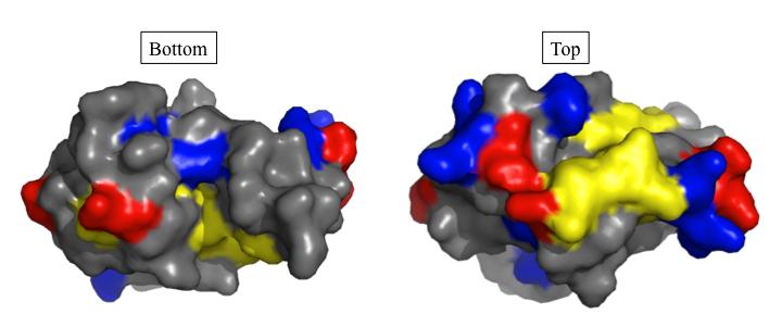
157 143 A B
Review. Diverse Paths to Midcell: Assembly of the Bacterial Cell Division Machinery. Nathan W. Goehring and Jon Beckwith*
 , Vol. 15, R514 R526, July 12, 2005, 2005 Elsevier Ltd. All rights reserved. DOI 10.1016/j.cub.2005.06.038 Diverse Paths to Midcell: Assembly of the Bacterial Cell Division Machinery Review Nathan W. Goehring
A tale of two RLPAs : studies of cell division in Escherichia coli and Pseudomonas aeruginosa
 University of Iowa Iowa Research Online Theses and Dissertations Summer 2014 A tale of two RLPAs : studies of cell division in Escherichia coli and Pseudomonas aeruginosa Matthew Allan Jorgenson University
Cytokinesis in Bacteria
 MICROBIOLOGY AND MOLECULAR BIOLOGY REVIEWS, Mar. 2003, p. 52 65 Vol. 67, No. 1 1092-2172/03/$08.00 0 DOI: 10.1128/MMBR.67.1.52 65.2003 Copyright 2003, American Society for Microbiology. All Rights Reserved.
Introduction. Gene expression is the combined process of :
 1 To know and explain: Regulation of Bacterial Gene Expression Constitutive ( house keeping) vs. Controllable genes OPERON structure and its role in gene regulation Regulation of Eukaryotic Gene Expression
Role of Leucine Zipper Motifs in Association of the Escherichia coli Cell Division Proteins FtsL and FtsB
 JOURNAL OF BACTERIOLOGY, Sept. 2011, p. 4988 4992 Vol. 193, No. 18 0021-9193/11/$12.00 doi:10.1128/jb.00324-11 Copyright 2011, American Society for Microbiology. All Rights Reserved. Role of Leucine Zipper
University of Groningen. Bacterial Cell Wall Synthesis Scheffers, Dirk; Pinho, Mariana G. Published in: Microbiology and Molecular Biology Reviews
 University of Groningen Bacterial Cell Wall Synthesis Scheffers, Dirk; Pinho, Mariana G. Published in: Microbiology and Molecular Biology Reviews DOI: 10.1128/MMBR.69.4.585-607.2005 IMPORTANT NOTE: You
SUPPLEMENTARY INFORMATION
 doi:10.1038/nature10244 a O07391_MYCAV/127-243 NLPC_HAEIN/80-181 SPR_SHIFL/79-183 P74160_SYNY3/112-245 O24914_HELPY/301-437 Q51835_PORGI/68-178 DPP6_BACSH/163-263 YKFC_BACSU/185-292 YDHO_ECOLI/153-263
CHAPTER 13 PROKARYOTE GENES: E. COLI LAC OPERON
 PROKARYOTE GENES: E. COLI LAC OPERON CHAPTER 13 CHAPTER 13 PROKARYOTE GENES: E. COLI LAC OPERON Figure 1. Electron micrograph of growing E. coli. Some show the constriction at the location where daughter
The cell shape proteins MreB and MreC control cell morphogenesis by positioning cell wall synthetic complexes
 Molecular Microbiology (2007) 66(1), 174 188 doi:10.1111/j.1365-2958.2007.05910.x The cell shape proteins MreB and MreC control cell morphogenesis by positioning cell wall synthetic complexes Arun V. Divakaruni,
RNA Synthesis and Processing
 RNA Synthesis and Processing Introduction Regulation of gene expression allows cells to adapt to environmental changes and is responsible for the distinct activities of the differentiated cell types that
Transmembrane Domains (TMDs) of ABC transporters
 of ABC transporters") Transmembrane Domains (TMDs) of ABC transporters Most ABC transporters contain heterodimeric TMDs (e.g. HisMQ, MalFG) TMDs show only limited sequence homology (high diversity) High degree of conservation
Transmembrane Domains (TMDs) of ABC transporters Most ABC transporters contain heterodimeric TMDs (e.g. HisMQ, MalFG) TMDs show only limited sequence homology (high diversity) High degree of conservation
Morphogenesis of Escherichia coli
 MICROBIOLOGY AND MOLECULAR BIOLOGY REVIEWS, Mar. 1998, p. 110 129 Vol. 62, No. 1 1092-2172/98/$04.00 0 Copyright 1998, American Society for Microbiology Morphogenesis of Escherichia coli NANNE NANNINGA*
Supporting online material
 Supporting online material Materials and Methods Target proteins All predicted ORFs in the E. coli genome (1) were downloaded from the Colibri data base (2) (http://genolist.pasteur.fr/colibri/). 737 proteins
7.06 Problem Set #4, Spring 2005
 7.06 Problem Set #4, Spring 2005 1. You re doing a mutant hunt in S. cerevisiae (budding yeast), looking for temperaturesensitive mutants that are defective in the cell cycle. You discover a mutant strain
NIH Public Access Author Manuscript Mol Microbiol. Author manuscript; available in PMC 2011 March 15.
 NIH Public Access Author Manuscript Published in final edited form as: Mol Microbiol. 2010 July 1; 77(1): 90 107. doi:10.1111/j.1365-2958.2010.07224.x. DipM, a new factor required for peptidoglycan remodeling
Growth of the Stress-Bearing and Shape-Maintaining Murein Sacculus of Escherichia coli
 MICROBIOLOGY AND MOLECULAR BIOLOGY REVIEWS, Mar. 1998, p. 181 203 Vol. 62, No. 1 1092-2172/98/$04.00 0 Copyright 1998, American Society for Microbiology Growth of the Stress-Bearing and Shape-Maintaining
Nature Structural & Molecular Biology: doi: /nsmb Supplementary Figure 1
 Supplementary Figure 1 Chemical structure of LPS and LPS biogenesis in Gram-negative bacteria. a. Chemical structure of LPS. LPS molecule consists of Lipid A, core oligosaccharide and O-antigen. The polar
This article appeared in a journal published by Elsevier. The attached copy is furnished to the author for internal non-commercial research and
 This article appeared in a journal published by Elsevier. The attached copy is furnished to the author for internal non-commercial research and education use, including for instruction at the authors institution
Newly made RNA is called primary transcript and is modified in three ways before leaving the nucleus:
 m Eukaryotic mrna processing Newly made RNA is called primary transcript and is modified in three ways before leaving the nucleus: Cap structure a modified guanine base is added to the 5 end. Poly-A tail
The integral membrane FtsW protein and peptidoglycan synthase PBP3 form a subcomplex in Escherichia coli
 Microbiology (2011), 157, 251 259 DOI 10.1099/mic.0.040071-0 The integral membrane FtsW protein and peptidoglycan synthase PBP3 form a subcomplex in Escherichia coli Claudine Fraipont, 1 3 Svetlana Alexeeva,
Reading Assignments. A. Genes and the Synthesis of Polypeptides. Lecture Series 7 From DNA to Protein: Genotype to Phenotype
 Lecture Series 7 From DNA to Protein: Genotype to Phenotype Reading Assignments Read Chapter 7 From DNA to Protein A. Genes and the Synthesis of Polypeptides Genes are made up of DNA and are expressed
Mechanisms for Precise Positional Information in Bacteria: The Min system in E. coli and B. subtilis
 Mechanisms for Precise Positional Information in Bacteria: The Min system in E. coli and B. subtilis Martin Howard Imperial College London Bacterial Organization Many processes where bacterial cell needs
Physics of Bacterial Morphogenesis
 MICROBIOLOGY AND MOLECULAR BIOLOGY REVIEWS, Dec. 2011, p. 543 565 Vol. 75, No. 4 1092-2172/11/$12.00 doi:10.1128/mmbr.00006-11 Copyright 2011, American Society for Microbiology. All Rights Reserved. Physics
Analysis of Escherichia coli amino acid transporters
 Ph.D thesis Analysis of Escherichia coli amino acid transporters Presented by Attila Szvetnik Supervisor: Dr. Miklós Kálmán Biology Ph.D School University of Szeged Bay Zoltán Foundation for Applied Research
In Escherichia coli, MreB and FtsZ Direct the Synthesis of Lateral Cell Wall via Independent Pathways That Require PBP 2
 JOURNAL OF BACTERIOLOGY, June 2009, p. 3526 3533 Vol. 191, No. 11 0021-9193/09/$08.00 0 doi:10.1128/jb.01812-08 Copyright 2009, American Society for Microbiology. All Rights Reserved. In Escherichia coli,
ACCEPTED. Why spherical E. coli dies: The inside story. Kevin D. Young. Department of Microbiology and Immunology,
 JB Accepts, published online ahead of print on 28 December 2007 J. Bacteriol. doi:10.1128/jb.01975-07 Copyright 2007, American Society for Microbiology and/or the Listed Authors/Institutions. All Rights
Gene regulation I Biochemistry 302. Bob Kelm February 25, 2005
 Gene regulation I Biochemistry 302 Bob Kelm February 25, 2005 Principles of gene regulation (cellular versus molecular level) Extracellular signals Chemical (e.g. hormones, growth factors) Environmental
16 The Cell Cycle. Chapter Outline The Eukaryotic Cell Cycle Regulators of Cell Cycle Progression The Events of M Phase Meiosis and Fertilization
 The Cell Cycle 16 The Cell Cycle Chapter Outline The Eukaryotic Cell Cycle Regulators of Cell Cycle Progression The Events of M Phase Meiosis and Fertilization Introduction Self-reproduction is perhaps
7.06 Cell Biology EXAM #3 April 21, 2005
 7.06 Cell Biology EXAM #3 April 21, 2005 This is an open book exam, and you are allowed access to books, a calculator, and notes but not computers or any other types of electronic devices. Please write
BACTERIAL PHYSIOLOGY SMALL GROUP. Monday, August 25, :00pm. Faculty: Adam Driks, Ph.D. Alan Wolfe, Ph.D.
 BACTERIAL PHYSIOLOGY SMALL GROUP Monday, August 25, 2014 1:00pm Faculty: Adam Driks, Ph.D. Alan Wolfe, Ph.D. Learning Goal To understand how bacterial physiology applies to the diagnosis and treatment
Archaeal Cell Structure. Copyright McGraw-Hill Global Education Holdings, LLC. Permission required for reproduction or display.
 4 Archaeal Cell Structure Copyright McGraw-Hill Global Education Holdings, LLC. Permission required for reproduction or display. 1 4.1 A typical Archaeal Cell 2 Archaea Highly diverse with respect to morphology,
Lecture 10: Cyclins, cyclin kinases and cell division
 Chem*3560 Lecture 10: Cyclins, cyclin kinases and cell division The eukaryotic cell cycle Actively growing mammalian cells divide roughly every 24 hours, and follow a precise sequence of events know as
Scale in the biological world
 Scale in the biological world 2 A cell seen by TEM 3 4 From living cells to atoms 5 Compartmentalisation in the cell: internal membranes and the cytosol 6 The Origin of mitochondria: The endosymbion hypothesis
Gene Control Mechanisms at Transcription and Translation Levels
 Gene Control Mechanisms at Transcription and Translation Levels Dr. M. Vijayalakshmi School of Chemical and Biotechnology SASTRA University Joint Initiative of IITs and IISc Funded by MHRD Page 1 of 9
Copyright Mark Brandt, Ph.D A third method, cryogenic electron microscopy has seen increasing use over the past few years.
 Structure Determination and Sequence Analysis The vast majority of the experimentally determined three-dimensional protein structures have been solved by one of two methods: X-ray diffraction and Nuclear
MOLECULAR CELL BIOLOGY
 1 Lodish Berk Kaiser Krieger scott Bretscher Ploegh Matsudaira MOLECULAR CELL BIOLOGY SEVENTH EDITION CHAPTER 13 Moving Proteins into Membranes and Organelles Copyright 2013 by W. H. Freeman and Company
7.06 Cell Biology EXAM #3 KEY
 7.06 Cell Biology EXAM #3 KEY May 2, 2006 This is an OPEN BOOK exam, and you are allowed access to books, a calculator, and notes BUT NOT computers or any other types of electronic devices. Please write
Mechanical control of bacterial cell shape: supplemental material
 Mechanical control of bacterial cell shape: supplemental material Hongyuan Jiang 1, Fangwei Si 1, William Margolin 2, Sean X. Sun 1,3 I. Table of Contents II. Introduction...2 III. Geometry of the cell
Susceptibility to Antibiotics and -Lactamase Induction in Murein Hydrolase Mutants of Escherichia coli
 ANTIMICROBIAL AGENTS AND CHEMOTHERAPY, Apr. 2005, p. 1404 1409 Vol. 49, No. 4 0066-4804/05/$08.00 0 doi:10.1128/aac.49.4.1404 1409.2005 Copyright 2005, American Society for Microbiology. All Rights Reserved.
Chapter 17. From Gene to Protein. Biology Kevin Dees
 Chapter 17 From Gene to Protein DNA The information molecule Sequences of bases is a code DNA organized in to chromosomes Chromosomes are organized into genes What do the genes actually say??? Reflecting
Chapter 12. Genes: Expression and Regulation
 Chapter 12 Genes: Expression and Regulation 1 DNA Transcription or RNA Synthesis produces three types of RNA trna carries amino acids during protein synthesis rrna component of ribosomes mrna directs protein
Regulation of Gene Expression in Bacteria and Their Viruses
 11 Regulation of Gene Expression in Bacteria and Their Viruses WORKING WITH THE FIGURES 1. Compare the structure of IPTG shown in Figure 11-7 with the structure of galactose shown in Figure 11-5. Why is
Bacterial Genetics & Operons
 Bacterial Genetics & Operons The Bacterial Genome Because bacteria have simple genomes, they are used most often in molecular genetics studies Most of what we know about bacterial genetics comes from the
Introduction to Microbiology BIOL 220 Summer Session I, 1996 Exam # 1
 Name I. Multiple Choice (1 point each) Introduction to Microbiology BIOL 220 Summer Session I, 1996 Exam # 1 B 1. Which is possessed by eukaryotes but not by prokaryotes? A. Cell wall B. Distinct nucleus
From gene to protein. Premedical biology
 From gene to protein Premedical biology Central dogma of Biology, Molecular Biology, Genetics transcription replication reverse transcription translation DNA RNA Protein RNA chemically similar to DNA,
15.2 Prokaryotic Transcription *
 OpenStax-CNX module: m52697 1 15.2 Prokaryotic Transcription * Shannon McDermott Based on Prokaryotic Transcription by OpenStax This work is produced by OpenStax-CNX and licensed under the Creative Commons
THIN SECTIONS OF DIVIDING NEISSERIA GONORRHOEAE
 JOURNAL OF BACTERIOLOGY Vol. 87, No. 6, pp. 1477-1482 June, 1964 Copyright 1964 by the American Society for Microbiology Printed in U.S.A. THIN SECTIONS OF DIVIDING NEISSERIA GONORRHOEAE PHILIP FITZ-JAMES
Initiation of translation in eukaryotic cells:connecting the head and tail
 Initiation of translation in eukaryotic cells:connecting the head and tail GCCRCCAUGG 1: Multiple initiation factors with distinct biochemical roles (linking, tethering, recruiting, and scanning) 2: 5
Topic 4 - #14 The Lactose Operon
 Topic 4 - #14 The Lactose Operon The Lactose Operon The lactose operon is an operon which is responsible for the transport and metabolism of the sugar lactose in E. coli. - Lactose is one of many organic
CAP 5510 Lecture 3 Protein Structures
 CAP 5510 Lecture 3 Protein Structures Su-Shing Chen Bioinformatics CISE 8/19/2005 Su-Shing Chen, CISE 1 Protein Conformation 8/19/2005 Su-Shing Chen, CISE 2 Protein Conformational Structures Hydrophobicity
Chapter 6- An Introduction to Metabolism*
 Chapter 6- An Introduction to Metabolism* *Lecture notes are to be used as a study guide only and do not represent the comprehensive information you will need to know for the exams. The Energy of Life
MONTGOMERY COUNTY COMMUNITY COLLEGE BIO 140 CHAPTER 4. Functional Anatomy of Prokaryotic and Eukaryotic Cells
 MONTGOMERY COUNTY COMMUNITY COLLEGE BIO 140 CHAPTER 4 Functional Anatomy of Prokaryotic and Eukaryotic Cells I. PROKARYOTES A. Structure Of The Cell: Chemical Composition And Function 1. Cell Wall a. composition
BA, BSc, and MSc Degree Examinations
 Examination Candidate Number: Desk Number: BA, BSc, and MSc Degree Examinations 2017-8 Department : BIOLOGY Title of Exam: Molecular Biology and Biochemistry Part I Time Allowed: 1 hour and 30 minutes
THE BACTERIA: THEIR ORIGIN, STRUCTURE, FUNCTION AND ANTIBIOSIS
 THE BACTERIA: THEIR ORIGIN, STRUCTURE, FUNCTION AND ANTIBIOSIS The Bacteria: Their Origin, Structure, Function and Antibiosis by Arthur L. Koch Indiana University, Bloomington, IN, USA A C.I.P. Catalogue
Gene expression in prokaryotic and eukaryotic cells, Plasmids: types, maintenance and functions. Mitesh Shrestha
 Gene expression in prokaryotic and eukaryotic cells, Plasmids: types, maintenance and functions. Mitesh Shrestha Plasmids 1. Extrachromosomal DNA, usually circular-parasite 2. Usually encode ancillary
Bio Microbiology - Spring 2014 Learning Guide 04.
 Bio 230 - Microbiology - Spring 2014 Learning Guide 04 http://pessimistcomic.blogspot.com/ Cell division is a part of a replication cycle that takes place throughout the life of the bacterium A septum
Bio Microbiology - Spring 2012 Learning Guide 04.
 Bio 230 - Microbiology - Spring 2012 Learning Guide 04 http://pessimistcomic.blogspot.com/ A septum assembles at the center of the cell. This molecular "purse string" is linked to the inner surface of
Chapter 16 Lecture. Concepts Of Genetics. Tenth Edition. Regulation of Gene Expression in Prokaryotes
 Chapter 16 Lecture Concepts Of Genetics Tenth Edition Regulation of Gene Expression in Prokaryotes Chapter Contents 16.1 Prokaryotes Regulate Gene Expression in Response to Environmental Conditions 16.2
ROLE OF FtsA IN CELL DIVISION IN Neisseria gonorrhoeae
 ROLE OF FtsA IN CELL DIVISION IN Neisseria gonorrhoeae A Thesis Submitted to the College of Graduate Studies and Research In Partial Fulfillment of the Requirements For the Degree of Doctor of Philosophy
Three different fusions led to three basic ideas: 1) If one fuses a cell in mitosis with a cell in any other stage of the cell cycle, the chromosomes
 If one fuses a cell in mitosis with a cell in any other stage of the cell cycle, the chromosomes") Section Notes The cell division cycle presents an interesting system to study because growth and division must be carefully coordinated. For many cells it is important that it reaches the correct size
Section Notes The cell division cycle presents an interesting system to study because growth and division must be carefully coordinated. For many cells it is important that it reaches the correct size
Development Team. Regulation of gene expression in Prokaryotes: Lac Operon. Molecular Cell Biology. Department of Zoology, University of Delhi
 Paper Module : 15 : 23 Development Team Principal Investigator : Prof. Neeta Sehgal Department of Zoology, University of Delhi Co-Principal Investigator : Prof. D.K. Singh Department of Zoology, University
Protein Structure. W. M. Grogan, Ph.D. OBJECTIVES
 Protein Structure W. M. Grogan, Ph.D. OBJECTIVES 1. Describe the structure and characteristic properties of typical proteins. 2. List and describe the four levels of structure found in proteins. 3. Relate
Introduction to Molecular and Cell Biology
 Introduction to Molecular and Cell Biology Molecular biology seeks to understand the physical and chemical basis of life. and helps us answer the following? What is the molecular basis of disease? What
Name Period The Control of Gene Expression in Prokaryotes Notes
 Bacterial DNA contains genes that encode for many different proteins (enzymes) so that many processes have the ability to occur -not all processes are carried out at any one time -what allows expression
Analysis and Simulation of Biological Systems
 Analysis and Simulation of Biological Systems Dr. Carlo Cosentino School of Computer and Biomedical Engineering Department of Experimental and Clinical Medicine Università degli Studi Magna Graecia Catanzaro,
Transport between cytosol and nucleus
 of 60 3 Gated trans Lectures 9-15 MBLG 2071 The n GATED TRANSPORT transport between cytoplasm and nucleus (bidirectional) controlled by the nuclear pore complex active transport for macro molecules e.g.
2012 Univ Aguilera Lecture. Introduction to Molecular and Cell Biology
 2012 Univ. 1301 Aguilera Lecture Introduction to Molecular and Cell Biology Molecular biology seeks to understand the physical and chemical basis of life. and helps us answer the following? What is the
Cells to Tissues. Peter Takizawa Department of Cell Biology
 Cells to Tissues Peter Takizawa Department of Cell Biology From one cell to ensembles of cells. Multicellular organisms require individual cells to work together in functional groups. This means cells
Protein-Protein Interactions of the Cytoplasmic Loops of the Yeast Multidrug Resistance Protein,PDR 5: A Two -Hybrid Based Analysis.
 Researcher : Rao Subba G (2001) SUMMARY Guide :Gupta,C.M.(Dr.) Protein-Protein Interactions of the Cytoplasmic Loops of the Yeast Multidrug Resistance Protein,PDR 5: A Two -Hybrid Based Analysis. Mukidrug
Microbial Genetics, Mutation and Repair. 2. State the function of Rec A proteins in homologous genetic recombination.
 Answer the following questions 1. Define genetic recombination. Microbial Genetics, Mutation and Repair 2. State the function of Rec A proteins in homologous genetic recombination. 3. List 3 types of bacterial
7.06 Problem Set
 7.06 Problem Set 5 -- 2006 1. In the first half of the course, we encountered many examples of proteins that entered the nucleus in response to the activation of a cell-signaling pathway. One example of
UNIVERSITY OF YORK. BA, BSc, and MSc Degree Examinations Department : BIOLOGY. Title of Exam: Molecular microbiology
 Examination Candidate Number: Desk Number: UNIVERSITY OF YORK BA, BSc, and MSc Degree Examinations 2017-8 Department : BIOLOGY Title of Exam: Molecular microbiology Time Allowed: 1 hour 30 minutes Marking
purpose of this Chapter is to highlight some problems that will likely provide new
 119 Chapter 6 Future Directions Besides our contributions discussed in previous chapters to the problem of developmental pattern formation, this work has also brought new questions that remain unanswered.
Mouth animalcules (bacteria)
") Mouth animalcules (bacteria) 1684 http://en.citizendium.org/images/thumb/9/94/leeuwenhoek.jpg/300px-leeuwenhoek.jpg Prokaryotic Cell Shapes Coccus - cocci Bacillus - bacillus Spirillum - spirilli Vibrio
Mouth animalcules (bacteria) 1684 http://en.citizendium.org/images/thumb/9/94/leeuwenhoek.jpg/300px-leeuwenhoek.jpg Prokaryotic Cell Shapes Coccus - cocci Bacillus - bacillus Spirillum - spirilli Vibrio
Bacterial Morphology and Structure م.م رنا مشعل
 Bacterial Morphology and Structure م.م رنا مشعل SIZE OF BACTERIA Unit for measurement : Micron or micrometer, μm: 1μm=10-3 mm Size: Varies with kinds of bacteria, and also related to their age and external
Class IX: Biology Chapter 5: The fundamental unit of life. Chapter Notes. 1) In 1665, Robert Hooke first discovered and named the cells.
 In 1665, Robert Hooke first discovered and named the cells.") Class IX: Biology Chapter 5: The fundamental unit of life. Key learnings: Chapter Notes 1) In 1665, Robert Hooke first discovered and named the cells. 2) Cell is the structural and functional unit of all
Class IX: Biology Chapter 5: The fundamental unit of life. Key learnings: Chapter Notes 1) In 1665, Robert Hooke first discovered and named the cells. 2) Cell is the structural and functional unit of all
CE 421/521 Environmental Biotechnology. The Cell: The common denominator of all living things Chapter 4 in Vaccari et al. Tim Ellis August 24, 2006
 CE 421/521 Environmental Biotechnology The Cell: The common denominator of all living things Chapter 4 in Vaccari et al. Tim Ellis August 24, 2006 Introduction Cells were discovered around the same time
Helical Macrofiber Formation in Bacillus subtilis: Inhibition by Penicillin G
 JOURNAL OF BACTERIOLOGY, June 1984, p. 1182-1187 0021-9193/84/061182-06$02.00/0 Copyright C 1984, American Society for Microbiology Vol. 158, No. 3 Helical Macrofiber Formation in Bacillus subtilis: Inhibition
Molecular Cell Biology 5068 In Class Exam 1 September 30, Please print your name:
 Molecular Cell Biology 5068 In Class Exam 1 September 30, 2014 Exam Number: Please print your name: Instructions: Please write only on these pages, in the spaces allotted and not on the back. Write your
Characterization of two Bacillus subtilis penicillin-binding. protein-coding genes, pbph (ykua) and pbpi (yrrr)
 and pbpi (yrrr)") Characterization of two Bacillus subtilis penicillin-binding protein-coding genes, pbph (ykua) and pbpi (yrrr) Yuping Wei Thesis submitted to the Faculty of the Virginia Polytechnic Institute and State
Characterization of two Bacillus subtilis penicillin-binding protein-coding genes, pbph (ykua) and pbpi (yrrr) Yuping Wei Thesis submitted to the Faculty of the Virginia Polytechnic Institute and State
SECOND PUBLIC EXAMINATION. Honour School of Physics Part C: 4 Year Course. Honour School of Physics and Philosophy Part C C7: BIOLOGICAL PHYSICS
 2757 SECOND PUBLIC EXAMINATION Honour School of Physics Part C: 4 Year Course Honour School of Physics and Philosophy Part C C7: BIOLOGICAL PHYSICS TRINITY TERM 2013 Monday, 17 June, 2.30 pm 5.45 pm 15
Bacillus anthracis. Clostridium botulinum Clostridium perfringens and other, but never Gram-negative microbes
 SPORES (endospores) the spore is formed inside the parent vegetative cell hence the name endospores The spore is a dehydrated, multishelled structure that protects and allows the bacteria to exist in suspended
GCD3033:Cell Biology. Transcription
 Transcription Transcription: DNA to RNA A) production of complementary strand of DNA B) RNA types C) transcription start/stop signals D) Initiation of eukaryotic gene expression E) transcription factors
9/8/2010. Chapter 4. Structures Internal to the Cell Wall. The Plasma Membrane. Functional Anatomy of Prokaryotic and Eukaryotic Cells
 Chapter 4 Functional Anatomy of Prokaryotic and Eukaryotic Cells Johana Meléndez Part II slides 39-87 Lectures prepared by Christine L. Case Structures Internal to the Cell Wall Learning Objectives 4-8
Tellurite resistance protein/ethidium efflux transporter/ proflavin transporter. Putative inner membrane protein: function unknown
 Additional file 1. Table S1 and Figures S1-4 of Zhang et al. High-level production of membrane proteins in E. coli BL21(DE3) by omitting the inducer IPTG Table S1. Properties of the membrane proteins used
Bi 8 Lecture 11. Quantitative aspects of transcription factor binding and gene regulatory circuit design. Ellen Rothenberg 9 February 2016
 Bi 8 Lecture 11 Quantitative aspects of transcription factor binding and gene regulatory circuit design Ellen Rothenberg 9 February 2016 Major take-home messages from λ phage system that apply to many
Chapter 8 Lectures by Gregory Ahearn University of North Florida
 Chapter 8 The Continuity of Life: How Cells Reproduce Lectures by Gregory Ahearn University of North Florida Copyright 2009 Pearson Education, Inc. 8.1 Why Do Cells Divide? Cells reproduce by cell division.
C a h p a t p e t r e r 6 E z n y z m y e m s
 Chapter 6 Enzymes 4. Examples of enzymatic reactions acid-base catalysis: give and take protons covalent catalysis: a transient covalent bond is formed between the enzyme and the substrate metal ion catalysis:
The mechanisms that drive bacterial cell division have been
 Defining the rate-limiting processes of bacterial cytokinesis Carla Coltharp a, Jackson Buss a,1, Trevor M. Plumer a, and Jie Xiao a,2 a Department of Biophysics and Biophysical Chemistry, Johns Hopkins
Biology: Life on Earth
 Teresa Audesirk Gerald Audesirk Bruce E. Byers Biology: Life on Earth Eighth Edition Lecture for Chapter 4 Cell Structure and Function Copyright 2008 Pearson Prentice Hall, Inc. Chapter 4 Outline 4.1 What
Multiple Interactions between the Transmembrane Division Proteins of Bacillus subtilis and the Role of FtsL Instability in Divisome Assembly
 JOURNAL OF BACTERIOLOGY, Nov. 2006, p. 7396 7404 Vol. 188, No. 21 0021-9193/06/$08.00 0 doi:10.1128/jb.01031-06 Copyright 2006, American Society for Microbiology. All Rights Reserved. Multiple Interactions
I. Archaeal cell structure. (Chap 2 pg , Supplemental notes 3, 5)
") Thurs, Jan 23, 2003 I. Archaeal cell structure. (Chap 2 pg. 450-453, Supplemental notes 3, 5) The Archaea are a diverse group of prokaryotic organisms that are very different from bacteria and from eucaryotes.
Thurs, Jan 23, 2003 I. Archaeal cell structure. (Chap 2 pg. 450-453, Supplemental notes 3, 5) The Archaea are a diverse group of prokaryotic organisms that are very different from bacteria and from eucaryotes.
Bi 1x Spring 2014: LacI Titration
 Bi 1x Spring 2014: LacI Titration 1 Overview In this experiment, you will measure the effect of various mutated LacI repressor ribosome binding sites in an E. coli cell by measuring the expression of a
Substrate Requirements for Regulated Intramembrane Proteolysis of Bacillus subtilis Pro- K
 JOURNAL OF BACTERIOLOGY, Feb. 2005, p. 961 971 Vol. 187, No. 3 0021-9193/05/$08.00 0 doi:10.1128/jb.187.3.961 971.2005 Copyright 2005, American Society for Microbiology. All Rights Reserved. Substrate
Direct detection of antibodies in blood plasma using bioluminescent
 Supplementary information Direct detection of antibodies in blood plasma using bioluminescent sensor proteins and a smartphone. Remco Arts, Ilona den Hartog, Stefan Zijlema, Vito Thijssen, Stan van der
Peptidoglycan Hydrolases of Escherichia coli
 MICROBIOLOGY AND MOLECULAR BIOLOGY REVIEWS, Dec. 2011, p. 636 663 Vol. 75, No. 4 1092-2172/11/$12.00 doi:10.1128/mmbr.00022-11 Copyright 2011, American Society for Microbiology. All Rights Reserved. Peptidoglycan
Basic Structure of a Cell
 Basic Structure of a Cell Prokaryotic Cells No nucleus Archaea & Eubacteria One circular chromosome Extremely small Eukaryotic Cells Has a nucleus!!! Membrane-bound organelles Plants, Animals, Fungi, &
2011 The Simple Homeschool Simple Days Unit Studies Cells
 1 We have a full line of high school biology units and courses at CurrClick and as online courses! Subscribe to our interactive unit study classroom and make science fun and exciting! 2 A cell is a small
L I F E S C I E N C E S
 1a L I F E S C I E N C E S 5 -UUA AUA UUC GAA AGC UGC AUC GAA AAC UGU GAA UCA-3 5 -TTA ATA TTC GAA AGC TGC ATC GAA AAC TGT GAA TCA-3 3 -AAT TAT AAG CTT TCG ACG TAG CTT TTG ACA CTT AGT-5 NOVEMBER 7, 2006
2. In regards to the fluid mosaic model, which of the following is TRUE?
 General Biology: Exam I Sample Questions 1. How many electrons are required to fill the valence shell of a neutral atom with an atomic number of 24? a. 0 the atom is inert b. 1 c. 2 d. 4 e. 6 2. In regards