Characterization of two Bacillus subtilis penicillin-binding. protein-coding genes, pbph (ykua) and pbpi (yrrr)
|
|
|
- Dwight Sparks
- 5 years ago
- Views:
Transcription
1 Characterization of two Bacillus subtilis penicillin-binding protein-coding genes, pbph (ykua) and pbpi (yrrr) Yuping Wei Thesis submitted to the Faculty of the Virginia Polytechnic Institute and State University in partial fulfillment of the requirements for the degree of Master of Science in Biology Committee members: Chairman: David Popham Charles Rutherford Timothy Larson August 6 th, Blacksburg, VA Keywords: Bacillus subtilis, Bacterial cell wall, Penicillin-binding protein (PBP), ykua (pbph), yrrr (pbpi) Copyright 2002, Yuping Wei
2 Characterization of two Bacillus subtilis penicillin-binding protein-coding genes, pbph (ykua) and pbpi (yrrr) Yuping Wei David L. Popham, Chairman Department of Biology, Virginia Polytechnic Institute and State University ABSTRACT Penicillin-binding proteins (PBPs) are required in the synthesis of the cell wall of bacteria. In Bacillus subtilis, PBPs play important roles in the life cycle, including both vegetative growth and sporulation, and contribute to the formation of the different structures of vegetative cell wall and spore cortex. The B. subtilis genome sequencing project revealed there were two uncharacterized genes, ykua and yrrr, with extensive sequence similarity to class B PBPs. These two genes are renamed and referred to henceforth as pbph and pbpi, respectively. A sequence alignment of the predicted product of pbph against the microbial protein database demonstrated that the most similar protein in B. subtilis is PBP2A and in E. coli is PBP2. This suggested that PbpH belongs to a group of the genes required for maintaining the rod shape of the cell. Study of a pbph-lacz fusion showed that pbph was expressed weakly during vegetative growth and the expression reached the highest level at the transition from exponential phase to stationary phase. The combination of a pbpa deletion and the pbph deletion was lethal and double mutant strains lacking pbph and pbpc or pbpi (also named yrrr) were viable. The viable mutants were indistinguishable from the wild-type except that the vegetative PG of the pbpc pbph strain had a slightly
3 slightly lower amount of disaccharide tetrapeptide with 1 amidation and higher amount of disaccharide tripeptide tetrapeptide with 2 amidations when compared to others strains. This suggests that PbpC (PBP3) is involved in vegetative PG synthesis but only affects the PG structure with a very low efficiency. A pbpa pbph double mutant containing a xylose-regulated pbph gene inserted into the chromosome at the amye locus was constructed. Depletion of PbpH resulted in an arrest in cell growth and a dramatic morphological change in both vegetative cells and outgrowing spores. Vegetative cells lacking pbpa and pbph expression swelled and cell elongation was arrested, leading to the formation of pleiomorphic spherical cells and eventual lysis. In these cells, cell septations were randomly localized, cell walls and septa were thicker than those seen in wild type cells, and the average cell width and volume were larger than those of cells expressing pbpa or pbph. The vegetative PG had an increased abundance of one unidentified muropeptide. Spores produced by the pbpa pbph double mutant were able to initiate germination but the transition of the ovalshaped spores to rod-shape cells was blocked. The outgrowing cells were spherical, gradually enlarged, and eventually lysed. Outgrowth of these spores in the presence of xylose led to the formation of helical cells. Thus, PbpH is apparently required for maintenance of cell shape, specifically for cell elongation. PbpH and PBP2a play a redundant role homologous to that of PBP2 in E. coli. A sequence alignment of the predicted product of pbpi against the microbial protein database demonstrated that the most similar protein in B. subtilis is SpoVD and in E. coli is PBP3. This suggested that PbpI belongs to the group of the genes required for synthesis of the spore or septum PG. PbpI was identified using radio-labeled penicillin iii
4 and found to run underneath PBP4 on SDS-PAGE. PbpI is therefore renamed PBP4b. Study of a pbpi-lacz fusion showed that pbpi was expressed predominantly during early sporulation. A putative sigma F recognition site is present in the region upstream of pbpi and studies using mutant strains lacking sporulation-specific sigma factors demonstrated that the expression of pbpi is mainly dependent on sigma factor F. A pbpi single mutant, a pbpi pbpg double mutant, and a pbpi pbpf double mutant were indistinguishable from the wild-type. The sporulation defect of a pbpi pbpf pbpg triple mutant was indistinguishable from that of a pbpf pbpg double mutant. Structure parameters of the forespore PG in a pbpi spovd strain are similar to that of a spovd strain. These results indicate that PBP4b plays an unknown redundant role. iv
5 ACKNOWLEDGEMENT This thesis is the result of two years work whereby I have been accompanied and supported by many people. I would like to express my gratitude for all of them. The first person I would like to thank is my supervisor, Dr. David Popham, for all the instructions, helps and understandings. I am really happy I have come to his lab two years ago and known him in my life. I would like to thank to my committee members, Dr. Charles Rutherford and Dr. Timothy Larson for helps and valuable comments on the thesis. I would like to thank to Meghan Gilmore, Derrell McPherson, Amanda Dean, Amy Weaver, Nicole Ganzala, and Sarah Linnstaedt for help and making the lab fun. I would like to thank the people on the 4 th floor of Derring Hall for making the labs like a big family. I want to thank to my parents, Xuemei Wei and Yongmiao Zhang, for all the caring and worrying, and my parents-in law, Luangxian Wang and Qidong Lin, for the help during the preparation of thesis. Especially, I would like to give my special thanks to my husband, Qin Lin, for all the support. v
6 LIST OF FIGURES Page CHAPTER ONE Figure 1. The domain structures of the class A, class B high MW PBPs, 9 and low MW PBPs. Figure 2. Stages of the B. subtilis life cycle. 10 CHAPTER TWO Figure 3. Expression of a pbph-lacz fusion. 23 Figure 4. The germination kinetics of B. subtilis wild type and mutant 26 spores. Figure 5. Growth of DPVB207 and PS832 in the presence and absence 30 of xylose. Figure 6. Germination and outgrowth kinetics of spores produced by 31 DPVB207 and PS832. Figure 7. Phase contrast microscopy of wild type and DPVB207 (the 36 xylose regulated pbpa pbph double mutant) vegetative cells. Fig. 8. Phase contrast light microscopy of outgrowing DPVB207 (the 38 xylose regulated pbpa pbph double mutant) and wild-type spores. Figure 9. Electron microscopy of wild type and DPVB207 (the xylose 43 regulated pbpa pbph double mutant) vegetative cells. Figure 10. RP-HPLC Muropeptide elution patterns of peptidoglycan 47 from vegetative cells of various strains. vi
7 CHAPTER THREE Figure 11. Identification of PbpI. 68 Figure 12. Expression of a pbpi-lacz fusion. 69 Figure 13. Nucleotide sequence of the upstream region and the 71 beginning of pbpi. Figure 14. Sigma factor dependence of pbpi expression. 72 Figure 15. Heat-killing of wild type and mutant spores. 76 Figure 16. The germination and outgrowth kinetics of wild type and 77 mutant spores. vii
8 LIST OF TABLES Page CHAPTER TWO Table 1. Bacillus subtilis strains and plasmid used 18 Table 2. Primer sequences 20 Table 3. Growth and sporulation of B. subtilis wild type and mutant 25 strains. Table 4. The cell dimensions of wild type and pbpa pbph cells 34 grown in medium with the presence and absence of xylose CHAPTER THREE Table 5. Bacillus subtilis strains and plasmid used 63 Table 6. Primer sequences 65 Table 7. Growth and sporulation of pbpi mutant strains 74 Table 8. Structural parameters for the spore cortex PG of various 79 strains Table 9. Structural parameters for the forespore PG of various strains 80 viii
9 TABLE OF CONTENTS Page ABSTRACT ACKNOWLEDGEMENTS LIST OF FIGURES LIST OF TABLES ii v vi viii CHAPTER ONE. REVIEW OF THE LITERATURE Introduction 1 B. subtilis vegetative growth and cell wall structure 3 Sporulation, sporulation related sigma factors and spore cell wall 4 (cortex) structure PBPs and maintenance of the rod-shaped cell 6 CHAPTER TWO. CHARACTERIZATION OF THE PBP-CODING GENE pbph 11 (ykua) Materials and methods Plasmids, bacterial strains, and growth conditions Construction of plasmids and strains Enzyme assays, membrane preparation, and gel 14 identification of PBPs. 4. Phase contrast and electron microscopy Vegetative peptidoglycan analysis. 16 ix
10 Results 21 Identification of the pbph gene. 21 Production of an inducible xylap-pbph construct and 21 attempted identification of the gene product of pbph. Expression of pbph 22 Phenotypic properties of pbph single mutant and 24 multiple mutants. Non-viability of a pbpa pbph double mutant strain 27 Growth of DPVB207 in the presence and absence 27 of xylose. Spore germination and outgrowth in the presence 28 and absence of xylose. Cell morphology of the pbpa pbph double 32 mutant. Vegetative PG structure. 35 Discussion 50 The identification of gene product of pbph. 50 The function of pbph gene product. 50 Cell shape maintenance and PBPs. 52 Helical cells of inducable pbph pbpa double mutant 54 when germinated with xylose. The thicker cell wall of pbph pbpa double mutant. 54 Conclusions 56 x
11 CHAPTER THREE. THE CHARACTERIZATION OF PUTATIVE PBP- 58 CODING GENE pbpi (yrrr) Materials and Methods Plasmids, bacterial strains, and growth conditions Construction of plasmids and strains Enzyme assays, membrane preparation, and gel 61 identification of PBPs. 4. Immature forespore and spore peptidoglycan structure 61 analysis. 5. Other phenotypic and biochemical assays. 62 Results 66 Identification of the pbpi gene 66 Construction of an inducible xylap-pbpi and 66 identification of the product of pbpi Expression of pbpi 67 Sigma factor dependence of the expression of pbpi 70 Phenotypic properties of pbpi single mutant and multiple 73 mutants Phenotypic studies of a pbpi single mutant, a 73 pbpi pbpg double mutant and a pbpi pbpf double mutant. Phenotypic studies of a pbpi spovd double 78 mutant and a pbpi pbpf pbpg triple mutant. xi
12 Discussion 81 Conclusions 83 REFERENCES 84 CIRRICULUM VITAE 94 xii
13 CHAPTER ONE REVIEW OF THE LITERATURE Introduction Most bacteria have a rigid cell wall, whose indispensable function is providing support for the maintenance of bacterial morphology, protection of the integrity of the cell membrane, and resistance to the turgor pressure (27, 33). The main component of the bacteria cell wall is peptidoglycan (PG), which is a net-like macromolecule of glycan strands cross-linked by peptides. The glycan strands are made up of alternating N- acetylglucosamine (NAG) and N-acetylmuramic acid (NAM) residues. The carboxyl groups of NAM residues are linked to short peptides. The formation of this PG involves two steps (Reviewed in 19). The first step is the synthesis of the PG subunit in the cytoplasm, the disaccharide pentapeptide. In many organisms, including Bacillus subtilis and Escherichia coli, the pentapeptide is L-Ala D-Glu L-meso-diaminopimelic acid D-Ala D-Ala. The transport of the subunit through the membrane is mediated by attachment to 55-carbon isoprenoid lipid. The second step is the polymerization, which includes the elongation of the glycan strands and the cross-linking of the peptide side chains. This step happens on the outer surface of the membrane and requires two enzyme activities, glycosyl transferase and transpeptidase. Glycosyl transferases connect the disaccharide units together by the β-1,4-glycosidic linkage to form glycan strands. Transpeptidases carry out the function of cross-linking by removing the terminal D-Ala from the side chain of the donor glycan strand and linking the penultimate D-Ala to the free amino group of the diaminopimelic acid in the side chain of the acceptor glycan strand or to a bridge peptide which links to the peptide side chain of the acceptor glycan 1
14 strand (3). The maturation of the structure and some regulation of the level of crosslinking are carried out by D, D-carboxypeptidases (49) which remove the terminal D-Ala from the pentapeptide side chains and inhibit the cross-linking. A family of enzymes known as the penicillin-binding proteins (PBPs) is required in the assembly and metabolism of bacterial cell wall PG. Most of these proteins are membrane-bound with their active sites available on the outer surface of the membrane. Based on their structure, function, and similarities in peptide sequence, they are classified as multimodular class A or B high molecular-weight (MW) PBPs or monofunctional low molecular-weight PBPs (49). Most of the low MW PBPs (Fig. 1) (MW 55 KDa) have D, D-carboxypeptidase activity, whereas most high MW PBPs (generally 60kDa) (46) are transpeptidases (70); activities that are carried out by the C-terminal penicillinbinding domains. Class A PBPs are bifunctional proteins. Besides the transpeptidase activity in the C-terminal penicillin-binding domain, they possess an N-terminal glycosyl transferase domain. The N-terminal domain of the class B PBPs is believed to be a morphogenetic determinant (20), but the specific activity is unknown. A common feature of both high MW PBPs and low MW PBPs is they all have a penicillin-binding domain that interacts with β-lactam antibiotics, including penicillin, by covalent binding of these antibiotics to their active-site serine residues. The modified enzymes are inactive and deprived of their essential functions. All bacteria except those without PG synthesis possess multiple PBPs. Multiple PBPs of the same class within a species often exhibit redundant function (53, 54). B. subtilis is the most well-studied Gram positive bacterium that undergoes sporulation, which is the process of producing an endospore, in a nutrient deprived 2
15 environment. When the environmental conditions become favorable the spore can resume vegetative growth by germination. The PG structure of the spore cell wall (spore cortex) is different from that of the vegetative cell wall on the level of cross-linking and the peptide side chains of NAM (reviewed in (19). The PBPs of B. subtilis are involved in the synthesis of both the vegetative cell wall and the spore cortex. Research about PBPs of B. subtilis dates back to the 1970s (22). In the 1990s a great deal of progress was made. This progress was stimulated further after the entire genome sequence of this organism was published in 1997 (30). Analysis of the B. subtilis genome reveals 16 PBP-encoding genes recognizable from sequence similarities. These include 4 class A PBP-encoding genes: pona (9, 50), pbpf (29, 51), pbpd (9, 53) and pbpg (also called ywhe) (30, 36, 45), 6 class B PBP-encoding genes: pbpa (29, 41), pbpb (29, 41, 70), pbpc (8, 9, 42), spovd (13, 14), ykua (30) and yrrr (30), and 6 low MW PBP-encoding genes: pbpx (30), pbpe (51, 58, 64), daca (8, 9, 65), dacb (11, 12, 49, 58, 64) dacc (30, 44), dacf (46, 69). By cloning, mutagenesis, and construction of lacz fusions, thirteen of these genes have been studied extensively whereas genes pbpx, ykua and yrrr have not. The putative gene product of pbpx displays high sequence homology to low MW PBPs. The putative gene products of ykua and yrrr are similar to high MW class B PBPs. The studies presented here concern ykua, yrrr, and their gene products. B. subtilis vegetative growth and cell wall structure B. subtilis vegetative cells are rod-shaped and they grow by elongation in their long axis (17). Cell length is dependent on the growth rate. In general faster growing cells are longer than the slow growers. Cell division occurs at the midpoint of the rod, 3
16 generally after a doubling in length. Right after cell division the new wall material between the nascent sister cells holds the cells together. Later, hydrolysis of the septal PG separates the two cells and the new poles of the cells complete their maturation. The hydrolysis is influenced by the growth medium and growth phase. Under exponential growth conditions in a rich medium, long chains of unseparated cells can accumulate. In the vegetative cell wall, the peptide side chains of the PG are mainly tripeptides and the cross-linking level is between 29-33% of muramic acid residues (4, 19) Sporulation, sporulation related sigma factors and spore cell wall (cortex) structure As an adaptive response to starvation, some gram-positive bacteria, including B. subtilis, will undergo a complex survival mechanism called sporulation. The sporulation process of B. subtilis takes about 8 hours and has been described as a series of stages (Fig 2) (Reviewed in (16)). Vegetative cells are defined as being at stage 0. Under starvation, vegetative cells initiate sporulation which is characterized by the formation of an axial filament of DNA at the centers of cells. This is defined as stage I. Next, a septum is formed at an extreme polar position and lead to the partitioning of the cell into large and small compartments known as the mother cell and the prespore. Completion of the septation is defined as stage II. The prespore is then engulfed by the mother cell. When engulfment is completed (stage III), the forespore is surrounded by two membranes, the inner forespore membrane and the outer forespore membrane. Between these two membranes, two layers of PG are being synthesized (stage IV). The thinner inner layer, the germ cell wall or primordial cell wall, is adjacent to the inner membrane. The thicker outer layer, the cortex, is a modified form of PG. The spore coat proteins are deposited on the outside surface of the forespore (stage V). As the spore matures (stagevi), the 4
17 resistance, dormancy, and germination ability appear in sequence. Lysis of mother cell and the release of the mature spore is defined as stage VII. The process of sporulation involves over 125 genes (61), the transcription of which is temporally and spatially directed by DNA-binding proteins and five RNA polymerase sigma factors (sigmas H, F, E, G, and K), which are activated sequentially. First, in response to starvation, sigma A (the major sigma factor of growing cells), sigma H, and other transcription factors regulate the transcription of the genes that direct the formation of the asymmetric septum. After the septation, sigma F becomes active in the forespore and then sigma E is activated in the mother cell. These two sigma factors control the genes required for engulfment. After engulfment, sigma G is activated in the forespore and shortly thereafter, sigma K becomes active in the mother cell. Both sigma G and K are required for the synthesis of the spore cortex and coat proteins and the regulation of spore maturation. There are two layers of spore PG that have different structures. The germ cell wall has the same basic structure as vegetative cell wall PG (37). During germination, this layer will be developed into the vegetative cell wall. On the contrary, fifty percent of the muramic acid residues of cortex PG do not have side chains and are converted into muramic δ-lactam, which occurs at every alternate disaccharide (5, 47, 68). The muramic δ-lactam residues are not required for the spore dormancy but do play an important role as part of the substrate recognized by germination-specific lytic enzymes (48). About 23% of the NAM residues contain single L-ala side chains. The remaining 27% of the NAM residues contain peptide side chains, some of which are involved in cross-linking. The cross-linking index (the percentage of muramic acid residues with cross-linked 5
18 peptides) is only 2.9% (5, 47), a dramatic difference from the 29% found in the vegetative cell PG (4). PBPs and maintenance of the rod-shaped cell Class A PBPs have bi-functional activities as glycosyl transferase and transpeptidase, which are located in the N-terminal domain and C-terminal domain, respectively. The four B. subtilis class A PBP-encoding genes, pona, pbpd, pbpf, and pbpg appear to play partially redundant, yet distinct, roles in the life cycle. Deletion of pona, which codes for PBP1 (50), causes decreases in growth rate and sporulation efficiencies and the production of cells that are longer, thinner, and slightly bent. Two other genes, pbpd and pbpf, play some redundant function of pona, the further loss of these genes from a pona mutant exacerbates the phenotypic changes (54). The deletion of both pbpf and pbpg leads to the severe sporulation defect (36), which indicates these two genes play a redundant role in sporulation. Class B PBPs are monofunctional transpeptidases. Some are essential for septation and regulation of cell shape. PBP2b, the gene product of pbpb, is essential for septation (70), and its sequence is very similar to that of a protein with the analogous function in E. coli, PBP3 (gene product of pbpb). PBP2a, the gene product of pbpa, is involved in the formation of the rod-shaped cell from oval spores during the spore germination and out-growth (41), and is most similar to the E. coli PBP2, which is required for the maintenance of the rod shape of the cell (59). The deletion of B. subtilis pbpa is not lethal and has no effect on vegetative growth and sporulation, which suggests there is another gene in the same class carrying out the redundant function of pbpa. The other gene with known function is spovd, which codes for SpoVD and is required for the 6
19 spore PG synthesis. PBP3, encoded by pbpc, was found to be dispensable under all growth, sporulation, and germination conditions tested (42). The functions of the two remaining class B PBP-encoding genes, ykua and yrrr, are undefined. Among the six low-molecular-weight class PBPs, PBP5 (encoded by daca) and PBP5* (encoded by dacb) are D,D- carboxypeptidase, and DacC and DacF are predicted to have the same activity based on sequence similarity (reviewed in (19). The two remaining gene products (those encoded by pbpe (51) and pbpx (30)) are predicted to be D, D-endopeptidases according to their sequence similarity to the D, D-endopeptidase, PBP4, of E.coli (19). The cell envelope is important in determining the cell shape in bacteria. In E. coli and B. subtilis, most of the spherical morphology mutants have mutations in genes associated with cell envelope synthesis, mostly PG synthesis. A hypothesis is that during specific periods of the cell cycle, the cell shape is associated with the cognate PBPs to control the synthesis of PG. To maintain their shape, it has been proposed that rod-shaped bacteria have at least two systems, one for cell elongation and the other for the formation of the new poles by septation. The final shape is determined by the balance of these two systems (26). Each of the systems probably contains PG synthesis enzymes (PBPs), autolysins, and regulation enzymes. In E. coli, two proteins, RodA (mrdb, an integral membrane protein, encoded by roda) (35, 63) and class B high MW PBP2 (encoded by pbpa, also called mrda)(59), are involved in maintenance of the rod shape of cells during cell elongation. Mutations in roda or pbpa, or the inactivation of PBP2, block elongation and cause the production of spherical cells. These two genes are in an operon (35), and their gene products are 7
20 expected to interact with each other (34). Two other proteins, FtsW (encoded by ftsw and with sequence similarity to RodA) (10, 28) and class B high MW PBP3 (encoded by pbpb), are required in formation of the division septum. In B. subtilis, there are two proteins, RodA (encoded by roda, (24)) and PBP2a (41), identified as the homologues of RodA and PBP2 in E. coli. RodA has an essential role in elongation. The loss of RodA causes the formation of non-viable spherical cells (24). PBP2a is required for the normal elongation of germinating cells but is not essential for viability, suggesting there is another gene having similar function (41). PBP2b has been identified as a homologue to PBP3 and partial loss-of-function mutation affecting PBP2b results in filamentous growth (70). A candidate for the B. subtilis homologue of E. coli FtsW is the product of an uncharacterized ORF designated as ylao (30). Through studies of the two uncharacterized putative class B HMW PBPs of B. subtilis we hope to gain further insight into the mechanisms of cell elongation and cell shape maintenance. 8
21 Class A High MW PBP Class B High MW PBP Glycosyl transferase? Low MW PBP Penicillin-binding SXXK Y S XN HS KT G Transpeptidase Transpeptidase Carboxypeptidase Endopeptidase Fig. 1. The domain structures of the class A, class B high MW PBPs, and low MW PBPs. Class A and class B high MW PBPs have two function domains. Class A PBPs have glycosyl transferase and transpeptidase activities located in the N-terminal domain and C-terminal domain, respectively. Class B PBPs have transpeptidase activity in the C-terminal domain while the function of N-terminal domain is unknown. Most of the low MW PBPs have D, D-carboxypeptidase activity, and some of them are endopeptidases. A common feature of all three class PBPs is the penicillin-binding domain that interacts with β-lactam antibiotics, including penicillin, by covalent binding of these antibiotics to their active-site serine residues. The most highly conserved sequences within these domains are shown as black boxes. 9
22 Stage VII germination and outgrowth Stage V-VI Stage IV vegetative growth sporulation forespore Stage III mother cell Stage 0 Stage I Stage II Fig. 2. Stages of the B. subtilis life cycle. The vegetative cell wall is shown in dark green. The developing spore (stage IV- VI) is surrounded by the cortex PG (light green). The coat protein layers are shown as a blue line. Membranes are represented as thin black lines. The filamentous structures within the cytoplasm represent the chromosomal DNA. 10
23 CHAPTER TWO CHARACTERIZATION OF THE PBP-CODING GENE pbph (ykua) Extensive sequence similarity indicated that ykua encodes a class B PBP. This gene was renamed and is now referred to as pbph. The first goal was the identification of the gene product of pbph by comparing the PBP profiles of a pbph over-expressing strain and a wild type strain using radio-labeled penicillin. The second goal was examination of the expression of pbph. The final goal was identification of the functions of pbph. Materials and Methods 1. Plasmids, bacterial strains, and growth conditions. All plasmids and B. subtilis strains used in this study are listed in Table 1. E. coli strain JM109 was used for cloning. All B. subtilis strains used were derived from strain 168. Transformation with plasmid DNA or chromosomal DNA was performed as described previously (2). Transformants were selected on 2xSG (32) plates containing appropriate antibiotics: chloramphenicol (3 µg/ml), spectinomycin (100 µg/ml), kanamycin (10 µg/ml), tetracycline (10 µg/ml), and erythromycin (0.5 µg/ml) plus lincomycin (12.5 µg/ml) (macrolide-lincosamide-streptogramin B resistance). Vegetative growth for determining growth rate and sporulation for obtaining spores was routinely done in 2xSG at 37 C. Growth rate was measured by plotting the optical density at 600 nm (OD) of the growing culture versus time on a semi-log plot and reading off the generation time. Spores were purified by water washing (43). Spore germination and outgrowth was analyzed in 2xYT medium (48) containing 4 mm L-ala after a 30 min 11
24 heat shock in water at 65 C (43). The OD of all vegetative, germinating, and outgrowing cultures were determined using a Genesys 5 spectrophotometer. Spore heat resistance and chloroform resistance was measured after sporulation for 24 hours as previously described (43). For induction of the xylose-regulated promoter, strains were grown at 37 C in 2xSG lacking glucose with the concentration of xylose at 0.4% (for the study of pbph function) or 2% (for over-expression). To resuspend an exponentially growing culture into a new medium, the culture was centrifuged at 5,000xg for 5 min at 24 C and resuspended into prewarmed (37 C) fresh medium. 2. Construction of plasmids and strains. Primers pbph1 and pbph2 (Table 2) were used to amplify a 2628 bp fragment containing 272 bp of upstream sequence, the coding sequence, and 302 bp of sequence downstream of pbph from the chromosomal DNA of PS832. This fragment was cloned into pgem-t (Promega), generating plasmid pdpv103, in which the pbph sequence is in the same orientation as lacz. The insert of this plasmid was sequenced using SP6 and T7 primers to confirm that the PCR product had the correct sequence. To construct a deletion mutation in pbph, pdpv103 was digested with HindIII and BamHI to remove 40.3% of the coding sequence, including the penicillin-binding active site. The deleted region was replaced with a spectinomycin resistance gene cassette obtained from pjl73 (31) by digested with HindIII and BamHI. In this way plasmid pdpv113 ( pbph::sp) was generated. The plasmid was linearized by restriction digestion at an XmnI site within the pgem-t vector sequence. The linearized DNA was transformed into the wild type B. subtilis strain, PS832, with selection for the spectinomycin resistant marker to allow the 12
25 mutated gene to integrate into the chromosome via double crossover. Thus the pbph deletion mutant (DPVB133) was obtained. Primer pbpha was designed to contain an added EcoRI site to assist in cloning a fragment for constructing lacz-fusion. Primer pbpha and pbph2 were used to PCR amplified the 2628 bp insert in pdpv103. The 2637 bp PCR product was cut with EcoRI and HindIII to get a 682 bp fragment that contained the upstream region and the first 402 bp of the pbph coding sequence. The fragment was cloned into EcoRI- and HindIIIdigested pdpc87 (52) to generate pdpv125, in which the 682 bp fragment was placed in front of a promoterless lacz gene. The insert of the plasmid was sequenced and confirmed to be identical to the sequence from the genome. The plasmid was used in transforming B. subtilis PS832 to allow the lacz fusion to recombine into the chromosome via a single crossover. Transformants were selected on 2xSG plates containing chloramphenicol. In this way strain DPVB168 was generated, containing the transcriptional lacz fusion to the pbph promoter. DPVB203 was constructed by transforming PS2465 (pbpa::cm) using plasmid pcm::erm r (60) to switch the antibiotic resistance that is inserted in the pbpa locus from Cm to Erm r. Primers pbph5 and pbph3 (Table 2) were designed with added PacI site and BglII sites, respectively. They were used to PCR amplify a 2162 bp fragment containing 80 bp upstream, the coding sequence, and 25 bp downstream of pbph from chromosomal DNA of PS832. The resulting 2176 bp PacI-BglII fragment was cloned into PacI- and BglII-digested psweet-bgab (7) which contains a xylose-regulated expression system. The pbph gene in the resulting plasmid, pdpv138, was sequenced and found to be 13
26 identical to the sequence from the genome. The plasmid was linearized at the PstI site in the vector and used to transform DPVB133 to generate a xylap-pbph fusion at the amye locus. The resulting Amy -, Cm, Sp strain DPVB202 ( pbph::sp amye::xylap-pbph::cm) was obtained. It was transformed with chromosomal DNA from DPVB203 with the selection for Erm r and Sp in the presence of 0.2% xylose. The resulting transformant, DPVB 207, can only survive in medium containing xylose. As a control, DPVB213 was generated by transforming PS832 using the plasmid psweet-bgab. 3. Enzyme assays, membrane preparation, and gel identification of PBPs. β-galactosidase assays of vegetative cells, sporulating cells, and germinating spores were done using the substrate o-nitrophenyl-β-d-galactopyranoside (43, 52) and the activity was expressed in Miller units (38). Glucose dehydrogenase activity was assayed as previously described (43). For over-expression of PbpH, strains were grown at 37 C to an OD of 0.1. Xylose was added to the culture to make the final concentration of 2%, and incubation was continued until the OD reached 1.0. Membranes were prepared as previously described (50). PBPs were detected using 125 I-labeled penicillin X as previously described (36). 4. Phase contrast and electron microscopy. For phase contrast light microscopy, 0.5ml samples of germinating and outgrowing spores or vegetative cells of PS832 and DPVB207 were centrifuged, resuspended in 0.5 ml of fixing solution (2.4% paraformaldehyde and 0.01% glutaraldehyde in 30 mm sodium phosphate buffer ph 7.0) for 15 min at room temperature and 30 min on ice, washed twice with 0.5 ml of phosphate-buffered saline, ph 7.4 (PBS; 137 mm NaCl, 2.7 mm KCl, 10.1 mm Na 2 HPO 4, 1.76 mm KH 2 PO 4 ), and 14
27 resuspended in µl PBS. Fixed cell suspensions were placed on slides with polylysine-coated cover slips (One drop of 0.1% polylysine was air-dried on each cover slip.) and were photographed on an Olympus Provis AX70 microscope equipped with an Olypus UPlanF1 100X/1.30 Oil Ph3 objective. Images were processed for publishing using Adobe Photoshop software on a Macintosh computer. For electron microscopy, 10 ml samples of vegetative cells of PS832 and DPVB207 were centrifuged; resuspended gently in the fixing solution (840 µl of 0.5 M sodium phosphate buffer, ph7.0 plus 50 µl of 25% EM grade glutaraldehyde); placed at 4 C overnight; washed 4 times with 1 ml cold 0.1 M sodium phosphate buffer, ph 6.7; resuspended in 1% osmium in cold 0.1 M sodium phosphate buffer, ph 6.7; and stored at 4 C overnight. Fixed cells were than washed in 0.5 M NH 4 Cl, suspended in 2% warm agar, and immediately spun at 10,000xg for 3-5 min while cooling to pellet the cells in agar. A standard dehydration was performed at 30%, 50%, 70%, 95% and 100% EtOH, then 1: 1 with EtOH and Spurr s resin overnight, and finally suspended in 100% Spurr s resin overnight. Consequently the samples were embedded in Spurr s resin and cut with a diamond knife. The sections were placed on 200 mesh copper grids and stained with 1% uranyl acetate for 12 min and Reynold s lead for 5 min. Samples were viewed and photographed using a JEOL 100 CX-II transmission electron microscope at an accelerating voltage of 80kV. Cell length and width were measured from electron micrographs. The negatives of photographs were scanned to produce digital images and processed for publication using Adobe Photoshop software. 15
28 5. Vegetative peptidoglycan analysis. Cultures (100 ml) of PS832 and DPVB207 were grown to OD of cultures of DPVB133 and DPVB171 were grown to an OD of 0.5. The cultures were then chilled in ice water with swirling for 5 min, and centrifuged at 15,000xg for 10 min at 4 C. Pellets were resuspended in 2 ml of 4 C water, added dropwise to 50 ml of boiling 4% SDS, and boiled for 30 min. The cooled suspensions were centrifuged at 12,000xg for 10 min at room temperature and washed with water until free of SDS. Pellets were resuspended in 1 ml 100 mm Tris-HCl, ph7.5 and digested with α-amylase for 2 hr, then with DNase I (10 µg) and RNase A (50 µg) for 2 hr in the presence of 20 mm MgSO 4, and finally with trypsin (100 µg) overnight after the addition of CaCl 2 to a final concentration of 10 mm. SDS was added to a final concentration of 1% and the mixture was boiled for 15 min, diluted into 7 ml warm H 2 O, and then centrifuged at 12,000xg for 10 min at 20 C. The pellets were washed twice in 8 ml H 2 O, once in 8 M LiCl, twice in H 2 O, and resuspended in a small amount of H 2 O. The suspensions were transferred to 2 ml screw-cap microcentrifuge tubes and centrifuged at 13,000xg for 5 min. The pellets were lyophilized for 1 hr, resuspended in 2 ml 49% HF, and then placed on a rocker at 4 C for 48 hr to cleave off teichoic acids. The suspensions were centrifuged at 13,000xg for 5 min. The pellets were washed three times in 1 ml of H 2 O and resuspended in fresh 100 mm NH 4 HCO 3. The suspensions were transferred to microcentrifuge tubes, digested with 5 units of alkaline phosphatase (Boehringer Mannheim, calf intestinal) at 37 C overnight, boiled for 5 min, and centrifuged at 13,000xg for 15 min. The pellets were washed three times in H 2 O and digested with 125 U of a muramidase (Mutanolysin; Sigma) in a total volume of 250 µl of 12.5 mm sodium phosphate (ph 5.5) for 16 hr at 16
29 37 C. The later steps of the lyophilization and the reduction (15 min) of the muropeptides were performed as previously described (47). The separation of the muropeptides by reverse-phase HPLC was carried out as described previously (4). Analysis of the chromatograms was done using Powerchrom V2.2.2 software on a Macintosh OS 9.1 computer. 17
30 Table 1. Bacillus subtilis strains and plasmids B. subtilis strain Genotype a construction Source or reference DPVB133 pbph::sp pdpv113 PS832 This work DPVB168 pbph-lacz pdpv125 PS832 This work DPVB171 pbph::sp pbpc::cm DPVB133 PS2352 This work DPVB172 pbph::sp spovd::kn DPVB133 DPVB64 This work DPVB173 pbph::sp pbpi::erm r DPVB133 DPVB160 This work DPVB175 pbpi::erm r pbpc::cm DPVB160 PS2352 This work DPVB187 pbph::sp pbpc::cm spovd::kn DPVB64 DPVB171 This work DPVB190 pbph::sp pbpi::erm r pbpc::cm DPVB133 DPVB175 This work DPVB202 pbph::sp xylp-pbph at amye pdpv138 DPVB133 This work DPVB203 pbpa::erm r pcm::erm r PS2465 This work DPVB207 pbph::sp pbpa::erm r xylp-pbph at amye DPVB203 DPVB202 This work DPVB213 xylp-bgab at amye psweet-bagb PS832 This work PS832 Wild type, trp + revertant of 168 Laboratory stock PS2352 pbpc::cm (42) PS2465 pbpa::ptma4 Cm (41) Plasmid Construction or description of use Source or reference pcm::erm Cm::Erm r, for switching the antibiotic resistance from Cm to Erm r (60) pdpc87 B. subtilis integrating lacz transcriptional fusion vector (52) pdpv103 Upstream, downstream, and coding region of pbph in pgem-t This work pdpv113 pdpv125 pdpv138 Deletion of pbph and the replacement of Sp cassette, pdpv103 digested with HindIII and BamHI, ligated with a 1200 bp HindIII- BamHI Sp cassette from pjl73 PpbpH-lacZ, pdpc87 carrying 682 bp EcoRI-HindIII fragment of pbph from pdpv103 xylap- pbph, psweet carrying 2170 bp PacI-BglII PCR fragment of pbph This work This work This work 18
31 pjf751 B. subtilis integrating lacz fusion vector (18) (45) pjl73 Carrying the Sp cassette (31) psweet-bgab xylap- bgab, psweet carrying bgab (7) a Abbreviations for antibiotic resistance: Cm, chloramphenicol; Erm r, lincomycin and erythromycin; Kn, kanamycin; Sp, spectinomycin. 19
32 Table 2. Primer sequences Name Primer sequence Restriction Uses Position in chromosome enzyme site added pbph1 5' CGT TGA ATC ACT AAG AAT AAG G For constructing -272 upstream of pbph pdpv103 pbph 2 5' TCA TCT CTC CTT GGA GAT AGC C For constructing pdpv downstream of pbph pbph a 5' CGG AAT TCC* GTT GAA TCA CTA EcoRI LacZ fusion -272 upstream of pbph AGA ATA AGG pbph 5 5' GCG CTT AAT TAA* TGA TTT GAG PacI psweet-ykua -80 upstream of pbph AGG GGT A pbph 3 5' GAA G*AT CTT TTA GTT GTG CAC CCT G BglII psweet-ykua 28 downstream of pbph * The added restriction enzyme site. 20
33 Results Identification of the pbph gene A sequence alignment of the predicted gene product of pbph against the microbial protein database was done using the BLAST software (1). The most similar protein in B. subtilis is PBP2a (42% identical and 60% similar) and in E. coli is PBP2 (22% identical and 40% similar). PBP2a is the high molecular weight (MW) class B PBP that is required for the normal outgrowth of spores. Insertional mutagenesis of its coding gene, pbpa, had no effect on the phenotype of cells in log phase or sporulation, and pbpa spores initiated germination normally, but these spores had difficulty in the transition from a spherical germinated spore to a cylindrical cell (41). PBP2 of E. coli, the gene product of pbpa, is a HMW class B PBP and is required for cell elongation and maintenance of the rod shape (59). A strain lacking pbpa grew as spherical cells and was not viable (66). These data highly suggest that pbph belong to the group of the genes required for maintaining the rod shape of the cell. Production of an inducible xylap-pbph construct and attempted identification of the gene product of pbph Plasmid psweet contains a xylose-dependent expression system that can be integrated into the chromosome of B. subtilis at the amye locus (7). In order to clone the whole coding sequence of pbph into the plasmid, two primers were designed and the gene was PCR-amplified and ligated into the digested vector. This produced the plasmid pdpv138 (psweet-pbph), in which pbph is under the control of the xylose-regulated promoter. Plasmid pdpv138 was linearized and used to transform strain DPVB133. Strain DPVB202 ( pbph::sp, amye::xylp-pbph) was obtained in which the level of 21
34 pbph expression could be controlled by adjusting the xylose concentration. Plasmid psweet-bgab was linearized and used to transform PS832 to produce strain DPVB213 (xylap-bgab) as a control. Radioactively labeled penicillin was used to visualize the PBPs present in the xylose-induced DPVB202, DPVB213, and our wild type strain PS832. The predicted gene product of pbph has a molecular mass of 77.0 kd. A PBP that appeared to be the product of pbph is not visualized in membranes prepared from these cells. Expression of pbph A PCR-amplified fragment containing 402 bp of the N-terminal coding region and 272 bp upstream of pbph was used to generate a transcriptional lacz fusion. This construct was used to transform B. subtilis PS832 to allow the lacz fusion to recombine into the pbph locus via a single crossover, producing strain DPVB168. To determine at which stage pbph was expressed, the activity of β-galactosidase in vegetative cells, sporulating cells, and outgrowing spores was assayed (Fig. 3). Transcription of pbph was very low, even when compared to that of other PBP-encoding genes (50, 52, 53), increased during vegetative growth, and reached the highest level at the transition from exponential phase to stationary phase. Expression was minimal during sporulation and in outgrowing spores. According to the time of its expression and its upstream sequence, it is hard to predict which sigma factor controls pbph transcription. Further work is needed to define the transcription start site. 22
35 9 2 Belta-galactosidqse activity (Miller Unites) A Belta-galactosidqse activity (Miller Unites) B Time(minutes) time(minutes) Fig. 3. Expression of a pbph-lacz fusion. (A) Expression during vegetative growth and sporulation in 2xSG medium at 37 C. The beginning of the sporulation was at 210 min. (B) Expression during spore germination and outgrowth in 2xYT medium with 4 mm L-Ala at 37 C. Symbols:, wild type (strain PS832);, pbph-lacz (strain DPVB168). 23
36 Phenotypic properties of pbph single mutant and multiple mutants PBPs of the same group often have function redundancy. To identify the function of pbph, we constructed a strain with a pbph deletion (DPVB133), and multiple mutant strains containing the pbph deletion and the deletions of other class B PBP-coding genes. It was found that the combination of a pbpa deletion and the pbph deletion was lethal. Double mutant strains lacking pbph and pbpc or pbpi were viable. The phenotypic properties of the mutant strains were compared to those of the wild type strain PS832.There were no significant changes in the doubling times of the mutant strains (Table 3). Differences of 1 or 2 min were within experimental error. No distinguishable difference was found between mutant and the wild type cell morphologies when observed under the phase contrast microscope (data not shown). Sporulation efficiency was assayed by determining the numbers of chloroform- and heat-resistant spores per ml of the culture after sporulation for 24 hours. The results showed that all three mutant strains sporulated as efficiently as the wild type (Table 3). Spores were heat activated for 30 minutes at 65 C and germinated in 2xYT medium containing L-Ala. The progression of germination and outgrowth was measured by the change in OD over time (Fig. 4). All mutants spores were able to initiate spore germination as shown by the decrease in OD of spore suspensions. The germination kinetics of mutants were very close to that of the wild type. The outgrowth kinetics were also indistinguishable from those of the wild type. 24
37 Table 3. Growth and sporulation of B. subtilis wild type and mutant strains. Strains Genotype a Generation time (min) Viable Cell counts (x10 9 cfu/ml) Chloroformresistant spores Heatresistant spores PS832 Wild type PS2352 pbpc::cm DPVB133 pbph::sp DPVB160 pbpi:: Erm r DPVB171 pbpc::cm pbph::sp DPVB173 pbph::sp pbpi:: Erm r DPVB175 pbpc::cm pbpi:: Erm r DPVB190 pbpc::cm pbph::sp pbpi:: Erm r a Abbreviations for antibiotic resistance: Cm, chloramphenicol; Erm r, lincomycin and erythromycin;sp, spectinomycin. 25
38 350 The percentages of initial OD 600nm DPVB133 DPVB160 DPVB171 DPVB173 DPVB175 DPVB190 PS2352 PS Germination time(minutes) Fig. 4. The germination kinetics of B. subtilis wild type and mutant spores. The spores were heat activated and germinated in 2xYT medium with 4 mm L-Ala. There was no distinguishable difference of the germination kinetics among wild-type (PS832), a pbph mutant (DPVB133), a pbpi mutant (DPVB160), a pbpc single mutant (DPVB2352), a pbpc pbph mutant (DPVB171), a pbph pbpi mutant (DPVB173), a pbpc pbpi mutant (DPVB175) and a pbpc pbph pbpi mutant (DPVB190). 26
39 Non-viability of a pbpa pbph double mutant strain Our inability to obtain the double mutant lacking pbpa and pbph suggested that such a strain was not viable. To study the reason for the non-viability and the phenotype of this double mutant, we constructed a double mutant containing a xylose-regulated pbph gene inserted into the chromosome at the amye locus. A PCR fragment containing the pbph putative ribosome binding site and coding sequence was cloned into the xyloseregulated promoter vector psweet (7), creating pdpv138 (psweet-pbph). The plasmid pdpv138 was linearized and used to transform DPVB133. Strain DPVB202 was obtained as a result of the integration of pdpv138 into the chromosome by a double cross-over at the amye locus. Chromosome of DPVB202 was used to transform DPVB203 (pbpa::erm), and the transformants were selected on LB plates containing 0.4% xylose and antibiotics. Strain DPVB207 was obtained, which has null mutations in both pbpa and pbph and the xylose-inducible copy of pbph inserted at the amye locus. On solid medium, in the absence of xylose, DPVB207 did not grow and only a few suppressor colonies were formed. These most likely resulted from a mutation inactivating the xylose repressor or from recombination events that exchanged the pbph null and wild type alleles between the normal and the xylose-inducible constructs. With the addition of 0.4% xylose to plates, DPVB207 grew and sporulated normally. This xylose concentration was used to grow this strain on both solid and liquid media. Growth of DPVB207 in the presence and absence of xylose. Cultures of DPVB207 and PS832, inoculated from overnight plates containing xylose, were grown to an OD of 0.5 in 2xSG liquid medium containing xylose and lacking glucose, then 5 ml of the culture was centrifuged and diluted 20-fold during resuspension into fresh medium 27
40 with the presence or absence of xylose. The vegetative growth of DPVB207 and wild type strain PS832 was studied (Fig 5). Growth of the wild type was not affected by the presence or absence of xylose. In the presence of xylose, the growth of DPVB207 was almost the same as that of wild type. However, when the culture of DPVB207 was resuspended into medium lacking xylose, the cells resumed their growth for only three generations. These results indicate that pbpa and pbph have redundant function and these two genes together have an essential role in vegetative growth. Spore germination and outgrowth in the presence and absence of xylose. When a culture of DPVB207, at an OD of 0.5 in medium containing xylose, was spun down and diluted two-fold into fresh xylose-containing and xylose-free medium, the cells resumed growth, entered into sporulation, and generated apparently normal spores. DPVB207 and PS832 spores were purified following sporulation in both medium containing and lacking 0.4% xylose. Germination was initiated in both the presence and absence of xylose in 2xYT medium containing 4 mm L-ala (Fig. 6). The germination and outgrowth of wild type spores was the same under all conditions. The initiation of germination (visualized by the decrease in OD) of the xylose-grown (xylose-positive) spores of DPVB207 was delayed whereas that of the spores produced in the absence of xylose (xylose-negative) was indistinguishable from the wild type. The early stage of outgrowth (the initial slow increase in OD) of xylose-negative spores was similar to that of the wild type spores while that of the xylose-positive spores was still delayed. Subsequently, the outgrowth of both xylose-negative and -positive spores were significantly impaired in the medium lacking xylose, whereas all the spores exhibited similar rates of growth in the presence of xylose. This indicates that either PbpH or 28
41 PBP2a is required for outgrowth and vegetative growth but not for sporulation or initiation of spore germination. The delayed germination of DPVB207 spores produced in the presence of xylose could be due to a change in the level of cross-linking of the spore cortex due to the activity of PbpH. However, the cortex PG of DPVB207 xylose-positive and -negative spores was indistinguishable from that of wild type (data not shown). Further work is required to answer this question. 29
42 10 Log OD 600nm Time(minutes) Fig. 5. Growth of DPVB207 and PS832 in the presence and absence of xylose. Filled symbols are for PS832 and open symbols are for DPVB207. Both strains were grown in the presence of xylose (diamonds) to an OD of ~0.7. The cultures were centrifuged and resuspended with a 20-fold dilution in medium containing (triangles) or lacking (circles) xylose and incubation was continued at 37 C. 30
43 400 The percentages of initial OD 600nm DPVB207++ DPVB207-+ DPVB207-- PS832++ DPVB207+- PS Time (minutes) Fig. 6. Germination and outgrowth kinetics of spores produced by DPVB207 and PS832. '++', the spores were produced in the presence of xylose and germinated in the presence of xylose; ' + ', the spores were produced in the presence of xylose and germinated without xylose; ' + ', the spores were produced without xylose and germinated in the presence of xylose. 31
44 Cell morphology of the pbpa pbph double mutant. Cell samples from both vegetative growth and spore germination were fixed for examination under phase contrast microscopy. Examination of vegetative growth samples revealed that the wild type cells in the presence and absence of xylose and the pbpa pbph cells in the medium containing xylose looked similar (Fig. 7). However, between 60 and 80 minutes after resuspension in the absence of the xylose (about two to three doubling of mass), cells of strain DPVB207 started to swell. After 2 h a few cells lysed, and many cells swelled dramatically and had a cone shape. Some cell divisions were asymmetric. After 3 h many cells had lysed, and most cells were nearly spherical. After 4 h, most cells had lysed and within the remaining cells, division septa were extremely irregular. The morphologies of germinating and outgrowing wild type spores were identical in the presence and absence of xylose (Fig. 8 and data not shown). Examination of pbpa pbph outgrowing spores revealed that those spores produced in xylose-containing medium were able to initiate germination either with or without xylose even though the initiation was delayed (Fig. 8). From then on, the outgrowth was different. Cells germinated without xylose were spherical, gradually enlarged, and eventually lysed (Fig. 8). Few cells were able to divide and the septation was asymmetrical. At later stages (240 min), a few rod-shaped cells appeared, apparently due to the acquisition of suppressor mutations. For spores germinated in xylose-containing medium, the outgrowing cells had a greater width than those of the wild type and were highly bent. Some of these cells became helical but all eventually became shorter bent rods and grew in a manner similar to the wild type. Sporulation without xylose apparently led to the production of a significant number of spores with suppressor mutations that allowed them to carry out 32
45 outgrowth in a manner similar to the wild type. When this spore suspension was germinated without xylose, the growth of the suppressor strains was highly selected. So in the beginning, the growth of the culture was like a mixture of wild type and DPVB207 xylose-negative spores germinated without xylose, and later the fast-growing mutants predominated. When this spore suspension was germinated with xylose, the cell morphology was similar to the DPVB207 xylose-negative spores germinated with xylose except a few normal rod shaped cells were present. This data suggest that in the pbpa strain PbpH is required in the transition of an oval-shaped outgrowing spore to a rodshaped cell and for the maintenance of the rod-shape. It also indicates that the overproduction of PbpH during early outgrowth leads to the distortion of the normal regular rod-shape of cells and the formation of bent or helical cells. Vegetative cells were examined in greater details by transmission electron microscopy (Fig. 9). The observations were consistent with phase contrast microscopy results. Cells of the pbpa pbph strain incubated in the presence of xylose had the same morphology as those of the wild type. However, these cells had a pleiomorphic spherical or distorted irregular shape when incubated in the absence of xylose. Cell septations were randomly localized. In some cells successive divisions seem to occur in perpendicularly alternating planes. Most cell walls and septa were thicker than those seen in wild type cells. Eventually many of the cells lysed. The average cell width and volume were larger than those of the wild type and pbpa pbph cells grown in xylose-containing medium (Table 4). 33
46 Table 4. The cell dimensions of wild type and pbpa pbph cells grown in medium with the presence and absence of xylose Strains Presence or absence of xylose Cell width (µm) Cell length (µm) PS832 +xylose a xylose b DPVB207 +xylose c (pbpa pbph) -xylose d a the values here are the average of 40 cells; b the values here are the average of 21 cells; c the values here are the average of 10 cells; d the values here are the average of 56 cells. 34
47 Vegetative PG structure To determine if PG structural changes could be correlated with the altered morphology of the pbpa pbph double mutant, the muropeptides derived from vegetative PG were separated and quantified using reversed-phase HPLC. The PG of DPVB133 (pbph), DPVB207 grown in the presence and absence of xylose, and wild type were analyzed. In addition, the PG of DPVB171 (pbpc pbph) was also studied. Analysis of the muropeptide chromatograms (Fig. 10) revealed no difference among the vegetative PG structures of VB133, DPVB207 with xylose, PS832 with xylose, and PS832 without xylose. DPVB207 without xylose and DPVB171 produced slightly different muropeptide profiles than the others. DPVB207 minus xylose had an increased abundance of one unidentified muropeptide. DPVB171 had a slightly lower amount of disaccharide tetrapeptide with 1 amidation (peak 7) and higher amount of disaccharide tripeptide tetrapeptide with 2 amidations (peak 10) when compared to others strains. This suggests PBP3 is involved in vegetative PG synthesis but only affects the PG structure with a very low efficiency. 35

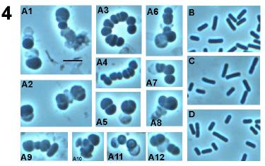

48 36
49 Fig. 7. Phase contrast microscopy of wild type and DPVB207 (the xylose regulated pbpa pbph double mutant) vegetative cells. (A), DPVB207 without xylose; (B), DPVB207 with xylose; (C), PS832 with xylose; (D), PS832 without xylose. (1), (2), (3) and (4) are 80, 120, 180 and 240min, respectively, after the resuspension into the new medium. Scale bars, 4µm. 37


50 38

51 39

52 40

53 41
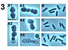
54 Fig. 8. Phase contrast light microscopy of outgrowing DPVB207 (xylose regulated pbpa pbph double mutant) and wildtype spores. A, the spores of DPVB207 were from the medium without xylose and germinated with the presence of xylose; B, the spores of DPVB207 were from the medium without xylose and germinated without xylose; C, the spores of DPVB207, were obtained in the medium with the presence of xylose and germinated in the presence of xylose; D, the spores of DPVB207, were from the medium with xylose and germinated without xylose; E, the spores of PS832 were from the medium without xylose and germinated with xylose; F, the spores of PS832 were obtained in the medium without xylose and germinated without xylose; G, the spores of PS832, were from the medium with the presence of xylose and germinated with xylose; H, the spores of PS832, were from the medium with xylose and germinated without xylose. 1, 2, 3, 4, 5, and 6, are 45, 60, 90,120, 180, and 240min, respectively, after initiation of germination. Scale bars, 4µm. 42

55 43

56 44

57 45
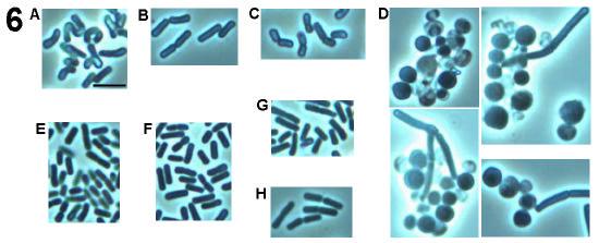
58 Fig. 9. Electron microscopy of wild type and DPVB207 (xylose regulated pbpa pbph double mutant) vegetative cells. (A), DPVB207 without xylose; (B), DPVB207 with xylose; (C), PS832 with xylose. (1), (2) and (3) are 80, 180 and 240min, respectively, after the resuspension into the new medium. Scale bars, 1µm. 46
59 47
60 48
61 Fig. 10. RP-HPLC Muropeptide elution patterns of peptidoglycan from vegetative cells of various strains. (A), PS832 (w.t.) with xylose; (B), PS832 without xylose; (C), DPVB133 (pbph) without xylose; (D), DPVB171 (pbpc pbph) without xylose; (E), DPVB207 (pbpa pbph) with xylose; (F), DPVB207 without xylose. The Y- axis is the Absorbance at 206 nm. The arrows point at the new peak or the peaks of different size. 49
62 Discussion The identification and characterization of pbph was carried out through the study of the putative coding sequence and the construction and analysis of a series of B. subtilis mutant and altered gene expression strains. The identification of gene product of pbph. Analysis of the expression of a transcriptional lacz fusion to pbph showed that transcription of pbph is at a very low level. Over-expression of the gene was attempted in order to detect the gene product. In this study, strain DPVB202, which contains an inducible xylap-pbph fusion, was used to overexpress pbph. Membranes were purified from the induced strain, incubated with labeled penicillin, run on a SDS-PAGE gel, and scanned on a phosphoimager. However, we could not visualize the gene product. The chromosome of DPVB202 was used to transform a pbpa mutant to generate a pbph pbpa double mutant in which viability was maintained only upon xylose induction of pbph expression, indicting that pbph was being expressed at some level from the xyla promoter construct. In the other part of this project the gene product of pbpi was identified using labeled penicillin, indicating that our performance of this method was successful. The inability to detect PbpH may due to a low binding affinity for penicillin, a low level of pbph expression upon xylose induction, or instability of the protein. The function of pbph gene product. Analysis of the coding sequence of pbph revealed its gene product s sequence similarity to class B high-molecular weight PBPs, especially to PBP2a in B. subtilis. The non-viability of a pbpa pbph double mutant indicated that pbpa and pbph play a redundant essential function. Our studies of the inducible pbpa pbph double mutant verified that these two genes are determinants of cell 50
63 elongation and maintenance of the rod-shape of B. subtilis. These two proteins, therefore, appear to carry out a role homologous to that of PBP2 in E. coli. E. coli cells in which PBP2 is inactivated grow as spherical cells and are not viable (59, 66). Insertional mutagenesis of B. subtilis pbpa had no effect on the phenotype of cells in log phase or sporulation or on the properties of the dormant spores. Spores produced by the pbpa strain initiated germination normally, but these spores had difficulty in cell elongation and the determination of the cell diameter during the spore outgrowth (41). However, pbpa spores eventually gave rise to vegetative cells that were indistinguishable from those of the wild-type strain. It was suggested that PBP2a s role in vegetative elongation is minimal or that other PBPs are able to compensate for PBP2a s contribution to vegetative cell wall elongation in its absence. Our results confirmed that this other PBP is the product of pbph. PBP2a and PbpH have the same indispensable function of contribution to the elongation of the cylindrical cell wall and maintenance of the normal rod shape of B. subtilis. The non-viability of a pbph pbpa strain may be due to an inability to properly segregate the cell contents and to carry out cell division without causing a wall disruption resulting in lysis. Analysis of the expression of a transcriptional lacz fusion to pbph showed that the expression of pbph was increased during the vegetative growth and reached the highest level during the transition to the stationary phase. Then, the expression of pbph dropped to a very low level during sporulation. It was not expressed during germination. On the contrary, the expression of pbpa began to increase 30 to 40 min after the initiation of germination, increased throughout vegetative growth, and then decreased upon entry into stationary phase and sporulation (41). The expression pattern of pbph may explain 51
64 the phenotype of the pbpa mutant. The initiation of germination doesn t require the activity of any wall synthesis protein, thus both the pbpa and pbpa pbph strains can initiate spore germination. At the stage of spore outgrowth in the pbpa mutant, pbph is expressed at an extremely low level, thus PbpH cannot fulfill its function in maintenance of rod shape, leading to the delayed outgrowth and defective cell morphology. Eventually, the expression of pbph is increased to the level that pbpa's function is compensated and the cell resumes normal shape and growth. In the pbpa pbph double mutant, after the initiation of spore germination, the cells never change to rod shape due to the loss of the cell elongation function carried out by PbpH and/or PBP2a. In our study we found that the expression level of pbph is much less than that of pbpa (41). It will be interesting to study the expression level of pbph in the pbpa mutant and the expression level of pbpa in the pbph mutant. It is possible that in the absence of one of these gene products, expression of the other may be increased. Cell shape maintenance and PBPs. It is believed that cell shape is determined by the balance of cell elongation and cell septation systems (26). Each of these systems is predicted to contain PG synthesis enzymes (PBPs), autolysins, and possibly regulation proteins. During the normal cell cycle, cells appear to switch between these systems, elongating until the cell is prepared to divide and then temporarily directing the majority of the PG synthetic activity to septum production. In E. coli, RodA (an integral membrane protein, encoded by roda) (35, 63) and class B high MW PBP2 (encoded by pbpa)(59), are involved in the maintenance of the rod shape during cell elongation. Mutations in roda or pbpa, or the inactivation of PBP2 block elongation and cause the production of spherical cells. (These mutant cells look 52
65 similar to some of spherical cells of our B. subtilis pbpa pbph double mutant (24, 55).) These two genes are in an operon (35), and their gene products are believed to interact with each other (34). Two other proteins, FtsW (also an integral membrane protein, encoded by ftsw) (10, 28) and class B HMW PBP3 (encoded by pbpb), are required for cell division. It has been suggested that there is an interaction between RodA and PBP3 (6) and PBP2 is essential for both elongation and division (67). Thus the cell elongation system and cell division system are associated. FtsW and RodA have similar sequences (24). Other proteins belonging to this FtsW/RodA (mrdb) family seem to be associated with the function of every class B protein that has been examined in detail (25). This study has given some insights to B. subtilis cell elongation and cell shape maintenance. In B. subtilis, there are three proteins, RodA (encoded by roda, (24)), PBP2a (41), and PbpH as the homologous system of RodA and PBP2 in E. coli. RodA has an essential role in elongation. The mutation of roda causes the formation of nonviable spherical cells (24). PBP2a and pbph are required in the elongation of cells as specific transpeptidases. In B. subtilis cell division, PBP2b (encoded by pbpb) is essential for septation and has been identified as a homologue to PBP3 (70). Based on sequence similarities, a candidate for the B. subtilis homologue of E. coli FtsW is the product of an uncharacterized ORF designated as ylao. It is not known at this point whether these two systems are associated or not. In the spherical cells produced by a B. subtilis pbpa pbph double mutant there is still septum formation, but the septa are formed extremely irregularly and cells have different sizes and shapes. This may be due to the activity of only the cell septation PG synthetic system. It is not clear why these cells lyse. The septation localization system 53
66 appears to be affected by the loss of the rod-shape. There can be several possibilities to explain this phenomenon. One hypothesis is that PbpH or PBP2a is required for localization of the division sites or for completion of septation. Another possibility is the larger-than-normal diameter of the mutant cells impedes the formation of a complete FtsZ ring as required for normal septation (15). Polymerization of the FtsZ at the division site may lead to partial rings or spirals, which are unable to direct a normal invagination of the envelope. Helical cells of inducable pbph pbpa double mutant when germinated with xylose. During spore germination and outgrowth, the cell shape of the induced xylosedependent pbpa pbph double mutant was helical or twisted. This helical phenotype has been previously reported in B. subtilis in several circumstances, such as wild-type cells grown in a chemostat with a low [Mg 2+ ] in the medium (56), cells treated with penicillin G or chlorpromazine (62), mutants resistant to Triton X-100 (62), strains with conditional mutation in pbpb (57), a strain with mutations in prfa, pona, pbpd and pbpf (54), and a strain with mutations in pona and pbpd grown in medium with a low [Mg 2+ ] (40). The twist of our cells indicates abnormal cell wall PG synthesis, arrangement or turn-over in the cell. The thicker cell wall of pbph pbpa double mutant. It was reported that the cell wall of outgrowing spores of pbpa mutants possess a thicker cell wall, which is probably due in part to the slower PG turnover early in pbpa spore outgrowth (39). The spores of this strain eventually outgrow to vegetative cells indistinguishable from wild-type cells. If the hypothesis is true then any imbalance between PG synthesis and degradation early in the outgrowth will be corrected to give rise to normal cell (39). And this will indicate that 54
67 PbpH is associated with the correction of this imbalance. The other hypothesis proposed to explain the increased cell wall thickness of pbpa spores early in outgrowth is that newly synthesized PG is inserted into a smaller surface area in the outgrowing spherical pbpa spore than in the more cylindrical outgrowing wild-type spore, since a sphere has a smaller surface area than a cylinder and outgrowing wild type and pbpa spores have similar volumes (39). The unevenly thicker cell wall of the pbpa pbph double mutant is more likely due to the imbalance of the synthesis and degradation of PG. Another explanation is that PG in the cell poles is more stable than PG in the cylindrical cell wall. If, in the pbpa outgrowing spores or vegetative cells, all the PG synthesis is carried out by the cell septation machinery then this PG will more closely resemble cell pole PG and will be degraded more slowly, and thus will be thicker. 55
68 Conclusions A sequence alignment of the predicted product of pbph against the microbial protein database demonstrated that the most similar protein in B. subtilis is PBP2A and in E. coli is PBP2. This suggested that PbpH belongs to a group of the genes required for maintaining the rod shape of the cell. Study of a pbph-lacz fusion showed that pbph was expressed weakly during vegetative growth and the expression reached the highest level at the transition from exponential phase to stationary phase. Construction of mutant strain lacking PbpH and mutant strains lacking multiple class B PBPs including PbpH revealed that the combination of a pbpa deletion and the pbph deletion was lethal and double mutant strains lacking pbph and pbpc or pbpi (also named yrrr) were viable. The viable mutants were indistinguishable from the wild-type except that the vegetative PG of the pbpc pbph strain had a slightly slightly lower amount of disaccharide tetrapeptide with 1 amidation and higher amount of disaccharide tripeptide tetrapeptide with 2 amidations when compared to others strains. This suggests that PbpC is involved in vegetative PG synthesis but only affects the PG structure with a very low efficiency. A pbpa pbph double mutant containing a xylose-regulated pbph gene inserted into the chromosome at the amye locus was constructed to study the mechanism of the non-viability and the phenotype of this double mutant. Depletion of PbpH resulted in an arrest in cell growth and a dramatic morphological change in both vegetative cells and outgrowing spores. Vegetative cells lacking pbpa and pbph expression swelled and cell elongation was arrested, leading to the formation of pleiomorphic spherical cells and eventual lysis. In these cells, cell septations were randomly localized, cell walls and septa 56
69 were thicker than those seen in wild type cells, and the average cell width and volume were larger than those of cells expressing pbpa or pbph. The vegetative PG had an increased abundance of one unidentified muropeptide. Spores produced by the pbpa pbph double mutant were able to initiate germination but the transition of the ovalshaped spores to rod-shape cells was blocked. The outgrowing cells were spherical, gradually enlarged, and eventually lysed. Outgrowth of these spores in the presence of xylose led to the formation of helical cells. Thus, PbpH is apparently required for maintenance of cell shape, specifically for cell elongation. PbpH and PBP2a play a redundant role homologous to that of PBP2 in E. coli. 57
70 CHAPTER THREE CHARACTERIZATION OF PBP-CODING GENE pbpi (yrrr) Extensive sequence similarity indicates that yrrr encodes a class B PBP. This gene has been renamed and will now be referred to as pbpi. The first goal was the identification of the gene product of pbpi by comparing the PBP profile of a PbpI overexpressing strain to that of the wild type strain using radio-labeled penicillin. The second goal was examination of the expression of pbpi. The final goal was the identification of the function of pbpi. Materials and Methods 1. Plasmids, bacterial strains, and growth conditions. All plasmids and strains used in this study are listed in Table 5. E. coli strain JM109 was used for cloning. All B. subtilis strains used were derived from strain 168. Transformation with either plasmid DNA or chromosomal DNA was performed as described previously (2). Transformants were selected on 2xSG (32) plates containing appropriate antibiotics: chloramphenicol (3 µg/ml), spectinomycin (100 µg/ml), kanamycin (10 µg/ml), tetracycline (10 µg/ml), and erythromycin (0.5 µg/ml) plus lincomycin (12.5 µg/ml; macrolide-lincosamide-streptogramin B resistance). Vegetative growth and sporulation was routinely carried out in 2xSG liquid medium at 37 C. Spores were purified by water washing (43). Spore germination and outgrowth was analyzed in 2xYT medium (48) containing 4 mm L-ala after a 30 min heat shock in water at 65 C (43). The optical densities at 600nm (OD) of all cultures were determined using a 58
71 Genesys 5 spectrophotometer. Spore heat- and chloroform-resistance assays were carried out after sporulation for 24 hours as previously described (43). For induction of the xylose-regulated promoter, the strains were grown at 37 C in 2xSG lacking glucose (16 g nutrient broth, 2 ml 1M MgSO 4, 10.7 ml 2M KCl, 100 µl 1M MnCl 2, 2.72 µl 0.36M FeSO 4, and 20 ml 1.18% Ca(NO 3 ) 2 per liter) with a final xylose concentration of 2%. 2. Construction of plasmids and strains. Primers pbpi1 and pbpi2 (Table 6) were used to amplify a 2451 bp fragment containing 345 bp of upstream sequence, the coding sequence, and 355 bp of downstream sequence. This fragment was cloned into pgemt, generating plasmid pdpv107. The insert of this plasmid was sequenced using SP6 and T7 promoter primers and compared to the genome sequence of B. subtilus. The cloned pbpi gene was 100% identical to that in the genome sequence. To construct a deletion mutation in pbpi, pdpv107 was digested with EcoRV and HindIII to remove 89.2% of the coding sequence, including the conserved penicillin-binding active site sequences. The deleted region was replaced with an erythromycin resistance gene cassette obtained from pdg646 (21) by digestion with HindIII and SmaI. In this way we generated plasmid pdpv114 ( pbpi::erm). The plasmid was linearized by restriction digestion at a ScaI site within the pgemt vector sequence. The linearized DNA was transformed into our wild type B. subtilis strain, PS832, to allow the mutated gene to integrate into the chromosome via double crossover with selection for the erythromycin resistant marker. Thus, the pbpi deletion mutant strain (DPVB160) was obtained. 59
72 Primer pbpia (Table 6) was designed to contain an added EcoRI site on pbpi1 to assist in cloning a fragment for constructing a lacz-fusion. Primers pbpia and pbpi2 were used to PCR amplify the 2451 bp insert in pdpv107. The 2459 bp PCR product was cut with EcoRI, HindIII and DraI to get a 975 bp frament which contains the upstream region and the first 622 bp of the pbpi coding sequence. The fragment was cloned into EcoRIand HindIII-digested pdpc87 to generate pdpv126, in which the 975 bp fragment was placed in front of a promoterless lacz gene. The insert of the plasmid was sequenced and confirmed to be identical to the sequence from the genome. The supercoiled plasmid was used in transforming B. subtilis PS832 to allow the lacz fusion to recombine into the chromosomal copy of pbpi via a single crossover. Transformants were selected on 2xSG plates containing chloramphenical. In this way we generated strain DPVB169, containing a transcriptional fusion of lacz to the pbpi promoter. The pbpi-lacz fusion was transformed into a set of strains carrying null mutations in genes encoding sporulation-specific sigma factors: SC1159 (spoiiac1=σf - ), SC137 (spoiigb::tn917=σe - ::MLS R ) and SC500 (spoiiig 1=σG - ) using chromosomal DNA of DPVB169. Thus, DPVB184 (spoiiac1 pbpi-lacz), DPVB185 (spoiigb::tn917 pbpi-lacz), DPVB186 (spoiiig 1 pbpi-lacz) were generated. These sigma factor mutants isogenic wild type background is strain PY79. As a control, DPVB183 was generated by transforming PY79 using the chromosomal DNA of DPVB169. Primers pbpi5 and pbpi3 (Table 6) were designed with added PacI site and BglII sites, respectively. They were used to PCR amplify a 1802 bp fragment containing 25 bp of upstream sequence, the pbpi coding sequence, and 26 bp of downsteam sequence. The resulting 1820 bp PacI-BglII fragment was cloned into PacI- and BglII-digested 60
73 psweet-bgab (7) which contains a xylose-regulated expression system. The pbpi gene in the resulting plasmid, pdpv146, was sequenced and found to be identical to the sequence from the genome. The plasmid was linearlized at the PstI site in the vector and used to transform PS832 to generate a xylap-pbpi fusion at the amye locus. The resulting AmyE - and Cm R strain, DPVB210 (amye::xylap-pbpi), was obtained. As a control, DPVB213 was generated by transforming PS832 using plasmid psweet-bgab. 3. Enzyme assays, membrane preparation, and gel identification of PBPs β-galactosidase assays of vegetative cells, sporulating cells, and germinating spores were done by using the substrate o-nitrophenyl-β-d-galactopyranoside (43, 52) and the activity was expressed in Miller units. Glucose dehydrogenase activity was assayed as previously described (43). For small-scale membrane preparation, strains were grown in 2xSG medium at 37 C to an OD of 0.1. Then xylose was added to the culture to a final concentration of 2%, and incubation was continued until the OD reached 1.0. Membranes were prepared as previously described (49). PBPs were detected using 125 I-labeled penicillin X as previously described (36). 4. Immature forespore and spore peptidoglycan structure analysis. Hexosamines were assayed as described (37). The preparation and analysis of immature forespore PG were performed as described in detail previously (37). Spore PG structure were determined as described (47). The analysis of the chromotagrams was done using Powerchrom V2.2.2 software on a Machintosh OS 9.1 computer. 61
74 5. Other phenotypic and biochemical assays. Dipicolinic acid (DPA) accumulation was assayed as previously described (43). An assay of spore heat resistance was carried out as described previously (49). 62
75 Table 5. Bacillus subtilis strains and plasmid used Strains Genotype b Construction Source or reference DPVB45 pbpg::kn (36) DPVB56 pbpg::kn pbpf::erm r (36) DPVB64 spovd::kn Laboratory stock DPVB160 pbpi::erm r pdpv114 PS832 This work DPVB169 pbpi-lacz pdpv126 PS832 This work DPVB176 pbpi::erm r spovd::kn DPVB160 DPVB64 This work DPVB183 pbpi-lacz DPVB169 PY79 This work DPVB184 spoiiac1 pbpi-lacz DPVB169 SC1159 This work DPVB185 spoiigb::tn917ωnv325 pbpi-lacz DPVB169 SC137 This work DPVB186 spoiiig 1 pbpi-lacz DPVB169 SC500 This work DPVB198 pbpi::erm r pbpg::kn DPVB45 DPVB160 This work DPVB199 pbpi::erm r pbpf::cm PS1838 DPVB160 This work DPVB200 pbpi::erm r pbpg::kn pbpf::cm PS1838 DPVB199 This work DPVB210 xylap-pbpi at amye pdpv146 PS832 This work DPVB213 xylap-bgab at amye psweet-bagb PS832 This work PS832 Wild type, trp + revertant of 168 Laboratory stock PS1838 pbpf::cm (52) PY79 a wild type (45, 71) SC137 a spoiigb::tn917ωnv325 S. Cutting (45) SC500 a spoiiig 1 S. Cutting (45) SC1159 a spoiiac1 S. Cutting (45) Plasmid Construction or description of uses Source or reference pdg646 Carrying Erm r cassette (21) pdpc87 B. subtilis integrating lacz trascriptional fusion vector (52) pdpv107 Upstream, downstream, and coding region of pbpi in pgem-t This work 63
76 pdpv114 pdpv126 pdpv146 This work This work This work pjf751 B. subtilid integrating lacz fusion vector (18) (45) xylap- bgab, psweet carrying bgab (7) Deletion of pbpi and the replacement of Erm r cassette, pdpv107 digested with SmaI and HindIII, ligated with a 1603 EcoRV- HindIII bp Erm r cassette from pdg646 PpbpI-lacZ, pdpc87 carrying 975 bp EcoRI-HindIII fragment of pbpi from pdpv107 xylap- pbpi, psweet carrying 1819 bp PacI-BamHI PCR fragment of pbph psweetbgab a Genetic background is PY79. The other strains' genetic background is PS832 b Abbreviations for antibiotic resistance: Cm, chloramphenicol; Erm r, lincomycin and erythromycin; Kn, kanamycin; Sp, spectinomycin.. 64
77 Table 6. Primer sequences Name Primer sequence Restriction enzyme site added pbpi1 5' AGA GGG CCG CGT GAC GAC TCT TG pbpi2 5' ATC AGA GTC AGA AGA CTT CTC AG Uses For constructing pdpv103 For constructing pdpv103 Position in chromosome -345 upstream of pbpi 355 downstream of pbpi pbpia 5' CGG AAT TC*A GAG GGC CGC GTG ACG ACT CTT G EcoRI LacZ fusion -345 upstream of pbpi pbpi5 pbpi3 5' GCG CTT AAT TAA* CAC AAT GTG GGT GAG GTG TTT 5' CGG GAT CC*T TAA CAT GTG CTG AGA AGT TG PacI PSWEET-yrrR -25 upstream of pbpi BamHI PSWEET-yrrR 26 downstream of pbpi * The added restriction enzyme site. 65
78 Results Identification of the pbpi gene A sequence alignment of the predicted product of pbpi against a microbial protein database was done using the BLAST software (1). The most similar proteins in B. subtilis are the high-molecular-weight class B PBPs, among which SpoVD is the most similar one (27% identical and 42% similar). In E. coli the most similar protein is PBP3 (22% identical and 38% similar). SpoVD is transcribed from a sigmae-dependent promoter in the mother cell of a sporulating sporangium and is required for synthesis of the modified peptidoglycan of the spore cortex (14). PBP3 of E. coli, the gene product of pbpb (ftsi), is a class B PBP and is essential for synthesis of septal peptidoglycan during cell division (59). These data suggest that the product of pbpi belongs to the group of genes required for synthesis of the spore or septum PG. Construction of an inducible xylap-pbpi and identification of the product of pbpi Plasmid psweet contains a xylose-dependent expression system that can integrate into the chromosome of B. subtilis at the amye locus. In order to clone the whole coding sequence of pbpi into the plasmid, two primers were designed and the gene was PCR-amplified and ligated into the digested vector. Thus we constructed the plasmid pdpv146 (psweet-pbpi) in which pbpi is under the control of the xylose-regulated promoter. Then pdpv146 was linearized and used to transform strain PS832 to produce strain DPVB210 (amye::xylap-pbpi). PbpI was over-expressed upon the addition of xylose. Plasmid psweet-bgab was linearized and used to transform PS832 to produce strain DPVB213 (xylap-bgab) as a control. 2% xylose was added to cultures at an OD of 0.1 and membranes were prepared following growth to an OD of 1.0. Radioactively- 66
79 labeled penicillin was used to visualize the PBPs present in xylose-induced DPVB210, DPVB213, and our wild type PS832. In DPVB210 (over-expressing pbpi) we identified a new PBP with an apparent mass of 65 kda which matches the predicted molecular mass of the pbpi product (65.3 kda) (Fig. 11). This PBP appeared to be the product of pbpi and we will refer to it as PBP4b since it runs just underneath PBP4. This follows the convention of naming PBPs based upon their position on an SDS-PAGE gel. Expression of pbpi A PCR amplified fragment containing 622 bp of the N-terminal coding region and 345 bp upstream of pbpi was used to generate a transcriptional lacz fusion. This construct was transformed into B. subtilis PS832 to allow the lacz fusion to recombine into the pbpi locus in the chromosome via a single crossover, producing strain DPVB169. To determine at which stage of the B. subtilis life cycle that pbpi was expressed, the activity of β-galactosidase in vegetative cells, sporulating cells, and outgrowing spores was assayed (Fig. 12). 67
; lane 2, DPVB213 (over-expressed BgaB); lane 3, PS832 (wild-type).")
80 Fig. 11. Identification of PbpI. Membranes were purified from cultures at an OD of 1.0 and were incubated with 125 I-labeled penicillin X. Proteins were separated on an SDS-7.5% PAGE gel, and PBPs were detected using a phosphorimager. Lane 1, DPVB210 (over-expressed pbpi); lane 2, DPVB213 (over-expressed BgaB); lane 3, PS832 (wild-type). Calibrated molecular mass standards (MWM; in kilodaltons) were Bio-Rad low-range-prestained SDS-PAGE standards. 68
Two Class A High-Molecular-Weight Penicillin-Binding Proteins of Bacillus subtilis Play Redundant Roles in Sporulation
 JOURNAL OF BACTERIOLOGY, Oct. 2001, p. 6046 6053 Vol. 183, No. 20 0021-9193/01/$04.00 0 DOI: 10.1128/JB.183.20.6046 6053.2001 Copyright 2001, American Society for Microbiology. All Rights Reserved. Two
SUPPORTING INFORMATION FOR. SEquence-Enabled Reassembly of β-lactamase (SEER-LAC): a Sensitive Method for the Detection of Double-Stranded DNA
: a Sensitive Method for the Detection of Double-Stranded DNA") SUPPORTING INFORMATION FOR SEquence-Enabled Reassembly of β-lactamase (SEER-LAC): a Sensitive Method for the Detection of Double-Stranded DNA Aik T. Ooi, Cliff I. Stains, Indraneel Ghosh *, David J. Segal
SUPPORTING INFORMATION FOR SEquence-Enabled Reassembly of β-lactamase (SEER-LAC): a Sensitive Method for the Detection of Double-Stranded DNA Aik T. Ooi, Cliff I. Stains, Indraneel Ghosh *, David J. Segal
Dynamic localization of penicillin-binding proteins during spore development in Bacillus subtilis
 Microbiology (2005), 151, 999 1012 DOI 10.1099/mic.0.27692-0 Dynamic localization of penicillin-binding proteins during spore development in Bacillus subtilis Dirk-Jan Scheffers3 Correspondence Dirk-Jan
Introduction. Gene expression is the combined process of :
 1 To know and explain: Regulation of Bacterial Gene Expression Constitutive ( house keeping) vs. Controllable genes OPERON structure and its role in gene regulation Regulation of Eukaryotic Gene Expression
SUPPLEMENTARY DATA - 1 -
 - 1 - SUPPLEMENTARY DATA Construction of B. subtilis rnpb complementation plasmids For complementation, the B. subtilis rnpb wild-type gene (rnpbwt) under control of its native rnpb promoter and terminator
Chapter 12. Genes: Expression and Regulation
 Chapter 12 Genes: Expression and Regulation 1 DNA Transcription or RNA Synthesis produces three types of RNA trna carries amino acids during protein synthesis rrna component of ribosomes mrna directs protein
Analysis of Peptidoglycan Structural Changes and Cortex Lytic Enzymes during Germination of Bacillus anthracis Spores. Melissa M.
 Analysis of Peptidoglycan Structural Changes and Cortex Lytic Enzymes during Germination of Bacillus anthracis Spores Melissa M. Dowd Thesis submitted to the Faculty of the Virginia Polytechnic Institute
Biology 112 Practice Midterm Questions
 Biology 112 Practice Midterm Questions 1. Identify which statement is true or false I. Bacterial cell walls prevent osmotic lysis II. All bacterial cell walls contain an LPS layer III. In a Gram stain,
SUPPLEMENTARY INFORMATION
 doi:10.1038/nature10244 a O07391_MYCAV/127-243 NLPC_HAEIN/80-181 SPR_SHIFL/79-183 P74160_SYNY3/112-245 O24914_HELPY/301-437 Q51835_PORGI/68-178 DPP6_BACSH/163-263 YKFC_BACSU/185-292 YDHO_ECOLI/153-263
Helical Macrofiber Formation in Bacillus subtilis: Inhibition by Penicillin G
 JOURNAL OF BACTERIOLOGY, June 1984, p. 1182-1187 0021-9193/84/061182-06$02.00/0 Copyright C 1984, American Society for Microbiology Vol. 158, No. 3 Helical Macrofiber Formation in Bacillus subtilis: Inhibition
ydci GTC TGT TTG AAC GCG GGC GAC TGG GCG CGC AAT TAA CGG TGT GTA GGC TGG AGC TGC TTC
 Table S1. DNA primers used in this study. Name ydci P1ydcIkd3 Sequence GTC TGT TTG AAC GCG GGC GAC TGG GCG CGC AAT TAA CGG TGT GTA GGC TGG AGC TGC TTC Kd3ydcIp2 lacz fusion YdcIendP1 YdcItrgP2 GAC AGC
LABORATORY 7 ENDOSPORE STAIN AND BACTERIAL MOTILITY
 LABORATORY 7 ENDOSPORE STAIN AND BACTERIAL MOTILITY A. Endospore Stain B. Bacterial Motility A. ENDOSPORE STAIN DISCUSSION A few genera of bacteria, such as Bacillus and Clostridium have the ability to
University of Groningen. Bacterial Cell Wall Synthesis Scheffers, Dirk; Pinho, Mariana G. Published in: Microbiology and Molecular Biology Reviews
 University of Groningen Bacterial Cell Wall Synthesis Scheffers, Dirk; Pinho, Mariana G. Published in: Microbiology and Molecular Biology Reviews DOI: 10.1128/MMBR.69.4.585-607.2005 IMPORTANT NOTE: You
CHAPTER 13 PROKARYOTE GENES: E. COLI LAC OPERON
 PROKARYOTE GENES: E. COLI LAC OPERON CHAPTER 13 CHAPTER 13 PROKARYOTE GENES: E. COLI LAC OPERON Figure 1. Electron micrograph of growing E. coli. Some show the constriction at the location where daughter
Bacterial Genetics & Operons
 Bacterial Genetics & Operons The Bacterial Genome Because bacteria have simple genomes, they are used most often in molecular genetics studies Most of what we know about bacterial genetics comes from the
RNA Synthesis and Processing
 RNA Synthesis and Processing Introduction Regulation of gene expression allows cells to adapt to environmental changes and is responsible for the distinct activities of the differentiated cell types that
Microbial Genetics, Mutation and Repair. 2. State the function of Rec A proteins in homologous genetic recombination.
 Answer the following questions 1. Define genetic recombination. Microbial Genetics, Mutation and Repair 2. State the function of Rec A proteins in homologous genetic recombination. 3. List 3 types of bacterial
Introduction to Microbiology BIOL 220 Summer Session I, 1996 Exam # 1
 Name I. Multiple Choice (1 point each) Introduction to Microbiology BIOL 220 Summer Session I, 1996 Exam # 1 B 1. Which is possessed by eukaryotes but not by prokaryotes? A. Cell wall B. Distinct nucleus
CHAPTER : Prokaryotic Genetics
 CHAPTER 13.3 13.5: Prokaryotic Genetics 1. Most bacteria are not pathogenic. Identify several important roles they play in the ecosystem and human culture. 2. How do variations arise in bacteria considering
Bacillus anthracis. Clostridium botulinum Clostridium perfringens and other, but never Gram-negative microbes
 SPORES (endospores) the spore is formed inside the parent vegetative cell hence the name endospores The spore is a dehydrated, multishelled structure that protects and allows the bacteria to exist in suspended
BACTERIAL PHYSIOLOGY SMALL GROUP. Monday, August 25, :00pm. Faculty: Adam Driks, Ph.D. Alan Wolfe, Ph.D.
 BACTERIAL PHYSIOLOGY SMALL GROUP Monday, August 25, 2014 1:00pm Faculty: Adam Driks, Ph.D. Alan Wolfe, Ph.D. Learning Goal To understand how bacterial physiology applies to the diagnosis and treatment
Bacterial Morphology and Structure م.م رنا مشعل
 Bacterial Morphology and Structure م.م رنا مشعل SIZE OF BACTERIA Unit for measurement : Micron or micrometer, μm: 1μm=10-3 mm Size: Varies with kinds of bacteria, and also related to their age and external
Topic 4 - #14 The Lactose Operon
 Topic 4 - #14 The Lactose Operon The Lactose Operon The lactose operon is an operon which is responsible for the transport and metabolism of the sugar lactose in E. coli. - Lactose is one of many organic
Microbiology. Definition of a Microorganism. Microorganisms in the Lab. The Study of Microorganisms
 Microbiology The Study of Microorganisms Definition of a Microorganism Derived from the Greek: Mikros, «small» and Organismos, organism Microscopic organism which is single celled (unicellular) or a mass
Role of GerD in Germination of Bacillus subtilis Spores
 JOURNAL OF BACTERIOLOGY, Feb. 2007, p. 1090 1098 Vol. 189, No. 3 0021-9193/07/$08.00 0 doi:10.1128/jb.01606-06 Copyright 2007, American Society for Microbiology. All Rights Reserved. Role of GerD in Germination
Regulation of Gene Expression in Bacteria and Their Viruses
 11 Regulation of Gene Expression in Bacteria and Their Viruses WORKING WITH THE FIGURES 1. Compare the structure of IPTG shown in Figure 11-7 with the structure of galactose shown in Figure 11-5. Why is
Substrate Requirements for Regulated Intramembrane Proteolysis of Bacillus subtilis Pro- K
 JOURNAL OF BACTERIOLOGY, Feb. 2005, p. 961 971 Vol. 187, No. 3 0021-9193/05/$08.00 0 doi:10.1128/jb.187.3.961 971.2005 Copyright 2005, American Society for Microbiology. All Rights Reserved. Substrate
Germination of Individual Bacillus subtilis Spores with Alterations in the GerD and SpoVA Proteins, Which Are Important in Spore Germination
 JOURNAL OF BACTERIOLOGY, May 2011, p. 2301 2311 Vol. 193, No. 9 0021-9193/11/$12.00 doi:10.1128/jb.00122-11 Copyright 2011, American Society for Microbiology. All Rights Reserved. Germination of Individual
Optimization of Immunoblot Protocol for Use with a Yeast Strain Containing the CDC7 Gene Tagged with myc
 OPTIMIZATION OF IMMUNOBLOT PROTOCOL 121 Optimization of Immunoblot Protocol for Use with a Yeast Strain Containing the CDC7 Gene Tagged with myc Jacqueline Bjornton and John Wheeler Faculty Sponsor: Anne
DNA Technology, Bacteria, Virus and Meiosis Test REVIEW
 Be prepared to turn in a completed test review before your test. In addition to the questions below you should be able to make and analyze a plasmid map. Prokaryotic Gene Regulation 1. What is meant by
Supporting Information for. Initial Biochemical and Functional Evaluation of Murine Calprotectin Reveals Ca(II)-
-") Supporting Information for Initial Biochemical and Functional Evaluation of Murine Calprotectin Reveals Ca(II)- Dependence and Its Ability to Chelate Multiple Nutrient Transition Metal Ions Rose C. Hadley,
Supporting Information for Initial Biochemical and Functional Evaluation of Murine Calprotectin Reveals Ca(II)- Dependence and Its Ability to Chelate Multiple Nutrient Transition Metal Ions Rose C. Hadley,
Cortex Peptidoglycan Lytic Activity in Germinating Bacillus anthracis Spores
 JOURNAL OF BACTERIOLOGY, July 2008, p. 4541 4548 Vol. 190, No. 13 0021-9193/08/$08.00 0 doi:10.1128/jb.00249-08 Copyright 2008, American Society for Microbiology. All Rights Reserved. Cortex Peptidoglycan
Number-controlled spatial arrangement of gold nanoparticles with
 Electronic Supplementary Material (ESI) for RSC Advances. This journal is The Royal Society of Chemistry 2016 Number-controlled spatial arrangement of gold nanoparticles with DNA dendrimers Ping Chen,*
Cannibalism by Sporulating Bacteria
 Cannibalism by Sporulating Bacteria José E. González-Pastor, Erret C. Hobbs, Richard Losick 2003. Science 301:510-513 Introduction Some bacteria form spores. Scientist are intrigued by them. Bacillus subtilis
Corrected by : Shady Soghayr
 Done by : Renad Abu Rumman Corrected by : Shady Soghayr ممكن تفقد البكتيريا هذه الطبقه عند التعرض لظروف مختبريه S layer is different from slime layer sex pili (common pili) :help in genetic transfer between
A Gene (sleb) Encoding a Spore Cortex-Lytic Enzyme from Bacillus subtilis and Response of the Enzyme to
 Encoding a Spore Cortex-Lytic Enzyme from Bacillus subtilis and Response of the Enzyme to") JOURNAL OF BACTERIOLOGY, Oct. 1996, p. 6059 6063 Vol. 178, No. 20 0021-9193/96/$04.00 0 Copyright 1996, American Society for Microbiology A Gene (sleb) Encoding a Spore Cortex-Lytic Enzyme from Bacillus
JOURNAL OF BACTERIOLOGY, Oct. 1996, p. 6059 6063 Vol. 178, No. 20 0021-9193/96/$04.00 0 Copyright 1996, American Society for Microbiology A Gene (sleb) Encoding a Spore Cortex-Lytic Enzyme from Bacillus
AP Bio Module 16: Bacterial Genetics and Operons, Student Learning Guide
 Name: Period: Date: AP Bio Module 6: Bacterial Genetics and Operons, Student Learning Guide Getting started. Work in pairs (share a computer). Make sure that you log in for the first quiz so that you get
Supplemental Figure 1.
 A wt spoiiiaδ spoiiiahδ bofaδ B C D E spoiiiaδ, bofaδ Supplemental Figure 1. GFP-SpoIVFA is more mislocalized in the absence of both BofA and SpoIIIAH. Sporulation was induced by resuspension in wild-type
7.06 Cell Biology EXAM #3 April 21, 2005
 7.06 Cell Biology EXAM #3 April 21, 2005 This is an open book exam, and you are allowed access to books, a calculator, and notes but not computers or any other types of electronic devices. Please write
3.B.1 Gene Regulation. Gene regulation results in differential gene expression, leading to cell specialization.
 3.B.1 Gene Regulation Gene regulation results in differential gene expression, leading to cell specialization. We will focus on gene regulation in prokaryotes first. Gene regulation accounts for some of
Newly made RNA is called primary transcript and is modified in three ways before leaving the nucleus:
 m Eukaryotic mrna processing Newly made RNA is called primary transcript and is modified in three ways before leaving the nucleus: Cap structure a modified guanine base is added to the 5 end. Poly-A tail
Killing of Bacillus Spores by High-Intensity Ultraviolet Light
 Killing of Bacillus Spores by High-Intensity Ultraviolet Light STUDY ON EFFECTS OF PULSED LIGHT Abraham L. Sonenshein, PhD Professor and Deputy Chair Department of Molecular Biology and Microbiology Tufts
Ch 3. Bacteria and Archaea
 Ch 3 Bacteria and Archaea SLOs for Culturing of Microorganisms Compare and contrast the overall cell structure of prokaryotes and eukaryotes. List structures all bacteria possess. Describe three basic
7.06 Problem Set #4, Spring 2005
 7.06 Problem Set #4, Spring 2005 1. You re doing a mutant hunt in S. cerevisiae (budding yeast), looking for temperaturesensitive mutants that are defective in the cell cycle. You discover a mutant strain
Modulation of the ComA-Dependent Quorum Response in Bacillus subtilis by Multiple Rap Proteins and Phr Peptides
 JOURNAL OF BACTERIOLOGY, July 2006, p. 5273 5285 Vol. 188, No. 14 0021-9193/06/$08.00 0 doi:10.1128/jb.00300-06 Copyright 2006, American Society for Microbiology. All Rights Reserved. Modulation of the
Supporting online material
 Supporting online material Materials and Methods Target proteins All predicted ORFs in the E. coli genome (1) were downloaded from the Colibri data base (2) (http://genolist.pasteur.fr/colibri/). 737 proteins
Clay Carter. Department of Biology. QuickTime and a TIFF (Uncompressed) decompressor are needed to see this picture.
 decompressor are needed to see this picture.") QuickTime and a TIFF (Uncompressed) decompressor are needed to see this picture. Clay Carter Department of Biology QuickTime and a TIFF (LZW) decompressor are needed to see this picture. Ornamental tobacco
QuickTime and a TIFF (Uncompressed) decompressor are needed to see this picture. Clay Carter Department of Biology QuickTime and a TIFF (LZW) decompressor are needed to see this picture. Ornamental tobacco
Gene expression in prokaryotic and eukaryotic cells, Plasmids: types, maintenance and functions. Mitesh Shrestha
 Gene expression in prokaryotic and eukaryotic cells, Plasmids: types, maintenance and functions. Mitesh Shrestha Plasmids 1. Extrachromosomal DNA, usually circular-parasite 2. Usually encode ancillary
Mouth animalcules (bacteria)
") Mouth animalcules (bacteria) 1684 http://en.citizendium.org/images/thumb/9/94/leeuwenhoek.jpg/300px-leeuwenhoek.jpg Prokaryotic Cell Shapes Coccus - cocci Bacillus - bacillus Spirillum - spirilli Vibrio
Mouth animalcules (bacteria) 1684 http://en.citizendium.org/images/thumb/9/94/leeuwenhoek.jpg/300px-leeuwenhoek.jpg Prokaryotic Cell Shapes Coccus - cocci Bacillus - bacillus Spirillum - spirilli Vibrio
evoglow - express N kit distributed by Cat.#: FP product information broad host range vectors - gram negative bacteria
 evoglow - express N kit broad host range vectors - gram negative bacteria product information distributed by Cat.#: FP-21020 Content: Product Overview... 3 evoglow express N -kit... 3 The evoglow -Fluorescent
Supplementary Information
 Electronic Supplementary Material (ESI) for RSC Advances. This journal is The Royal Society of Chemistry 2014 Directed self-assembly of genomic sequences into monomeric and polymeric branched DNA structures
Copyright is owned by the Author of the thesis. Permission is given for a copy to be downloaded by an individual for the purpose of research and
 Copyright is owned by the Author of the thesis. Permission is given for a copy to be downloaded by an individual for the purpose of research and private study only. The thesis may not be reproduced elsewhere
REGULATION OF GENE EXPRESSION. Bacterial Genetics Lac and Trp Operon
 REGULATION OF GENE EXPRESSION Bacterial Genetics Lac and Trp Operon Levels of Metabolic Control The amount of cellular products can be controlled by regulating: Enzyme activity: alters protein function
Three types of RNA polymerase in eukaryotic nuclei
 Three types of RNA polymerase in eukaryotic nuclei Type Location RNA synthesized Effect of α-amanitin I Nucleolus Pre-rRNA for 18,.8 and 8S rrnas Insensitive II Nucleoplasm Pre-mRNA, some snrnas Sensitive
Mag-Bind Soil DNA Kit. M preps M preps M preps
 Mag-Bind Soil DNA Kit M5635-00 5 preps M5635-01 50 preps M5635-02 200 preps January 2013 Mag-Bind Soil DNA Kit Table of Contents Introduction and Overview...2 Kit Contents/Storage and Stability...3 Preparing
YodL and YisK possess shape-modifying activities that are suppressed by mutations in Bacillus subtilis mreb and mbl
 JB Accepted Manuscript Posted Online 23 May 2016 J. Bacteriol. doi:10.1128/jb.00183-16 Copyright 2016, American Society for Microbiology. All Rights Reserved. 1 2 3 YodL and YisK possess shape-modifying
REVIEW SESSION. Wednesday, September 15 5:30 PM SHANTZ 242 E
 REVIEW SESSION Wednesday, September 15 5:30 PM SHANTZ 242 E Gene Regulation Gene Regulation Gene expression can be turned on, turned off, turned up or turned down! For example, as test time approaches,
Identification of a New Gene Essential for Germination of Bacillus subtilis Spores with Ca 2 -Dipicolinate
 JOURNAL OF BACTERIOLOGY, Apr. 2003, p. 2315 2329 Vol. 185, No. 7 0021-9193/03/$08.00 0 DOI: 10.1128/JB.185.7.2315 2329.2003 Copyright 2003, American Society for Microbiology. All Rights Reserved. Identification
BACTERIA. CLS 212: Medical Microbiology Miss Zeina Alkudmani
 BACTERIA CLS 212: Medical Microbiology Miss Zeina Alkudmani Prokaryotes Prokaryotic cells possess simpler structures than eukaryotic cells, since they do not have a nucleus or a lot of cytoplasmic organelles.
Production of Recombinant Annexin V from plasmid pet12a-papi
 Tait Research Laboratory Page 1 of 5 Principle Production of Recombinant Annexin V from plasmid pet12a-papi Annexin V is expressed cytoplasmically in BL21(DE3) E. coli (Novagen) with the pet vector system
15.2 Prokaryotic Transcription *
 OpenStax-CNX module: m52697 1 15.2 Prokaryotic Transcription * Shannon McDermott Based on Prokaryotic Transcription by OpenStax This work is produced by OpenStax-CNX and licensed under the Creative Commons
THIN SECTIONS OF DIVIDING NEISSERIA GONORRHOEAE
 JOURNAL OF BACTERIOLOGY Vol. 87, No. 6, pp. 1477-1482 June, 1964 Copyright 1964 by the American Society for Microbiology Printed in U.S.A. THIN SECTIONS OF DIVIDING NEISSERIA GONORRHOEAE PHILIP FITZ-JAMES
Data Sheet. Azide Cy5 RNA T7 Transcription Kit
 Cat. No. Size 1. Description PP-501-Cy5 10 reactions à 40 µl For in vitro use only Quality guaranteed for 12 months Store all components at -20 C. Avoid freeze and thaw cycles. DBCO-Sulfo-Cy5 must be stored
I. Archaeal cell structure. (Chap 2 pg , Supplemental notes 3, 5)
") Thurs, Jan 23, 2003 I. Archaeal cell structure. (Chap 2 pg. 450-453, Supplemental notes 3, 5) The Archaea are a diverse group of prokaryotic organisms that are very different from bacteria and from eucaryotes.
Thurs, Jan 23, 2003 I. Archaeal cell structure. (Chap 2 pg. 450-453, Supplemental notes 3, 5) The Archaea are a diverse group of prokaryotic organisms that are very different from bacteria and from eucaryotes.
MORPHOLOGY: the study of form and structure
 MICROBIOLOGY CHAPTER 3 Bacteria Morphology 3:1 Bacteria Structure and Function MORPHOLOGY: the study of form and structure Structure of Bacteria 1. PROKARYOTIC no membrane bound nucleus nor other organelles
Name Period The Control of Gene Expression in Prokaryotes Notes
 Bacterial DNA contains genes that encode for many different proteins (enzymes) so that many processes have the ability to occur -not all processes are carried out at any one time -what allows expression
BME 5742 Biosystems Modeling and Control
 BME 5742 Biosystems Modeling and Control Lecture 24 Unregulated Gene Expression Model Dr. Zvi Roth (FAU) 1 The genetic material inside a cell, encoded in its DNA, governs the response of a cell to various
Component Product # Product # Cell Lysis Reagent 100 ml 500 ml Product Insert 1 1
 3430 Schmon Parkway Thorold, ON, Canada L2V 4Y6 Phone: 866-667-4362 (905) 227-8848 Fax: (905) 227-1061 Email: techsupport@norgenbiotek.com Cell Lysis Reagent Product # 18800 (100 ml) Product # 18801 (500
Molecular Biology, Genetic Engineering & Biotechnology Operons ???
 1 Description of Module Subject Name?? Paper Name Module Name/Title XV- 04: 2 OPERONS OBJECTIVES To understand how gene is expressed and regulated in prokaryotic cell To understand the regulation of Lactose
evoglow - express N kit Cat. No.: product information broad host range vectors - gram negative bacteria
 evoglow - express N kit broad host range vectors - gram negative bacteria product information Cat. No.: 2.1.020 evocatal GmbH 2 Content: Product Overview... 4 evoglow express N kit... 4 The evoglow Fluorescent
Lecture 10: Cyclins, cyclin kinases and cell division
 Chem*3560 Lecture 10: Cyclins, cyclin kinases and cell division The eukaryotic cell cycle Actively growing mammalian cells divide roughly every 24 hours, and follow a precise sequence of events know as
chapter one: the history of microbiology
 chapter one: the history of microbiology Revised 6/19/2018 microbes microscopic (small) organisms, viruses, prions prefix sci. notation frac. equivalent dec. equivalent kilo- (k) 1 10 3 1000/1 = 1000 1000
Big Idea 3: Living systems store, retrieve, transmit and respond to information essential to life processes. Tuesday, December 27, 16
 Big Idea 3: Living systems store, retrieve, transmit and respond to information essential to life processes. Enduring understanding 3.B: Expression of genetic information involves cellular and molecular
Protocol for 2D-E. Protein Extraction
 Protocol for 2D-E Protein Extraction Reagent 1 inside the ReadyPrep TM Sequential Extraction kit (in powder form) 50ml of deionized water is used to dissolve all the Reagent 1. The solution is known as
Contains ribosomes attached to the endoplasmic reticulum. Genetic material consists of linear chromosomes. Diameter of the cell is 1 m
 1. (a) Complete each box in the table, which compares a prokaryotic and a eukaryotic cell, with a tick if the statement is correct or a cross if it is incorrect. Prokaryotic cell Eukaryotic cell Contains
Review. Diverse Paths to Midcell: Assembly of the Bacterial Cell Division Machinery. Nathan W. Goehring and Jon Beckwith*
 , Vol. 15, R514 R526, July 12, 2005, 2005 Elsevier Ltd. All rights reserved. DOI 10.1016/j.cub.2005.06.038 Diverse Paths to Midcell: Assembly of the Bacterial Cell Division Machinery Review Nathan W. Goehring
mrna Isolation Kit for Blood/Bone Marrow For isolation mrna from blood or bone marrow lysates Cat. No
 For isolation mrna from blood or bone marrow lysates Cat. No. 1 934 333 Principle Starting material Application Time required Results Key advantages The purification of mrna requires two steps: 1. Cells
Analysis of Escherichia coli amino acid transporters
 Ph.D thesis Analysis of Escherichia coli amino acid transporters Presented by Attila Szvetnik Supervisor: Dr. Miklós Kálmán Biology Ph.D School University of Szeged Bay Zoltán Foundation for Applied Research
Effects of the Chromosome Partitioning Protein Spo0J (ParB) on oric Positioning and Replication Initiation in Bacillus subtilis
 on oric Positioning and Replication Initiation in Bacillus subtilis") JOURNAL OF BACTERIOLOGY, Feb. 2003, p. 1326 1337 Vol. 185, No. 4 0021-9193/03/$08.00 0 DOI: 10.1128/JB.185.4.1326 1337.2003 Copyright 2003, American Society for Microbiology. All Rights Reserved. Effects
JOURNAL OF BACTERIOLOGY, Feb. 2003, p. 1326 1337 Vol. 185, No. 4 0021-9193/03/$08.00 0 DOI: 10.1128/JB.185.4.1326 1337.2003 Copyright 2003, American Society for Microbiology. All Rights Reserved. Effects
UNIT 6 PART 3 *REGULATION USING OPERONS* Hillis Textbook, CH 11
 UNIT 6 PART 3 *REGULATION USING OPERONS* Hillis Textbook, CH 11 REVIEW: Signals that Start and Stop Transcription and Translation BUT, HOW DO CELLS CONTROL WHICH GENES ARE EXPRESSED AND WHEN? First of
MULTIPLE CHOICE. Choose the one alternative that best completes the statement or answers the question. 1)
") MULTIPLE CHOICE. Choose the one alternative that best completes the statement or answers the question. 1) 1) Which of the following statements about the atom A) It has 12 neutrons in its nucleus. B) It
MULTIPLE CHOICE. Choose the one alternative that best completes the statement or answers the question. 1) 1) Which of the following statements about the atom A) It has 12 neutrons in its nucleus. B) It
Practical Bioinformatics
 5/2/2017 Dictionaries d i c t i o n a r y = { A : T, T : A, G : C, C : G } d i c t i o n a r y [ G ] d i c t i o n a r y [ N ] = N d i c t i o n a r y. h a s k e y ( C ) Dictionaries g e n e t i c C o
Development Team. Regulation of gene expression in Prokaryotes: Lac Operon. Molecular Cell Biology. Department of Zoology, University of Delhi
 Paper Module : 15 : 23 Development Team Principal Investigator : Prof. Neeta Sehgal Department of Zoology, University of Delhi Co-Principal Investigator : Prof. D.K. Singh Department of Zoology, University
Magnetic Janus Nanorods for Efficient Capture, Separation. and Elimination of Bacteria
 Electronic Supplementary Material (ESI) for RSC Advances. This journal is The Royal Society of Chemistry 2016 Magnetic Janus Nanorods for Efficient Capture, Separation and Elimination of Bacteria Zhi-min
Meiosis and Tetrad Analysis Lab
 Meiosis and Tetrad Analysis Lab Objectives: - Explain how meiosis and crossing over result in the different arrangements of ascospores within asci. - Learn how to calculate the map distance between a gene
Gene Switches Teacher Information
 STO-143 Gene Switches Teacher Information Summary Kit contains How do bacteria turn on and turn off genes? Students model the action of the lac operon that regulates the expression of genes essential for
MULTIPLE CHOICE. Choose the one alternative that best completes the statement or answers the question. Figure 2.1
 Exam Name MULTIPLE CHOICE. Choose the one alternative that best completes the statement or answers the question. Figure 2.1 1) Which compound in Figure 2.1 is an ester? 1) A) a b c d e Answer: D 2) A scientist
Fitness constraints on horizontal gene transfer
 Fitness constraints on horizontal gene transfer Dan I Andersson University of Uppsala, Department of Medical Biochemistry and Microbiology, Uppsala, Sweden GMM 3, 30 Aug--2 Sep, Oslo, Norway Acknowledgements:
This article appeared in a journal published by Elsevier. The attached copy is furnished to the author for internal non-commercial research and
 This article appeared in a journal published by Elsevier. The attached copy is furnished to the author for internal non-commercial research and education use, including for instruction at the authors institution
Molecular Biology (9)
") Molecular Biology (9) Translation Mamoun Ahram, PhD Second semester, 2017-2018 1 Resources This lecture Cooper, Ch. 8 (297-319) 2 General information Protein synthesis involves interactions between three
Molecular Biology (9) Translation Mamoun Ahram, PhD Second semester, 2017-2018 1 Resources This lecture Cooper, Ch. 8 (297-319) 2 General information Protein synthesis involves interactions between three
Determinants for the Subcellular Localization and Function of a Nonessential SEDS Protein
 zjs / DCHEAD ARTICLE zjss / DCTPIC GENETICS AND MLECULAR BILGY FRM CVER SHEET: Editor: Moran Article No.: T Section: Genetics JURNAL F BACTERILGY, Jan. 2008, p. 000 Vol. 190, No. 1 0021-9193/08/$08.00
Phenol-Chloroform reagents. Selection guide. OH ; MW : High quality reagents for use in nucleic acid purification.
 Phenol-Chloroform reagents Extraction with phenol and phenol/chloroform mixtures is a universal method for purification of DNA and RNA. Proteins and restriction enzymes are removed by phenol and chloroform
Microbiology Helmut Pospiech
 Microbiology 20.03.2018 Helmut Pospiech The control of what gets in Passive transport along a concentration gradient often inefficient Active transport Requires energy consumption and what gets out ABC
DNA can be extracted from the following sample types using this procedure: Archived
 Sample types Principle Safety Equipment and supplies DNA can be extracted from the following sample types using this procedure: concentrated DNA samples (e.g., blood, saliva, non-contact samples) hair
Nature Structural & Molecular Biology: doi: /nsmb Supplementary Figure 1
 Supplementary Figure 1 Zn 2+ -binding sites in USP18. (a) The two molecules of USP18 present in the asymmetric unit are shown. Chain A is shown in blue, chain B in green. Bound Zn 2+ ions are shown as
The two daughter cells are genetically identical to each other and the parent cell.
 Prokaryote Growth and Reproduction This micrograph shows a bacillus bacteria (probably E. coli) undergoing binary fission. This is a form of asexual reproduction. During prokaryotic binary fission, as
2. What was the Avery-MacLeod-McCarty experiment and why was it significant? 3. What was the Hershey-Chase experiment and why was it significant?
 Name Date Period AP Exam Review Part 6: Molecular Genetics I. DNA and RNA Basics A. History of finding out what DNA really is 1. What was Griffith s experiment and why was it significant? 1 2. What was
Bacillus subtilis Food Grade Expression System
 MoBiTec GmbH 2017 Page 1 Bacillus subtilis Food Grade Expression System MoBiTec GmbH 2017 Page 2 Contents Introduction... 3 The Food Grade Selection System... 3 Storage and Handling Instructions... 5 Protocols...
Multiple Septation in Variants of Bacillus cereus
 JOURNAL OF BACTERIOLOGY, Nov., 1965 Copyright @ 1965 American Society for Microbiology Vol. 90, No. 5 Printed in U.S.A. Multiple Septation in Variants of Bacillus cereus C. C. REMSEN AND D. G. LUNDGREN
Reading Assignments. A. Genes and the Synthesis of Polypeptides. Lecture Series 7 From DNA to Protein: Genotype to Phenotype
 Lecture Series 7 From DNA to Protein: Genotype to Phenotype Reading Assignments Read Chapter 7 From DNA to Protein A. Genes and the Synthesis of Polypeptides Genes are made up of DNA and are expressed