Chapter 3: Novel Regulators of the C. elegans Caspase CED-3
|
|
|
- Claude Merritt
- 5 years ago
- Views:
Transcription
1 53 Chapter 3: Novel Regulators of the C. elegans Caspase CED Abstract The hallmark of apoptosis is the presence of activated caspases, a conserved family of cysteine proteases. Proper regulation of caspases is crucial to maintain cell viability. IAPs are a class of proteins known to inhibit caspases in Drosophila and mammals, though they do not regulate the C. elegans caspase CED-3. To understand how CED-3 is regulated, we have performed a yeast-based screen for CED-3 suppressors and isolated two previously uncharacterized genes, Y39B6A.12 and T23G11.7b. Characterization of these genes in Drosophila shows that T23G11.7b and its ortholog CG7967 are regulators of gene expression and not caspases. Y39B6A.12, on the other hand, inhibits the Bcl-2 gene Debcl, but fails to regulate the pro-apoptotic genes Grim, Hid, or Dronc. It still remains unclear if Y39B6A.12 is a critical regulator of CED-3 in viable C. elegans cells. 3.2 Introduction Programmed cell death is a regulated process used by an organism for the removal of deleterious cells or developmentally transient cells. Characterization of programmed cell
2 54 death began in the nematode Caenorhabditis elegans and led to the eventual identification of the genes involved (Ellis and Horvitz, 1986). ced-3 and ced-4 were shown to be necessary for cell death while the gene ced-9 blocks their function (reviewed in Metzstein et al., 1998). Later studies from other model systems showed that the ced genes were highly conserved in apoptosis. ced-3 belongs to a family of proteases now known as caspases, which are the executioners of programmed cell death (Yuan et al., 1993). The mitochondrial localized CED-4 facilitates the irreversible activation of CED-3 (Seshagiri and Miller, 1997). Subsequent studies show that Apaf-1 is structurally similar to CED-4 and promotes the activation of the apical caspase Caspase-9 (Zou et al., 1997). ced-9 is a member of the Bcl-2 family of mitochondrial proteins and inhibits ced-4 (Hentgartner and Horvitz, 1994). While the genes involved in apoptosis are highly conserved, the inhibition of caspases appears to vary in different organisms. In flies and mammals, caspases are inhibited by the IAP group of proteins. Removal of IAPs causes rapid cellular destruction, thus proving that they are crucial for maintaining cell viability (Wang et al., 1999). Importantly, no functional caspase inhibitor has yet been identified in C. elegans. The two genes with structural motifs similar to other IAPs in worms, the genes bir-1 and bir-2, are not involved in apoptosis, but instead are required for midzone spindle formation and cytokinesis (Fraser et al, 1999, Speliotes, et al, 2000). To explain how CED-3 is kept from chewing up the cell, two models have been proposed. One model states that an
3 55 alternative anti-apoptotic splice form of CED-4 keeps CED-3 down, though this splice form is relatively rare when compared to the pro-apoptotic one (Shaham and Horvitz, 1996). Another model suggests that the Bcl-like protein CED-9 binds CED-3 and CED-4 in a ternary complex and prevents CED-3 activation (Xue and Horvitz, 1997). A necessary tenet of this model is that CED-3 is localized to the mitochondria along with CED-4 and CED-9, yet no one has shown the localization pattern of CED-3. Furthermore, recent work in a heterologous assay has shown that CED-9 cannot inhibit CED-3 mediated cell death (Jabbour et al., 2004). If these models are flawed, the question remains: What keeps CED-3 from killing the cell? To probe this question, we used a function-based assay in S. cerevisiae to screen for inhibitors of CED-3. Two C. elegans genes were isolated from this screen, though subsequent studies in Drosophila eliminated one candidate from consideration. The remaining candidate, Y39B6A.12, weakly suppressed death induced by the Bcl2 gene Debcl in the Drosophila eye. Though Y39B6A.12 still needs to be characterized further in C. elegans, this heterologous screen provides a novel way to screen for regulators of the caspase CED Materials and Methods Yeast protocols
4 56 S. cerevisiae were transformed using a standard LiAc protocol. The W303α strain (MATα, can1-100, leu2-3, his3-11, -15, ura3-1, ade2-1) was grown on selective media containing 2% glucose or 2% galactose/1% raffinose for gene induction. ced-3 and ced-4 were induced using an 815 bp fragment of the Gal1 promoter region (Hawkins et al., 1997). The caspase-lacz reporter assay used to test caspase suppression independent of galactose has been previously described (Hawkins et al., 1997). C. elegans cdna library construction Mixed-stage C. elegans polya+ RNA was converted into cdna using the Superscript cdna synthesis kit (GIBCO). cdnas were size-fractioned to select clones greater than 500 bp, and were ligated into the NotI SalI sites of the gpat-his vector. Transformation of the ligation mix into the bacteria strain DH10B (GIBCO) yielded approximately 5 x 10 5 colonies. The cdna library was amplified by growing the colonies in LB with carbecillin and isolating their plasmid DNA (Qiagen). Drosophila genetics Drosophila strains were grown at 25 0 C. CG7967, T23G11.7b, and Y39B6A.12 were subcloned into the UASt vector and injected using standard embryo microinjection techniques. Recombinants of the UAS transgenics with the GMR-gal4 chromosome were made. UAS-Q78 was kindly provided by N. Bonini. UAS-Wg and UAS-pygo were provided by K. Cadigan. Pictures of Drosophila retina were taken using an Olympus DP10 digital camera.
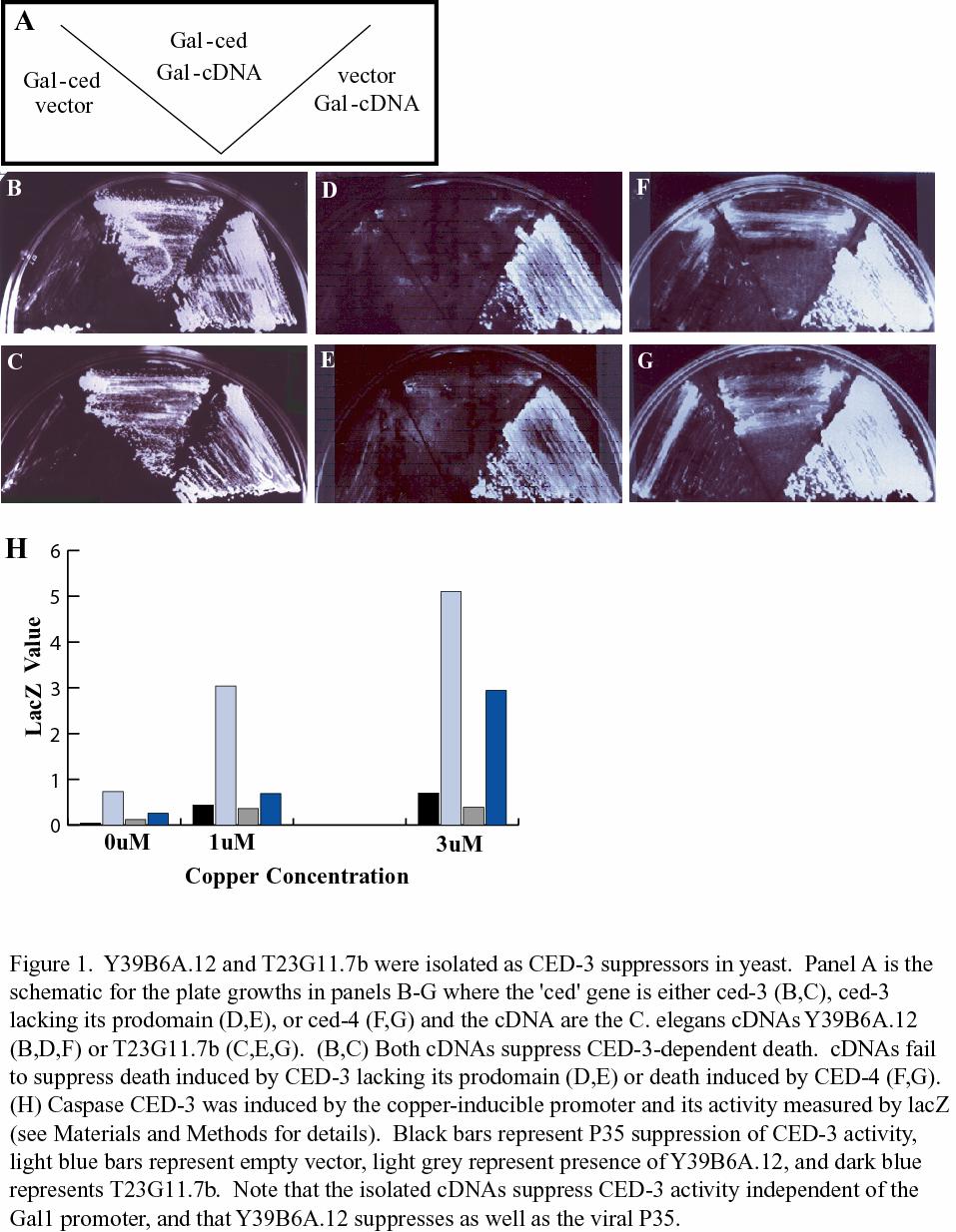
5 C. elegans RNAi 57 cdnas for T23G11.7b and Y39B6A.12 were cloned into the BamHI XbaI sites of ppd RNAi knockdown of these genes was conducted by feeding C. elegans on bacteria producing dsrna from these constructs. 3.4 Results Isolation of two ced-3 suppressors in yeast CED-3 is the only C. elegans caspase known to be involved in programmed cell death. To test whether C. elegans does encode a novel CED-3 inhibitor we screened an inducible C. elegans cdna library for genes that suppress ced-3 in S. cerevisiae. As previously reported, overexpression of ced-3, under the control of the galactose-inducible promoter, kills S. cerevisiae cells (Figure 1, Jabbour et al., 2004). This killing is complete when two Gal-ced-3 plasmids are present. The C. elegans genes T23G11.7b and Y39B6A.12 were both repeatedly isolated as suppressors of CED-3 mediated death (Fig. 1B, C). Reintroduction of the cdnas encoding Y39B6A.12 and T23G11.7b back into S. cerevisiae verified their suppression. The CARD domain found in the prodomain of CED-3 is necessary for its regulation, though not essential for proteolytic activity. Overexpression of CED-3 lacking its CARD
6 58
.")
7 59 domain (ced-3 Δprodomain) can still kill yeast cells. Interestingly, both T23G11.7b and Y39B6A.12 can no longer function as CED-3 inhibitors when the CED-3 CARD domain is missing, suggesting that the CARD domain is important for regulation (Fig 1D, E). CED-4 also encodes a CARD domain, and CED-4 has been shown to activate apoptosis and kill S. pombe (James et al., 1997). In S. cerevisiae, galactose-induced ced- 4 can also kill, though this death cannot be suppressed by T23G11.7b or Y39B6A.12 (Fig 1F, G). Because the two cdnas failed to suppress CED-3 (Δprodomain) and CED-4 dependent death in yeast, it is reasonable to suggest that the cdnas are CED-3 inhibitors and not suppressors of the galactose-inducible promoter. To test this hypothesis another way, we
. Notably, Y39B6A.12 suppressed just as well as P35. T23G11.")
8 60 tested whether T23G11.7b and Y39B6A.12 could suppress ced-3 controlled by the copper-inducible promoter. Increasing the concentration of copper led to an increase in CED-3 activity. Y39B6A.12 and T23G11.7b, as well as the pan-caspase suppressor P35, suppressed CED-3 activity independent of the Gal1 promoter (Fig. 1H). Notably, Y39B6A.12 suppressed just as well as P35. T23G11.7b/ CG7967 suppresses the Gal4/UAS system and does regulate apoptosis To determine whether the isolated cdnas are genuine caspase suppressors in vivo, we used the Drosophila eye for our assays. Using the GMR (Glass multiple repeat)-gal4 line to drive gene expression in the eye, ectopic expression of CG7967, the Drosophila ortholog of T23G11.7b, showed no phenotype (Fig. 2A). hid, grim, and reaper are
.")
9 61 activators of apoptosis and cause a small eye phenotype when overexpressed using the GMR enhancer. Crosses of GMR-gal4, UAS-CG7967 to GMR-hid, GMR-grim, and GMR-reaper showed no changes in eye size (Fig 2). In a similar fashion, expression of GMR-Dronc, which also induces death, could not be suppressed by GMR-gal4, UAS- CG7967 (Fig 2D, E). Though the Bcl proteins are much more extensively characterized in humans and mice, the Drosophila members, Debcl and Buffy, have been shown to regulate apoptosis. Ablation of the eye by overexpressed Debcl can be restored by
10 62 overexpression of Buffy (Quinn et al., 2003). CG7967 can suppress excess death phenotypes associated with ectopically expressed Debcl both in the eye and the wing (Fig 2, data not shown). During the course of experiments, it became apparent that CG7967 could suppress genes expressed using the Gal4/UAS system, but not genes controlled directly by the GMR enhancer (Fig 2, 3). Once again, to test whether CG7967 and T23G11.7b are genuine inhibitors of apoptosis, we tested their ability to suppress genes expressed using the GMR enhancer and the Gal4/UAS system. The caspase Dronc expressed using the Gal4/UAS system gives a stronger phenotype than GMR-Dronc. CG7967 could not suppress GMR- Dronc, as previously mentioned, though it could suppress GMR-gal4, UAS-Dronc (Fig. 4). Along with other members of the JNK pathway, overexpression of the kinase dtak1 and its mouse counterpart mtak1 activates apoptosis in the Drosophila eye. Overexpression of CG7967 failed to suppress GMR-mTak, while it suppressed GMRgal4, UAS-dTak (Fig. 4). Taken together, these results show that CG7967, and, by implication, T23G11.7b are not genuine regulators of apoptosis. Y39B6A.12, a possible caspase inhibitor To determine whether Y39B6A.12 is a pan-caspase inhibitor, we resorted to the Drosophila eye once again. In yeast, Y39B6A.12 proved to be a CED-3 inhibitor. Drosophila encodes for seven caspases, with the downstream caspases DrICE, decay, and dcp-1 showing homology to ced-3 in their catalytic domains. Since at the time of this writing, recombinants of GMR-gal4; GMR-DrICE, -Decay, and dcp-1 do not exist, we

11 63 sought to test whether Y39B6A.12 could suppress activators of these caspases. GMRgal4, UAS-Y39B6A.12 could not suppress ectopic expression of grim nor hid (Fig. 5). The upstream caspase Dronc contains an N-terminal CARD motif, though with little similarity to the CARD domain in CED-3, and has been shown to activate DrICE. Like hid and grim, overexpression of Dronc could not be suppressed by ectopically expressed Y39B6A.12. However, Y39B6A.12 did slightly suppress the ablated eye caused by overexpression of Debcl (Fig. 5). Finally, unlike T23G11.7b, Y39B6A.12 failed to counteract the JNK pathway components, Eiger, hep, and dtak1 (Fig. 5, data not shown). Crosses to establish the GMR-gal4, UAS-Y39B6A.12 recombinant are being made, so that we can test Y39B6A.12 suppression against the effector caspases DrICE and dcp-1.
12 64 To test for the function of Y39B6A.12 in C. elegans, worms were fed with bacteria expressing dsrna to Y39B6A.12. RNAi knockdown of Y39B6A.12 in N2 worms caused a sterile phenotype that was not suppressed by mutants for ced-3 or ced-4 (B. Derry, personal communication). Loss of the upstream inhibitor ced-9 shows partial sterility, among other effects (Hentgartner et al., 1992). The results of the Y39B6A.12 RNAi and epistasis analysis suggest that either Y39B6A.12 is a tissue-specific inhibitor of an unidentified caspase or that the sterility is not related to cell death genes at all. Protein characteristics and homology Y39B6A.12 encodes a previously uncharacterized gene. With an ortholog found only in C. briggsae, Y39B6A.12 is a nematode-specific gene characterized only by its BED Znfinger domain. BED Zn-finger proteins are a diverse class of nuclear proteins thought to bind to DNA. C. elegans encodes several BED proteins, including dpy-20 and the transposase tam-3. The best-characterized BED proteins in Drosophila are the transcriptional activator Dref and the chromatin insulator Beaf. How a DNA-binding protein like Y39B6A.12 functions in relation to CED-3 is yet to be determined. 3.5 Discussion Y39B6A.12 shows anti-apoptotic activity in yeast and Drosophila CED-3 remains the only characterized caspase in C. elegans apoptosis, and to date no proteins have been identified as CED-3 inhibitors. In this report, we describe the two
13 65 genes identified in a yeast-based screen performed to isolate CED-3 suppressors. The C. elegans gene T23G11.7b and its Drosophila ortholog proved to be general suppressors of gene induction, while preliminary work on the C. elegans-specific gene Y39B6A.12 shows conserved function, as it partially suppressed death induced by expression of the pro-apoptotic Bcl2 protein Debcl. Because Y39B6A.12 was isolated in S. cerevisiae, which is devoid of the core components of the apoptosis machinery, it stands to reason that Y39B6A.12 functions to inhibit caspases. Though Y39B6A.12 could not suppress the overexpressed Drosophila caspase Dronc, Y39B6A.12 may still inhibit caspases that have not been tested. Drosophila encodes seven caspases, and the downstream caspases DrICE, DCP-1, and Decay share the most sequence similarity to CED-3. Tests to determine if Y39B6A.12 is a pan-caspase inhibitor and can inhibit any Drosophila caspase are currently underway. Another possibility is that Y39B6A.12 could function only with CED-3 and not show any activity in Drosophila. To test for in vivo function in C. elegans, we knocked-down Y39B6A.12 levels by the feeding RNAi method. If Y39B6A.12 inhibits apoptosis, then loss of its function would cause spurious cell death. Reduced Y39B6A.12 function caused sterility, though this phenotype could not be suppressed by mutants for ced-3 or ced-4 (B. Derry, personal communication). Whether this sterility is caused by ectopic cell death is still to be determined. The ability of Y39B6A.12 to prevent cell death in C. elegans is currently being examined. Complete story of developmental apoptosis in C. elegans remains to be resolved
14 66 C. elegans was used first to understand the molecular components of programmed cell death. Screens pulled out 13 genes involved in killing the cell and in corpse removal. Even though worms have the simplest cell killing machinery it is not completely understood. For example it is not understood how the fragmentation of the mitochondria, important in mammalian apoptosis, is involved in worm cell death (Jagasia et al., 2005). The homologous apoptotic components AIF (apoptosis inducing factor) and endonuclease G are important in C. elegans programmed cell death, though the mechanism of their mitochondrial release has not been determined (Wang et al., 2002; Parrish et al., 2001). Anecdotal evidence also suggests that even the list of players mediating C. elegans apoptosis is incomplete. A deficiency screen looking at abnormal corpse numbers isolated many genomic regions containing no previously characterized CED genes (Sugimoto et al., 2001). In addition, mutants for ced-4 but not ced-3 suppress the embryonic cell death caused by loss of icd-1, a mitochondrial localized βnac ortholog. Finally, ced-3 is not essential for the death of the male linker-cells, the socalled murders ; linker-cell death requires the presence of another cell (Sulston and White, 1980, Ellis and Horvitz, 1986). 3.6 Conclusion We report the isolation of C. elegans genes that can suppress the killing ability of the C. elegans caspase CED-3. One of these genes, T23G11.7b and its Drosophila ortholog CG7967 prove to be regulating Gal4 dependent gene expression. Y39B6A.12 is a
15 67 nematode-specific gene containing a BED Zn-finger domain. In the Drosophila retina, Y39B6A.12 shows weak ability to suppress ectopically expressed Debcl. Further tests are being conducted to determine if Y39B6A.12 encodes a novel inhibitor of CED Acknowledgments I would like to thank Chris Hawkins for providing most of the yeast constructs and establishing the caspase-lacz reporter assay, Jackie Barton and Kim Copeland for use of their radioactive hoods, Brent Derry for performing RNAi analysis for Y39B6A.12 and T23G11.7b, and Jun Huh for providing the JNK pathway strains. 3.8 References Ellis, H. M. and Horvitz, H. R. (1986) Genetic control of programmed cell death in the nematode C. elegans. Cell 44, Fraser, A. G., James, C., Evan, G. I., Hentgartner, M. O. (1999) Caenorhabditis elegans inhibitor of apoptosis protein (IAP) homologue BIR-1 plays a conserved role in cytokinesis. Curr. Biol. 9, Hawkins, C. J., Wang, S. L., and Hay, B. A. (1999) A cloning method to identify caspases and their regulators in yeast: Indentification of Drosophila IAP1 as an inhibitor of the Drosophila caspase DCP-1. Proc. Natl. Acad. Sci. USA 96,
16 68 Hentgartner, M. O., Ellis, R. E., and Horvitz, H. R. (1992) Caenorhabditis elegans gene ced-9 protects cells from programmed cell death. Nature 356, Jabbour, A. M., Ho, P. K., Puryer, M. A., Ashley, D. M., Ekert, P. G., and Hawkins, C. J. (2004) The Caenorhabditis elegans CED-9 protein does not directly inhibit the caspase CED-3, in vitro nor in yeast. Cell Death and Diff. 11, Jagasia, R., Grote, P., Westermann, B., and Conradt, B. (2005) DRP-1-mediated mitochondrial fragmentation during EGL-1-induced cell death in C. elegans. Nature 433, James, C., Gschmeissner, S., Fraser, A., and Evans, G. I. (1997) CED-4 induces chromatin condensation in Schizosaccharomyces pombe and is inhibited by direct physical associations with CED-9. Curr. Biol. 7, Metzstein, M. M., Stanfield, G. M., and Horvitz, H. R. (1998) Genetics of programmed cell death in C. elegans: past, present, and future. Trends Genet. 14, Parrish, J., Li, L., Klotz, K., Ledwich, D., Wang, X., and Xue, D. (2001) Mitochondrial endonuclease G is important of apoptosis in C. elegans. Nature 412, Quinn, L., Coombe, M., Mills, K., Daish, T., Colussi, P., Kumar, S., and Richardson, H. (2003) Buffy, a Drosophila Bcl-2 protein, has anti-apoptotic and cell cycle inhibitory functions. EMBO J., 22, Seshagiri, S., and Miller, L. K. (1997) Caenorhabditis elegans CED-4 stimulates CED- 3 processing and CED-3-induced apoptosis. Curr Biol. 7,
17 69 Shaham, S., and Horvitz, H. R. (1996) An alternatively spliced C. elegans ced-4 RNA encodes a novel cell death inhibitor. Cell 86, Speliotes, E. K., Uren, A., Vaux, D., and Horvitz, H. R. (2000) The Survivin-like C. elegans BIR-1 protein acts with the Aurora-like kinase AIR-2 to affect chromosomes and the spindle midzone. Mol. Cell 6, Sugimoto, A., Kusano, A., Hozak, R. R., Derry, W. B., Zhu, J., and Rothman, J. H. (2001) Many genomic regions are required for normal embryonic programmed cell death in Caenorhabditis elegans. Genetics 158, Sulston, J. E., and White, J. G. (1980) Regulation and cell autonomy during postembryonic development of Caenorhabditis elegans. Dev. Biol. 78, Wang, S. L., Hawkins, C. J., Yoo, S. J., Muller, H. A. J., and Hay, B. A. (1999) The Drosophila caspase inhibitor DIAP1 is essential for cell survival and is negatively regulated by HID. Cell 98, Wang, X., Yang, C., Chai, J., Shi, Y., and Xue, D. (2002) Mechanisms of AIFmediated apoptotic DNA degradation in Caenorhabditis elegans. Science 298, Xue, D., and Horvitz, H. R. (1997) Caenorhabditis elegans CED-9 protein is a bifunctional cell-death inhibitor. Nature 390, Zou, H., Henzel, W. J., Liu, X., Lutschg, A., and Wang, X. (1997) Apaf-1, a human protein homologous to C. elegans CED-4, participates in cytochrome c-dependent activation of Caspase-3. Cell 90,
Drosophila Apoptosis and the Regulation of the Caspase Cascade
 Drosophila Apoptosis and the Regulation of the Caspase Cascade Kate Stafford March 18, 2005 Abstract The caspase cascade in Drosophila is controlled primarily by DIAP1 (Drosophila inhibitor of apoptosis),
Chapter 4: A Deficiency Screen to Isolate Novel Regulators of DIAP1
 70 Chapter 4: A Deficiency Screen to Isolate Novel Regulators of DIAP1 4.1 Abstract DIAP1, a ubiquitin E3 ligase, is the key Drosophila regulator of the family of cysteine proteases known as caspases.
TO DIE OR TO DIFFERENTIATE: APOPTOTIC AND NON-APOPTOTIC ROLES OF DEATH MOLECULES IN DROSOPHILA MELANOGASTER
 TO DIE OR TO DIFFERENTIATE: APOPTOTIC AND NON-APOPTOTIC ROLES OF DEATH MOLECULES IN DROSOPHILA MELANOGASTER Thesis by Jun Ryul Huh In Partial Fulfillment of the Requirements for the Degree of Doctor of
Apoptosis in Mammalian Cells
 Apoptosis in Mammalian Cells 7.16 2-10-05 Apoptosis is an important factor in many human diseases Cancer malignant cells evade death by suppressing apoptosis (too little apoptosis) Stroke damaged neurons
Cell Death & Trophic Factors II. Steven McLoon Department of Neuroscience University of Minnesota
 Cell Death & Trophic Factors II Steven McLoon Department of Neuroscience University of Minnesota 1 Remember? Neurotrophins are cell survival factors that neurons get from their target cells! There is a
Mechanism of Dronc activation in Drosophila cells
 Research Article 5035 Mechanism of Dronc activation in Drosophila cells Israel Muro*, Kristin Monser* and Rollie J. Clem Molecular, Cellular, and Developmental Biology Program, Division of Biology, Ackert
Caspase-Dependent Cell Death in Drosophila
 Annu. Rev. Cell Dev. Biol. 2006. 22:623 50 First published online as a Review in Advance on July 14, 2006 The Annual Review of Cell and Developmental Biology is online at http://cellbio.annualreviews.org
Chapter 1: Developmentally Regulated Programmed Cell Death in C.
 1 Chapter 1: Developmentally Regulated Programmed Cell Death in C. elegans and Drosophila Death is essential to life. Programmed cell death, the removal of certain cells, allows multicellular organisms
The Caenorhabditis elegans CED-9 protein does not directly inhibit the caspase CED-3, in vitro nor in yeast
 (2004) 11, 1309 1316 & 2004 Nature Publishing Group All rights reserved 1350-9047/04 $30.00 www.nature.com/cdd The Caenorhabditis elegans CED-9 protein does not directly inhibit the caspase CED-3, in vitro
Caenorhabditis elegans CED-4 stimulates CED-3 processing and CED-3-induced apoptosis Somasekar Seshagiri* and Lois K. Miller*
 Research Paper 455 Caenorhabditis elegans CED-4 stimulates CED-3 processing and CED-3-induced apoptosis Somasekar Seshagiri* and Lois K. Miller* Background: Programmed cell death or apoptosis is a key
Bio 3411, Fall 2006, Lecture 19-Cell Death.
 Types of Cell Death Questions : Apoptosis (Programmed Cell Death) : Cell-Autonomous Stereotypic Rapid Clean (dead cells eaten) Necrosis : Not Self-Initiated Not Stereotypic Can Be Slow Messy (injury can
C. elegans apoptosis pathway CED-4 APAF-1. Ced-1 Ced-2 Ced-5 Ced-6 Ced-7 Ced-10 Ced-12 PSR Lrp CrkII Dock180 Gulp ABC-1 Rac Elmo PSR
 C. elegans apoptosis pathway EGL-1 BH3 CED-9 BCL2 CED-4 APAF-1 CED-3 CASPASE-9 Effector caspases Subtrates cleavage CELL DEATH Phagocytosis Ced-1 Ced-2 Ced-5 Ced-6 Ced-7 Ced-10 Ced-12 PSR Lrp CrkII Dock180
Chang 1. Characterization of the Drosophila Ortholog of the Mammalian Anti-apoptotic Protein Aven
 Chang 1 Characterization of the Drosophila Ortholog of the Mammalian Anti-apoptotic Protein Aven Joy Chang Georgetown University Senior Digital Thesis May 1, 2006 Chang 2 Table of Contents BACKGROUND 4
Drosophila IAP1-mediated ubiquitylation controls activation of the initiator caspase DRONC independent of protein degradation
 University of Massachusetts Medical School escholarship@umms Molecular, Cell and Cancer Biology Publications Molecular, Cell and Cancer Biology 9-1-2011 Drosophila IAP1-mediated ubiquitylation controls
Regulation of the Drosophila Initiator Caspase Dronc through Ubiquitylation
 University of Massachusetts Medical School escholarship@umms GSBS Dissertations and Theses Graduate School of Biomedical Sciences 1-17-2017 Regulation of the Drosophila Initiator Caspase Dronc through
Identification of Novel Cell Death Regulators in C. elegans and Drosophila
 Identification of Novel Cell Death Regulators in C. elegans and Drosophila Thesis by Jeffrey Michael Copeland In Partial Fulfillment of the Requirements for the Degree of Doctor of Philosophy CALIFORNIA
Howard Hughes Medical Institute and Department of Biology, Massachusetts Institute of Technology, Cambridge, Massachusetts, United States of America
 Both the Caspase CSP-1 and a Caspase-Independent Pathway Promote Programmed Cell Death in Parallel to the Canonical Pathway for Apoptosis in Caenorhabditis elegans Daniel P. Denning, Victoria Hatch, H.
2. Yeast two-hybrid system
 2. Yeast two-hybrid system I. Process workflow a. Mating of haploid two-hybrid strains on YPD plates b. Replica-plating of diploids on selective plates c. Two-hydrid experiment plating on selective plates
Massive loss of neurons in embryos occurs during normal development (!)
") Types of Cell Death Apoptosis (Programmed Cell Death) : Cell-Autonomous Stereotypic Rapid Clean (dead cells eaten) Necrosis : Not Self-Initiated Not Stereotypic Can Be Slow Messy (injury can spread) Apoptosis
Types of Cell Death Apoptosis (Programmed Cell Death) : Cell-Autonomous Stereotypic Rapid Clean (dead cells eaten) Necrosis : Not Self-Initiated Not Stereotypic Can Be Slow Messy (injury can spread) Apoptosis
Developmental apoptosis in C. elegans: a complex CEDnario
 DEVELOPMENTAL CELL BIOLOGY Developmental apoptosis in C. elegans: a complex CEDnario Guillaume Lettre* and Michael O. Hengartner Abstract Apoptosis, an evolutionarily conserved programme of cellular self-destruction,
A complementation test would be done by crossing the haploid strains and scoring the phenotype in the diploids.
 Problem set H answers 1. To study DNA repair mechanisms, geneticists isolated yeast mutants that were sensitive to various types of radiation; for example, mutants that were more sensitive to UV light.
Bypass and interaction suppressors; pathway analysis
 Bypass and interaction suppressors; pathway analysis The isolation of extragenic suppressors is a powerful tool for identifying genes that encode proteins that function in the same process as a gene of
Characterization of head involution defective (hid) as a pro-apoptotic gene in Megasalia scalaris
 as a pro-apoptotic gene in Megasalia scalaris") University of Tennessee, Knoxville Trace: Tennessee Research and Creative Exchange University of Tennessee Honors Thesis Projects University of Tennessee Honors Program 5-2015 Characterization of head
University of Tennessee, Knoxville Trace: Tennessee Research and Creative Exchange University of Tennessee Honors Thesis Projects University of Tennessee Honors Program 5-2015 Characterization of head
Proteases for Cell Suicide: Functions and Regulation of Caspases
 MICROBIOLOGY AND MOLECULAR BIOLOGY REVIEWS, Dec. 2000, p. 821 846 Vol. 64, No. 4 1092-2172/00/$04.00 0 Copyright 2000, American Society for Microbiology. All Rights Reserved. Proteases for Cell Suicide:
Eukaryotic Gene Expression
 Eukaryotic Gene Expression Lectures 22-23 Several Features Distinguish Eukaryotic Processes From Mechanisms in Bacteria 123 Eukaryotic Gene Expression Several Features Distinguish Eukaryotic Processes
Systematic in vivo RNAi analysis of putative components of the Drosophila cell death machinery Introduction UNCORRECTED PROOF
 (2006) 00, 1 12 & 2006 Nature Publishing Group All rights reserved 1350-9047/06 $30.00 www.nature.com/cdd Systematic in vivo RNAi analysis of putative components of the Drosophila cell death machinery
DPL-1 DP, LIN-35 Rb, and EFL-1 E2F act with the MCD-1 Zinc-finger protein to promote programmed cell death in C. elegans
 Genetics: Published Articles Ahead of Print, published on January 21, 2007 as 10.1534/genetics.106.068148 DPL-1 DP, LIN-35 Rb, and EFL-1 E2F act with the MCD-1 Zinc-finger protein to promote programmed
Drosophila Bruce Can Potently Suppress Rpr- and Grim-Dependent but Not Hid-Dependent Cell Death
 Current Biology, Vol. 12, 1164 1168, July 9, 2002, 2002 Elsevier Science Ltd. All rights reserved. PII S0960-9822(02)00935-1 Drosophila Bruce Can Potently Suppress Rpr- and Grim-Dependent but Not Hid-Dependent
APOPTOSIS REGULATOR BCL-2
 Papers on Anthropology Apoptosis regulator XXII, 2013, BCL-2 pp. 63 67 P. Hussar, M. Žuravskaja, M. Kärner APOPTOSIS REGULATOR BCL-2 Piret Hussar 1, Maria Žuravskaja 2, Martin Kärner 2 1 Institute of Anatomy,
The CARD-carrying caspase Dronc is essential for most, but not all, developmental cell death in Drosophila
 2125 The CARD-carrying caspase Dronc is essential for most, but not all, developmental cell death in Drosophila Dongbin Xu 1, *, Ying Li 1, *, Michael Arcaro 1, Melinda Lackey 1 and Andreas Bergmann 1,
2. Der Dissertation zugrunde liegende Publikationen und Manuskripte. 2.1 Fine scale mapping in the sex locus region of the honey bee (Apis mellifera)
") 2. Der Dissertation zugrunde liegende Publikationen und Manuskripte 2.1 Fine scale mapping in the sex locus region of the honey bee (Apis mellifera) M. Hasselmann 1, M. K. Fondrk², R. E. Page Jr.² und
2. Der Dissertation zugrunde liegende Publikationen und Manuskripte 2.1 Fine scale mapping in the sex locus region of the honey bee (Apis mellifera) M. Hasselmann 1, M. K. Fondrk², R. E. Page Jr.² und
RNA Synthesis and Processing
 RNA Synthesis and Processing Introduction Regulation of gene expression allows cells to adapt to environmental changes and is responsible for the distinct activities of the differentiated cell types that
12/5/2014. The cell cycle and cell death. The cell cycle: cells duplicate their contents and divide
 The cell cycle and cell death The cell cycle: cells duplicate their contents and divide 1 The cell cycle may be divided into 4 phases Eucaryotic cell division: Mitosis (nuclear division) Cytokinesis (cell
Characterization of caspase regulation and Hepatitis B Virus induced cell death in the nematode Caenorhabditis elegans
 University of Colorado, Boulder CU Scholar Molecular, Cellular, and Developmental Biology Graduate Theses & Dissertations Molecular, Cellular, and Developmental Biology Spring 1-1-2011 Characterization
Introduction. Gene expression is the combined process of :
 1 To know and explain: Regulation of Bacterial Gene Expression Constitutive ( house keeping) vs. Controllable genes OPERON structure and its role in gene regulation Regulation of Eukaryotic Gene Expression
REVIEW SESSION. Wednesday, September 15 5:30 PM SHANTZ 242 E
 REVIEW SESSION Wednesday, September 15 5:30 PM SHANTZ 242 E Gene Regulation Gene Regulation Gene expression can be turned on, turned off, turned up or turned down! For example, as test time approaches,
Apoptosis: Death Comes for the Cell
 Apoptosis: Death Comes for the Cell Joe W. Ramos joeramos@hawaii.edu From Ingmar Bergman s The Seventh Seal 1 2 Mutations in proteins that regulate cell proliferation, survival and death can contribute
Lecture 10: Cyclins, cyclin kinases and cell division
 Chem*3560 Lecture 10: Cyclins, cyclin kinases and cell division The eukaryotic cell cycle Actively growing mammalian cells divide roughly every 24 hours, and follow a precise sequence of events know as
purpose of this Chapter is to highlight some problems that will likely provide new
 119 Chapter 6 Future Directions Besides our contributions discussed in previous chapters to the problem of developmental pattern formation, this work has also brought new questions that remain unanswered.
PROGRAMMED cell death is important for many
 Copyright Ó 2007 by the Genetics Society of America DOI: 10.1534/genetics.106.068148 DPL-1 DP, LIN-35 Rb and EFL-1 E2F Act With the MCD-1 Zinc-Finger Protein to Promote Programmed Cell Death in Caenorhabditis
Timing of the onset of a developmental cell death is controlled by transcriptional induction of the C. elegans ced-3 caspase-encoding gene
 RESEARCH ARTICLE 1357 Development 134, 1357-1368 (2007) doi:10.1242/dev.02818 Timing of the onset of a developmental cell death is controlled by transcriptional induction of the C. elegans ced-3 caspase-encoding
Unit 5: Cell Division and Development Guided Reading Questions (45 pts total)
") Name: AP Biology Biology, Campbell and Reece, 7th Edition Adapted from chapter reading guides originally created by Lynn Miriello Chapter 12 The Cell Cycle Unit 5: Cell Division and Development Guided
Name: AP Biology Biology, Campbell and Reece, 7th Edition Adapted from chapter reading guides originally created by Lynn Miriello Chapter 12 The Cell Cycle Unit 5: Cell Division and Development Guided
S Phase Coupled E2f1 Destruction Ensures Homeostasis in Proliferating Tissues
 S Phase Coupled E2f1 Destruction Ensures Homeostasis in Proliferating Tissues Jean M. Davidson 1, Robert J. Duronio 1,2,3 * 1 Department of Biology, The University of North Carolina at Chapel Hill, Chapel
Small RNA in rice genome
 Vol. 45 No. 5 SCIENCE IN CHINA (Series C) October 2002 Small RNA in rice genome WANG Kai ( 1, ZHU Xiaopeng ( 2, ZHONG Lan ( 1,3 & CHEN Runsheng ( 1,2 1. Beijing Genomics Institute/Center of Genomics and
Life Sciences 1a: Section 3B. The cell division cycle Objectives Understand the challenges to producing genetically identical daughter cells
 Life Sciences 1a: Section 3B. The cell division cycle Objectives Understand the challenges to producing genetically identical daughter cells Understand how a simple biochemical oscillator can drive the
16 The Cell Cycle. Chapter Outline The Eukaryotic Cell Cycle Regulators of Cell Cycle Progression The Events of M Phase Meiosis and Fertilization
 The Cell Cycle 16 The Cell Cycle Chapter Outline The Eukaryotic Cell Cycle Regulators of Cell Cycle Progression The Events of M Phase Meiosis and Fertilization Introduction Self-reproduction is perhaps
Chapter 4 Evaluating a potential interaction between deltex and git in Drosophila: genetic interaction, gene overexpression and cell biology assays.
 Evaluating a potential interaction between deltex and git in Drosophila: genetic interaction, gene overexpression and cell biology assays. The data described in chapter 3 presented evidence that endogenous
A structural view of mitochondria-mediated apoptosis
 A structural view of mitochondria-mediated apoptosis Yigong Shi Mitochondria-mediated apoptosis plays a central role in animal development and tissue homeostasis, and its alteration results in a range
Upstream Elements Regulating mir-241 and mir-48 Abstract Introduction
 Upstream Elements Regulating mir-241 and mir-48 Hanna Vollbrecht, Tamar Resnick, and Ann Rougvie University of Minnesota: Twin Cities Undergraduate Research Scholarship 2012-2013 Abstract Caenorhabditis
Apoptosis in Drosophila: neither fish nor fowl (nor man, nor worm)
") Commentary 1779 Apoptosis in Drosophila: neither fish nor fowl (nor man, nor worm) Sally Kornbluth 1, * and Kristin White 2 1 Department of Pharmacology and Cancer Biology, Duke University Medical Center,
Commentary 1779 Apoptosis in Drosophila: neither fish nor fowl (nor man, nor worm) Sally Kornbluth 1, * and Kristin White 2 1 Department of Pharmacology and Cancer Biology, Duke University Medical Center,
Interactions of DNR1 with the apoptotic machinery of Drosophila melanogaster
 Research Article 1189 Interactions of DNR1 with the apoptotic machinery of Drosophila melanogaster David A. Primrose, Sidharth Chaudhry, A. George D. Johnson, Adam Hrdlicka, Anja Schindler, Dave Tran and
Gene Control Mechanisms at Transcription and Translation Levels
 Gene Control Mechanisms at Transcription and Translation Levels Dr. M. Vijayalakshmi School of Chemical and Biotechnology SASTRA University Joint Initiative of IITs and IISc Funded by MHRD Page 1 of 9
Death signaling. Color PDF file of handouts can be found at Wu lab web-page:
 Death signaling Hao Wu References 1. Wang, X. The expanding role of mitochondria in apoptosis. Genes Dev 15, 2922-2933. (2001). 2. Fesik, S. W. Insights into programmed cell death through structural biology.
The geneticist s questions. Deleting yeast genes. Functional genomics. From Wikipedia, the free encyclopedia
 From Wikipedia, the free encyclopedia Functional genomics..is a field of molecular biology that attempts to make use of the vast wealth of data produced by genomic projects (such as genome sequencing projects)
Induction of cap-independent BiP (hsp-3) and Bcl-2 (ced-9) translation in response to eif4g (IFG-1) depletion in C. elegans
 and Bcl-2 (ced-9) translation in response to eif4g (IFG-1) depletion in C. elegans") Research Paper Translation 2, e28935; April; 2014 Landes Bioscience Research Paper Induction of cap-independent BiP (hsp-3) and Bcl-2 (ced-9) translation in response to eif4g (IFG-1) depletion in C. elegans
Research Paper Translation 2, e28935; April; 2014 Landes Bioscience Research Paper Induction of cap-independent BiP (hsp-3) and Bcl-2 (ced-9) translation in response to eif4g (IFG-1) depletion in C. elegans
NOPO modulates Egr-induced JNK-independent cell death in Drosophila
 ORIGINAL ARTICLE Cell Research (2012) 22:425-431. 2012 IBCB, SIBS, CAS All rights reserved 1001-0602/12 $ 32.00 www.nature.com/cr npg NOPO modulates Egr-induced JNK-independent cell death in Drosophila
Mechanisms of Cell Death
 Mechanisms of Cell Death CELL DEATH AND FORMATION OF THE SEMICIRCULAR CANALS Carol M. Troy, MD PhD August 24, 2009 FROM: Fekete et al., Development 124: 2451 (1997) PHENOMENOLOGY OF CELL DEATH I. DEVELOPMENT
Mechanisms. Cell Death. Carol M. Troy, MD PhD August 24, 2009 PHENOMENOLOGY OF CELL DEATH I. DEVELOPMENT 8/20/2009
 Mechanisms of Cell Death Carol M. Troy, MD PhD August 24, 2009 PHENOMENOLOGY OF CELL DEATH I. DEVELOPMENT A. MORPHOGENESIS: SCULPTING/SHAPING STRUCTURES CREATION OF CAVITIES AND TUBES FUSION OF TISSUE
CASPASES AND CASPASE REGULATORS IN LEPIDOPTERA AND DIPTERA WILLIAM BARTON BRYANT. B.S., Middle Tennessee State University, 2002
 CASPASES AND CASPASE REGULATORS IN LEPIDOPTERA AND DIPTERA by WILLIAM BARTON BRYANT B.S., Middle Tennessee State University, 2002 AN ABSTRACT OF A DISSERTATION submitted in partial fulfillment of the requirements
ADAM FAMILY. ephrin A INTERAZIONE. Eph ADESIONE? PROTEOLISI ENDOCITOSI B A RISULTATO REPULSIONE. reverse. forward
 ADAM FAMILY - a family of membrane-anchored metalloproteases that are known as A Disintegrin And Metalloprotease proteins and are key components in protein ectodomain shedding Eph A INTERAZIONE B ephrin
MBios 401/501: Lecture 14.2 Cell Differentiation I. Slide #1. Cell Differentiation
 MBios 401/501: Lecture 14.2 Cell Differentiation I Slide #1 Cell Differentiation Cell Differentiation I -Basic principles of differentiation (p1305-1320) -C-elegans (p1321-1327) Cell Differentiation II
DAP genes: novel apoptotic genes isolated by a functional approach to gene cloning
 Ž. Biochimica et Biophysica Acta 1377 1998 F13 F33 DAP genes: novel apoptotic genes isolated by a functional approach to gene cloning Adi Kimchi ) Department of Molecular Genetics, Weizmann Institute of
Proteome-wide High Throughput Cell Based Assay for Apoptotic Genes
 John Kenten, Doug Woods, Pankaj Oberoi, Laura Schaefer, Jonathan Reeves, Hans A. Biebuyck and Jacob N. Wohlstadter 9238 Gaither Road, Gaithersburg, MD 2877. Phone: 24.631.2522 Fax: 24.632.2219. Website:
The Eukaryotic Genome and Its Expression. The Eukaryotic Genome and Its Expression. A. The Eukaryotic Genome. Lecture Series 11
 The Eukaryotic Genome and Its Expression Lecture Series 11 The Eukaryotic Genome and Its Expression A. The Eukaryotic Genome B. Repetitive Sequences (rem: teleomeres) C. The Structures of Protein-Coding
Characterization of the apoptotic functions of the HID hmolog isolated from Megaselia scalaris
 University of Tennessee, Knoxville Trace: Tennessee Research and Creative Exchange University of Tennessee Honors Thesis Projects University of Tennessee Honors Program 5-2013 Characterization of the apoptotic
Shavenbaby Couples Patterning to Epidermal Cell Shape Control. Chanut-Delalande H, Fernandes I, Roch F, Payre F, Plaza S (2006) PLoS Biol 4(9): e290
 PLoS Biol 4(9): e290") Shavenbaby Couples Patterning to Epidermal Cell Shape Control. Chanut-Delalande H, Fernandes I, Roch F, Payre F, Plaza S (2006) PLoS Biol 4(9): e290 Question (from Introduction): How does svb control the
Shavenbaby Couples Patterning to Epidermal Cell Shape Control. Chanut-Delalande H, Fernandes I, Roch F, Payre F, Plaza S (2006) PLoS Biol 4(9): e290 Question (from Introduction): How does svb control the
Green Fluorescent Protein (GFP) Today s Nobel Prize in Chemistry
 Today s Nobel Prize in Chemistry") In the news: High-throughput sequencing using Solexa/Illumina technology The copy number of each fetal chromosome can be determined by direct sequencing of DNA in cell-free plasma from pregnant women Confession:
In the news: High-throughput sequencing using Solexa/Illumina technology The copy number of each fetal chromosome can be determined by direct sequencing of DNA in cell-free plasma from pregnant women Confession:
Cell Survival Curves (Chap. 3)
") Cell Survival Curves (Chap. 3) Example of pedigress of EMT6 mouse cells Clone The in vitro cell survival curve Cell Survival Assay Dye exclusion assay: membrane integrity MTT assay: respiratory activity
Cell Survival Curves (Chap. 3) Example of pedigress of EMT6 mouse cells Clone The in vitro cell survival curve Cell Survival Assay Dye exclusion assay: membrane integrity MTT assay: respiratory activity
Caenorhabditis elegans
 Caenorhabditis elegans Why C. elegans? Sea urchins have told us much about embryogenesis. They are suited well for study in the lab; however, they do not tell us much about the genetics involved in embryogenesis.
Welcome to Class 21!
 Welcome to Class 21! Introductory Biochemistry! Lecture 21: Outline and Objectives l Regulation of Gene Expression in Prokaryotes! l transcriptional regulation! l principles! l lac operon! l trp attenuation!
Bcl-2 proteins and autophagy regulate mitochondrial dynamics during programmed cell death in the Drosophila ovary
 RESEARCH ARTICLE 327 Development 138, 327-338 (2011) doi:10.1242/dev.057943 2011. Published by The Company of Biologists Ltd Bcl-2 proteins and autophagy regulate mitochondrial dynamics during programmed
The Caspase System: a potential role in muscle proteolysis and meat quality? Tim Parr
 The Caspase System: a potential role in muscle proteolysis and meat quality? Tim Parr Caroline Kemp, Ron Bardsley,, Peter Buttery Division of Nutritional Sciences, School of Biosciences, University of
A cloning method to identify caspases and their regulators in yeast: Identification of Drosophila IAP1 as an inhibitor of the Drosophila caspase DCP-1
 Proc. Natl. Acad. Sci. USA Vol. 96, pp. 2885 2890, March 1999 Genetics A cloning method to identify caspases and their regulators in yeast: Identification of Drosophila IAP1 as an inhibitor of the Drosophila
Plant Molecular and Cellular Biology Lecture 10: Plant Cell Cycle Gary Peter
 Plant Molecular and Cellular Biology Lecture 10: Plant Cell Cycle Gary Peter 9/10/2008 1 Learning Objectives Explain similarities and differences between fungal, mammalian and plant cell cycles Explain
A. Incorrect! The Cell Cycle contains 4 distinct phases: (1) G 1, (2) S Phase, (3) G 2 and (4) M Phase.
 G 1, (2) S Phase, (3) G 2 and (4) M Phase.") Molecular Cell Biology - Problem Drill 21: Cell Cycle and Cell Death Question No. 1 of 10 1. Which of the following statements about the cell cycle is correct? Question #1 (A) The Cell Cycle contains 3
Molecular Cell Biology - Problem Drill 21: Cell Cycle and Cell Death Question No. 1 of 10 1. Which of the following statements about the cell cycle is correct? Question #1 (A) The Cell Cycle contains 3
Wan-Ju Liu 1,2, John S Reece-Hoyes 3, Albertha JM Walhout 3 and David M Eisenmann 1*
 Liu et al. BMC Developmental Biology 2014, 14:17 RESEARCH ARTICLE Open Access Multiple transcription factors directly regulate Hox gene lin-39 expression in ventral hypodermal cells of the C. elegans embryo
Principles of Genetics
 Principles of Genetics Snustad, D ISBN-13: 9780470903599 Table of Contents C H A P T E R 1 The Science of Genetics 1 An Invitation 2 Three Great Milestones in Genetics 2 DNA as the Genetic Material 6 Genetics
Programmed cell death or apoptosis, an evolutionarily conserved. Drob-1, a Drosophila member of the Bcl-2 CED-9 family that promotes cell death
 Drob-1, a Drosophila member of the Bcl-2 CED-9 family that promotes cell death Tatsushi Igaki*, Hirotaka Kanuka*, Naohiro Inohara, Kazunobu Sawamoto*, Gabriel Núñez, Hideyuki Okano*, and Masayuki Miura*
Initiation of translation in eukaryotic cells:connecting the head and tail
 Initiation of translation in eukaryotic cells:connecting the head and tail GCCRCCAUGG 1: Multiple initiation factors with distinct biochemical roles (linking, tethering, recruiting, and scanning) 2: 5
Pathways of apoptosis and importance in development. Ciara Twomey, J.V. McCarthy *
 JCMM JCMM Apoptosis Review Series Available online at www.jcmm.ro www.jcmm.org J. Cell. Mol. Med. Vol 9, No 2, 2005 pp. 345-359 Pathways of apoptosis and importance in development Ciara Twomey, J.V. McCarthy
 http://cwp.embo.org/w09-13/index.html Roads to Ruin is s o t p o ap healthy cell autophagic cell death ne cr os is Thanks to Seamus Martin! Evolution of apoptosis signalling cascades Inhibitor Adopted
Lecture 18 June 2 nd, Gene Expression Regulation Mutations
 Lecture 18 June 2 nd, 2016 Gene Expression Regulation Mutations From Gene to Protein Central Dogma Replication DNA RNA PROTEIN Transcription Translation RNA Viruses: genome is RNA Reverse Transcriptase
Chapter 18 Lecture. Concepts of Genetics. Tenth Edition. Developmental Genetics
 Chapter 18 Lecture Concepts of Genetics Tenth Edition Developmental Genetics Chapter Contents 18.1 Differentiated States Develop from Coordinated Programs of Gene Expression 18.2 Evolutionary Conservation
A pathway of signals regulating effector and initiator caspases in the developing Drosophila eye
 Development 129, 3269-3278 (2002) Printed in Great Britain The Company of Biologists Limited 2002 DEV5018 3269 A pathway of signals regulating effector and initiator caspases in the developing Drosophila
Evolution of the Complex Eye and Pax6 Gene
 Evolution of the Complex Eye and Pax6 Gene Rachel Thomsen Sarah Kim Jenia Ostrovskaya Key Points Definition of an Eye o Types of Eyes Origin of Species: Difficulties on Theory Pax family Pax6 gene why
An in vivo model of apoptosis: linking cell behaviours and caspase substrates in embryos lacking DIAP1
 2594 Research Article An in vivo model of apoptosis: linking cell behaviours and caspase substrates in embryos lacking DIAP1 Dhianjali Chandraratna 1, Nicola Lawrence 1, *, David P. Welchman 2 and Bénédicte
Supplementary Materials for
 www.sciencesignaling.org/cgi/content/full/6/301/ra98/dc1 Supplementary Materials for Regulation of Epithelial Morphogenesis by the G Protein Coupled Receptor Mist and Its Ligand Fog Alyssa J. Manning,
Frequently Asked Questions (FAQs)
") Frequently Asked Questions (FAQs) Q1. What is meant by Satellite and Repetitive DNA? Ans: Satellite and repetitive DNA generally refers to DNA whose base sequence is repeated many times throughout the
Frequently Asked Questions (FAQs) Q1. What is meant by Satellite and Repetitive DNA? Ans: Satellite and repetitive DNA generally refers to DNA whose base sequence is repeated many times throughout the
SUPPLEMENTARY INFORMATION
 Supplementary Discussion Rationale for using maternal ythdf2 -/- mutants as study subject To study the genetic basis of the embryonic developmental delay that we observed, we crossed fish with different
09/30/2017. Kyu-Sun Lee, Ph.D. Metabolism & Neurophysiology Research Group Hazard Monitoring BNT Res Center
 09/30/2017 Kyu-Sun Lee, Ph.D. Metabolism & Neurophysiology Research Group Hazard Monitoring BNT Res Center Conflict of interest disclosure None Committee of Scientific Affairs Committee of Scientific Affairs
RANK. Alternative names. Discovery. Structure. William J. Boyle* SUMMARY BACKGROUND
 RANK William J. Boyle* Department of Cell Biology, Amgen, Inc., One Amgen Center Drive, Thousand Oaks, CA 91320-1799, USA * corresponding author tel: 805-447-4304, fax: 805-447-1982, e-mail: bboyle@amgen.com
CHAPTER 13 PROKARYOTE GENES: E. COLI LAC OPERON
 PROKARYOTE GENES: E. COLI LAC OPERON CHAPTER 13 CHAPTER 13 PROKARYOTE GENES: E. COLI LAC OPERON Figure 1. Electron micrograph of growing E. coli. Some show the constriction at the location where daughter
This is an open access publisher version of an article that appears in:
 This is an open access publisher version of an article that appears in: JOURNAL OF CELL BIOLOGY The internet address for this paper is: https://publications.icr.ac.uk/5622/ Published text: PS Ribeiro,
Molecular Developmental Physiology and Signal Transduction
 Prof. Dr. J. Vanden Broeck (Animal Physiology and Neurobiology - Dept. of Biology - KU Leuven) Molecular Developmental Physiology and Signal Transduction My Research Team Insect species under study +
University of Massachusetts Medical School Wan-Ju Liu University of Maryland
 University of Massachusetts Medical School escholarship@umms Program in Systems Biology Publications and Presentations Program in Systems Biology 5-13-2014 Multiple transcription factors directly regulate
Chromosome duplication and distribution during cell division
 CELL DIVISION AND HEREDITY Student Packet SUMMARY IN EUKARYOTES, HERITABLE INFORMATION IS PASSED TO THE NEXT GENERATION VIA PROCESSES THAT INCLUDE THE CELL CYCLE, MITOSIS /MEIOSIS AND FERTILIZATION Mitosis
Lecture Series 5 Cell Cycle & Cell Division
 Lecture Series 5 Cell Cycle & Cell Division Reading Assignments Read Chapter 18 Cell Cycle & Cell Division Read Chapter 19 pages 651-663 663 only (Benefits of Sex & Meiosis sections these are in Chapter
5/4/05 Biol 473 lecture
 5/4/05 Biol 473 lecture animals shown: anomalocaris and hallucigenia 1 The Cambrian Explosion - 550 MYA THE BIG BANG OF ANIMAL EVOLUTION Cambrian explosion was characterized by the sudden and roughly simultaneous
GSBHSRSBRSRRk IZTI/^Q. LlML. I Iv^O IV I I I FROM GENES TO GENOMES ^^^H*" ^^^^J*^ ill! BQPIP. illt. goidbkc. itip31. li4»twlil FIFTH EDITION
 FIFTH EDITION IV I ^HHk ^ttm IZTI/^Q i I II MPHBBMWBBIHB '-llwmpbi^hbwm^^pfc ' GSBHSRSBRSRRk LlML I I \l 1MB ^HP'^^MMMP" jflp^^^^^^^^st I Iv^O FROM GENES TO GENOMES %^MiM^PM^^MWi99Mi$9i0^^ ^^^^^^^^^^^^^V^^^fii^^t^i^^^^^
Complete all warm up questions Focus on operon functioning we will be creating operon models on Monday
 Complete all warm up questions Focus on operon functioning we will be creating operon models on Monday 1. What is the Central Dogma? 2. How does prokaryotic DNA compare to eukaryotic DNA? 3. How is DNA