Procesamiento de Imágenes y Bioseñales
|
|
|
- Elaine Bradford
- 5 years ago
- Views:
Transcription
1 Procesamiento de Imágenes y Bioseñales Dr. Víctor Castañeda
2 Agenda Physical basis of X-ray- CT, NMR, Ultrasound, Nuclear Medicine Sensors (cameras, gamma probes, microphone) Computational Tomography (CT) Magnetic Resonance Imaging (MRI) Positron Emission Tomography (PET) Single-photon emission computed tomography (SPECT) Ultrasound

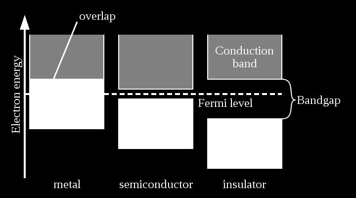
3 Sensors Metal, Semiconductor, Insulator

4 Sensors 1D Sensor Piezoelectric Measure mechanical motion Generate a voltage when deformed mechanical stress causes the charge separation in the individual atoms of the material Eg. Audio, autofocus objective Principle Strain Sensitivity [V/µ*] Threshold [µ*] Span to threshold ratio Piezoelectric ,000,000 Piezoresistive ,500,000 Inductive ,000,000 Capacitive ,000
 Sum signal of all detected photons Very")
5 Sensors 1D sensor Photomultiplier Detect photons and amplify the signal (by 100 million times) Sum signal of all detected photons Very sensitive


6 Sensors 2D-Sensor charge-coupled device (CCD) Low noise High power comsuption Need move charges Complementary metal oxide semiconductor (CMOS) Moderate noise Low power comsuption Region Of Interest Read directly from pixel storage
 Extremely low noise Rapid frame rates Wide dynamic range High quantum efficiency (QE) Solid state Electron Multiplying (EM) register to the end of the normal serial")
7 Sensors 2D-Sensor Electron multiplying charge-coupled device (EMCCD) Very low noise High and broad QE Single Photon Sensitive Good dynamic range possible Fast or slow readout Scientific complementary metal oxide semiconductor (scmos) Extremely low noise Rapid frame rates Wide dynamic range High quantum efficiency (QE) Solid state Electron Multiplying (EM) register to the end of the normal serial register It is a mix between CCD/CMOS

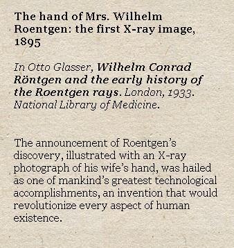
8 Radiology History

9 X-Ray Take a picture of inside body It emits a gamma ray The attenuation of the rays is the basis of working


10 X-Ray Principle Photons with energies kev are emitted by an X-Ray source Interaction with biological tissue absorbs & scatters some of the photons (photo-electric and Compton effect)» Attenuation through homogenous» medium Infinitesimally» small object Integration yields» Resulting in» or


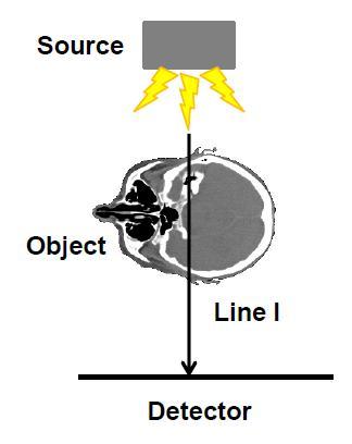
11 X-Ray Principle Attenuation through inhomogeneous medium Detector Intensity Attenuation Integral
12 X-Ray Principles Hounsfield Units Named after the inventor of CT, normalized reconstruction intensities where air=-1000 and water=0 considered during scanner calibration Typical X-Ray data has 12 bit precision [-1000, 3095] Blood Air Lung Fat Water Kidney Liver Bone Metal

")
13 Computed Tomography CT Principles X-Ray of different orientation -> Computed Tomography (CT) Geometric Principles:
14 CT Principles The Radon Transform in 2D Integral of a 2D function over straight lines With Dirac delta function over line equation: As a parameterized line:


15 CT Principles CT Example Original image and sinogram of the Shepp-Logan head phantom y Radon Transform θ? x t We have the sinogram how can we invert the Radon transform to yield the reconstructed image?
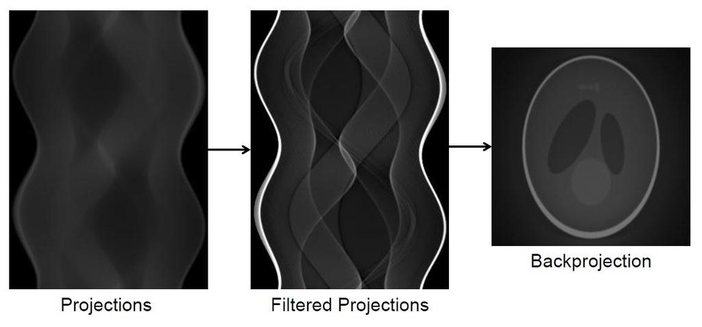
16 CT Principles
")
17 CT Principles Effect of number of projections (fan-beam case)

18 CT Principles Single-slice: ~1000 angles and detector bins, several revolutions per second Spiral CT geometry due to continuous table motion z-interpolation/filtering to use 2D FBP for spiral CT reconstruction! Multi-slice scanners: up to 256 detector lines Designated 3D reconstruction required!
 (Fourier Filter) Reconstruction")
19 CT Principles 3D-Reconstruction Workflow (X-Ray) (Fourier Filter) Reconstruction Method


20 Magnetic Resonance Imaging Principles Nuclear Magnetic Resonance (NMR) Quantum physics tells us that an atomic nucleus has a magnetic moment. Its precession in a magnetic field with a (or Larmor) frequency ω is defined as the product of the external magnetic field strength B and the so called gyromagnetic ratio : In nuclei with an uneven number of spins that remainder creates a magnetic moment. Examples for :


21 Magnetic Resonance Imaging Principles From the quantum mechanics point of view, only two states (parallel and anti parallel are possible for spins ; ) The transition between the states requires or gains the Bo energy. The macroscopic distribution is governed by the Boltzmann statistics. Under typical conditions, the net excess is in the ppm range (1.5 T equals 3000x the earth's magnetic field)


22 Magnetic Resonance Imaging Principles Bo In summary, external RF brings some spins into the anti-parallel state - which is left after a few moments under emission of a quant This quant is emitted in any direction but can be detected by an antenna! We normally capture only water signal

 90 180 Spin-")
23 Magnetic Resonance Imaging Principles Spin-Echo-Sequence (SE) Spin- Echo

. The relaxation time are usually named T1 and T2.")
24 Magnetic Resonance Imaging Principles Relaxation This relaxation process can be separated in two factors: longitudinal (or spin-lattice) and transversal (or spin-spin). The relaxation time are usually named T1 and T2. Spin-lattice Spin-spin

25 Magnetic Resonance Imaging Principles Relaxation The acquisition of the FID directly after the end of the HF pulse is technically very demanding as a high energy signal just ended and a very weak signal is occurring right after. The so called spin echo comes to help: (Echo Time) (Repetition time)

: Determine the")
26 Magnetic Resonance Imaging Principles Z Enconding (Gradient in Magnetic Field) RF Bandwidth (line slope): Determine the slice width
 As soon as a slice selective")

27 Magnetic Resonance Imaging Principles Y Enconding (Phase Gradient) As soon as a slice selective gradient was applied and the spin system is excited, a phase encoding gradient is applied in this preparation phase.

28 Magnetic Resonance Imaging Principles X Enconding (Frequency Gradient) Left Right After the preparation phase, the spin system is allowed to relax and the induced signal is measured. However, an x dependent gradient is applied during the read-out. Thus, the water molecules emit radiation with spatially varying frequency Water

29 Magnetic Resonance Imaging Principles How are all these individual steps combined?


30 Magnetic Resonance Imaging Image Reconstruction Using data only from one slice and coding this information in phase and frequency, we are able to sample to so called k-space. Its 2D Fourier transform yields the desired image. Essentially, the 2D FFT separates out the phase shift and the different frequencies


31 Magnetic Resonance Imaging Hardware 0.1 T Philips T Siemens T Siemens T Siemens 2005 Length: 2m Weight: 4 tons Length : > 3m Weight : 32 tons 15.2T, 11.7T, 9.4 T Bruker 2012 (Small Animal MRI) Wikipedia


32 Magnetic Resonance Imaging Why MRI? No ionizing radiation Hardly any side effects, few contra indications Works even in children and anesthetized patients May be applied in serial examinations Excellent image quality High biological contrast High resolution in arbitrary orientation Mature technology Relatively easy to set up Low maintenance Still advances happening and possible Good reimbursement

33 Ultrasound The energy used in US is sound (pressure) waves. Sound waves need material environment for propagation. Their motion in material depends on the materials property. They can be altered/reflected by the materials they are moving inside. We can use these alterations as an information source about the materials (tissues).

34 Ultrasound What is Ultrasound? Ultrasound is any sound emitted at a frequency > 20 KHz. Medical ultrasound uses frequencies in the range of 500 KHz to 30 MHz. For imaging 1MHz to 10 MHz. Intravascular imaging up to 30 MHz. (Higher the frequency better the resolution!!)
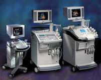
35 Ultrasound Examples of ultrasound probes (Piezoelectric elements)
 returned back from the tissue (by ultrasound recording element). 3. Process the RF signal and extract information. 4.")
36 Ultrasound Therefore, to generate an ultrasound image: 1. Generate ultrasound signal and send it into the tissue. 2. Record the echoes (RF signals) returned back from the tissue (by ultrasound recording element). 3. Process the RF signal and extract information. 4. Demonstrate the results (there are different methods).


37 Ultrasound Workflow 1. An ultrasound probe is used to generate a short burst of the ultrasound signal, which is sent into the tissue. 2. The same ultrasound probe is usually used to Record the echoes (RF signals) returned back from the tissue. Different tissues show different reflection properties.
38 Ultrasound The sound speed in soft tissue is about 1540 m/s As round-trip increases, reflector s distance increases For c = 1540 m/s = 1.54 mm/s: 3. Processing the RF signals Envelope detection by Hilbert Transform Dynamic range reduction by log function Decimation for data reduction (5100 samples into 300 samples)
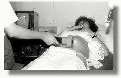

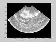
39 Ultrasound Examples:


40 Nuclear Medicine Activity distribution equivalent to distribution of the injected substance. Nuclear Medicine
41 Nuclear Medicine Radioisotop What do we want: not too short and not too long half life time only γ radiation (α & β would increase patient dose without gain for diagnosis)
 to analyze the Glucose metabolism")
42 Nuclear Medicine Radiopharmaceuticals The radioisotope has to be connected (labelling) to a pharmaceutical based on the organ specific question. Radiopharmaceuticals should not disturb the process under investigation. e.g. FDG (18F-Fluorodesoxyglucose) to analyze the Glucose metabolism

43 Nuclear Medicine Tracer principle The tracer (radiopharmaceutical) takes part in the metabolism of investigation The tracer may not disturb the process under investigation Possible due to the high sensitivity of the devices, e.g. 18F-FDG typical dose: 370 MBq

44 Nuclear Medicine Radiation-Detection Scintillators, e.g. NaI(Tl) Thallium doped Sodium Iodide

45 Nuclear Medicine Gamma camera Collimators are chosen depending on sensitivity and resolution necessary for an particular examination.

46 Nuclear Medicine Photo multiplier multiply the signal produced by incident light by as much as 100 million times


47 Nuclear Medicine Single Photon Emission Tomography SPECT Gamma camera rotating around the patient
 of coronar")
48 Nuclear Medicine Single Photon Emission Tomography - SPECT Myocard SPECT 99mTc MIBI (methoxyisobutylisonitrile) myocardial perfusion in cardial rest and stress for diagnosis and staging (prognostic factor) of coronar disease
49 Nuclear Medicine Positron Emission Tomography PET Positron decay:

50 Nuclear Medicine Positron Emission Tomography PET Coincidence events are registered by a detector ring around the patient: No collimator necessary - electronic collimation! Use of all coincidence lines ( projections)

51 Nuclear Medicine Positron Emission Tomography - PET Absolute activity concentration

52 Nuclear Medicine Positron Emission Tomography - PET Attenuation


53 Nuclear Medicine Multimodality PET/CT

54 Nuclear Medicine Multimodality PET/MR
 standard for handling, storing, printing, and transmitting information in medical imaging File")
55 3D imaging format? Digital Imaging and Communications in Medicine (DICOM) standard for handling, storing, printing, and transmitting information in medical imaging File format Network communications protocol Owned by National Electrical Manufacturers Association (NEMA)

56 Summary Tomographic Imaging

57 Summary Examples of Tomographic Imaging

58 Summary Tomographic Imaging
59 Summary CT Scan MRI Suited for bone injuries, Lung and Chest imaging, cancer detection Provides good details about bony structures Usually completed within 5 minutes Risk of irradiation (Moderate to high radiation) Suited for ligament and tendon injury, spinal cord injury, brain tumors Scanning typically run for about 30 minutes. High detail in the soft tissues No biological hazards have been reported Patients with Cardiac Pacemakers are not allowed
60 Reference Computer Assisted Medical Procedures Lecture, Dr. Prof. Navab, Technical University of Munich. PhD. Thesis, Dr. Lange, University of Siegen
Technical University of Denmark
 Technical University of Denmark Page 1 of 11 pages Written test, 9 December 2010 Course name: Introduction to medical imaging Course no. 31540 Aids allowed: none. "Weighting": All problems weight equally.
DEVIL PHYSICS THE BADDEST CLASS ON CAMPUS IB PHYSICS
 DEVIL PHYSICS THE BADDEST CLASS ON CAMPUS IB PHYSICS TSOKOS OPTION I-2 MEDICAL IMAGING Reading Activity Answers IB Assessment Statements Option I-2, Medical Imaging: X-Rays I.2.1. I.2.2. I.2.3. Define
Introduction to Medical Imaging. Medical Imaging
 Introduction to Medical Imaging BME/EECS 516 Douglas C. Noll Medical Imaging Non-invasive visualization of internal organs, tissue, etc. I typically don t include endoscopy as an imaging modality Image
ELG7173 Topics in signal Processing II Computational Techniques in Medical Imaging
 ELG7173 Topics in signal Processing II Computational Techniques in Medical Imaging Topic #1: Intro to medical imaging Medical Imaging Classifications n Measurement physics Send Energy into body Send stuff
Introduction to Biomedical Imaging
 Alejandro Frangi, PhD Computational Imaging Lab Department of Information & Communication Technology Pompeu Fabra University www.cilab.upf.edu MRI advantages Superior soft-tissue contrast Depends on among
Radioisotopes in action. Diagnostic application of radioisotopes. Steps of diagnostic procedure. Information from various medical imaging techniques
 Radioisotopes in action Diagnostic application of radioisotopes Steps of diagnostic procedure - Radioactive material introduced into the patient - Distribution and alteration of activity is detected -
Doppler echocardiography & Magnetic Resonance Imaging. Doppler echocardiography. History: - Langevin developed sonar.
 1 Doppler echocardiography & Magnetic Resonance Imaging History: - Langevin developed sonar. - 1940s development of pulse-echo. - 1950s development of mode A and B. - 1957 development of continuous wave
A Brief Introduction to Medical Imaging. Outline
 A Brief Introduction to Medical Imaging Outline General Goals Linear Imaging Systems An Example, The Pin Hole Camera Radiations and Their Interactions with Matter Coherent vs. Incoherent Imaging Length
Structure of Biological Materials
 ELEC ENG 3BA3: Structure of Biological Materials Notes for Lecture #19 Monday, November 22, 2010 6.5 Nuclear medicine imaging Nuclear imaging produces images of the distribution of radiopharmaceuticals
Technical University of Denmark
 Technical University of Denmark Page 1 of 10 pages Written test, 12 December 2012 Course name: Introduction to medical imaging Course no. 31540 Aids allowed: None. Pocket calculator not allowed "Weighting":
EL-GY 6813/BE-GY 6203 Medical Imaging, Fall 2016 Final Exam
 EL-GY 6813/BE-GY 6203 Medical Imaging, Fall 2016 Final Exam (closed book, 1 sheets of notes double sided allowed, no calculator or other electronic devices allowed) 1. Ultrasound Physics (15 pt) A) (9
22.56J Noninvasive Imaging in Biology and Medicine Instructor: Prof. Alan Jasanoff Fall 2005, TTh 1-2:30
 22.56J Noninvasive Imaging in Biology and Medicine Instructor: Prof. Alan Jasanoff Fall 2005, TTh 1-2:30 Sample problems HW1 1. Look up (e.g. in the CRC Manual of Chemistry and Physics www.hbcpnetbase.com)
Nuclear Medicine RADIOPHARMACEUTICAL CHEMISTRY
 Nuclear Medicine RADIOPHARMACEUTICAL CHEMISTRY An alpha particle consists of two protons and two neutrons Common alpha-particle emitters Radon-222 gas in the environment Uranium-234 and -238) in the environment
Magnetic Resonance Imaging (MRI)
") Magnetic Resonance Imaging Introduction The Components The Technology (MRI) Physics behind MR Most slides taken from http:// www.slideworld.org/ viewslides.aspx/magnetic- Resonance-Imaging- %28MRI%29-MR-Imaging-
Magnetic Resonance Imaging Introduction The Components The Technology (MRI) Physics behind MR Most slides taken from http:// www.slideworld.org/ viewslides.aspx/magnetic- Resonance-Imaging- %28MRI%29-MR-Imaging-
Magnetic Resonance Imaging. Pål Erik Goa Associate Professor in Medical Imaging Dept. of Physics
 Magnetic Resonance Imaging Pål Erik Goa Associate Professor in Medical Imaging Dept. of Physics pal.e.goa@ntnu.no 1 Why MRI? X-ray/CT: Great for bone structures and high spatial resolution Not so great
Radioisotopes and PET
 Radioisotopes and PET 1 Radioisotopes Elements are defined by their number of protons, but there is some variation in the number of neutrons. Atoms resulting from this variation are called isotopes. Consider
MEDICAL EQUIPMENT: NUCLEAR MEDICINE. Prof. Yasser Mostafa Kadah
 MEDICAL EQUIPMENT: NUCLEAR MEDICINE Prof. Yasser Mostafa Kadah www.k-space.org Recommended Textbook Introduction to Medical Imaging: Physics, Engineering and Clinical Applications, by Nadine Barrie Smith
This Week. 3/23/2017 Physics 214 Summer
 This Week Atoms and nuclei What are we made of? The periodic table Why does it stop? How were the elements made? Radioactive decay Useful but can be toxic Discovery of X Rays: Cathode Rays and TV sets
Magnetic resonance imaging MRI
 Magnetic resonance imaging MRI Introduction What is MRI MRI is an imaging technique used primarily in medical settings that uses a strong magnetic field and radio waves to produce very clear and detailed
BNG/ECE 487 FINAL (W16)
") BNG/ECE 487 FINAL (W16) NAME: 4 Problems for 100 pts This exam is closed-everything (no notes, books, etc.). Calculators are permitted. Possibly useful formulas and tables are provided on this page. Fourier
BNG/ECE 487 FINAL (W16) NAME: 4 Problems for 100 pts This exam is closed-everything (no notes, books, etc.). Calculators are permitted. Possibly useful formulas and tables are provided on this page. Fourier
Tomography is imaging by sections. 1
 Tomography is imaging by sections. 1 It is a technique used in clinical medicine and biomedical research to create images that show how certain tissues are performing their physiological functions. 1 Conversely,
Radioisotopes in action. Diagnostic application of radioisotopes. Steps of diagnostic procedure. Information from various medical imaging techniques
 Radioisotopes in action Diagnostic application of radioisotopes Steps of diagnostic procedure - Radioactive material introduced into the patient - Distribution and alteration of activity is detected -Monitoring
Electrical Engineering 3BA3: Structure of Biological Materials
 Electrical Engineering 3BA3: Structure of Biological Materials Day Class Instructor: Dr. I. C. BRUCE Duration of Examination: 3 Hours McMaster University Final Examination December, 2004 This examination
Part III Minor Option in Medical Physics 2018 Examples Sheet
 Part III Minor Option in Medical Physics 2018 Examples Sheet Any errors or comments should be addressed sent to: seb53@cam.ac.uk URLs that may be useful: Stanford Event Generation Simulator: http://tinyurl.com/pkg476r
(INCLUDING THIS FRONT PAGE)
") I'IFIITIIBIFI UNIVERSITY OF SCIEI'ICE RITD TECHNOLOGY FACULTY OF HEALTH AND APPLIED SCIENCES DEPARTMENT OF NATURAL AND APPLIED SCIENCES QUALIFICATION: BACHELOR OF SCIENCE (MAJOR AND MINOR) QUALIFICATION
I'IFIITIIBIFI UNIVERSITY OF SCIEI'ICE RITD TECHNOLOGY FACULTY OF HEALTH AND APPLIED SCIENCES DEPARTMENT OF NATURAL AND APPLIED SCIENCES QUALIFICATION: BACHELOR OF SCIENCE (MAJOR AND MINOR) QUALIFICATION
MEDICAL IMAGING. METHODS OF MODERN IMAGING, BASED ON ELECTRO-MAGNETIC RADIATION (radiowaves, infrared radiation, X-rays, γ-rays ) AND ULTRASOUND
 AND ULTRASOUND") MEDICAL IMAGING MEDICAL IMAGING METHODS OF MODERN IMAGING, BASED ON ELECTRO-MAGNETIC RADIATION (radiowaves, infrared radiation, X-rays, γ-rays ) AND ULTRASOUND MEDICAL IMAGING RADIOLOGY NUCLEAR MEDICINE
MEDICAL IMAGING MEDICAL IMAGING METHODS OF MODERN IMAGING, BASED ON ELECTRO-MAGNETIC RADIATION (radiowaves, infrared radiation, X-rays, γ-rays ) AND ULTRASOUND MEDICAL IMAGING RADIOLOGY NUCLEAR MEDICINE
What is scintigraphy? The process of obtaining an image or series of sequential images of the distribution of a radionuclide in tissues, organs, or
 Let's remind... What is nuclear medicine? Nuclear medicine can be broadly divided into two branches "in vitro" and "in vivo" procedures. There are numerous radioisotopic "in vitro" procedures for genotyping
Nuclear Physics and Astrophysics
 Nuclear Physics and Astrophysics PHY-302 Dr. E. Rizvi Lecture 24 Medical Imaging Effects of Radiation We now know what radiation is But what does it mean for our bodies? Radioactivity is quantified in
Introduction to SPECT & PET TBMI02 - Medical Image Analysis 2017
 Introduction to SPECT & PET TBMI02 - Medical Image Analysis 2017 Marcus Ressner, PhD, Medical Radiation Physicist, Linköping University Hospital Content What is Nuclear medicine? Basic principles of Functional
Radionuclide Imaging MII Positron Emission Tomography (PET)
") Radionuclide Imaging MII 3073 Positron Emission Tomography (PET) Positron (β + ) emission Positron is an electron with positive charge. Positron-emitting radionuclides are most commonly produced in cyclotron
Radionuclide Imaging MII 3073 Positron Emission Tomography (PET) Positron (β + ) emission Positron is an electron with positive charge. Positron-emitting radionuclides are most commonly produced in cyclotron
 www.aask24.com www.aask24.com www.aask24.com P=Positron E= Emission T=Tomography Positron emission or beta plus decay (+ ) is a particular type of radioactive decay, in which a proton inside a radionuclide
Nuclear Medicine Intro & Physics from Medical Imaging Signals and Systems, Chapter 7, by Prince and Links
 Nuclear Medicine Intro & Physics from Medical Imaging Signals and Systems, Chapter 7, by Prince and Links NM - introduction Relies on EMISSION of photons from body (versus transmission of photons through
ENG4BF3 Medical Image Processing
 ENG4BF3 Medical Image Processing Medical Imaging Modalities Imaging in Medical Sciences Imaging is an essential aspect of medical sciences for visualization of anatomical structures and functional or metabolic
Radiation Detectors. How do we detect ionizing radiation? What are these effects? Types of Ionizing Radiation Detectors
 Radiation Detectors 1 How do we detect ionizing radiation? Indirectly, by its effects as it traverses matter? What are these effects? Ionization and excitation of the atoms and molecules Heat 2 Types of
AQA Physics /7408
 AQA Physics - 7407/7408 Module 10: Medical physics You should be able to demonstrate and show your understanding of: 10.1 Physics of the eye 10.1.1 Physics of vision The eye as an optical refracting system,
This Week. 7/20/2016 Physics 214 Spring
 This Week Atoms and nuclei What are we made of? The periodic table Why does it stop? How were the elements made? Radioactive decay Useful but can be toxic Discovery of X Rays: Cathode Rays and TV sets
Nuclear Magnetic Resonance Imaging
 Nuclear Magnetic Resonance Imaging Jeffrey A. Fessler EECS Department The University of Michigan NSS-MIC: Fundamentals of Medical Imaging Oct. 20, 2003 NMR-0 Background Basic physics 4 magnetic fields
6: Positron Emission Tomography
 6: Positron Emission Tomography. What is the principle of PET imaging? Positron annihilation Electronic collimation coincidence detection. What is really measured by the PET camera? True, scatter and random
Biomedical Imaging Magnetic Resonance Imaging
 Biomedical Imaging Magnetic Resonance Imaging Charles A. DiMarzio & Eric Kercher EECE 4649 Northeastern University May 2018 Background and History Measurement of Nuclear Spins Widely used in physics/chemistry
CT-PET calibration : physical principles and operating procedures F.Bonutti. Faustino Bonutti Ph.D. Medical Physics, Udine University Hospital.
 CT-PET calibration : physical principles and operating procedures Faustino Bonutti Ph.D. Medical Physics, Udine University Hospital Topics Introduction to PET physics F-18 production β + decay and annichilation
The NMR Inverse Imaging Problem
 The NMR Inverse Imaging Problem Nuclear Magnetic Resonance Protons and Neutrons have intrinsic angular momentum Atoms with an odd number of proton and/or odd number of neutrons have a net magnetic moment=>
Index. p, lip, 78 8 function, 107 v, 7-8 w, 7-8 i,7-8 sine, 43 Bo,94-96
 p, lip, 78 8 function, 107 v, 7-8 w, 7-8 i,7-8 sine, 43 Bo,94-96 B 1,94-96 M,94-96 B oro!' 94-96 BIro!' 94-96 I/r, 79 2D linear system, 56 2D FFT, 119 2D Fourier transform, 1, 12, 18,91 2D sinc, 107, 112
Physics in Nuclear Medicine
 SIMON R. CHERRY, PH.D. Professor Department of Biomedical Engineering University of California-Davis Davis, California JAMES A. SORENSON, PH.D. Emeritus Professor of Medical Physics University of Wisconsin-Madison
Lecture 5: Tomographic nuclear systems: SPECT
 Lecture 5: Tomographic nuclear systems: SPECT Field trip this saturday at 11 AM at UWMC meet in main hospital lobby at 11 AM if you miss the 'boat', page me at 540-4950 should take ~1 to 1.5 hours, depending
NMR and MRI : an introduction
 Intensive Programme 2011 Design, Synthesis and Validation of Imaging Probes NMR and MRI : an introduction Walter Dastrù Università di Torino walter.dastru@unito.it \ Introduction Magnetic Resonance Imaging
Nuclear Magnetic Resonance Imaging
 Nuclear Magnetic Resonance Imaging Simon Lacoste-Julien Electromagnetic Theory Project 198-562B Department of Physics McGill University April 21 2003 Abstract This paper gives an elementary introduction
Bases of radioisotope diagnostic methods
 Medical, pharmaceutical applications of radioisotopes Bases of radioisotope diagnostic methods Dr. István Voszka Basis of application: radioisotopes have identical behavior in the organism to corresponding
Year 12 Notes Radioactivity 1/5
 Year Notes Radioactivity /5 Radioactivity Stable and Unstable Nuclei Radioactivity is the spontaneous disintegration of certain nuclei, a random process in which particles and/or high-energy photons are
β and γ decays, Radiation Therapies and Diagnostic, Fusion and Fission Final Exam Surveys New material Example of β-decay Beta decay Y + e # Y'+e +
 β and γ decays, Radiation Therapies and Diagnostic, Fusion and Fission Last Lecture: Radioactivity, Nuclear decay Radiation damage This lecture: nuclear physics in medicine and fusion and fission Final
The Basics of Magnetic Resonance Imaging
 The Basics of Magnetic Resonance Imaging Nathalie JUST, PhD nathalie.just@epfl.ch CIBM-AIT, EPFL Course 2013-2014-Chemistry 1 Course 2013-2014-Chemistry 2 MRI: Many different contrasts Proton density T1
3. Which of the following statements is (are) TRUE about detector crystals in Anger cameras?
 TRUE about detector crystals in Anger cameras?") BioE 1330 - Exam 2 11/13/2018 Answer Sheet - Correct answer is A for all questions 1. Unlike CT, in nuclear medicine A. Bremsstrahlung is not used to produce high-energy photons. B. signal can be increased
BioE 1330 - Exam 2 11/13/2018 Answer Sheet - Correct answer is A for all questions 1. Unlike CT, in nuclear medicine A. Bremsstrahlung is not used to produce high-energy photons. B. signal can be increased
A. I, II, and III B. I C. I and II D. II and III E. I and III
 BioE 1330 - Review Chapters 7, 8, and 9 (Nuclear Medicine) 9/27/2018 Instructions: On the Answer Sheet, enter your 2-digit ID number (with a leading 0 if needed) in the boxes of the ID section. Fill in
Outline Chapter 14 Nuclear Medicine
 Outline Chapter 14 uclear Medicine Radiation Dosimetry I Text: H.E Johns and J.R. Cunningham, The physics of radiology, 4 th ed. http://www.utoledo.edu/med/depts/radther Introduction Detectors for nuclear
The physics of medical imaging US, CT, MRI. Prof. Peter Bogner
 The physics of medical imaging US, CT, MRI Prof. Peter Bogner Clinical radiology curriculum blocks of lectures and clinical practice (7x2) Physics of medical imaging Neuroradiology Head and neck I. Head
Compton Camera with PositionSensitive Silicon Detectors
 University of Ljubljana Faculty of mathematics and physics Andrej Studen Compton Camera with PositionSensitive Silicon Detectors Doctoral thesis Supervisor: Professor Marko Mikuž Outline: Motivation Basic
Dana-Farber Cancer Institute, 44 Binney Street, Boston, MA 02115, USA ramsey
 SPECIAL FEATURE: MEDICAL PHYSICS www.iop.org/journals/physed Nuclear medicine Ramsey D Badawi Dana-Farber Cancer Institute, 44 Binney Street, Boston, MA 02115, USA E-mail: ramsey badawi@dfci.harvard.edu
PET/MRI Principle, History, and Perspective. Main Imaging Techniques. X-ray Tube. History of X-ray & CT. How to Look inside the Human Body
 PET/MRI Principle, History, and Perspective Jae Sung Lee, PhD Dept. of Nuclear Medicine and Biomedical Sciences WCU Dept. of Brain and Cognitive Sciences Seoul National University Basic Imaging Principles
Nuclear Reactions A Z. Radioactivity, Spontaneous Decay: Nuclear Reaction, Induced Process: x + X Y + y + Q Q > 0. Exothermic Endothermic
 Radioactivity, Spontaneous Decay: Nuclear Reactions A Z 4 P D+ He + Q A 4 Z 2 Q > 0 Nuclear Reaction, Induced Process: x + X Y + y + Q Q = ( m + m m m ) c 2 x X Y y Q > 0 Q < 0 Exothermic Endothermic 2
Fundamental MRI Principles Module Two
 Fundamental MRI Principles Module Two 1 Nuclear Magnetic Resonance There are three main subatomic particles: protons neutrons electrons positively charged no significant charge negatively charged Protons
2015 U N I V E R S I T I T E K N O L O G I P E T R O N A S
 Multi-Modality based Diagnosis: A way forward by Hafeez Ullah Amin Centre for Intelligent Signal and Imaging Research (CISIR) Department of Electrical & Electronic Engineering 2015 U N I V E R S I T I
Fundamental MRI Principles Module 2 N. Nuclear Magnetic Resonance. X-ray. MRI Hydrogen Protons. Page 1. Electrons
 Fundamental MRI Principles Module 2 N S 1 Nuclear Magnetic Resonance There are three main subatomic particles: protons positively charged neutrons no significant charge electrons negatively charged Protons
NMR Imaging in porous media
 NMR Imaging in porous media What does NMR give us. Chemical structure. Molecular structure. Interactions between atoms and molecules. Incoherent dynamics (fluctuation, rotation, diffusion). Coherent flow
Modern physics ideas are strange! L 36 Modern Physics [2] The Photon Concept. How are x-rays produced? The uncertainty principle
![Modern physics ideas are strange! L 36 Modern Physics [2] The Photon Concept. How are x-rays produced? The uncertainty principle](/thumbs/88/117098787.jpg "Modern physics ideas are strange! L 36 Modern Physics [2] The Photon Concept. How are x-rays produced? The uncertainty principle") L 36 Modern Physics [2] X-rays & gamma rays How lasers work Medical applications of lasers Applications of high power lasers Medical imaging techniques CAT scans MRI s Modern physics ideas are strange!
L 36 Modern Physics [2] X-rays & gamma rays How lasers work Medical applications of lasers Applications of high power lasers Medical imaging techniques CAT scans MRI s Modern physics ideas are strange!
Part II: Magnetic Resonance Imaging (MRI)
") Part II: Magnetic Resonance Imaging (MRI) Contents Magnetic Field Gradients Selective Excitation Spatially Resolved Reception k-space Gradient Echo Sequence Spin Echo Sequence Magnetic Resonance Imaging
Part II: Magnetic Resonance Imaging (MRI) Contents Magnetic Field Gradients Selective Excitation Spatially Resolved Reception k-space Gradient Echo Sequence Spin Echo Sequence Magnetic Resonance Imaging
12/1/17 OUTLINE KEY POINTS ELEMENTS WITH UNSTABLE NUCLEI Radioisotopes and Nuclear Reactions 16.2 Biological Effects of Nuclear Radiation
 OUTLINE 16.1 Radioisotopes and Nuclear Reactions 16.2 Biological Effects of Nuclear Radiation PET scan X-ray technology CT scan 2009 W.H. Freeman KEY POINTS Radioactivity is the consequence of an unstable
The physics US and MRI. Prof. Peter Bogner
 The physics US and MRI Prof. Peter Bogner Sound waves mechanical disturbance, a pressure wave moves along longitudinal wave compression rarefaction zones c = nl, (c: velocity, n: frequency, l: wavelength
1st Faculty of Medicine, Charles University in Prague Center for Advanced Preclinical Imaging (CAPI)
") Radioation Resolution and Sensitivity Nuclear Imaging PET + SPECT Radioactive Decay (EC,Ɣ), (β -,Ɣ), (I.T.,Ɣ) β + Projection imaging collimator needed one angular view Projection imaging coincidence imaging,
Radioation Resolution and Sensitivity Nuclear Imaging PET + SPECT Radioactive Decay (EC,Ɣ), (β -,Ɣ), (I.T.,Ɣ) β + Projection imaging collimator needed one angular view Projection imaging coincidence imaging,
CHIPP Plenary Meeting University of Geneva, June 12, 2008 W. Lustermann on behalf of the AX PET Collaboration
 CHIPP Plenary Meeting University of Geneva, June 12, 2008 W. Lustermann on behalf of the AX PET Collaboration INFN Bari, Ohio State University, CERN, University of Michigan, University of Oslo, INFN Roma,
PET scan simulation. Meysam Dadgar. UMSU, Iran. IFMP, Elbasan, Fig 1: PET camera simulation in gate by cylindrical phantom
 PET scan simulation Meysam Dadgar UMSU, Iran IFMP, Elbasan, 2016 Meysamdadgar10@gmail.com 1 Fig 1: PET camera simulation in gate by cylindrical phantom 2 What is PET? Positron emission tomography (PET),
Chapter 16 Nuclear Chemistry. An Introduction to Chemistry by Mark Bishop
 Chapter 16 Nuclear Chemistry An Introduction to Chemistry by Mark Bishop Chapter Map Nuclides Nuclide = a particular type of nucleus, characterized by a specific atomic number and nucleon number Nucleon
Principles of Magnetic Resonance Imaging
 Principles of Magnetic Resonance Imaging Hi Klaus Scheffler, PhD Radiological Physics University of 1 Biomedical Magnetic Resonance: 1 Introduction Magnetic Resonance Imaging Contents: Hi 1 Introduction
Compton Camera. Compton Camera
 Diagnostic Imaging II Student Project Compton Camera Ting-Tung Chang Introduction The Compton camera operates by exploiting the Compton Effect. It uses the kinematics of Compton scattering to contract
Differentiating Chemical Reactions from Nuclear Reactions
 Differentiating Chemical Reactions from Nuclear Reactions 1 CHEMICAL Occurs when bonds are broken or formed. Atoms remained unchanged, though may be rearranged. Involves valence electrons Small energy
Tomography and Reconstruction
 Tomography and Reconstruction Lecture Overview Applications Background/history of tomography Radon Transform Fourier Slice Theorem Filtered Back Projection Algebraic techniques Measurement of Projection
Principles of MRI EE225E / BIO265. Instructor: Miki Lustig UC Berkeley, EECS
 Principles of MRI EE225E / BIO265 Instructor: Miki Lustig UC Berkeley, EECS Today... Administration http://inst.eecs.berkeley.edu/~ee225e/sp16/ Intro to Medical Imaging and MRI Medical Imaging (Before
Medical Imaging Physics Spring Quarter Week 9-1
 Medical Imaging Physics Spring Quarter Week 9-1 NMR and MRI Davor Balzar balzar@du.edu www.du.edu/~balzar Intro MRI Outline NMR & MRI Guest lecturer fmri Thursday, May 22 Visit to CUHSC It s not mandatory
Magnetic Resonance Imaging
 http://www.qldxray.com.au/filelibrary/mri_cardiovascular_system_ca_0005.jpg Magnetic Resonance Imaging 1 Overview 1. The magnetic properties of nuclei, and how they behave in strong magnetic fields. 2.
Nuclear Radiation. Natural Radioactivity. A person working with radioisotopes wears protective clothing and gloves and stands behind a shield.
 Nuclear Radiation Natural Radioactivity A person working with radioisotopes wears protective clothing and gloves and stands behind a shield. 1 Radioactive Isotopes A radioactive isotope has an unstable
III. Proton-therapytherapy. Rome SB - 2/5 1
 Outline Introduction: an historical review I Applications in medical diagnostics Particle accelerators for medicine Applications in conventional radiation therapy II III IV Hadrontherapy, the frontier
Introduction to MRI. Spin & Magnetic Moments. Relaxation (T1, T2) Spin Echoes. 2DFT Imaging. K-space & Spatial Resolution.
 Spin Echoes. 2DFT Imaging. K-space & Spatial Resolution.") Introduction to MRI Spin & Magnetic Moments Relaxation (T1, T2) Spin Echoes 2DFT Imaging Selective excitation, phase & frequency encoding K-space & Spatial Resolution Contrast (T1, T2) Acknowledgement:
Introduction to MRI Spin & Magnetic Moments Relaxation (T1, T2) Spin Echoes 2DFT Imaging Selective excitation, phase & frequency encoding K-space & Spatial Resolution Contrast (T1, T2) Acknowledgement:
Radiochemistry and Radiopharmacy III
 Radiochemistry and Radiopharmacy III Compact course held at UFSCAR, September 20123 Ulrich Abram Freie Universität Berlin Institute of Chemistry and Biochemistry Radiochemistry and Radiopharmacy 1. Fundamentals
There are three mechanisms by which gamma rays interact with absorber atoms from which two are important for nuclear medicine.
 Measurement of radioactivity. Radioactive decay is a random process and therefore fluctuations are expected in the radioactivity measurement. That is why measurement of radioactivity must be treated by
Study of the feasibility of a compact gamma camera for real-time cancer assessment
 Study of the feasibility of a compact gamma camera for real-time cancer assessment L. Caballero Instituto de Física Corpuscular - CSIC - University of Valencia; C/Catedrático José Beltrán, 2; E-46980;
Elec Eng 3BA3: Structure of Biological Materials
 Elec Eng 3BA3: Structure of Biological Materials Page 1 of 12 Day Class Instructor: Dr. I. C. BRUCE Duration of Examination: 3 Hours McMaster University Final Examination December 5, 2008 This examination
Introduction to the Course and the Techniques. Jeffry R. Alger, PhD Ahmanson-Lovelace Brain Mapping Center Department of Neurology
 Introduction to the Course and the Techniques Jeffry R. Alger, PhD Ahmanson-Lovelace Brain Mapping Center Department of Neurology (jralger@ucla.edu) CTSI Neuroimaging April 2013 Rationale for the Course
RADIOLOGIV TECHNOLOGY 4912 COMPREHENSEIVE REVIEW/MRI WORSHEET #1- PATIENT CARE AND SAFETY/PHYSICAL PRINCIPLES
 RADIOLOGIV TECHNOLOGY 4912 COMPREHENSEIVE REVIEW/MRI WORSHEET #1- PATIENT CARE AND SAFETY/PHYSICAL PRINCIPLES 1. What are potential consequences to patients and personnel should there be a release of gaseous
Tissue Characteristics Module Three
 Tissue Characteristics Module Three 1 Equilibrium State Equilibrium State At equilibrium, the hydrogen vector is oriented in a direction parallel to the main magnetic field. Hydrogen atoms within the vector
1-D Fourier Transform Pairs
 1-D Fourier Transform Pairs The concept of the PSF is most easily explained by considering a very small point source being placed in the imaging field-of-view The relationship between the image, I, and
Chapter 2 PET Imaging Basics
 Chapter 2 PET Imaging Basics Timothy G. Turkington PET Radiotracers Positron emission tomography (PET) imaging is the injection (or inhalation) of a substance containing a positron emitter, the subsequent
University of Ljubljana Faculty of mathematics and physics Department of physics. Tomography. Mitja Eržen. August 6, Menthor: Dr.
 University of Ljubljana Faculty of mathematics and physics Department of physics Tomography Mitja Eržen August 6, 2009 Menthor: Dr. Matjaž Vencelj Abstract We ll describe some methods for medical imaging.i
Mayneord-Phillips Summer School St Edmund Hall, University of Oxford July Proton decays to n, e +, ν
 Positron Emission Tomography Physics & Instrumentation Dimitra G. Darambara, Ph.D Multimodality Molecular Imaging Joint Department of Physics RMH/ICR Outline Introduction PET Physics overview Types of
General Physics (PHY 2140)
") General Physics (PHY 2140) Lecture 19 Modern Physics Nuclear Physics Nuclear Reactions Medical Applications Radiation Detectors Chapter 29 http://www.physics.wayne.edu/~alan/2140website/main.htm 1 Lightning
General Physics (PHY 2140) Lecture 19 Modern Physics Nuclear Physics Nuclear Reactions Medical Applications Radiation Detectors Chapter 29 http://www.physics.wayne.edu/~alan/2140website/main.htm 1 Lightning
General Physics (PHY 2140)
") General Physics (PHY 2140) Lightning Review Lecture 19 Modern Physics Nuclear Physics Nuclear Reactions Medical Applications Radiation Detectors Chapter 29 http://www.physics.wayne.edu/~alan/2140website/main.htm
General Physics (PHY 2140) Lightning Review Lecture 19 Modern Physics Nuclear Physics Nuclear Reactions Medical Applications Radiation Detectors Chapter 29 http://www.physics.wayne.edu/~alan/2140website/main.htm
BME I5000: Biomedical Imaging
 BME I5000: Biomedical Imaging Lecture 9 Magnetic Resonance Imaging (imaging) Lucas C. Parra, parra@ccny.cuny.edu Blackboard: http://cityonline.ccny.cuny.edu/ 1 Schedule 1. Introduction, Spatial Resolution,
Radiation Detection and Measurement
 Radiation Detection and Measurement June 2008 Tom Lewellen Tkldog@u.washington.edu Types of radiation relevant to Nuclear Medicine Particle Symbol Mass (MeV/c 2 ) Charge Electron e-,! - 0.511-1 Positron
MRI Physics I: Spins, Excitation, Relaxation
 MRI Physics I: Spins, Excitation, Relaxation Douglas C. Noll Biomedical Engineering University of Michigan Michigan Functional MRI Laboratory Outline Introduction to Nuclear Magnetic Resonance Imaging
Field trip: Tuesday, Feb 5th
 Pulse Sequences Field trip: Tuesday, Feb 5th Hardware tour of VUIIIS Philips 3T Meet here at regular class time (11.15) Complete MRI screening form! Chuck Nockowski Philips Service Engineer Reminder: Project/Presentation
Positron Annihilation in Material Research
 Positron Annihilation in Material Research Introduction Positron sources, positron beams Interaction of positrons with matter Annihilation channels: Emission of 1, 2 or 3 γ-quanta Annihilation spectroscopies:
Radionuclide Imaging MII Detection of Nuclear Emission
 Radionuclide Imaging MII 3073 Detection of Nuclear Emission Nuclear radiation detectors Detectors that are commonly used in nuclear medicine: 1. Gas-filled detectors 2. Scintillation detectors 3. Semiconductor
Medical Physics. Nuclear Medicine Principles and Applications
 Medical Physics Nuclear Medicine Principles and Applications Dr Roger Fulton Department of PET & Nuclear Medicine Royal Prince Alfred Hospital Sydney Email: rfulton@mail.usyd.edu.au Lectures: http://www-personal.usyd.edu.au/~rfulton/medical_physics