The physics of medical imaging US, CT, MRI. Prof. Peter Bogner
|
|
|
- Justin Ellis
- 5 years ago
- Views:
Transcription
1 The physics of medical imaging US, CT, MRI Prof. Peter Bogner
2 Clinical radiology curriculum blocks of lectures and clinical practice (7x2) Physics of medical imaging Neuroradiology Head and neck I. Head and neck II. Thoracic radiology Gastrointestinal radiology Urogenital and interventional radiology Final exam: test + oral

3 Medical imaging Energy matter is required or not Imaging needs energy-matter interaction Part of the energy is deposited

4 Sound waves mechanical disturbance, a pressure wave moves along longitudinal wave compression rarefaction zones c = nl, (c: velocity, n: frequency, l: wavelength diagnostic range: MHz

5 Ultrasound velocity Relaitvely low in gases higher in solids Velocity changes with the wavelength, frequency remains approx. constant c = E/ (E ~ elasticity, ~ density) Variation in velocity artifacts
6 60-90% US interactions in tissues
7
 acoustic impedance ~ density + elasticity (similarly to velocity) impedance (Z) = ( )(v) (rayl = kgm 2 sec -1 )")
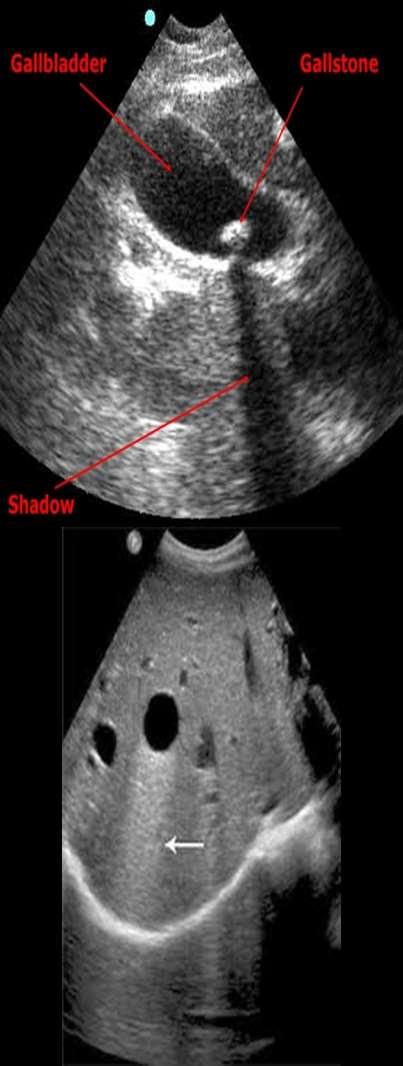
8 Reflection the basis of US imaging it occurs on the interface of two media, that have different acoustic impedances (Z) acoustic impedance ~ density + elasticity (similarly to velocity) impedance (Z) = ( )(v) (rayl = kgm 2 sec -1 )

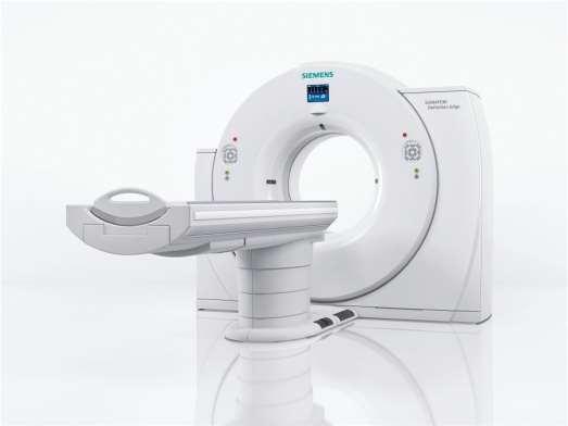
9
10

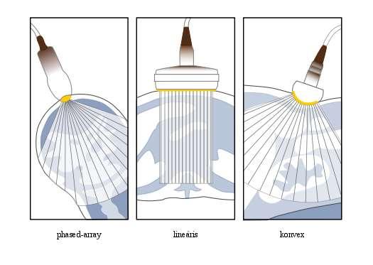
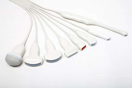
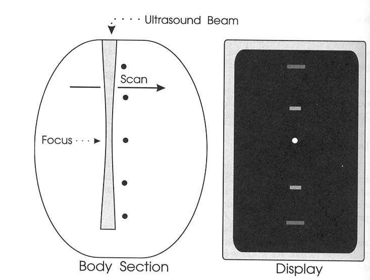
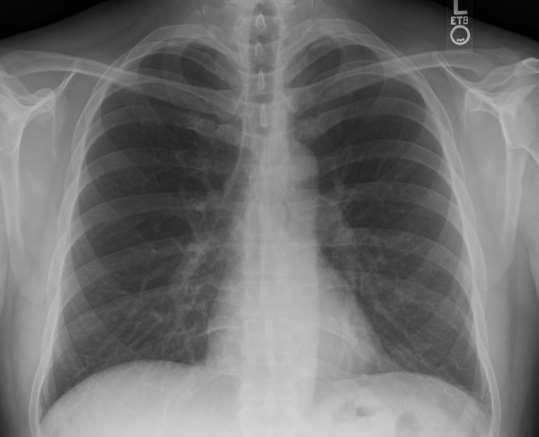
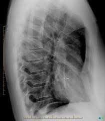
11 Transducer
12 Adjustable focus
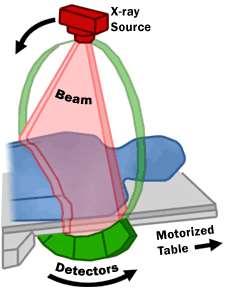
")

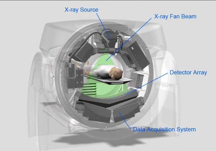
13 A(mplitude) B(rightness) M(otion) doppler Presentation modes
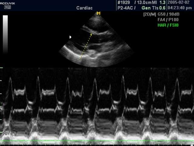
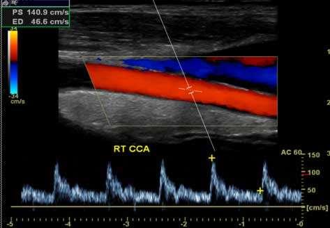
14 Presentation modes M doppler
15 Resolution
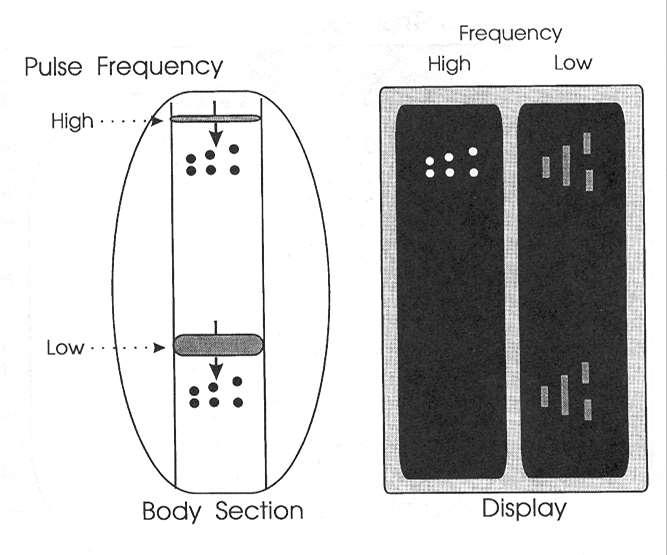
16 Resolution


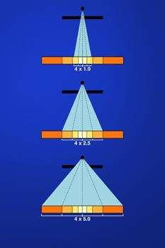
17 CT - computed tomography Godfrey N. Hounsfield Allan M. Cormack The Nobel Prize in Physiology or Medicine 1979


18 MRI - magnetic resonance imaging Sir Peter Mansfield Paul C. Lauterbur The Nobel Prize in Medicine 2003
19 Superimposition
20 Limitations of radiography superimposition slice thickness in direction! estimation of x-ray attenuation tissue contrast sensitivity: ~ 10%
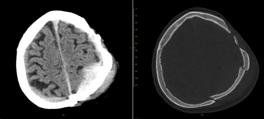
21 computer tomography minimal superimposition improves tissue contrast tissue characterization (CT number)
22 computed tomography
23


24 CT principles attenuation coefficient reconstruction algorithm CT number Hounsfield value

25 element of CT image: voxel FOV field of view image matrix slice thickness

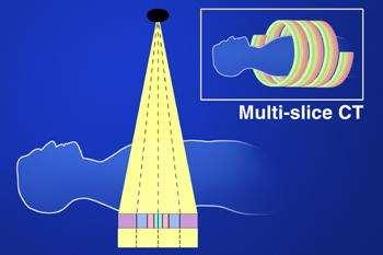
26 Spatial encoding detectors number of slices : 16,32,64,128,256,384
27 Detector configuration
28 CT contrast factor physical (electron) density 1 HU is 0.1% change in attenuation 4 HU can be differentiated 0,008 g/cm 3 between white matter and white matter edema: 2.6 HU 1 % difference in water content (% wet weight) cytotoxic edema can produce a 3% increase in water within 1 hour (hypoxia)


29 relationship between electron and physical density x-ray attenuation
30 windowing
31 windowing
32 window settings 1 HU is 0.1% change in attenuation 4 HU can be differentiated experienced reader can differentiate 10-15% change of contrast in radiography
, flow (ml/s) Adverse effects, contraindications, nephrotoxicity (ESUR")
33 CT contrast media Oral contains iodine or barium Intravenous iodinated, non-ionic Injector volume, timing (phases), flow (ml/s) Adverse effects, contraindications, nephrotoxicity (ESUR guidelines)

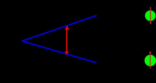
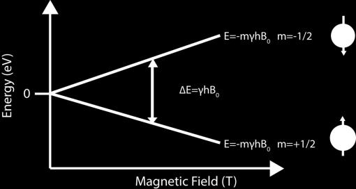
34 Pre- and post-contrast (iv.)
35 CT methods morphologic/structural imaging preand postcontrast (multiple phases) CT angiography, perfusion CT neuro, orbits, sinuses, cervical soft tissue, thorax, abdomen, pelvis, traumaemergency, staging, radiation oncology planning, surgical planning, cardiac, virtual colonoscopy, etc.

36 isotropic voxel reconstruction

37 cardiac CT


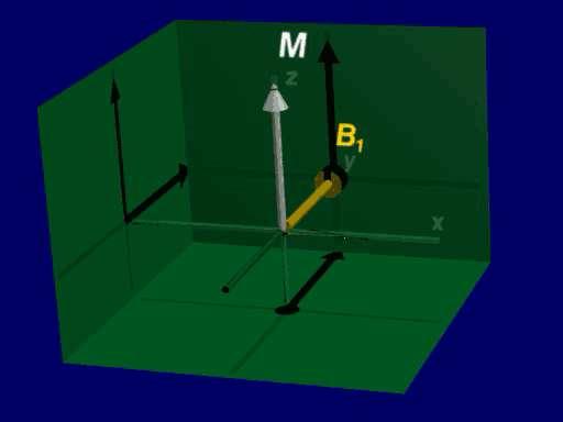
38 virtual endoscopy
39 CT angiography
40 characteristics of modern CT imaging isotropic voxels powerful reformatting possibilities high contrast good temporal resolution (e.g. cardiac imaging, CT-fluoroscopy) volume scanning: good spatial resolution lower patient dose

41 Damadian R Tumor detection by nuclear magnetic resonance. Science 1971, 171:
42

43 Hydrogen without magnetic field Normally, spins are randomly oriented No net magnetization (M=0)

44 Hydrogen in magnetic field Two possible orientations Paralell: lower-energy state Anti-paralell: higher-energy state Natural systems tend to have minimal energy Thermal agitation : only small imbalance
45 Hydrogen in magnetic field At 1.5 T only 1 spin in a contribute to the net magnetization (M) M
46
47 Larmor equation w=gxb 0
48 Excitation

49 Signal detection

50 Signal detection
51 Relaxation in z direction: T1
52 Relaxation in the x-y plane: T2
53 spin echo (SE) pulse sequence
54 sequence parameters TR 90º TE TR: time to repeat TE: time to echo
55 Basis of imaging : spatial localization
 Different spatial locations become associated with different precession")
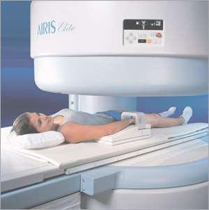
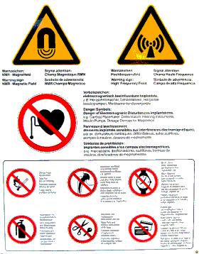
56 Gradient magnetic field Magnetic field gradients are used to change the strength of the main magnetic field (B0) Different spatial locations become associated with different precession frequencies
57 Spatial encoding It is done using magnetic gradients Slice selection - z Frequency encoding - x Phase encoding - y

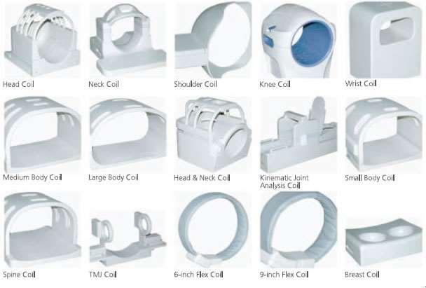
58 Slice selection Based on Larmour equation: ω 0 =γb 0 The central frequency of RF pulse determines the particular location excited Different slice positions are achieved by changing the central frequency Hydrogen nuclei located outside the slice plane are not excited, they will not emit a signal


59 Frequency encoding It causes range of Larmor frequencies to exist in the direction in which it is applied
60 Frequency encoding Fourier transform to separate the different frequencies out after the signal detection
61 Phase encoding Protons located at different positions in the phase encoding direction experience different amounts of phase shift. Repeating the signal detection multiple times with different amplitude of phase-encoding gradient.
 Gradients")
 Image is Fourier transform of")
62 Review: Image Formation Fourier transform k y k x k-space image space Data gathered in k-space (Fourier domain of image) Gradients change position in k-space during data acquisition (location in k-space is integral of gradients) Image is Fourier transform of acquired data
63 The timing of spin echo sequence
64 Magnet types in MRI In Permanent magnets the magnetic field is always there and always at full strength (<0,5T). Resistive magnets are made from many coils of wire wrapped around a cylinder through which an electric current is passed. This generates a magnetic field. When the electricity is shut off, the magnetic field dies. Superconducting magnets are somewhat similar to resistive magnets - coils of wire with a passing electrical current create the magnetic field. The important difference is that in a superconducting magnet the wire is continually bathed in liquid helium. Always on at full strength.
65
66
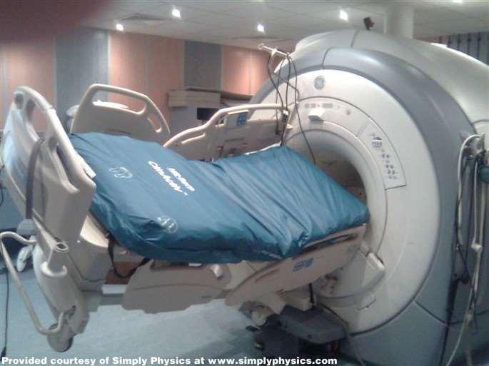
67 MR safety!
68 MR safety!
69 Coils
70 MR contrast, methods T1W pre- and post-contrast T2W T1/T2W mixed Fat or water sturation (pl. FLAIR, STIR, Dixon ) Diffusion weighted (DWI, DTI) Susceptibility weighted T2* (SWI, fmri) Flow-sensitive MR angiography (TOF, PC, 3D CE) in vivo spectroscopy
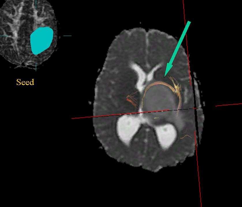
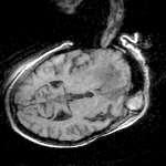
71 molecular basis of MRI tissue contrast 1. water content Relaxation rate is directly proportional to water content 2. restricted water movement Originates from the interaction of water and macromolecules. This phenomenon is common in pathologic tissues. 3. macromolecular motion It also influences water motion. Other parameters might also be important, like ph, ion concentration, polimerisation of macromolecules, etc. 4. lipid content Hidrophobic lipids membranes 5. paramagnetic ions Primarily paramagnetic iron; contrast agents..

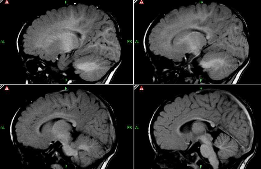
72 T1 weighted

73 T2 weighted

74 FLAIR & T2 weighted
75 Myelinisation T1 weighted

76 T2 weighted submilimeter slice thickness

77 Pre- and postcontrast T1 weighted Note the metallic artifact in the mouth!

78 T2 weighted and DWI
 50 100 250 500")
79 Inversion Recovery TR = 2000 ms TI (ms)

80 1.5T vs. 3T
81
82 fat suppression


83 Late enhancement method myocardial infarction
84 MR-guided neurosurgery

85 Fetal imaging
The physics US and MRI. Prof. Peter Bogner
 The physics US and MRI Prof. Peter Bogner Sound waves mechanical disturbance, a pressure wave moves along longitudinal wave compression rarefaction zones c = nl, (c: velocity, n: frequency, l: wavelength
MRI in Practice. Catherine Westbrook MSc, DCRR, CTC Senior Lecturer Anglia Polytechnic University Cambridge UK. John Talbot MSc, DCRR
 MRI in Practice Third edition Catherine Westbrook MSc, DCRR, CTC Senior Lecturer Anglia Polytechnic University Cambridge UK and Carolyn Kaut RothRT(R) (MR) (CT) (M) (CV) Fellow SMRT (Section for Magnetic
Nuclear Magnetic Resonance Imaging
 Nuclear Magnetic Resonance Imaging Jeffrey A. Fessler EECS Department The University of Michigan NSS-MIC: Fundamentals of Medical Imaging Oct. 20, 2003 NMR-0 Background Basic physics 4 magnetic fields
MRI Physics I: Spins, Excitation, Relaxation
 MRI Physics I: Spins, Excitation, Relaxation Douglas C. Noll Biomedical Engineering University of Michigan Michigan Functional MRI Laboratory Outline Introduction to Nuclear Magnetic Resonance Imaging
Introduction to Biomedical Imaging
 Alejandro Frangi, PhD Computational Imaging Lab Department of Information & Communication Technology Pompeu Fabra University www.cilab.upf.edu MRI advantages Superior soft-tissue contrast Depends on among
EL-GY 6813/BE-GY 6203 Medical Imaging, Fall 2016 Final Exam
 EL-GY 6813/BE-GY 6203 Medical Imaging, Fall 2016 Final Exam (closed book, 1 sheets of notes double sided allowed, no calculator or other electronic devices allowed) 1. Ultrasound Physics (15 pt) A) (9
BMB 601 MRI. Ari Borthakur, PhD. Assistant Professor, Department of Radiology Associate Director, Center for Magnetic Resonance & Optical Imaging
 BMB 601 MRI Ari Borthakur, PhD Assistant Professor, Department of Radiology Associate Director, Center for Magnetic Resonance & Optical Imaging University of Pennsylvania School of Medicine A brief history
Rad Tech 4912 MRI Registry Review. Outline of the Registry Exam: Certification Fees
 Rad Tech 4912 MRI Registry Review Outline of the Registry Exam: Category: # of questions: A. Patient Care 30 B. Imaging Procedures 62 C. Data Acquisition and Processing 65 D. Physical Principles of Image
Magnetic Resonance Imaging
 Magnetic Resonance Imaging History Nuclear magnetic resonance was first described by Isidor Rabi in 1938 - Columbia University, New York City, (Nobel Prize Nobel Prize in Physics 1944) 1946 - Edward Mills
RADIOLOGIV TECHNOLOGY 4912 COMPREHENSEIVE REVIEW/MRI WORSHEET #1- PATIENT CARE AND SAFETY/PHYSICAL PRINCIPLES
 RADIOLOGIV TECHNOLOGY 4912 COMPREHENSEIVE REVIEW/MRI WORSHEET #1- PATIENT CARE AND SAFETY/PHYSICAL PRINCIPLES 1. What are potential consequences to patients and personnel should there be a release of gaseous
MRI in Review: Simple Steps to Cutting Edge Part I
 MRI in Review: Simple Steps to Cutting Edge Part I DWI is now 2 years old... Mike Moseley Radiology Stanford DWI, b = 1413 T2wt, 28/16 ASN 21 San Francisco + Disclosures: Funding NINDS, NCRR, NCI 45 minutes
ENG4BF3 Medical Image Processing
 ENG4BF3 Medical Image Processing Medical Imaging Modalities Imaging in Medical Sciences Imaging is an essential aspect of medical sciences for visualization of anatomical structures and functional or metabolic
Part III: Sequences and Contrast
 Part III: Sequences and Contrast Contents T1 and T2/T2* Relaxation Contrast of Imaging Sequences T1 weighting T2/T2* weighting Contrast Agents Saturation Inversion Recovery JUST WATER? (i.e., proton density
Principles of MRI EE225E / BIO265. Instructor: Miki Lustig UC Berkeley, EECS
 Principles of MRI EE225E / BIO265 Instructor: Miki Lustig UC Berkeley, EECS Today... Administration http://inst.eecs.berkeley.edu/~ee225e/sp16/ Intro to Medical Imaging and MRI Medical Imaging (Before
Introduction to Medical Imaging. Medical Imaging
 Introduction to Medical Imaging BME/EECS 516 Douglas C. Noll Medical Imaging Non-invasive visualization of internal organs, tissue, etc. I typically don t include endoscopy as an imaging modality Image
Technical University of Denmark
 Technical University of Denmark Page 1 of 10 pages Written test, 12 December 2012 Course name: Introduction to medical imaging Course no. 31540 Aids allowed: None. Pocket calculator not allowed "Weighting":
The Basics of Magnetic Resonance Imaging
 The Basics of Magnetic Resonance Imaging Nathalie JUST, PhD nathalie.just@epfl.ch CIBM-AIT, EPFL Course 2013-2014-Chemistry 1 Course 2013-2014-Chemistry 2 MRI: Many different contrasts Proton density T1
Technical University of Denmark
 Technical University of Denmark Page 1 of 11 pages Written test, 9 December 2010 Course name: Introduction to medical imaging Course no. 31540 Aids allowed: none. "Weighting": All problems weight equally.
Introductory MRI Physics
 C HAPR 18 Introductory MRI Physics Aaron Sodickson EXRNAL MAGNETIC FIELD, PROTONS AND EQUILIBRIUM MAGNETIZATION Much of the bulk of the magnetic resonance imaging (MRI) scanner apparatus is dedicated to
Physics of MR Image Acquisition
 Physics of MR Image Acquisition HST-583, Fall 2002 Review: -MRI: Overview - MRI: Spatial Encoding MRI Contrast: Basic sequences - Gradient Echo - Spin Echo - Inversion Recovery : Functional Magnetic Resonance
DEVIL PHYSICS THE BADDEST CLASS ON CAMPUS IB PHYSICS
 DEVIL PHYSICS THE BADDEST CLASS ON CAMPUS IB PHYSICS TSOKOS OPTION I-2 MEDICAL IMAGING Reading Activity Answers IB Assessment Statements Option I-2, Medical Imaging: X-Rays I.2.1. I.2.2. I.2.3. Define
Chapter 14:Physics of Magnetic Resonance
 Chapter 14:Physics of Magnetic Resonance Slide set of 141 slides based on the chapter authored by Hee Kwon Song of the publication (ISBN 978-92-0-131010-1): Diagnostic Radiology Physics: A Handbook for
Magnetic Resonance Imaging. Pål Erik Goa Associate Professor in Medical Imaging Dept. of Physics
 Magnetic Resonance Imaging Pål Erik Goa Associate Professor in Medical Imaging Dept. of Physics pal.e.goa@ntnu.no 1 Why MRI? X-ray/CT: Great for bone structures and high spatial resolution Not so great
Doppler echocardiography & Magnetic Resonance Imaging. Doppler echocardiography. History: - Langevin developed sonar.
 1 Doppler echocardiography & Magnetic Resonance Imaging History: - Langevin developed sonar. - 1940s development of pulse-echo. - 1950s development of mode A and B. - 1957 development of continuous wave
AQA Physics /7408
 AQA Physics - 7407/7408 Module 10: Medical physics You should be able to demonstrate and show your understanding of: 10.1 Physics of the eye 10.1.1 Physics of vision The eye as an optical refracting system,
MEDICAL IMAGING. METHODS OF MODERN IMAGING, BASED ON ELECTRO-MAGNETIC RADIATION (radiowaves, infrared radiation, X-rays, γ-rays ) AND ULTRASOUND
 AND ULTRASOUND") MEDICAL IMAGING MEDICAL IMAGING METHODS OF MODERN IMAGING, BASED ON ELECTRO-MAGNETIC RADIATION (radiowaves, infrared radiation, X-rays, γ-rays ) AND ULTRASOUND MEDICAL IMAGING RADIOLOGY NUCLEAR MEDICINE
MEDICAL IMAGING MEDICAL IMAGING METHODS OF MODERN IMAGING, BASED ON ELECTRO-MAGNETIC RADIATION (radiowaves, infrared radiation, X-rays, γ-rays ) AND ULTRASOUND MEDICAL IMAGING RADIOLOGY NUCLEAR MEDICINE
Basic MRI physics and Functional MRI
 Basic MRI physics and Functional MRI Gregory R. Lee, Ph.D Assistant Professor, Department of Radiology June 24, 2013 Pediatric Neuroimaging Research Consortium Objectives Neuroimaging Overview MR Physics
Contrast Mechanisms in MRI. Michael Jay Schillaci
 Contrast Mechanisms in MRI Michael Jay Schillaci Overview Image Acquisition Basic Pulse Sequences Unwrapping K-Space Image Optimization Contrast Mechanisms Static and Motion Contrasts T1 & T2 Weighting,
NMR and MRI : an introduction
 Intensive Programme 2011 Design, Synthesis and Validation of Imaging Probes NMR and MRI : an introduction Walter Dastrù Università di Torino walter.dastru@unito.it \ Introduction Magnetic Resonance Imaging
Rochester Institute of Technology Rochester, New York. COLLEGE of Science Department of Chemistry. NEW (or REVISED) COURSE:
 COURSE:") Rochester Institute of Technology Rochester, New York COLLEGE of Science Department of Chemistry NEW (or REVISED) COURSE: 1014-730 1.0 Title: Magnetic Resonance Imaging (MRI) Date: July 2006 Credit Hours:
Rochester Institute of Technology Rochester, New York COLLEGE of Science Department of Chemistry NEW (or REVISED) COURSE: 1014-730 1.0 Title: Magnetic Resonance Imaging (MRI) Date: July 2006 Credit Hours:
The NMR Inverse Imaging Problem
 The NMR Inverse Imaging Problem Nuclear Magnetic Resonance Protons and Neutrons have intrinsic angular momentum Atoms with an odd number of proton and/or odd number of neutrons have a net magnetic moment=>
Magnetic resonance imaging MRI
 Magnetic resonance imaging MRI Introduction What is MRI MRI is an imaging technique used primarily in medical settings that uses a strong magnetic field and radio waves to produce very clear and detailed
Medical Imaging Physics Spring Quarter Week 9-1
 Medical Imaging Physics Spring Quarter Week 9-1 NMR and MRI Davor Balzar balzar@du.edu www.du.edu/~balzar Intro MRI Outline NMR & MRI Guest lecturer fmri Thursday, May 22 Visit to CUHSC It s not mandatory
ELG7173 Topics in signal Processing II Computational Techniques in Medical Imaging
 ELG7173 Topics in signal Processing II Computational Techniques in Medical Imaging Topic #1: Intro to medical imaging Medical Imaging Classifications n Measurement physics Send Energy into body Send stuff
G Medical Imaging. Outline 4/13/2012. Physics of Magnetic Resonance Imaging
 G16.4426 Medical Imaging Physics of Magnetic Resonance Imaging Riccardo Lattanzi, Ph.D. Assistant Professor Department of Radiology, NYU School of Medicine Department of Electrical and Computer Engineering,
Magnetic Resonance Imaging (MRI)
") Magnetic Resonance Imaging Introduction The Components The Technology (MRI) Physics behind MR Most slides taken from http:// www.slideworld.org/ viewslides.aspx/magnetic- Resonance-Imaging- %28MRI%29-MR-Imaging-
Magnetic Resonance Imaging Introduction The Components The Technology (MRI) Physics behind MR Most slides taken from http:// www.slideworld.org/ viewslides.aspx/magnetic- Resonance-Imaging- %28MRI%29-MR-Imaging-
Introduction to MRI Acquisition
 Introduction to MRI Acquisition James Meakin FMRIB Physics Group FSL Course, Bristol, September 2012 1 What are we trying to achieve? 2 What are we trying to achieve? Informed decision making: Protocols
Introduction to MRI. Spin & Magnetic Moments. Relaxation (T1, T2) Spin Echoes. 2DFT Imaging. K-space & Spatial Resolution.
 Spin Echoes. 2DFT Imaging. K-space & Spatial Resolution.") Introduction to MRI Spin & Magnetic Moments Relaxation (T1, T2) Spin Echoes 2DFT Imaging Selective excitation, phase & frequency encoding K-space & Spatial Resolution Contrast (T1, T2) Acknowledgement:
Introduction to MRI Spin & Magnetic Moments Relaxation (T1, T2) Spin Echoes 2DFT Imaging Selective excitation, phase & frequency encoding K-space & Spatial Resolution Contrast (T1, T2) Acknowledgement:
Part III Minor Option in Medical Physics 2018 Examples Sheet
 Part III Minor Option in Medical Physics 2018 Examples Sheet Any errors or comments should be addressed sent to: seb53@cam.ac.uk URLs that may be useful: Stanford Event Generation Simulator: http://tinyurl.com/pkg476r
Fundamental MRI Principles Module 2 N. Nuclear Magnetic Resonance. X-ray. MRI Hydrogen Protons. Page 1. Electrons
 Fundamental MRI Principles Module 2 N S 1 Nuclear Magnetic Resonance There are three main subatomic particles: protons positively charged neutrons no significant charge electrons negatively charged Protons
Field trip: Tuesday, Feb 5th
 Pulse Sequences Field trip: Tuesday, Feb 5th Hardware tour of VUIIIS Philips 3T Meet here at regular class time (11.15) Complete MRI screening form! Chuck Nockowski Philips Service Engineer Reminder: Project/Presentation
FREQUENCY SELECTIVE EXCITATION
 PULSE SEQUENCES FREQUENCY SELECTIVE EXCITATION RF Grad 0 Sir Peter Mansfield A 1D IMAGE Field Strength / Frequency Position FOURIER PROJECTIONS MR Image Raw Data FFT of Raw Data BACK PROJECTION Image Domain
Basic Principles of MRI
 MRI for Technologists Basic Principles of MRI PROGRAM INFORMATION MRI for Technologists is a training program designed to meet the needs of radiologic technologists entering or working in the field of
K-space. Spin-Warp Pulse Sequence. At each point in time, the received signal is the Fourier transform of the object s(t) = M( k x
 = M( k x") Bioengineering 280A Principles of Biomedical Imaging Fall Quarter 2015 MRI Lecture 4 k (t) = γ 2π k y (t) = γ 2π K-space At each point in time, the received signal is the Fourier transform of the object
Bioengineering 280A Principles of Biomedical Imaging Fall Quarter 2015 MRI Lecture 4 k (t) = γ 2π k y (t) = γ 2π K-space At each point in time, the received signal is the Fourier transform of the object
Structure of Biological Materials
 ELEC ENG 3BA3: Structure of Biological Materials Notes for Lecture #19 Monday, November 22, 2010 6.5 Nuclear medicine imaging Nuclear imaging produces images of the distribution of radiopharmaceuticals
Nuclear Magnetic Resonance Imaging
 Nuclear Magnetic Resonance Imaging Simon Lacoste-Julien Electromagnetic Theory Project 198-562B Department of Physics McGill University April 21 2003 Abstract This paper gives an elementary introduction
Lecture #7 In Vivo Water
 Lecture #7 In Vivo Water Topics Hydration layers Tissue relaxation times Magic angle effects Magnetization Transfer Contrast (MTC) CEST Handouts and Reading assignments Mathur-De Vre, R., The NMR studies
Magnetic Resonance Imaging. Qun Zhao Bioimaging Research Center University of Georgia
 Magnetic Resonance Imaging Qun Zhao Bioimaging Research Center University of Georgia The Nobel Prize in Physiology or Medicine 2003 "for their discoveries concerning magnetic resonance imaging" Paul C.
ROCHESTER INSTITUTE OF TECHNOLOGY COURSE OUTLINE FORM COLLEGE OF SCIENCE. Chester F. Carlson Center for Imaging Science
 ROCHESTER INSTITUTE OF TECHNOLOGY COURSE OUTLINE FORM COLLEGE OF SCIENCE Chester F. Carlson Center for Imaging Science NEW COURSE: COS-IMGS-730 Magnetic Resonance Imaging 1.0 Course Designations and Approvals
Fundamental MRI Principles Module Two
 Fundamental MRI Principles Module Two 1 Nuclear Magnetic Resonance There are three main subatomic particles: protons neutrons electrons positively charged no significant charge negatively charged Protons
(INCLUDING THIS FRONT PAGE)
") I'IFIITIIBIFI UNIVERSITY OF SCIEI'ICE RITD TECHNOLOGY FACULTY OF HEALTH AND APPLIED SCIENCES DEPARTMENT OF NATURAL AND APPLIED SCIENCES QUALIFICATION: BACHELOR OF SCIENCE (MAJOR AND MINOR) QUALIFICATION
I'IFIITIIBIFI UNIVERSITY OF SCIEI'ICE RITD TECHNOLOGY FACULTY OF HEALTH AND APPLIED SCIENCES DEPARTMENT OF NATURAL AND APPLIED SCIENCES QUALIFICATION: BACHELOR OF SCIENCE (MAJOR AND MINOR) QUALIFICATION
Master s Program in Medical Physics. Physics of Imaging Systems Basic Principles of Magnetic Resonance Imaging I. Prof. Dr. Lothar Schad.
 1 12/9/2008 Page 1 Master s Program in Medical Physics Physics of Imaging Systems Basic Principles of Magnetic Resonance Imaging I Chair in Faculty of Medicine Mannheim University of Heidelberg Theodor-Kutzer-Ufer
Apodization. Gibbs Artifact. Bioengineering 280A Principles of Biomedical Imaging. Fall Quarter 2013 MRI Lecture 5. rect(k x )
") Bioengineering 280A Principles of Biomedical Imaging Fall Quarter 2013 MRI Lecture 5 GE Medical Systems 2003 Gibbs Artifact Apodization rect(k ) Hanning Window h(k )=1/2(1+cos(2πk ) 256256 image 256128
Bioengineering 280A Principles of Biomedical Imaging Fall Quarter 2013 MRI Lecture 5 GE Medical Systems 2003 Gibbs Artifact Apodization rect(k ) Hanning Window h(k )=1/2(1+cos(2πk ) 256256 image 256128
NMR/MRI examination (8N080 / 3F240)
") NMR/MRI examination (8N080 / 3F240) Remarks: 1. This test consists of 3 problems with at total of 26 sub-questions. 2. Questions are in English. You are allowed to answer them in English or Dutch. 3. Please
NMR/MRI examination (8N080 / 3F240) Remarks: 1. This test consists of 3 problems with at total of 26 sub-questions. 2. Questions are in English. You are allowed to answer them in English or Dutch. 3. Please
Introduction to Magnetic Resonance Imaging (MRI) Pietro Gori
 Pietro Gori") Introduction to Magnetic Resonance Imaging (MRI) Pietro Gori Enseignant-chercheur Equipe IMAGES - Télécom ParisTech pietro.gori@telecom-paristech.fr September 20, 2017 P. Gori BIOMED 20/09/2017 1 / 76
Introduction to Magnetic Resonance Imaging (MRI) Pietro Gori Enseignant-chercheur Equipe IMAGES - Télécom ParisTech pietro.gori@telecom-paristech.fr September 20, 2017 P. Gori BIOMED 20/09/2017 1 / 76
Nuclei, Excitation, Relaxation
 Outline 4.1 Principles of MRI uclei, Excitation, Relaxation Carolyn Kaut Roth, RT (R)(MR)(CT)(M)(CV) FSMRT CEO Imaging Education Associates www.imaginged.com candi@imaginged.com What nuclei are MR active?
MR Advance Techniques. Flow Phenomena. Class I
 MR Advance Techniques Flow Phenomena Class I Flow Phenomena In this class we will explore different phenomenona produced from nuclei that move during the acquisition of data. Flowing nuclei exhibit different
Tissue Characteristics Module Three
 Tissue Characteristics Module Three 1 Equilibrium State Equilibrium State At equilibrium, the hydrogen vector is oriented in a direction parallel to the main magnetic field. Hydrogen atoms within the vector
Biomedical Engineering Image Formation
 Biomedical Engineering Image Formation PD Dr. Frank G. Zöllner Computer Assisted Clinical Medicine Medical Faculty Mannheim Learning objectives! Understanding the process of image formation! Point spread
M R I Physics Course. Jerry Allison Ph.D., Chris Wright B.S., Tom Lavin B.S., Nathan Yanasak Ph.D. Department of Radiology Medical College of Georgia
 M R I Physics Course Jerry Allison Ph.D., Chris Wright B.S., Tom Lavin B.S., Nathan Yanasak Ph.D. Department of Radiology Medical College of Georgia M R I Physics Course Spin Echo Imaging Hahn Spin Echo
Introduction to the Course and the Techniques. Jeffry R. Alger, PhD Ahmanson-Lovelace Brain Mapping Center Department of Neurology
 Introduction to the Course and the Techniques Jeffry R. Alger, PhD Ahmanson-Lovelace Brain Mapping Center Department of Neurology (jralger@ucla.edu) CTSI Neuroimaging April 2013 Rationale for the Course
Biomedical Imaging Magnetic Resonance Imaging
 Biomedical Imaging Magnetic Resonance Imaging Charles A. DiMarzio & Eric Kercher EECE 4649 Northeastern University May 2018 Background and History Measurement of Nuclear Spins Widely used in physics/chemistry
Sequence Overview. Gradient Echo Spin Echo Magnetization Preparation Sampling and Trajectories Parallel Imaging. B.Hargreaves - RAD 229
 Sequence Overview Gradient Echo Spin Echo Magnetization Preparation Sampling and Trajectories Parallel Imaging 75 Pulse Sequences and k-space RF k y G z k x G x 3D k-space G y k y k z Acq. k x 76 Gradient
Physics and Brain Imaging
 Physics and Brain Imaging Nuclear Magnetic Resonance (NMR) Magnetic Resonance Imaging (MRI) Functional MRI (fmri) Talk at Quarknet FSU Summer Workshop, July 24, 2017 Per Arne Rikvold Leonardo da Vinci
Introduction to Magnetic Resonance Imaging
 Introduction to Magnetic Resonance Imaging MRI of the brain, ca. 1978. ca. 1993 ca. 2006 2014 Modality Characteristics and Comparison Radiography CT scanning Nuclear medicine MRI transmission modalities
Advanced Topics and Diffusion MRI
 Advanced Topics and Diffusion MRI Slides originally by Karla Miller, FMRIB Centre Modified by Mark Chiew (mark.chiew@ndcn.ox.ac.uk) Slides available at: http://users.fmrib.ox.ac.uk/~mchiew/teaching/ MRI
BNG/ECE 487 FINAL (W16)
") BNG/ECE 487 FINAL (W16) NAME: 4 Problems for 100 pts This exam is closed-everything (no notes, books, etc.). Calculators are permitted. Possibly useful formulas and tables are provided on this page. Fourier
BNG/ECE 487 FINAL (W16) NAME: 4 Problems for 100 pts This exam is closed-everything (no notes, books, etc.). Calculators are permitted. Possibly useful formulas and tables are provided on this page. Fourier
MRI Physics II: Gradients, Imaging. Douglas C. Noll, Ph.D. Dept. of Biomedical Engineering University of Michigan, Ann Arbor
 MRI Physics II: Gradients, Imaging Douglas C., Ph.D. Dept. of Biomedical Engineering University of Michigan, Ann Arbor Magnetic Fields in MRI B 0 The main magnetic field. Always on (0.5-7 T) Magnetizes
Spin Echo Imaging Sequence
 1 MRI In Stereotactic Procedures Edward F. Jackson, Ph.D. The University of Texas M.D. Anderson Cancer Center Houston, Texas 2 RF G slice G phase G freq Signal k-space Spin Echo Imaging Sequence TE 1st
Lecture 12 February 11, 2016
 MATH 262/CME 372: Applied Fourier Analysis and Winter 2016 Elements of Modern Signal Processing Lecture 12 February 11, 2016 Prof. Emmanuel Candes Scribe: Carlos A. Sing-Long, Edited by E. Bates 1 Outline
QUALITY ASSURANCE OF MAGNETIC RESONANCE IMAGING FOR ADAPTIVE RADIOTHERAPY: PRELIMINARY INVESTIGATIONS TREVOR THANG 1 Supervisors: Dr.
 QUALITY ASSURANCE OF MAGNETIC RESONANCE IMAGING FOR ADAPTIVE RADIOTHERAPY: PRELIMINARY INVESTIGATIONS TREVOR THANG 1 Supervisors: Dr. Eugene Wong 2, Dr. Rob Bartha 1 Department of Medical Biophysics 1,
Chapter 15:Magnetic Resonance Imaging
 Chapter 15:Magnetic Resonance Imaging Slide set of 242 slides based on the chapter authored by Martin O. Leach of the publication (ISBN 978-92-0-131010-1): Diagnostic Radiology Physics: A Handbook for
Chapter 24 MRA and Flow quantification. Yongquan Ye, Ph.D. Assist. Prof. Radiology, SOM Wayne State University
 Chapter 24 MRA and Flow quantification Yongquan Ye, Ph.D. Assist. Prof. Radiology, SOM Wayne State University Previous classes Flow and flow compensation (Chap. 23) Steady state signal (Cha. 18) Today
Procesamiento de Imágenes y Bioseñales
 Procesamiento de Imágenes y Bioseñales Dr. Víctor Castañeda Agenda Physical basis of X-ray- CT, NMR, Ultrasound, Nuclear Medicine Sensors (cameras, gamma probes, microphone) Computational Tomography (CT)
Principles of MRI. Vinyl Record. Last time: Today: Homework Due tonight! EE225E / BIO265. Transforms a temporal signal to a spatial signal
 What is this? ` Principles of MRI Lecture 05 EE225E / BIO265 Instructor: Miki Lustig UC Berkeley, EECS The first NMR spectrum of ethanol 1951. 1 2 Today Last time: Linear systems, Fourier Transforms, Sampling
Basis of MRI Contrast
 Basis of MRI Contrast MARK A. HORSFIELD Department of Cardiovascular Sciences University of Leicester Leicester LE1 5WW UK Tel: +44-116-2585080 Fax: +44-870-7053111 e-mail: mah5@le.ac.uk 1 1.1 The Magnetic
Part II: Magnetic Resonance Imaging (MRI)
") Part II: Magnetic Resonance Imaging (MRI) Contents Magnetic Field Gradients Selective Excitation Spatially Resolved Reception k-space Gradient Echo Sequence Spin Echo Sequence Magnetic Resonance Imaging
Part II: Magnetic Resonance Imaging (MRI) Contents Magnetic Field Gradients Selective Excitation Spatially Resolved Reception k-space Gradient Echo Sequence Spin Echo Sequence Magnetic Resonance Imaging
Electrical Engineering 3BA3: Structure of Biological Materials
 Electrical Engineering 3BA3: Structure of Biological Materials Day Class Instructor: Dr. I. C. BRUCE Duration of Examination: 3 Hours McMaster University Final Examination December, 2004 This examination
Background II. Signal-to-Noise Ratio (SNR) Pulse Sequences Sampling and Trajectories Parallel Imaging. B.Hargreaves - RAD 229.
 Pulse Sequences Sampling and Trajectories Parallel Imaging. B.Hargreaves - RAD 229.") Background II Signal-to-Noise Ratio (SNR) Pulse Sequences Sampling and Trajectories Parallel Imaging 1 SNR: Signal-to-Noise Ratio Signal: Desired voltage in coil Noise: Thermal, electronic Noise Thermal
Background II Signal-to-Noise Ratio (SNR) Pulse Sequences Sampling and Trajectories Parallel Imaging 1 SNR: Signal-to-Noise Ratio Signal: Desired voltage in coil Noise: Thermal, electronic Noise Thermal
Introduction to the Physics of NMR, MRI, BOLD fmri
 Pittsburgh, June 13-17, 2011 Introduction to the Physics of NMR, MRI, BOLD fmri (with an orientation toward the practical aspects of data acquisition) Pittsburgh, June 13-17, 2001 Functional MRI in Clinical
Navigator Echoes. BioE 594 Advanced Topics in MRI Mauli. M. Modi. BioE /18/ What are Navigator Echoes?
 Navigator Echoes BioE 594 Advanced Topics in MRI Mauli. M. Modi. 1 What are Navigator Echoes? In order to correct the motional artifacts in Diffusion weighted MR images, a modified pulse sequence is proposed
Index. p, lip, 78 8 function, 107 v, 7-8 w, 7-8 i,7-8 sine, 43 Bo,94-96
 p, lip, 78 8 function, 107 v, 7-8 w, 7-8 i,7-8 sine, 43 Bo,94-96 B 1,94-96 M,94-96 B oro!' 94-96 BIro!' 94-96 I/r, 79 2D linear system, 56 2D FFT, 119 2D Fourier transform, 1, 12, 18,91 2D sinc, 107, 112
Electron Spin Resonance, Basic principle of NMR, Application of NMR in the study of Biomolecules, NMR imaging and in vivo NMR spectromicroscopy
 Electron Spin Resonance, Basic principle of NMR, Application of NMR in the study of Biomolecules, NMR imaging and in vivo NMR spectromicroscopy Mitesh Shrestha Electron Spin Resonance Electron paramagnetic
Magnetic Resonance Elastography for Liver Fibrosis. By: Larissa Schudlo and Natalie Tong
 Magnetic Resonance Elastography for Liver Fibrosis By: Larissa Schudlo and Natalie Tong Outline Introduction Importance of Elasticity in Biological Tissues Liver Disease Ultrasound Elastography (UE) Basics
Pulse Sequences: RARE and Simulations
 Pulse Sequences: RARE and Simulations M229 Advanced Topics in MRI Holden H. Wu, Ph.D. 2018.04.19 Department of Radiological Sciences David Geffen School of Medicine at UCLA Class Business Final project
How is it different from conventional MRI? What is MR Spectroscopy? How is it different from conventional MRI? MR Active Nuclei
 What is MR Spectroscopy? MR-Spectroscopy (MRS) is a technique to measure the (relative) concentration of certain chemical or biochemical molecules in a target volume. MR-Spectroscopy is an in vivo (in
BME I5000: Biomedical Imaging
 BME I5000: Biomedical Imaging Lecture 9 Magnetic Resonance Imaging (imaging) Lucas C. Parra, parra@ccny.cuny.edu Blackboard: http://cityonline.ccny.cuny.edu/ 1 Schedule 1. Introduction, Spatial Resolution,
Outlines: (June 11, 1996) Instructor:
 Instructor:") Magnetic Resonance Imaging (June 11, 1996) Instructor: Tai-huang Huang Institute of Biomedical Sciences Academia Sinica Tel. (02) 2652-3036; Fax. (02) 2788-7641 E. mail: bmthh@ibms.sinica.edu.tw Reference:
Magnetic Resonance Imaging (June 11, 1996) Instructor: Tai-huang Huang Institute of Biomedical Sciences Academia Sinica Tel. (02) 2652-3036; Fax. (02) 2788-7641 E. mail: bmthh@ibms.sinica.edu.tw Reference:
Modern physics ideas are strange! L 36 Modern Physics [2] The Photon Concept. How are x-rays produced? The uncertainty principle
![Modern physics ideas are strange! L 36 Modern Physics [2] The Photon Concept. How are x-rays produced? The uncertainty principle](/thumbs/88/117098787.jpg "Modern physics ideas are strange! L 36 Modern Physics [2] The Photon Concept. How are x-rays produced? The uncertainty principle") L 36 Modern Physics [2] X-rays & gamma rays How lasers work Medical applications of lasers Applications of high power lasers Medical imaging techniques CAT scans MRI s Modern physics ideas are strange!
L 36 Modern Physics [2] X-rays & gamma rays How lasers work Medical applications of lasers Applications of high power lasers Medical imaging techniques CAT scans MRI s Modern physics ideas are strange!
Relaxation times in nuclear magnetic resonance
 Relaxation times in TEP Related topics Nuclear spins, atomic nuclei with a magnetic moment, precession movement of the nuclear spins, Landau-Lifshitz equation, Bloch equation, magnetisation, resonance
Principles of Nuclear Magnetic Resonance Microscopy
 Principles of Nuclear Magnetic Resonance Microscopy Paul T. Callaghan Department of Physics and Biophysics Massey University New Zealand CLARENDON PRESS OXFORD CONTENTS 1 PRINCIPLES OF IMAGING 1 1.1 Introduction
2.1.1 A Brief History of NMR The conception of NMR sprouted after the Pauli s prediction of nuclear spin in
 CHAPTER--2 BASICS OF NMR IMAGING AND SPECTROSCOPY 2.1 Introduction 2.1.1 A Brief History of NMR The conception of NMR sprouted after the Pauli s prediction of nuclear spin in 1924. Later Gorter (1936)
HY Ιατρική Απεικόνιση. Διδάσκων: Kώστας Μαριάς
 HY 571 - Ιατρική Απεικόνιση Διδάσκων: Kώστας Μαριάς 11. MRI Τ1,Τ2, PD and physiological parameter imaging Summary and Clarifications Resonance is referred to as the property of an atom to absorb energy
Tissue Parametric Mapping:
 Tissue Parametric Mapping: Contrast Mechanisms Using SSFP Sequences Jongho Lee Department of Radiology University of Pennsylvania Tissue Parametric Mapping: Contrast Mechanisms Using bssfp Sequences Jongho
PETER PAZMANY CATHOLIC UNIVERSITY Consortium members SEMMELWEIS UNIVERSITY, DIALOG CAMPUS PUBLISHER
 PETER PAZMANY CATHOLIC UNIVERSITY SEMMELWEIS UNIVERSITY Development of Complex Curricula for Molecular Bionics and Infobionics Programs within a consortial* framework** Consortium leader PETER PAZMANY
Magnetic Resonance Imaging: Window to a Watery World
 Magnetic Resonance Imaging Magnetic Resonance Imaging: Window to a Watery World ~. ~~'
Exam 8N080 - Introduction to MRI
 Exam 8N080 - Introduction to MRI Friday April 10 2015, 18.00-21.00 h For this exam you may use an ordinary calculator (not a graphical one). In total there are 5 assignments and a total of 50 points can
6/22/2011. RT 4912 Review. Rex T. Christensen MHA RT (R) (MR) (CT)
 (MR) (CT)") RT 4912 Review Rex T. Christensen MHA RT (R) (MR) (CT) 1 Questions? ARRT Content Specifications: https://www.arrt.org/pdfs/disciplines/content-specification/mri- Content-Specification.pdf Tests Can take
RT 4912 Review Rex T. Christensen MHA RT (R) (MR) (CT) 1 Questions? ARRT Content Specifications: https://www.arrt.org/pdfs/disciplines/content-specification/mri- Content-Specification.pdf Tests Can take
Elec Eng 3BA3: Structure of Biological Materials
 Elec Eng 3BA3: Structure of Biological Materials Page 1 of 12 Day Class Instructor: Dr. I. C. BRUCE Duration of Examination: 3 Hours McMaster University Final Examination December 5, 2008 This examination
Basic Pulse Sequences I Saturation & Inversion Recovery UCLA. Radiology
 Basic Pulse Sequences I Saturation & Inversion Recovery Lecture #5 Learning Objectives Explain what the most important equations of motion are for describing spin systems for MRI. Understand the assumptions