Introduction to SPECT & PET TBMI02 - Medical Image Analysis 2017
|
|
|
- Felix Hill
- 5 years ago
- Views:
Transcription
1 Introduction to SPECT & PET TBMI02 - Medical Image Analysis 2017 Marcus Ressner, PhD, Medical Radiation Physicist, Linköping University Hospital Content What is Nuclear medicine? Basic principles of Functional Imaging with radioisotopes Basic principals of SPECT and PET Camera technology Image acquisition Image reconstruction Clinical applications 1
 Study a tracers way through the body Gives quantitative functional")
2 What is Nuclear Medicine? CT SPECT (or PET) Nuclear Medicine Functional imaging Visualization of physiological processes i.e study how the body works, not how it looks (anatomical imaging) Study a tracers way through the body Gives quantitative functional information 2


3 Basic principle SPECT 99m Tc Radioactiv isotope Isotope PET 18 F 123 I 11 C 131 I 13 N 111 In 15 O Target molecule Tracer Salt analog: 99 Tc m HDP Glucos analog: 18 F FDG oraly injected inhaled Diagnostic imaging Illustration modified from Mehran M. Sadeghi et al. J Nucl Med 2010;51:51S-65S 3

4 Radioactive nuclides Instable nuclides that decays and emit its surplus energy by radiation Types of ionisation radiation: alfa( beta( gamma ( Radionuclides in nuclear medicin Photons within the energy range of kev Half life in the range hours Minimal emission of other ionising particles Stabilty in the chemical bond Easy chemical label procedures 4
 123 I 159 kev 13 h Adrenal")


5 Radionuclides in nuclear medicin 99m Tc 140 kev 6 h Various (almost all) 123 I 159 kev 13 h Adrenal glands, Thyroid 111 In 171, 245 kev 67,9 h Neuroendocrine tumors 18 F 511 kev 110 m Tumordiagnostics 11 C 511 kev 20 m Neuro imaging 13 N 511 kev 10 m Cardiac imaging 15 O 511 kev 2 m Hypoxia, Cardiac Hotlab 99m Tc generator 5

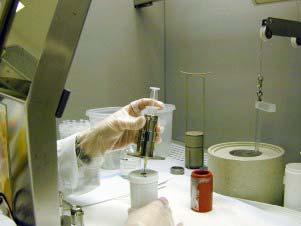
6 99m Tc eluate activity Production of PET radioisotopes Cyclotron 6


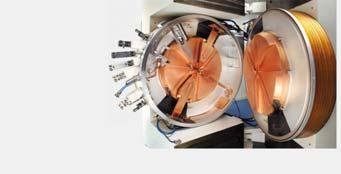
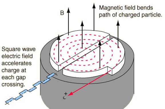
7 Cyclotron vid US 2015 PET/CT CYCLOTRON The Accelerator 14 7

8 Production of F O 18 F, p(p,n)n 16 8


9 Which Imaging Modality? ANTERIOR POSTERIOR Bone Scintigraphy Tracer: Sodium oxidronate Accumulates in active osteogenisis (bone metabolism) ANTERIOR POSTERIOR 9

10 Neuroendocrine tumors In 111 Octreotid SPECT Treatment response monitoring Somatostatin receptor imaging In 111 Octreotid SPECT Ga 68 DOTA-TOC PET 2 receptor types 5 receptor types 10

11 Gamma camera GE NM Discovery Gamma camera principles 11

12 The collimator Gamma detection Position X Position Y Energi Z 12


13 Energy resolution Peak energy Count rate Compton scatter Energy window ± 10% 140 kev Voltage pulse hight / Energy Energy window 13
14 X,Y and Z Y The Z-dimension is given by the pulse height -Y -X X Acquisition types Planar or Static Scanning Dynamic Time interval ECG tigger Tomographic 14


15 Static acquisition Perfusionsscintigraphy of the lungs ANT POST WB scanning Tracer: Natrium oxidronate Radioisotope: Tc-99m The distribution of the radiotracer show the osteoblast activity, and blood perfusion of the bone tissue 15


16 Dynamic acquisition A group of images taken at set time intervals e.g sec images over the kidneys showing the distribution of the radiotracer as a function of time. Dynamic acquisition 16
 The same activity distribution renders different projections.")
17 Distance - resolution FWHM Tomographic acquisition detector (projection) Planar images from different angles around the patient (projections) The same activity distribution renders different projections. object 17
18 Sinogram Projection data Original data Sinogram Sinogram Projection data Original data Sinogram 18

 Ordered Subset Expectation")
19 Sinogram Projection data Data acq Sinogram Image reconconstruction Analytical Filtered Back Projection (FBP) Statistical Maximum Likelihood Expectation Maximization (MLEM) Ordered Subset Expectation Maximization (OSEM) 19


20 FBP - Forward projection view 1 view 2 view 3 Back projection view 1 view 2 view 3 Using 3 views Using many views 20


21 Back projection blurred by 1/r Back projection original 8 proj 16 proj 32 proj 64 proj 128 proj 256 proj 21


22 Filtered Back projection filtered view 1 filtered view 2 filtered view 3 Using 3 views Using many views Filtered back projection 8 proj 32 proj 128 proj 256 proj 22
 original projections")


23 Iterative reconstruction (MLEM) original projections BP original NO CHANGE estimate patient update (x ratio) FP Estimated projections Estimate Contrast recovery Contrast [%] iterations Noise [%] 23


24 Post filter FBP vs OSEM Advanced image processing. S. Ted Treves et al. J Nucl Med 2011;52:

25 Projection overview 3D Representation CBF examination with 120 projections 25


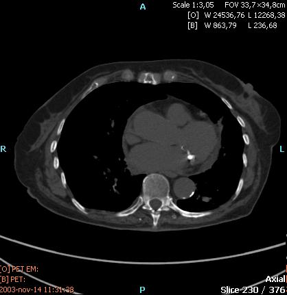
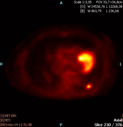
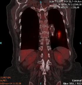
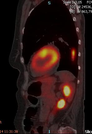
26 SPECT/CT Image Fusion Attenuation effects Reduction of the number of detected photons. Absorption Spread Depend on Photonenergy Density of tissues 26

27 Attenuation effects Water filled cylinder, well mixed. Transaxial image with intensity profile Count statistics Injected activity Image aquisition time More pulses less noise and better contrast 27


 Semiconductor")
28 Antal pulser i bilden 5 s 1 x Aktivitetsmängd 30 s 6 x Aktivitetsmängd 300 s 60 x Aktivitetsmängd New technologies Natriumjodid (NaI) PM-tube Cadmium Zink Tellurid(CZT) Semiconductor Kan arbeta i rum 56 28

29 Semiconductor collimation Holes match CZT pixels Collimator to match semiconductor energies: kev Semiconductor detectors: Higher signal-to-noise Better spatial resolution Better energy resolution 57 29


30 Dedicated heart cameras D-SPECT from Spectrum Dynamics Installed in Linköping summer 2013 Discovery NM 530c GE 59 Alcyone Technology with Lightspeed VCT Discovery NM/CT 570c Courtesy of University Hospital, Zurich, Switzerland 60 30

31 PET (/CT) Principle Radioactive isotope Address Tracer SPECT 99m Tc PET 18 F 123 I 11 C 131 I 13 N 111 In 15 O Tracers Fluorodeoxyglucose 31

 + e -")
32 Metabolic Imaging PET Pre-treatment Post-treatment Positron decay and annihilations (511 kev) + e - (511 kev) Radio nuclide 32
33 PET radioisotopes Radioisotope Half-life Energy (mean) C min 0.39 MeV N min 0.50 MeV O min 0.72 MeV F min 0.25 MeV Cu min 1.3 MeV Ga min 0.83 MeV Rb min 1.5 MeV The PET camera 33


34 Data acquisition 511 kev e+ e- 180 degrees 511 kev 34
35 Coincidence Positron annihilation Detection within coincidence window Energy within energy window Bild från Siemens Coincidence Philips 35


")
36 Coincidence Bild från Siemens Reconstruction FBP, OSEM r Data Acquisition Sinogram (raw data) Reconstructed Image 36


37 ToF Time of Flight ToF Improved SNR 37


 PET 3D")
38 SUV Standard Uptake Value C(t) (kbq/ml) SUV = Dose (kbq)/ Weight (kq) PET 3D MIP Image 38

39 PET/CT 39
Nuclear Medicine Intro & Physics from Medical Imaging Signals and Systems, Chapter 7, by Prince and Links
 Nuclear Medicine Intro & Physics from Medical Imaging Signals and Systems, Chapter 7, by Prince and Links NM - introduction Relies on EMISSION of photons from body (versus transmission of photons through
Radioisotopes in action. Diagnostic application of radioisotopes. Steps of diagnostic procedure. Information from various medical imaging techniques
 Radioisotopes in action Diagnostic application of radioisotopes Steps of diagnostic procedure - Radioactive material introduced into the patient - Distribution and alteration of activity is detected -
Radionuclide Imaging MII Positron Emission Tomography (PET)
") Radionuclide Imaging MII 3073 Positron Emission Tomography (PET) Positron (β + ) emission Positron is an electron with positive charge. Positron-emitting radionuclides are most commonly produced in cyclotron
Radionuclide Imaging MII 3073 Positron Emission Tomography (PET) Positron (β + ) emission Positron is an electron with positive charge. Positron-emitting radionuclides are most commonly produced in cyclotron
Chapter 2 PET Imaging Basics
 Chapter 2 PET Imaging Basics Timothy G. Turkington PET Radiotracers Positron emission tomography (PET) imaging is the injection (or inhalation) of a substance containing a positron emitter, the subsequent
1st Faculty of Medicine, Charles University in Prague Center for Advanced Preclinical Imaging (CAPI)
") Radioation Resolution and Sensitivity Nuclear Imaging PET + SPECT Radioactive Decay (EC,Ɣ), (β -,Ɣ), (I.T.,Ɣ) β + Projection imaging collimator needed one angular view Projection imaging coincidence imaging,
Radioation Resolution and Sensitivity Nuclear Imaging PET + SPECT Radioactive Decay (EC,Ɣ), (β -,Ɣ), (I.T.,Ɣ) β + Projection imaging collimator needed one angular view Projection imaging coincidence imaging,
MEDICAL EQUIPMENT: NUCLEAR MEDICINE. Prof. Yasser Mostafa Kadah
 MEDICAL EQUIPMENT: NUCLEAR MEDICINE Prof. Yasser Mostafa Kadah www.k-space.org Recommended Textbook Introduction to Medical Imaging: Physics, Engineering and Clinical Applications, by Nadine Barrie Smith
CT-PET calibration : physical principles and operating procedures F.Bonutti. Faustino Bonutti Ph.D. Medical Physics, Udine University Hospital.
 CT-PET calibration : physical principles and operating procedures Faustino Bonutti Ph.D. Medical Physics, Udine University Hospital Topics Introduction to PET physics F-18 production β + decay and annichilation
A. I, II, and III B. I C. I and II D. II and III E. I and III
 BioE 1330 - Review Chapters 7, 8, and 9 (Nuclear Medicine) 9/27/2018 Instructions: On the Answer Sheet, enter your 2-digit ID number (with a leading 0 if needed) in the boxes of the ID section. Fill in
Radioisotopes in action. Diagnostic application of radioisotopes. Steps of diagnostic procedure. Information from various medical imaging techniques
 Radioisotopes in action Diagnostic application of radioisotopes Steps of diagnostic procedure - Radioactive material introduced into the patient - Distribution and alteration of activity is detected -Monitoring
Radioisotopes and PET
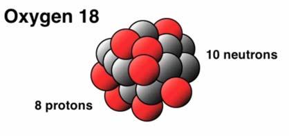
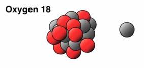
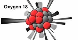
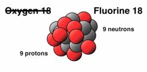
 Radioisotopes and PET 1 Radioisotopes Elements are defined by their number of protons, but there is some variation in the number of neutrons. Atoms resulting from this variation are called isotopes. Consider
Dana-Farber Cancer Institute, 44 Binney Street, Boston, MA 02115, USA ramsey
 SPECIAL FEATURE: MEDICAL PHYSICS www.iop.org/journals/physed Nuclear medicine Ramsey D Badawi Dana-Farber Cancer Institute, 44 Binney Street, Boston, MA 02115, USA E-mail: ramsey badawi@dfci.harvard.edu
Dosimetry of patients injected with tracers Ga-68, Zr-89 and Lu-177. Bruno Vanderlinden
 Dosimetry of patients injected with tracers Ga-68, Zr-89 and Lu-177 Bruno Vanderlinden What is NM speciality? Imaging radiology Physics Diagnostic Treatment assessment Clinical pathology Biological marker
6: Positron Emission Tomography
 6: Positron Emission Tomography. What is the principle of PET imaging? Positron annihilation Electronic collimation coincidence detection. What is really measured by the PET camera? True, scatter and random
Tomography is imaging by sections. 1
 Tomography is imaging by sections. 1 It is a technique used in clinical medicine and biomedical research to create images that show how certain tissues are performing their physiological functions. 1 Conversely,
CLINICALLY USEFUL RADIONUCLIDES:
 INTRODUCTION It is important that Nuclear Medicine Technologists be familiar with the imaging properties of all commonly used radionuclides to insure correct choice of isotope for a particular study as
What is scintigraphy? The process of obtaining an image or series of sequential images of the distribution of a radionuclide in tissues, organs, or
 Let's remind... What is nuclear medicine? Nuclear medicine can be broadly divided into two branches "in vitro" and "in vivo" procedures. There are numerous radioisotopic "in vitro" procedures for genotyping
Mayneord-Phillips Summer School St Edmund Hall, University of Oxford July Proton decays to n, e +, ν
 Positron Emission Tomography Physics & Instrumentation Dimitra G. Darambara, Ph.D Multimodality Molecular Imaging Joint Department of Physics RMH/ICR Outline Introduction PET Physics overview Types of
Lecture 5: Tomographic nuclear systems: SPECT
 Lecture 5: Tomographic nuclear systems: SPECT Field trip this saturday at 11 AM at UWMC meet in main hospital lobby at 11 AM if you miss the 'boat', page me at 540-4950 should take ~1 to 1.5 hours, depending
Outline Chapter 14 Nuclear Medicine
 Outline Chapter 14 uclear Medicine Radiation Dosimetry I Text: H.E Johns and J.R. Cunningham, The physics of radiology, 4 th ed. http://www.utoledo.edu/med/depts/radther Introduction Detectors for nuclear
 www.aask24.com www.aask24.com www.aask24.com P=Positron E= Emission T=Tomography Positron emission or beta plus decay (+ ) is a particular type of radioactive decay, in which a proton inside a radionuclide
Nuclear Medicine: Physics and Imaging Methods (SPECT and PET)
") EL-GY 6813 / BE-GY 6203 / G16.4426 Medical Imaging Nuclear Medicine: Physics and Imaging Methods (SPECT and PET) Jonathan Mamou and Yao Wang Polytechnic School of Engineering New York University, Brooklyn,
EL-GY 6813 / BE-GY 6203 / G16.4426 Medical Imaging Nuclear Medicine: Physics and Imaging Methods (SPECT and PET) Jonathan Mamou and Yao Wang Polytechnic School of Engineering New York University, Brooklyn,
Bases of radioisotope diagnostic methods
 Medical, pharmaceutical applications of radioisotopes Bases of radioisotope diagnostic methods Dr. István Voszka Basis of application: radioisotopes have identical behavior in the organism to corresponding
Nuclear Medicine: Physics and Imaging Methods (SPECT and PET)
") EL-GY 6813 / BE-GY 6203 / G16.4426 Medical Imaging Nuclear Medicine: Physics and Imaging Methods (SPECT and PET) Yao Wang Polytechnic School of Engineering New York University, Brooklyn, NY 11201 Based
EL-GY 6813 / BE-GY 6203 / G16.4426 Medical Imaging Nuclear Medicine: Physics and Imaging Methods (SPECT and PET) Yao Wang Polytechnic School of Engineering New York University, Brooklyn, NY 11201 Based
Radiation Detectors. How do we detect ionizing radiation? What are these effects? Types of Ionizing Radiation Detectors
 Radiation Detectors 1 How do we detect ionizing radiation? Indirectly, by its effects as it traverses matter? What are these effects? Ionization and excitation of the atoms and molecules Heat 2 Types of
3. Which of the following statements is (are) TRUE about detector crystals in Anger cameras?
 TRUE about detector crystals in Anger cameras?") BioE 1330 - Exam 2 11/13/2018 Answer Sheet - Correct answer is A for all questions 1. Unlike CT, in nuclear medicine A. Bremsstrahlung is not used to produce high-energy photons. B. signal can be increased
BioE 1330 - Exam 2 11/13/2018 Answer Sheet - Correct answer is A for all questions 1. Unlike CT, in nuclear medicine A. Bremsstrahlung is not used to produce high-energy photons. B. signal can be increased
Study of the feasibility of a compact gamma camera for real-time cancer assessment
 Study of the feasibility of a compact gamma camera for real-time cancer assessment L. Caballero Instituto de Física Corpuscular - CSIC - University of Valencia; C/Catedrático José Beltrán, 2; E-46980;
22.56J Noninvasive Imaging in Biology and Medicine Instructor: Prof. Alan Jasanoff Fall 2005, TTh 1-2:30
 22.56J Noninvasive Imaging in Biology and Medicine Instructor: Prof. Alan Jasanoff Fall 2005, TTh 1-2:30 Sample problems HW1 1. Look up (e.g. in the CRC Manual of Chemistry and Physics www.hbcpnetbase.com)
Physics in Nuclear Medicine
 SIMON R. CHERRY, PH.D. Professor Department of Biomedical Engineering University of California-Davis Davis, California JAMES A. SORENSON, PH.D. Emeritus Professor of Medical Physics University of Wisconsin-Madison
Positron Emission Tomography
 Positron Emission Tomography CERN Accelerator School Small Accelerators Zeegse, the Netherlands A.M.J. Paans Nuclear Medicine & Molecular Imaging UMC Groningen Elements of Life PET-nuclide Hydrogen Carbon
PET. Technical aspects
 PET Technical aspects 15 N 15 O Detector 1 β+ Detector 2 e- Evolution of PET Detectors CTI/Siemens 15 N 15 O Detector block 1 β+ Detector block 2 x e- x y y location line of response Constant fraction
11/10/2014. Chapter 1: Introduction to Medical Imaging. Projection (Transmission) vs. Emission Imaging. Emission Imaging
 vs. Emission Imaging. Emission Imaging") Chapter 1: Introduction to Medical Imaging Overview of Modalities Properties of an Image: Limitations on Information Content Contrast (both object & image): Brightness difference Sharpness (blur): Smallest
Chapter 1: Introduction to Medical Imaging Overview of Modalities Properties of an Image: Limitations on Information Content Contrast (both object & image): Brightness difference Sharpness (blur): Smallest
Nuclear Medicine RADIOPHARMACEUTICAL CHEMISTRY
 Nuclear Medicine RADIOPHARMACEUTICAL CHEMISTRY An alpha particle consists of two protons and two neutrons Common alpha-particle emitters Radon-222 gas in the environment Uranium-234 and -238) in the environment
Introduction to Medical Imaging. Medical Imaging
 Introduction to Medical Imaging BME/EECS 516 Douglas C. Noll Medical Imaging Non-invasive visualization of internal organs, tissue, etc. I typically don t include endoscopy as an imaging modality Image
Essentials of nuclear medicine
 Essentials of nuclear medicine Medical imaging CT Rtg X- rays usg Ultrasound MR Nuclear Magnetic Resonance Nuclear Medicine SPECT PET A conventional radiological, ultrasound and magnetic resonance diagnostics
Compton Camera. Compton Camera
 Diagnostic Imaging II Student Project Compton Camera Ting-Tung Chang Introduction The Compton camera operates by exploiting the Compton Effect. It uses the kinematics of Compton scattering to contract
Detector technology. Aim of this talk. Principle of a radiation detector. Interactions of gamma photons (gas) Gas-filled detectors: examples
 Gas-filled detectors: examples") Aim of this tal Detector technology WMIC Educational Program Nuclear Imaging World Molecular Imaging Congress, Dublin, Ireland, Sep 5-8, 202 You can now the name of a bird in all the languages of the world,
Aim of this tal Detector technology WMIC Educational Program Nuclear Imaging World Molecular Imaging Congress, Dublin, Ireland, Sep 5-8, 202 You can now the name of a bird in all the languages of the world,
Bioimage Informatics. Lecture 23, Spring Emerging Applications: Molecular Imaging
 Bioimage Informatics Lecture 23, Spring 2012 Emerging Applications: Molecular Imaging Lecture 23 April 25, 2012 1 Outline Overview of molecular imaging Molecular imaging modalities Molecular imaging applications
Structure of Biological Materials
 ELEC ENG 3BA3: Structure of Biological Materials Notes for Lecture #19 Monday, November 22, 2010 6.5 Nuclear medicine imaging Nuclear imaging produces images of the distribution of radiopharmaceuticals
PET scan simulation. Meysam Dadgar. UMSU, Iran. IFMP, Elbasan, Fig 1: PET camera simulation in gate by cylindrical phantom
 PET scan simulation Meysam Dadgar UMSU, Iran IFMP, Elbasan, 2016 Meysamdadgar10@gmail.com 1 Fig 1: PET camera simulation in gate by cylindrical phantom 2 What is PET? Positron emission tomography (PET),
12/1/17 OUTLINE KEY POINTS ELEMENTS WITH UNSTABLE NUCLEI Radioisotopes and Nuclear Reactions 16.2 Biological Effects of Nuclear Radiation
 OUTLINE 16.1 Radioisotopes and Nuclear Reactions 16.2 Biological Effects of Nuclear Radiation PET scan X-ray technology CT scan 2009 W.H. Freeman KEY POINTS Radioactivity is the consequence of an unstable
DEVIL PHYSICS THE BADDEST CLASS ON CAMPUS IB PHYSICS
 DEVIL PHYSICS THE BADDEST CLASS ON CAMPUS IB PHYSICS TSOKOS OPTION I-2 MEDICAL IMAGING Reading Activity Answers IB Assessment Statements Option I-2, Medical Imaging: X-Rays I.2.1. I.2.2. I.2.3. Define
The Physics of PET/CT scanners
 The Physics of PET/CT scanners Ruth E. Schmitz, Adam M. Alessio, and Paul E. Kinahan Imaging Research Laboratory Department of Radiology University of Washington What Makes PET Useful? Positron emission
Comparison of image quality of different iodine isotopes (I-123, I-124 and I-131)
") Comparison of image quality of different iodine isotopes (I-123, I-124 and I-131) Erwann Rault, Stefaan Vandenberghe, Roel Van Holen, Jan De Beenhouwer, Steven Staelens, and Ignace Lemahieu MEDISIP, ELIS
Comparison of image quality of different iodine isotopes (I-123, I-124 and I-131) Erwann Rault, Stefaan Vandenberghe, Roel Van Holen, Jan De Beenhouwer, Steven Staelens, and Ignace Lemahieu MEDISIP, ELIS
Development of Radioactivity Standards for Quantitative Positron Emission Tomography
 Development of Radioactivity Standards for Quantitative Positron Emission Tomography Brian E. Zimmerman, PhD Radiation Physics Division National Institute of Standards and Technology Gaithersburg, MD 20899-8462
List of Nuclear Medicine Radionuclides. Nuclear Medicine Imaging Systems: The Scintillation Camera. Crystal and light guide
 Nuclear Medicine Imaging Systems: The Scintillation Camera List of Nuclear Medicine Radionuclides Tc99m 140.5 kev 6.03 hours I-131 364, 637 kev 8.06 days I-123 159 kev 13.0 hours I-125 35 kev 60.2 days
Procesamiento de Imágenes y Bioseñales
 Procesamiento de Imágenes y Bioseñales Dr. Víctor Castañeda Agenda Physical basis of X-ray- CT, NMR, Ultrasound, Nuclear Medicine Sensors (cameras, gamma probes, microphone) Computational Tomography (CT)
LUND UNIVERSITY MASTER THESIS. Dissertation of Edita MJEKIQI
 LUND UNIVERSITY MASTER THESIS Estimation of the absorbed dose to patients treated with 177 Lu- Dotatate with regards to the long-term retention and radionuclide impurity in the form of 177m Lu Dissertation
Design of a virtual model of a hand-held Germanium detector and a voxelized ICRP whole body phantom: A Monte Carlo study
 Design of a virtual model of a hand-held Germanium detector and a voxelized ICRP whole body phantom: A Monte Carlo study ASM SABBIR AHMED 1, Gary H Kramer 2, Kurt Ungar 2 1 University of Saskatchewan,
Positron Emission Tomography
 Positron Emission Tomography Presenter: Difei Wang June,2018 Universität Bonn Contents 2 / 24 1 2 3 4 Positron emission Detected events Detectors and configuration Data acquisition Positron emission Positron
This Week. 3/23/2017 Physics 214 Summer
 This Week Atoms and nuclei What are we made of? The periodic table Why does it stop? How were the elements made? Radioactive decay Useful but can be toxic Discovery of X Rays: Cathode Rays and TV sets
MEDICAL IMAGING. METHODS OF MODERN IMAGING, BASED ON ELECTRO-MAGNETIC RADIATION (radiowaves, infrared radiation, X-rays, γ-rays ) AND ULTRASOUND
 AND ULTRASOUND") MEDICAL IMAGING MEDICAL IMAGING METHODS OF MODERN IMAGING, BASED ON ELECTRO-MAGNETIC RADIATION (radiowaves, infrared radiation, X-rays, γ-rays ) AND ULTRASOUND MEDICAL IMAGING RADIOLOGY NUCLEAR MEDICINE
MEDICAL IMAGING MEDICAL IMAGING METHODS OF MODERN IMAGING, BASED ON ELECTRO-MAGNETIC RADIATION (radiowaves, infrared radiation, X-rays, γ-rays ) AND ULTRASOUND MEDICAL IMAGING RADIOLOGY NUCLEAR MEDICINE
III. Proton-therapytherapy. Rome SB - 2/5 1
 Outline Introduction: an historical review I Applications in medical diagnostics Particle accelerators for medicine Applications in conventional radiation therapy II III IV Hadrontherapy, the frontier
A Brief Introduction to Medical Imaging. Outline
 A Brief Introduction to Medical Imaging Outline General Goals Linear Imaging Systems An Example, The Pin Hole Camera Radiations and Their Interactions with Matter Coherent vs. Incoherent Imaging Length
Year 12 Notes Radioactivity 1/5
 Year Notes Radioactivity /5 Radioactivity Stable and Unstable Nuclei Radioactivity is the spontaneous disintegration of certain nuclei, a random process in which particles and/or high-energy photons are
Technical University of Denmark
 Technical University of Denmark Page 1 of 11 pages Written test, 9 December 2010 Course name: Introduction to medical imaging Course no. 31540 Aids allowed: none. "Weighting": All problems weight equally.
There are three mechanisms by which gamma rays interact with absorber atoms from which two are important for nuclear medicine.
 Measurement of radioactivity. Radioactive decay is a random process and therefore fluctuations are expected in the radioactivity measurement. That is why measurement of radioactivity must be treated by
Quantitative Imaging with Y-90 Bremsstrahlung SPECT (Single Positron Emission Computed Tomography)
") Quantitative Imaging with Y-90 Bremsstrahlung SPECT (Single Positron Emission Computed Tomography) S. Beykan, M. Lassmann, S. Schlögl Klinik und Poliklinik für Nuklearmedizin Direktor: Prof. Dr. A. Buck
Quantitative Imaging with Y-90 Bremsstrahlung SPECT (Single Positron Emission Computed Tomography) S. Beykan, M. Lassmann, S. Schlögl Klinik und Poliklinik für Nuklearmedizin Direktor: Prof. Dr. A. Buck
ELG7173 Topics in signal Processing II Computational Techniques in Medical Imaging
 ELG7173 Topics in signal Processing II Computational Techniques in Medical Imaging Topic #1: Intro to medical imaging Medical Imaging Classifications n Measurement physics Send Energy into body Send stuff
Medical Physics. Nuclear Medicine Principles and Applications
 Medical Physics Nuclear Medicine Principles and Applications Dr Roger Fulton Department of PET & Nuclear Medicine Royal Prince Alfred Hospital Sydney Email: rfulton@mail.usyd.edu.au Lectures: http://www-personal.usyd.edu.au/~rfulton/medical_physics
Radiochemistry and Radiopharmacy III
 Radiochemistry and Radiopharmacy III Compact course held at UFSCAR, September 20123 Ulrich Abram Freie Universität Berlin Institute of Chemistry and Biochemistry Radiochemistry and Radiopharmacy 1. Fundamentals
Part III Minor Option in Medical Physics 2018 Examples Sheet
 Part III Minor Option in Medical Physics 2018 Examples Sheet Any errors or comments should be addressed sent to: seb53@cam.ac.uk URLs that may be useful: Stanford Event Generation Simulator: http://tinyurl.com/pkg476r
69 Ga Ga
 Stable isotope Relative atomic mass Mole fraction 69 Ga 68.925 574 0.601 08 71 Ga 70.924 703 0.398 92 Gallium isotopes in medicine 68 Ga is a radioactive isotope that emits positrons, which are used to
Technical University of Denmark
 Technical University of Denmark Page 1 of 10 pages Written test, 12 December 2012 Course name: Introduction to medical imaging Course no. 31540 Aids allowed: None. Pocket calculator not allowed "Weighting":
Nuclear Physics and Astrophysics
 Nuclear Physics and Astrophysics PHY-302 Dr. E. Rizvi Lecture 24 Medical Imaging Effects of Radiation We now know what radiation is But what does it mean for our bodies? Radioactivity is quantified in
β and γ decays, Radiation Therapies and Diagnostic, Fusion and Fission Final Exam Surveys New material Example of β-decay Beta decay Y + e # Y'+e +
 β and γ decays, Radiation Therapies and Diagnostic, Fusion and Fission Last Lecture: Radioactivity, Nuclear decay Radiation damage This lecture: nuclear physics in medicine and fusion and fission Final
Chapter. Nuclear Chemistry
 Chapter Nuclear Chemistry Nuclear Reactions 01 Chapter 22 Slide 2 Chapter 22 Slide 3 Alpha Decay: Loss of an α-particle (a helium nucleus) 4 2 He 238 92 U 234 4 U He 90 + 2 Chapter 22 Slide 4 Beta Decay:
Solid State LightBurst New PET Technology GE PET/CT and PET/MR
 Solid State LightBurst New PET Technology GE PET/CT and PET/MR Osama Mawlawi PhD. Dept. of Imaging Physics MD Anderson Cancer Center Disclosures SIEMENS Research grant GE research grant Discovery MI LYSO
AQA Physics /7408
 AQA Physics - 7407/7408 Module 10: Medical physics You should be able to demonstrate and show your understanding of: 10.1 Physics of the eye 10.1.1 Physics of vision The eye as an optical refracting system,
Theoretical questions for the final exam ED 2012.
 Theoretical questions for the final exam ED 2012. 1. Radiation a) Properties and types of radiation b) Physical parameters of radiation 2. Law of attenuation of radiation a) Experimental interpretation
Differentiating Chemical Reactions from Nuclear Reactions
 Differentiating Chemical Reactions from Nuclear Reactions 1 CHEMICAL Occurs when bonds are broken or formed. Atoms remained unchanged, though may be rearranged. Involves valence electrons Small energy
Sample Spectroscopy System Hardware
 Semiconductor Detectors vs. Scintillator+PMT Detectors Semiconductors are emerging technology - Scint.PMT systems relatively unchanged in 50 years. NaI(Tl) excellent for single-photon, new scintillation
Synthesis and stability of [ 77 Br]-m-Bromobenzylguanidine ( 77 Br-MBBG)
![Synthesis and stability of [ 77 Br]-m-Bromobenzylguanidine ( 77 Br-MBBG)](/thumbs/86/94173113.jpg "Synthesis and stability of [ 77 Br]-m-Bromobenzylguanidine ( 77 Br-MBBG)") Synthesis and stability of [ 77 Br]-m-Bromobenzylguanidine ( 77 Br-MBBG) Shigeki Watanabe 1 [ watanabe.shigeki@jaea.go.jp ] Noriko S. Ishioka 1, Ji Xin Liang 1, Hirofumi Hanaoka 2, Yasuhiko iida 2,3, Keigo
Synthesis and stability of [ 77 Br]-m-Bromobenzylguanidine ( 77 Br-MBBG) Shigeki Watanabe 1 [ watanabe.shigeki@jaea.go.jp ] Noriko S. Ishioka 1, Ji Xin Liang 1, Hirofumi Hanaoka 2, Yasuhiko iida 2,3, Keigo
Nuclear Radiation. Natural Radioactivity. A person working with radioisotopes wears protective clothing and gloves and stands behind a shield.
 Nuclear Radiation Natural Radioactivity A person working with radioisotopes wears protective clothing and gloves and stands behind a shield. 1 Radioactive Isotopes A radioactive isotope has an unstable
Introduction to Medical Physics
 Introduction to Medical Physics Ab branch of applied physics concerning the application of physics to medicine or, in other words The application of physics techniques to the human health Marco Silari,
Radiochemistry in nuclear medicine
 Lecce, Jan 14 2011 Radiochemistry in nuclear medicine Giancarlo Pascali, PhD radiochemist IFC-CNR, Pisa pascali@ifc.cnr.it Do not say contrast media (but Contrast media Mainly anatomical High injected
MINERVA MEDICA COPYRIGHT
 Q J NUCL MED MOL IMAGING 2008;52:151-8 Image quality with non-standard nuclides in PET Non-standard positron emission tomography (PET) nuclides bring with them the prospect of new chemistry leading the
PANEL DISCUSSION. Radionuclides and Health A promising future! OCTOBER 14-16, 2014 PARIS LE BOURGET FRANCE
 PANEL DISCUSSION Radionuclides and Health A promising future! Hosted by Richard Zimmermann, Chrysalium Consulting Discussion coordinated by François Sarkozy, President of FSNB Health & Care Speakers: Remigiusz
Radionuclide Imaging MII Detection of Nuclear Emission
 Radionuclide Imaging MII 3073 Detection of Nuclear Emission Nuclear radiation detectors Detectors that are commonly used in nuclear medicine: 1. Gas-filled detectors 2. Scintillation detectors 3. Semiconductor
Nuclear Medicine Treatments and Clinical Applications
 INAYA MEDICAL COLLEGE (IMC) RAD 243- LECTURE 2 Nuclear Medicine Treatments and Clinical Applications DR. MOHAMMED MOSTAFA EMAM Next Lectures Outlines Introduction to Nuclear Physics Physics of Radioactivity
QUIZ: Physics of Nuclear Medicine Atomic Structure, Radioactive Decay, Interaction of Ionizing Radiation with Matter
 QUIZ: Physics of Nuclear Medicine Atomic Structure, Radioactive Decay, Interaction of Ionizing Radiation with Matter 1. An atomic nucleus contains 39 protons and 50 neutrons. Its mass number (A) is a)
Positron Emission Tomography (PET)
") Positron Emission Tomography (PET) A radiological technique for functional imaging Please note that this exercise takes place at the Stockholm Centre for Physics, Astronomy and Biotechniques (Alba Nova).
Positron Emission Tomography (PET) A radiological technique for functional imaging Please note that this exercise takes place at the Stockholm Centre for Physics, Astronomy and Biotechniques (Alba Nova).
Expand the probe idea functionality (above points) to larger imaging systems. First Applications, Probes, Isotopes
 to larger imaging systems. First Applications, Probes, Isotopes") Gamma Ray and Beta Ray Probes Larry MacDonald Imaging Research Laboratory University of Washington Radiology http://depts.washington.edu/nucmed/irl/ 23 Oct. 2007 Oct. 2007 1 Gamma Ray and Beta Ray Probes
Gamma Ray and Beta Ray Probes Larry MacDonald Imaging Research Laboratory University of Washington Radiology http://depts.washington.edu/nucmed/irl/ 23 Oct. 2007 Oct. 2007 1 Gamma Ray and Beta Ray Probes
Positron emission tomography with additional c-ray detectors for multipletracer
 Positron emission tomography with additional c-ray detectors for multipletracer imaging Tomonori Fukuchi, a) Takashi Okauchi, and Mika Shigeta RIKEN Center for Life Science Technologies, Kobe 650-0047,
This Week. 7/20/2016 Physics 214 Spring
 This Week Atoms and nuclei What are we made of? The periodic table Why does it stop? How were the elements made? Radioactive decay Useful but can be toxic Discovery of X Rays: Cathode Rays and TV sets
Nuclear Chemistry. Background Radiation. Three-fourths of all exposure to radiation comes from background radiation.
 Chapter 11 Nuclear Chemistry Background Radiation Three-fourths of all exposure to radiation comes from background radiation. Most of the remaining one-fourth comes from medical irradiation such as X-rays.
Final exam questions ED
 Final exam questions ED 2015-2016 1. Radiation a) Properties and types of radiation b) Physical parameters of radiation 2. Law of attenuation of radiation a) Experimental interpretation of the law b) Forms
Isotope Production for Nuclear Medicine
 Isotope Production for Nuclear Medicine Eva Birnbaum Isotope Program Manager February 26 th, 2016 LA-UR-16-21119 Isotopes for Nuclear Medicine More than 20 million nuclear medicine procedures are performed
A dual scintillator - dual silicon photodiode detector module for intraoperative gamma\beta probe and portable anti-compton spectrometer
 University of Wollongong Research Online Faculty of Engineering - Papers (Archive) Faculty of Engineering and Information Sciences 2008 A dual scintillator - dual silicon photodiode detector module for
Chap. 15 Radiation Imaging
 Chap. 15 Radiation Imaging 15.1 INTRODUCTION Modern Medical Imaging Devices Incorporating fundamental concepts in physical science and innovations in computer technology Nobel prize (physics) : 1895 Wilhelm
Laboratory 3: Kit Preparation and Chromatography. Design Considerations for a Radiopharmaceutical
 Laboratory 3: Kit Preparation and Chromatography PART 1: KIT PREPARATION Introduction In nuclear medicine, radionuclides are rarely used in their simplest chemical form. Instead they are incorporated into
PHY138Y Nuclear and Radiation Section
 PHY138Y Supplementary Notes V: Radioisotopes in Medicine. A.W. Key Page 1 of 10 PHY138Y Nuclear and Radiation Section Supplementary Notes V Radioisotopes in Medicine Contents. 5.1 Introduction 5.2 Radioisotopes
Mitigation of External Radiation Exposures
 Mitigation of External Radiation Exposures The three (3) major principles to assist with maintaining doses ALARA are :- 1) Time Minimizing the time of exposure directly reduces radiation dose. 2) Distance
PRODUCTION OF RADIOISOTOPES FOR IMAGING AND THERAPY AT LOW ENERGY
 PRODUCTION OF RADIOISOTOPES FOR IMAGING AND THERAPY AT LOW ENERGY THOMAS J. RUTH TRIUMF Vancouver, BC, Canada truth@triumf.ca 1 Introduction The production of radioisotopes for use in biomedical procedures
Medical Neutron Science
 Medical Neutron Science 03 Neutron Activation Analysis The use of NAA techniques for medical applications was first reported in 1964 for measurement of sodium in the body J. Anderson, SB S.B. Ob Osborn,
Medical Biophysics II. Final exam theoretical questions 2013.
 Medical Biophysics II. Final exam theoretical questions 2013. 1. Early atomic models. Rutherford-experiment. Franck-Hertz experiment. Bohr model of atom. 2. Quantum mechanical atomic model. Quantum numbers.
Application of Nuclear Physics
 Application of Nuclear Physics Frontier of gamma-ray spectroscopy 0.1 IR visible light UV soft X-ray X-ray hard X-ray gamma-ray 1 10 100 1e3 1e4 1e5 1e6 energy [ev] Photoelectric effect e - Compton scattering
The Nuclear Imaging Uncertainty Principle. Do Nuclear Cameras Really Work? Richard M. Fleming
 The Nuclear Imaging Uncertainty Principle. Do Nuclear Cameras Really Work? Richard M. Fleming Address correspondence to: Dr. R.M. Fleming 1697 Lone Oak Trail Reno, NV 89523 Rmfmd7@hotmail.com 12 October
Travels with a Cyclotron. David Parker University of Birmingham
 Travels with a Cyclotron David Parker University of Birmingham Quick history Current uses of the cyclotron Transfer from Minneapolis 2 History of accelerators at Birmingham 60 Nuffield cyclotron (1948-1999)
Low Energy Medical Isotope Production. Naomi Ratcliffe IIAA, University of Huddersfield UK
 Low Energy Medical Isotope Production Naomi Ratcliffe naomi.ratcliffe@hud.ac.uk IIAA, University of Huddersfield UK Overview: Nuclear Medicine Cover the use of radioactive isotopes for diagnostic and therapy
Overview of Nuclear Medical Imaging Instrumentation and Techniques*
 Overview of Nuclear Medical Imaging Instrumentation and Techniques* William W. Moses Lawrence Berkeley National Laboratory, University of California, Berkeley, CA 94720 USA Abstract. Nuclear medical imaging