What is scintigraphy? The process of obtaining an image or series of sequential images of the distribution of a radionuclide in tissues, organs, or
|
|
|
- Barrie Dean
- 5 years ago
- Views:
Transcription
1
2 Let's remind... What is nuclear medicine? Nuclear medicine can be broadly divided into two branches "in vitro" and "in vivo" procedures. There are numerous radioisotopic "in vitro" procedures for genotyping and molecular profiling applicable to clinical molecular biology.
3 The majority of nuclear medicine procedures are "in vivo" non-invasive procedures. After administration of the radiopharmaceutical typically by intravenous route (sometimes locally) to a patient, its distribution and localization provides functional or metabolic information. This helps doctors to make critical decisions based on objective information about the status and function of a particular organ or disease. The data is depicted with the aide of imaging systems called gamma-cameras (be they planar or SPECT systems) and transformed into images which allow visual determination and staging of the disease. One of the fastest growing techniques is positron emission tomography (PET) that requires special instrumentations called PET tomographs. This technique allows clinicians to track organ function at a molecular level, therefore revealing intricate health changes earlier in individual patients than other diagnostic methods.
4 What is scintigraphy? The process of obtaining an image or series of sequential images of the distribution of a radionuclide in tissues, organs, or body systems using a scintillation gamma camera,e.g. hepatic scintigraphy, renal scintigraphy etc. Also known as radionuclide scintigraphy.
5 Departments of Nuclear Medicine major equipment: activity meters, monitoring equipment, probes, scanners, gamma cameras, SPECT(Single photon emission computed tomography ) and PET (Positron emission tomography).
6 RADIATION DETECTORS As a detector of ionizing radiation any substance may be used that produces a measurable signal as a result of energy deposition in the material. The signal can be electrical charge, light, chemically changed molecules, etc. Some materials will emit the signal during the exposure to ionizing radiation, others can retain the changes and be measured a long time after the exposure. According to their uses detectors for ionizing radiation are divided into counters, dosimeters and spectrometers.
7 DETECTORS USED FOR RADIOACTIVITY MEASUREMENT
8 Gas-filled detectors Activity meter Radioactive sample (placed inside the meter) emits radiation. It can ionize some of the gas molecules in the tube. In the ionization process an ion-pair will be produced consisting of a negative electron and a positive atom (ion). If an electric field is applied between two electrodes then the electrons will move towards the positive electrode and the positive ions towards the negative electrode. A current will appear in the outer circuit which is proportional to the number of ion pairs produced per second. A scheme of the meter Photo
. The ionization chamber can be used as a activity meter.")
9 Depending on the strength of the electric field (high voltage) and the design of the detector the properties of the gas detector will be different. Usually a distinction is made between the: 1. ionization chamber 2. proportional counter 3. Geiger-Müller counter (GM-counter). The ionization chamber can be used as a activity meter. The proportional counter can be used as a spectrometer while the GM-counter is used as a survey meter.

10 ACTIVITY METER DOSE CALIBRATOR
11 ACTIVITY METER Well-shaped ionization chamber Proportionality between the number of photons emitted and the ionization current SC97
12 ACTIVITY METER The response of the detector will depend on: Radionuclide (energy and abundance of photons). Geometry of the detector. Geometry of the source. The condition of the instrument (QC).
13 GEOMETRIC EFFICIENCY The quotient: number of photons reaching the detector over the number of photons emitted from the sample Increasing geometric efficiency
14 QUALITY CONTROL OF ACTIVITY METER (WHAT SHOULD BE DONE AND WHO SHOULD DO IT) This is a suggestion of who is going to do what and when. It depends on the instrument and the people available. Daily Monthly Yearly Zero adjust T T P Background T T P Precision T P Linearity P Electrical safety P P: physicist T: technician
, Ba133 (barium) Cs137")
15 SEALED SOURCES FOR CALIBRATION OF ACTIVITY METERS Long half-life Range of photon energies Range of activities Calibrated within 5% Co57 (cobalt), Ba133 (barium) Cs137 (caesium),
16 Sealed sources for calibration of activity meters Radionuclide Photon energy (kev) Half-life Activity (MBq) Co d 185 Ba , y 9.3 Cs y 7.4 Other...

17 GM-counter A counter is a device that will only count the number of particles and photons interacting with the detector. It will not provide information about the type and energy of the radiation. This type of detector is generally used as a survey meter to determine if radiation is present or not and to check for contamination of radionuclides.

")

18 DIFFERENT TYPES OF G-M counters Purpose: 1. determine if radiation is present or not (survey meter ) 2. check for contamination of radionuclides 3. measurement of radiation dose rate

19 What is a gamma camera? An imaging apparatus used to visualize the distribution of radionuclides within the body. Siemens Used to measure the spatial and temporal distribution of a radiopharmaceutical
20 GAMMA CAMERA Position X Position Y Energy Z PM-tubes Detector Collimator


21 For PET detectors LSO PbWO4 Luminescence detectors Upon deexcitation some organic molecules and inorganic crystals can emit visible light. This phenomenon is called radioluminescence. This property is used in scintillations detectors which are commonly used in medical applications. The gamma camera detector is a scintillation detector made of a crystal of sodium iodide activated with thallium (NaI(Tl)). The sodium iodide crystal is mostly used to detect gamma-radiation.


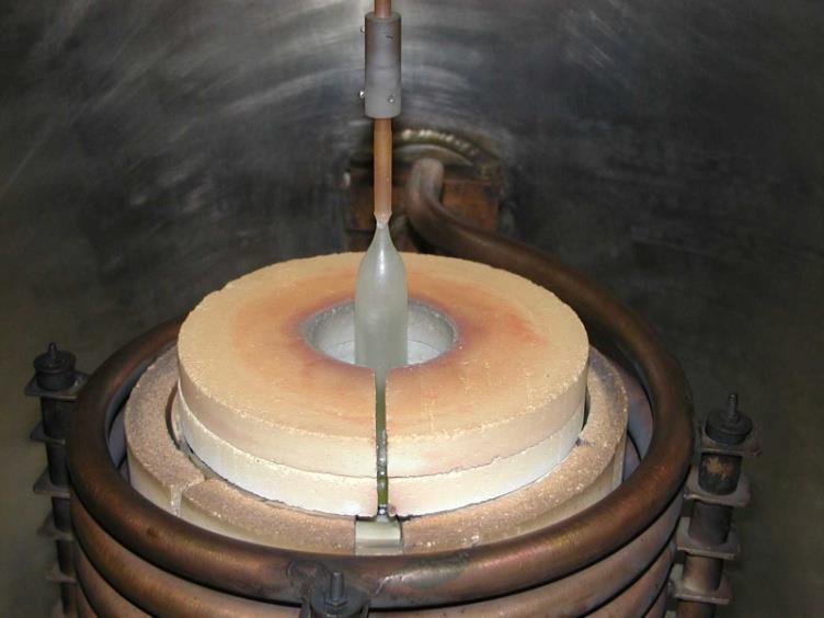
22 Crystal production process
 photomultiplier tubes detects the light quanta emitted by the crystal and converts their energies into electrical pulses.")

23 Radiation emanating from the patient is detected by a single, large, NaI (Tl) scintillation crystal. An array of (37, 61 or 91) photomultiplier tubes detects the light quanta emitted by the crystal and converts their energies into electrical pulses. Associated electronic circuitry determines the x, y coordinates of each scintillation event, the outputs of all the tubes being summed to provide a z pulse, the amplitude of which corresponds to the total energy of the scintillation event. The gamma camera is connected to a dedicated computer, radioactive distributions being displayed on a monitor.


24 PM-TUBES
25 PULSE HEIGHT ANALYZER UL Pulse height (V) For example: Tc99m ±10% window Peak energy140 Kev UL-154 kev LL- 126 kev LL Time The pulse height analyzer allows only pulses of a certain height (energy) to be counted. counted not counted

26 PULSE-HEIGHT DISTRIBUTION NAI(TL)
27 GAMMA CAMERA DATA ACQUISITION Static Dynamic ECG-gated Wholebody scanning Tomography ECG-gated tomography

28 PLANAR AND SPECT IMAGING
 where each frame then represents a certain phase (t) of the heart cycle.")
29 The ECG gated acquisition is a type of dynamic acquisition. The RR-interval is divided into a certain number of frames (16 to 32) where each frame then represents a certain phase (t) of the heart cycle. Each time the R-peak in the ECG is detected the collection of data starts in frame 1 during time t, then in frame 2 during time t and so on. The total acquisition time is generally about 10 min so the whole study represents several hundred heart cycles. ECG-GATED ACQUISITION R Interval n Image n
 is the end diastolic image and the sixth image (2nd row) is the end systolic image.")
30 ECG-GATED BLOODPOOL SCINTIGRAPHY Why such a study? Example of 16 frames of an ECG gated acquisition in LAO projection. The first image (upper left) is the end diastolic image and the sixth image (2nd row) is the end systolic image. This is the method of ejection fraction measurement


31 LEFT VENTRICLE TIME-ACTIVITY CURVE EjectEjection Fraction ~58 %


32 WHOLE BODY SCANNING Either the camera or the examination table moves

33

34

35 TOMOGRAPHIC ACQUISITION

36 TOMOGRAPHIC PLANES Gamma camera tomography is imaging of a volume where slices can be displayed in any plane

37 FACTORS AFFECTING IMAGE FORMATION Distribution of radiopharmaceutical Collimator selection and sensitivity Spatial resolution Energy resolution Uniformity Count rate performance Spatial positioning at different energies Center of rotation Scattered radiation Attenuation Noise
38 SPATIAL RESOLUTION Sum of intrinsic resolution and the collimator resolution Intrinsic resolution depends on the positioning of the scintillation events (detector thickness, number of PM-tubes, photon energy) Collimator resolution depends on the collimator geometry (size, shape and length of the holes)

39 SPATIAL RESOLUTION Object point Image stain Intensity IMAGING WITH GAMMA CAMERA IS NOT PERFECT!

40 SPATIAL RESOLUTION - DISTANCE RESOLUTION DEPENDS ON THE DISTANCE Optimal Large distance

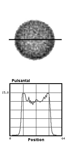
41 NON-UNIFORMITY The image to the right is aquired after cleaning of the collimator (Contamination of collimator)

42 NON UNIFORMITY RING ARTIFACTS The lower image is the absolute difference between the upper two images. It clearly shows the ring artifacts. Good uniformity Bad uniformity Difference

43 NON-UNIFORMITY The images in the lower row are acquired using a collimator with 50% lower sensitivity in an 1cm 3 area in the center of the field of view. The images in the upper row are from the same patient acquired with a good collimator. It is important to point out the risk of false positive results if the camera is not working perfectly, Defect collimator
44 SCATTERED RADIATION Remember that compton scattering is the dominating process in the attenuation of photons in soft tissue. Scattered photon photon electron
45 THE AMOUNT OF SCATTERED PHOTONS REGISTERED depends on: Patient size Energy resolution of the gammacamera Window setting
46 In the case of a big patient some of the full energy photons that should have reached the gamma camera will be scattered in the patient. The relation scattered/full energy photons will increase with the volume of the patient. PATIENT SIZE
47 ATTENUATION CORRECTION


48 ATTENUATION CORRECTION Transmission measurements CT

49 Count density NOISE


50 VARIETY OF GAMMA CAMERAS scanners Division with regard for number of 1. One head rotating gamma camera 2. One head gamma camera 3. Two head rotating gamma camera 4. Three head gamma camera P. Zorga
51 Two head gamma camera for cardio studies
52
53 SKYLight Above the Crowd & Beyond the Possible A revolution in HOW we do nuclear medicine. A revolution in WHAT we do in nuclear medicine.

54 Skylight Gamma Camera Philips Skylight gamma camera showing pre-program motion From Park to Patient Loading

55 Skylight Gamma Camera Philips Skylight gamma camera showing pre-program motion From Park to Patient Loading


56 INTRODUCING THE MOST COMPLETE VARIABLE ANGLE FAMILY IN NUCLEAR MEDICINE... Ring Gantry economical solution advanced imaging small footprint Vertex V60 Forte No Gantry infinite flexibility advanced imaging expansion beyond NM Open Gantry SKYLight true openness advanced imaging bed imaging


57 GANTRY IS FASTENED INTO THE CEILING SKYLIGHT HOW IS THIS SYSTEM UNIQUE? Infinite Flexibility Free-Dimensional positioning image in any position- trauma image on any bed smallest & largest detector radius minimum: 0, maximum: room width unlimited upright imaging extreme high & low detector heights no limits on patient size excellent access & openness lymphoscintigraphy localization, pediatrics, claustrophobic patients


58 High Throughput improved efficiency bed imaging flexibility faster set-up without patient transfer to imaging table stage next patient with second table automatic collimator exchange one-touch gantry robotics


59 BEYOND THE POSSIBLE RADIOLOGY CT ROOM

60 ADAC Forte FAST, EASY COLLIMATOR EXCHANGE ADAC Forte Integrated design Fast & easy, 4 min per pair No carts to store or move No table removal Customer Benefit: Reduced floor space requirement Greater throughput Easier for technologist Efficiency

61 HIGH QUALITY CARDIAC SPECT ADAC Forte No dead space Superior depth resolution No effect on acquisition time Consistent set-up with AC Customer Benefit: Higher resolution Better deep lesion image quality High throughput No learning curve for techs
62 POSITRON EMISSION TOMOGRAPHY PET
63 Positron emission tomography (PET) One of the fastest growing techniques is, positron emission tomography (PET) that requires special instrumentations called PET tomographs. This technique allows clinicians to track organ function at a molecular level, therefore revealing intricate health changes earlier in individual patients than other diagnostic method.
 injected into patients to provide images of the body using specialized")
64 PET POSITRON EMISSION TOMOGRAPHY PET is an imaging technique that uses radioactive substances (positrons emitters) injected into patients to provide images of the body using specialized scanners.
65 RADIONUCLIDES POSITRON EMITTERS Radionuclide Halftime Particle energy (mean) C min 0.39 MeV N min 0.50 MeV O min 0.72 MeV F min 0.25 MeV Cu min 1.3 MeV Ga min 0.83 MeV Rb min 1.5 MeV
66 ANNIHILATION THE POSITIVE ELECTRON TRAVELS A CERTAIN DISTANCE (1-3 MM) BEFORE IT UNDERGOES ANIHILATIONS WITH A NEGATIVE ELECTRON, CREATING A PAIR OF COLLINEAR GAMMA RAYS. (511 kev) + + e - (511 kev) + (1-3 mm) Radionuclide


67 PET-SCANNER PRINCIPLE Detector Detector

68 M Dahlbom, UCLA PET-SCANNER

69

")
70 TUMOUR STAGING WITH PET (F18-FDG) CLINICAL USE

71

72

73 CLINICAL BENEFITS OF PET IMAGING Impact on patient management Reduced patient risk/improved patient outcome Increased revenue for diagnostic services Aid to defining appropriate care pathways Decreased overall healthcare costs

74 Reliable Performance ECAT EXACT

75 Case Study 1 ECAT EXACT

76 Highest Performance ECAT EXACT HR+


77 ECAT EXACT HR+ Case Study 1


78 RDS SOLUTIONS RDS Eclipse * RDS 111 *Works-in-progress

79 PET-MRI

80 PET-MRI

81 PET-MRI
Radionuclide Imaging MII Positron Emission Tomography (PET)
") Radionuclide Imaging MII 3073 Positron Emission Tomography (PET) Positron (β + ) emission Positron is an electron with positive charge. Positron-emitting radionuclides are most commonly produced in cyclotron
Radionuclide Imaging MII 3073 Positron Emission Tomography (PET) Positron (β + ) emission Positron is an electron with positive charge. Positron-emitting radionuclides are most commonly produced in cyclotron
MEDICAL EQUIPMENT: NUCLEAR MEDICINE. Prof. Yasser Mostafa Kadah
 MEDICAL EQUIPMENT: NUCLEAR MEDICINE Prof. Yasser Mostafa Kadah www.k-space.org Recommended Textbook Introduction to Medical Imaging: Physics, Engineering and Clinical Applications, by Nadine Barrie Smith
Radioisotopes in action. Diagnostic application of radioisotopes. Steps of diagnostic procedure. Information from various medical imaging techniques
 Radioisotopes in action Diagnostic application of radioisotopes Steps of diagnostic procedure - Radioactive material introduced into the patient - Distribution and alteration of activity is detected -
Radionuclide Imaging MII Detection of Nuclear Emission
 Radionuclide Imaging MII 3073 Detection of Nuclear Emission Nuclear radiation detectors Detectors that are commonly used in nuclear medicine: 1. Gas-filled detectors 2. Scintillation detectors 3. Semiconductor
A. I, II, and III B. I C. I and II D. II and III E. I and III
 BioE 1330 - Review Chapters 7, 8, and 9 (Nuclear Medicine) 9/27/2018 Instructions: On the Answer Sheet, enter your 2-digit ID number (with a leading 0 if needed) in the boxes of the ID section. Fill in
Radiation Detection and Measurement
 Radiation Detection and Measurement June 2008 Tom Lewellen Tkldog@u.washington.edu Types of radiation relevant to Nuclear Medicine Particle Symbol Mass (MeV/c 2 ) Charge Electron e-,! - 0.511-1 Positron
There are three mechanisms by which gamma rays interact with absorber atoms from which two are important for nuclear medicine.
 Measurement of radioactivity. Radioactive decay is a random process and therefore fluctuations are expected in the radioactivity measurement. That is why measurement of radioactivity must be treated by
List of Nuclear Medicine Radionuclides. Nuclear Medicine Imaging Systems: The Scintillation Camera. Crystal and light guide
 Nuclear Medicine Imaging Systems: The Scintillation Camera List of Nuclear Medicine Radionuclides Tc99m 140.5 kev 6.03 hours I-131 364, 637 kev 8.06 days I-123 159 kev 13.0 hours I-125 35 kev 60.2 days
Nuclear Medicine Intro & Physics from Medical Imaging Signals and Systems, Chapter 7, by Prince and Links
 Nuclear Medicine Intro & Physics from Medical Imaging Signals and Systems, Chapter 7, by Prince and Links NM - introduction Relies on EMISSION of photons from body (versus transmission of photons through
Radiation Detectors. How do we detect ionizing radiation? What are these effects? Types of Ionizing Radiation Detectors
 Radiation Detectors 1 How do we detect ionizing radiation? Indirectly, by its effects as it traverses matter? What are these effects? Ionization and excitation of the atoms and molecules Heat 2 Types of
Detector technology. Aim of this talk. Principle of a radiation detector. Interactions of gamma photons (gas) Gas-filled detectors: examples
 Gas-filled detectors: examples") Aim of this tal Detector technology WMIC Educational Program Nuclear Imaging World Molecular Imaging Congress, Dublin, Ireland, Sep 5-8, 202 You can now the name of a bird in all the languages of the world,
Aim of this tal Detector technology WMIC Educational Program Nuclear Imaging World Molecular Imaging Congress, Dublin, Ireland, Sep 5-8, 202 You can now the name of a bird in all the languages of the world,
Introduction to SPECT & PET TBMI02 - Medical Image Analysis 2017
 Introduction to SPECT & PET TBMI02 - Medical Image Analysis 2017 Marcus Ressner, PhD, Medical Radiation Physicist, Linköping University Hospital Content What is Nuclear medicine? Basic principles of Functional
Chemical Engineering 412
 Chemical Engineering 412 Introductory Nuclear Engineering Lecture 26 Radiation Detection & Measurement II Spiritual Thought 2 I would not hold the position in the Church I hold today had I not followed
Compton Camera. Compton Camera
 Diagnostic Imaging II Student Project Compton Camera Ting-Tung Chang Introduction The Compton camera operates by exploiting the Compton Effect. It uses the kinematics of Compton scattering to contract
1st Faculty of Medicine, Charles University in Prague Center for Advanced Preclinical Imaging (CAPI)
") Radioation Resolution and Sensitivity Nuclear Imaging PET + SPECT Radioactive Decay (EC,Ɣ), (β -,Ɣ), (I.T.,Ɣ) β + Projection imaging collimator needed one angular view Projection imaging coincidence imaging,
Radioation Resolution and Sensitivity Nuclear Imaging PET + SPECT Radioactive Decay (EC,Ɣ), (β -,Ɣ), (I.T.,Ɣ) β + Projection imaging collimator needed one angular view Projection imaging coincidence imaging,
Physics in Nuclear Medicine
 SIMON R. CHERRY, PH.D. Professor Department of Biomedical Engineering University of California-Davis Davis, California JAMES A. SORENSON, PH.D. Emeritus Professor of Medical Physics University of Wisconsin-Madison
Radioisotopes in action. Diagnostic application of radioisotopes. Steps of diagnostic procedure. Information from various medical imaging techniques
 Radioisotopes in action Diagnostic application of radioisotopes Steps of diagnostic procedure - Radioactive material introduced into the patient - Distribution and alteration of activity is detected -Monitoring
6: Positron Emission Tomography
 6: Positron Emission Tomography. What is the principle of PET imaging? Positron annihilation Electronic collimation coincidence detection. What is really measured by the PET camera? True, scatter and random
Technical University of Denmark
 Technical University of Denmark Page 1 of 11 pages Written test, 9 December 2010 Course name: Introduction to medical imaging Course no. 31540 Aids allowed: none. "Weighting": All problems weight equally.
Radioactivity. Lecture 6 Detectors and Instrumentation
 Radioactivity Lecture 6 Detectors and Instrumentation The human organs Neither humans nor animals have an organ for detecting radiation from radioactive decay! We can not hear it, smell it, feel it or
Radioisotopes and PET
 Radioisotopes and PET 1 Radioisotopes Elements are defined by their number of protons, but there is some variation in the number of neutrons. Atoms resulting from this variation are called isotopes. Consider
 www.aask24.com www.aask24.com www.aask24.com P=Positron E= Emission T=Tomography Positron emission or beta plus decay (+ ) is a particular type of radioactive decay, in which a proton inside a radionuclide
Nuclear Medicine RADIOPHARMACEUTICAL CHEMISTRY
 Nuclear Medicine RADIOPHARMACEUTICAL CHEMISTRY An alpha particle consists of two protons and two neutrons Common alpha-particle emitters Radon-222 gas in the environment Uranium-234 and -238) in the environment
GLOSSARY OF BASIC RADIATION PROTECTION TERMINOLOGY
 GLOSSARY OF BASIC RADIATION PROTECTION TERMINOLOGY ABSORBED DOSE: The amount of energy absorbed, as a result of radiation passing through a material, per unit mass of material. Measured in rads (1 rad
Structure of Biological Materials
 ELEC ENG 3BA3: Structure of Biological Materials Notes for Lecture #19 Monday, November 22, 2010 6.5 Nuclear medicine imaging Nuclear imaging produces images of the distribution of radiopharmaceuticals
Nuclear Radiation. Natural Radioactivity. A person working with radioisotopes wears protective clothing and gloves and stands behind a shield.
 Nuclear Radiation Natural Radioactivity A person working with radioisotopes wears protective clothing and gloves and stands behind a shield. 1 Radioactive Isotopes A radioactive isotope has an unstable
Sample Spectroscopy System Hardware
 Semiconductor Detectors vs. Scintillator+PMT Detectors Semiconductors are emerging technology - Scint.PMT systems relatively unchanged in 50 years. NaI(Tl) excellent for single-photon, new scintillation
CT-PET calibration : physical principles and operating procedures F.Bonutti. Faustino Bonutti Ph.D. Medical Physics, Udine University Hospital.
 CT-PET calibration : physical principles and operating procedures Faustino Bonutti Ph.D. Medical Physics, Udine University Hospital Topics Introduction to PET physics F-18 production β + decay and annichilation
Bases of radioisotope diagnostic methods
 Medical, pharmaceutical applications of radioisotopes Bases of radioisotope diagnostic methods Dr. István Voszka Basis of application: radioisotopes have identical behavior in the organism to corresponding
PET scan simulation. Meysam Dadgar. UMSU, Iran. IFMP, Elbasan, Fig 1: PET camera simulation in gate by cylindrical phantom
 PET scan simulation Meysam Dadgar UMSU, Iran IFMP, Elbasan, 2016 Meysamdadgar10@gmail.com 1 Fig 1: PET camera simulation in gate by cylindrical phantom 2 What is PET? Positron emission tomography (PET),
The Nuclear Imaging Uncertainty Principle. Do Nuclear Cameras Really Work? Richard M. Fleming
 The Nuclear Imaging Uncertainty Principle. Do Nuclear Cameras Really Work? Richard M. Fleming Address correspondence to: Dr. R.M. Fleming 1697 Lone Oak Trail Reno, NV 89523 Rmfmd7@hotmail.com 12 October
Dana-Farber Cancer Institute, 44 Binney Street, Boston, MA 02115, USA ramsey
 SPECIAL FEATURE: MEDICAL PHYSICS www.iop.org/journals/physed Nuclear medicine Ramsey D Badawi Dana-Farber Cancer Institute, 44 Binney Street, Boston, MA 02115, USA E-mail: ramsey badawi@dfci.harvard.edu
PET. Technical aspects
 PET Technical aspects 15 N 15 O Detector 1 β+ Detector 2 e- Evolution of PET Detectors CTI/Siemens 15 N 15 O Detector block 1 β+ Detector block 2 x e- x y y location line of response Constant fraction
Radiation Dose, Biology & Risk
 ENGG 167 MEDICAL IMAGING Lecture 2: Sept. 27 Radiation Dosimetry & Risk References: The Essential Physics of Medical Imaging, Bushberg et al, 2 nd ed. Radiation Detection and Measurement, Knoll, 2 nd Ed.
hν' Φ e - Gamma spectroscopy - Prelab questions 1. What characteristics distinguish x-rays from gamma rays? Is either more intrinsically dangerous?
 Gamma spectroscopy - Prelab questions 1. What characteristics distinguish x-rays from gamma rays? Is either more intrinsically dangerous? 2. Briefly discuss dead time in a detector. What factors are important
Study of the feasibility of a compact gamma camera for real-time cancer assessment
 Study of the feasibility of a compact gamma camera for real-time cancer assessment L. Caballero Instituto de Física Corpuscular - CSIC - University of Valencia; C/Catedrático José Beltrán, 2; E-46980;
Chapter 2 PET Imaging Basics
 Chapter 2 PET Imaging Basics Timothy G. Turkington PET Radiotracers Positron emission tomography (PET) imaging is the injection (or inhalation) of a substance containing a positron emitter, the subsequent
Detection and measurement of gamma-radiation by gammaspectroscopy
 Detection and measurement of gamma-radiation by gammaspectroscopy Gamma-radiation is electromagnetic radiation having speed equal to the light in vacuum. As reaching a matter it interact with the different
Mitigation of External Radiation Exposures
 Mitigation of External Radiation Exposures The three (3) major principles to assist with maintaining doses ALARA are :- 1) Time Minimizing the time of exposure directly reduces radiation dose. 2) Distance
3. Which of the following statements is (are) TRUE about detector crystals in Anger cameras?
 TRUE about detector crystals in Anger cameras?") BioE 1330 - Exam 2 11/13/2018 Answer Sheet - Correct answer is A for all questions 1. Unlike CT, in nuclear medicine A. Bremsstrahlung is not used to produce high-energy photons. B. signal can be increased
BioE 1330 - Exam 2 11/13/2018 Answer Sheet - Correct answer is A for all questions 1. Unlike CT, in nuclear medicine A. Bremsstrahlung is not used to produce high-energy photons. B. signal can be increased
DEVIL PHYSICS THE BADDEST CLASS ON CAMPUS IB PHYSICS
 DEVIL PHYSICS THE BADDEST CLASS ON CAMPUS IB PHYSICS TSOKOS OPTION I-2 MEDICAL IMAGING Reading Activity Answers IB Assessment Statements Option I-2, Medical Imaging: X-Rays I.2.1. I.2.2. I.2.3. Define
Lecture 5: Tomographic nuclear systems: SPECT
 Lecture 5: Tomographic nuclear systems: SPECT Field trip this saturday at 11 AM at UWMC meet in main hospital lobby at 11 AM if you miss the 'boat', page me at 540-4950 should take ~1 to 1.5 hours, depending
Medical Physics. Nuclear Medicine Principles and Applications
 Medical Physics Nuclear Medicine Principles and Applications Dr Roger Fulton Department of PET & Nuclear Medicine Royal Prince Alfred Hospital Sydney Email: rfulton@mail.usyd.edu.au Lectures: http://www-personal.usyd.edu.au/~rfulton/medical_physics
Chapter Seven (Nuclear Detectors)
") Al-Mustansiriyah University College of Science Physics Department Fourth Grade Nuclear Physics Dr. Ali A. Ridha Chapter Seven (Nuclear Detectors) Ionizing radiation is rarely detected directly. Instead,
Al-Mustansiriyah University College of Science Physics Department Fourth Grade Nuclear Physics Dr. Ali A. Ridha Chapter Seven (Nuclear Detectors) Ionizing radiation is rarely detected directly. Instead,
(INCLUDING THIS FRONT PAGE)
") I'IFIITIIBIFI UNIVERSITY OF SCIEI'ICE RITD TECHNOLOGY FACULTY OF HEALTH AND APPLIED SCIENCES DEPARTMENT OF NATURAL AND APPLIED SCIENCES QUALIFICATION: BACHELOR OF SCIENCE (MAJOR AND MINOR) QUALIFICATION
I'IFIITIIBIFI UNIVERSITY OF SCIEI'ICE RITD TECHNOLOGY FACULTY OF HEALTH AND APPLIED SCIENCES DEPARTMENT OF NATURAL AND APPLIED SCIENCES QUALIFICATION: BACHELOR OF SCIENCE (MAJOR AND MINOR) QUALIFICATION
Differentiating Chemical Reactions from Nuclear Reactions
 Differentiating Chemical Reactions from Nuclear Reactions 1 CHEMICAL Occurs when bonds are broken or formed. Atoms remained unchanged, though may be rearranged. Involves valence electrons Small energy
EEE4106Z Radiation Interactions & Detection
 EEE4106Z Radiation Interactions & Detection 2. Radiation Detection Dr. Steve Peterson 5.14 RW James Department of Physics University of Cape Town steve.peterson@uct.ac.za May 06, 2015 EEE4106Z :: Radiation
FXA UNIT G485 Module X-Rays. Candidates should be able to : I = I 0 e -μx
 1 Candidates should be able to : HISTORY Describe the nature of X-rays. Describe in simple terms how X-rays are produced. X-rays were discovered by Wilhelm Röntgen in 1865, when he found that a fluorescent
Outline Chapter 14 Nuclear Medicine
 Outline Chapter 14 uclear Medicine Radiation Dosimetry I Text: H.E Johns and J.R. Cunningham, The physics of radiology, 4 th ed. http://www.utoledo.edu/med/depts/radther Introduction Detectors for nuclear
Basic physics of nuclear medicine
 Basic physics of nuclear medicine Nuclear structure Atomic number (Z): the number of protons in a nucleus; defines the position of an element in the periodic table. Mass number (A) is the number of nucleons
Gamma ray coincidence and angular correlation
 University of Cape Town Department of Physics Course III laboratory Gamma ray coincidence and angular correlation Introduction Medical imaging based on positron emission tomography (PET) continues to have
Scintillation Detector
 Scintillation Detector Introduction The detection of ionizing radiation by the scintillation light produced in certain materials is one of the oldest techniques on record. In Geiger and Marsden s famous
Procesamiento de Imágenes y Bioseñales
 Procesamiento de Imágenes y Bioseñales Dr. Víctor Castañeda Agenda Physical basis of X-ray- CT, NMR, Ultrasound, Nuclear Medicine Sensors (cameras, gamma probes, microphone) Computational Tomography (CT)
City University of Hong Kong
 City University of Hong Kong Information on a Course offered by the Department of Physics and Materials Science with effect from Semester A in 2013 / 2014 Part I Course Title: Radiological Physics and
CLINICALLY USEFUL RADIONUCLIDES:
 INTRODUCTION It is important that Nuclear Medicine Technologists be familiar with the imaging properties of all commonly used radionuclides to insure correct choice of isotope for a particular study as
Essentials of nuclear medicine
 Essentials of nuclear medicine Medical imaging CT Rtg X- rays usg Ultrasound MR Nuclear Magnetic Resonance Nuclear Medicine SPECT PET A conventional radiological, ultrasound and magnetic resonance diagnostics
CHAPTER 5. Department of Medical Physics, University of the Free State, Bloemfontein, South Africa
 CHAPTE 5 STATISTICS FO ADIATIO MEASUEMET M.G. LÖTTE Department of Medical Physics, University of the Free State, Bloemfontein, South Africa 5.1. SOUCES OF EO I UCLEA MEDICIE MEASUEMET Measurement errors
11/10/2014. Chapter 1: Introduction to Medical Imaging. Projection (Transmission) vs. Emission Imaging. Emission Imaging
 vs. Emission Imaging. Emission Imaging") Chapter 1: Introduction to Medical Imaging Overview of Modalities Properties of an Image: Limitations on Information Content Contrast (both object & image): Brightness difference Sharpness (blur): Smallest
Chapter 1: Introduction to Medical Imaging Overview of Modalities Properties of an Image: Limitations on Information Content Contrast (both object & image): Brightness difference Sharpness (blur): Smallest
Absolute activity measurement
 Absolute activity measurement Gábor Veres, Sándor Lökös Eötvös University, Department of Atomic Physics January 12, 2016 Financed from the financial support ELTE won from the Higher Education Restructuring
Positron Emission Tomography
 Positron Emission Tomography Presenter: Difei Wang June,2018 Universität Bonn Contents 2 / 24 1 2 3 4 Positron emission Detected events Detectors and configuration Data acquisition Positron emission Positron
Radiation Detection. 15 th Annual OSC Readiness Training Program.
 Radiation Detection 15 th Annual OSC Readiness Training Program www.oscreadiness.org GM Detectors 15 th Annual OSC Readiness Training Program www.oscreadiness.org 1 A closer look 15 th Annual OSC Readiness
Application of Nuclear Physics
 Application of Nuclear Physics Frontier of gamma-ray spectroscopy 0.1 IR visible light UV soft X-ray X-ray hard X-ray gamma-ray 1 10 100 1e3 1e4 1e5 1e6 energy [ev] Photoelectric effect e - Compton scattering
A Brief Introduction to Medical Imaging. Outline
 A Brief Introduction to Medical Imaging Outline General Goals Linear Imaging Systems An Example, The Pin Hole Camera Radiations and Their Interactions with Matter Coherent vs. Incoherent Imaging Length
Theoretical questions for the final exam ED 2012.
 Theoretical questions for the final exam ED 2012. 1. Radiation a) Properties and types of radiation b) Physical parameters of radiation 2. Law of attenuation of radiation a) Experimental interpretation
Rad T 290 Worksheet 2
 Class: Date: Rad T 290 Worksheet 2 1. Projectile electrons travel from a. anode to cathode. c. target to patient. b. cathode to anode. d. inner shell to outer shell. 2. At the target, the projectile electrons
AQA Physics /7408
 AQA Physics - 7407/7408 Module 10: Medical physics You should be able to demonstrate and show your understanding of: 10.1 Physics of the eye 10.1.1 Physics of vision The eye as an optical refracting system,
Tomography is imaging by sections. 1
 Tomography is imaging by sections. 1 It is a technique used in clinical medicine and biomedical research to create images that show how certain tissues are performing their physiological functions. 1 Conversely,
NORM and TENORM: Occurrence, Characterizing, Handling and Disposal
 NORM and TENORM: Occurrence, Characterizing, Handling and Disposal Ionizing Radiation and Hazard Potential John R. Frazier, Ph.D. Certified Health Physicist May 12, 2014 Radiation Radiation is a word that
ความร พ นฐานและเทคน คการบ าร งร กษา เคร องโดสคาล เบรเตอร (Dose Calibrator) ส ว ทย ป ณณช ยยะ
 ส ว ทย ป ณณช ยยะ") ความร พ นฐานและเทคน คการบ าร งร กษา เคร องโดสคาล เบรเตอร (Dose Calibrator) ส ว ทย ป ณณช ยยะ Principle & Maintenance Technique of Dose Calibrator Contents : 1. General principle 2. Basic system operation
ความร พ นฐานและเทคน คการบ าร งร กษา เคร องโดสคาล เบรเตอร (Dose Calibrator) ส ว ทย ป ณณช ยยะ Principle & Maintenance Technique of Dose Calibrator Contents : 1. General principle 2. Basic system operation
Introduction to Medical Imaging. Medical Imaging
 Introduction to Medical Imaging BME/EECS 516 Douglas C. Noll Medical Imaging Non-invasive visualization of internal organs, tissue, etc. I typically don t include endoscopy as an imaging modality Image
SCINTILLATION DETECTORS & GAMMA SPECTROSCOPY: AN INTRODUCTION
 SCINTILLATION DETECTORS & GAMMA SPECTROSCOPY: AN INTRODUCTION OBJECTIVE The primary objective of this experiment is to use an NaI(Tl) detector, photomultiplier tube and multichannel analyzer software system
Wiper Gold Edition. Premium Wipe Test Counter. Setting the bar higher
 Wiper Gold Edition Premium Wipe Test Counter Setting the bar higher Software - Simplicity, Re-defined Load your wipe into the well and press the button. Your wipe is counted and automatically converted
INTERACTIONS OF RADIATION WITH MATTER
 INTERACTIONS OF RADIATION WITH MATTER Renée Dickinson, MS, DABR Medical Physicist University of Washington Medical Center Department of Radiology Diagnostic Physics Section Outline Describe the various
Dosimetry. Sanja Dolanski Babić May, 2018.
 Dosimetry Sanja Dolanski Babić May, 2018. What s the difference between radiation and radioactivity? Radiation - the process of emitting energy as waves or particles, and the radiated energy Radioactivity
SCINTILLATION DETECTORS AND PM TUBES
 SCINTILLATION DETECTORS AND PM TUBES General Characteristics Introduction Luminescence Light emission without heat generation Scintillation Luminescence by radiation Scintillation detector Radiation detector
MEDICAL IMAGING. METHODS OF MODERN IMAGING, BASED ON ELECTRO-MAGNETIC RADIATION (radiowaves, infrared radiation, X-rays, γ-rays ) AND ULTRASOUND
 AND ULTRASOUND") MEDICAL IMAGING MEDICAL IMAGING METHODS OF MODERN IMAGING, BASED ON ELECTRO-MAGNETIC RADIATION (radiowaves, infrared radiation, X-rays, γ-rays ) AND ULTRASOUND MEDICAL IMAGING RADIOLOGY NUCLEAR MEDICINE
MEDICAL IMAGING MEDICAL IMAGING METHODS OF MODERN IMAGING, BASED ON ELECTRO-MAGNETIC RADIATION (radiowaves, infrared radiation, X-rays, γ-rays ) AND ULTRASOUND MEDICAL IMAGING RADIOLOGY NUCLEAR MEDICINE
THE ACTIVITY CALIBRATOR
 A.O.U. OSPEDALI RIUNITI di TRIESTE S.C. di FISICA SANITARIA THE ACTIVITY CALIBRATOR Dr. Maria Rosa Fornasier 1 INDEX GENERAL FEATURES DETECTOR DESIGN CALIBRATION PROCEDURE - EFFECTS OF AN EXTERNAL SHIELD
Jazan University College of Science Physics Department. Lab Manual. Nuclear Physics (2) 462 Phys. 8 th Level. Academic Year: 1439/1440
 462 Phys. 8 th Level. Academic Year: 1439/1440") Jazan University College of Science Physics Department جاهعة جازان كلية العل وم قسن الفيزياء Lab Manual Nuclear Physics (2) 462 Phys 8 th Level Academic Year: 1439/1440 1 Contents No. Name of the Experiment
Jazan University College of Science Physics Department جاهعة جازان كلية العل وم قسن الفيزياء Lab Manual Nuclear Physics (2) 462 Phys 8 th Level Academic Year: 1439/1440 1 Contents No. Name of the Experiment
Experiment 6 1. The Compton Effect Physics 2150 Experiment No. 6 University of Colorado
 Experiment 6 1 Introduction The Compton Effect Physics 2150 Experiment No. 6 University of Colorado In some situations, electromagnetic waves can act like particles, carrying energy and momentum, which
Overview of Nuclear Medical Imaging Instrumentation and Techniques*
 Overview of Nuclear Medical Imaging Instrumentation and Techniques* William W. Moses Lawrence Berkeley National Laboratory, University of California, Berkeley, CA 94720 USA Abstract. Nuclear medical imaging
Final exam questions ED
 Final exam questions ED 2015-2016 1. Radiation a) Properties and types of radiation b) Physical parameters of radiation 2. Law of attenuation of radiation a) Experimental interpretation of the law b) Forms
Mayneord-Phillips Summer School St Edmund Hall, University of Oxford July Proton decays to n, e +, ν
 Positron Emission Tomography Physics & Instrumentation Dimitra G. Darambara, Ph.D Multimodality Molecular Imaging Joint Department of Physics RMH/ICR Outline Introduction PET Physics overview Types of
DETECTORS. I. Charged Particle Detectors
 DETECTORS I. Charged Particle Detectors A. Scintillators B. Gas Detectors 1. Ionization Chambers 2. Proportional Counters 3. Avalanche detectors 4. Geiger-Muller counters 5. Spark detectors C. Solid State
Activities at the Laboratory of the Nuclear Engineering Department of the Polytechnic University of Valencia
 7 th Workshop on European Collaboration for Higher Education and Research in Nuclear Engineering & Radiological Protection Bruxelles, Belgique 30 May - 1 June 2011 Activities at the Laboratory of the Nuclear
EXPERIMENTAL DETERMINATION OF SHIELDING REQUIREMENTS FOR PET MEDICAL FACILITIES BRADLEY S. BRINKLEY
 EXPERIMENTAL DETERMINATION OF SHIELDING REQUIREMENTS FOR PET MEDICAL FACILITIES by BRADLEY S. BRINKLEY CLAUDIU T. LUNGU, COMMITTEE CHAIR ALFRED A. BARTOLUCCI STEVEN M. BECKER RIEDAR K. OESTENSTAD SHARON
GUIDE TO LABORATORY SURVEYS. Introduction
 APPENDIX - V GUIDE TO LABORATORY SURVEYS Introduction Routine laboratory surveys are an important part of the overall radiation safety program in a laboratory. Surveys provide a direct measure of the presence
University of Ljubljana Faculty of mathematics and physics Department of physics. Tomography. Mitja Eržen. August 6, Menthor: Dr.
 University of Ljubljana Faculty of mathematics and physics Department of physics Tomography Mitja Eržen August 6, 2009 Menthor: Dr. Matjaž Vencelj Abstract We ll describe some methods for medical imaging.i
The Physics of PET/CT scanners
 The Physics of PET/CT scanners Ruth E. Schmitz, Adam M. Alessio, and Paul E. Kinahan Imaging Research Laboratory Department of Radiology University of Washington What Makes PET Useful? Positron emission
Revision checklist. Step Learning outcome Had a look Nearly there Nailed it!
 Radioactivity a Atomic models Describe the structure of an atom (in terms of nucleus and electrons). State where most of the mass of an atom is found. State the sizes of atoms and small molecules. Describe
ELG7173 Topics in signal Processing II Computational Techniques in Medical Imaging
 ELG7173 Topics in signal Processing II Computational Techniques in Medical Imaging Topic #1: Intro to medical imaging Medical Imaging Classifications n Measurement physics Send Energy into body Send stuff
Compton Camera with PositionSensitive Silicon Detectors
 University of Ljubljana Faculty of mathematics and physics Andrej Studen Compton Camera with PositionSensitive Silicon Detectors Doctoral thesis Supervisor: Professor Marko Mikuž Outline: Motivation Basic
Photon Instrumentation. First Mexican Particle Accelerator School Guanajuato Oct 6, 2011
 Photon Instrumentation First Mexican Particle Accelerator School Guanajuato Oct 6, 2011 Outline The Electromagnetic Spectrum Photon Detection Interaction of Photons with Matter Photoelectric Effect Compton
Name: COMBINED SCIENCE Topics 4, 5 & 6 LEARNING OUTCOMES. Maintain a record of your progress Use the booklet to guide revision
 Name: COMBINED SCIENCE Topics 4, 5 & 6 LEARNING OUTCOMES Maintain a record of your progress Use the booklet to guide revision Close the Gap Contemporary record of the Topics / Learning outcomes that I
Application Note. Understanding Performance Specifications for Low Background Alpha Beta Counters. FOM What Is It and Is It Useful?
 Application Note Understanding Performance Specifications for Low Background Alpha Beta Counters Comparisons between vendors systems, often a tedious task, can lead to frustration and confusion. This application
Analytical Technologies in Biotechnology Prof. Dr. Ashwani K. Sharma Department of Biotechnology Indian Institute of Technology, Roorkee
 Analytical Technologies in Biotechnology Prof. Dr. Ashwani K. Sharma Department of Biotechnology Indian Institute of Technology, Roorkee Module - 2 Radioisotopes Techniques Lecture - 3 GM Counting and
Copyright 2008, University of Chicago, Department of Physics. Experiment VI. Gamma Ray Spectroscopy
 Experiment VI Gamma Ray Spectroscopy 1. GAMMA RAY INTERACTIONS WITH MATTER In order for gammas to be detected, they must lose energy in the detector. Since gammas are electromagnetic radiation, we must
General Overview of Radiation Detection and Equipment
 www.inl.gov INL/MIS-11-22727 General Overview of Radiation Detection and Equipment International Nuclear Safeguards Policy and Information Analysis Course Monterey Institute of International Studies June
Contents. Charged Particles. Coulomb Interactions Elastic Scattering. Coulomb Interactions - Inelastic Scattering. Bremsstrahlung
 Contents Marcel MiGLiERiNi Nuclear Medicine, Radiology and Their Metrological Aspects. Radiation in Medicine. Dosimetry 4. Diagnostics & Therapy 5. Accelerators in Medicine 6. Therapy Planning 7. Nuclear
QUIZ: Physics of Nuclear Medicine Atomic Structure, Radioactive Decay, Interaction of Ionizing Radiation with Matter
 QUIZ: Physics of Nuclear Medicine Atomic Structure, Radioactive Decay, Interaction of Ionizing Radiation with Matter 1. An atomic nucleus contains 39 protons and 50 neutrons. Its mass number (A) is a)
Gamma Spectroscopy. References: Objectives:
 Gamma Spectroscopy References: G.F. Knoll, Radiation Detection and Measurement (John Wiley & Sons, New York, 2000) W. R. Leo, Techniques for Nuclear and Particle Physics Experiments: A How-to Approach,