Radionuclide Imaging MII Detection of Nuclear Emission
|
|
|
- Alaina Franklin
- 6 years ago
- Views:
Transcription
1 Radionuclide Imaging MII 3073 Detection of Nuclear Emission
2 Nuclear radiation detectors Detectors that are commonly used in nuclear medicine: 1. Gas-filled detectors 2. Scintillation detectors 3. Semiconductor detectors 4. Film badge and Thermoluminescent dosimeters (TLD) Gas-filled detectors: 1. Geiger-Mueller (GM) counter 2. Ionization chamber 3. Dose calibrator Scintillation detectors: 1. Sodium iodide well counter 2. Single probe counting system 3. Dose calibrator
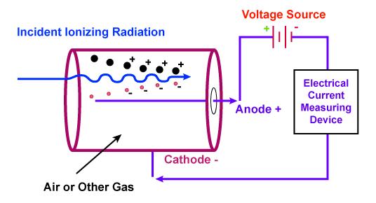
3 Gas-filled detectors Operational principle: measuring the ionization that radiation produces within the gas. Commonly for monitoring α and β radiations. Typical gases used are argon and helium. The central electrode is an anode, that has been insulated from the chamber walls and the cathode. A voltage is applied to the anode and the chamber walls. As a charged particle passes through, it ionizes some of the gas (air). The positive anode attracts the electrons, or negative particles. The detector wall, or cathode, attracts the positive charges. This movement of ions/charges is an electric current, which can be detected by a sensitive meter. The current between the electrodes is a measure of the amount of incoming radiation.
4 Gas-filled detectors
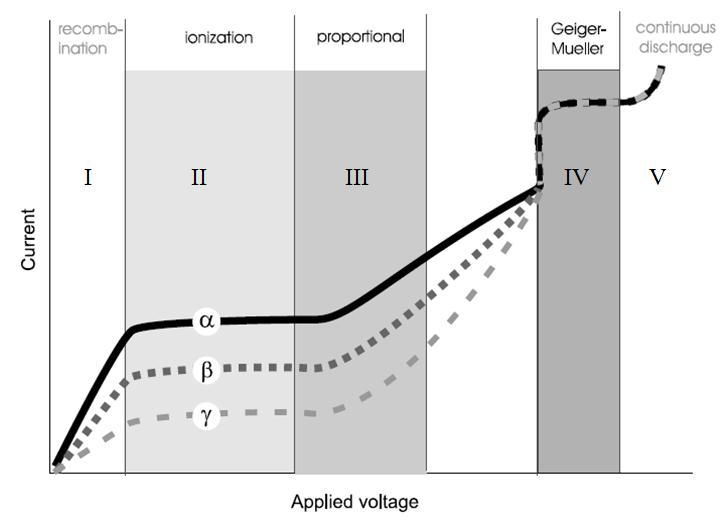
5 Gas-filled detectors The amount of current produced depends on several factors: 1. The applied voltage between the two electrodes 2. Distance between the two electrodes 3. Type of gas 4. Volume, pressure and temperature of the gas 5. Geometry and shape of the electrodes Typically for a gas-filled detector, the amount of the current produced by a single radiation is a function of the applied voltage. Their relationship can be divided to 5 distinct regions.
6 Gas-filled detectors
7 Gas-filled detectors Region I: recombination The voltage is low, some ion pairs are still able to recombine and form neutral atoms or molecules. Incomplete collection of primary ion pairs by the electrodes. As the voltage increases, more primary ion pairs are collected and more current flows. Region II: ionization plateau The voltage is sufficiently high to attract all primary ion pairs. Region III: proportional The higher voltage is able to attract all primary ion pairs and sufficient to provide energy to some primary ion pairs for producing secondary ion pairs through collisions with neutral atoms and molecules of the gas (gas amplification). The amount of secondary ion pairs produced depends on the energy acquired by primary ion pairs. The amount of current produced by a radiation increases with voltage increasing.
8 Gas-filled detectors Region IV: Geiger Muller As the voltage is increased, a point is reached at which most of the gas within the detector is massively involved in the multiple, successive ionizations (no more gas amplification). The pulse of current is larger but becomes independent of number of primary ion pairs produced. Region V: Continuous discharge The voltage is so high that radiation is not necessary to produce discharge. Under this high electric field, the electrons are pulled out form the atomic shells, the atoms and molecules become ionized and a discharge may be established even without radiation (spontaneous and continuous ionization). This interaction stops only when voltage is lowered.
9
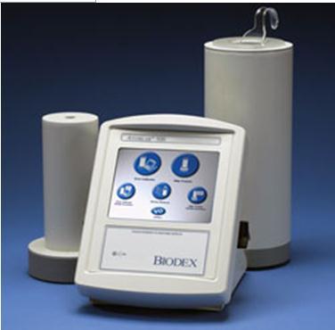
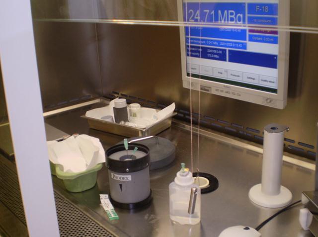
10 Dose calibrator A dose calibrator (activity meter) consists of: a cylindrically shaped, gas filled sealed chamber with a well, high voltage supply applied to electrodes, specific energy settings for different radionuclides, an activity readout (e.g. in MBq, GBq, etc). How to use dose calibrator? 1. Turn on the main power and wait for any self checks or warm-up up to complete. 2. Place the syringe or vial holder in the detector well. 3. Select appropriate (nuclide, energy) settings. 4. Zero the dose calibrator. 5. Measure the activity of the radionuclide in the syringe or vial. 6. Read the activity from the display console and record.
11
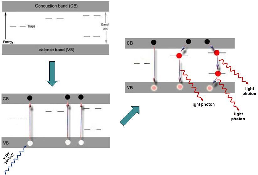
12 Scintillation detectors Scintillators are materials that emit visible or UV light following the interaction of ionizing radiation with material. The most widely used crystals are made of sodium iodide (NaI); clear glass-like structure, fragile and sealed in an airtight aluminum container. NaI crystals are doped with small amounts of stable thallium (Tl); improve response to gamma ray photons. When an incoming x or gamma ray hits the scintillation detector, it will interact with an electron (from the valence band) in the crystal, by either a Compton or PE process (energy transfer). Each of these energetic electrons distributes its energy among electrons in the crystals, leaving them in ionized and excited states.
13 Scintillation detectors These electrons may move to higher energy levels, known as the conduction band, until they fall into certain impurity centers, which act as energy traps. These traps are produced by the addition of chemical impurities into the crystal at the time of manufacture, called activators. For NaI, small amounts of thallium produce the trapping centers; (thallium-activated sodium iodide). For returning to the original state, the trapped electron may give up its energy in the form of a light photon. This light photon will then be detected and converted into electrical signal by photomultiplier tube (PMT).
14
15 Scintillation detectors The desirable properties of a scintillator are: 1. The conversion efficiency: the fraction of deposited energy that is converted into light should be high. (The conversion efficiency should not be confused with detection efficiency) 2. For many applications, the decay times of excited states should be short. (Light is emitted promptly after an interaction). 3. The material should be transparent to own emissions. (Most emitted light escapes reabsorption). 4. The frequency spectrum (color) of emitted light should match the spectral sensitivity of the light receptor (PMT, photodiode or film). 5. If used for x- and gamma-ray detection, the attenuation coefficient (µ) should be large, so that the scintillation detectors have high detection efficiency. (Materials with large atomic numbers and high densities have large attenuation coefficients). 6. The material should be rough, unaffected by moisture and inexpensive to manufacture.
16 Photomultiplier tube (PMT) The amount of light produced in Nai(Tl) crystals or any other scintillator is very small in volume. PMT is a light sensitive device that converts light into measurable electronic pulses. It consists of a photocathode facing the window through which light enters, a series of metallic electrodes known as dynodes arranged in special geometry and pattern, and an anode. All of these are enclosed in vacuum in a glass tube. Photocathode is a clear photosensitive glass surface that has been coupled with a light-conductive transparent gel to the surface of the crystal. The transparent gel has the same refractive index as the crystal and the PMT window.
17 PMT When the light photon hits the photocathode, it produces an electron of low energy through PE interaction; called photoelectron. This photoelectron is accelerated by a potential difference ( range of V) between the emitting surface and the 1 st dynode. Upon collision with the dynode, the electron acquires sufficient kinetic energy to create a number of secondary electrons. These secondary electrons are then accelerated toward a 2 nd dynode, with a similar electron multiplication. Eventually, at the last dynode (generally 10 th ) the total electron gain of about is produced. These electrons generate a current pulse of a few microamperes in amplitude and less than a microsecond in duration at the anode.
18 Anode Dynode Photo-electron Photo-cathode Visible light photon
19 Sodium iodide well counter Well counters are common in nuclear medicine laboratories, for performing in vitro studies as well as QC and QC procedures. Many NaI well counters are designed for counting radioactive samples in standard test tubes. Generally, there is a solid cylindrical NaI crystal with a cylindrical well cut into the crystal, into which the test tube is placed. PMT is optically coupled to the crystal base. Radiation from the sample interacts with the crystal and is detected by the PMT, which feeds into a scalar. The scalar readout directly reflects the amount of radioactivity in the sample and is usually recorded in counts for the period of measurement.

20 Sodium iodide well counter
21 Single probe counting system (thyroid probe) A thyroid probe has a single NaI crystal, a PMT at the end, and a single-hole collimator. Single probe counting systems using only 1 crystalline detector are useful for measuring not only thyroid uptake of radioactive iodine but also cardiac output. The probe used for thyroid counting is actually similar to the standard well counter, although it does not have the central hole in the NaI crystal. The typical crystal is 5 cm in diameter and 5 cm in thickness, with a cone-shaped collimator. Again, a PMT is located at the crystal base. When this probe is used, it is important for quantitative consistency to maintain a fixed distance from the object being measured to the face of the crystal and to eliminate all extraneous sources of background radiation.

22 Thyroid probe

23 Thyroid probe
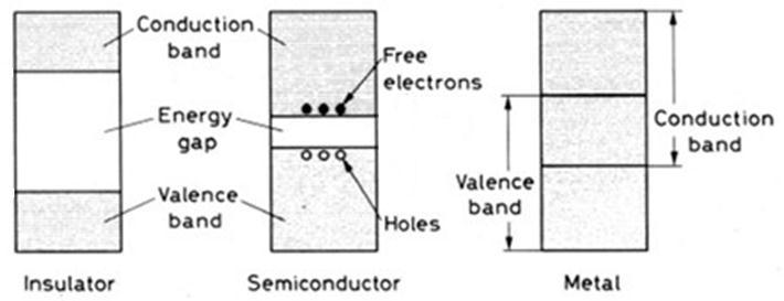
24 Semiconductor detectors In metals, the valence band is partially filled. However, in semiconductors and insulators, the valence band is completely filled and the conduction band is completely empty. The energy gap between the valence and the conduction bands of semiconductors is smaller than that of insulators. Thus, in semiconductors, electrons (in valence band) can be easily excited to the conduction band. When a photon enters a semiconductor, the energy of the photon is absorbed (PE, Compton or PP). The electrons produced by the primary interaction of photons with the semiconductor will transfer their energy to the valence electrons, thus elevating them into the conduction band.
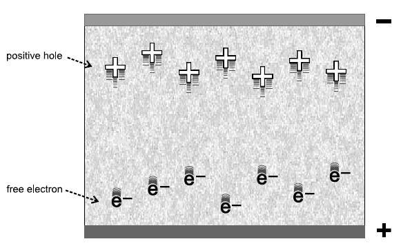
25 Semiconductor detectors This leaves equal numbers of holes in the valence band. These holes act as positively charged particles. If a voltage is applied across the semiconductor, the electrons in the conduction band will move towards the positive electrode and the holes in the valence band are for the negative electrode. Since the number of electron-hole pairs produced is proportional to the energy of the incident photon, the collection of charges on the respective electrodes results in a pulse whose height is proportional to the photon energy. This pulse can be amplified and energy-discriminated for counting purposes.
26
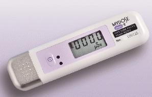
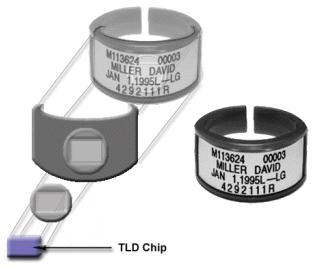
27 Film badge External radiation monitoring system. Film badge is the most common and economical, although not the most accurate. It consists of a small film enclosed in a plastic container with 4 windows of the covered with different radiation filters to identify the nature and energy exposing radiation. When the badge is exposed to ionizing radiation, the film emulsion darkens in proportion to the degree of radiation exposure received. The resultant optical density can be measured with a densitometer and calibrated to the degree of radiation exposure received. Film badge is capable of measuring exposures ranging from msv. The film is normally changed each month.
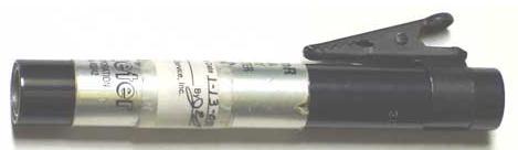
28 Thermoluminescent Dosimeter (TLD) Contains small chips of a thermoluminescent material, usually lithium fluoride (LiF). When exposed to radiation, a portion of the absorbed energy is stored in the crystal structure of the LiF chips in metastable states. If the LiF chips are heated, the absorbed energy is released as visible light. The heating and measurement of LiF chips are carried out in a device called a reader. The amount of measured light is proportional to the absorbed radiation dose.
29
30 Collimators The collimator is made of perforated or folded lead and is interposed between the patient and the scintillation crystal. It allows the gamma camera to accurately localize the radionuclide in the patient s body. Collimators perform this function by absorbing and stopping most radiation except that arriving perpendicular to the detector face.
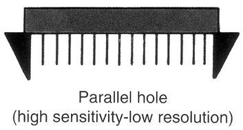
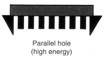
31 Collimators The collimator is made of perforated or folded lead and is interposed between the patient and the scintillation crystal. Nuclides emit gamma ray photons in all directions. The collimator allows only those photons travelling directly along the long axis of each hole to reach the crystal. Photons emitted in any other direction are absorbed by the septa between the holes. Without a collimator in front of the crystal, the image would be indistinct. Collimator is the rate limiting step in the imaging chain of gamma camera technology. Thus, by appropriate choice of collimator, it is possible to magnify of minify images and to select between imaging quality (resolution) and imaging speed (sensitivity).
32 Collimators Four types of collimators are commonly used with the gamma camera: 1. Parallel-hole 2. Pinhole 3. Converging 4. Diverging A parallel-hole collimator is made of a large number (many thousands) of small holes in a lead disc. The diameter of the lead disc is the same as the scintillation crystal used. Thickness of the lead disc and diameter of the holes depend on the desired spatial resolution and sensitivity of the collimators.
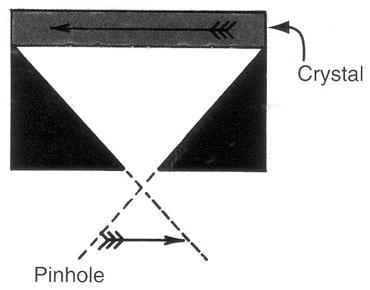
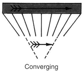
33 Collimators Pinhole collimator consists of a single hole, usually 2-4 mm in diameter. The image is projected upside down and reversed right to left at the crystal. However, it is usually corrected electronically on the viewing screen. A pinhole collimator generates magnified images of a small organ like the thyroid or a joint. In converging collimator, the holes are angled inward, toward the organ/patient. All holes focus at an axial point, outside the collimator. Therefore, the organ appears larger at the face of the crystal. A converging collimator may be used for examination of small areas.
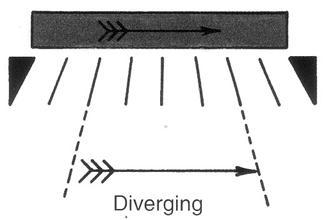
34 Collimators A diverging collimator, has holes and septa that begin to diverge from the crystal face. Generally, use of a diverging collimator increases the imaged are by about 30% over that obtained with a parallelhole. However, the image itself is slightly minified. Diverging collimator is used particularly on cameras with small crystal faces to image large organs, such as the lungs. Commercially, the collimators are also classified according to their spatial resolution or sensitivity as high sensitivity (for dynamic studies), all purpose (for most clinical applications), or high spatial resolution (for fine details) collimators and according to the energies of rays low (0-200 kev), medium ( kev) and high ( kev)
35
36


37 Effect of septal length on collimator sensitivity and resolution Effect of different source-to-camera distances
38
Chapter 4 Scintillation Detectors
 Med Phys 4RA3, 4RB3/6R03 Radioisotopes and Radiation Methodology 4-1 4.1. Basic principle of the scintillator Chapter 4 Scintillation Detectors Scintillator Light sensor Ionizing radiation Light (visible,
Radiation Dose, Biology & Risk
 ENGG 167 MEDICAL IMAGING Lecture 2: Sept. 27 Radiation Dosimetry & Risk References: The Essential Physics of Medical Imaging, Bushberg et al, 2 nd ed. Radiation Detection and Measurement, Knoll, 2 nd Ed.
Chapter Seven (Nuclear Detectors)
") Al-Mustansiriyah University College of Science Physics Department Fourth Grade Nuclear Physics Dr. Ali A. Ridha Chapter Seven (Nuclear Detectors) Ionizing radiation is rarely detected directly. Instead,
Al-Mustansiriyah University College of Science Physics Department Fourth Grade Nuclear Physics Dr. Ali A. Ridha Chapter Seven (Nuclear Detectors) Ionizing radiation is rarely detected directly. Instead,
Radiation Detectors. How do we detect ionizing radiation? What are these effects? Types of Ionizing Radiation Detectors
 Radiation Detectors 1 How do we detect ionizing radiation? Indirectly, by its effects as it traverses matter? What are these effects? Ionization and excitation of the atoms and molecules Heat 2 Types of
GLOSSARY OF BASIC RADIATION PROTECTION TERMINOLOGY
 GLOSSARY OF BASIC RADIATION PROTECTION TERMINOLOGY ABSORBED DOSE: The amount of energy absorbed, as a result of radiation passing through a material, per unit mass of material. Measured in rads (1 rad
EEE4106Z Radiation Interactions & Detection
 EEE4106Z Radiation Interactions & Detection 2. Radiation Detection Dr. Steve Peterson 5.14 RW James Department of Physics University of Cape Town steve.peterson@uct.ac.za May 06, 2015 EEE4106Z :: Radiation
Analytical Technologies in Biotechnology Prof. Dr. Ashwani K. Sharma Department of Biotechnology Indian Institute of Technology, Roorkee
 Analytical Technologies in Biotechnology Prof. Dr. Ashwani K. Sharma Department of Biotechnology Indian Institute of Technology, Roorkee Module - 2 Radioisotopes Techniques Lecture - 3 GM Counting and
Radioactivity. Lecture 6 Detectors and Instrumentation
 Radioactivity Lecture 6 Detectors and Instrumentation The human organs Neither humans nor animals have an organ for detecting radiation from radioactive decay! We can not hear it, smell it, feel it or
hν' Φ e - Gamma spectroscopy - Prelab questions 1. What characteristics distinguish x-rays from gamma rays? Is either more intrinsically dangerous?
 Gamma spectroscopy - Prelab questions 1. What characteristics distinguish x-rays from gamma rays? Is either more intrinsically dangerous? 2. Briefly discuss dead time in a detector. What factors are important
There are three mechanisms by which gamma rays interact with absorber atoms from which two are important for nuclear medicine.
 Measurement of radioactivity. Radioactive decay is a random process and therefore fluctuations are expected in the radioactivity measurement. That is why measurement of radioactivity must be treated by
Chemical Engineering 412
 Chemical Engineering 412 Introductory Nuclear Engineering Lecture 26 Radiation Detection & Measurement II Spiritual Thought 2 I would not hold the position in the Church I hold today had I not followed
Gamma and X-Ray Detection
 Gamma and X-Ray Detection DETECTOR OVERVIEW The kinds of detectors commonly used can be categorized as: a. Gas-filled Detectors b. Scintillation Detectors c. Semiconductor Detectors The choice of a particular
Scintillation Detector
 Scintillation Detector Introduction The detection of ionizing radiation by the scintillation light produced in certain materials is one of the oldest techniques on record. In Geiger and Marsden s famous
Radiation Protection & Radiation Therapy
 Radiation Protection & Radiation Therapy For Medical Students Professor of Medical Physics Radiation Units Activity Number disintegrations per second (Curie, Becquerel) Exposure (Roentgen, C/kg) Absorbed
Radiation Detection and Measurement
 Radiation Detection and Measurement June 2008 Tom Lewellen Tkldog@u.washington.edu Types of radiation relevant to Nuclear Medicine Particle Symbol Mass (MeV/c 2 ) Charge Electron e-,! - 0.511-1 Positron
Contents. Charged Particles. Coulomb Interactions Elastic Scattering. Coulomb Interactions - Inelastic Scattering. Bremsstrahlung
 Contents Marcel MiGLiERiNi Nuclear Medicine, Radiology and Their Metrological Aspects. Radiation in Medicine. Dosimetry 4. Diagnostics & Therapy 5. Accelerators in Medicine 6. Therapy Planning 7. Nuclear
PHYS 3446 Lecture #12
 PHYS 3446 Lecture #12 Wednesday, Oct. 18, 2006 Dr. 1. Particle Detection Ionization Detectors MWPC Scintillation Counters Time of Flight 1 Announcements Next LPCC Workshop Preparation work Each group to
Radiation Detection. 15 th Annual OSC Readiness Training Program.
 Radiation Detection 15 th Annual OSC Readiness Training Program www.oscreadiness.org GM Detectors 15 th Annual OSC Readiness Training Program www.oscreadiness.org 1 A closer look 15 th Annual OSC Readiness
Gamma Spectroscopy. References: Objectives:
 Gamma Spectroscopy References: G.F. Knoll, Radiation Detection and Measurement (John Wiley & Sons, New York, 2000) W. R. Leo, Techniques for Nuclear and Particle Physics Experiments: A How-to Approach,
Energetic particles and their detection in situ (particle detectors) Part II. George Gloeckler
 Part II. George Gloeckler") Energetic particles and their detection in situ (particle detectors) Part II George Gloeckler University of Michigan, Ann Arbor, MI University of Maryland, College Park, MD Simple particle detectors Gas-filled
Energetic particles and their detection in situ (particle detectors) Part II George Gloeckler University of Michigan, Ann Arbor, MI University of Maryland, College Park, MD Simple particle detectors Gas-filled
DETECTORS. I. Charged Particle Detectors
 DETECTORS I. Charged Particle Detectors A. Scintillators B. Gas Detectors 1. Ionization Chambers 2. Proportional Counters 3. Avalanche detectors 4. Geiger-Muller counters 5. Spark detectors C. Solid State
electrons out of, or ionize, material in their paths as they pass. Such radiation is known as
 Detecting radiation It is always possible to detect charged particles moving through matter because they rip electrons out of, or ionize, material in their paths as they pass. Such radiation is known as
Copyright 2008, University of Chicago, Department of Physics. Experiment VI. Gamma Ray Spectroscopy
 Experiment VI Gamma Ray Spectroscopy 1. GAMMA RAY INTERACTIONS WITH MATTER In order for gammas to be detected, they must lose energy in the detector. Since gammas are electromagnetic radiation, we must
Nuclear Physics Laboratory. Gamma spectroscopy with scintillation detectors. M. Makek Faculty of Science Department of Physics
 Nuclear Physics Laboratory Gamma spectroscopy with scintillation detectors M. Makek Faculty of Science Department of Physics Zagreb, 2015 1 1 Introduction The goal of this excercise is to familiarize with
Introduction. Principle of Operation
 Introduction Ionizing radiation that is associated with radioactivity cannot be directly detected by our senses. Ionization is the process whereby the radiation has sufficient energy to strip electrons
Nuclear Physics and Astrophysics
 Nuclear Physics and Astrophysics PHY-30 Dr. E. Rizvi Lecture 4 - Detectors Binding Energy Nuclear mass MN less than sum of nucleon masses Shows nucleus is a bound (lower energy) state for this configuration
Diffractometer. Geometry Optics Detectors
 Diffractometer Geometry Optics Detectors Diffractometers Debye Scherrer Camera V.K. Pecharsky and P.Y. Zavalij Fundamentals of Powder Diffraction and Structural Characterization of Materials. Diffractometers
SCINTILLATION DETECTORS AND PM TUBES
 SCINTILLATION DETECTORS AND PM TUBES General Characteristics Introduction Luminescence Light emission without heat generation Scintillation Luminescence by radiation Scintillation detector Radiation detector
Chemistry Instrumental Analysis Lecture 19 Chapter 12. Chem 4631
 Chemistry 4631 Instrumental Analysis Lecture 19 Chapter 12 There are three major techniques used for elemental analysis: Optical spectrometry Mass spectrometry X-ray spectrometry X-ray Techniques include:
MEDICAL EQUIPMENT: NUCLEAR MEDICINE. Prof. Yasser Mostafa Kadah
 MEDICAL EQUIPMENT: NUCLEAR MEDICINE Prof. Yasser Mostafa Kadah www.k-space.org Recommended Textbook Introduction to Medical Imaging: Physics, Engineering and Clinical Applications, by Nadine Barrie Smith
SCINTILLATION DETECTORS & GAMMA SPECTROSCOPY: AN INTRODUCTION
 SCINTILLATION DETECTORS & GAMMA SPECTROSCOPY: AN INTRODUCTION OBJECTIVE The primary objective of this experiment is to use an NaI(Tl) detector, photomultiplier tube and multichannel analyzer software system
Radioactivity and Ionizing Radiation
 Radioactivity and Ionizing Radiation QuarkNet summer workshop June 24-28, 2013 1 Recent History Most natural phenomena can be explained by a small number of simple rules. You can determine what these rules
Science of Nuclear Energy and Radiation a Comprehensive Course for Science Teachers June 22-25, 1998 McMaster University
 Science of Nuclear Energy and Radiation a Comprehensive Course for Science Teachers June 22-25, 1998 McMaster University Notes to accompany Lab demonstrations by Barry Diacon, Technician, Department of
Dosimetry. Sanja Dolanski Babić May, 2018.
 Dosimetry Sanja Dolanski Babić May, 2018. What s the difference between radiation and radioactivity? Radiation - the process of emitting energy as waves or particles, and the radiated energy Radioactivity
Scintillators General Characteristics
 Scintillators General Characteristics Principle: de/dx converted into visible light Detection via photosensor [e.g. photomultiplier, human eye ] Main Features: Sensitivity to energy Fast time response
NATO HANDBOOK ON THE MEDICAL ASPECTS OF NBC DEFENSIVE OPERATIONS AMedP-6(B) PART I - NUCLEAR ANNEX A RADIATION DETECTION AND MEASUREMENT
 PART I - NUCLEAR ANNEX A RADIATION DETECTION AND MEASUREMENT") NATO HANDBOOK ON THE MEDICAL ASPECTS OF NBC DEFENSIVE OPERATIONS AMedP-6(B) PART I - NUCLEAR RADIATION DETECTION AND MEASUREMENT 1 FEBRUARY 1996 NATO UNCLASSIFIED A ORIGINAL (Reverse Blank) TABLE OF CONTENTS
NATO HANDBOOK ON THE MEDICAL ASPECTS OF NBC DEFENSIVE OPERATIONS AMedP-6(B) PART I - NUCLEAR RADIATION DETECTION AND MEASUREMENT 1 FEBRUARY 1996 NATO UNCLASSIFIED A ORIGINAL (Reverse Blank) TABLE OF CONTENTS
Applied Nuclear Physics (Fall 2006) Lecture 21 (11/29/06) Detection of Nuclear Radiation: Pulse Height Spectra
 Lecture 21 (11/29/06) Detection of Nuclear Radiation: Pulse Height Spectra") 22.101 Applied Nuclear Physics (Fall 2006) Lecture 21 (11/29/06) Detection of Nuclear Radiation: Pulse Height Spectra References: W. E. Meyerhof, Elements of Nuclear Physics (McGraw-Hill, New York, 1967),
22.101 Applied Nuclear Physics (Fall 2006) Lecture 21 (11/29/06) Detection of Nuclear Radiation: Pulse Height Spectra References: W. E. Meyerhof, Elements of Nuclear Physics (McGraw-Hill, New York, 1967),
Chemistry 311: Instrumentation Analysis Topic 2: Atomic Spectroscopy. Chemistry 311: Instrumentation Analysis Topic 2: Atomic Spectroscopy
 Topic 2b: X-ray Fluorescence Spectrometry Text: Chapter 12 Rouessac (1 week) 4.0 X-ray Fluorescence Download, read and understand EPA method 6010C ICP-OES Winter 2009 Page 1 Atomic X-ray Spectrometry Fundamental
Analysis of γ spectrum
 IFM The Department of Physics, Chemistry and Biology LAB 26 Analysis of γ spectrum NAME PERSONAL NUMBER DATE APPROVED I. OBJECTIVES - To understand features of gamma spectrum and recall basic knowledge
Unit 2. Instrumentation. Experts Teaching from Practical Experience
 Unit 2 Instrumentation Experts Teaching from Practical Experience Gas-Filled Detectors Gas-filled detectors measure the charge released when radiation interacts with the gas Three types: Ion Chambers,
Experiment 6 1. The Compton Effect Physics 2150 Experiment No. 6 University of Colorado
 Experiment 6 1 Introduction The Compton Effect Physics 2150 Experiment No. 6 University of Colorado In some situations, electromagnetic waves can act like particles, carrying energy and momentum, which
Absorption and Backscattering ofβrays
 Experiment #54 Absorption and Backscattering ofβrays References 1. B. Brown, Experimental Nucleonics 2. I. Kaplan, Nuclear Physics 3. E. Segre, Experimental Nuclear Physics 4. R.D. Evans, The Atomic Nucleus
Dosimetry of ionizing radiation
 Dosimetry of ionizing radiation Estimated average of annual dose from natural background and man-made sources is 3.6 msv. environmental occupation military nuclear industry medical use Distribution of
Detector technology. Aim of this talk. Principle of a radiation detector. Interactions of gamma photons (gas) Gas-filled detectors: examples
 Gas-filled detectors: examples") Aim of this tal Detector technology WMIC Educational Program Nuclear Imaging World Molecular Imaging Congress, Dublin, Ireland, Sep 5-8, 202 You can now the name of a bird in all the languages of the world,
Aim of this tal Detector technology WMIC Educational Program Nuclear Imaging World Molecular Imaging Congress, Dublin, Ireland, Sep 5-8, 202 You can now the name of a bird in all the languages of the world,
Detection and measurement of gamma-radiation by gammaspectroscopy
 Detection and measurement of gamma-radiation by gammaspectroscopy Gamma-radiation is electromagnetic radiation having speed equal to the light in vacuum. As reaching a matter it interact with the different
Jazan University College of Science Physics Department. Lab Manual. Nuclear Physics (2) 462 Phys. 8 th Level. Academic Year: 1439/1440
 462 Phys. 8 th Level. Academic Year: 1439/1440") Jazan University College of Science Physics Department جاهعة جازان كلية العل وم قسن الفيزياء Lab Manual Nuclear Physics (2) 462 Phys 8 th Level Academic Year: 1439/1440 1 Contents No. Name of the Experiment
Jazan University College of Science Physics Department جاهعة جازان كلية العل وم قسن الفيزياء Lab Manual Nuclear Physics (2) 462 Phys 8 th Level Academic Year: 1439/1440 1 Contents No. Name of the Experiment
PARTICLES REVELATION THROUGH SCINTILLATION COUNTER
 14-25 JUNE 2004 SUMMER STAGE PARTICLES REVELATION THROUGH SCINTILLATION COUNTER by Flavio Cavalli and Marcello De Vitis Liceo Scientifico Statale Farnesina Tutor: Marco Mirazita 1) COSMIC RAYS - The Muons
Chapter 6: Basic radiation detectors
 Chapter 6: Basic radiation detectors Set of 60 slides based on the chapter authored by C.W.E. VAN EIJK Faculty of Applied Sciences, Delft University of Technology, Delft, Netherlands of the publication
General Overview of Gas Filled Detectors
 GAS-FILLED DETECTOR General Overview of Gas Filled Detectors Gas-Filled Detectors Ion chamber Proportional counter G-M (Geiger-Miller) counter Diagram of a Generic Gas-Filled Detector A Anode High-voltage
Radionuclide Imaging MII Positron Emission Tomography (PET)
") Radionuclide Imaging MII 3073 Positron Emission Tomography (PET) Positron (β + ) emission Positron is an electron with positive charge. Positron-emitting radionuclides are most commonly produced in cyclotron
Radionuclide Imaging MII 3073 Positron Emission Tomography (PET) Positron (β + ) emission Positron is an electron with positive charge. Positron-emitting radionuclides are most commonly produced in cyclotron
"Neutron Flux Distribution"
 TECHNICAL UNIVERSITY DRESDEN Institute of Power Engineering Training Reactor Reactor Training Course Experiment "Neutron Flux Distribution" Instruction for Experiment Neutron Flux Distribution Content:
Absorption and Backscattering of β-rays
 Experiment #54 Absorption and Backscattering of β-rays References 1. B. Brown, Experimental Nucleonics 2. I. Kaplan, Nuclear Physics 3. E. Segre, Experimental Nuclear Physics 4. R.D. Evans, The Atomic
EXPERIMENTS CHARACTERIZING THE X-RAY EMISSION FROM A SOLID-STATE CATHODE USING A HIGH-CURRENT GLOW DISCHARGE
 EXPERIMENTS CHARACTERIZING THE X-RAY EMISSION FROM A SOLID-STATE CATHODE USING A HIGH-CURRENT GLOW DISCHARGE A.B. KARABUT AND S.A. KOLOMEYCHENKO FSUE SIA LUCH 24 Zheleznodorozhnaja Street, Podolsk, Moscow
Particle Energy Loss in Matter
 Particle Energy Loss in Matter Charged particles loose energy when passing through material via atomic excitation and ionization These are protons, pions, muons, The energy loss can be described for moderately
Lecture # 3. Muhammad Irfan Asghar National Centre for Physics. First School on LHC physics
 Lecture # 3 Muhammad Irfan Asghar National Centre for Physics Introduction Gaseous detectors Greater mobility of electrons Obvious medium Charged particles detection Particle information easily transformed
Basic physics Questions
 Chapter1 Basic physics Questions S. Ilyas 1. Which of the following statements regarding protons are correct? a. They have a negative charge b. They are equal to the number of electrons in a non-ionized
Gamma Ray Spectroscopy
 Gamma Ray Spectroscopy Uzair Latif, Imran Younus Department of Physics Lahore University of Management Sciences November 4, 2014 1 Objectives 1. To acquaint the students with some of the basic techniques
Scintillation Detectors
 Scintillation Detectors Introduction Components Scintillator Light Guides Photomultiplier Tubes Formalism/Electronics Timing Resolution Elton Smith JLab 2006 Detector/Computer Summer Lecture Series Experiment
List of Nuclear Medicine Radionuclides. Nuclear Medicine Imaging Systems: The Scintillation Camera. Crystal and light guide
 Nuclear Medicine Imaging Systems: The Scintillation Camera List of Nuclear Medicine Radionuclides Tc99m 140.5 kev 6.03 hours I-131 364, 637 kev 8.06 days I-123 159 kev 13.0 hours I-125 35 kev 60.2 days
Detection and Measurement of Radiation
 Chapter 8 Detection and Measurement of Radiation Ionizing radiation is rarely detected directly. Instead, detectors usually measure the secondary products arising from the interactions of the radiation
Lecture 16 Light transmission and optical detectors
 Lecture 6 Light transmission and optical detectors Charged particle traversing through a material can generate signal in form of light via electromagnetic interactions with orbital electrons of the atoms
Platinum resistance. also wirewound versions. eg
 Platinum resistance Platinum resistance Very stable and reproducible, wide T range (~ -200 C to 1000 C) T coefficient ~ +0.4%/ C Bulky and expensive for some applications (~ 2-3) need wires (R) or local
Diffraction: spreading of waves around obstacles (EM waves, matter, or sound) Interference: the interaction of waves
 Interference: the interaction of waves") Diffraction & Interference Diffraction: spreading of waves around obstacles (EM waves, matter, or sound) Interference: the interaction of waves Diffraction in Nature What is Interference? The resultant
Diffraction & Interference Diffraction: spreading of waves around obstacles (EM waves, matter, or sound) Interference: the interaction of waves Diffraction in Nature What is Interference? The resultant
Anwendungen von Radionukliden und Strahlung (FS2011) Radiation Detectors (Week 2, 2 nd part)
 Radiation Detectors (Week 2, 2 nd part)") UNI BASEL www.unibas.ch Anwendungen von Radionukliden und Strahlung (FS2011) Radiation Detectors (Week 2, 2 nd part) Edwin Kolbe Departement Physik Uni Basel ARS, 09.03.11 Radiation Detectors: Table of
UNI BASEL www.unibas.ch Anwendungen von Radionukliden und Strahlung (FS2011) Radiation Detectors (Week 2, 2 nd part) Edwin Kolbe Departement Physik Uni Basel ARS, 09.03.11 Radiation Detectors: Table of
Nuclear Physics Lab I: Geiger-Müller Counter and Nuclear Counting Statistics
 Nuclear Physics Lab I: Geiger-Müller Counter and Nuclear Counting Statistics PART I Geiger Tube: Optimal Operating Voltage and Resolving Time Objective: To become acquainted with the operation and characteristics
The Compton Effect. Martha Buckley MIT Department of Physics, Cambridge, MA (Dated: November 26, 2002)
") The Compton Effect Martha Buckley MIT Department of Physics, Cambridge, MA 02139 marthab@mit.edu (Dated: November 26, 2002) We measured the angular dependence of the energies of 661.6 kev photons scattered
The Compton Effect Martha Buckley MIT Department of Physics, Cambridge, MA 02139 marthab@mit.edu (Dated: November 26, 2002) We measured the angular dependence of the energies of 661.6 kev photons scattered
Radiation Safety Office
 Radiation Safety Office Table of Contents Introduction..1 Review of Scientific Notation and Unit Prefixes.3 Sources of Ionizing Radiation..5 Interaction of Radiation with Matter..13 Radiation Shielding
Chapter 3 Gas Filled Detectors
 Med Phys 4RA3, 4RB3/6R03 Radioisotopes and Radiation Methodology 3-1 Chapter 3 Gas Filled Detectors 3.1. Ionization chamber A. Ionization process and charge collection The interactions of charged particles
What is scintigraphy? The process of obtaining an image or series of sequential images of the distribution of a radionuclide in tissues, organs, or
 Let's remind... What is nuclear medicine? Nuclear medicine can be broadly divided into two branches "in vitro" and "in vivo" procedures. There are numerous radioisotopic "in vitro" procedures for genotyping
GEM: A new concept for electron amplification in gas detectors
 GEM: A new concept for electron amplification in gas detectors F. Sauli, Nucl. Instr. & Methods in Physics Research A 386 (1997) 531-534 Contents 1. Introduction 2. Two-step amplification: MWPC combined
Experimental Particle Physics
 Experimental Particle Physics Particle Interactions and Detectors Lecture 2 2nd May 2014 Fergus Wilson, RAL 1/31 How do we detect particles? Particle Types Charged (e - /K - /π - ) Photons (γ) Electromagnetic
Scintillation Detectors
 Scintillation Detectors Introduction Components Scintillator Light Guides Photomultiplier Tubes Formalism/Electronics Timing Resolution Elton Smith JLab 2009 Detecto Summer Lecture Series Experiment basics
Introduction to Radiation Monitoring
 Introduction to Radiation Monitoring Iain Darby Honorary Research Fellow, University of Glasgow iain.darby@glasgow.ac.uk https://at.linkedin.com/in/idarby https://www.facebook.com/iain.darby.662 Outline
Proportional Counters
 Proportional Counters 3 1 Introduction 3 2 Before we can look at individual radiation processes, we need to understand how the radiation is detected: Non-imaging detectors Detectors capable of detecting
Radiation (Particle) Detection and Measurement
 Detection and Measurement") Radiation (Particle) Detection and Measurement Radiation detection implies that the radiation interacts (e.g. leaves at least part of its energy) in the material. A specific material is chosen, because
Radiation (Particle) Detection and Measurement Radiation detection implies that the radiation interacts (e.g. leaves at least part of its energy) in the material. A specific material is chosen, because
Sensors and Detectors Part 2
 Revision 1 December 2014 Sensors and Detectors Part 2 Instructor Guide Reviewed by: Cassandra Bitler 11/3/2014 Project Manager, OGF Date Approved by: Robert Coovert 11/3/2014 Manager, INPO Learning Development
Particle Energy Loss in Matter
 Particle Energy Loss in Matter Charged particles, except electrons, loose energy when passing through material via atomic excitation and ionization These are protons, pions, muons, The energy loss can
Neutron Detection. n interactions with matter n detection High/low energy n detectors
 Neutron Detection Example of n detection: Well logging Reservoir/Formation Evaluation Brief introduction to neutron generation Continuous sources Large accelerators Pulsed neutron generators n interactions
HPGe/NaI Simulation Summer 2012 UC Davis REU Program
 HPGe/NaI Simulation Summer 2012 UC Davis REU Program Kaitlin Howell George Mason University September 18, 2012 Abstract A simulation code was initialized for a HPGe detector surrounded by a NaI compton
Radioisotopes in action. Diagnostic application of radioisotopes. Steps of diagnostic procedure. Information from various medical imaging techniques
 Radioisotopes in action Diagnostic application of radioisotopes Steps of diagnostic procedure - Radioactive material introduced into the patient - Distribution and alteration of activity is detected -
Experimental Particle Physics
 Experimental Particle Physics Particle Interactions and Detectors 20th February 2007 Fergus Wilson, RAL 1 How do we detect Particles? Particle Types Charged (e - /K - /π - ) Photons (γ) Electromagnetic
Particle Detectors Tools of High Energy and Nuclear Physics Detection of Individual Elementary Particles
 Particle Detectors Tools of High Energy and Nuclear Physics Detection of Individual Elementary Particles Howard Fenker Jefferson Lab May 31, 2006 Outline of Talk Interactions of Particles with Matter Atomic
The Nuclear Imaging Uncertainty Principle. Do Nuclear Cameras Really Work? Richard M. Fleming
 The Nuclear Imaging Uncertainty Principle. Do Nuclear Cameras Really Work? Richard M. Fleming Address correspondence to: Dr. R.M. Fleming 1697 Lone Oak Trail Reno, NV 89523 Rmfmd7@hotmail.com 12 October
08 - Miscellaneous and historical detectors
 08 - Miscellaneous and historical detectors Jaroslav Adam Czech Technical University in Prague Version 2 Jaroslav Adam (CTU, Prague) DPD_08, Miscellaneous and historical detectors Version 2 1 / 25 Streamer
Experimental Particle Physics
 Experimental Particle Physics Particle Interactions and Detectors Lecture 2 17th February 2010 Fergus Wilson, RAL 1/31 How do we detect particles? Particle Types Charged (e - /K - /π - ) Photons (γ) Electromagnetic
4.1. The Positron Sources
 4.1. The Positron Sources More than 300 positron-emitting nuclides are known, of which about a dozen can be used as sources in positron annihilation experiments [1]. The relevant properties of a positron
Photon Instrumentation. First Mexican Particle Accelerator School Guanajuato Oct 6, 2011
 Photon Instrumentation First Mexican Particle Accelerator School Guanajuato Oct 6, 2011 Outline The Electromagnetic Spectrum Photon Detection Interaction of Photons with Matter Photoelectric Effect Compton
Reference literature. (See: CHEM 2470 notes, Module 8 Textbook 6th ed., Chapters )
") September 17, 2018 Reference literature (See: CHEM 2470 notes, Module 8 Textbook 6th ed., Chapters 13-14 ) Reference.: https://slideplayer.com/slide/8354408/ Spectroscopy Usual Wavelength Type of Quantum
September 17, 2018 Reference literature (See: CHEM 2470 notes, Module 8 Textbook 6th ed., Chapters 13-14 ) Reference.: https://slideplayer.com/slide/8354408/ Spectroscopy Usual Wavelength Type of Quantum
Physics of X-RAY Production
 Physics of X-RAY Production When fast-moving electrons slam into a metal object, x-rays are produced. The kinetic energy of the electron is transformed into electromagnetic energy. The function of the
RADIATION DETECTION AND MEASUREMENT
 RADIATION DETECTION AND MEASUREMENT SECOND EDITION GLENN F. KNOLL Professor of Nuclear Engineering The University of Michigan Ann Arbor, Michigan WILEY JOHN WILEY & SONS New York Chichester Brisbane Toronto
Activities at the Laboratory of the Nuclear Engineering Department of the Polytechnic University of Valencia
 7 th Workshop on European Collaboration for Higher Education and Research in Nuclear Engineering & Radiological Protection Bruxelles, Belgique 30 May - 1 June 2011 Activities at the Laboratory of the Nuclear
Calibration & Use of a Capintec CAPTUS 3000 Portable Thyroid Uptake System for Iodine-125 Bioassay Measurements Todd W.
 Calibration & Use of a Capintec CAPTUS 3000 Portable Thyroid Uptake System for Iodine-125 Bioassay Measurements Todd W. Baker, MSPH, CHP Photo image area measures 2 H x 6.93 W and can be masked by a collage
Large area scintillation detector for dosimetric stand with improved light collection
 NUKLEONIKA 2011;56(4):317 321 ORIGINAL PAPER Large area scintillation detector for dosimetric stand with improved light collection Bronisław Machaj, Jan Mirowicz, Ewa Kowalska Abstract. In order to improve
CHEM*3440. X-Ray Energies. Bremsstrahlung Radiation. X-ray Line Spectra. Chemical Instrumentation. X-Ray Spectroscopy. Topic 13
 X-Ray Energies very short wavelength radiation 0.1Å to 10 nm (100 Å) CHEM*3440 Chemical Instrumentation Topic 13 X-Ray Spectroscopy Visible - Ultraviolet (UV) - Vacuum UV (VUV) - Extreme UV (XUV) - Soft
Scintillation Detectors
 Scintillation Detectors J.L. Tain Jose.Luis.Tain@ific.uv.es http://ific.uv.es/gamma/ Instituto de Física Corpuscular C.S.I.C - Univ. Valencia Scintillation detector: SCINTILLATION MATERIAL LIGHT-GUIDE
General Overview of Radiation Detection and Equipment
 www.inl.gov INL/MIS-11-22727 General Overview of Radiation Detection and Equipment International Nuclear Safeguards Policy and Information Analysis Course Monterey Institute of International Studies June
INAYA MEDICAL COLLEGE (IMC) RAD LECTURE 1 RADIATION PHYSICS DR. MOHAMMED MOSTAFA EMAM
 RAD LECTURE 1 RADIATION PHYSICS DR. MOHAMMED MOSTAFA EMAM") INAYA MEDICAL COLLEGE (IMC) RAD 232 - LECTURE 1 RADIATION PHYSICS DR. MOHAMMED MOSTAFA EMAM LECTURES & CLASS ACTIVITIES https://inayacollegedrmohammedemam.wordpress.com/ Password: drmohammedemam 16-02-2015
INAYA MEDICAL COLLEGE (IMC) RAD 232 - LECTURE 1 RADIATION PHYSICS DR. MOHAMMED MOSTAFA EMAM LECTURES & CLASS ACTIVITIES https://inayacollegedrmohammedemam.wordpress.com/ Password: drmohammedemam 16-02-2015
Single Photon detectors
 Single Photon detectors Outline Motivation for single photon detection Semiconductor; general knowledge and important background Photon detectors: internal and external photoeffect Properties of semiconductor
Absolute activity measurement
 Absolute activity measurement Gábor Veres, Sándor Lökös Eötvös University, Department of Atomic Physics January 12, 2016 Financed from the financial support ELTE won from the Higher Education Restructuring
Radiation Detector 2016/17 (SPA6309)
") Radiation Detector 2016/17 (SPA6309) Semiconductor detectors (Leo, Chapter 10) 2017 Teppei Katori Semiconductor detectors are used in many situations, mostly for some kind of high precision measurement.
Radiation Detector 2016/17 (SPA6309) Semiconductor detectors (Leo, Chapter 10) 2017 Teppei Katori Semiconductor detectors are used in many situations, mostly for some kind of high precision measurement.