Regulation of Protein Kinase Dbf2 in Mitotic Exit
|
|
|
- Denis Gray
- 5 years ago
- Views:
Transcription
1 Regulation of Protein Kinase Dbf2 in Mitotic Exit Thesis by Angie S. Mah In Partial Fulfillment of the Requirements For the Degree of Doctor of Philosophy California Institute of Technology Pasadena, California May 18 th, 2005
2 ii 2005 Angie S. Mah All Rights Reserved
3 iii Acknowledgements I came to Ray Deshaies lab hoping to gain the skills to become a good scientist, a neverending, always challenging process. Ray provided an environment where my expectations were exceeded by all that I ve learned from his amazing insights, rigorous methods, and brilliant approaches. I ve learned a tremendous amount, from proper controls to selling your presentation. Many thanks to Ray for teaching me so much. Huge thanks to William Dunphy, Paul Sternberg, Bruce Hay, and David Chan for being a supportive committee during my years here. Wenying Shou and Renny Feldman provided me with a lot of initial guidance, as has Rati Verma throughout my years here. I greatly appreciate their words of advice as well as their technical expertise. I am also grateful for having had the opportunity to work with Ramzi Azzam. Not only for the many FEAR and MEN discussions, but I m also grateful for his friendship, his continual encouragement and support. I also thank Johannes Graumann (minus the ice throwing) and Robert Oania for their technical help and making the lab (and outside the lab) an amusing place. Thanks to all the friends I ve met at Caltech, especially Premal, where our many heated discussions led to greater appreciation of our friendship, though he overcoming his disdain of dogs (at least Mack) may have had a role. Thanks also to Jessica for providing an ear, and to Catherine and Vanessa, for being more than just roommates and Mackenzie-sitters.
4 iv I am also extremely appreciative of the friendships I ve formed outside of my Caltech life. Thanks to Fred, for the musicals, movies, even the comics. He would know the right thing to say (well, most of the time) and would always know how to make me laugh. I am very glad to have met Chris. He put up with my impatience and my many idiosyncrasies. Most importantly, I thank him for being just a phone call away. I cannot imagine what I would do without my amazing friends from up north. They have supported me not only through my years at Caltech, but also the many years prior to starting graduate school. I am sure they will continue to be there for me in the years ahead. My twin, Michelle, has always been there for me since our days at Windermere. Deep thanks goes to Sonya and Hubert, for putting up with my epic s while always offering encouragement and support in my personal and professional life since our time at McGill. I could never express enough gratitude and love for my wonderful family. Mom, Dad, Jason and Keith have supported my choices throughout my life and I know they will always continue to do so. And course I have to mention Mackenzie, the fuzzball of constant tail-wagging happiness that always puts a smile on my face.
5 v Abstract Cyclin-dependent kinases (Cdk) direct cell cycle transitions by associating with various cyclins throughout the cell cycle. For cells to exit mitosis, mitotic Cdk activity must be turned off. In Saccharomyces cerevisiae, the mitotic exit network, or MEN, comprises of a group of proteins that form a signaling pathway required for mitotic exit. The MEN regulates the activity of Cdc14, the protein phosphatase critical for inactivating mitotic Cdk. Components of the MEN include the protein kinases Cdc15 and Dbf2, as well as the Dbf2-associated protein Mob1. We determined how these proteins are organized within the MEN by determining the molecular mechanism of Dbf2 activation. Dbf2 requires Mob1 association in order to be active and Cdc15 phosphorylates and thereby activates the Dbf2-Mob1 protein kinase complex. We also determined that the conserved phosphorylation sites of the NDR protein kinase family are required for Dbf2 kinase activity in vitro as well as for DBF2 function in vivo. It is unknown how Dbf2-Mob1 leads to Cdc14 release or how the protein kinase complex functions in cytokinesis. As a result, we sought to identify physiological substrates of Dbf2-Mob1 which would provide insight to Dbf2-Mob1 function in both of these significant cell cycle processes. There is no known physiological substrate for Dbf2-Mob1 we first identified RXXS as the motif that Dbf2-Mob1 preferentially phosphorylates. We then identified a number of in vitro substrates for Dbf2-Mob1, of which the majority contains the RXXS motif. The mechanism of Dbf2 activity has been shown to be conserved in a number of other NDR kinase family members, which have roles in morphogenesis and cell division, and have been implicated in tumorigenesis. Studies on Dbf2 will provide insight into cell cycle processes in budding yeast as well as in higher eukaryotes.
6 vi Table of Contents Acknowledgements...iii Abstract... v Table of Contents... vi List of Figures and Tables...viii Chapter 1 : Introduction... 1 Cell Cycle Regulation... 1 FEAR (Cdc Fourteen Early Anaphase Release) Network... 3 Other Roles for FEAR... 6 MEN (Mitotic Exit Network)... 7 Other Regulators of MEN... 9 MEN in Cytokinesis MEN Conservation Conservation of Dbf2-Related Kinases Dbf2 Regulation References Chapter 2 : Protein Kinase Cdc15 Activates the Dbf2-Mob1 Kinase Complex Summary Introduction Results Discussion Materials and Methods Acknowledgements... 58
7 vii References Figures Tables Chapter 3 : Substrate specificity analysis of protein kinase complex Dbf2-Mob1 by peptide library and proteome array screening Summary Introduction Results Discussion Materials and Methods Acknowledgements References Figures Tables Chapter 4 : Future Direction
8 viii List of Figures and Tables Figures Figure 2-1: Dbf2 kinase activity in MEN mutants Figure 2-2: Active Dbf2 is obtained from okadaic acid-treated insect cells when coexpressed with Mob Figure 2-3: Dbf2 kinase activity is dependent on Mob1 and is activated by Cdc15 protein kinase Figure 2-4: Direct phosphorylation of Dbf2 by Cdc15 is required for Dbf2 kinase activity and DBF2 function Figure 3-1: Dbf2-Mob1 peptide substrate requires arginine at position Figure 3-2: Yeast proteins phosphorylated by Dbf2-Mob Tables Table 2-1: Yeast strains used for this study Table 2-2: Plasmids constructed for this study Table 3-1: Relative phosphate incorporation into peptide libraries by Dbf2-Mob1 kinase complex Table 3-2: Amino acids selected in a peptide library screen for Dbf2-Mob1 substrates101 Table 3-3: Putative Dbf2-Mob1 substrates from proteome chip screen Table 3-4: Proteins with highest phosphorylation signal as selected in a proteome chip screen for Dbf2-Mob1 substrates
9 1 Chapter 1: Introduction Cell Cycle Regulation The cell cycle is a complex process that leads to the production of two daughter cells. The mammalian cell cycle is involved in processes that range from the growth and proliferation of cells to organism development. An aberrant cell cycle can lead to disease such as cancer. The eukaryotic cell cycle is divided into four stages: G1, S, G2, and M. The G1 and G2 phases are gap phases, where the cell prepares for the S and M phases, respectively. The S phase, or synthesis phase, is where DNA is replicated. The M phase, or mitosis, is where the replicated chromosomes are segregated into separate nuclei before cytokinesis can occur. Cells that exit the cell cycle, such as cells undergoing differentiation, enter a quiescent phase called G0. Transition from one stage of the cell cycle to the next is regulated by cyclindependent kinases, or Cdks. Cdks are serine/threonine protein kinases that become activated when complexed to cyclins. The periodic association with cyclins thereby regulates Cdk activity, which in turns controls cell cycle progression. There have been at least nine Cdks and sixteen cyclins identified so far in mammalian cells, though not all are involved in cell cycle regulation (reviewed in Johnson and Walker, 1999; Schafer, 1998). The complexity in mammalian Cdk-cyclin complexes makes cell cycle analysis difficult. As key components of cell cycle regulation are conserved in higher and lower eukaryotes, much can be learned from studying the cell cycle in budding yeast. The
10 2 budding yeast, Saccharomyces cerevisiae, provides a much simpler system to study the cell cycle as it contains one Cdk, Cdc28. Cdc28 becomes activated in late G1 by associating with G1 cyclins, Cln1, Cln2, and Cln3. In both S-phase and M-phase, Cdc28 associates with B-type cyclins, or Clbs. Promotion into S-phase is governed by Cdc28 association with S-phase cyclins, Clb5 and Clb6. Mitotic entry is controlled by Cdc28 activation when the Cdk is complexed to the mitotic cyclins Clb1, Clb2, Clb3, and Clb4. Unlike most other eukaryotic cells, there is no clear distinction between the S-phase and M-phase, and thus no real G2, in budding yeast. As a result, the two main states of the cell cycle in budding yeast is an oscillation between high and low Clb-Cdk activity. In G1 there is low Clb-Cdk activity and in S- phase and mitosis, there is high Clb-Cdk activity (reviewed in Bardin and Amon, 2001; Deshaies, 1997; Miller and Cross, 2001). For subsequent re-entry into G1 after mitosis, mitotic Clb-Cdk activity must be turned off. Inactivation of Cdc28 at the end of mitosis occurs through two distinct mechanisms: Clb2 degradation and Sic1 accumulation. Clb2 is the main mitotic cyclin and Sic1 is a Cdk inhibitor. The phosphatase Cdc14 plays a critical role in both processes (reviewed in Bardin and Amon, 2001; Stegmeier and Amon, 2004). Cdc14 dephosphorylates Hct1/Cdh1, a specificity factor required for activation of the APC (anaphase-promoting complex), a ubiquitin ligase (Jaspersen et al., 1999; Schwab et al., 1997; Visintin et al., 1998; Zachariae et al., 1998). The APC ubiquitinates Clb2, targeting Clb2 for degradation by the proteasome. Cdc14 also desphosphorylates Sic1, which stabilizes the Cdk inhibitor by preventing its ubiquitination by the ubiquitin ligase SCF, and allows Sic1 to bind Cdc28. Cdc14 also promotes Sic1 accumulation by
11 3 dephosphorylating Swi5, a Sic1 transcription factor, permitting Swi5 entry into the nucleus to increase transcription of Sic1 (Moll et al., 1991; Visintin et al., 1998). Cdc14 activity is regulated by its localization. It is held in an inactive state by Net1 in the nucleolus throughout G1, S, G2 and early mitosis (Shou et al., 1999; Visintin et al., 1999). Two signaling networks, FEAR (Cdc14 Early Anaphase Release) and MEN (Mitotic Exit Network), control Cdc14 release from Net1 in late mitosis/anaphase (reviewed in Bardin and Amon, 2001; D'Amours and Amon, 2004; Stegmeier and Amon, 2004). The FEAR network causes a transient release of Cdc14 in early anaphase (Pereira et al., 2002; Stegmeier et al., 2002). This release of Cdc14 is localized to the nucleus, not throughout the cell. The early release of Cdc14 is essential for timely exit from mitosis, but is not sufficient for Clb-Cdk inactivation, and therefore insufficient for mitotic exit. MEN, on the other hand, causes a sustained release of Cdc14 throughout the cell and is essential for mitotic exit (Jaspersen et al., 1998; Pereira et al., 2002; Shou et al., 1999; Stegmeier et al., 2002; Visintin et al., 1999). FEAR (Cdc Fourteen Early Anaphase Release) Network The FEAR network initiates early anaphase release of Cdc14 by promoting Net1 phosphorylation by mitotic Cdks, weakening the interaction between Cdc14 and Net1 (Azzam et al., 2004). The FEAR network comprises of seven proteins (reviewed in D'Amours and Amon, 2004; Stegmeier and Amon, 2004). The positive regulators of the early anaphase release of Cdc14 are Esp1, Slk19, Spo12, Bns1, and Cdc5. The remaining two members, Pds1 and Fob1, inhibit the transient release of Cdc14.
12 4 Esp1 is the budding yeast equivalent of separase in higher eukaryotes. Separase is a protease that is required for the metaphase to anaphase transition as it cleaves Scc1, a cohesin that holds sister chromatids together (Uhlmann et al., 2000). The protease activity of Esp1 is not required for its activity in FEAR however, consistent with the observation that cleavage of Slk19 is not required for its FEAR activity (Sullivan and Uhlmann, 2003). Slk19 is a kinetochore binding protein that is cleaved in anaphase by Esp1. Pds1, or securin, inhibits Esp1 function both at promoting sister chromatid separation as well as its FEAR function (Cohen-Fix and Koshland, 1999; Stegmeier et al., 2002; Sullivan and Uhlmann, 2003; Tinker-Kulberg and Morgan, 1999). Cdc5, a polo kinase, acts at multiple points of mitotic exit. It is a member of the MEN and FEAR network, and thus dissecting its role in either pathway has proven difficult. Spo12 and its homologue Bns1 are small proteins with unknown enzymatic function. It is thought that Spo12 may act as a scaffolding protein in the nucleolus. Spo12, a phosphoprotein, binds Fob1, an inhibitor of FEAR (Stegmeier et al., 2004). Fob1 binds Net1, preventing early anaphase release of Cdc14 (Stegmeier et al., 2004). It has been proposed that Spo12 releases this inhibition when phosphorylated. Phosphorylation of Spo12 would cause a conformational change decreasing the binding affinity of Fob1 to Net1, therefore permitting Cdc14 release. Though the components of the FEAR network are known to be required for early anaphase release of Cdc14, the molecular nature of how this network functions and interacts is unclear. Current data consists of genetic experiments that suggest a model in which the FEAR is controlled by two pathways in which both are required for early anaphase release of Cdc14.
13 5 One path is proposed to consist of Esp1, Slk19, Pds1 and Cdc5, the other with Spo12, Bns1, and Fob1 (reviewed in D'Amours and Amon, 2004; Stegmeier and Amon, 2004). The double mutant slk19 esp1-1 has the same Cdc14 release defect as that of the single mutants which suggests Slk19 and Esp1 function in the same pathway (Visintin et al., 2003). Slk19 was also shown to be required for Cdc14 release from the nucleolus when Esp1 is overexpressed (Sullivan and Uhlmann, 2003; Visintin et al., 2003). Overexpression of Spo12 and Cdc5 could overcome this phenotype which suggested either both acted downstream or in parallel to Esp1 and Slk19. However, Cdc5 overexpression causes a stronger early anaphase Cdc14 release response than that of Spo12, which further supports Cdc5 function in both MEN and FEAR (Visintin et al., 2003). Future biochemical studies and more genetic data will provide more insight as to how the FEAR network components interact and function. It is proposed the FEAR network is essential for timely exit because the early release of Cdc14 has the ability to activate itself by forming a positive feedback loop through its activation of MEN. In support of this model, phosphorylation of Cdc15 is inhibitory and its dephosphorylation dependent on Cdc14 (Jaspersen and Morgan, 2000; Menssen et al., 2001; Stegmeier et al., 2002; Xu et al., 2000). Recent work has suggested that other than stimulating MEN activity, the FEAR network functions in regulating other anaphase events to ensure proper progression of anaphase prior to mitotic exit (reviewed in D'Amours and Amon, 2004; Stegmeier and Amon, 2004).
14 6 Other Roles for FEAR The FEAR network has a role in regulating the anaphase spindle (reviewed in D'Amours and Amon, 2004; Stegmeier and Amon, 2004). The Esp1-dependent release of Cdc14 dephosphorylates Sli15, a component of the Sli15-Ipl1 complex (Pereira and Schiebel, 2003). The Sli15-Ipl1 complex is the budding yeast equivalent of the INCENP(inner centromere-like protein)-aurora B complex, which ensures proper chromosome segregation (reviewed in Adams et al., 2001; Tanaka, 2002). The INCENP-Aurora B complex is comprised of chromosomal passenger proteins, so termed because they are localized along the length of the chromosome until prometaphase/metaphase, where they relocalize to the inner centromere and then relocalize to the spindle at the onset of anaphase, stabilizing the spindle (reviewed in Adams et al., 2001; Tanaka, 2002). The dephosphorylation of Sli15 by Cdc14 is essential for targeting the INCENP-Aurora B complex to the anaphase spindle (Pereira and Schiebel, 2003). Both Sli15 and Cdc14 are required for the spindle midzone localization of Slk19, a component required for spindle stability (Pereira and Schiebel, 2003). Similar to the situation where there is a delay in mitotic exit without FEAR function, Sli15 association with the spindle is delayed when FEAR is compromised. The MEN-released Cdc14 targets the Sli15-Ipl1 complex to late anaphase spindles (Pereira and Schiebel, 2003). A recent study further shows a role for FEAR released Cdc14 in spindle stabilization, by regulating localization of microtubule stabilizing proteins to the anaphase spindle (Higuchi and Uhlmann, 2005). The segregation of telomeres and rdna is also regulated by the FEAR network (reviewed in D'Amours and Amon, 2004; Stegmeier and Amon, 2004). Chromosomes are separated when cohesin is cleaved by separase, with the exception of telomeres and
15 7 rdna, which remain connected until mid-anaphase (D'Amours et al., 2004; Sullivan et al., 2004). The protease-independent function of Esp1, required for FEAR, activates Cdc14, which is essential for the condensation and separation of rdna and telomeres (D'Amours et al., 2004; Sullivan et al., 2004). This separation of rdna and telomeres by Cdc14 is dependent on condensin, and does not require the MEN (D'Amours et al., 2004; Sullivan et al., 2004). The mechanism as to how Cdc14 regulates rdna and telomere separation is unknown. One possible mechanism of rdna separation by Cdc14 is through Cdc14 action on Ycs4, a condensin subunit. Cdc14 targets Ycs4 to rdna during anaphase and induces sumoylation of Ycs4 (D'Amours et al., 2004). Proper meiosis has also been revealed to require the FEAR network (reviewed in D'Amours and Amon, 2004; Stegmeier and Amon, 2004). The stages of meiosis are uncoupled when cells are deficient of Cdc14, Spo12, or Slk19 (Buonomo et al., 2003; Marston et al., 2003). Cells undergo the first meiotic division, but with chromosomes segregating both reductionally and equatorially (Buonomo et al., 2003; Marston et al., 2003). Cdc14, Spo12, and Slk19 are also required for anaphase I spindle disassembly (Buonomo et al., 2003; Marston et al., 2003). The MEN is dispensable for these functions in meiosis I (Buonomo et al., 2003; Marston et al., 2003). MEN (Mitotic Exit Network) The MEN is required for cells to exit mitosis. The MEN regulates late anaphase release of Cdc14 from the nucleolus. In contrast to the FEAR network, the MEN causes sustained release of Cdc14, allowing Cdc14 to act on its substrates leading to Cdk
16 8 inactivation and mitotic exit. The MEN consists of the GTPase Tem1, the putative guanine-nucleotide exchange factor (GEF) Lte1, the two-component GTPase activating proteins (GAP) Bub2-Bfa1, the protein kinases Cdc5, Cdc15, Dbf2, the Dbf2 binding protein Mob1, and the scaffolding protein, Nud1 (reviewed in Bardin and Amon, 2001; D'Amours and Amon, 2004; Stegmeier and Amon, 2004). Mutations in components that form this cascade cause cells to arrest in late anaphase with stable Clb2 and high Cdk activity (Jaspersen et al., 1998; Shirayama et al., 1994b; Surana et al., 1993; Toyn and Johnston, 1994). Genetic and biochemical data has revealed how the MEN components interact and form a signaling cascade. At the top of this cascade is the Ras-like GTPase, Tem1. Tem1 is regulated by Bub2-Bfa1 and Lte1 (Bardin et al., 2000; Fesquet et al., 1999; Geymonat et al., 2002; Pereira et al., 2000). Tem1 preferentially localizes to the spindle pole body (SPB), the yeast equivalent of the mammalian centrosome, that will migrate into the bud that becomes the future daughter cell (Bardin et al., 2000; Pereira et al., 2000). Bub2-Bfa1 colocalizes with Tem1, negatively regulating Tem1 by holding Tem1 in an inactive GDP-bound form (Bardin et al., 2000; Pereira et al., 2000). Nud1, a component of the SPB, acts as a scaffold for the Bub2-Bfa1-Tem1 complex and recruits the other MEN components (Bardin et al., 2000; Gruneberg et al., 2000; Pereira et al., 2000; Visintin and Amon, 2001). When the spindle elongates and the SPB with Tem1 enters the bud, Tem1 comes into contact with Lte1, which is localized at the bud cortex (Bardin et al., 2000; Pereira et al., 2000). Lte1, presumably the GEF for Tem1, is now in the proximity of its substrate and can activate Tem1. Active Tem1 in turn activates Cdc15, the kinase that physically binds Tem1 (Asakawa et al., 2001; Bardin et al., 2003).
17 9 Activated Cdc15 then phosphorylates and thereby activates another kinase, Dbf2 (Mah et al., 2001). Dbf2 kinase activity requires its binding partner, Mob1 (Mah et al., 2001). How the signal from the Dbf2-Mob1 kinase complex is then propagated to Cdc14 release is still unknown. Also unclear is the role of Cdc5 in the MEN. Cdc5 is a pololike kinase that functions at multiple points of the cell cycle (reviewed in Nigg, 2001). Even in mitotic exit, other than functioning in the MEN, Cdc5 is also a component of the FEAR network (Stegmeier et al., 2002). Studies have shown Cdc5 acting on Dbf2-Mob1 and Bub2-Bfa1 to promote mitotic exit, activating the former, inhibiting the latter (Geymonat et al., 2003; Hu et al., 2001; Lee et al., 2001a; Lee et al., 2001b; Visintin and Amon, 2001). Cdc5 can also directly phosphorylate Net1 and Cdc14 in vitro, promoting Cdc14 release, however, it is unclear whether this direct action occurs in vivo (Shou et al., 2002; Visintin et al., 2003; Yoshida and Toh-e, 2002). The MEN forms a novel mitotic checkpoint, one that detects spindle position (reviewed in Nigg, 2001). A misaligned spindle prevents Tem1 from entering the bud, and therefore cannot be activated by Lte1. It has also been proposed that the MEN may have a role in the spindle damage checkpoint through Bub2-Bfa1 (Hu et al., 2001; Krishnan et al., 2000; Wang et al., 2000). Other Regulators of MEN Other than the FEAR network and spindle elongation/position that regulates the MEN, there is data suggesting other determinants that control mitotic exit (reviewed in Stegmeier and Amon, 2004).
18 10 Cell polarity proteins are thought to contribute to the spindle position checkpoint by regulating the MEN. Cdc42, a Rho-like GTPase, its GEF, Cdc24, and its effectors Cla4, Ste20, Gic1, and Gic2 have been shown to regulate Lte1 and Bub2-Bfa1 (Hofken and Schiebel, 2002; Hofken and Schiebel, 2004; Jensen et al., 2002; Seshan et al., 2002). Activation of the kinase Cla4 by Cdc42 is required and sufficient for the phosphorylation and localization of Lte1 to the bud cortex (Hofken and Schiebel, 2002; Seshan et al., 2002). Gic1 and Gic2, also Cdc42 substrates, inhibit Bub2-Bfa1, thereby promoting mitotic exit (Hofken and Schiebel, 2004). Ste20, also activated by Cdc42, is believed to function in parallel to Lte1, but how Ste20 acts on the MEN is unknown (Hofken and Schiebel, 2002). Two other cell polarity proteins, Kel1 and Kel2, have been found to associate with both Lte1 and Tem1 (Hofken and Schiebel, 2002; Seshan et al., 2002). However, there are conflicting reports on the effects of the Kel proteins on mitotic exit (Hofken and Schiebel, 2002; Seshan et al., 2002). There has also been data suggesting the Ras pathway regulates mitotic exit (reviewed in Stegmeier and Amon, 2004). Cells lacking RAS1 and RAS2, but have bypassed the essential requirement of the Ras pathway in camp formation, are deficient in mitotic exit at compromised temperatures (Morishita et al., 1995). It is thought that this is due to Ras being required for the Lte1 localization to the bud cortex (Yoshida et al., 2003). The MEN must be inactivated for the next cell cycle and Cdc14 appears to play a key role in its own inactivation (reviewed in Stegmeier and Amon, 2004). Cdc14 dephosphorylates Bfa1 and Lte1, which is thought to restore the GAP activity of Bub2- Bfa1 and release Lte1 from the bud cortex (Jensen et al., 2002; Pereira et al., 2002;
19 11 Seshan et al., 2002). It is thought that concentrated Lte1 at the bud cortex is important for its activity, though dephosphorylation may also have a role in lowering its activity (Bardin et al., 2000; Jensen et al., 2002; Seshan et al., 2002). Amn1, an inhibitor of MEN that competes with Cdc15 for Tem1 binding, is expressed only in the daughter cell after Cdc14 activation (Wang et al., 2003). Cdc14 also turns off the MEN through degradation of Cdc5 by activating the APC/C Cdh1 (Charles et al., 1998; Cheng et al., 1998; Shirayama et al., 1998). MEN in Cytokinesis The MEN is also believed to play a crucial role in cytokinesis (reviewed in Bardin and Amon, 2001; D'Amours and Amon, 2004; Stegmeier and Amon, 2004). Tem1, Cdc5, Cdc15, Dbf2, and Mob1 localize to the SPB in anaphase (Bardin et al., 2000; Cenamor et al., 1999; Frenz et al., 2000a; Hwa Lim et al., 2003; Menssen et al., 2001; Pereira et al., 2000; Visintin and Amon, 2001; Xu et al., 2000; Yoshida and Toh-e, 2001). During late mitosis, Cdc15, Dbf2, Mob1, and Cdc5 are localized to the bud neck (Frenz et al., 2000a; Hwa Lim et al., 2003; Luca et al., 2001; Song et al., 2000; Xu et al., 2000; Yoshida and Toh-e, 2001). The translocation of the MEN components from the SPB to the bud neck suggested a link between mitotic exit and cytokinesis. The bud neck localization of Dbf2 and Mob1 are dependent on each other as well as Cdc5, Cdc14, Cdc15, Nud1, and the septins Cdc12 and Cdc3 (Frenz et al., 2000b; Luca et al., 2001; Yoshida and Toh-e, 2001). This data supported the idea that the MEN form a signaling pathway that regulates cytokinesis. These MEN proteins were also found to be required for actin ring formation (Frenz et al., 2000a; Lee et al., 2001a; Luca et al., 2001; Yoshida and Toh-e,
20 ), whereas the MEN inhibitor Bub2 inhibits actin ring formation (Lee et al., 2001a; Lee et al., 2001b). Further evidence confirming a cytokinetic role for the MEN has been established by the identification of MEN mutants with defects in cytokinesis. The net1-1 tem1 and net1-1 cdc15, where the lethality of the deletion of TEM1 and CDC15 is bypassed by the net1-1 mutant, exhibit cytokinetic defects (Lippincott et al., 2001; Shou et al., 1999). Mutants of mob1 and the cdc15-lyt1 mutant have also been isolated and shown to have defects in cytokinesis (Jimenez et al., 1998; Lippincott et al., 2001; Luca et al., 2001; Shou et al., 1999). It is proposed the MEN release and activation of Cdc14 ensures mitotic exit occurs prior to cytokinesis. Cdc14 released by MEN then acts on MEN components that regulate cytokinesis. Supporting this hypothesis, Dbf2-Mob1 bud neck localization is dependent on Cdc14 (Frenz et al., 2000b; Luca et al., 2001; Yoshida and Toh-e, 2001), whereas its kinase activity is not (Lee et al., 2001a; Mah et al., 2001). How Cdc14 regulates the MEN in cytokinesis or the molecular function of any MEN component in cytokinesis is largely unknown. MEN Conservation There is a pathway analogous to the MEN in Schizosaccharomyces pombe termed the septation initiation network (SIN), as mutants in this network have defects in septation. The SIN is comprised of Plo1, Cdc11, Cdc16-Byr4, Spg1, Cdc7, Sid2-Mob1, and Clp1/Flp1, which are analogous to Cdc5, Nud1, Bub2-Bfa1, Tem1, Cdc15, Dbf2-Mob1, and Cdc14 in the MEN (reviewed in Bardin and Amon, 2001; Krapp et al., 2004;
21 13 Stegmeier and Amon, 2004). The fission yeast equivalent of Lte1 remains elusive. Budding yeast orthologues of Sid4, a scaffolding protein, and Sid1-Cdc14, another protein kinase complex that acts between Cdc7 and Sid2-Mob1, have not been identified (reviewed in Krapp et al., 2004; Stegmeier and Amon, 2004). Unlike the MEN, the SIN is not essential for mitotic exit (reviewed in Krapp et al., 2004; Stegmeier and Amon, 2004). Similar to Cdc14, the S. pombe orthologue Clp1 is localized at the nucleolus during G1 and S phases (Cueille et al., 2001; Trautmann et al., 2001). However, Clp1 is released at the onset of mitosis rather than during anaphase (Cueille et al., 2001; Trautmann et al., 2001). Also in contrast to Cdc14 and the MEN, Clp1 release from the nucleolus does not require the SIN (Cueille et al., 2001; Trautmann et al., 2001). Instead, the SIN is required to maintain Clp1 release (Cueille et al., 2001; Trautmann et al., 2001). Inactivation of the SIN would signal the end of mitosis and Clp1 reenters the nucleolus. Another difference between Cdc14 and Clp1 is the mode in which they inactivate mitotic Cdks. Clp1 is not required for the accumulation of Rum1, the Cdk1/Cdc2 (the Cdc28 equivalent in fission yeast) inhibitor, the degradation of Cdc13, the B-type cyclin, or the activation of the APC/C specificity factor Ste9 (Cueille et al., 2001; Trautmann et al., 2001). Instead, there is evidence suggesting Clp1 inhibits Cdk activity by dephosphorylating, thereby targeting Cdc25, a phosphatase that activates Cdc2, for degradation (Esteban et al., 2004; Trautmann et al., 2001; Wolfe and Gould, 2004). It is proposed that Clp1 maintains low mitotic Cdk activity during G2, allowing cytokinesis to be completed. Consistent with this view, overexpression of clp1 causes a G2 arrest (Cueille et al., 2001; Trautmann et al., 2001; Wolfe and Gould, 2004). Recent data has
22 14 suggested a possible role for Clp1, in conjunction with other regulators, in mitotic exit (Esteban et al., 2004; Guertin et al., 2002; Wolfe and Gould, 2004). Orthologues of Cdc14 have also been identified in Caenorhabditis elegans, Xenopus laevis, and humans (Gruneberg et al., 2002; Kaiser et al., 2004; Li et al., 1997). Similar to S. pombe, Cdc14 orthologues do not appear to be required for mitotic exit, but essential for cytokinesis (reviewed in Trautmann and McCollum, 2002). CeCdc14 localizes to the central spindle in anaphase and functions with Zen-4, a mitotic kinesin (Gruneberg et al., 2002; Saito et al., 2004). CeCdc14 dephosphorylates Zen-4, promoting its localization to the microtubules to create the central spindle (Saito et al., 2004). However, one study revealed an essential role for CeCdc14 in embryonic division and cytokinesis (Gruneberg et al., 2002), whereas the other study showed no essential role for CeCdc14 in mitosis (Saito et al., 2004). Further studies will provide some clarification to CeCdc14 function. In Xenopus, two homologues of Cdc14 have been identified, XCdc14α and XCdc14β (Kaiser et al., 2004). It appears that XCdc14α/β may have a role in normal cell division as antibodies against a region of the proteins when injected blocks cell division (Kaiser et al., 2004). XCdc14α/β may be regulated by phosphorylation and localization, as it is detected at the nucleolus and centrosome, the latter having been implicated in having a critical role in cytokinesis (Kaiser et al., 2004; Piel et al., 2001). XCdc14α/β was identified by its similarity to one of the two human Cdc14 paralogues, hcdc14a (Kaiser et al., 2004). Both hcdc14a and hcdc14b localization is cell cycle dependent; hcdc14a localizes to interphase but not mitotic centrosomes and hcdc14b localizes to the nucleolus in interphase (Kaiser et al., 2002). It is possible that Cdc14 function has diverged in higher eukaryotes such that two
23 15 paralogues control different aspects of mitosis. The hcdc14a paralogue had been shown to complement the lethality of the budding yeast temperature sensitive cdc14-1 mutant (Li et al., 1997). Similar to budding yeast Cdc14, hcdc14a has also been shown to dephosphorylate Cdk substrates in vitro, including hcdh1, thereby activating APC Cdh1 (Bembenek and Yu, 2001; Kaiser et al., 2002). Overexpression or downregulation of hcdc14a causes a number of mitotic and cytokinetic defects, revealing a key role for Cdc14 in maintaining genomic stability (Kaiser et al., 2002; Mailand et al., 2002). Other conserved MEN components include Cdc5, Nud1, Bub2, Dbf2, and Mob1 (reviewed in Bardin and Amon, 2001; D'Amours and Amon, 2004; Stegmeier and Amon, 2004). Cdc5 is a member of the polo-like kinase family that includes mammalian Plk, Xenopus Plx, Drosophila Polo, C. elegans Plc, which regulate the cell cycle at multiple points, including mitotic exit and cytokinesis (reviewed in Barr et al., 2004). Centriolin and GAPCenA are the putative human orthologues of Nud1 and Bub2, respectively (Cuif et al., 1999; Gromley et al., 2003). Consistent with a role in cytokinesis, both localize to the centrosome, and centriolin has been shown to be required for cytokinesis (Cuif et al., 1999; Gromley et al., 2003). Putative homologues of Dbf2 and Mob1 have also been identified and characterized in a number of eukaryotic systems (reviewed in Tamaskovic et al., 2003a). Conservation of Dbf2-Related Kinases Dbf2 belongs to the NDR (nuclear, Dbf2-related) family of kinases, which is a subgroup of the AGC (protein kinases A, G, and C) kinase family (reviewed in Tamaskovic et al.,
24 a). The kinases in this subfamily contain an insert between their conserved kinase catalytic subdomains VII and VIII and the function of these kinases appear to be conserved, with roles in cell division and morphogenesis (reviewed in Tamaskovic et al., 2003a). Recent work suggests that the NDR family of kinases may have a conserved mechanism of activation. Cbk1, another NDR member in S. cerevisiae, requires Mob2 for its activity and may also be regulated by a network of proteins similar to the MEN (Colman-Lerner et al., 2001; Nelson et al., 2003; Weiss et al., 2002). This kinase complex controls daughter specific expression of genes, regulating polarized cell growth and separation (Bidlingmaier et al., 2001; Colman-Lerner et al., 2001). The Cbk1 orthologue in S. pombe, Orb6, interacts with its Mob2 counterpart to regulate cell polarity (Hou et al., 2003). Members of the NDR kinase family in C. elegans and Drosophila also regulate cell morphogenesis. Sax-1 in C. elegans regulates neuronal cell shape and polarity, while Trc (tricornered) in Drosophila regulates polarized structures (Geng et al., 2000; Zallen et al., 2000). Recently, Trc has been shown to be regulated by same conserved phosphorylation sites as Dbf2 and Ndr (He et al., 2005; Mah et al., 2001; Millward et al., 1999). Two-hybrid analysis suggests that Trc also binds a Mob protein (He et al., 2005). The WARTS(Wts)/Lats(large tumour suppressor) kinases in Drosophila and humans also belong to the NDR family. They were first identified in a Drosophila screen for tumour suppressors (reviewed in Hay and Guo, 2003). Wts inhibits cell proliferation and promotes apoptosis, by inhibiting Cyclin E transcription and promoting DIAP1 loss (reviewed in Hay and Guo, 2003). Wts, Hippo (Hpo), and Salvador (Sav) interact, permitting Hpo phosphorylation of Wts (reviewed in Hay and Guo, 2003). Interestingly,
25 17 Hpo is a Ste20-like kinase, as is Cdc15, the kinase that activates Dbf2 (Mah et al., 2001; Manning et al., 2002a; Wu et al., 2003). Wts has now been shown to associate with a Mob protein, Mats (Mob as Tumour Suppressor), which stimulates Wts kinase activity (Lai et al., 2005). Loss of Mats function showed similar phenotypes as lats mutants (Lai et al., 2005). Human Lats1 has been shown to rescue lats alleles in flies, and similarly, human Mats1 was found to rescue the mats defects in flies (Lai et al., 2005; Tao et al., 1999). They also found Mats1 mutated in human and mice tumours, suggesting a role for the Hpo-Sav-Wts pathway in tumorigenesis (Lai et al., 2005). In support of this, human Lats1 has been shown to suppress tumour growth (Tao et al., 1999). Further evidence for the role of Lats kinases in tumorigenesis is obtained from knockout mice studies. Lats1 -/- mice developed soft-tissue sarcomas and ovarian stromal cell tumours and were highly sensitive to carcinogenic treatments (St John et al., 1999). These mice had defects in mammary gland development, infertility, and growth retardation (St John et al., 1999). Lat2 -/- mice were not even viable past embryonic day 12.5 (McPherson et al., 2004). Mouse embryonic fibroblasts (MEFs) showed defects in contact inhibition, defective cytokinesis, centrosome amplification, and genomic instability (McPherson et al., 2004). Mammalian Lats1 is phosphorylated and localized in a cell cycle dependent manner (Nishiyama et al., 1999; Yang et al., 2004). Lats1 is phosphorylated in mitotic cells, localized to the centrosomes in interphase, to the spindle in metaphase and anaphase, then to the midbody in telophase (Nishiyama et al., 1999; Yang et al., 2004). Lats2 also localizes to centrosomes (McPherson et al., 2004). Lats1 may inhibit cell proliferation by inhibiting Cdc2 activity (Tao et al., 1999). Lats1 also regulates
26 18 cytokinesis, through inhibition of LIMK1, which in turn inhibits cofilin, an actin depolymerizing protein (Yang et al., 2004). Much less is known about the function of the Ndr kinases. The Ndr kinases have homologues in mammals, C. elegans, and Drosophila (Millward et al., 1995). Similar to Dbf2, human Mob proteins have been found to bind human Ndr kinases, stimulating Ndr kinase activity (Bichsel et al., 2004; Devroe et al., 2004). Crystal and NMR structures of hmob1 and XMob1 have provided insight to the Mob-Ndr kinase interaction (Ponchon et al., 2004; Stavridi et al., 2003). The conserved acidic interface of Mob1 was shown to bind a peptide corresponding to the basic N- terminus of Ndr1 (Ponchon et al., 2004; Stavridi et al., 2003). In support of this model, conditional mutant alleles of ScMob1 decrease the negative charge of this conserved surface (Ponchon et al., 2004; Stavridi et al., 2003). Further confirmation was obtained from point mutants of conserved residues within this region in hmob1, which disrupted hmob1-ndr1 binding, reducing Ndr1 kinase activity (Bichsel et al., 2004). The N- terminus of Ndr1 is required for its kinase activity and is also highly conserved (Bichsel et al., 2004; Stavridi et al., 2003). The conserved region between subdomains of VII and VIII in the catalytic domain, unique to NDR kinases, serves an autoinhibitory function (Bichsel et al., 2004). Mob protein binding may relieve autoinhibition by conformational change or perhaps by permitting phosphorylation within this region (Bichsel et al., 2004; Mah et al., 2001). Mob proteins may also control localization of its associated NDR kinases. Ndr2 was found to be excluded from the nucleus, in contrast to Ndr1 (Devroe et al., 2004; Stegert et al., 2004). Perhaps like hcdc14, two paralogues of the Ndr kinases have been evolved in higher eukaryotes for separate cell cycle functions.
27 19 The Ndr kinases are also regulated by S100B, a Ca 2+ -binding protein (reviewed in Tamaskovic et al., 2003a). S100B also interacts with the N-terminal domain of Ndr, which has been termed the SMA (S100B and Mob1 association) domain (Millward et al., 1998; Stegert et al., 2004; Tamaskovic et al., 2003a). Ndr kinase activity is stimulated by Ca 2+ in a S100B dependent manner (Millward et al., 1998; Stegert et al., 2004). Ndr1 activity, like Dbf2, is regulated by phosphorylation on two conserved residues, Ser-281 and Thr-444 (Mah et al., 2001; Millward et al., 1999). It is proposed that binding of S100B induces a conformation change that allows Ndr to autophosphorylate its conserved Ser281, whereas the other conserved phosphorylation site, Thr-444, is phosphorylated by another kinase (Bhattacharya et al., 2003; Tamaskovic et al., 2003b). Though there is data about the molecular mechanism of Ndr activation, very little is known about the cellular function of the Ndr kinases. Recently, preliminary work has revealed that the Ndr kinases are incorporated into HIV-1 particles and are cleaved by the HIV-1 protease (Devroe et al., 2005). It will be interesting to determine whether the Ndr kinases are bona fide targets of the HIV-1 protease and have a direct role in the HIV-1 life cycle. It is likely that the Ndr kinases have a role in controlling cell division and morphogenesis, similar to other NDR kinases. The Ndr kinases may have a role in tumour progression (reviewed in Tamaskovic et al., 2003a). S100B has shown to be a prognostic factor for survival in melanoma patients, where survival is significantly longer for patients with a normal level of S100B compared to those with elevated amounts of S100B (reviewed in Harpio and Einarsson, 2004). Ndr mrna has been shown to be upregulated in higher risk ductal carcinoma in situ (DCIS), a marker for an increased risk for the recurrence or progression of invasive breast cancer (Adeyinka et al., 2002). Also,
28 20 both Ndr1 and Ndr2 are located in regions that are cancer amplicons (Manning et al., 2002b). Further studies will reveal whether the Ndr kinases have a role in cell cycle regulation. Dbf2 Regulation To understand the molecular mechanism of mitotic exit signaling, we determined how Cdc15, Dbf2, and Mob1 interact and are organized within the MEN (Chapter 2). Our studies revealed a conserved mechanism in which the NDR family of kinases is activated. The only substrate known for Dbf2-Mob1 is the commonly used artificial substrate histone H1. We identified the substrate specificity of Dbf2-Mob1 as well as a number of in vitro substrates for Dbf2-Mob1 (Chapter 3). The peptide and yeast protein substrates for Dbf2-Mob1 will serve as critical tools for providing insight into the regulation and function of Dbf2, as well as other NDR kinases.
29 21 References Adams, R.R., Carmena, M. and Earnshaw, W.C. (2001) Chromosomal passengers and the (aurora) ABCs of mitosis. Trends Cell Biol, 11, Adeyinka, A., Emberley, E., Niu, Y., Snell, L., Murphy, L.C., Sowter, H., Wykoff, C.C., Harris, A.L. and Watson, P.H. (2002) Analysis of gene expression in ductal carcinoma in situ of the breast. Clin Cancer Res, 8, Asakawa, K., Yoshida, S., Otake, F. and Toh-e, A. (2001) A novel functional domain of Cdc15 kinase is required for its interaction with Tem1 GTPase in Saccharomyces cerevisiae. Genetics, 157, Azzam, R., Chen, S.L., Shou, W., Mah, A.S., Alexandru, G., Nasmyth, K., Annan, R.S., Carr, S.A. and Deshaies, R.J. (2004) Phosphorylation by cyclin B-Cdk underlies release of mitotic exit activator Cdc14 from the nucleolus. Science, 305, Bardin, A.J. and Amon, A. (2001) Men and sin: what's the difference? Nat Rev Mol Cell Biol, 2, Bardin, A.J., Boselli, M.G. and Amon, A. (2003) Mitotic exit regulation through distinct domains within the protein kinase Cdc15. Mol Cell Biol, 23, Bardin, A.J., Visintin, R. and Amon, A. (2000) A mechanism for coupling exit from mitosis to partitioning of the nucleus. Cell, 102,
30 22 Barr, F.A., Sillje, H.H. and Nigg, E.A. (2004) Polo-like kinases and the orchestration of cell division. Nat Rev Mol Cell Biol, 5, Bembenek, J. and Yu, H. (2001) Regulation of the anaphase-promoting complex by the dual specificity phosphatase human Cdc14a. J Biol Chem, 276, Bhattacharya, S., Large, E., Heizmann, C.W., Hemmings, B. and Chazin, W.J. (2003) Structure of the Ca2+/S100B/NDR kinase peptide complex: insights into S100 target specificity and activation of the kinase. Biochemistry, 42, Bichsel, S.J., Tamaskovic, R., Stegert, M.R. and Hemmings, B.A. (2004) Mechanism of activation of NDR (nuclear Dbf2-related) protein kinase by the hmob1 protein. J Biol Chem, 279, Bidlingmaier, S., Weiss, E.L., Seidel, C., Drubin, D.G. and Snyder, M. (2001) The Cbk1p pathway is important for polarized cell growth and cell separation in Saccharomyces cerevisiae. Mol Cell Biol, 21, Buonomo, S.B., Rabitsch, K.P., Fuchs, J., Gruber, S., Sullivan, M., Uhlmann, F., Petronczki, M., Toth, A. and Nasmyth, K. (2003) Division of the nucleolus and its release of CDC14 during anaphase of meiosis I depends on separase, SPO12, and SLK19. Dev Cell, 4, Cenamor, R., Jimenez, J., Cid, V.J., Nombela, C. and Sanchez, M. (1999) The budding yeast Cdc15 localizes to the spindle pole body in a cell-cycle-dependent manner. Mol Cell Biol Res Commun, 2,
31 23 Charles, J.F., Jaspersen, S.L., Tinker-Kulberg, R.L., Hwang, L., Szidon, A. and Morgan, D.O. (1998) The Polo-related kinase Cdc5 activates and is destroyed by the mitotic cyclin destruction machinery in S. cerevisiae. Curr Biol, 8, Cheng, L., Hunke, L. and Hardy, C.F. (1998) Cell cycle regulation of the Saccharomyces cerevisiae polo-like kinase cdc5p. Mol Cell Biol, 18, Cohen-Fix, O. and Koshland, D. (1999) Pds1p of budding yeast has dual roles: inhibition of anaphase initiation and regulation of mitotic exit. Genes Dev, 13, Colman-Lerner, A., Chin, T.E. and Brent, R. (2001) Yeast Cbk1 and Mob2 activate daughter-specific genetic programs to induce asymmetric cell fates. Cell, 107, Cueille, N., Salimova, E., Esteban, V., Blanco, M., Moreno, S., Bueno, A. and Simanis, V. (2001) Flp1, a fission yeast orthologue of the s. cerevisiae CDC14 gene, is not required for cyclin degradation or rum1p stabilisation at the end of mitosis. J Cell Sci, 114, Cuif, M.H., Possmayer, F., Zander, H., Bordes, N., Jollivet, F., Couedel-Courteille, A., Janoueix-Lerosey, I., Langsley, G., Bornens, M. and Goud, B. (1999) Characterization of GAPCenA, a GTPase activating protein for Rab6, part of which associates with the centrosome. EMBO J, 18, D'Amours, D. and Amon, A. (2004) At the interface between signaling and executing anaphase--cdc14 and the FEAR network. Genes Dev, 18,
32 24 D'Amours, D., Stegmeier, F. and Amon, A. (2004) Cdc14 and condensin control the dissolution of cohesin-independent chromosome linkages at repeated DNA. Cell, 117, Deshaies, R.J. (1997) Phosphorylation and proteolysis: partners in the regulation of cell division in budding yeast. Curr Opin Genet Dev, 7, Devroe, E., Erdjument-Bromage, H., Tempst, P. and Silver, P.A. (2004) Human Mob proteins regulate the NDR1 and NDR2 serine-threonine kinases. J Biol Chem, 279, Devroe, E., Silver, P.A. and Engelman, A. (2005) HIV-1 incorporates and proteolytically processes human NDR1 and NDR2 serine-threonine kinases. Virology, 331, Esteban, V., Blanco, M., Cueille, N., Simanis, V., Moreno, S. and Bueno, A. (2004) A role for the Cdc14-family phosphatase Flp1p at the end of the cell cycle in controlling the rapid degradation of the mitotic inducer Cdc25p in fission yeast. J Cell Sci, 117, Fesquet, D., Fitzpatrick, P.J., Johnson, A.L., Kramer, K.M., Toyn, J.H. and Johnston, L.H. (1999) A Bub2p-dependent spindle checkpoint pathway regulates the Dbf2p kinase in budding yeast. EMBO J, 18, Frenz, L.M., Lee, S.E., Fesquet, D. and Johnston, L.H. (2000a) The budding yeast Dbf2 protein kinase localises to the centrosome and moves to the bud neck in late mitosis. J Cell Sci, 113,
33 25 Frenz, L.M., Lee, S.E., Fesquet, D. and Johnston, L.H. (2000b) The budding yeast Dbf2 protein kinase localises to the centrosome and moves to the bud neck in late mitosis. J Cell Sci, 113, Geng, W., He, B., Wang, M. and Adler, P.N. (2000) The tricornered gene, which is required for the integrity of epidermal cell extensions, encodes the Drosophila nuclear DBF2-related kinase. Genetics, 156, Geymonat, M., Spanos, A., Smith, S.J., Wheatley, E., Rittinger, K., Johnston, L.H. and Sedgwick, S.G. (2002) Control of mitotic exit in budding yeast. In vitro regulation of Tem1 GTPase by Bub2 and Bfa1. J Biol Chem, 277, Geymonat, M., Spanos, A., Walker, P.A., Johnston, L.H. and Sedgwick, S.G. (2003) In vitro regulation of budding yeast Bfa1/Bub2 GAP activity by Cdc5. J Biol Chem, 278, Gromley, A., Jurczyk, A., Sillibourne, J., Halilovic, E., Mogensen, M., Groisman, I., Blomberg, M. and Doxsey, S. (2003) A novel human protein of the maternal centriole is required for the final stages of cytokinesis and entry into S phase. J Cell Biol, 161, Gruneberg, U., Campbell, K., Simpson, C., Grindlay, J. and Schiebel, E. (2000) Nud1p links astral microtubule organization and the control of exit from mitosis. EMBO J, 19,
34 26 Gruneberg, U., Glotzer, M., Gartner, A. and Nigg, E.A. (2002) The CeCDC-14 phosphatase is required for cytokinesis in the Caenorhabditis elegans embryo. J Cell Biol, 158, Guertin, D.A., Venkatram, S., Gould, K.L. and McCollum, D. (2002) Dma1 prevents mitotic exit and cytokinesis by inhibiting the septation initiation network (SIN). Dev Cell, 3, Harpio, R. and Einarsson, R. (2004) S100 proteins as cancer biomarkers with focus on S100B in malignant melanoma. Clin Biochem, 37, Hay, B.A. and Guo, M. (2003) Coupling cell growth, proliferation, and death. Hippo weighs in. Dev Cell, 5, He, Y., Fang, X., Emoto, K., Jan, Y.N. and Adler, P.N. (2005) The tricornered Ser/Thr protein kinase is regulated by phosphorylation and interacts with furry during Drosophila wing hair development. Mol Biol Cell, 16, Higuchi, T. and Uhlmann, F. (2005) Stabilization of microtubule dynamics at anaphase onset promotes chromosome segregation. Nature, 433, Hofken, T. and Schiebel, E. (2002) A role for cell polarity proteins in mitotic exit. EMBO J, 21, Hofken, T. and Schiebel, E. (2004) Novel regulation of mitotic exit by the Cdc42 effectors Gic1 and Gic2. J Cell Biol, 164,
35 27 Hou, M.C., Wiley, D.J., Verde, F. and McCollum, D. (2003) Mob2p interacts with the protein kinase Orb6p to promote coordination of cell polarity with cell cycle progression. J Cell Sci, 116, Hu, F., Wang, Y., Liu, D., Li, Y., Qin, J. and Elledge, S.J. (2001) Regulation of the Bub2/Bfa1 GAP complex by Cdc5 and cell cycle checkpoints. Cell, 107, Hwa Lim, H., Yeong, F.M. and Surana, U. (2003) Inactivation of mitotic kinase triggers translocation of MEN components to mother-daughter neck in yeast. Mol Biol Cell, 14, Jaspersen, S.L., Charles, J.F. and Morgan, D.O. (1999) Inhibitory phosphorylation of the APC regulator Hct1 is controlled by the kinase Cdc28 and the phosphatase Cdc14. Curr Biol, 9, Jaspersen, S.L., Charles, J.F., Tinker-Kulberg, R.L. and Morgan, D.O. (1998) A late mitotic regulatory network controlling cyclin destruction in Saccharomyces cerevisiae. Mol Biol Cell, 9, Jaspersen, S.L. and Morgan, D.O. (2000) Cdc14 activates cdc15 to promote mitotic exit in budding yeast. Curr Biol, 10, Jensen, S., Geymonat, M., Johnson, A.L., Segal, M. and Johnston, L.H. (2002) Spatial regulation of the guanine nucleotide exchange factor Lte1 in Saccharomyces cerevisiae. J Cell Sci, 115,
36 28 Jimenez, J., Cid, V.J., Cenamor, R., Yuste, M., Molero, G., Nombela, C. and Sanchez, M. (1998) Morphogenesis beyond cytokinetic arrest in Saccharomyces cerevisiae. J Cell Biol, 143, Johnson, D.G. and Walker, C.L. (1999) Cyclins and cell cycle checkpoints. Annu Rev Pharmacol Toxicol, 39, Kaiser, B.K., Nachury, M.V., Gardner, B.E. and Jackson, P.K. (2004) Xenopus Cdc14 alpha/beta are localized to the nucleolus and centrosome and are required for embryonic cell division. BMC Cell Biol, 5, 27. Kaiser, B.K., Zimmerman, Z.A., Charbonneau, H. and Jackson, P.K. (2002) Disruption of centrosome structure, chromosome segregation, and cytokinesis by misexpression of human Cdc14A phosphatase. Mol Biol Cell, 13, Krapp, A., Gulli, M.P. and Simanis, V. (2004) SIN and the art of splitting the fission yeast cell. Curr Biol, 14, R Krishnan, R., Pangilinan, F., Lee, C. and Spencer, F. (2000) Saccharomyces cerevisiae BUB2 prevents mitotic exit in response to both spindle and kinetochore damage. Genetics, 156, Lai, Z.C., Wei, X., Shimizu, T., Ramos, E., Rohrbaugh, M., Nikolaidis, N., Ho, L.L. and Li, Y. (2005) Control of cell proliferation and apoptosis by mob as tumor suppressor, mats. Cell, 120,
37 29 Lee, S.E., Frenz, L.M., Wells, N.J., Johnson, A.L. and Johnston, L.H. (2001a) Order of function of the budding-yeast mitotic exit-network proteins Tem1, Cdc15, Mob1, Dbf2, and Cdc5. Curr Biol, 11, Lee, S.E., Jensen, S., Frenz, L.M., Johnson, A.L., Fesquet, D. and Johnston, L.H. (2001b) The Bub2-dependent mitotic pathway in yeast acts every cell cycle and regulates cytokinesis. J Cell Sci, 114, Li, L., Ernsting, B.R., Wishart, M.J., Lohse, D.L. and Dixon, J.E. (1997) A family of putative tumor suppressors is structurally and functionally conserved in humans and yeast. J Biol Chem, 272, Lippincott, J., Shannon, K.B., Shou, W., Deshaies, R.J. and Li, R. (2001) The Tem1 small GTPase controls actomyosin and septin dynamics during cytokinesis. J Cell Sci, 114, Luca, F.C., Mody, M., Kurischko, C., Roof, D.M., Giddings, T.H. and Winey, M. (2001) Saccharomyces cerevisiae Mob1p is required for cytokinesis and mitotic exit. Mol Cell Biol, 21, Mah, A.S., Jang, J. and Deshaies, R.J. (2001) Protein kinase Cdc15 activates the Dbf2- Mob1 kinase complex. Proc Natl Acad Sci U S A, 98, Mailand, N., Lukas, C., Kaiser, B.K., Jackson, P.K., Bartek, J. and Lukas, J. (2002) Deregulated human Cdc14A phosphatase disrupts centrosome separation and chromosome segregation. Nat Cell Biol, 4,
38 30 Manning, G., Plowman, G.D., Hunter, T. and Sudarsanam, S. (2002a) Evolution of protein kinase signaling from yeast to man. Trends Biochem Sci, 27, Manning, G., Whyte, D.B., Martinez, R., Hunter, T. and Sudarsanam, S. (2002b) The protein kinase complement of the human genome. Science, 298, Marston, A.L., Lee, B.H. and Amon, A. (2003) The Cdc14 phosphatase and the FEAR network control meiotic spindle disassembly and chromosome segregation. Dev Cell, 4, McPherson, J.P., Tamblyn, L., Elia, A., Migon, E., Shehabeldin, A., Matysiak-Zablocki, E., Lemmers, B., Salmena, L., Hakem, A., Fish, J., Kassam, F., Squire, J., Bruneau, B.G., Hande, M.P. and Hakem, R. (2004) Lats2/Kpm is required for embryonic development, proliferation control and genomic integrity. EMBO J, 23, Menssen, R., Neutzner, A. and Seufert, W. (2001) Asymmetric spindle pole localization of yeast Cdc15 kinase links mitotic exit and cytokinesis. Curr Biol, 11, Miller, M.E. and Cross, F.R. (2001) Cyclin specificity: how many wheels do you need on a unicycle? J Cell Sci, 114, Millward, T., Cron, P. and Hemmings, B.A. (1995) Molecular cloning and characterization of a conserved nuclear serine(threonine) protein kinase. Proc Natl Acad Sci U S A, 92,
39 31 Millward, T.A., Heizmann, C.W., Schafer, B.W. and Hemmings, B.A. (1998) Calcium regulation of Ndr protein kinase mediated by S100 calcium-binding proteins. EMBO J, 17, Millward, T.A., Hess, D. and Hemmings, B.A. (1999) Ndr protein kinase is regulated by phosphorylation on two conserved sequence motifs. J Biol Chem, 274, Moll, T., Tebb, G., Surana, U., Robitsch, H. and Nasmyth, K. (1991) The role of phosphorylation and the CDC28 protein kinase in cell cycle-regulated nuclear import of the S. cerevisiae transcription factor SWI5. Cell, 66, Morishita, T., Mitsuzawa, H., Nakafuku, M., Nakamura, S., Hattori, S. and Anraku, Y. (1995) Requirement of Saccharomyces cerevisiae Ras for completion of mitosis. Science, 270, Nelson, B., Kurischko, C., Horecka, J., Mody, M., Nair, P., Pratt, L., Zougman, A., McBroom, L.D., Hughes, T.R., Boone, C. and Luca, F.C. (2003) RAM: a conserved signaling network that regulates Ace2p transcriptional activity and polarized morphogenesis. Mol Biol Cell, 14, Nigg, E.A. (2001) Mitotic kinases as regulators of cell division and its checkpoints. Nat Rev Mol Cell Biol, 2, Nishiyama, Y., Hirota, T., Morisaki, T., Hara, T., Marumoto, T., Iida, S., Makino, K., Yamamoto, H., Hiraoka, T., Kitamura, N. and Saya, H. (1999) A human homolog of
40 32 Drosophila warts tumor suppressor, h-warts, localized to mitotic apparatus and specifically phosphorylated during mitosis. FEBS Lett, 459, Pereira, G., Hofken, T., Grindlay, J., Manson, C. and Schiebel, E. (2000) The Bub2p spindle checkpoint links nuclear migration with mitotic exit. Mol Cell, 6, Pereira, G., Manson, C., Grindlay, J. and Schiebel, E. (2002) Regulation of the Bfa1p- Bub2p complex at spindle pole bodies by the cell cycle phosphatase Cdc14p. J Cell Biol, 157, Pereira, G. and Schiebel, E. (2003) Separase regulates INCENP-Aurora B anaphase spindle function through Cdc14. Science, 302, Piel, M., Nordberg, J., Euteneuer, U. and Bornens, M. (2001) Centrosome-dependent exit of cytokinesis in animal cells. Science, 291, Ponchon, L., Dumas, C., Kajava, A.V., Fesquet, D. and Padilla, A. (2004) NMR solution structure of Mob1, a mitotic exit network protein and its interaction with an NDR kinase peptide. J Mol Biol, 337, Saito, R.M., Perreault, A., Peach, B., Satterlee, J.S. and van den Heuvel, S. (2004) The CDC-14 phosphatase controls developmental cell-cycle arrest in C. elegans. Nat Cell Biol, 6, Schafer, K.A. (1998) The cell cycle: a review. Vet Pathol, 35,
41 33 Schwab, M., Lutum, A.S. and Seufert, W. (1997) Yeast Hct1 is a regulator of Clb2 cyclin proteolysis. Cell, 90, Seshan, A., Bardin, A.J. and Amon, A. (2002) Control of Lte1 localization by cell polarity determinants and Cdc14. Curr Biol, 12, Shirayama, M., Matsui, Y. and Toh, E.A. (1994) The yeast TEM1 gene, which encodes a GTP-binding protein, is involved in termination of M phase. Mol Cell Biol, 14, Shirayama, M., Zachariae, W., Ciosk, R. and Nasmyth, K. (1998) The Polo-like kinase Cdc5p and the WD-repeat protein Cdc20p/fizzy are regulators and substrates of the anaphase promoting complex in Saccharomyces cerevisiae. EMBO J, 17, Shou, W., Azzam, R., Chen, S.L., Huddleston, M.J., Baskerville, C., Charbonneau, H., Annan, R.S., Carr, S.A. and Deshaies, R.J. (2002) Cdc5 influences phosphorylation of Net1 and disassembly of the RENT complex. BMC Mol Biol, 3, 3. Shou, W., Seol, J.H., Shevchenko, A., Baskerville, C., Moazed, D., Chen, Z.W., Jang, J., Charbonneau, H. and Deshaies, R.J. (1999) Exit from mitosis is triggered by Tem1- dependent release of the protein phosphatase Cdc14 from nucleolar RENT complex. Cell, 97, Song, S., Grenfell, T.Z., Garfield, S., Erikson, R.L. and Lee, K.S. (2000) Essential function of the polo box of Cdc5 in subcellular localization and induction of cytokinetic structures. Mol Cell Biol, 20,
42 34 St John, M.A., Tao, W., Fei, X., Fukumoto, R., Carcangiu, M.L., Brownstein, D.G., Parlow, A.F., McGrath, J. and Xu, T. (1999) Mice deficient of Lats1 develop soft-tissue sarcomas, ovarian tumours and pituitary dysfunction. Nat Genet, 21, Stavridi, E.S., Harris, K.G., Huyen, Y., Bothos, J., Verwoerd, P.M., Stayrook, S.E., Pavletich, N.P., Jeffrey, P.D. and Luca, F.C. (2003) Crystal structure of a human Mob1 protein: toward understanding Mob-regulated cell cycle pathways. Structure (Camb), 11, Stegert, M.R., Tamaskovic, R., Bichsel, S.J., Hergovich, A. and Hemmings, B.A. (2004) Regulation of NDR2 protein kinase by multi-site phosphorylation and the S100B calcium-binding protein. J Biol Chem, 279, Stegmeier, F. and Amon, A. (2004) Closing Mitosis: The Functions of the Cdc14 Phosphatase and Its Regulation. Annu Rev Genet, 38, Stegmeier, F., Huang, J., Rahal, R., Zmolik, J., Moazed, D. and Amon, A. (2004) The replication fork block protein Fob1 functions as a negative regulator of the FEAR network. Curr Biol, 14, Stegmeier, F., Visintin, R. and Amon, A. (2002) Separase, polo kinase, the kinetochore protein Slk19, and Spo12 function in a network that controls Cdc14 localization during early anaphase. Cell, 108,
43 35 Sullivan, M., Higuchi, T., Katis, V.L. and Uhlmann, F. (2004) Cdc14 phosphatase induces rdna condensation and resolves cohesin-independent cohesion during budding yeast anaphase. Cell, 117, Sullivan, M. and Uhlmann, F. (2003) A non-proteolytic function of separase links the onset of anaphase to mitotic exit. Nat Cell Biol, 5, Surana, U., Amon, A., Dowzer, C., McGrew, J., Byers, B. and Nasmyth, K. (1993) Destruction of the CDC28/CLB mitotic kinase is not required for the metaphase to anaphase transition in budding yeast. EMBO J, 12, Tamaskovic, R., Bichsel, S.J. and Hemmings, B.A. (2003a) NDR family of AGC kinases--essential regulators of the cell cycle and morphogenesis. FEBS Lett, 546, Tamaskovic, R., Bichsel, S.J., Rogniaux, H., Stegert, M.R. and Hemmings, B.A. (2003b) Mechanism of Ca2+-mediated regulation of NDR protein kinase through autophosphorylation and phosphorylation by an upstream kinase. J Biol Chem, 278, Tanaka, T.U. (2002) Bi-orienting chromosomes on the mitotic spindle. Curr Opin Cell Biol, 14, Tao, W., Zhang, S., Turenchalk, G.S., Stewart, R.A., St John, M.A., Chen, W. and Xu, T. (1999) Human homologue of the Drosophila melanogaster lats tumour suppressor modulates CDC2 activity. Nat Genet, 21,
44 36 Tinker-Kulberg, R.L. and Morgan, D.O. (1999) Pds1 and Esp1 control both anaphase and mitotic exit in normal cells and after DNA damage. Genes Dev, 13, Toyn, J.H. and Johnston, L.H. (1994) The Dbf2 and Dbf20 protein kinases of budding yeast are activated after the metaphase to anaphase cell cycle transition. EMBO J, 13, Trautmann, S. and McCollum, D. (2002) Cell cycle: new functions for Cdc14 family phosphatases. Curr Biol, 12, R Trautmann, S., Wolfe, B.A., Jorgensen, P., Tyers, M., Gould, K.L. and McCollum, D. (2001) Fission yeast Clp1p phosphatase regulates G2/M transition and coordination of cytokinesis with cell cycle progression. Curr Biol, 11, Uhlmann, F., Wernic, D., Poupart, M.A., Koonin, E.V. and Nasmyth, K. (2000) Cleavage of cohesin by the CD clan protease separin triggers anaphase in yeast. Cell, 103, Visintin, R. and Amon, A. (2001) Regulation of the mitotic exit protein kinases Cdc15 and Dbf2. Mol Biol Cell, 12, Visintin, R., Craig, K., Hwang, E.S., Prinz, S., Tyers, M. and Amon, A. (1998) The phosphatase Cdc14 triggers mitotic exit by reversal of Cdk-dependent phosphorylation. Mol Cell, 2, Visintin, R., Hwang, E.S. and Amon, A. (1999) Cfi1 prevents premature exit from mitosis by anchoring Cdc14 phosphatase in the nucleolus. Nature, 398,
45 37 Visintin, R., Stegmeier, F. and Amon, A. (2003) The role of the polo kinase Cdc5 in controlling Cdc14 localization. Mol Biol Cell, 14, Wang, Y., Hu, F. and Elledge, S.J. (2000) The Bfa1/Bub2 GAP complex comprises a universal checkpoint required to prevent mitotic exit. Curr Biol, 10, Wang, Y., Shirogane, T., Liu, D., Harper, J.W. and Elledge, S.J. (2003) Exit from exit: resetting the cell cycle through Amn1 inhibition of G protein signaling. Cell, 112, Weiss, E.L., Kurischko, C., Zhang, C., Shokat, K., Drubin, D.G. and Luca, F.C. (2002) The Saccharomyces cerevisiae Mob2p-Cbk1p kinase complex promotes polarized growth and acts with the mitotic exit network to facilitate daughter cell-specific localization of Ace2p transcription factor. J Cell Biol, 158, Wolfe, B.A. and Gould, K.L. (2004) Fission yeast Clp1p phosphatase affects G(2)/M transition and mitotic exit through Cdc25p inactivation. EMBO J, 23, Wu, S., Huang, J., Dong, J. and Pan, D. (2003) hippo encodes a Ste-20 family protein kinase that restricts cell proliferation and promotes apoptosis in conjunction with salvador and warts. Cell, 114, Xu, S., Huang, H.K., Kaiser, P., Latterich, M. and Hunter, T. (2000) Phosphorylation and spindle pole body localization of the Cdc15p mitotic regulatory protein kinase in budding yeast. Curr Biol, 10,
46 38 Yang, X., Yu, K., Hao, Y., Li, D.M., Stewart, R., Insogna, K.L. and Xu, T. (2004) LATS1 tumour suppressor affects cytokinesis by inhibiting LIMK1. Nat Cell Biol, 6, Yoshida, S., Ichihashi, R. and Toh-e, A. (2003) Ras recruits mitotic exit regulator Lte1 to the bud cortex in budding yeast. J Cell Biol, 161, Yoshida, S. and Toh-e, A. (2001) Regulation of the localization of Dbf2 and Mob1 during cell division of Saccharomyces cerevisiae. Genes Genet Syst, 76, Yoshida, S. and Toh-e, A. (2002) Budding yeast Cdc5 phosphorylates Net1 and assists Cdc14 release from the nucleolus. Biochem Biophys Res Commun, 294, Zachariae, W., Schwab, M., Nasmyth, K. and Seufert, W. (1998) Control of cyclin ubiquitination by CDK-regulated binding of Hct1 to the anaphase promoting complex. Science, 282, Zallen, J.A., Peckol, E.L., Tobin, D.M. and Bargmann, C.I. (2000) Neuronal cell shape and neurite initiation are regulated by the Ndr kinase SAX-1, a member of the Orb6/COT-1/warts serine/threonine kinase family. Mol Biol Cell, 11,
47 39 Chapter 2: Protein Kinase Cdc15 Activates the Dbf2-Mob1 Kinase Complex Angie S. Mah, Joanne Jang, and Raymond J. Deshaies. Published in the Proceedings of the National Academy of Sciences, USA, 98: , (Contribution from JJ: Figure 2-1A, top panel) Summary Exit from mitosis in budding yeast requires inactivation of cyclin-dependent kinases through mechanisms triggered by the protein phosphatase Cdc14. Cdc14 activity, in turn, is regulated by a group of proteins, the mitotic exit network (MEN), which includes Lte1, Tem1, Cdc5, Cdc15, Dbf2/Dbf20, and Mob1. The direct biochemical interactions between the components of the MEN remain largely unresolved. Here, we investigate the mechanisms that underlie activation of the protein kinase Dbf2. Dbf2 kinase activity depended on Tem1, Cdc15, and Mob1 in vivo. In vitro, recombinant protein kinase Cdc15 activated recombinant Dbf2, but only when Dbf2 was bound to Mob1. Conserved phosphorylation sites Ser-374 and Thr-544, present in the human, Caenorhabditis elegans, and Drosophila melanogaster relatives of Dbf2 were required for DBF2 function in vivo and activation of Dbf2-Mob1 by Cdc15 in vitro. Whereas Cdc15 phosphorylated Dbf2, Dbf2 Mob1, and Dbf2(S374A/T544A) Mob1, the pattern of phosphate incorporation into Dbf2 was substantially altered by either the S374A T544A mutations or omission of Mob1. Thus, Cdc15 promotes the exit from mitosis by directly switching on the kinase activity of Dbf2. We propose that Mob1 promotes this activation process
48 40 by enabling Cdc15 to phosphorylate the critical Ser-374 and Thr-544 phosphoacceptor sites of Dbf2. Introduction The cell cycle in Saccharomyces cerevisiae is controlled by a single cyclin-dependent kinase (Cdk), Cdc28. By associating with different cyclins, Cdc28 activity orchestrates cell cycle progression. Following sister chromatid separation, mitotic Cdk activity is extinguished and cells exit mitosis. Separation of sister chromatids and inactivation of mitotic Cdk are both regulated by proteolysis via the anaphase-promoting complex/cyclosome (APC/C) ubiquitin ligase. The APC/C in cooperation with the substrate specificity factor, Cdc20, initiates anaphase by targeting the anaphase inhibitor Pds1 for degradation (reviewed in Morgan, 1999). Pds1 forms a complex with the separin Esp1, thereby preventing Esp1 from cleaving Scc1, a cohesin subunit that helps hold sister chromatids together (Uhlmann et al., 1999; Uhlmann et al., 2000). Pds1 also impedes Cdk inactivation by preventing release of protein phosphatase Cdc14 from the nucleolus (Shirayama et al., 1999). Cdc14, which is tethered to the nucleolus in an inactive state by Net1/Cfi1 (Shou et al., 1999; Visintin et al., 1999), promotes cyclin degradation upon its release from the nucleolus in anaphase by removing inhibitory phosphates from Cdh1/Hct1, which then targets Clb2 to the APC/C for ubiquitinmediated proteolysis (Jaspersen et al., 1999; Visintin et al., 1998; Zachariae et al., 1998). Cdc14 also promotes the accumulation of the Cdk inhibitor Sic1 by dephosphorylating
49 41 Sic1 and its transcription factor, Swi5, thereby stabilizing the former and allowing the latter to enter the nucleus (Visintin et al., 1998). Mobilization of Cdc14 activity during anaphase depends on a group of genes (LTE1, TEM1, CDC5, CDC15, DBF2/DBF20, and MOB1) that comprise the mitotic exit network (MEN). Loss of function of the MEN causes cells to arrest in late anaphase/telophase, similar to the arrest caused by overexpression of non-degradable B- type cyclin (reviewed in Hoyt, 2000). Components of the MEN interact genetically with each other and act upstream of Cdc14 as suggested by epistasis studies (Grandin et al., 1998; Jaspersen et al., 1998; Kitada et al., 1993; Komarnitsky et al., 1998; Luca and Winey, 1998; Shirayama et al., 1994b; Shirayama et al., 1996; Visintin et al., 1998). Lte1, a putative guanine-nucleotide exchange factor (GEF), and Tem1, a GTP-binding protein, are likely to be at the top of this pathway (Jaspersen et al., 1998; Shirayama et al., 1994a; Shirayama et al., 1994b). Lte1 is concentrated at the bud cortex, and Tem1 associates with the heterodimeric GTPase activating protein (GAP) Bub2-Bfa1/Byr4 at the cytoplasmic face of the SPB. Tem1 presumably becomes activated when the spindle pole is translocated into the bud, which brings it into proximity with Lte1 (Bardin et al., 2000; Pereira et al., 2000). The functional organization of the remaining components of the MEN is unclear. Protein kinase Cdc15 associates with Tem1 at the spindle pole in mitotic cells, and epistasis analyses suggest that it acts downstream of Lte1 and Tem1 (Bardin et al., 2000; Jaspersen et al., 1998; Shirayama et al., 1994b; Shirayama et al., 1996). Cdc5, Dbf2, and Dbf20 are also protein kinases (Kitada et al., 1993; Toyn and Johnston, 1994), but their functional relationship to other MEN components remains obscure. Cdc5 is implicated in
50 42 a variety of processes in other organisms that do not appear to involve the MEN, including activation of both APC/C Cdc20 and the protein phosphatase Cdc25 (reviewed in Nigg, 2001). Dbf2 and Dbf20 are functionally-redundant homologues, but Dbf2, which is abruptly activated during anaphase, accounts for the majority of the Dbf2/Dbf20- associated kinase activity (Toyn et al., 1991; Toyn and Johnston, 1994). The biochemical function of Mob1 is unknown, but Mob1 binds Dbf2 and the lethality of a dbf2 /dbf20 is rescued by Mob1 overexpression (Komarnitsky et al., 1998; Luca and Winey, 1998). To gain greater insight into the functional organization of the MEN, we examined the interaction between Dbf2, Mob1, and Cdc15. Here, we provide both genetic and biochemical evidence that Cdc15 directly phosphorylates and activates Dbf2. Surprisingly, this activation step is dependent on Mob1. Results Dbf2 kinase activity is dependent on Tem1, Cdc15, and Mob1 The proteins Tem1, Cdc15, Cdc5, Dbf2, and Mob1 form a poorly understood regulatory network that controls exit from mitosis (Grandin et al., 1998; Jaspersen et al., 1998; Kitada et al., 1993; Komarnitsky et al., 1998; Luca and Winey, 1998; Shirayama et al., 1994b; Shirayama et al., 1996; Shou et al., 1999; Visintin et al., 1998; Visintin et al., 1999). To gain insight into the regulation of exit from mitosis, we sought to position Dbf2 within this network by measuring Dbf2 kinase activity in various mutant strains.
51 43 The DBF2 locus in the temperature sensitive mutants tem1-3, cdc5-1, cdc15-2, and cdc14-1 was modified to encode a protein tagged at its amino terminus with the tripartite FLAG-His6-HA3 epitope. The FHH DBF2 mob1-77 combination was synthetic lethal, and therefore a mob1-77/mob1-77 FHH DBF2/+ diploid strain was generated. FHH Dbf2 immunoprecipitated from mutants grown at the permissive and non-permissive temperatures was assayed for kinase activity using histone H1 as an artificial substrate. In the tem1-3 and cdc15-2 mutants, FHH Dbf2 activity was sharply decreased at the restrictive temperature, though at the permissive temperature of tem1-3, kinase activity was also reduced in comparison to wild-type (Figure 2-1A). FHH Dbf2 kinase activity was also significantly diminished in both the cdc5-1 and mob1-77/mob1-77 mutants, but the reduction was observed at both permissive and restrictive temperatures (Figure 2-1). The latter result was unexpected, because Mob1 has been proposed to function downstream of Dbf2 (Komarnitsky et al., 1998). It was also noted in repeated experiments that the amount of FHH Dbf2 immunoprecipitated from the mob1-77 diploid strain was reduced in comparison to other strains. In contrast to the above mutants, FHH Dbf2 activity remained unchanged in cdc14-1 and wild type cells. Taken together, these observations suggested that Dbf2 required signaling by MEN proteins to become activated, and that Dbf2 acted at or near the terminus of the MEN, prior to the mobilization of Cdc14. Mob1 is required for obtaining active Dbf2 from okadaic acid-treated insect cells Tem1 is thought to act near the top of the MEN, and a combination of genetic and biochemical evidence suggests strongly that Cdc15 acts directly downstream of Tem1 (Bardin et al., 2000; Jaspersen et al., 1998; Shirayama et al., 1994b; Shirayama et al.,
52 ). Thus, we reasoned that Cdc15, Cdc5, and/or Mob1 might directly activate Dbf2. To distinguish between these possibilities, we sought to reconstitute Dbf2 activation in vitro with recombinant proteins. Because Cdc15 and Cdc5 are protein kinases, we first tested the possibility that Dbf2 is activated by phosphorylation. FHH Dbf2 was expressed in Hi5 insect cells by infection with a recombinant baculovirus, and cells were either incubated with or without the phosphatase inhibitor okadaic acid prior to harvesting. FHH Dbf2 recovered on Anti-FLAG resin from okadaic acid-treated cells migrated more slowly upon SDS-PAGE, suggesting that okadaic acid promoted accumulation of phosphate on FHH Dbf2 (Figure 2-2A, lanes 1 and 2, top panel). Nevertheless, this material did not exhibit kinase activity (bottom panel). As Dbf2 kinase activity was dependent on Mob1 in vivo (Figure 2-1B), we tested if Mob1 influenced the recovery of Dbf2 kinase activity from insect cells. Hi5 cells were co-infected with baculoviruses that expressed FHH Dbf2 and H6 Mob1 TM9, and FHH Dbf2 was retrieved on Anti-FLAG resin. H6 Mob1 TM9 was recovered in association with FHH Dbf2, indicating that the recombinant proteins form a complex (Figure 2-2A, lane 3, top panel). H6 Mob1 TM9 association with FHH Dbf2 was specific as the FLAG resin did not bind H6 Mob1 TM9 alone (data not shown). Importantly, the FHH Dbf2- H6 Mob1 TM9 complex retrieved from okadaic acid-treated Hi5 cells had histone H1 kinase activity, whereas the protein complex from untreated cells was inactive (Figure 2-2A, lanes 3 and 4, bottom panel). To determine if the kinase activity was due to FHH Dbf2, a mutant Dbf2 lacking an asparagine residue that is conserved in protein kinase active sites (Hanks et al., 1988), FHH Dbf2(N305A), was co-expressed with H6 Mob1 TM9. Whereas Dbf2 has both autophosphorylation (Komarnitsky et al., 1998) and histone H1 kinase activities,
53 45 FHH Dbf2(N305A)- H6 Mob1 TM9 was inactive in both assays (Figure 2-2B). These results suggest that FHH Dbf2 was activated only when bound to H6 Mob1 TM9, and that activation of the FHH Dbf2 H6 Mob1 TM9 complex was promoted by phosphorylation. Baculovirus-expressed Cdc15 activates Dbf2 in an ATP- and Mob1-dependent manner To determine the identity of the putative Dbf2 Mob1-activating kinase, H6HA3 Cdc5 and Cdc15 H6 were expressed in insect cells from recombinant baculoviruses, purified, and incubated in the presence of ATP with various FHH Dbf2 substrates ( FHH Dbf2, FHH Dbf2- H6 Mob1 TM9 and FHH Dbf2(N305A)- H6 Mob1 TM9 ) retrieved from untreated insect cells on Anti-FLAG resin. Following incubation, soluble H6HA3 Cdc5 and Cdc15 H6 were washed away, and the kinase activity of immobilized FHH Dbf2 was measured. Recombinant H6HA3 Cdc5 had no effect on the activity of recombinant FHH Dbf2 substrates (data not shown). In contrast, both FHH Dbf2 and H6 Mob1 TM9 were modified in an ATP-dependent manner upon incubation with Cdc15 H6 (Figure 2-3A, lanes 3, 6, and 9). Moreover, Cdc15 H6 activated FHH Dbf2 in an ATP-dependent manner, but only when FHH Dbf2 was bound to H6 Mob1 TM9 (Figure 2-3A, lane 6). To address whether Cdc15 H6 phosphorylated FHH Dbf2 and H6 Mob1 TM9, we incubated Cdc15 H6 with FHH Dbf2, FHH Dbf2- H6 Mob1 TM9, and FHH Dbf2(N305A)- H6 Mob1 TM9 in the presence of [γ- 32 P]ATP. Cdc15 H6 incorporated label into all three substrates (Figure 2-3B). The most intense incorporation was obtained with FHH Dbf2- H6 Mob1 TM9, suggesting that upon its activation by Cdc15, FHH Dbf2 may phosphorylate itself and its Mob1 partner. To confirm that Cdc15 H6 activated FHH Dbf2- H6 Mob1 TM9 by
54 46 phosphorylation, we obtained from D. Morgan (UCSF) a recombinant baculovirus that expressed a Cdc15 H6 mutant previously shown to lack kinase activity, in which the conserved lysine was mutated to leucine (Jaspersen et al., 1998). As shown in Figure 3C, Cdc15(K54L) H6 neither phosphorylated nor activated FHH Dbf2- H6 Mob1 TM9. We conclude that Cdc15 phosphorylated both Dbf2 and Mob1, and that phosphorylation of one or both of these proteins switched on the kinase activity of Dbf2. Cdc15 phosphorylates Dbf2 on Ser-374 and Thr-544 Activation of the human serine/threonine protein kinase Ndr (nuclear, Dbf2-related) requires its phosphorylation on Ser-281 and Thr-444 (Millward et al., 1995). These residues are conserved in the relatives of Ndr, including Dbf2 (Millward et al., 1999). To test whether these residues might correspond to sites of phosphorylation by Cdc15, we mutated the corresponding residues in FHH Dbf2 (Ser-374 and Thr-544) to alanine to create single and double phosphorylation site mutants that were co-expressed with H6 Mob1 TM9 in insect cells. All three mutants were incapable of being activated by Cdc15 H6 (Figure 2-4A, lanes 8, 9, and 10, bottom panel). The double mutant, FHH Dbf2(S374A/T544A), was still up-shifted in MW (Figure 2-4A, lane 10, top panel) and incorporated [γ- 32 P]ATP (data not shown; see Figure 2-4B) when treated with Cdc15 H6, suggesting that Cdc15 H6 phosphorylated FHH Dbf2 on multiple sites. To address whether S374 and T544 were phosphorylated by Cdc15 H6, FHH Dbf2 H6 Mob1 TM9 complexes were treated with Cdc15 H6 plus [γ- 32 P]ATP and fractionated by SDS-PAGE. Radiolabeled FHH Dbf2 was excised from the gel, digested with trypsin, and resolved into phosphopeptides by TLC (Figure 2-
55 47 4B). Six major phosphopeptides were recovered from wild-type FHH Dbf2, all of which were also recovered from the kinase mutant, FHH Dbf2(N305A) (Figure 2-4B, panels I and II). By contrast, the phosphopeptide map of FHH Dbf2(S374A/T544A) (Figure 2-4B, panel III) lacked four of the major phosphopeptides (peptides 1, 2, 3, and 4). To test whether Mob1 promotes activation of Dbf2 by enabling phosphorylation of the critical Ser-374 and Thr-544 residues, we prepared in parallel a phosphopeptide map for FHH Dbf2 treated with Cdc15 H6 in the absence of H6 Mob1 TM9 (Figure 2-4B, panel IV). Interestingly, the resulting pattern was reminiscent of that obtained for the FHH Dbf2(S374A/T544A) H6 Mob1 TM9 complex, in that peptides 2, 3, and 4 were absent, while peptide 1 was reduced in amount. Taken together, these data suggest that binding of Mob1 to Dbf2 enables Cdc15 to phosphorylate Dbf2 on Ser-374 and Thr-544, and that phosphorylation of these residues underlies the mobilization of Dbf2 kinase activity. Dbf2 phosphorylation sites Ser-374 and Thr-544 are required in vivo To determine whether Ser-374 and Thr-544 were important for Dbf2 function in vivo, we examined whether DBF2 phosphorylation site mutants, upon expression from the GAL1 promoter, were able to rescue the conditional lethal dbf2-2 strain. Wild type FHH DBF2, but not the single or double phosphorylation site mutants, nor the kinase-dead FHH DBF2(N305A) mutant, restored growth of dbf2-2 on both glucose (Figure 2-4C) and galactose (not shown)-based media at the nonpermissive temperature. Thus, Ser-374 and Thr-544 were essential for Dbf2 function in vivo, presumably because Cdc15 must phosphorylate these sites to activate Dbf2 kinase activity.
56 48 Discussion Organizing the MEN Spindle pole body-associated Tem1 is presumably activated when, upon spindle elongation, it contacts guanine nucleotide exchange factor Lte1 in the cortex of the bud (Bardin et al., 2000; Pereira et al., 2000). Activated Tem1 may enable recruitment of Cdc15 to the SPB, possibly through other proteins such as Nud1 (Gruneberg et al., 2000). Mob1 would also need to be recruited to the SPB, if as in Schizosaccharomyces pombe, Mob1 localizes to the SPB only during mitosis (Hou et al., 2000; Salimova et al., 2000). Cdc15 would then be able to activate Dbf2, which appears to be present at the SPB throughout most of the cell cycle (Frenz et al., 2000a). Activated Dbf2 presumably phosphorylates one or more substrates, ultimately leading to release of Cdc14 from the nucleolus and exit from mitosis. It remains completely unclear how Cdc5 fits into the mitotic exit network. Dbf2 kinase activity was reduced in cdc5-1 cells grown at the permissive temperature, but in contrast to tem1-3 and cdc15-1 mutants, was not significantly reduced upon shift to the nonpermissive temperature. Whereas our observations are consistent with several scenarios, analysis of Cdc5 homologues that regulate cytokinesis place the polo-like kinase upstream in the pathway (reviewed in Nigg, 2001). Further genetic, biochemical, and cytological studies will be required to order Cdc5 function within the MEN.
57 49 What does Mob1 do? Our data point to Mob1 as playing a key role in the activation of the Dbf2. Dbf2 kinase activity was dramatically lowered at both the permissive and restrictive temperatures in the mob1-77/mob1-77 strain, and Mob1 was absolutely required for the Cdc15-dependent activation of Dbf2 in a reconstituted system that employed purified recombinant proteins. How does Mob1 promote Dbf2 activation? Although Cdc15 phosphorylated Dbf2 in the absence of Mob1, phosphorylation of the functionally critical Ser-374 or Thr-544 (or both) residues of Dbf2 was strongly stimulated by Mob1. In addition, Mob1 appears to stabilize Dbf2, in that Dbf2 levels were significantly reduced in mob1 mutant cells, and accumulation of Dbf2 in insect cells was significantly improved upon co-expression with Mob1 (data not shown). Upon binding to Dbf2, Mob1 may promote a change in conformation that both stabilizes Dbf2 and facilitates its phosphorylation on Ser-374 and Thr-544 by Cdc15. By analogy, binding of cyclin A causes major conformational changes in Cdk2 (Jeffrey et al., 1995) that are thought to enable phosphorylation of the critical T160 residue of Cdk2 by the Cdk-activating kinase p40(mo15) (Kaldis et al., 1998). Contrary to our results, previous studies have shown that overexpression of Mob1 rescues the lethality of dbf2 /dbf20 (Komarnitsky et al., 1998), which would place Mob1 downstream of Dbf2. Mob1 may be a downstream substrate for Dbf2, as phosphorylation of Mob1 was reduced in the kinase-dead Dbf2(N305A) complex (Figure 3B). Phosphorylation of Mob1 by Dbf2 may enhance, but may not be essential for a downstream effector function of Mob1, such that overexpression of Mob1 can overcome the normally essential requirement for Dbf2/Dbf20. Studies in S. pombe, however, have
58 50 shown that mob1 mutants are not rescued by overexpression of Sid2 (the S. pombe orthologue of Dbf2), and vice versa (Hou et al., 2000; Salimova et al., 2000). Our data, while arguing that Mob1 functions upstream of Dbf2, do not exclude a downstream function. A final resolution of this issue will require the identification of direct physiological targets of Dbf2-Mob1, which to date have remained elusive. MEN and SIN The SIN (septation initiation network) in S. pombe is an analogous pathway to the MEN. The SIN includes Plo1, Spg1, Cdc7, Sid2, and Mob1, which correspond to the S. cerevisiae homologues Cdc5, Tem1, Cdc15, Dbf2, and Mob1 (Hou et al., 2000). As in budding yeast, the fission yeast Spg1 is regulated by a two-component GAP consisting of Byr4-Cdc16, which holds Spg1 in an inactive GDP-state at the SPB (Balasubramanian et al., 2000). The SIN contains several components with no known counterparts in the MEN, including Sid1, Cdc14, Sid4, and Cdc11 (Hou et al., 2000). Sid1, associated with Cdc14 (no relation to S. cerevisiae Cdc14), localizes to the SPB with Cdc7-Spg1 during late anaphase, and epistasis analysis places it upstream of Sid2 (Guertin et al., 2000). Taken together, these observations suggest that unidentified Sid1 and Cdc14 homologues in S. cerevisiae act between Cdc15 and Dbf2 (reviewed in Nigg, 2001). It remains possible, albeit unlikely, that an insect cell protein kinase binds tightly to either Cdc15 or Dbf2- Mob1, and is directly responsible for the activation of Dbf2. Such a hypothetical kinase would need to be activated in a Cdc15-dependent manner, because kinase-dead Cdc15
59 51 was unable to sustain activation of Dbf2-Mob1 (Figure 2-3C). Another possibility is that the in vitro activation of Dbf2-Mob1 by Cdc15 is incomplete, and that Sid1/Cdc14 homologues or other factors (e.g. Sid4, Cdc11) may be required for optimal activation. This possibility is supported by quantitative considerations: the maximal k cat we observed for active Dbf2-Mob1 produced in our reconstituted system was 0.15 min -1 (R. Azzam, unpublished data), though this was based on histone H1 and not a physiological substrate, which remain unknown. We wish to stress that the placement of Sid1 upstream of Sid2 was based on evaluating the localization of Sid2 in sid1 mutant strains, and not on direct biochemical analysis (Guertin et al., 2000). In budding yeast, similar logic would place Cdc14 upstream of Dbf2 (Frenz et al., 2000a), even though it is well-accepted that Dbf2 is upstream of Cdc14 in the sense that it is required for the release of active Cdc14 from the nucleolus during anaphase. Interpreting the results of such experiments may be confounded by the operation of positive feedback loops within the MEN (Jaspersen and Morgan, 2000), so perhaps localization studies alone do not warrant placement of Sid1- Cdc14 upstream of Sid2. At first glance the MEN and SIN pathways regulate different processes: mitotic exit in budding yeast, and cytokinesis in fission yeast. However, various lines of evidence point to a role for the MEN in cytokinesis, including the prominent cell separation defect of net1-1 tem1 and net1-1 cdc15 strains (Lippincott et al., 2001; Shou et al., 1999), and the re-localization of MEN components from the SPB to the bud neck during late anaphase (Frenz et al., 2000a; Song et al., 2000; Xu et al., 2000). An intriguing hypothesis is that the mobilization of Cdc14 activity by the MEN helps ensure
60 52 the sequential progression of mitotic exit and cytokinesis. Specifically, the liberated Cdc14 dephosphorylates and hyperactivates Cdc15 (Jaspersen and Morgan, 2000), which may serve to re-direct the activity of Cdc15/Mob1/Dbf2 from the spindle pole body to the cleavage furrow. Materials and Methods Yeast Methods The yeast strains used in this study are listed in Table 1. All strains are derivatives of W303 (can1-100, leu2-3, -112, his3-11, -15, trp1-1, ura3-1, ade2-1). Chromosomal DBF2 in haploid strains was tagged with FLAGHis6HA3 (FHH) by transformation with StuI-linearized p03-dbf2ts (RDB884), a prs303 (Sikorski and Hieter, 1989) plasmid with nucleotides (nt) of DBF2, the PCR-generated FHH tag, followed by nt of DBF2 inserted into the PvuII site. Diploid strains were constructed by mating chromosomally tagged FHH DBF2 strains with untagged strains, with the exception of YAM2, in which the untagged strains were mated and the resulting diploid strain (YAM10) was transformed with p03-dbf2ts. The dbf2-2 strain, RJD1218, was transformed with pam17, pam18, pam19, pam20, pam21, and prs129, to obtain strains YAM4, YAM5, YAM6, YAM7, YAM8, and YAM9, respectively, and grown on minimal media (SD) supplemented with the appropriate nutrients, excluding tryptophan. All other strains were grown in 1% yeast extract, 2% peptone, 2% dextrose (YPD).
61 53 Plasmid and Baculovirus Constructions The plasmids constructed for this study are listed in Table 2. Sequences of oligonucleotides and details of constructs are available upon request. All PCR-amplified coding sequences were verified by DNA sequencing. FHH and the N-terminal portion of DBF2 (nt ) were amplified by PCR from p03dbf2ts with primers that generated RsrII and EcoRI sites at the 5' and 3 ends, respectively. The remainder of DBF2 was obtained as an EcoRI fragment from prs304- DBF2-myc (a gift from C. L. Denis, U. of New Hampshire). The two fragments were ligated into the RsrII/EcoRI sites of pfastbac1 (Gibco-BRL) to yield pam1. DBF2 was subjected to site-directed mutagenesis using pam1 as a template, to create a kinase inactive mutant (pam7) and the phosphorylation site mutants (pam8, pam9, pam10). MOB1 was cloned into pfastbachta(-xhoi), which contains a His6 (H6) tag and lacks the XhoI site, removed by XhoI cleavage, followed by Klenow fill-in before religation. MOB1 was amplified from prseta-mob1(1-314) (a gift from F. Luca, U. of Penn.) by PCR, creating a BamHI site at the 5 end and XhoI and EcoRI sites at the 3 end. MOB1 was ligated into the BamHI/EcoRI sites of pfastbachta(-xhoi) to yield pam4. An XhoI-EcoRI fragment that contained the TEV2Myc9 (TM9) tagging cassette (two tobacco etch virus protease sites followed by nine copies of the Myc epitope and a stop codon) was ligated into the XhoI/EcoRI sites of pam4 to yield pam5. The Bac-to- Bac baculovirus system (Gibco-BRL) was subsequently used to generate recombinant baculoviruses.
62 54 DBF2 was subcloned into prs129, a prs314-based (CEN ARS) plasmid with a GAL1 promoter, for RJD1218 transformations. pam1 was cleaved with RsrII, the ends blunted, and then digested with SpeI. FHH DBF2 was ligated into the SmaI/SpeI sites of prs129, yielding pam17. The same procedure was used for obtaining pam18, pam19, pam20, and pam21 (Table 2). Yeast Extract Immunoprecipitation and Kinase Assay Yeast strains were grown in YPD at 25 C to an OD 600 of 0.5, and half of each culture was shifted to 37 C for ~3 hr, until greater than 90% of the cells at 37 C had arrested. ~25 OD 600 units of cells were harvested and washed with Solution A (150 mm NaCl, 50mM Tris [ph 7.4]) and resuspended in 200 µl Lysis Buffer (150 mm NaCl, 50 mm Tris [ph 7.4], 2 mm EDTA [ph 8.0], 1% Triton, 10% glycerol, 2 mm dithiothreitol (DTT), 25 µg/ml aprotinin, 10 µg/ml pepstatin, 10 µg/ml chymostatin, 10 µg/ml leupeptin, 1 mm benzamidine, 1 mm PMSF, 10 mm NaF, 60 mm β-glycerophosphate, 10 mm sodium pyrophosphate, 2 mm sodium vanadate). An equal volume of glass beads was added, and the cells were lysed by 8 cycles of vortexing and icing, at 30 sec intervals. The lysates were clarified by centrifugation and supplemented with 1µl 12CA5 (anti-ha monoclonal antibody), incubated on ice for 1 hr, then added to 10 µl of Protein A beads (Sigma). The samples were rotated for 1 hr at 4 C, and the beads were washed 3 times with Lysis Buffer, twice with Solution B (150 mm NaCl, 50 mm Tris [ph 7.4], 2 mm EDTA [ph 8.0], 1% Triton, 10% glycerol, 2 mm DTT), and once with Solution A, then washed with Dbf2 Kinase Buffer (DKB: 50 mm Tris [ph 7.4], 60 mm potassium
63 55 acetate, 10 mm magnesium chloride, 1 mm DTT, 10 µm ATP) before incubation at room temperature for 30 min in 10 µl of DKB with 2 µci of [γ- 32 P]ATP and 5 µg of calf thymus H1 histone (Sigma). Reactions were stopped with 2X SDS-PAGE sample buffer and analyzed by SDS-PAGE. FHH Dbf2 levels were detected by Western blotting using the primary antibody 12CA5, followed by goat anti-mouse horse radish peroxidase (HRP)-conjugate (Biorad), and ECL+ (Amersham). The H1 kinase assay was evaluated by Phosphorimager. Baculovirus Protein Expression and Purification Hi5 insect cells were infected with the appropriate baculovirus at an MOI of 10. Cells infected with the recombinant Cdc15 H6 or Cdc15(K54L) H6 baculoviruses (gifts from D. O. Morgan, UCSF) were collected after 72 hr. FHH Dbf2 baculovirus-infected cells were harvested 48 hr post-infection, as were cells co-infected (at a 1:1 ratio) with H6 Mob1 TM9 baculovirus. Insect cells treated with okadaic acid were incubated for 3 hr in 0.1µM okadaic acid (Gibco-BRL) prior to harvesting. Harvested cells were washed once in 1X PBS and resuspended in 500 µl of cold Buffer A (10 mm HEPES-KOH [ph 7.5], 150 mm NaCl, 20 mm β-glycerophosphate, 5 mm EGTA, 5 mm β-mercaptoethanol, 1% CHAPS, 1 mm phenylmethylsulfonylfluoride (PMSF), 10 µg/ml pepstatin, 10 µg/ml leupeptin, and 10 µg/ml chymostatin; 0.1 mm sodium orthovanadate and 3 µm microcystin were supplemented in Buffer A for okadaic acid-treated cells) per 10 7 cells, then incubated on ice for 15 min.
64 56 Cdc15 H6 was purified by adding the clarified lysate to 100 µl Ni-NTA beads (Qiagen) and incubating at 4 C for 1 hr on a rotator. The beads were washed 6 times with Buffer B (10 mm HEPES-KOH [ph 7.5], 500 mm NaCl, 20 mm β- glycerophosphate, 5 mm EGTA, 5 mm β-mercaptoethanol, 0.1% CHAPS; 0.1 mm sodium orthovanadate was supplemented in Buffer B for okadaic acid-treated cells), then eluted with 100 µl of Buffer C (10 mm HEPES-KOH [ph 7.5], 25 mm NaCl, 20 mm β- glycerophosphate, 5 mm EGTA, 5 mm β-mercaptoethanol, 200 mm imidazole [ph 7.5]) for 10 min at 4 C. The eluted protein was dialyzed twice for 2 hr in 500 ml HBS (10 mm HEPES-KOH [ph 7.5] and 150 mm NaCl) supplemented with 1 mm DTT and 1 mm EDTA. The same procedure was used in the purification of Cdc15(K54L) H6. Purification of FHH Dbf2 and FHH Dbf2- H6 Mob1 TM9 was as for Cdc15 H6, but Anti-FLAG M2 beads (Sigma) were used instead and not eluted. Protein Kinase Assays with Recombinant Proteins Bead-bound FHH Dbf2 and FHH Dbf2- H6 Mob1 TM9 were freeze-thawed once and washed three times with TBST (50 mm Tris [ph 7.6], 150 mm NaCl, 0.2% Triton) before being used in kinase assays. H1 kinase assays with baculovirus-expressed FHH Dbf2 were performed as described in the section 'Yeast Extract Immunoprecipitation and Kinase Assay'. Phosphorylation of various FHH Dbf2-based substrates by Cdc15 H6 was performed by washing beads with Cdc15 Kinase Buffer (CKB: 50 mm HEPES-KOH [ph 7.5], 5 mm MgCl 2, 2.5 mm MnCl 2, 5 mm β-glycerophosphate, 1 mm DTT, 20 µm ATP) and incubating in CKB and 2 µci of [γ- 32 P]ATP for 30 min at room temperature. To monitor
65 57 activation of FHH Dbf2 substrates by Cdc15 H6, immobilized FHH Dbf2 substrates were washed with CKB and then incubated with CKB (with 1 mm ATP) and Cdc15 H6 for 30 min at room temperature. The beads were washed 3 times with TBST to remove cold ATP and Cdc15 H6, washed once with DKB, then incubated with DKB, 2 µci of [γ- 32 P]ATP, and 5 µg histone H1 for 30 min at room temperature. HBS was substituted for Cdc15 H6 in controls. Kinase reactions were stopped with 2X SDS-PAGE sample buffer and analyzed by SDS-PAGE. Protein levels were detected by Western blotting with anti- His6 (Santa Cruz Biotechnology) followed by goat anti-rabbit HRP-conjugate (Biorad) and ECL+ (Amersham). Phosphorimager analysis was used to evaluate kinase assays. Phosphopeptide Mapping FHH Dbf2- H6 Mob1 TM9 and the FHH Dbf2 mutants were retrieved from insect cells on Anti- FLAG beads and were incubated with Cdc15 H6 in CKB containing 1 µm of ATP and 2 µci of [γ- 32 P]ATP for 30 min at room temperature. The samples were fractionated by SDS-PAGE and transferred to nitrocellulose. [γ- 32 P]-labeled FHH Dbf2 was detected by autoradiography and excised. The samples were then digested with trypsin and prepared for phosphopeptide mapping as described (Boyle et al., 1991). The digested peptides were resuspended in 2.2% formic acid, 7.8% glacial acetic acid (ph 1.9 Buffer) and resolved on thin-layer cellulose plates (EM Science) by electrophoresis in the first dimension (ph 1.9 Buffer) and chromatography (37.5% 1-butanol, 25% pyridine, 7.5% glacial acetic acid) in the second dimension. Peptides were visualized by autoradiography.
66 58 Acknowledgements We would like to thank C. L. Denis, and F. Luca for plasmids. We are particularly indebted to S. L. Jaspersen and D. O. Morgan for unpublished Cdc15 and Cdc15(K54L) baculoviruses. A. S. M. thanks W. Shou for her insight and encouragement, R. Azzam for helpful discussions, and R. Verma for help with peptide mapping. This work was supported by a grant from the NIH (GM59940) and a Beckman Young Investigator Award to R.J.D. A. S. M. and R.J.D. are supported by the Howard Hughes Medical Institute.
67 59 References Balasubramanian, M.K., McCollum, D. and Surana, U. (2000) Tying the knot: linking cytokinesis to the nuclear cycle. J Cell Sci, 113, Bardin, A.J., Visintin, R. and Amon, A. (2000) A mechanism for coupling exit from mitosis to partitioning of the nucleus. Cell, 102, Boyle, W.J., van der Geer, P. and Hunter, T. (1991) Phosphopeptide mapping and phosphoamino acid analysis by two-dimensional separation on thin-layer cellulose plates. Methods Enzymol, 201, Frenz, L.M., Lee, S.E., Fesquet, D. and Johnston, L.H. (2000) The budding yeast Dbf2 protein kinase localises to the centrosome and moves to the bud neck in late mitosis. J Cell Sci, 113, Grandin, N., de Almeida, A. and Charbonneau, M. (1998) The Cdc14 phosphatase is functionally associated with the Dbf2 protein kinase in Saccharomyces cerevisiae. Mol Gen Genet, 258, Gruneberg, U., Campbell, K., Simpson, C., Grindlay, J. and Schiebel, E. (2000) Nud1p links astral microtubule organization and the control of exit from mitosis. EMBO J, 19, Guertin, D.A., Chang, L., Irshad, F., Gould, K.L. and McCollum, D. (2000) The role of the sid1p kinase and cdc14p in regulating the onset of cytokinesis in fission yeast. EMBO J, 19,
68 60 Hanks, S.K., Quinn, A.M. and Hunter, T. (1988) The protein kinase family: conserved features and deduced phylogeny of the catalytic domains. Science, 241, Hou, M.C., Salek, J. and McCollum, D. (2000) Mob1p interacts with the Sid2p kinase and is required for cytokinesis in fission yeast. Curr Biol, 10, Hoyt, M.A. (2000) Exit from mitosis: spindle pole power. Cell, 102, Jaspersen, S.L., Charles, J.F. and Morgan, D.O. (1999) Inhibitory phosphorylation of the APC regulator Hct1 is controlled by the kinase Cdc28 and the phosphatase Cdc14. Curr Biol, 9, Jaspersen, S.L., Charles, J.F., Tinker-Kulberg, R.L. and Morgan, D.O. (1998) A late mitotic regulatory network controlling cyclin destruction in Saccharomyces cerevisiae. Mol Biol Cell, 9, Jaspersen, S.L. and Morgan, D.O. (2000) Cdc14 activates cdc15 to promote mitotic exit in budding yeast. Curr Biol, 10, Jeffrey, P.D., Russo, A.A., Polyak, K., Gibbs, E., Hurwitz, J., Massague, J. and Pavletich, N.P. (1995) Mechanism of CDK activation revealed by the structure of a cyclina-cdk2 complex. Nature, 376, Kaldis, P., Russo, A.A., Chou, H.S., Pavletich, N.P. and Solomon, M.J. (1998) Human and yeast cdk-activating kinases (CAKs) display distinct substrate specificities. Mol Biol Cell, 9,
69 61 Kitada, K., Johnson, A.L., Johnston, L.H. and Sugino, A. (1993) A multicopy suppressor gene of the Saccharomyces cerevisiae G1 cell cycle mutant gene dbf4 encodes a protein kinase and is identified as CDC5. Mol Cell Biol, 13, Komarnitsky, S.I., Chiang, Y.C., Luca, F.C., Chen, J., Toyn, J.H., Winey, M., Johnston, L.H. and Denis, C.L. (1998) DBF2 protein kinase binds to and acts through the cell cycle-regulated MOB1 protein. Mol Cell Biol, 18, Lippincott, J., Shannon, K.B., Shou, W., Deshaies, R.J. and Li, R. (2001) The Tem1 small GTPase controls actomyosin and septin dynamics during cytokinesis. J Cell Sci, 114, Luca, F.C. and Winey, M. (1998) MOB1, an essential yeast gene required for completion of mitosis and maintenance of ploidy. Mol Biol Cell, 9, Millward, T., Cron, P. and Hemmings, B.A. (1995) Molecular cloning and characterization of a conserved nuclear serine(threonine) protein kinase. Proc Natl Acad Sci U S A, 92, Millward, T.A., Hess, D. and Hemmings, B.A. (1999) Ndr protein kinase is regulated by phosphorylation on two conserved sequence motifs. J Biol Chem, 274, Morgan, D.O. (1999) Regulation of the APC and the exit from mitosis. Nat Cell Biol, 1, E Nigg, E.A. (2001) Mitotic kinases as regulators of cell division and its checkpoints. Nat Rev Mol Cell Biol, 2,
70 62 Pereira, G., Hofken, T., Grindlay, J., Manson, C. and Schiebel, E. (2000) The Bub2p spindle checkpoint links nuclear migration with mitotic exit. Mol Cell, 6, Salimova, E., Sohrmann, M., Fournier, N. and Simanis, V. (2000) The S. pombe orthologue of the S. cerevisiae mob1 gene is essential and functions in signalling the onset of septum formation. J Cell Sci, 113, Shirayama, M., Matsui, Y., Tanaka, K. and Toh-e, A. (1994a) Isolation of a CDC25 family gene, MSI2/LTE1, as a multicopy suppressor of ira1. Yeast, 10, Shirayama, M., Matsui, Y. and Toh, E.A. (1994b) The yeast TEM1 gene, which encodes a GTP-binding protein, is involved in termination of M phase. Mol Cell Biol, 14, Shirayama, M., Matsui, Y. and Toh-e, A. (1996) Dominant mutant alleles of yeast protein kinase gene CDC15 suppress the lte1 defect in termination of M phase and genetically interact with CDC14. Mol Gen Genet, 251, Shirayama, M., Toth, A., Galova, M. and Nasmyth, K. (1999) APC(Cdc20) promotes exit from mitosis by destroying the anaphase inhibitor Pds1 and cyclin Clb5. Nature, 402, Shou, W., Seol, J.H., Shevchenko, A., Baskerville, C., Moazed, D., Chen, Z.W., Jang, J., Charbonneau, H. and Deshaies, R.J. (1999) Exit from mitosis is triggered by Tem1- dependent release of the protein phosphatase Cdc14 from nucleolar RENT complex. Cell, 97,
71 63 Sikorski, R.S. and Hieter, P. (1989) A system of shuttle vectors and yeast host strains designed for efficient manipulation of DNA in Saccharomyces cerevisiae. Genetics, 122, Song, S., Grenfell, T.Z., Garfield, S., Erikson, R.L. and Lee, K.S. (2000) Essential function of the polo box of Cdc5 in subcellular localization and induction of cytokinetic structures. Mol Cell Biol, 20, Toyn, J.H., Araki, H., Sugino, A. and Johnston, L.H. (1991) The cell-cycle-regulated budding yeast gene DBF2, encoding a putative protein kinase, has a homologue that is not under cell-cycle control. Gene, 104, Toyn, J.H. and Johnston, L.H. (1994) The Dbf2 and Dbf20 protein kinases of budding yeast are activated after the metaphase to anaphase cell cycle transition. EMBO J, 13, Uhlmann, F., Lottspeich, F. and Nasmyth, K. (1999) Sister-chromatid separation at anaphase onset is promoted by cleavage of the cohesin subunit Scc1. Nature, 400, Uhlmann, F., Wernic, D., Poupart, M.A., Koonin, E.V. and Nasmyth, K. (2000) Cleavage of cohesin by the CD clan protease separin triggers anaphase in yeast. Cell, 103, Visintin, R., Craig, K., Hwang, E.S., Prinz, S., Tyers, M. and Amon, A. (1998) The phosphatase Cdc14 triggers mitotic exit by reversal of Cdk-dependent phosphorylation. Mol Cell, 2,
72 64 Visintin, R., Hwang, E.S. and Amon, A. (1999) Cfi1 prevents premature exit from mitosis by anchoring Cdc14 phosphatase in the nucleolus. Nature, 398, Xu, S., Huang, H.K., Kaiser, P., Latterich, M. and Hunter, T. (2000) Phosphorylation and spindle pole body localization of the Cdc15p mitotic regulatory protein kinase in budding yeast. Curr Biol, 10, Zachariae, W., Schwab, M., Nasmyth, K. and Seufert, W. (1998) Control of cyclin ubiquitination by CDK-regulated binding of Hct1 to the anaphase promoting complex. Science, 282,
73 65 Figures Figure 2-1: Dbf2 kinase activity in MEN mutants. (A) Dbf2 kinase activity is decreased in the tem1-3 and cdc5-1 strains at the permissive temperature, but severely reduced in the tem1-3 and cdc15-2 strains at the restrictive temperature. The temperature sensitive strains tem1-3, cdc5-1, cdc15-2, and cdc14-1 with chromosomally tagged DBF2 ( FHH DBF2) were grown at the permissive temperature of 25 C, and then half of each culture was shifted to 37 C, the restrictive temperature, for ~3 hr. An untagged strain was used as a negative control. Cells were lysed with glass beads, FHH Dbf2 was immunoprecipitated with anti-ha monoclonal antibody 12CA5 bound to Protein A beads, and bead-bound histone H1 kinase activity was evaluated (top panel) and quantitated by Phosphorimager. Phosphorimager analysis of an anti-ha immunoblot (bottom panel) was used to estimate FHH Dbf2 levels. The ratios of histone H1 kinase activity/ FHH Dbf2 antigen level are plotted in the graph. (B) In the mob1-77/mob1-77 mutant, Dbf2 kinase activity is greatly diminished. Immunoprecipitations, H1 kinase assays, and quantitations were performed as in (A), but with diploid strains.


74 66 A B Figure 2-1
75 67 Figure 2-2: Active Dbf2 is obtained from okadaic acid-treated insect cells when coexpressed with Mob1. (A) Okadaic acid- and Mob1-dependent activation of Dbf2 expressed in insect cells. Hi5 insect cells were infected with FHH Dbf2 or co-infected with FHH Dbf2 and H6 Mob1 TM9 baculoviruses. Okadaic acid (O.A.) treatment (lanes 2 and 4) was for 3 hr prior to harvesting cells. Anti-FLAG beads were used for immunoprecipitation, and bead-bound FHH Dbf2 and FHH Dbf2- H6 Mob1 TM9 were subjected to an H1 kinase assay with [γ- 32 P]ATP, which was evaluated by phosphorimaging (lower panel). FHH Dbf2 and H6 Mob1 TM9 proteins were evaluated by immunoblotting with anti-his6 (top panel). The samples shown in lanes 1 and 2 of the upper panel were overloaded 6-fold to compensate for the reduced accumulation of Dbf2 in insect cells. (B) H1 phosphorylation is attributed to Dbf2. The indicated FHH Dbf2 and H6 Mob1 TM9 complexes were isolated from insect cells treated with (even-numbered lanes) or without (odd-numbered lanes) okadaic acid, and evaluated for autophosphorylation (lanes 1-4) or histone H1 kinase activity (lanes 5-8) as described in (A) The identity of the band marked with the asterisk is not known.


76 68 A B Figure 2-2
77 69 Figure 2-3: Dbf2 kinase activity is dependent on Mob1 and is activated by Cdc15 protein kinase. (A) Cdc15 phosphorylates Dbf2, activating its Mob1-dependent kinase activity in the presence of ATP. Baculovirus-expressed FHH Dbf2, FHH Dbf2- H6 Mob1 TM9, and FHH Dbf2(N305A)- H6 Mob1 TM9 attached to beads were incubated with (+) or without (-) Cdc15 H6 purified from insect cells, in Cdc15 Kinase Buffer (CKB) containing no ATP (-) or 1 mm ATP (+). The beads were then washed and half were subjected to immunoblotting with anti-his6 (top and middle panels). The other half was incubated with Dbf2 Kinase Buffer containing histone H1 and [γ- 32 P]ATP. Reaction products were detected by Phosphorimager (bottom panel). (B) Dbf2 and Mob1 are phosphorylated by Cdc15. Same as (A), except that FHH Dbf2 substrates were incubated with Cdc15 H6 in the presence of 20 µm ATP plus [γ- 32 P]ATP. The top panel shows Phosphorimager analysis of the kinase reaction. The bottom three panels are the protein levels as detected by staining with Coomassie Blue. (C) Kinase inactive Cdc15 neither phosphorylates nor activates Dbf2. FHH Dbf2- H6 Mob1 TM9 was incubated with buffer (lane 1), Cdc15(K54L) H6, or Cdc15 H6 as in (B) (top panel), or subjected to an H1 kinase assay as in (A) (middle panel). Reaction products were visualized by phosphorimaging. The bottom panel shows Cdc15 H6 levels as detected by immunoblotting with anti-his6.

78 70 A B C Figure 2-3
79 71 Figure 2-4: Direct phosphorylation of Dbf2 by Cdc15 is required for Dbf2 kinase activity and DBF2 function. (A) S374A and T544A mutations prevent Dbf2 activation by Cdc15. Baculovirusexpressed wild-type FHH Dbf2, kinase inactive FHH Dbf2(N305A), and the phosphorylation site mutants FHH Dbf2(S374A), FHH Dbf2(T544A), and FHH Dbf2(S374A/T544A) were coexpressed with H6 Mob1 TM9, purified on FLAG resin, treated with Cdc15 H6 and assayed for H1 kinase activity as in Figure 3A. The top two panels are anti-his6 Western blots and the bottom panel displays the kinase activity, detected by autoradiography. (B) Tryptic map of Dbf2 phosphorylation sites. Bead-bound (I) FHH Dbf2- H6 Mob1 TM9, (II) FHH Dbf2(N305A)- H6 Mob1 TM9, (III) FHH Dbf2(S374A/T544A)- H6 Mob1 TM9, and (IV) FHH Dbf2 from insect cells were phosphorylated by Cdc15 H6 as in Figure 3B with [γ- 32 P]ATP. The proteins were transferred onto nitrocellulose and FHH Dbf2 was excised and digested into peptides, which were separated in two dimensions and detected by autoradiography. (I) and (II) were exposed for 1 day, (III) and (IV) for 5 days, though the intensity was normalized with Photoshop using peptide 6. (C) Dbf2 phosphorylation sites S374 and T544 are required in vivo. Plasmids containing (1) FHH DBF2, (2) FHH DBF2(N305A), (3) FHH DBF2(S374A), (4) FHH DBF2(T544A), (5) FHH DBF2(S374A/T544A), or (6) vector alone, were introduced into a dbf2-2 strain, and transformants were grown on SD -TRP plates at 25 C and 37 C. Sector 7 was streaked with the untransformed strain.


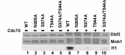
80 72 A B C Figure 2-4
81 73 Tables Table 2-1: Yeast strains used for this study. Strain YAM1 YAM2 YAM3 Relevant Genotype MATa/MATα DBF2:: FHH DBF2 (HIS3)/+ MATa/MATα mob1-77/mob1-77 DBF2:: FHH DBF2 (HIS3)/+ MATa/MATα cdc15-2/cdc15-2 pep4::trp1/pep4::trp1 bar1::leu2/+ DBF2:: FHH DBF2 (HIS3)/+ YAM4 YAM5 YAM6 YAM7 YAM8 YAM9 RJD361 RJD381 RJD1218 RJD1224 RJD1225 RJD1228 RJD1264 RJD1265 MATα dbf2-2 [pgal1- FHH DBF2/TRP1] MATα dbf2-2 [pgal1- FHH DBF2(N305A)/TRP1] MATα dbf2-2 [pgal1- FHH DBF2(S374A)/TRP1] MATα dbf2-2 [pgal1- FHH DBF2(T544A)/TRP1] MATα dbf2-2 [pgal1- FHH DBF2(S374A/T544A)/TRP1] MATα dbf2-2 [prs129] MATα MATa/MATα MATα dbf2-2 MATa DBF2:: FHH DBF2 (HIS3) MATα cdc5-1 DBF2:: FHH DBF2 (HIS3) MATα cdc14-1 DBF2:: FHH DBF2 (HIS3) MATa tem1-3 bar1::hisg DBF2:: FHH DBF2 (HIS3) MATa cdc15-2 bar1::leu2 pep4::trp1 DBF2:: FHH DBF2 (HIS3) a All strains are in the W303 background (ura3, leu2, trp1, his3, ade2).
82 74 Table 2-2: Plasmids constructed for this study. Plasmid Construct Alias RDB884 prs303- FHH DBF2(nt ) RDB1267 pfastbac1- FHH DBF2 AM1 RDB1331 pfastbachta(-xhoi)- H6 Mob1 AM4 RDB1335 pfastbachta(-xhoi)- H6 Mob1 TM9 AM5 RDB1337 pfastbac1- FHH DBF2(N305A) AM7 RDB1351 pfastbac1- FHH DBF2(S374A) AM8 RDB1352 pfastbac1- FHH DBF2(T544A) AM9 RDB1354 pfastbac1- FHH DBF2(S374A/T544A) AM10 RDB1414 prs129- FHH DBF2 AM17 RDB1415 prs129- FHH DBF2(N305A) AM18 RDB1416 prs129- FHH DBF2(S374A) AM19 RDB1417 prs129- FHH DBF2(T544A) AM20 RDB1418 prs129- FHH DBF2(S374A/T544A) AM21
83 75 Chapter 3: Substrate Specificity Analysis of Protein Kinase Complex Dbf2-Mob1 by Peptide Library and Proteome Array Screening Angie S. Mah, Andrew E. H. Elia, Geeta Devgan, Jason Ptacek, Michael Snyder, Michael B. Yaffe, Raymond J. Deshaies. Manuscript in preparation. (Contribution from AEHE: Table 1; GD and JP: proteome chip screening; MBY: Figure 3-1A). Summary The mitotic exit network (MEN) is a group of proteins that form a signaling cascade that is essential for cells to exit mitosis in Saccharomyces cerevisiae. The MEN has also been implicated in playing a role in cytokinesis. Two components of this signaling pathway are the protein kinase Dbf2 and its binding partner essential for its kinase activity, Mob1. The components of MEN that act upstream of Dbf2-Mob1 have been characterized, but physiological substrates for Dbf2-Mob1 have yet to be identified. Using a combination of peptide library scanning and evaluation of chemically synthesized peptides, we found that Dbf2-Mob1 preferentially phosphorylated serine over threonine and required an arginine three residues upstream of the phosphorylated serine in its substrate. This requirement for arginine in peptide substrates could not be substituted with the similarly charged lysine. This specificity determined for peptide substrates was also evident in many of the proteins phosphorylated by Dbf2-Mob1 in a proteome chip analysis. We have determined that the protein kinase Dbf2-Mob1 preferentially phosphorylated substrates that contain an RXXS motif. A combination of peptide library and proteome
84 76 microarray screening has identified putative Dbf2-Mob1 substrates and phosphorylation sites that may play critical roles in mitotic exit and cytokinesis. Introduction In the budding yeast Saccharomyces cerevisiae the protein phosphatase Cdc14 must be activated to turn off mitotic Cdk activity for cells to exit mitosis. There are two groups of proteins that regulate Cdc14 activity, the Cdc14 early anaphase release (FEAR) network and the mitotic exit network (MEN) (reviewed in D'Amours and Amon, 2004). There is one cyclin-dependent kinase (Cdk), Cdc28, that controls cell cycle progression in S. cerevisiae. By associating with mitotic cyclins, Cdc28 promotes entry into mitosis. Cdc14 plays a critical role in inactivating mitotic Cdk activity, thereby promoting exit from mitosis. Cdc14 dephosphorylates Hct1/Cdh1 which can then target Clb2, the main mitosis-specific cyclin, for degradation (Jaspersen et al., 1999; Schwab et al., 1997; Visintin et al., 1998; Zachariae et al., 1998). Cdc14 also promotes the accumulation of Sic1, a Cdk inhibitor, by acting on Sic1 as well as transcriptional activator Swi5, which promotes transcription of SIC1 (Moll et al., 1991; Visintin et al., 1998). Together, the degradation of mitotic cyclins and the production of active Sic1 conspire to down-regulate Cdc28 activity and return the cell cycle to an interphase state. Cdc14 is held in an inactive state in the nucleolus by its inhibitor, Net1 (Shou et al., 1999; Visintin et al., 1999). Cdc14 is tethered to Net1 throughout the cell cycle until the onset of anaphase, at which time it is released. The FEAR network and MEN both regulate Cdc14 release and therefore its activity (reviewed in D'Amours and Amon, 2004;
85 77 Stegmeier and Amon, 2004). The FEAR network initiates early anaphase release of Cdc14 from the nucleolus by promoting Net1 phosphorylation by mitotic Cdks, weakening the interaction between Cdc14 and Net1 (Azzam et al., 2004). The FEAR network consists of Esp1 (also known as separase), polo-like kinase Cdc5, the kinetochore protein Slk19, the nuclear protein Spo12, and its homologue Bns1. The action of these proteins is opposed by Pds1 (also known as securin) and the nucleolar replication fork block protein Fob1 (Stegmeier et al., 2004; Stegmeier et al., 2002; Sullivan and Uhlmann, 2003). However, it is still unclear how the factors that promote and restrain FEAR interact. Although the release of Cdc14 in early anaphase by the FEAR network is transient and insufficient for mitotic exit, exit is delayed when the FEAR network is compromised by mutation. One possible explanation is that the FEAR network weakens the Cdc14-Net1 interaction, enabling the MEN to more rapidly cause a sustained release of Cdc14. The FEAR network also has other mitotic functions that may play an important role in coordinating events during exit from mitosis (reviewed in D'Amours and Amon, 2004; Stegmeier and Amon, 2004). In contrast to the FEAR network, the MEN is essential for mitotic exit (reviewed in Bardin and Amon, 2001; D'Amours and Amon, 2004; Stegmeier and Amon, 2004). This pathway consists of the GTPase Tem1, the putative guanine-nucleotide exchange factor (GEF) Lte1, the two-component GTPase activating protein (GAP) Bub2-Bfa1, the protein kinases Cdc5, Cdc15, Dbf2, the Dbf2 binding protein Mob1, and the scaffolding protein, Nud1. Genetic and biochemical data have provided significant insight into how this signaling cascade is activated by the localization of its components. Bub2-Bfa1 binds and inhibits Tem1 at the spindle pole body (SPB) that enters the daughter cell
86 78 during nuclear division. As the spindle elongates into the bud, Tem1 is presumably activated by Lte1, which is localized in the bud (Bardin et al., 2000; Pereira et al., 2000). The activated GTP-bound form of Tem1 then somehow activates bound Cdc15, which then phosphorylates and activates the Dbf2-Mob1 kinase complex (Mah et al., 2001). However, how activated Dbf2-Mob1 affects Cdc14 release from Net1 and mitotic exit is unknown. In addition to their role in mitotic exit, Dbf2-Mob1 and the other MEN proteins function in cytokinesis. Dbf2 localizes to the SPB in anaphase as do Tem1, Cdc5, Cdc15, and Mob1 (Bardin et al., 2000; Frenz et al., 2000a; Hwa Lim et al., 2003; Pereira et al., 2000; Visintin and Amon, 2001; Xu et al., 2000). During late mitosis, Dbf2 and Mob1 migrate to the bud neck. Bud neck localization of Dbf2 and Mob1 are dependent on each other as well as the MEN proteins Cdc5, Cdc14, Cdc15, Nud1, and the septins Cdc12 and Cdc3 (Frenz et al., 2000a; Luca et al., 2001; Yoshida and Toh-e, 2001). Several lines of evidence suggest that localization of MEN proteins to the bud neck is crucial for cytokinesis. Mutant mob1 ts cells, as well as tem1 net1-1 and cdc15 net1-1 cells whose mitotic exit defects are bypassed by the net1-1 allele fail to undergo cytokinesis (Lippincott et al., 2001; Luca et al., 2001; Shou et al., 1999). Interestingly, localization of Dbf2 Mob1 to the bud neck depends upon Cdc14 (Frenz et al., 2000b; Luca et al., 2001). MEN-dependent release and activation of Cdc14 may help to ensure that mitotic exit occurs prior to cytokinesis. The function of Dbf2-Mob1 in cytokinesis is unclear. Also unknown is how Dbf2-Mob1 ultimately leads to release of Cdc14 from the nucleolus during mitotic exit. To give us insight into these two key cell cycle processes, we sought to identify potential
87 79 substrates and phosphorylation sites of Dbf2-Mob1. Here, we report the substrate specificity of Dbf2-Mob1 and a number of putative substrates that contain a Dbf2 phosphorylation motif and are phosphorylated by Dbf2 Mob1 in vitro. Results Determination of optimal peptide sequence motif phosphorylated by Dbf2-Mob1 To identify potential physiological substrates of Dbf2-Mob1, we first proceeded to determine an optimal substrate sequence by using an oriented degenerate peptide library technique (Obata et al., 2000). Dbf2, a Ser/Thr kinase, was initially tested to determine whether there was a preference for phosphorylation on Ser or Thr residues. Degenerate peptide libraries containing either a fixed Ser residue, XXXXSXXXX, or a fixed Thr residue, XXXXTXXXX, were incubated with [γ- 32 P]ATP and Dbf2-Mob1 that was expressed in insect cells, purified, and activated by recombinant Cdc15. All amino acids except Cys, Ser, Thr, and Tyr are represented by X, where the last 3 residues were omitted to limit phosphorylation to the fixed Ser or Thr. The level of phosphorylation was determined by the amount of radioactive phosphate incorporated in the peptides. Dbf2-Mob1 had a two-fold preference for Ser phosphorylation over Thr (Table 1). As Dbf2-Mob1 had a preference for Ser, two other degenerate peptide libraries with fixed Ser residues (position 0) were tested for the amount of phosphate incorporated after treatment with Dbf2-Mob1. One of the libraries tested was the RS library with degenerate sequence of XXXXRXXSXXXX, containing the fixed Ser along with Arg
88 80 fixed at position -3. The other library tested was the Cdk site library, with the fixed Ser residue along with a fixed Pro in position +1, resulting in the degenerate sequence of XXXXSPXXXX. The RS library was found to incorporate 8 times more phosphate than the SP library (Table 1). As a result, the RS library was used to determine an optimal Dbf2-Mob1 substrate motif. Sequencing a pool of Dbf2-phosphorylated peptides enriched from the RS library revealed a strong preference for Ile and Phe at the -2 and -1 positions, respectively (Table 2). There was a moderate selection for Met at both the -4 and +1 positions (Table 2). The predicted optimal consensus motif for Dbf2-Mob1 substrates was determined to be MRIFSM. Optimal sequence phosphorylation efficiency To evaluate the contribution of each residue in the predicted optimal consensus motif, we synthesized a peptide based on the consensus motif, as well as a set of variants in which each position was substituted by an alanine residue (Figure 1A). Peptides with Tyr at 1 (F-1Y) and Lys at -3 (R-3K) were also synthesized to determine whether the selection of Phe at position -1 could be replaced by another bulky residue like Tyr, or if the Arg at position -3 could be substituted with the similarly charged Lys. The NT-Control peptide contains a substitution in the Arg residue that lies outside of the predicted consensus motif to determine whether there is a selection at that position. Finally, the inhibitor peptide is the same as the consensus except that the Ser phosphorylation site was replaced with Ala.
89 81 The synthetic peptides were treated with Dbf2-Mob1 in vitro for 0, 4, 8 or 12 minutes and the amount of phosphorylation was determined (Figure 1B). As a negative control, the optimal consensus peptide was treated in parallel with the kinase-inactive Dbf2(N305A)-Mob1 that had undergone the same treatment with Cdc15 as the wild type Dbf2-Mob1 complex (Opt D2M). As expected, the inhibitor peptide, as well as the R-3A peptide both had negligible levels of phosphorylation (Figure 1B; Inh, R-3A). The optimal peptide treated with kinase inactive Dbf2(N305A)-Mob1 was also negligibly phosphorylated (Figure 1B; Inh). For the remaining peptides in which measurable levels of phosphorylation were detected, the V max and k cat for Dbf2-Mob1 were calculated (Figure 1C). There did not appear to be selection for Ile at the -2 position nor selection for the Arg in the -7 position outside of the consensus motif, as both mutant peptides were phosphorylated to a similar degree as the optimal peptide (Figure 1B; I-2A, NT, Opt), with similar V max and k cat values for Dbf2-Mob1 (Figure 1C). Mutations of the bulkier groups at positions -1, -4, and +1 to Ala actually increased the amount of phosphorylation of the peptides (Figure 1B; F-1A, M-4A, M+1A), increasing the V max and k cat values for Dbf2-Mob1 (Figure 1C). This was surprising because the peptide library screen had indicated a strong selection for Phe at position -1. Interestingly, the substitution of Arg by Lys in position -3 decreased the amount of peptide phosphorylation to a level comparable to the negative controls (Figure 1B; R-3K). Taken together, these results revealed a preference for non-bulky residues proximal to the Ser phosphorylation site and that the critical Arg required for substrate phosphorylation cannot be substituted with Lys, at least in the context of a peptide substrate.
90 82 Proteome array screen identifies in vitro Dbf2-Mob1 substrates The relatively low sequence complexity of the Dbf2 phosphorylation motif diminished the power of using genome-wide bioinformatics screens to identify putative substrates. Accordingly, we carried out a proteome array screen to identify putative yeast substrates. Proteome chips spotted with ~4,400 different glutathione-s-transferase (GST) fusion proteins purified from yeast were probed with activated Dbf2-Mob1, kinase-inactive Dbf2(N305A)-Mob1, and buffer alone. After taking into account the proteins phosphorylated in the negative controls, 67 proteins were determined to be putative Dbf2-Mob1 substrates (Table 3). To confirm that the proteins identified in the proteome array screen could be phosphorylated by Dbf2-Mob1 as opposed to being bound to Dbf2 substrates, we performed further analysis on the 25 proteins with the highest incorporation signal relative to protein amount (relative protein amounts were determined by anti-gst immunoblot of the proteome chip). Interestingly, three or more copies of this motif were found in 16, or 64%, of the proteins in the list (Table 4), compared to only 29% of the proteins encoded in the yeast genome. This enrichment for proteins with three or more copies of the RXXS motif is highly significant (p = 1.2 X 10⁻⁴ ; G. Kleiger, unpublished data). TAP-tagged strains were obtained for 22 of the 25 top candidates. TAP-tagged alleles for three of the genes (YJL108C, YBR285W, and YBR108W) were not available, and therefore these candidates were not subjected to further analysis. Asynchronous cultures of the 22 TAP-tagged strains were grown and the TAP-tagged proteins
91 83 immunoprecipitated with IgG sepharose and analyzed by Western blotting (Figure 2A). Candidates were determined to be phosphorylated if a radioactive signal was detected at the molecular weight predicted for the tagged protein. Of the 22 strains in which immunoprecipitations were performed, YBR138C (HDR1), YAL051W (OAF1), YNL101W (AVT4), YIL135C (VHS2), and YMR184W did not have detectable protein expression. The 17 TAP-tagged proteins that were expressed and purified were used in Dbf2-Mob1 kinase assays (Figure 2B). Of the 17, 10 were determined to be phosphorylated. To determine whether the phosphorylation of these proteins was specific to Dbf2-Mob1, rather than due to a co-precipitating protein kinase or residual Cdc15 used to activate Dbf2-Mob1, kinase assays were performed using kinase-inactive Dbf2(N305A-Mob1) as a negative control (Figure 2C). In all cases, there was a strong decrease in incorporation when kinase-inactive Dbf2-Mob1 was used. These results suggest that the proteins identified by the proteome chip screen were indeed substrates of Dbf2-Mob1. Discussion The substrate used to test Dbf2-Mob1 kinase activity, histone H1, is a commonly used artificial substrate for many protein kinases. We wanted to find physiological protein substrates of Dbf2-Mob1. To do so, we first sought to define the optimal phosphorylation site motif for Dbf2-Mob1 substrates. Peptide library screening revealed the putative optimal substrate motif to be MRIFSM (Figure 1A). However, when we tested each residue in in vitro Dbf2-Mob1 peptide kinase assays, the only substitutions
92 84 that diminished incorporation were swapping the Arg in position -3 for Ala or Lys (Figure 2B). The latter result was unexpected, because Ndr1, a human homologue of Dbf2, was proposed to phosphorylate sequences with either Lys or Arg in the 3 position (Millward et al., 1998). Surprisingly, several substitutions actually resulted in an increase in activity. Contrary to the predicted optimal motif with strong selectivity for Phe at position 1, we found that Dbf2 Mob1 prefers Ala in this position. The proteins identified by proteome chip screening gave further evidence that the RXXS motif serves as a substrate for Dbf2-Mob1 phosphorylation, as 80% of the top 25 proteins that were identified in the proteome chip analysis contained this motif and 64% contained more than 3 copies of the motif. Of these proteins, 17 were tested further and 10 of these were identified as in vitro substrates, 8 of which have the RXXS motif. Pre10 and Adk1 were phosphorylated by Dbf2-Mob1 but not its kinase inactive form despite not having the RXXS motif (Figure 2C; Table 4). One reason may be that both proteins are immunoprecipitated at high levels and therefore may serve as non-specific substrates of Dbf2-Mob1. Recently, the results of a systematic proteome array analysis of yeast protein kinases have been posted online (J. Ptacek et al., submitted). There were a few discrepancies between the results posted online for Dbf2-Mob1 and those of Table 3. The discrepancies were based on the methodology for identifying positive signals. In our original data set, the results were obtained by computer analysis. Further visual analysis to confirm positives was performed for the data available online. There were minor differences in the results, with the exception of Cbf1, Oaf1, and Htz1. These 3 proteins were within the top 4 proteins that gave the highest signal in our original data set (Table
93 85 3) yet were not in the data set posted online. They had further been identified as false positives (J. Ptacek, personal communication). Our in vitro analysis confirmed that two of these proteins (Oaf1 was not tested because a TAP-tagged strain was not available) were indeed negatives as neither Cbf1 nor Htz1 were found to be phosphorylated by Dbf2-Mob1 (Figure 2B). In attempts to confirm whether any of the Dbf2 Mob1 substrates identified by our analyses are true physiological substrates of this complex, we took four approaches. First, we examined whether any of the substrates undergo a molecular weight shift upon their phosphorylation by Dbf2 Mob1, which might serve as a simple diagnostic to evaluate phosphorylation in vivo. Next, we immunoprecipitated each protein from yeast cells via the TAP tag, and immunoblotted for Mob1 to determine if the putative substrates were associated with the Dbf2 complex. Third, we queried the yeast GFP localization database, to see if any of the candidates display SPB or bud neck localization characteristic of Dbf2 Mob1. Finally, we searched a database of 700 mapped yeast phosphorylation sites to see if any of them reside in our candidate substrates. Unfortunately, none of these efforts yielded a positive result. This experience highlights that validation of putative protein kinase substrates identified by proteome chip analysis may require considerable investment in the mapping of in vivo phosphorylation sites. We have determined that protein kinase Dbf2 Mob1 has a preference for phosphorylating peptides and proteins that bear one or more RXXS motifs. Future analysis of candidate Dbf2 Mob1 substrates reported here may provide significant insights into understanding the mechanism by which the Mitotic Exit Network brings about the exit from mitosis and cytokinesis.
94 86 Materials and Methods Purification and activation of Dbf2-Mob1 kinase complex FlagHis6HA3 Dbf2 ( FHH Dbf2) was co-immunoprecipitated with His6 Mob1 TEVmyc9 ( H6 Mob1 TM9 ) from Hi5 insect cells as previously described (Mah et al., 2001). To activate FHH Dbf2- H6 Mob1 TM9, the protein complex bound to anti-flag M2 beads (Sigma) was incubated with baculovirus-expressed Cdc15 His6 in the presence of kinase buffer containing 50 mm HEPES (ph 7.5), 5 mm MgCl 2, 2.5 mm MnCl 2, 5 mm β-glycerophosphate, 1 mm DTT, and 1 mm ATP for 30 min at room temperature. The beads were washed three times with buffer containing 50 mm Tris (ph 7.6), 150 mm NaCl, 0.2% Triton X-100 to remove ATP and Cdc15 His6. FHH Dbf2- H6 Mob1 TM9 was then eluted from the beads with 1 µg/ml FLAG peptide (Sigma) in Dbf2 kinase buffer (DKB) containing 50 mm Tris (ph 7.4), 60 mm potassium acetate, 10 mm MgCl 2, 1 mm DTT, and 10 µm ATP for four hours at 4 C. The same procedure was used to produce FHH Dbf2(N305A)- H6 Mob1 TM9, the kinase inactive point mutant of Dbf2. The eluted active or inactive FHH Dbf2- H6 Mob1 TM9 was used for subsequent assays. Peptide library screening Baculovirus-derived FHH Dbf2- H6 Mob1 TM9 was used for peptide library screening. Peptide library screening and data analysis were performed as previously described (Obata et al., 2000; Songyang and Cantley, 1998; Yaffe et al., 1997). Briefly, the RS peptide library
95 87 was used for screening. This library consists of peptides with the general sequence MAXXXXRXXSXXXXAKKK, where X represents all amino acids except Cys, Ser, Thr, and Tyr. The total library is predicted to contain 1.1 X distinct sequences. Peptides (~1 mg) were incubated with FHH Dbf2- H6 Mob1 TM9, 100 µm unlabeled ATP, and tracer amounts of [γ- 32 P]ATP at 30 C for 2 h until ~0.5-1% of peptides were phosphorylated. A ferric-iminodiacetic acid (IDA) column was used to separate the phosphopeptides from non-phosphorylated peptides. The phosphopeptides were then sequenced in batch by automated Edman degradation. Data analysis was as described (Yaffe et al., 1997). Selectivity values refer to the relative preference for an amino acid at a given degenerate position, based on the amount of each amino acid recovered at that position compared to the amount in the starting library (Songyang and Cantley, 1998). Peptide kinase assays Synthetic peptides (Abgent) were used as substrates for FHH Dbf2- H6 Mob1 TM9 kinase assays. Reactions containing 250 µm of peptide substrate and ~13 ng of FHH Dbf2 bound to H6 Mob1 TM9 were incubated in the presence of 30 µl of DKB and 2 µci [γ- 32 P]ATP at room temperature. Reactions were terminated at indicated timepoints with addition of 10 µl of stop solution (8 N HCl, 1 mm ATP). Phosphate incorporation was determined by spotting reactions on P81-phosphocellulose paper (Whatman), washing with 0.5% phosphoric acid, air-drying the filters, and then quantifying the bound radioactivity by scintillation counting. For each individual peptide, values were normalized to time zero.
96 88 Proteome chip assays Yeast proteome microarrays were prepared as previously described (Zhu and Snyder, 2001). Overexpressed GST-tagged yeast proteins were purified from ~4,400 yeast strains and spotted on slides. To determine the optimal amount of kinase to use for probing proteome chips, we performed trial assays as described (J. Ptacek et al., submitted). Multiple dilutions of FHH Dbf2- H6 Mob1 TM9 and kinase inactive FHH Dbf2(N305A)- H6 Mob1 TM9 (~20 ng/µl of Dbf2) in DKB buffer containing 2 µl [γ- 33 P]ATP were used on trial proteome chips before using on the full proteome array. The full proteome array was probed with 4 µl FHH Dbf2- H6 Mob1 TM9 in 200 µl of DKB supplemented with [γ- 33 P]ATP. As a control, FHH Dbf2(N305A)- H6 Mob1 TM9 was used to probe the proteome array. To normalize the background signal, 2 µl of FHH Dbf2(N305A)- H6 Mob1 TM9 in 200 µl DKB supplemented with [γ- 33 P]ATP was used. To control for autophosphorylated proteins, the proteome array was probed with 200 µl of DKB supplemented with [γ- 33 P]ATP. Proteome chips were assayed in duplicate in each case. Data analysis was performed as described (J. Ptacek et al., submitted). Briefly, signals were analyzed by a computer algorithm designed to normalize background and identify signals as positive if 3 of 4 spots (each protein is spotted twice on each slide and each kinase or control was tested on 2 slides) were 2 standard deviations above background and the fourth spot was 1.5 standard deviations above background.
97 89 Immunoprecipitations and kinase assays of TAP-tagged proteins Yeast strains containing TAP-tagged genes (Open Biosystems) were grown to OD 600 ~2.0 in 25 ml of YPD. Cells were harvested by centrifugation and washed in buffer containing 150 mm NaCl and 50 mm Tris (ph 7.4). Cells were then resuspended in 600 µl lysis buffer containing 150 mm NaCl, 50 mm Tris (ph 7.4), 2 mm EDTA (ph 8.0), 1% Triton-X 100, 10% glycerol, 2 mm DTT, 5 µg/ml aprotinin, 5 µg/ml pepstatin, 5 µg/ml l chymostatin, 5 µg/ml leupeptin, 0.5 mm AEBSF, 1 mm PMSF, 10 mm NaF, 60 mm β-glycerophosphate,. 10 mm sodium pyrophosphate, 2 mm sodium vanadate). An equal volume of glass beads was added. The cells were then lysed by 4 cycles of vortexing (ThermoSavant FastPrep) at 4 C for 45 s at setting 5.5 alternating with cycles of icing samples for 1 min. Lysates were clarified by centrifugation then added to 60 µl IgG sepharose beads (Amersham) for 1 h at 4 C on a rotator. Beads were then washed 3 times with lysis buffer, twice with buffer containing 150 mm NaCl, 50 mm Tris (ph 7.4), 2 mm EDTA (ph 8.0), 1% Triton-X 100, 10% glycerol, and 2 mm DTT, and a final wash with buffer containing 150 mm NaCl and 50 mm Tris (ph 7.4). Immunoprecipitated TAP-tagged proteins were analyzed by SDS-PAGE and detected by Western blotting using the primary antibody anti-tap (Open Biosystems) followed by goat anti-rabbit horseradish peroxidase (HRP)-conjugate (Bio-Rad), and ECL. For kinase assays, TAP-tagged proteins bound to 20 µl beads were washed with DKB then incubated with FHH Dbf2- H6 Mob1 TM9 or FHH Dbf2(N305A)- H6 Mob1 TM9 (~13 ng of Dbf2) with 2 µci [γ- 32 P]ATP for 30 min at room temperature. Kinase reactions were stopped
98 90 by addition of 2X SDS-PAGE sample buffer, fractionated on SDS-PAGE and detected by autoradiography. Acknowledgements We thank Ramzi Azzam for his invaluable insight and enthusiasm for initiating this project. We also thank Dane Mohl and William Ja for their thoughts and comments on this work. We are also grateful to Heng Zhu for performing initial proteome chip experiments and Gary Kleiger for providing bioinformatics expertise. This research was supported by an NIH grant to RJD (GM059940).
99 91 References Azzam, R., Chen, S.L., Shou, W., Mah, A.S., Alexandru, G., Nasmyth, K., Annan, R.S., Carr, S.A. and Deshaies, R.J. (2004) Phosphorylation by cyclin B-Cdk underlies release of mitotic exit activator Cdc14 from the nucleolus. Science, 305, Bardin, A.J. and Amon, A. (2001) Men and sin: what's the difference? Nat Rev Mol Cell Biol, 2, Bardin, A.J., Visintin, R. and Amon, A. (2000) A mechanism for coupling exit from mitosis to partitioning of the nucleus. Cell, 102, D'Amours, D. and Amon, A. (2004) At the interface between signaling and executing anaphase--cdc14 and the FEAR network. Genes Dev, 18, Frenz, L.M., Lee, S.E., Fesquet, D. and Johnston, L.H. (2000) The budding yeast Dbf2 protein kinase localises to the centrosome and moves to the bud neck in late mitosis. J Cell Sci, 113 Pt 19, Hwa Lim, H., Yeong, F.M. and Surana, U. (2003) Inactivation of mitotic kinase triggers translocation of MEN components to mother-daughter neck in yeast. Mol Biol Cell, 14, Jaspersen, S.L., Charles, J.F. and Morgan, D.O. (1999) Inhibitory phosphorylation of the APC regulator Hct1 is controlled by the kinase Cdc28 and the phosphatase Cdc14. Curr Biol, 9,
100 92 Lee, S.E., Frenz, L.M., Wells, N.J., Johnson, A.L. and Johnston, L.H. (2001a) Order of function of the budding-yeast mitotic exit-network proteins Tem1, Cdc15, Mob1, Dbf2, and Cdc5. Curr Biol, 11, Lee, S.E., Jensen, S., Frenz, L.M., Johnson, A.L., Fesquet, D. and Johnston, L.H. (2001b) The Bub2-dependent mitotic pathway in yeast acts every cell cycle and regulates cytokinesis. J Cell Sci, 114, Lippincott, J., Shannon, K.B., Shou, W., Deshaies, R.J. and Li, R. (2001) The Tem1 small GTPase controls actomyosin and septin dynamics during cytokinesis. J Cell Sci, 114, Luca, F.C., Mody, M., Kurischko, C., Roof, D.M., Giddings, T.H. and Winey, M. (2001) Saccharomyces cerevisiae Mob1p is required for cytokinesis and mitotic exit. Mol Cell Biol, 21, Mah, A.S., Jang, J. and Deshaies, R.J. (2001) Protein kinase Cdc15 activates the Dbf2- Mob1 kinase complex. Proc Natl Acad Sci U S A, 98, Millward, T.A., Heizmann, C.W., Schafer, B.W. and Hemmings, B.A. (1998) Calcium regulation of Ndr protein kinase mediated by S100 calcium-binding proteins. EMBO J, 17, Moll, T., Tebb, G., Surana, U., Robitsch, H. and Nasmyth, K. (1991) The role of phosphorylation and the CDC28 protein kinase in cell cycle-regulated nuclear import of the S. cerevisiae transcription factor SWI5. Cell, 66,
101 93 Obata, T., Yaffe, M.B., Leparc, G.G., Piro, E.T., Maegawa, H., Kashiwagi, A., Kikkawa, R. and Cantley, L.C. (2000) Peptide and protein library screening defines optimal substrate motifs for AKT/PKB. J Biol Chem, 275, Pereira, G., Hofken, T., Grindlay, J., Manson, C. and Schiebel, E. (2000) The Bub2p spindle checkpoint links nuclear migration with mitotic exit. Mol Cell, 6, Schwab, M., Lutum, A.S. and Seufert, W. (1997) Yeast Hct1 is a regulator of Clb2 cyclin proteolysis. Cell, 90, Shou, W., Seol, J.H., Shevchenko, A., Baskerville, C., Moazed, D., Chen, Z.W., Jang, J., Charbonneau, H. and Deshaies, R.J. (1999) Exit from mitosis is triggered by Tem1- dependent release of the protein phosphatase Cdc14 from nucleolar RENT complex. Cell, 97, Songyang, Z. and Cantley, L.C. (1998) The use of peptide library for the determination of kinase peptide substrates. Methods Mol Biol, 87, Stegmeier, F. and Amon, A. (2004) Closing Mitosis: The Functions of the Cdc14 Phosphatase and Its Regulation. Annu Rev Genet, 38, Stegmeier, F., Huang, J., Rahal, R., Zmolik, J., Moazed, D. and Amon, A. (2004) The replication fork block protein Fob1 functions as a negative regulator of the FEAR network. Curr Biol, 14,
102 94 Stegmeier, F., Visintin, R. and Amon, A. (2002) Separase, polo kinase, the kinetochore protein Slk19, and Spo12 function in a network that controls Cdc14 localization during early anaphase. Cell, 108, Sullivan, M. and Uhlmann, F. (2003) A non-proteolytic function of separase links the onset of anaphase to mitotic exit. Nat Cell Biol, 5, Visintin, R. and Amon, A. (2001) Regulation of the mitotic exit protein kinases Cdc15 and Dbf2. Mol Biol Cell, 12, Visintin, R., Craig, K., Hwang, E.S., Prinz, S., Tyers, M. and Amon, A. (1998) The phosphatase Cdc14 triggers mitotic exit by reversal of Cdk-dependent phosphorylation. Mol Cell, 2, Visintin, R., Hwang, E.S. and Amon, A. (1999) Cfi1 prevents premature exit from mitosis by anchoring Cdc14 phosphatase in the nucleolus. Nature, 398, Xu, S., Huang, H.K., Kaiser, P., Latterich, M. and Hunter, T. (2000) Phosphorylation and spindle pole body localization of the Cdc15p mitotic regulatory protein kinase in budding yeast. Curr Biol, 10, Yaffe, M.B., Schutkowski, M., Shen, M., Zhou, X.Z., Stukenberg, P.T., Rahfeld, J.U., Xu, J., Kuang, J., Kirschner, M.W., Fischer, G., Cantley, L.C. and Lu, K.P. (1997) Sequence-specific and phosphorylation-dependent proline isomerization: a potential mitotic regulatory mechanism. Science, 278,
103 95 Yoshida, S. and Toh-e, A. (2001) Regulation of the localization of Dbf2 and Mob1 during cell division of Saccharomyces cerevisiae. Genes Genet Syst, 76, Zachariae, W., Schwab, M., Nasmyth, K. and Seufert, W. (1998) Control of cyclin ubiquitination by CDK-regulated binding of Hct1 to the anaphase promoting complex. Science, 282, Zhu, H. and Snyder, M. (2001) Protein arrays and microarrays. Curr Opin Chem Biol, 5,
104 96 Figures Figure 3-1: Dbf2-Mob1 peptide substrate requires arginine at position -3. (A) Synthetic peptides based on the predicted optimal substrate of Dbf2-Mob1. The underlined residues represent the predicted preferred amino acids for Dbf2-Mob1 substrate specificity, the asterik denotes the single amino acid substitution. (B) The various peptides denoted in (A) at a concentration of 250 µm were treated with ~13 ng of FHH Dbf2 bound to H6 Mob1 TM9. Reactions were halted at the indicated timepoints to determine the amount of phosphorylation by liquid scintillation. The Optimal peptide was also treated with the kinase inactive FHH Dbf2(N305A)- H6 Mob1 TM9 complex, as denoted by D2M. (C) Using the conditions in (B), K m and V max was determined for each peptide with the exception of R-3K, R-3A, Inhibitor and the Optimal peptide treated with kinase inactive FHH Dbf2(N305A)- H6 Mob1 TM9, due to low phosphorylation.
105 97 A C B Phosphorylation (X10 3 cpm) Phosphorylation (X10 3 cpm) Time (min) Figure 3-1
106 98 Figure 3-2: Yeast proteins phosphorylated by Dbf2-Mob1. (A) Of the 25 proteins with the highest phosphorylation signal as shown in Table 3, 22 of these genes were TAP-tagged in the Open Biosystems TAP-tagged yeast library. The TAP-tagged proteins were immunoprecipitated with IgG sepharose beads from asynchronous cultures, fractionated on SDS-PAGE and immunoblotted with anti-tap. Of the 22 strains, 5 did not have detectable protein expression, such as VHS2 as shown. (B) The TAP-tagged proteins expressed in (A) were treated with FHH Dbf2- H6 Mob1 TM9 in the presence of [γ- 32 P]ATP, fractionated on SDS-PAGE and detected by autoradiography. (C) The TAP-tagged proteins phosphorylated by FHH Dbf2- H6 Mob1 TM9 in (B) were treated with either FHH Dbf2- H6 Mob1 TM9 or the kinase inactive FHH Dbf2(N305A)- H6 Mob1 TM9 in the presence of [γ- 32 P]ATP, fractionated on SDS-PAGE and detected by autoradiography.


107 99 A B C Figure 3-2
Department of Pharmacology, UT Southwestern Medical Center, 5323 Harry Hines Blvd., Dallas, TX
 [Frontiers in Bioscience 8, d1275-1287, September 1, 2003] REGULATION OF CDC14: PATHWAYS AND CHECKPOINTS OF MITOTIC EXIT Joshua Bembenek and Hongtao Yu Department of Pharmacology, UT Southwestern Medical
7.06 Problem Set #4, Spring 2005
 7.06 Problem Set #4, Spring 2005 1. You re doing a mutant hunt in S. cerevisiae (budding yeast), looking for temperaturesensitive mutants that are defective in the cell cycle. You discover a mutant strain
The FEAR network. Citation. As Published Publisher. Version
 The FEAR network The MIT Faculty has made this article openly available. lease share how this access benefits you. Your story matters. Citation As ublished ublisher Rock, Jeremy M., and Angelika Amon.
Life Sciences 1a: Section 3B. The cell division cycle Objectives Understand the challenges to producing genetically identical daughter cells
 Life Sciences 1a: Section 3B. The cell division cycle Objectives Understand the challenges to producing genetically identical daughter cells Understand how a simple biochemical oscillator can drive the
Lecture 10: Cyclins, cyclin kinases and cell division
 Chem*3560 Lecture 10: Cyclins, cyclin kinases and cell division The eukaryotic cell cycle Actively growing mammalian cells divide roughly every 24 hours, and follow a precise sequence of events know as
16 The Cell Cycle. Chapter Outline The Eukaryotic Cell Cycle Regulators of Cell Cycle Progression The Events of M Phase Meiosis and Fertilization
 The Cell Cycle 16 The Cell Cycle Chapter Outline The Eukaryotic Cell Cycle Regulators of Cell Cycle Progression The Events of M Phase Meiosis and Fertilization Introduction Self-reproduction is perhaps
Plant Molecular and Cellular Biology Lecture 8: Mechanisms of Cell Cycle Control and DNA Synthesis Gary Peter
 Plant Molecular and Cellular Biology Lecture 8: Mechanisms of Cell Cycle Control and DNA Synthesis Gary Peter 9/10/2008 1 Learning Objectives Explain why a cell cycle was selected for during evolution
12/5/2014. The cell cycle and cell death. The cell cycle: cells duplicate their contents and divide
 The cell cycle and cell death The cell cycle: cells duplicate their contents and divide 1 The cell cycle may be divided into 4 phases Eucaryotic cell division: Mitosis (nuclear division) Cytokinesis (cell
Cdk-counteracting phosphatases unlock mitotic exit Ethel Queralt 1 and Frank Uhlmann 2
 Available online at www.sciencedirect.com Cdk-counteracting phosphatases unlock mitotic exit Ethel Queralt 1 and Frank Uhlmann 2 Entry into mitosis of the eukaryotic cell cycle is driven by rising cyclin-dependent
The Cell Cycle/Le Cycle cellulaire SMC6052/BIM6028 IRCM
 The Cell Cycle/Le Cycle cellulaire SMC6052/BIM6028 IRCM 1 février 2018 Benjamin H. Kwok, Ph.D. Chercheur principal, Institut de recherche en immunologie et en cancérologie Professeur sous octroi agrégé,
The cell cycle entails an ordered series of macromolecular
 21 REGULATING THE EUKARYOTIC CELL CYCLE This cultured rat kidney cell in metaphase shows condensed chromosomes (blue), microtubules of the spindle apparatus (red), and the inner nuclear envelope protein
Overview of the cell cycle
 Chapter 2 Overview of the cell cycle 2.1 The organisation of cell cycle in eukaryotes During the cell cycle, the typical eukaryotic cell goes through a series of well defined phases, to divide into two
The FEAR network. Jeremy M. Rock and Angelika Amon *
 Magazine R1063 documented among orchid bees. Two out of five orchid bee genera are obligate cleptoparasites (or cuckoo parasites) of other nest-building euglossine genera. Do orchid bees sting? Female
A Mathematical Model of Mitotic Exit in Budding Yeast: The Role of Polo Kinase
 A Mathematical Model of Mitotic Exit in Budding Yeast: The Role of Polo Kinase Baris Hancioglu*, John J. Tyson Department of Biological Sciences, Virginia Polytechnic Institute and State University, Blacksburg,
Phosphatase 2A negatively regulates mitotic exit in
 Phosphatase 2A negatively regulates mitotic exit in Saccharomyces cerevisiae Yanchang Wang* and Tuen-Yung Ng Department of Biomedical Sciences, College of Medicine, Florida State University 1115 West Call
Dr. Fred Cross, Rockefeller (KITP Bio Networks 3/26/2003) 1
 1") Outline Cell growth as the driver for cell cycle (in microbes): coordination of growth and division A basic principle organizing cell cycle control: why cyclin-dependent kinase activity must oscillate
Outline Cell growth as the driver for cell cycle (in microbes): coordination of growth and division A basic principle organizing cell cycle control: why cyclin-dependent kinase activity must oscillate
CELL CYCLE AND GROWTH REGULATION
 CELL CYCLE AND GROWTH REGULATION The cell cycle is the set of stages through which a cell progresses from one division to the next. Interphase is the period between mitotic cell divisions; divided into
3.a.2- Cell Cycle and Meiosis
 Big Idea 3: Living systems store, retrieve, transmit and respond to information essential to life processes. 3.a.2- Cell Cycle and Meiosis EU 3.A: Heritable information provides for continuity of life.
Analysis and Simulation of Biological Systems
 Analysis and Simulation of Biological Systems Dr. Carlo Cosentino School of Computer and Biomedical Engineering Department of Experimental and Clinical Medicine Università degli Studi Magna Graecia Catanzaro,
Essential Knowledge: In eukaryotes, heritable information is passed to the next generation via processes that include the cell cycle and mitosis OR
 Essential Knowledge: In eukaryotes, heritable information is passed to the next generation via processes that include the cell cycle and mitosis OR meiosis plus fertilization Objective: You will be able
Reading Assignments. A. Systems of Cell Division. Lecture Series 5 Cell Cycle & Cell Division
 Lecture Series 5 Cell Cycle & Cell Division Reading Assignments Read Chapter 18 Cell Cycle & Cell Death Read Chapter 19 Cell Division Read Chapter 20 pages 659-672 672 only (Benefits of Sex & Meiosis sections)
Lecture Series 5 Cell Cycle & Cell Division
 Lecture Series 5 Cell Cycle & Cell Division Reading Assignments Read Chapter 18 Cell Cycle & Cell Death Read Chapter 19 Cell Division Read Chapter 20 pages 659-672 672 only (Benefits of Sex & Meiosis sections)
Regulation of the Cell Cycle by Protein Phosphatase 2A in Saccharomyces cerevisiae
 MICROBIOLOGY AND MOLECULAR BIOLOGY REVIEWS, June 2006, p. 440 449 Vol. 70, No. 2 1092-2172/06/$08.00 0 doi:10.1128/mmbr.00049-05 Copyright 2006, American Society for Microbiology. All Rights Reserved.
Lecture Series 5 Cell Cycle & Cell Division
 Lecture Series 5 Cell Cycle & Cell Division Reading Assignments Read Chapter 18 Cell Cycle & Cell Division Read Chapter 19 pages 651-663 663 only (Benefits of Sex & Meiosis sections these are in Chapter
Biology: Life on Earth
 Biology: Life on Earth Eighth Edition Lecture for Chapter 11 The Continuity of Life: Cellular Reproduction Cellular Reproduction Intracellular activity between one cell division to the next is the cell
Three different fusions led to three basic ideas: 1) If one fuses a cell in mitosis with a cell in any other stage of the cell cycle, the chromosomes
 If one fuses a cell in mitosis with a cell in any other stage of the cell cycle, the chromosomes") Section Notes The cell division cycle presents an interesting system to study because growth and division must be carefully coordinated. For many cells it is important that it reaches the correct size
Section Notes The cell division cycle presents an interesting system to study because growth and division must be carefully coordinated. For many cells it is important that it reaches the correct size
CHAPTER 12 - THE CELL CYCLE (pgs )
") CHAPTER 12 - THE CELL CYCLE (pgs. 228-245) CHAPTER SEVEN TARGETS I. Describe the importance of mitosis in single-celled and multi-cellular organisms. II. Explain the organization of DNA molecules and their
CHAPTER 12 - THE CELL CYCLE (pgs. 228-245) CHAPTER SEVEN TARGETS I. Describe the importance of mitosis in single-celled and multi-cellular organisms. II. Explain the organization of DNA molecules and their
Honors Biology Test Chapter 8 Mitosis and Meiosis
 Honors Biology Test Chapter 8 Mitosis and Meiosis 1. In mitosis, if a parent cell has 16 chromosomes, each daughter cell will have how many chromosomes? a. 64 b. 32 c. 16 d. 8 e. 4 2. Chromatids that are
Cell Division (Outline)
") Cell Division (Outline) 1. Overview of purpose and roles. Comparison of prokaryotic and eukaryotic chromosomes and relation between organelles and cell division. 2. Eukaryotic cell reproduction: asexual
Cell Division (Outline) 1. Overview of purpose and roles. Comparison of prokaryotic and eukaryotic chromosomes and relation between organelles and cell division. 2. Eukaryotic cell reproduction: asexual
Control of the mitotic exit network during meiosis
 Control of the mitotic exit network during meiosis The MIT Faculty has made this article openly available. Please share how this access benefits you. Your story matters. Citation As Published Publisher
Edinburgh Research Explorer
 Edinburgh Research Explorer Meiosis Citation for published version: Marston, AL 2009, 'Meiosis: DDK is not just for replication' Current biology : CB, vol. 19, no. 2, pp. R74-6. DOI: 10.1016/j.cub.2008.12.003
CELL CYCLE AND DIFFERENTIATION
 CELL CYCLE AND DIFFERENTIATION Dewajani Purnomosari Department of Histology and Cell Biology Faculty of Medicine Universitas Gadjah Mada d.purnomosari@ugm.ac.id WHAT IS CELL CYCLE? 09/12/14 d.purnomosari@ugm.ac.id
Answer Key. Cell Growth and Division
 Cell Growth and Division Answer Key SECTION 1. THE CELL CYCLE Cell Cycle: (1) Gap1 (G 1): cells grow, carry out normal functions, and copy their organelles. (2) Synthesis (S): cells replicate DNA. (3)
What happens to the replicated chromosomes? depends on the goal of the division
 Segregating the replicated chromosomes What happens to the replicated chromosomes? depends on the goal of the division - to make more vegetative cells: mitosis daughter cells chromosome set should be identical
Functions of the Cdc14-Family Phosphatase Clp1p in the Cell Cycle Regulation of Schizosaccharomyces pombe: A Dissertation
 University of Massachusetts Medical School escholarship@umms GSBS Dissertations and Theses Graduate School of Biomedical Sciences 2005-05-20 Functions of the Cdc14-Family Phosphatase Clp1p in the Cell
DNA replication. M (mitosis)
") Introduction to cell cycle modeling Attila Csikász-Nagy G1 The cell cycle is the sequence of events whereby a growing cell replicates all its components and divides them more-or-less evenly between two
Introduction to cell cycle modeling Attila Csikász-Nagy G1 The cell cycle is the sequence of events whereby a growing cell replicates all its components and divides them more-or-less evenly between two
Name Chapter 10: Chromosomes, Mitosis, and Meiosis Mrs. Laux Take home test #7 DUE: MONDAY, NOVEMBER 16, 2009 MULTIPLE CHOICE QUESTIONS
 MULTIPLE CHOICE QUESTIONS 1. A bacterial chromosome consists of: A. a linear DNA molecule many times larger than the cell. B. a circular DNA molecule many times larger than the cell. C. a circular DNA
Regulation of the Localization of Ltel, a S. cerevisiae Mitotic Exit Activator
 Regulation of the Localization of Ltel, a S. cerevisiae Mitotic Exit Activator Anupama Seshan B.A. Biological Sciences Cornell University College of Arts and Sciences, 1999 Submitted to the Department
Lecture #13 10/3 Dr. Wormington
 Lecture #13 10/3 Dr. Wormington The Molecular "Logic" of the Cell Cycle Recap 1. Cdks generally present throughout cell cycle but are inactive w/o cyclin subunits. 2. Cyclin subunits synthesized in discrete
1- Below is a list of cell cycle phases matched with specific processes. Choose the correct pairing:
 Name: NetID: Exam 4 - Version 2 November 13, 2018 Dr. A. Pimentel Instructions: 1- Select the BEST answer for each question 2- Use pencil to mark your responses in the answer sheet. 3- You can mark your
Claudia Wurzenberger and Daniel W. Gerlich
 Phosphatases: providing safe passage through mitotic exit Claudia Wurzenberger and Daniel W. Gerlich Abstract The mitosis-to-interphase transition involves dramatic cellular reorganization from a state
ACCELERATE ITS BIOCHEMICAL PROCESSES WHICH WERE SLOWED DOWN BY MITOSIS. THE LENGTH OF THE G1 PHASE CREATES THE DIFFERENCE BETWEEN FAST DIVIDING
 CHAPTER 1: OVERVIEW OF THE CELL CYCLE THE THREE STAGES OF INTERPHASE: INTERPHASE BEFORE A CELL CAN ENTER CELL DIVISION, IT NEEDS TO PREPARE ITSELF BY REPLICATING ITS GENETIC INFORMATION AND ALL OF THE
cellular division cell division cell cycle cell cycle kinases chapter 18-19
 cellular division chapter 18-19 cell division when? growth replacement of older cells production of specialized cells asexual reproduction sexual reproduction production of gametes prokaryotic binary fission
Sic1 as a timer of Clb cyclin waves in the yeast cell cycle design principle of not just an inhibitor
 REVIEW ARTICLE Sic1 as a timer of Clb cyclin waves in the yeast cell cycle design principle of not just an inhibitor Matteo Barberis 1,2 1 Institute for Biology, Theoretical Biophysics, Humboldt University
Chapter 12: The Cell Cycle
 Name Period Chapter 12: The Cell Cycle Overview: 1. What are the three key roles of cell division? State each role, and give an example. Key Role Example 2. What is meant by the cell cycle? Concept 12.1
Study Guide 11 & 12 MULTIPLE CHOICE. Choose the one alternative that best completes the statement or answers the question.
 Study Guide 11 & 12 MULTIPLE CHOICE. Choose the one alternative that best completes the statement or answers the question. 1) The receptors for a group of signaling molecules known as growth factors are
A. Incorrect! The Cell Cycle contains 4 distinct phases: (1) G 1, (2) S Phase, (3) G 2 and (4) M Phase.
 G 1, (2) S Phase, (3) G 2 and (4) M Phase.") Molecular Cell Biology - Problem Drill 21: Cell Cycle and Cell Death Question No. 1 of 10 1. Which of the following statements about the cell cycle is correct? Question #1 (A) The Cell Cycle contains 3
Molecular Cell Biology - Problem Drill 21: Cell Cycle and Cell Death Question No. 1 of 10 1. Which of the following statements about the cell cycle is correct? Question #1 (A) The Cell Cycle contains 3
A diploid somatic cell from a rat has a total of 42 chromosomes (2n = 42). As in humans, sex chromosomes determine sex: XX in females and XY in males.
. As in humans, sex chromosomes determine sex: XX in females and XY in males.") Multiple Choice Use the following information for questions 1-3. A diploid somatic cell from a rat has a total of 42 chromosomes (2n = 42). As in humans, sex chromosomes determine sex: XX in females and
Multiple Choice Use the following information for questions 1-3. A diploid somatic cell from a rat has a total of 42 chromosomes (2n = 42). As in humans, sex chromosomes determine sex: XX in females and
The Cell Cycle and Cell Division
 11 The Cell Cycle and Cell Division Why and how Cells divide because? DNA/cell DNA Replication DNA Replication DNA replication requires deoxyribonucleoside triphosphates, DNA polymerase, and a DNA template.
AP Biology Unit 6 Practice Test 1. A group of cells is assayed for DNA content immediately following mitosis and is found to have an average of 8
 AP Biology Unit 6 Practice Test Name: 1. A group of cells is assayed for DNA content immediately following mitosis and is found to have an average of 8 picograms of DNA per nucleus. How many picograms
5.1. Cells have distinct phases of growth, reproduction, and normal functions. G 1. Cell Growth and Division CHAPTER 5 THE CELL CYCLE KEY CONCEPT
 SECTION 5.1 THE CELL CYCLE Study Guide KEY CONCEPT Cells have distinct phases of growth, reproduction, and normal functions. VOCABULARY cell cycle mitosis cytokinesis The cell cycle has four main stages.
V5 Cell Cycle. In cells with a nucleus (eukaryotes), the cell cycle can be divided in 2 brief periods:
, the cell cycle can be divided in 2 brief periods:") V5 Cell Cycle The cell cycle, or cell-division cycle, is the series of events that takes place in a cell leading to its division and duplication (replication). In cells without a nucleus (prokaryotes),
V5 Cell Cycle The cell cycle, or cell-division cycle, is the series of events that takes place in a cell leading to its division and duplication (replication). In cells without a nucleus (prokaryotes),
Chapter 12: The Cell Cycle. 2. What is the meaning of genome? Compare your genome to that of a prokaryotic cell.
 Name: AP Bio Chapter 12: The Cell Cycle 12.1 Cell division results in genetically identical daughter cells 1. What is meant by the cell cycle? 2. What is the meaning of genome? Compare your genome to that
Chapter 9 Active Reading Guide The Cell Cycle
 Name: AP Biology Mr. Croft Chapter 9 Active Reading Guide The Cell Cycle 1. Give an example of the three key roles of cell division. Key Role Reproduction Example Growth and Development Tissue Renewal
Study Guide A. Answer Key. Cell Growth and Division. SECTION 1. THE CELL CYCLE 1. a; d; b; c 2. gaps 3. c and d 4. c 5. b and d 6.
 Cell Growth and Division Answer Key SECTION 1. THE CELL CYCLE 1. a; d; b; c 2. gaps 3. c and d 4. c 5. b and d 6. G 1 7. G 0 8. c 9. faster; too large 10. volume 11. a and b 12. repeating pattern or repetition
Dr. Mahmood S. Choudhery, PhD, Postdoc (USA) Assistant Professor Tissue Engineering & Regenerative Medicine King Edward Medical University
 Assistant Professor Tissue Engineering & Regenerative Medicine King Edward Medical University") CELL DIVISION Dr. Mahmood S. Choudhery, PhD, Postdoc (USA) Assistant Professor Tissue Engineering & Regenerative Medicine King Edward Medical University Cell Division The key roles of cell division Unicellular
CELL DIVISION Dr. Mahmood S. Choudhery, PhD, Postdoc (USA) Assistant Professor Tissue Engineering & Regenerative Medicine King Edward Medical University Cell Division The key roles of cell division Unicellular
Key Concepts. n Cell Cycle. n Interphase. n Mitosis. n Cytokinesis
 The Cell Cycle B-2.6: Summarize the characteristics of the cell cycle: interphase (G 1, S, G 2 ); the phases of mitosis (prophase, metaphase, anaphase, telophase); and plant and animal cytokinesis. Key
Chapter 2: Chromosomes and cellular reproduction
 Chapter 2: Chromosomes and cellular reproduction I. Contrast between prokaryotes and eukaryotes. See Figure 2.1 Nucleus absent Small diameter 1 to 10 µm Genome usually 1 circular molecule Small genome;
AURORA-B KINASE AS A POSSIBLE SENSOR OF TENSION IN MEIOSIS
 AURORA-B KINASE AS A POSSIBLE SENSOR OF TENSION IN MEIOSIS Master of Science Thesis CG&DB S.A.A.C. van Heesch Utrecht University 2009 Student Number: 0357472 Supervisor: Dr. Susanne M.A. Lens Department
Why do we have to cut our hair, nails, and lawn all the time?
 Chapter 5 Cell Reproduction Mitosis Think about this Why do we have to cut our hair, nails, and lawn all the time? EQ: Why is cell division necessary for the growth & development of living organisms? Section
E. Incorrect! At telophase II, cells are nearly completed with meiosis, with no cross-over.
 OAT Biology - Problem Drill 06: Mitosis and Meiosis Question No. 1 of 10 1. During meiosis, cross-over between homologous chromosomes occurs at the end of. Question #01 (A) Anaphase II (B) Metaphase I
AP Biology Fall Semester Set 1
 1. During which stage does DNA replication occur? A. Prophase B. Metaphase C. Anaphase D. none of these 2. At what phase in the cell cycle does DNA replication occur? A. G1 B. S C. G2 D. M 3. Which of
Chemical aspects of the cell. Compounds that induce cell proliferation, differentiation and death
 Chemical aspects of the cell Compounds that induce cell proliferation, differentiation and death Cell proliferation The cell cycle Chapter 17 Molecular Biology of the Cell 2 Cell cycle Chapter 17 Molecular
Introduction: The Cell Cycle and Mitosis
 Contents 1 Introduction: The Cell Cycle and Mitosis 2 Mitosis Review Introduction: The Cell Cycle and Mitosis The cell cycle refers to the a series of events that describe the metabolic processes of growth
cellular division cell division cell cycle cell cycle kinases chapter 18-19
 cellular division chapter 18-19 cell division when? growth replacement of older cells production of specialized cells asexual reproduction sexual reproduction production of gametes prokaryotic binary fission
Cell cycle regulation in the budding yeast
 Cell cycle regulation in the budding yeast Bởi: TS. Nguyen Cuong Introduction The cell cycle is the sequence of events by which a growing cell duplicates all its components and then divides into two daughter
Learning Objectives Chapter 8
 Learning Objectives Chapter 8 Brief overview of prokaryotic cell replication The three main phases of eukaryotic cell division: Interphase, M phase, C phase Interphase is broken down into three sub-phases
Describe the process of cell division in prokaryotic cells. The Cell Cycle
 The Cell Cycle Objective # 1 In this topic we will examine the cell cycle, the series of changes that a cell goes through from one division to the next. We will pay particular attention to how the genetic
Spindle Position Is Coordinated with Cell-Cycle Progression through Establishment of Mitotic Exit-Activating and -Inhibitory Zones
 Article Spindle Position Is Coordinated with Cell-Cycle Progression through Establishment of Mitotic Exit-Activating and -Inhibitory Zones Leon Y. Chan 1 and Angelika Amon 1, * 1 David H. Koch Institute
AP Biology - Cell cycle / division
 AP Biology - Cell cycle / division Quiz Directions 1. During which stage does DNA replication occur? A. Prophase B. Metaphase C. Anaphase D. none of these 2. At what phase in the cell cycle does DNA replication
Chapter 6: Cell Growth and Reproduction Lesson 6.1: The Cell Cycle and Mitosis
 Chapter 6: Cell Growth and Reproduction Lesson 6.1: The Cell Cycle and Mitosis No matter the type of cell, all cells come from preexisting cells through the process of cell division. The cell may be the
Transport between cytosol and nucleus
 of 60 3 Gated trans Lectures 9-15 MBLG 2071 The n GATED TRANSPORT transport between cytoplasm and nucleus (bidirectional) controlled by the nuclear pore complex active transport for macro molecules e.g.
10 CELL DIVISION AND MITOSIS
 10 CELL DIVISION AND MITOSIS Chapter Outline Why It Matters 10.1 THE CYCLE OF CELL GROWTH AND DIVISION: OVERVIEW The products of mitosis are genetic duplicates of the dividing cell Chromosomes are the
The Cell Cycle. Chapter 12
 The Cell Cycle Chapter 12 Why are cells small? As cells get bigger they don t work as well WHY? Difficulties Larger Cells Have: More demands on its DNA Less efficient in moving nutrients/waste across its
The anaphase promoting complex/ cyclosome: a machine designed to destroy
 The anaphase promoting complex/ cyclosome: a machine designed to destroy Jan-Michael Peters Abstract The anaphase promoting complex/cyclosome (APC/C) is a ubiquitin ligase that has essential functions
nutrients growth & division repellants movement
 Network Dynamics and Cell Physiology John J. Tyson Department of Biological Sciences & Virginia Bioinformatics Institute Outline 1. Cell Signaling: Physiology 2. Cell Signaling: Molecular Biology 3. Chemical
8.8 Growth factors signal the cell cycle control system
 The Cellular Basis of Reproduction and Inheritance : part II PowerPoint Lectures for Campbell Biology: Concepts & Connections, Seventh Edition Reece, Taylor, Simon, and Dickey Biology 1408 Dr. Chris Doumen
CELL CYCLE UNIT GUIDE- Due January 19, 2016
 CELL CYCLE UNIT GUIDE- Due January 19, 2016 Monday Tuesday Wednesday Thursday Friday January 4- No School 5-Cell Cycle/Mitosis 6-Cell Cycle/ Mitosis 7-Mitosis 8-Meiosis Reading Check Quiz #1 sections 5.1-5.5
Chromosome duplication and distribution during cell division
 CELL DIVISION AND HEREDITY Student Packet SUMMARY IN EUKARYOTES, HERITABLE INFORMATION IS PASSED TO THE NEXT GENERATION VIA PROCESSES THAT INCLUDE THE CELL CYCLE, MITOSIS /MEIOSIS AND FERTILIZATION Mitosis
2. Which of the following are NOT prokaryotes? A) eubacteria B) archaea C) viruses D) ancient bacteria
 eubacteria B) archaea C) viruses D) ancient bacteria") 1. Which of the following statements is FALSE? A) Errors in chromosome separation are rarely a problem for an organism. B) Errors in chromosome separation can result in a miscarriage. C) Errors in chromosome
1. Which of the following statements is FALSE? A) Errors in chromosome separation are rarely a problem for an organism. B) Errors in chromosome separation can result in a miscarriage. C) Errors in chromosome
Cellular Growth & Reproduction. Biology 1B Ms. Morris
 Cellular Growth & Reproduction Biology 1B Ms. Morris Friday, February 7, 2014 Warm Up: Look around at the other people in the classroom. What types of variation (differences) do you see? What similarities
Unit 5: Cell Division and Development Guided Reading Questions (45 pts total)
") Name: AP Biology Biology, Campbell and Reece, 7th Edition Adapted from chapter reading guides originally created by Lynn Miriello Chapter 12 The Cell Cycle Unit 5: Cell Division and Development Guided
Name: AP Biology Biology, Campbell and Reece, 7th Edition Adapted from chapter reading guides originally created by Lynn Miriello Chapter 12 The Cell Cycle Unit 5: Cell Division and Development Guided
Cellular Division. copyright cmassengale
 Cellular Division 1 Cell Division All cells are derived from pre- existing cells New cells are produced for growth and to replace damaged or old cells Differs in prokaryotes (bacteria) and eukaryotes (protists,
A Few Terms: When and where do you want your cells to divide?
 Today: - Lab 4 Debrief - Mitosis - Lunch -Meiosis Other: Blood Drive Today! TIME: 11:00am 1:00pm + 2:00pm 5:00pm PLACE: Baxter Events Center Thinking About Mitosis When and where do you want your cells
Biol403 - Receptor Serine/Threonine Kinases
 Biol403 - Receptor Serine/Threonine Kinases The TGFβ (transforming growth factorβ) family of growth factors TGFβ1 was first identified as a transforming factor; however, it is a member of a family of structurally
Meiosis. Bởi: OpenStaxCollege
 Meiosis Bởi: OpenStaxCollege Sexual reproduction requires fertilization, a union of two cells from two individual organisms. If those two cells each contain one set of chromosomes, then the resulting cell
EMBO. Cell cycle trials in Salamanca. reports. Workshop on G 2 /M progression and associated checkpoints. Jonathon Pines +
 EMBO reports Cell cycle trials in Salamanca Workshop on G 2 /M progression and associated checkpoints Jonathon Pines + Wellcome/CRC Institute, Tennis Court Road, Cambridge CB2 1QR, UK Received October
Unit 6 Test: The Cell Cycle
 Name Date Class Mrs. Knight Biology EHS Unit 6 Test: The Cell Cycle 1. What are the four main stages of the cell cycle (correct order)? A. G 1, S, G 0, M C. G 2, S, G 1, M B. G 1, S, G 2, M D. M, G 2,
The Cell Cycle and Cell Division
 The Cell Cycle and Cell Division «The cell cycle is a regular pattern of growth, DNA replication, and cell division. The cell cycle has four main stages. «The main stages of the cell cycle are G1 (gap
BIOH111. o Cell Biology Module o Tissue Module o Integumentary system o Skeletal system o Muscle system o Nervous system o Endocrine system
 BIOH111 o Cell Biology Module o Tissue Module o Integumentary system o Skeletal system o Muscle system o Nervous system o Endocrine system Endeavour College of Natural Health endeavour.edu.au 1 Textbook
Cell Growth and Reproduction Module B, Anchor 1
 Cell Growth and Reproduction Module B, Anchor 1 Key Concepts: - The larger a cell becomes, the more demands the cell places on its DNA. In addition, a larger cell is less efficient in moving nutrients
CELL DIVISION MITOSIS & MEIOSIS
 CELL DIVISION MITOSIS & MEIOSIS Cell Cycle 2 distinct phases S Chromosome duplication Interphase G 2 Mitotic What's the most important event of interphase? What is significant about DNA in the S and G
Mitosis and Meiosis Cell growth and division
 LIMITS TO CELL GROWTH Mitosis and Meiosis Cell growth and division The larger the cell, the more trouble the cell has moving nutrients and waste across the cell membrane. LIMITS TO CELL GROWTH 1. DNA/information
Class XI Chapter 10 Cell Cycle and Cell Division Biology
 Question 1: What is the average cell cycle span for a mammalian cell? The average cell cycle span for a mammalian cell is approximately 24 hours. Question 2: Distinguish cytokinesis from karyokinesis.
Biology 2018 Final Review. Miller and Levine
 Biology 2018 Final Review Miller and Levine bones blood cells elements All living things are made up of. cells If a cell of an organism contains a nucleus, the organism is a(n). eukaryote prokaryote plant
Getting In and Out of Mitosis*
 Open Access NOBEL LAUREATE PERSPECTIVE Rambam Maimonides Medical Journal Getting In and Out of Mitosis* Tim Hunt, Ph.D., F.R.S. Nobel Laureate in Physiology or Medicine, 2001; Cancer Research UK, London
Roles of Cell Division. Reproduction - Like begets like, more or less. Examples of Cell Numbers. Outline Cell Reproduction
 Outline Cell Reproduction 1. Overview of Cell Reproduction 2. Cell Reproduction in Prokaryotes 3. Cell Reproduction in Eukaryotes 1. Chromosomes 2. Cell Cycle 3. Mitosis and Cytokinesis 4. Sexual Life
Role of the mitotic cyclin Clb2 in mitotic regulation
 University of Tennessee, Knoxville Trace: Tennessee Research and Creative Exchange Masters Theses Graduate School 8-2010 Role of the mitotic cyclin Clb2 in mitotic regulation Dustin K. Stutts dkimbrou@utk.edu
Class XI Chapter 10 Cell Cycle and Cell Division Biology
 Question 1: What is the average cell cycle span for a mammalian cell? The average cell cycle span for a mammalian cell is approximately 24 hours. Question 2: Distinguish cytokinesis from karyokinesis.
Cell Cycle Regulation by Chlamydomonas Cyclin-Dependent Protein Kinases
 Plant Cell Advance Publication. Published on February 5, 2018, doi:10.1105/tpc.18.00103 1 2 3 4 5 6 7 8 9 10 11 12 13 14 15 16 17 18 19 20 21 22 23 24 25 26 IN BRIEF Cell Cycle Regulation by Chlamydomonas