EGF Signal Propagation during C. elegans Vulval Development Mediated by ROM-1 Rhomboid
|
|
|
- Morgan Shelton
- 5 years ago
- Views:
Transcription
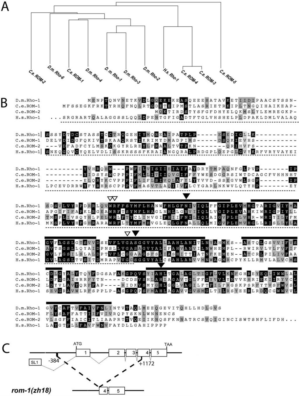
1 Open access, freely available online EGF Signal Propagation during C. elegans Vulval Development Mediated by ROM-1 Rhomboid PLoS BIOLOGY Amit Dutt [, Stefano Canevascini [, Erika Froehli-Hoier, Alex Hajnal * Zoologisches Institut, Universität Zurich, Zurich, Switzerland During Caenorhabditis elegans vulval development, the anchor cell (AC) in the somatic gonad secretes an epidermal growth factor (EGF) to activate the EGF receptor (EGFR) signaling pathway in the adjacent vulval precursor cells (VPCs). The inductive AC signal specifies the vulval fates of the three proximal VPCs P5.p, P6.p, and P7.p. The C. elegans Rhomboid homolog ROM-1 increases the range of EGF, allowing the inductive signal to reach the distal VPCs P3.p, P4.p and P8.p, which are further away from the AC. Surprisingly, ROM-1 functions in the signal-receiving VPCs rather than the signal-sending AC. This observation led to the discovery of an AC independent activity of EGF in the VPCs that promotes vulval cell fate specification and depends on ROM-1. Of the two previously reported EGF splice variants, the longer one requires ROM-1 for its activity, while the shorter form acts independently of ROM-1. We present a model in which ROM-1 relays the inductive AC signal from the proximal to the distal VPCs by allowing the secretion of the LIN-3L splice variant. These results indicate that, in spite of their structural diversity, Rhomboid proteins play a conserved role in activating EGFR signaling in C. elegans, Drosophila, and possibly also in mammals. Citation: Dutt A, Canevascini S, Froehli-Hoier E, Hajnal A (2004) EGF signal propagation during C. elegans vulval development mediated by ROM-1 rhomboid. PLoS Biol 2(11): e334. Introduction Intercellular signaling pathways control many diverse processes, such as cell proliferation, differentiation, survival, migration, shape changes, and responses to the environment. In most instances, the release of the signaling molecules by the signal-sending cells constitutes a rate-limiting step that determines the spatial distribution and temporal duration of the response (Freeman and Gurdon 2002). On the other hand, the binding sites on the receptors that are presented by the signal-receiving cells are usually in excess of the available ligands (Freeman and Gurdon 2002). Specificity is achieved by the tissue-specific expression of the signal or by the regulated activation of an inactive precursor molecule. For example, growth factor peptides are often produced as inactive precursors that need to be processed before they can be released and activate their cognate receptors on the signalreceiving cells (Arribas et al. 1996). The epidermal growth factor (EGF) receptor (EGFR) acts in a highly conserved signal transduction pathway that controls various cell fate decisions in metazoans (Bogdan and Klambt 2001). EGFR ligands of the transforming growth factor-a (TGF-a) family are produced as membrane-tethered precursor proteins with a single extracellular EGF repeat that is cleaved off the membrane anchor (Pandiella and Massague 1991; Bosenberg et al. 1993). The best-studied example of EGF processing is probably the Drosophila growth factor Spitz, which activates the EGFR in multiple developmental processes (Rutledge et al. 1992; Golembo et al. 1996). Genetic analysis of Drosophila EGFR signaling has identified Rhomboid-1 as a protein necessary for Spitz activation in the signal-sending cell (Bier et al. 1990; Golembo et al. 1996; Guichard et al. 2000; Wasserman et al. 2000). Drosophila Rhomboid-1 is the founding member of a family of seven-pass transmembrane proteins that function as intramembrane serine proteases (Urban et al. 2001). Site-specific cleavage of Spitz by Rhomboid-1 in the Golgi apparatus allows the secretion of the extracellular portion of Spitz by the signalsending cell (Lee et al. 2001; Urban and Freeman 2003). The Drosophila genome encodes a total of seven rhomboid genes with partially overlapping functions in different tissues that utilize the EGFR pathway (Wasserman et al. 2000; Urban et al. 2002). There are four predicted Rhomboid homologs in humans and five in C. elegans (Wasserman et al. 2000). Rhomboid-like proteins are even found in yeast and bacteria (Gallio et al. 2002; McQuibban et al. 2003). Rhomboids are part of the larger family of I-Clip proteases that includes the aspartyl protease Presenilin (Wolfe et al. 1999) and the Zn 2þ metalloprotease S2P (Urban and Freeman 2002). On the other hand, the secretion of vertebrate TGF-a involves Received April 27, 2004; Accepted August 3, 2004; Published September 28, 2004 DOI: /journal.pbio Copyright: Ó 2004 Dutt et al. This is an open-access article distributed under the terms of the Creative Commons Attribution License, which permits unrestricted use, distribution, and reproduction in any medium, provided the original work is properly cited. Abbreviations: AC, anchor cell; ACEL, AC-specific enhancer; ADAM, a disintegrin and metalloprotease; EGF, epidermal growth factor; EGFR, EGF receptor; Egl, egglaying defective; GFP, green fluorescent protein; hyp7, hypodermal syncytium; L[N], larval stage [N]; MAPK, mitogen-activated protein kinase; Muv, multivulva; NLS, nuclear localization signal; ORF, open reading frame; 18, primary; RNAi, RNA interference; RTK, receptor tyrosine kinase; 28, secondary; 38, tertiary; TGF, transforming growth factor; VPC, vulval precursor cell; Vul, vulvaless Academic Editor: Julie Ahringer, University of Cambridge *To whom correspondence should be addressed. ahajnal@zool.unizh.ch [These authors contributed equally to this work. Current address: Friedrich Miescher Institute for Biomedical Research, Basel, Switzerland 1799
2 processing by a disintegrin metalloprotease (ADAM), TNF-aconverting enzyme (Peschon et al. 1998). Whether a Rhomboid protease is also involved in the processing of TGF-a is currently unknown. The development of the C. elegans hermaphrodite vulva serves as a simple model by which to study signal transduction and cell fate determination during organogenesis (Kornfeld 1997; Sternberg and Han 1998). During C. elegans postembryonic development, the anchor cell (AC) in the somatic gonad induces three out of six equivalent vulval precursor cells (the VPCs, termed P3.p through P8.p) in the ventral hypodermis to adopt vulval cell fates (Sulston and White 1980; Kimble 1981). The AC produces the LIN-3 growth factor, which is similar to Drosophila Spitz and mammalian TGF-a (Hill and Sternberg 1992). The VPCs express the EGFR homolog LET-23 on the basolateral surface that faces the AC (Whitfield et al. 1999), and they are all competent to activate the RAS/mitogen-activated protein kinase (MAPK) signaling pathway in response to the inductive LIN-3 EGF signal. The VPC closest to the AC, P6.p, adopts the primary (18) cell fate characterized by a symmetrical cell lineage leading to eight 18 vulval cells (Sternberg and Horvitz 1986). The neighbors of P6.p, P5.p and P7.p, adopt the secondary (28) cell fate, which is characterized by an asymmetrical lineage leading to seven 28 vulval cells. The more distally located VPCs, P3.p, P4.p, and P8.p, adopt the uninduced tertiary (38) cell fate. After dividing once, they fuse with the surrounding hypodermal syncytium (hyp7). LIN-3 EGF dosage experiments have suggested that the inductive signal acts in a graded manner (Katz et al. 1995). According to this model, P6.p receives the highest amount of the inductive signal and thus adopts the 18 fate, while an intermediate level of the LIN-3 signal specifies the 28 fate in P5.p and P7.p. The distal VPCs, P3.p, P4.p, and P8.p, receive too little signal to adopt vulval cell fates. However, in response to the inductive signal, P6.p produces a lateral signal that activates the LIN-12 NOTCH signaling pathway in the neighboring VPCs, P5.p and P7.p (Greenwald et al. 1983). The LIN-12 NOTCH signal is both necessary and sufficient to induce the 28 cell fate (Sternberg 1988; Simske and Kim 1995). Thus, the graded LIN-3 EGF signal may act redundantly with the lateral LIN-12 NOTCH signal to specify the 28 vulval cell fate in the neighbors of P6.p (Kenyon 1995). Like its Drosophila and vertebrate homologs, LIN-3 EGF is synthesized as a transmembrane precursor protein (Hill and Sternberg 1992). Experiments with dig-1 mutants in which the AC is dorsally displaced indicate that the AC is capable of inducing vulval cell fates from a distance, suggesting that proteolytic cleavage of membrane-bound LIN-3 occurs in the AC (Thomas et al. 1990). Here, we report the identification of the C. elegans Rhomboid homolog ROM-1 as a positive regulator of vulval induction. Surprisingly, we find that ROM-1 acts in the signal-receiving VPCs rather than in the signal-sending AC. Furthermore, we uncover an AC-independent function of LIN-3 EGF that depends on ROM-1 activity in the VPCs. Two LIN-3 splice variants, termed LIN- 3S and LIN-3L, that differ by an insertion of 15 amino acids in the region in the juxtamembrane domain critical for processing, have been described (Hill and Sternberg 1992). Genetic epistasis experiments indicate that LIN-3L activity in the VPCs depends on ROM-1, while LIN-3S or a truncated form of LIN-3 lacking the transmembrane domain act independently of ROM-1. We propose a relay model in which ROM-1 is required for the activation of LIN-3L in the proximal VPCs to transmit the inductive AC signal to the distal VPCs. Results Five Rhomboid-Like Proteins in C. elegans Since the LIN-3 EGF growth factor is produced as a transmembrane precursor protein (Hill and Sternberg 1992), we asked whether an intramembrane serine protease of the Rhomboid family is involved in the proteolytic processing of LIN-3 EGF. Rhomboid proteins in metazoans share a characteristic secondary structure consisting of seven transmembrane domains (Bier et al. 1990; Urban et al. 2001). We searched the complete C. elegans genome sequence for genes with similarity to Drosophila rhomboid-1 and identified five rhomboid-like genes termed rom-1 (F26F4.3), rom-2 (C48B4.2), rom-3 (Y116A8C.14), rom-4 (Y116A8C.16), and rom-5 (Y54E10A.14) (Figure 1A). All five C. elegans ROM proteins display the typical secondary structure of Rhomboids (Wasserman et al. 2000). The transmembrane domains show the highest degree of sequence conservation, while the hydrophilic N termini are more divergent (Figure 1B). ROM-1 is most similar to Drosophila Rhomboid-1 (35% identity), followed by ROM-2 (29% identity) and the more diverged ROM-3 (24% identity), ROM-4 (26% identity), and ROM-5 (29% identity). Mutagenesis experiments with Drosophila Rhomboid-1 have identified a catalytic triad formed by conserved asparagine, serine, and histidine residues that are necessary for the serine protease activity (Urban et al. 2001). This catalytic triad is conserved only in ROM-1 (black triangles in Figure 1B), suggesting that the other four Rhomboid-like proteins do not function as serine proteases. In order to confirm the predicted intron-exon structure of rom-1, we isolated rom-1 cdna by RT-PCR. An SL1 transspliced leader sequence was identified at the 59 end of the message that was spliced to the second of the six exons predicted by the C. elegans genome project (Figure 1C) (see The remaining intron-exon boun- Figure 1. The C. elegans Rhomboid Genes (A) Dendogram showing the relation between the seven-pass transmembrane domains of Rhomboids from C. elegans (C.e.), Drosophila melanogaster (D.m.), and Homo sapiens (H.s.) calculated with the neighbor joining method using CLUSTAL X (Thompson et al. 1997). (B) Alignment of C. elegans (C.e.) ROM-1 and ROM-2 and Homo sapiens (H.s.) Rho-1 relative to Drosophila melanogaster (D.m.) Rho-1. Residues identical to those of Drosophila Rho-1 are highlighted in black, and similar residues are highlighted in grey. The thick black lines indicate the predicted seven-pass transmembrane domains. The three black triangles point at the residues forming a catalytic triad that forms a charge-relay system to activate the essential serine residue during peptide bond cleavage, and the three open triangles indicate other conserved residues necessary for the enzymatic activity as identified in D.m. Rho-1 (Urban et al. 2001). The region underlined with a dotted line indicates the extent of deletion in the rom-1(zh18) allele. (C) Intron-exon structure of the rom-1 locus and extent of the deletion in the rom-1(zh18) strain. The numbers indicate the position of the deletion break-points relative to the A in the ATG start codon. DOI: /journal.pbio g
3 1801
4 daries were confirmed experimentally and corresponded exactly to the predicted boundaries. The conceptual translation of the 1,071-bp open reading frame (ORF) predicts a protein of 356 amino acids, with very short stretches of hydrophilic amino acids between the seven-pass transmembrane domains, except for a longer loop consisting of 43 amino acids between the first and second transmembrane domains (see Figure 1B). ROM-1 and ROM-2 Are Not Essential for Normal Vulval Development As a first step to examine the biological function of the rom genes, we used RNA interference (RNAi) to transiently knock down their expression (Fire et al. 1998; Fraser et al. 2000; Kamath et al. 2001). Double-stranded RNA derived from a 352-bp rom-1 or a 718-bp rom-2 cdna fragment of the divergent N-terminal portion was injected into the hermaphrodite gonads, and vulval development was examined in the F1 progeny under Nomarski optics. No obvious vulval phenotype was observed when rom-1 or rom-2 RNAi was performed in a wild-type background. Also, feeding wild-type or let-60(n1046gf) animals with bacteria producing rom-3 dsrna had no effect on vulval development (unpublished data). Due to the high degree of sequence similarity between rom-3 and rom-4 (69.8% identity), rom-3 RNAi is likely to simultaneously reduce rom-4 function. Using a PCR-based assay to screen a library of mutagenized worms, we isolated a 1,556-bp deletion in the rom-1 gene (see Figure 1C) (Jansen et al. 1997; Berset et al. 2001). The zh18 deletion removes 206 amino acids from the N terminus, including the first three transmembrane domains and 384 bp of promoter sequences. Thus, the zh18 deletion probably results in a complete loss of rom-1 function and will be referred to as rom-1(0). The rom-1(0) single mutants exhibited no obvious phenotype; they were healthy and fertile. In addition, we obtained the rom-2(ok966) allele from the C. elegans Gene Knockout Consortium. The rom-2(ok966) animals carry a 530-bp deletion that removes the fifth exon, which contains the predicted catalytic center with the essential histidine residue (see Figure 1B) (Urban et al. 2001). Since this allele is predicted to inactivate any potential protease activity of ROM-2, we refer to it as rom-2(rf). Consistent with the RNAi experiments, both rom-1(0) and rom-2(rf) single mutants exhibited normal vulval development (Table 1, rows 2 and 3). Also, in rom-1(0) rom-2(rf) double mutants, no defects in vulval development were observed, ruling out a possible redundant function of the two genes (Table 1, row 4). Thus, neither ROM-1 nor ROM-2 are required for vulval induction under normal conditions. ROM-1 Positively Regulates the EGFR/RAS/MAPK Pathway in Distal VPCs Next, we examined whether loss of rom-1 or rom-2 function affects vulval induction in a sensitized genetic background by using mutations that hyperactivate the EGFR/RAS/MAPK pathway. The rom-1(0) mutation as well as rom-1 RNAi partially suppressed the multivulva (Muv) phenotype caused by overexpression of the LIN-3 EGF growth factor [lin-3(þ)] (Hill and Sternberg 1992) or by the n1046 gain-of-function (gf) mutation in the let-60 ras gene, which renders vulval development partially independent of upstream signaling (Beitel et al. 1990; Chang et al. 2000) (Table 1, rows 5 7 and 12 14). In addition, the rom-1(0) mutation suppressed the Muv phenotype of hs::mpk-1 animals that overexpress the wild-type MAPK MPK-1 under control of a heat-shock promoter together with Drosophila MEK-2 under control of the interferon-1a promoter (Lackner and Kim 1998) (Table 1, rows 16 and 17). In contrast to rom-1, neither the rom-2(rf) mutation nor rom-2 RNAi affected the Muv phenotype of let- 60(gf) animals (Table 1, row 15; unpublished data). The rom-1(0) mutation did not significantly enhance the vulvaless (Vul) phenotype caused by the lin-3(e1417), lin- 2(n397), sem-5(n2019), or let-60(n2021) mutations that reduce the activity of the receptor tyrosine kinase (RTK)/RAS/MAPK pathway (unpublished data). Since these Vul mutants affect the cell fates of only the proximal VPCs (P5.p, P6.p, and P7.p), ROM-1 plays no role in the induction of the proximal VPCs by the AC. Thus, ROM-1 enhances the activity of the EGFR/ RAS/MAPK pathway to allow the induction of the distal VPCs P3.p, P4.p, and P8.p. ROM-1 Regulates LIN-3 EGF Activity during Vulval Induction A soluble form of LIN-3 that consists of the extracellular domain with the EGF repeat but lacks the transmembrane and intracellular domains is biologically active and causes a Muv phenotype when overexpressed under control of a heatshock promoter (hs::lin-3extra) (Katz et al. 1995). Unlike fulllength LIN-3, the Muv phenotype induced by a low or high dosage of LIN-3extra was not suppressed by rom-1(0) (Table 1, rows 8 11). In lin-15(rf) mutants, all VPCs adopt vulval cell fates independently of the LIN-3 signal, though induction in lin-15(rf) mutants depends on the activity of LET-23 and the other components of the EGFR/RAS/MAPK pathway (Clark et al. 1994; Huang et al. 1994). The rom-1(0) mutation did not suppress the Muv phenotype of lin-15(rf) animals, suggesting that loss of rom-1 function affects the LIN-3-dependent induction of vulval cell fates rather than the LIN-3- independent activity of the EGFR/RAS/MAPK pathway (Table 1, rows 18 and 19). Finally, we examined the genetic interaction between rom-1 and the Notch and Wnt pathways, since both pathways control vulval cell fate specification in parallel with the RTK/ RAS/MAPK pathway (Wang and Sternberg 2001). In lin-12 notch(gf) animals, no AC is formed, and all VPCs adopt the 28 cell fate (Sternberg and Horvitz 1989). The same phenotype was observed in rom-1(0) lin-12(gf) double mutants (Table 1, rows 20 and 21). In addition, the Muv phenotype caused by hyperactivation of the Wnt pathway through a reduction-offunction mutation in pry-1 axin or by overexpression of a N- terminally truncated BAR-1 b-catenin protein was not suppressed by the rom-1(0) mutation (Table 1, rows 22 25) (Gleason et al. 2002). In summary, these experiments suggest that ROM-1 promotes the LIN-3-dependent activation of the EGFR/RAS/ MAPK signaling pathway. ROM-1 likely acts at the level or upstream of LIN-3 since full-length but not a soluble form of LIN-3 was sensitive to loss of rom-1 function. ROM-1 Is Required to Transmit the Inductive Signal to the Distal VPCs To assess how much inductive signal each VPC receives, we examined the expression pattern of the egl-17::cfp reporter, which is a transcriptional target of the EGFR/RAS/MAPK 1802
5 Table 1. Suppression of Multivulva Mutants by rom-1(0) Row Genotype % Muv % Vul Induction Index n 1 wild type many 2 rom-1(0) rom-2(rf) rom-1(0) rom-2(rf) [lin-3(þ)] a [lin-3(þ)]; rom-1 RNAi b 56 0 n.d rom-1(0); [lin-3(þ)] *** (3) 66 8 [hs::lin-3extra] low c rom-1(0);[hs::lin-3extra] low c [hs::lin-3extra] high d rom-1(0); [hs::lin-3extra] high d let-60(gf) let-60(gf); rom-1 RNAi b 37 0 n.d rom-1(0); let-60(gf) *** (10) rom-2(rf); let-60(gf) [hs::mpk-1] d rom-1(0); [hs:: mpk-1] d ** (15) lin-15(rf) rom-1(0); lin-15(rf) lin-12(gf) e lin-12(gf) rom-1(0) e pry-1(rf) pry-1(rf); rom-1(0) [hs::bar-1dnt] d [hs::bar-1dnt] rom-1(0) d Vulval induction was scored using Nomarski optics as described in Materials and Methods. % Vul indicates the fraction of animals with fewer than three induced VPCs, % Muv indicates the fraction of animals with more than three induced VPCs, and the induction index indicates the average number of VPCs per animal that had adopted 18 or 28 vulval fates. Number of animals scored is designated by n. Alleles used: pry-1(mu38), rom-1(zh18), rom-2(ok996), lin-12(n137gf), dpy-19(e1259), huis7[hs::bar-1dnt], let- 60(n1046gf), gais36[hs::mpk-1, D-mek-2(gf)], lin-15(n309), zhex22[lin-3(þ), sur-5::gfp, unc-119(þ)], and syis12[hs::lin-3extra]. Statistical analysis was done as described in Materials and Methods. a Five independent transgenic lines generated with this construct displayed induction indices ranging from 4.1 to 5.4, and one of the lines displaying a penetrant Muv phenotype (zhex22) was used for the further analysis in the different backgrounds. b dsrna was injected into the syncytial gonad of the parents, and vulval induction in the F1 progeny was scored for rows 4 and 11 by inspection under a dissecting microscope. c To provide a low dose of LIN-3extra, L1 larvae were heat-shocked for 5 min at 33 8C and grown at 25 8C until L4. d To provide a high dose of LIN-3extra, or MPK-1 or BAR-1DNT, respectively, early L21 larvae were heat-shocked for 30 min at 33 8C and grown at 25 8C until L4. e These strains carried the dpy-19(e1259) mutation in cis to lin-12(n137gf). ** p 0.001; *** p ; numbers in brackets next to asterisks indicate the row to which a dataset was compared. n.d., no data. DOI: /journal.pbio t001 pathway (Yoo et al. 2004). In mid L2 larvae, before the LIN-12 NOTCH-mediated lateral inhibition becomes effective, egl- 17::cfp is expressed in a graded manner with highest levels in P6.p, intermediate levels in P5.p and P7.p (Yoo et al. 2004), and lower levels in P3.p, P4.p, and P8.p (Figure 2A and 2B). We therefore examined the effect of the rom-1(0) mutation on the egl-17::cfp expression pattern in mid L2 larvae. For this purpose, larvae were synchronized in the mid L1 stage at 13 h of development by letting them hatch in the absence of food, and then development was allowed to proceed by adding food for another 24 h until they reached the mid L2 stage (approximately 37 h of development). Loss of ROM-1 function had no effect on egl-17::cfp expression in the proximal VPCs (P5.p, P6.p, and P7.p), but significantly reduced egl-17::cfp expression in the distal VPCs when compared to wild-type animals (Figure 2C and 2D). Thus, ROM-1 increases the range of the inductive LIN-3 signal, allowing the distal VPCs to activate the EGFR/RAS/MAPK pathway. The altered egl-17::cfp expression pattern in rom-1(0) animals is consistent with the epistasis data, which showed that loss of rom-1 function affects the induction of only the distal VPCs (see above). ROM-1 Is Expressed in the VPCs but Not in the AC during Vulval Induction To analyze the expression pattern of ROM-1, we generated a transcriptional rom-1 reporter by fusing 6.9 kb of the 59 rom- 1 promoter/enhancer region to the green fluorescent protein (gfp) ORF carrying a nuclear localizing signal (zhis5[rom-1::nls::gfp]). With a translational full-length rom-1::gfp fusion construct, we failed to obtain transgenic lines that consistently expressed ROM-1::GFP. Moreover, a genomic DNA fragment encompassing the entire rom-1 locus failed to produce stable transgenic lines even when injected at relatively low concentrations (1 10 ng/ll), suggesting that elevated levels of ROM-1 are toxic to the animals. 1803
 and lin-3(rf) Mutants Photographic images on the left (A, C, E, G, and I) show the expression of the aris92[egl-17::cfp] reporter in the VPCs of")
6 Figure 2. Expression of the egl-17::cfp Reporter in rom-1(0) and lin-3(rf) Mutants Photographic images on the left (A, C, E, G, and I) show the expression of the aris92[egl-17::cfp] reporter in the VPCs of mid-l2 larvae of the different genotypes indicated. Pie graphs on the right (B, D, F, H, and J) show semi-quantitative representations of the expression levels observed in individual VPCs in the different backgrounds. A solid black color indicates the strongest expression of EGL-17::CFP as it was observed in P6.p of many (59%) wild-type animals; dark grey indicates intermediate, light grey weak, and white undetectable expression. The numbers inside the pie charts are the corresponding percentage values, and n refers to the number of animals examined for each case. EGL-17::CFP expression in each VPC of rom-1(0) or lin-3(e1417rf) animals was compared against the same VPC in wild-type animals (considered as expected value) with a Chi 2 test for its independence; *** p , ** p The row to which a dataset was compared is indicated on the right. All photographs were taken with identical exposure and contrast settings. The scale bar in (I) is 20 lm. DOI: /journal.pbio g002 The transcriptional rom-1::nls::gfp reporter was widely expressed in somatic cells throughout development. Surprisingly, we did not detect any rom-1::nls::gfp expression in the gonadal AC before the L4 stage, while consistent expression was observed in the Pn.p cells and the Pn.a-derived neurons from the L1 stage on. In early L2 zhis5 larvae, the six VPCs expressed rom-1::nls::gfp at equal levels (Figure 3A and 3B). The Pn.p cells that are not part of the vulval equivalence group and had fused to hyp7 at the end of the L1 stage showed relatively higher rom-1::nls::gfp expression than the VPCs (for example, P1.p, P2.p, and P9.p in Figure 3C and 3D). Toward the end of the L2 stage, rom-1::nls::gfp expression decreased in distal VPCs adopting the 38 uninduced fate and persisted in the proximal VPCs adopting induced vulval fates (Figure 3C and 3D). In 60% of zhis5 animals, we observed an upregulation of rom-1::nls::gfp in P6.p, and in 35% and 45% of the cases, rom-1::nls::gfp expression was higher in P5.p and P7.p, respectively (n = 20). After vulval induction, rom- 1::nls::gfp was down-regulated in the 18 and 28 descendants of P5.p, P6.p and P7.p, while the 38 descendants of P3.p, P4.p, and P8.p again expressed high levels of rom-1::nls::gfp after they had fused with hyp7 (Figure 3E and 3F). Expression of rom-1::nls::gfp was observed in the AC and other cells of the somatic gonad only beginning in the L4 stage, before the AC fused with the uterine seam cell and persisting after fusion (Figure 3G and 3H) (Sulston and Horvitz 1977; Newman et al. 1804
![Figure 3. Expression Pattern of rom- 1::nls::gfp Expression pattern of the zhis5[rom- 1::nls::gfprom-1::] transcriptional reporter during vulval development.](/docs-images/82/86660341/images/7-0.jpg "Images on the left (A, C, E, G, and I) show the corresponding Nomarski pictures with the arrows pointing at the Pn.p cell nuclei and the arrowhead indicating the position of the AC nucleus.")
7 Figure 3. Expression Pattern of rom- 1::nls::gfp Expression pattern of the zhis5[rom- 1::nls::gfprom-1::] transcriptional reporter during vulval development. Images on the left (A, C, E, G, and I) show the corresponding Nomarski pictures with the arrows pointing at the Pn.p cell nuclei and the arrowhead indicating the position of the AC nucleus. (B) A mid L2 larva before vulval induction with uniform rom-1::nls::gfp expression in all the Pn.p cells. (D) An early L3 larva in which rom-1::nls::gfp expression was decreased in all VPCs except P6.p (see text for a quantification of the expression pattern). Note that the nuclei of hyp7 and the Pn.p cells that had fused to hyp7 displayed strong rom- 1::nls::gfp expression (P1.p, P2.p, P3.p and P9.p in the example shown). (F) A mid to late L3 larva in which P6.p had generated four descendants. Expression of rom-1::nls::gfp occurred only in the 38 descendants of P.4.p and P8.p after they fused to hyp7. (H) An L4 larva during vulval invagination. No rom-1::nls::gfp was detectable in the 18 and 28 descendants of P5.p, P6.p, and P7.p, but the AC and the surrounding uterine cells displayed strong rom-1::nls::gfp expression. (K) A late L2 to early L3 larva following the ablation of the precursors of the somatic gonad. No up-regulation of rom-1::nls::gfp in P5.p, P6.p, or P7.p was observed. The scale bar in (K) is 10 lm. DOI: /journal.pbio g
8 1996). To test whether the inductive AC signal is required for the elevated rom-1::nls::gfp expression in the proximal VPCs, we ablated in zhis5 animals the precursors of the somatic gonad Z1 and Z4 (Kimble 1981). Uniformly low rom-1::nls::gfp expression was found in all six VPCs of gonad-ablated zhis5 animals at the late L2 to early L3 stage, before the descendants of the 38 VPCs had fused to hyp7 (Figure 3J and 3K, n = 20). To test whether rom-1::nls::gfp expression depends on RTK/RAS/MAPK signaling in the VPCs, we introduced the zhis5 transgene into lin-7(e1413) mutants that exhibit a penetrant Vul phenotype due to reduced LET-23 EGFR activity (Simske et al. 1996). In lin-7(e1413); zhis5 animals, the up-regulation of rom-1::nls::gfp occurred less frequently (in 13%, 33%, and 7% of the cases in P5.p, P6.p, and P7.p, respectively, n = 15). Thus, the AC signal upregulates rom-1::nls::gfp expression in the VPCs that adopt vulval cell fates. ROM-1 Acts in an AC-Independent Pathway that Promotes Vulval Induction To examine whether ROM-1 acts in cells other than the AC (which is part of the somatic gonad), we tested the effect of loss of rom-1(þ) function on vulval induction in gonad-ablated animals. If ROM-1 acts exclusively in the AC, then the rom-1(0) mutation should not affect vulval induction in gonad-ablated animals. On the other hand, if ROM-1 acts in cells other than the AC, then the rom-1(0) mutation should suppress vulval induction even in the absence of the AC. Since the inductive AC signal is absolutely required to initiate vulval development (Kimble 1981), we performed the gonad ablation experiments in let-60(gf) or hs::mpk-1 animals that exhibit a hyperactive EGFR/RAS/MAPK signaling pathway causing AC-independent vulval induction (Table 2, rows 5 and 8) (Beitel et al. 1990; Lackner and Kim 1998; Chang et al. 2000). In addition, we examined lin-3(þ) animals because, as reported previously by Hill and Sternberg (1992), in animals that overexpress wildtype lin-3 under control of its own promoter, some vulval differentiation could still be observed in the absence of the AC, pointing at an additional source of LIN-3 from the transgene in cells outside of the gonad (Table 2, row 2). Loss of rom-1 function in gonad-ablated lin-3(þ), let-60(gf), or hs::mpk-1 animals caused a strong further reduction in vulval induction (Table 2, compare rows 2 with 3, 5 with 6, and 8 with 9). In contrast, vulval induction in gonad-ablated lin-15(rf) animals that exhibit lin-3 independent vulval differentiation was not affected by the rom-1(0) mutation (Table2, rows 13 and 14). Analogous results were obtained by examining the egl- 17::cfp expression pattern after removal of the AC. In gonadablated animals, residual egl-17::cfp expression was observed in all VPCs (Figure 2E and 2F). In many cases, P6.p expressed higher levels of the reporter than did the other VPCs despite the absence of the AC. Loss of rom-1 function in gonadablated animals caused a further decrease in egl-17::cfp expression in all VPCs (Figure 2G and 2H). Thus, ROM-1 acts in cells outside of the somatic gonad to promote vulval induction. An AC-Independent Activity of LIN-3 EGF Next, we used an analogous strategy to test whether endogenous LIN-3 acts with ROM-1 in an AC-independent pathway. The decrease in vulval induction in hs::mpk-1 animals that was caused by the lin-3(n1049) loss-of-function mutation [lin-3(0)] was much stronger than the decrease observed in gonad-ablated lin-3(þ); hs::mpk-1 animals (Table 2, compare rows 8 and 10; the L1 larval lethal phenotype caused by the lin-3(0) mutation was suppressed by the hs::mpk-1 transgene). Vulval induction in lin-3(0); hs::mpk-1 animals was not affected by gonad ablation since the lin-3(0) allele eliminated lin-3 function in the AC (Table 2, compare rows 10 and 11). Thus, a complete loss of lin-3 function had a more severe effect on vulval induction than did just the removal of the AC. Table 2. Gonad-Independent Function of rom-1 and lin-3 Row Genotype Gonad a % Muv % Vul Induction Index n 1 [lin-3(þ)] þ [lin-3(þ)] *** (1) 23 3 rom-1(0); [lin-3(þ)] *** (2) 20 4 let-60(gf) þ let-60(gf) *** (4) 30 6 rom-1(0); let-60(gf) *** (5) 21 7 [hs::mpk-1] b þ [hs::mpk-1] b *** (7) 14 9 rom-1(0); [hs::mpk-1] b *** (8) lin-3(0); [hs::mpk-1] b þ *** (8) lin-3(0); [hs::mpk-1] b *** (8) rom-1(0); lin-3(0); [hs::mpk-1] b þ *** (8) lin-15(rf) rom-1(0); lin-15(rf) Vulval induction was scored as described in the legend to Table 1. See Table 1 legend for key to abbreviations and terminology. Alleles used: rom-1(zh18), let-60(n1046gf), gais36[hs::mpk-1, D-mek-2(gf)], lin-3(n1049null) [the sterile Dpy nonunc progeny segregated by dpy-20(e1282) lin-3(n1049)/ unc-44(e362) unc24(e138); gais36 mothers was examined], lin-15(n309), zhex22[lin-3(þ), and sur-5::gfp, unc-119(þ)]. a The gonad precursors Z1 through Z4 were ablated in L1 larvae where indicated. b L2 larvae were heat-shocked for 30 min at 33 8C and grown at 25 8C until L4. DOI: /journal.pbio t
9 Likewise, the lin-3(e1417) reduction-of-function mutation almost completely abolished the expression of the egl-17::cfp marker (Figure 2I and 2J). Thus, LIN-3 is also necessary for the AC-independent egl-17::cfp expression in the VPCs. Loss of rom-1 function in a lin-3(0); hs::mpk-1 background caused no further decrease in vulval induction, suggesting that ROM-1 does not affect vulval development in the absence of LIN-3 (Table 2, compare rows 10 and 12). Taken together, these experiments indicate that not only the AC but also cells outside of the gonad produce LIN-3 to promote vulval fate specification. This AC-independent activity of LIN-3 requires ROM-1 function. ROM-1 Can Act in the Pn.p Cells The absence of detectable rom-1::nls::gfp expression in the AC around the time of vulval induction and the ACindependent function of rom-1 and lin-3 suggested that rom- 1 may act cell-autonomously in the VPCs. To test this hypothesis, we expressed rom-1 under control of the Pn.p cell-specific lin-31 promoter (lin-31::rom-1) (Tan et al. 1998). The lin-31::rom-1 transgene restored vulval induction in rom- 1(0); let-60(gf) and rom-1(0); hs::mpk-1 double mutants to levels comparable to those found in let-60(gf) and hs::mpk-1 single mutants (Table 3, rows 1 3 and 8 10). A transgene encoding bacterial Cre recombinase under control of the lin-31 promoter (lin-31::cre) that was used as a negative control had no effect on vulval induction (Table 3, rows 4 and 11) (Hoier et al. 2000). Consistent with a function of rom-1 in an ACindependent pathway, the lin-31::rom-1 transgene also increased induction in gonad-ablated rom-1(0); let-60(gf) animals (Table 3, rows 5 7). Finally, we expressed rom-1 in the AC under control of the AC-specific enhancer (ACEL) (ACEL::rom-1), which is located in the third intron of the lin-3 locus (Hwang and Sternberg 2003). In contrast to lin-31::rom- 1, the ACEL::rom-1 transgene did not rescue the suppression of the let-60(gf) Muv phenotype by rom-1(0) (Table 3, rows 12 and 13). Thus, the tissue-specific expression of ROM-1 in the Pn.p cells efficiently rescues a loss of rom-1 function. LIN-3 EGF from the Pn.p Cells Amplifies the AC Signal To examine whether the VPCs or their descendants are the source of the AC-independent LIN-3 signal, we expressed lin-3 dsrna in the Pn.p cells in order to down-regulate by RNAi any possible lin-3 expression in the VPCs (Timmons et al. 2003). For this purpose, a vector consisting of an inverted repeat of a 921-bp lin-3 cdna fragment under control of the same Pn.p cell-specific lin-31 promoter used above (lin-31::lin- 3i) was introduced into wild-type animals (Tan et al. 1998). Vulval induction occurred normally in lin-31::lin-3i animals (Table 4, row 1), although the adult animals displayed an 80% penetrant egg-laying defective (Egl) phenotype due to a defect in vulval morphogenesis (n = 122). In wild-type L4 larvae, the 18 descendants of P6.p in the vulf toroid ring (P6.papl/r and P6.ppal/r,), secrete LIN-3 to specify the ventral uterine (uv1) cell fate in the somatic gonad (Chang et al. 1999). If LIN-3 expression in the F cells is blocked through a mutation in the egl-38 pax transcription factor, then the uv1 cell adopts a uterine seam fate, resulting in an Egl phenotype. Thus, lin- 31::lin-3i appeared to efficiently reduce LIN-3 expression in the vulval F cells without reducing the activity of LIN-3 in the AC. To further authenticate the efficiency of this approach, we crossed animals carrying the lin-31::lin-3i transgene to animals expressing the short splice variant of lin-3 cdna in the Pn.p cells under control of the lin-31 promoter (lin-31::lin- 3S, see below). The lin-31::lin-3i transgene almost completely suppressed the Muv phenotype caused by the lin-31::lin-3s transgene, while the lin-31::cre transgene that was used as negative control had no effect (Table 4, rows 2 4). Furthermore, the lin-31::lin-3i transgene significantly reduced vulval induction in lin-3s animals that carry a lin-3 minigene encoding the short splice variant (Figure 4A), as well as in let-60 ras(gf) and hs::mpk-1 animals (Table 4, rows 5 14). Table 3. Expression of rom-1 in the Pn.p Cells but Not in the AC Rescues the rom-1(0) Phenotype Row Genotype Gonad a % Muv Induction Index n 1 let-60(gf) þ rom-1(0); let-60(gf) þ *** (1) 30 3 rom-1(0); let-60(gf); [lin-31::rom-1] b þ *** (2) rom-1(0); let-60(gf); [lin-31::cre] þ let-60(gf) *** (1) 30 6 rom-1(0); let-60(gf) 0 0.9*** (2) 21 7 rom-1(0); let-60(gf); [lin-31::rom-1] 0 2.1*** (6) 14 8 [hs::mpk-1] c þ rom-1(0); [hs::mpk-1] c þ ** (8) rom-1(0); [hs::mpk-1]; [lin-31::rom-1] c þ *** (9) rom-1(0); [hs::mpk-1]; [lin-31::cre] c þ rom-1(0); let-60(gf); [ACEL::rom-1] d þ rom-1(0); let-60(gf); [ACEL::rom-1] Vulval induction was scored as described in the legend to Table 1. See Table 1 legend for key to abbreviations and terminology. Alleles used: rom-1(zh18), let-60(n1046gf), gais36[hs::mpk-1, D-mek-2(gf)], zhex66[lin-31::rom-1, unc-119(þ), sur-5::gfp], zhex81[lin- zhex81[lin-31::cre, unc-119(þ), myo-3::gfp], and zhex89[acel::rom-1, sur-5::gfp]. a The gonad precursor cells Z1 through Z4 were ablated in L1 larvae where indicated. b All three independent transgenic lines examined increased the induction index of rom-1(0); let-60(gf) animals to c L2 larvae were heat-shocked for 30 min at 33 8C and grown at 25 8C until L4. d Two independent transgenic lines examined displayed an induction index in rom-1(0); let-60(gf) animals of 3.3 and 3.5. DOI: /journal.pbio t
10 Table 4. Pn.p Cell-Specific Function of lin-3 Row Genotype Gonad a % Muv % Vul Induction Index n 1 [lin-31::lin-3i] b þ Many 2 [lin-31::lin-3s] þ [lin-31::lin-3s]; [lin-31::lin-3i] þ *** (2) 23 4 [lin-31::lin-3s]; [lin-31::cre] þ [lin-3s] þ [lin-3s]; [lin-31::lin-3i] þ ** (5) 19 7 [lin-3s]; [lin-31::cre] þ let-60(gf) þ let-60(gf); [lin-31::lin-3i] þ *** (8) let-60(gf); [lin-31::cre] þ let-60(gf) *** (8) let-60(gf); [lin-31::lin-3i] *** (11) [hs::mpk-1] 3 þ [hs::mpk-1]; [lin-31::lin-3i] c þ *** (13) egl-38(rf) þ Many 16 egl-38(rf); [hs::mpk-1] 3 þ *** (13) 30 Vulval induction was scored as described in the legend to Table 1. See Table 1 legend for key to abbreviations and terminology. Alleles used: let-60(n1046gf), egl-38(n578), gais36[hs::mpk-1, D-mek-2(gf)], zhex72[lin-31::lin-3s, unc-119(þ), sur-5::gfp], zhex68[lin-3s, unc-119(þ), sur-5::gfp], zhex88[lin-31::lin-3i, unc-119(þ), myo-3::gfp],and zhex81[lin- 31::cre, unc-119(þ), myo-3::gfp]. a The gonad precursor cells Z1 through Z4 were ablated in L1 larvae where indicated. b Three independent transgenic lines displayed a penetrant Egl phenotype and suppressed the let-60(gf) phenotype to induction indices L1 ranging from 3.1 to 3.3, and one line was used for further analysis. Two lines displayed no Egl phenotype and did not suppress the let-60(gf) phenotype (induction index 4.2 and 4.3). All five lines exhibited normal vulval induction in a wild-type background. c L1 and L2 larvae were heat-shocked for 30 min at 33 8C and grown at 25 8C until L4. DOI: /journal.pbio t004 Consistent with an AC-independent function of LIN-3 in the VPCs, the lin-31::lin-3i transgene also affected vulval induction in let-60(gf) animals lacking a gonad (Table 4, rows 11 and 12). As an independent test to determine if a reduction of LIN- 3 expression in the vulval cell lineage affects induction, we used the n578 reduction-of-function mutation in the egl-38 pax transcription factor, because this egl-38 allele has been shown to eliminate LIN-3 expression in the 18 cell lineage (Chang et al. 1999). Although egl-38(rf) single mutants exhibited wild-type levels of vulval induction, the egl-38(rf) mutation reduced the Muv phenotype of hs::mpk-1 animals to a similar degree as the rom-1(0) mutation or the lin-31::lin-3i transgene (Table 4, rows 15 and 16). Thus, EGL-38 is necessary for the AC-independent function of LIN-3. In summary, these experiments indicated that, during the process of vulval cell fate specification, some of the Pn.p cells (probably the VPCs) produce LIN-3 to amplify the inductive signal. Three LIN-3 EGF Splice Variants that Differ in the Juxtamembrane Domain The lin-3 locus encodes two splice variants termed LIN-3S (short) and LIN-3L (long) that are generated by the differential choice of the splice donor of exon 6 (Figure 4B) (Hill and Sternberg 1992). While performing RT-PCR experiments using a primer pair flanking the differentially spliced exons, we discovered a third splice variant, termed LIN-3XL, that is generated by the insertion of an additional exon (6b) between exons 6 and 7 (Figure 4A and 4B). The LIN-3XL splice variant was independently isolated from the yk1053b07est clone. LIN-3XL contains a 41 amino acid insert, and LIN-3L contains a 15 amino acid insert, in the region between the EGF repeat and the transmembrane domain, when compared to LIN-3S (Figure 4C). Since the analogous region in Drosophila Spitz EGF is required for the proteolytic processing of Spitz by Rhomboid (Bang and Kintner 2000; Lee et al. 2001; Urban and Freeman 2003), we sought to determine which of the LIN-3 splice variants depend on ROM-1 activity. To address this question, we first constructed lin-3 minigenes by replacing the differentially spliced exons with cdna fragments encoding either of the splice forms (see Figure 4B). Both lin-3l and lin-3s minigenes were capable of inducing a Muv phenotype, but we observed a marked difference in the dosages required to elicit this phenotype. All (12 out of 12) transgenic lines generated by injection of a relatively low (1 ng/ll) or high (100 ng/ll) concentration of the lin-3s minigene exhibited a strong Muv phenotype (with induction indices ranging from 4.1 to 5.6). In contrast, the lin-3l construct caused a Muv phenotype only when injected at a high concentration. (None of the seven lin-3l lines obtained by injecting 1 ng/ll exhibited a Muv phenotype, while all nine lines obtained by injecting 100 ng/ll exhibited a Muv phenotype, with induction indices ranging from 4.2 to 5.0). For the lin-3xl construct, we obtained variable results; some lines exhibited a weak Muv and others no or even a Vul phenotype (unpublished data). Since we failed to observe a consistent phenotype with this minigene construct, we did not further pursue the analysis of the lin-3xl minigene. ROM-1 Is Necessary for the Activation of the Long LIN-3 Splice Variant To investigate the genetic interactions between rom-1 and the lin-3 splice variants, we compared one line for each of the lin-3s and lin-3l minigenes that displayed a similar degree of 1808
11 Figure 4. Alternative Splicing of lin-3 mrna (A) RT-PCR amplification of lin-3 mrna from mixed-stage N2 cdna before (left) and after (right) size fractionation by preparative agarose gel electrophoresis. The lowest band corresponding to LIN-3S is most prominent, and the two upper bands correspond to LIN-3L and LIN-3XL. (B) Intron-exon structure of the lin-3 locus. The lin-3l splice variant is generated by the usage of an alternative (more 39 located) splice donor in exon 6a. The lin-3xl variant contains the additional exon 6b inserted between exons 6a and 7. The regions encoding the EGF repeat in exon 5 and part of 6a and the transmembrane domain in exon 7 are outlined, and the positions of the PstI sites used for the construction of the minigenes are indicated (see Materials and Methods). The structure of the lin-3s and lin-3l minigenes is shown in the lower part of the graphic. (C) Sequence alignment of the alternatively spliced region in LIN-3 with the corresponding region in Drosophila Spitz. The 15 and 41 amino acids in LIN-3L and LIN-3XL, respectively, in the juxtamembrane region break the alignment of LIN-3 with Spitz. The C-terminal end of the EGF domain is underlined with a horizontally hatched bar, and the beginning of the transmembrane domain is underlined by a diagonally hatched line. DOI: /journal.pbio g004 vulval induction (Table 5, rows 1 and 4). Since the presence of endogenous LIN-3 might mask a specific requirement of either LIN-3 splice variant, we introduced the two minigenes into a lin-3(0) background. The lin-3s and lin-3l transgenes both rescued the larval lethality of lin-3(0) mutants, yielding adult Muv animals (Table 5, rows 2 and 5 and Table 6, rows 1 and 3). Since we used multicopy arrays that are silenced in the germ cells and LIN-3 is required in the oocytes to induce ovulation (Clandinin et al. 1998), the rescued lin-3(0); lin-3s and lin-3(0); lin-3l animals were sterile. Loss of rom-1 function did not affect the viability or the Muv phenotype of lin-3(0); lin-3s animals (Table 5, rows 2 and 3 and Table 6, row 2). In contrast, the efficiency of the lin-3l transgene in rescuing the larval lethality of lin-3(0) mutants was reduced by loss of rom-1 function (Table 6, row 4). Moreover, the rare rom-1(0), lin-3(0); lin-3l animals that escaped the larval lethality exhibited a weaker Muv phenotype than lin-3(0); lin-3l animals, suggesting that the function of LIN-3L during vulval induction partially depends on ROM-1 activity (Table 5, rows 5 and 6). To specifically test the function of the LIN-3 splice variants in the Pn.p cells, we cloned full-length cdnas encoding the LIN-3S and LIN-3L splice variants under control of the Pn.p cell-specific lin-31 promoter (lin-31::lin-3s and lin-31::lin-3l). The lin-31::lin-3s and lin-31::lin-3l transgenes both caused a strong Muv phenotype in the presence and absence of the AC (Table 5, rows 7, 9, 11, and 13). Loss of rom-1 function did not change the phenotype of lin-31::lin-3s animals (Table 5, rows 8 10), but it strongly suppressed the Muv phenotype of lin- 31::lin-3L animals (Table 5, rows 12 and 14). Thus, ROM-1 is required for the activity of the LIN-3L splice variant in the Pn.p cells, while LIN-3S functions independently of ROM-1. Discussion ROM-1 Positively Regulates LIN-3 EGF Mediated Vulval Induction Of the five Rhomboid proteins predicted by the complete C. elegans genome sequence, only ROM-1, the closest homolog of Drosophila Rhomboid-1, possesses the hallmarks of a serine protease with an intact catalytic center (Urban et al. 2001). Here, we show that ROM-1 acts as a positive regulator of the EGFR/RAS/MAPK signaling pathway during vulval induction, 1809
12 Table 5. The lin-3l Splice Form Depends on rom-1 Activity Row Genotype Gonad a % Muv % Vul Induction Index n 1 [lin-3s] a þ lin-3(0); [lin-3s] þ rom-1(0); lin-3(0); [lin-3s] þ [lin-3l] 2 þ lin-3(0); [lin-3l] þ rom-1(0); lin-3(0); [lin-3l] þ ** (5) 6 7 [lin-31::lin-3s] þ rom-1(0); [lin-31::lin-3s] þ [lin-31::lin-3s] ** (7) 9 10 rom-1(0); [lin-31::lin-3s] [lin-31::lin-3l] þ rom-1(0); [lin-31::lin-3l] þ *** (11) [lin-31::lin-3l] rom-1(0); [lin-31::lin-3l] *** (13) 16 Vulval induction was scored as described in the legend to Table 1. See Table 1 legend for key to abbreviations and terminology. Alleles used: rom-1(zh18), lin-3(n1049null) [the sterile Dpy nonunc progeny segregated by dpy-20(e1282) lin-3(n1049)/ unc24(e138) unc-unc-44(e362) mothers were examined], zhex68[lin-3s, sur-5::gfp, unc-119(þ)], zhex69[lin-3l, sur-5::gfp, unc-119(þ)], zhex72[lin-31::lin-3s, unc-119(þ), sur-5::gfp], and zhex73[lin-31::lin-3l, unc-119(þ) sur-5::gfp]. a The gonad precursor cells Z1 through Z4 were ablated in L1 larvae where indicated. DOI: /journal.pbio t005 as loss of rom-1 function partially suppresses ectopic vulval induction caused by hyperactivation of the EGFR/RAS/MAPK pathway. Our epistasis analysis points at a role of ROM-1 in activating LIN-3 EGF. The activity of a soluble form of LIN-3 lacking the transmembrane and intracellular domains is completely independent of ROM-1 activity, but the activity of full-length LIN-3 EGF is sensitive to loss of ROM-1 function. Moreover, a mutation in lin-15, which renders vulval induction independent of LIN-3 activity, is not suppressed by loss of rom-1 function, but mutations in LET-23 or downstream components of the EGFR/RAS/MAPK pathway Table 6. Rescue of the Larval Lethality in lin-3(0) Mutants by the lin-3s and lin-3l Minigenes Row Genotype % Viable a n 1 lin-3(0); [lin-3s] rom-1(0); lin-3(0); [lin-3s] lin-3(0); [lin-3l] rom-1(0); lin-3(0); [lin-3l] 4*** (3) 127 See Table 1 legend for key to abbreviations and terminology. To confirm that the viable Dpy nonunc animals were the rescued lin-3(0) homozygotes and not recombinants between dpy-20 and lin-3, they were scored for fertility 4 5 d later. All animals counted for this table developed into sterile adults due to the silencing of the rescuing transgenes in the germline. In contrast, all viable Dpy nonunc progeny segregated by dpy-20(e1282) lin-3(n1049)/ unc-44(e362) unc24(e138) mothers lacking a lin-3 minigene developed into fertile adults, indicating recombination between dpy-20 and lin-3. (Seven recombinants were found among 1,000 F1 progeny animals.) Alleles used: rom-1(zh18), lin-3(n1049), dpy-20(1282), unc24(e138), unc-44(e362), zhex68[lin-3s, sur-5::gfp, unc-119(þ)], and zhex69[lin- 3L,sur- sur-5::gfp, unc-119(þ)]. a To score viability, GFP-positive embryos segregated by dpy-20(e1282) lin- 3(n1049)/ unc24(e138) unc-44(e262) mothers carrying the indicated minigenes were placed on NGM plates. After 24 and 48 h, the viable Dpy nonunc larvae representing the rescued lin-3(0) animals and the dead larvae were counted, and the % viability was calculated as 1003 [Dpy nonuncs/(dpy nonuncsþ dead larvae)]. DOI: /journal.pbio t006 efficiently suppress the lin-15 Muv phenotype (Clark et al. 1994; Huang et al. 1994). Although ROM-1 enhances the activity of the inductive LIN-3 EGF signal, ROM-1 is not required for vulval induction under normal growth conditions. Loss of rom-1 function does not enhance the Vul phenotype caused by mutations that reduce RTK/RAS/MAPK signaling in the proximal VPCs, indicating that ROM-1 is not required for the induction of the proximal VPCs by the AC. These observations point at the existence of another, yet unidentified protease that mediates the release of LIN-3 from the AC. Like vertebrate TGF-a, and unlike Drosophila Spitz, which absolutely depends on Rhomboid function, membranebound LIN-3 might be cleaved at the surface of the AC by an ADAM family metalloprotease (Peschon et al. 1998). Another possibility our experiments have not ruled out is that in rom- 1(0) mutants an unprocessed, membrane-bound form of LIN- 3 that is retained on the plasma membrane of the AC induces the 18 fate in the adjacent VPC P6.p through juxtacrine signaling (Anklesaria et al. 1990). Once the AC has induced the 18 cell fate in P6.p, the 28 cell fate specification in the neighboring VPCs P5.p and P7.p can occur exclusively through lateral LIN-12 NOTCH signaling, resulting in a wild-type vulva (Greenwald et al. 1983; Kenyon 1995; Simske and Kim 1995). However, the AC is separated from the VPCs by two adjacent basal laminas that dissolve only after the vulval cell fates have been induced (Sherwood and Sternberg 2003). It is therefore difficult to predict whether LIN-3 anchored in the plasma membrane of the AC could reach its receptor LET-23 EGFR on the basolateral surface of P6.p. ROM-1 Is Required for LIN-3 EGF Activity in the Pn.p Cells Three lines of evidence indicate that ROM-1 functions in the signal-receiving VPCs rather than the signal-sending AC. First, a rom-1::nls::gfp transcriptional reporter is expressed in the VPCs but not in the AC around the time of vulval induction. The rom-1 gene appears to be a transcriptional 1810
Nature Biotechnology: doi: /nbt Supplementary Figure 1. Overexpression of YFP::GPR-1 in the germline.
 Supplementary Figure 1 Overexpression of YFP::GPR-1 in the germline. The pie-1 promoter and 3 utr were used to express yfp::gpr-1 in the germline. Expression levels from the yfp::gpr-1(cai 1.0)-expressing
Chapter 18 Lecture. Concepts of Genetics. Tenth Edition. Developmental Genetics
 Chapter 18 Lecture Concepts of Genetics Tenth Edition Developmental Genetics Chapter Contents 18.1 Differentiated States Develop from Coordinated Programs of Gene Expression 18.2 Evolutionary Conservation
The β-catenin homolog BAR-1 and LET-60 Ras coordinately regulate the Hox gene lin-39 during Caenorhabditis elegans vulval development
 Development 125, 3667-3680 (1998) Printed in Great Britain The Company of Biologists Limited 1998 DEV5223 3667 The β-catenin homolog BAR-1 and LET-60 Ras coordinately regulate the Hox gene lin-39 during
Chapter 11. Development: Differentiation and Determination
 KAP Biology Dept Kenyon College Differential gene expression and development Mechanisms of cellular determination Induction Pattern formation Chapter 11. Development: Differentiation and Determination
Cell fate specification by Ras-mediated cell signalling in C. elegans. Teresa Tiensuu
 Cell fate specification by Ras-mediated cell signalling in C. elegans Teresa Tiensuu Umeå Centre for Molecular Pathogenesis Umeå University Umeå Sweden 2003 Copyright Teresa Tiensuu 2003 ISBN 91-7305-525-5
CHAPTER 2. Wnt and EGF pathways act together to induce C. elegans. male hook development
 II-1 CHAPTER 2 Wnt and EGF pathways act together to induce C. elegans male hook development Adeline Seah *, Hui Yu *, Michael A. Herman, Edwin L. Ferguson, H. Robert Horvitz and Paul W. Sternberg *Equal
Wan-Ju Liu 1,2, John S Reece-Hoyes 3, Albertha JM Walhout 3 and David M Eisenmann 1*
 Liu et al. BMC Developmental Biology 2014, 14:17 RESEARCH ARTICLE Open Access Multiple transcription factors directly regulate Hox gene lin-39 expression in ventral hypodermal cells of the C. elegans embryo
with%dr.%van%buskirk%%%
 with%dr.%van%buskirk%%% How$to$do$well?$ Before$class:$read$the$corresponding$chapter$ Come$to$class$ready$to$par9cipate$in$Top$Hat$ Don t$miss$an$exam!!!!!!!!!!!!!!!!!!!!!!!!!!$ But$I m$not$good$with$science
CHAPTER 3. EGF and Wnt signaling during patterning of the C. elegans
 III-1 CHAPTER 3 EGF and Wnt signaling during patterning of the C. elegans Bγ/δ Equivalence Group Adeline Seah and Paul W. Sternberg Abstract III-2 During development, different signaling pathways interact
Caenorhabditis elegans Development
 MOLECULAR REPRODUCTION AND DEVELOPMENT 42523528 (1995) LET-23-Mediated Signal Transduction During Caenorhabditis elegans Development PAUL W. STERNBERG, GIOVANNI LESA, JUNHO LEE, WENDY S. KATZ, CHARLES
ADAM FAMILY. ephrin A INTERAZIONE. Eph ADESIONE? PROTEOLISI ENDOCITOSI B A RISULTATO REPULSIONE. reverse. forward
 ADAM FAMILY - a family of membrane-anchored metalloproteases that are known as A Disintegrin And Metalloprotease proteins and are key components in protein ectodomain shedding Eph A INTERAZIONE B ephrin
University of Massachusetts Medical School Wan-Ju Liu University of Maryland
 University of Massachusetts Medical School escholarship@umms Program in Systems Biology Publications and Presentations Program in Systems Biology 5-13-2014 Multiple transcription factors directly regulate
SUPPLEMENTARY MATERIAL
 1 SUPPLEMENTARY MATERIAL TABLE OF CONTENTS: Overview of VPC fate specification Biological sources of information for the Core Model Model structure: Objects GUI objects Internal objects Allele representation
Adam J. Schindler, L. Ryan Baugh, David R. Sherwood* Abstract. Introduction
 Identification of Late Larval Stage Developmental Checkpoints in Caenorhabditis elegans Regulated by Insulin/IGF and Steroid Hormone Signaling Pathways Adam J. Schindler, L. Ryan Baugh, David R. Sherwood*
THE epidermal growth factor (EGF) family com- anchor cell (AC) immediately dorsal to the VPCs (Hill
 family com- anchor cell (AC) immediately dorsal to the VPCs (Hill") Copyright 1999 by the Genetics Society of America Structural Requirements for the Tissue-Specific and Tissue-General Functions of the Caenorhabditis elegans Epidermal Growth Factor LIN-3 Jing Liu, 1 Phoebe
Copyright 1999 by the Genetics Society of America Structural Requirements for the Tissue-Specific and Tissue-General Functions of the Caenorhabditis elegans Epidermal Growth Factor LIN-3 Jing Liu, 1 Phoebe
A complementation test would be done by crossing the haploid strains and scoring the phenotype in the diploids.
 Problem set H answers 1. To study DNA repair mechanisms, geneticists isolated yeast mutants that were sensitive to various types of radiation; for example, mutants that were more sensitive to UV light.
Conservation and diversification of Wnt signaling function during the evolution of nematode vulva development
 Conservation and diversification of Wnt signaling function during the evolution of nematode vulva development Min Zheng 1, Daniel Messerschmidt 1, Benno Jungblut 1,2 & Ralf J Sommer 1 Cell-fate specification
The two steps of vulval induction in Oscheius tipulae CEW1 recruit common regulators including a MEK kinase
 Developmental Biology 265 (2004) 113 126 www.elsevier.com/locate/ydbio The two steps of vulval induction in Oscheius tipulae CEW1 recruit common regulators including a MEK kinase Marie-Laure Dichtel-Danjoy
GENETIC CONTROL OF CELL INTERACTIONS IN NEMATODE DEVELOPMENT
 Annu. Rev. Genet. 1991. 25. 411-36 Copyright 199l by Annual Reviews Inc. All rights reserved GENETIC CONTROL OF CELL INTERACTIONS IN NEMATODE DEVELOPMENT E. J. Lambie and Judith Kimble Laboratory of Molecular
SUPPLEMENTARY INFORMATION
 med!1,2 Wild-type (N2) end!3 elt!2 5 1 15 Time (minutes) 5 1 15 Time (minutes) med!1,2 end!3 5 1 15 Time (minutes) elt!2 5 1 15 Time (minutes) Supplementary Figure 1: Number of med-1,2, end-3, end-1 and
Multiple Choice Review- Eukaryotic Gene Expression
 Multiple Choice Review- Eukaryotic Gene Expression 1. Which of the following is the Central Dogma of cell biology? a. DNA Nucleic Acid Protein Amino Acid b. Prokaryote Bacteria - Eukaryote c. Atom Molecule
Developmental Biology
 Developmental Biology 327 (2009) 419 432 Contents lists available at ScienceDirect Developmental Biology jour nal homepage: www. elsevier. com/ developmentalbiology Wnt and EGF pathways act together to
Genetics: Early Online, published on December 29, 2015 as /genetics
 Genetics: Early Online, published on December, 01 as.1/genetics..10 1 The Mediator kinase module restrains epidermal growth factor receptor signaling and represses vulval cell fate specification in Caenorhabditis
Multiple levels of regulation specify the polarity of an asymmetric cell division in C. elegans
 Development 127, 4587-4598 (2) Printed in Great Britain The Company of Biologists Limited 2 DEV4424 4587 Multiple levels of regulation specify the polarity of an asymmetric cell division in C. elegans
Upstream Elements Regulating mir-241 and mir-48 Abstract Introduction
 Upstream Elements Regulating mir-241 and mir-48 Hanna Vollbrecht, Tamar Resnick, and Ann Rougvie University of Minnesota: Twin Cities Undergraduate Research Scholarship 2012-2013 Abstract Caenorhabditis
Green Fluorescent Protein (GFP) Today s Nobel Prize in Chemistry
 Today s Nobel Prize in Chemistry") In the news: High-throughput sequencing using Solexa/Illumina technology The copy number of each fetal chromosome can be determined by direct sequencing of DNA in cell-free plasma from pregnant women Confession:
In the news: High-throughput sequencing using Solexa/Illumina technology The copy number of each fetal chromosome can be determined by direct sequencing of DNA in cell-free plasma from pregnant women Confession:
Predictive Modeling of Signaling Crosstalk... Model execution and model checking can be used to test a biological hypothesis
 Model execution and model checking can be used to test a biological hypothesis The model is an explanation of a biological mechanism and its agreement with experimental results is used to either validate
MIR-237 is Likely a Developmental Timing Gene that Regulates the L2-to-L3 Transition in C. Elegans
 Marquette University e-publications@marquette Master's Theses (2009 -) Dissertations, Theses, and Professional Projects MIR-237 is Likely a Developmental Timing Gene that Regulates the L2-to-L3 Transition
Zinc Ions and Cation Diffusion Facilitator Proteins Regulate Ras-Mediated Signaling
 Developmental Cell, Vol. 2, 567 578, May, 2002, Copyright 2002 by Cell Press Zinc Ions and Cation Diffusion Facilitator Proteins Regulate Ras-Mediated Signaling Janelle J. Bruinsma, 1 Tanawat Jirakulaporn,
Wnt and EGF Pathways Act Together to Induce C. elegans Male Hook Development
 University of Nebraska - Lincoln DigitalCommons@University of Nebraska - Lincoln Faculty Publications in the Biological Sciences Papers in the Biological Sciences 3-2009 Wnt and EGF Pathways Act Together
Caenorhabditis elegans
 Caenorhabditis elegans Why C. elegans? Sea urchins have told us much about embryogenesis. They are suited well for study in the lab; however, they do not tell us much about the genetics involved in embryogenesis.
INTERCELLULAR signaling is one of the primary descendants) (reviewed by Horvitz and Sternberg
 (reviewed by Horvitz and Sternberg") Copyright 1998 by the Genetics Society of America Gain-of-Function Mutations in the Caenorhabditis elegans lin-1 ETS Gene Identify a C-Terminal Regulatory Domain Phosphorylated by ERK MAP Kinase Dave Jacobs,*
Copyright 1998 by the Genetics Society of America Gain-of-Function Mutations in the Caenorhabditis elegans lin-1 ETS Gene Identify a C-Terminal Regulatory Domain Phosphorylated by ERK MAP Kinase Dave Jacobs,*
1. What are the three general areas of the developing vertebrate limb? 2. What embryonic regions contribute to the developing limb bud?
 Study Questions - Lecture 17 & 18 1. What are the three general areas of the developing vertebrate limb? The three general areas of the developing vertebrate limb are the proximal stylopod, zeugopod, and
Chapter 4 Evaluating a potential interaction between deltex and git in Drosophila: genetic interaction, gene overexpression and cell biology assays.
 Evaluating a potential interaction between deltex and git in Drosophila: genetic interaction, gene overexpression and cell biology assays. The data described in chapter 3 presented evidence that endogenous
Ring Formation Drives Invagination of the Vulva in Caenorhabditis elegans: Ras, Cell Fusion, and Cell Migration Determine Structural Fates
 Developmental Biology 221, 233 248 (2000) doi:10.1006/dbio.2000.9657, available online at http://www.idealibrary.com on Ring Formation Drives Invagination of the Vulva in Caenorhabditis elegans: Ras, Cell
Regulation and signaling. Overview. Control of gene expression. Cells need to regulate the amounts of different proteins they express, depending on
 Regulation and signaling Overview Cells need to regulate the amounts of different proteins they express, depending on cell development (skin vs liver cell) cell stage environmental conditions (food, temperature,
Bypass and interaction suppressors; pathway analysis
 Bypass and interaction suppressors; pathway analysis The isolation of extragenic suppressors is a powerful tool for identifying genes that encode proteins that function in the same process as a gene of
Mutations in cye-1, a Caenorhabditis elegans cyclin E homolog, reveal coordination between cell-cycle control and vulval development
 Development 127, 4049-4060 (2000) Printed in Great Britain The Company of Biologists Limited 2000 DEV8754 4049 Mutations in cye-1, a Caenorhabditis elegans cyclin E homolog, reveal coordination between
Axis Specification in Drosophila
 Developmental Biology Biology 4361 Axis Specification in Drosophila November 2, 2006 Axis Specification in Drosophila Fertilization Superficial cleavage Gastrulation Drosophila body plan Oocyte formation
elegans vulva precursor cells
 Development 126, 1947-1956 (1999) Printed in Great Britain The Company of Biologists Limited 1999 DEV5288 1947 Cell cycle-dependent sequencing of cell fate decisions in Caenorhabditis elegans vulva precursor
The Worm, Ceanorhabditis elegans
 1 1 Institute of Biology University of Iceland October, 2005 Lecture outline The problem of phenotype Dear Max Sidney Brenner A Nobel Prize in Medicine Genome sequence Some tools Gene structure Genomic
V- 1. Chapter 5. Summary
 V- 1 Chapter 5 Summary V- 2 This body of work combines molecular and genetic techniques to analyze IP 3 signaling downstream of the Caenorhabditis elegans LET-23 epidermal growth factor receptor homolog.
Supplementary Materials for
 www.sciencesignaling.org/cgi/content/full/6/301/ra98/dc1 Supplementary Materials for Regulation of Epithelial Morphogenesis by the G Protein Coupled Receptor Mist and Its Ligand Fog Alyssa J. Manning,
DURING the development of the Caenorhabditis
 Copyright Ó 2008 by the Genetics Society of America DOI: 10.1534/genetics.108.087221 Caenorhabditis elegans Genes Required for the Engulfment of Apoptotic Corpses Function in the Cytotoxic Cell Deaths
Cell Death & Trophic Factors II. Steven McLoon Department of Neuroscience University of Minnesota
 Cell Death & Trophic Factors II Steven McLoon Department of Neuroscience University of Minnesota 1 Remember? Neurotrophins are cell survival factors that neurons get from their target cells! There is a
Investigating C. elegans development through mosaic analysis
 Primer 4761 Investigating C. elegans development through mosaic analysis John Yochem and Robert K. Herman Department of Genetics, Cell Biology and Development, University of Minnesota, 6-160 Jackson Hall,
Organization of Genes Differs in Prokaryotic and Eukaryotic DNA Chapter 10 p
 Organization of Genes Differs in Prokaryotic and Eukaryotic DNA Chapter 10 p.110-114 Arrangement of information in DNA----- requirements for RNA Common arrangement of protein-coding genes in prokaryotes=
OSM-11 Facilitates LIN-12 Notch Signaling during Caenorhabditis elegans Vulval Development
 OSM-11 Facilitates LIN-12 Notch Signaling during Caenorhabditis elegans Vulval Development Hidetoshi Komatsu 1,2[ a, Michael Y. Chao 3[, Jonah Larkins-Ford 1 b, Mark E. Corkins 1, Gerard A. Somers 1, Tim
Midterm 1. Average score: 74.4 Median score: 77
 Midterm 1 Average score: 74.4 Median score: 77 NAME: TA (circle one) Jody Westbrook or Jessica Piel Section (circle one) Tue Wed Thur MCB 141 First Midterm Feb. 21, 2008 Only answer 4 of these 5 problems.
Bi Lecture 8 Genetic Pathways and Genetic Screens
 Bi190-2013 Lecture 8 Genetic Pathways and Genetic Screens WT A 2X:2A her-1 tra-1 1X:2A her-1 tra-1 Female body Male body Female body Male body her-1(lf) B 2X:2A her-1(lf) tra-1 1X:2A her-1(lf) tra-1 Female
Illegitimate translation causes unexpected gene expression from on-target out-of-frame alleles
 Illegitimate translation causes unexpected gene expression from on-target out-of-frame alleles created by CRISPR-Cas9 Shigeru Makino, Ryutaro Fukumura, Yoichi Gondo* Mutagenesis and Genomics Team, RIKEN
bi lecture 17 integration of genetic interaction data
 bi190 2013 lecture 17 integration of genetic interaction data Genes interact to specify phenotype 20,000 genes 200,000,000 potential binary interactions (2 locus physiological epistasis) How will we efficiently
Axis Specification in Drosophila
 Developmental Biology Biology 4361 Axis Specification in Drosophila November 6, 2007 Axis Specification in Drosophila Fertilization Superficial cleavage Gastrulation Drosophila body plan Oocyte formation
Structure and Function Analysis of LIN-14, a Temporal Regulator of Postembryonic Developmental Events in Caenorhabditis elegans
 MOLECULAR AND CELLULAR BIOLOGY, Mar. 2000, p. 2285 2295 Vol. 20, No. 6 0270-7306/00/$04.00 0 Copyright 2000, American Society for Microbiology. All Rights Reserved. Structure and Function Analysis of LIN-14,
Development of Drosophila
 Development of Drosophila Hand-out CBT Chapter 2 Wolpert, 5 th edition March 2018 Introduction 6. Introduction Drosophila melanogaster, the fruit fly, is found in all warm countries. In cooler regions,
STUDY OF THE ROLE OF EGL-38 PAX IN THE DEVELOPING EGG-LAYING SYSTEM AND GERMLINE CELL SURVIVAL IN CAENORHABDITIS ELEGANS DISSERTATION
 STUDY OF THE ROLE OF EGL-38 PAX IN THE DEVELOPING EGG-LAYING SYSTEM AND GERMLINE CELL SURVIVAL IN CAENORHABDITIS ELEGANS DISSERTATION Presented in Partial Fulfillment of the Requirements for the Degree
COMPUTER SIMULATION OF DIFFERENTIAL KINETICS OF MAPK ACTIVATION UPON EGF RECEPTOR OVEREXPRESSION
 COMPUTER SIMULATION OF DIFFERENTIAL KINETICS OF MAPK ACTIVATION UPON EGF RECEPTOR OVEREXPRESSION I. Aksan 1, M. Sen 2, M. K. Araz 3, and M. L. Kurnaz 3 1 School of Biological Sciences, University of Manchester,
CHARACTERIZATION OF let-765/nsh-1 AND ITS ROLE IN RAS SIGNALLING IN Caenorhabditis elegans
 CHARACTERIZATION OF let-765/nsh-1 AND ITS ROLE IN RAS SIGNALLING IN Caenorhabditis elegans by Carrie Leanne Simms MSc, Simon Fraser University 2004 BSc, Simon Fraser University 1999 THESIS SUBMITTED IN
Lecture 7. Development of the Fruit Fly Drosophila
 BIOLOGY 205/SECTION 7 DEVELOPMENT- LILJEGREN Lecture 7 Development of the Fruit Fly Drosophila 1. The fruit fly- a highly successful, specialized organism a. Quick life cycle includes three larval stages
Chapter 18 Regulation of Gene Expression
 Chapter 18 Regulation of Gene Expression Differential gene expression Every somatic cell in an individual organism contains the same genetic information and replicated from the same original fertilized
Afundamental issue in developmental biology is how
 Copyright Ó 2006 by the Genetics Society of America DOI: 10.1534/genetics.106.056465 Identification and Classification of Genes That Act Antagonistically to let-60 Ras Signaling in Caenorhabditis elegans
Newly made RNA is called primary transcript and is modified in three ways before leaving the nucleus:
 m Eukaryotic mrna processing Newly made RNA is called primary transcript and is modified in three ways before leaving the nucleus: Cap structure a modified guanine base is added to the 5 end. Poly-A tail
Honors Biology Reading Guide Chapter 11
 Honors Biology Reading Guide Chapter 11 v Promoter a specific nucleotide sequence in DNA located near the start of a gene that is the binding site for RNA polymerase and the place where transcription begins
The Caenorhabditis elegans APC-related gene apr-1 is required for epithelial cell migration and Hox gene expression
 The Caenorhabditis elegans APC-related gene apr-1 is required for epithelial cell migration and Hox gene expression Erika Fröhli Hoier, 1 William A. Mohler, 2 Stuart K. Kim, 3 and Alex Hajnal 1,4 1 Division
Eukaryotic Gene Expression
 Eukaryotic Gene Expression Lectures 22-23 Several Features Distinguish Eukaryotic Processes From Mechanisms in Bacteria 123 Eukaryotic Gene Expression Several Features Distinguish Eukaryotic Processes
SUPPLEMENTARY INFORMATION
 Supplementary Discussion Rationale for using maternal ythdf2 -/- mutants as study subject To study the genetic basis of the embryonic developmental delay that we observed, we crossed fish with different
Developmental Biology Lecture Outlines
 Developmental Biology Lecture Outlines Lecture 01: Introduction Course content Developmental Biology Obsolete hypotheses Current theory Lecture 02: Gametogenesis Spermatozoa Spermatozoon function Spermatozoon
A predicted membrane protein, TRA-2A, directs hermaphrodite development in Caenorhabditis elegans
 Development 121, 2995-3004 (1995) Printed in Great Britain The Company of Biologists Limited 1995 2995 A predicted membrane protein,, directs hermaphrodite development in Caenorhabditis elegans Patricia
LIN-39 and the EGFR/RAS/MAPK pathway regulate C. elegans vulval morphogenesis via the VAB-23 zinc finger protein
 Zurich Open Repository and Archive University of Zurich Main Library Strickhofstrasse 39 CH-8057 Zurich www.zora.uzh.ch Year: 2011 LIN-39 and the EGFR/RAS/MAPK pathway regulate C. elegans vulval morphogenesis
Exam 1 ID#: October 4, 2007
 Biology 4361 Name: KEY Exam 1 ID#: October 4, 2007 Multiple choice (one point each) (1-25) 1. The process of cells forming tissues and organs is called a. morphogenesis. b. differentiation. c. allometry.
Unicellular: Cells change function in response to a temporal plan, such as the cell cycle.
 Spatial organization is a key difference between unicellular organisms and metazoans Unicellular: Cells change function in response to a temporal plan, such as the cell cycle. Cells differentiate as a
Follow this and additional works at:
 Washington University School of Medicine Digital Commons@Becker Open Access Publications 2014 The DAF-16 FOXO transcription factor regulates natc-1 to modulate stress resistance in Caenorhabditis elegans,
Big Idea 3: Living systems store, retrieve, transmit and respond to information essential to life processes. Tuesday, December 27, 16
 Big Idea 3: Living systems store, retrieve, transmit and respond to information essential to life processes. Enduring understanding 3.B: Expression of genetic information involves cellular and molecular
Why Flies? stages of embryogenesis. The Fly in History
 The Fly in History 1859 Darwin 1866 Mendel c. 1890 Driesch, Roux (experimental embryology) 1900 rediscovery of Mendel (birth of genetics) 1910 first mutant (white) (Morgan) 1913 first genetic map (Sturtevant
Nature Neuroscience: doi: /nn.2717
 Supplementary Fig. 1. Dendrite length is not secondary to body length. Dendrite growth proceeds independently of the rate of body growth and decreases in rate in adults. n 20 on dendrite measurement, n
purpose of this Chapter is to highlight some problems that will likely provide new
 119 Chapter 6 Future Directions Besides our contributions discussed in previous chapters to the problem of developmental pattern formation, this work has also brought new questions that remain unanswered.
The phenotype of this worm is wild type. When both genes are mutant: The phenotype of this worm is double mutant Dpy and Unc phenotype.
 Series 2: Cross Diagrams - Complementation There are two alleles for each trait in a diploid organism In C. elegans gene symbols are ALWAYS italicized. To represent two different genes on the same chromosome:
16 The Cell Cycle. Chapter Outline The Eukaryotic Cell Cycle Regulators of Cell Cycle Progression The Events of M Phase Meiosis and Fertilization
 The Cell Cycle 16 The Cell Cycle Chapter Outline The Eukaryotic Cell Cycle Regulators of Cell Cycle Progression The Events of M Phase Meiosis and Fertilization Introduction Self-reproduction is perhaps
EST1 Homology Domain. 100 aa. hest1a / SMG6 PIN TPR TPR. Est1-like DBD? hest1b / SMG5. TPR-like TPR. a helical. hest1c / SMG7.
 hest1a / SMG6 EST1 Homology Domain 100 aa 853 695 761 780 1206 hest1 / SMG5 -like? -like 109 145 214 237 497 165 239 1016 114 207 212 381 583 hest1c / SMG7 a helical 1091 Sc 57 185 267 284 699 Figure S1:
Coordinated Lumen Contraction and Expansion during Vulval Tube Morphogenesis in Caenorhabditis elegans
 Article Coordinated Lumen Contraction and Expansion during Vulval Tube Morphogenesis in Caenorhabditis elegans Sarfarazhussain Farooqui, 1,2 Mark W. Pellegrino, 1,3 Ivo Rimann, 1,4 Matthias K. Morf, 1,2
FGF signaling functions in the hypodermis to regulate fluid balance in C. elegans
 Research article 2595 FGF signaling functions in the hypodermis to regulate fluid balance in C. elegans Peng Huang and Michael J. Stern* Yale University School of Medicine, Department of Genetics, I-354
Novel heterochronic functions of the Caenorhabditis elegans period-related protein LIN-42
 Developmental Biology 289 (2006) 30 43 www.elsevier.com/locate/ydbio Novel heterochronic functions of the Caenorhabditis elegans period-related protein LIN-42 Jason M. Tennessen, Heather F. Gardner, Mandy
Hypodermis to Oppose Ras-Mediated Vulval Induction in C. elegans
 Developmental Cell, Vol. 8, 117 123, January, 2005, Copyright 2005 by Elsevier Inc. DOI 10.1016/j.devcel.2004.11.015 lin-35 Rb Acts in the Major Hypodermis to Oppose Ras-Mediated Vulval Induction in C.
Genetics 275 Notes Week 7
 Cytoplasmic Inheritance Genetics 275 Notes Week 7 Criteriafor recognition of cytoplasmic inheritance: 1. Reciprocal crosses give different results -mainly due to the fact that the female parent contributes
BIS &003 Answers to Assigned Problems May 23, Week /18.6 How would you distinguish between an enhancer and a promoter?
 Week 9 Study Questions from the textbook: 6 th Edition: Chapter 19-19.6, 19.7, 19.15, 19.17 OR 7 th Edition: Chapter 18-18.6 18.7, 18.15, 18.17 19.6/18.6 How would you distinguish between an enhancer and
Homology and the hierarchy of biological systems
 Homology and the hierarchy of biological systems Ralf J. Sommer Summary Homology is the similarity between organisms due to common ancestry. Introduced by Richard Owen in 1843 in a paper entitled Lectures
Principles of Genetics
 Principles of Genetics Snustad, D ISBN-13: 9780470903599 Table of Contents C H A P T E R 1 The Science of Genetics 1 An Invitation 2 Three Great Milestones in Genetics 2 DNA as the Genetic Material 6 Genetics
Cellular Neuroanatomy I The Prototypical Neuron: Soma. Reading: BCP Chapter 2
 Cellular Neuroanatomy I The Prototypical Neuron: Soma Reading: BCP Chapter 2 Functional Unit of the Nervous System The functional unit of the nervous system is the neuron. Neurons are cells specialized
Supporting Online Material for
 www.sciencemag.org/cgi/content/full/1178343/dc1 Supporting Online Material for Starvation Protects Germline Stem Cells and Extends Reproductive Longevity in C. elegans This PDF file includes: Giana Angelo
Mesoderm Induction CBT, 2018 Hand-out CBT March 2018
 Mesoderm Induction CBT, 2018 Hand-out CBT March 2018 Introduction 3. Books This module is based on the following books: - 'Principles of Developement', Lewis Wolpert, et al., fifth edition, 2015 - 'Developmental
BE 159: Signal Transduction and Mechanics in Morphogenesis
 BE 159: Signal Transduction and Mechanics in Morphogenesis Justin Bois Caltech Winter, 2018 2018 Justin Bois. This work is licensed under a Creative Commons Attribution License CC-BY 4.0. 5 Delta-Notch
Designer Genes C Test
 Northern Regional: January 19 th, 2019 Designer Genes C Test Name(s): Team Name: School Name: Team Number: Rank: Score: Directions: You will have 50 minutes to complete the test. You may not write on the
Drosophila Somatic Anterior-Posterior Axis (A-P Axis) Formation
 Formation") Home Biol 4241 Luria-Delbruck 1943 Hershey-Chase 1952 Meselson-Stahl 1958 Garapin et al. 1978 McClintock 1953 King-Wilson 1975 Sanger et al. 1977 Rothberg et al. 2011 Jeffreys et al. 1985 Bacterial Genetics
Home Biol 4241 Luria-Delbruck 1943 Hershey-Chase 1952 Meselson-Stahl 1958 Garapin et al. 1978 McClintock 1953 King-Wilson 1975 Sanger et al. 1977 Rothberg et al. 2011 Jeffreys et al. 1985 Bacterial Genetics
Cell Cell Communication in Development
 Biology 4361 Developmental Biology Cell Cell Communication in Development June 25, 2008 Cell Cell Communication Concepts Cells in developing organisms develop in the context of their environment, including
SUPPLEMENTARY INFORMATION
 doi:10.1038/nature11589 Supplementary Figure 1 Ciona intestinalis and Petromyzon marinus neural crest expression domain comparison. Cartoon shows dorsal views of Ciona mid gastrula (left) and Petromyzon
Bacterial diet affects vulval organogenesis in Caenorhabditis elegans Mediator kinase module mutants
 Correspondence taubert@cmmt.ubc.ca Disciplines Genetics Keywords Signal Transduction Transcription Gene Expression Models, Genetic Type of Observation Standalone Type of Link Orphan Data Submitted May
Synthesis of Biological Models from Mutation Experiments
 Synthesis of Biological Models from Mutation Experiments Ali Sinan Köksal, Saurabh Srivastava, Rastislav Bodík, UC Berkeley Evan Pu, MIT Jasmin Fisher, Microsoft Research Cambridge Nir Piterman, University
The Caenorhabditis elegans Presenilin sel-12 Is Required for Mesodermal Patterning and Muscle Function
 Developmental Biology 251, 178 192 (2002) doi:10.1006/dbio.2002.0782 The Caenorhabditis elegans Presenilin sel-12 Is Required for Mesodermal Patterning and Muscle Function Stefan Eimer, Roland Donhauser,
Cytokines regulate interactions between cells of the hemapoietic system
 Cytokines regulate interactions between cells of the hemapoietic system Some well-known cytokines: Erythropoietin (Epo) G-CSF Thrombopoietin IL-2 INF thrombopoietin Abbas et al. Cellular & Molecular Immunology
EGF, WNT & HOX INTERACTIONS DURING. PATTERNING OF Caenorhabditis elegans EQUIVALENCE GROUPS
 EGF, WNT & HOX INTERACTIONS DURING PATTERNING OF Caenorhabditis elegans EQUIVALENCE GROUPS Thesis by Adeline Seah In Partial Fulfillment of the Requirements for the Degree of Doctor of Philosophy California