Ring Formation Drives Invagination of the Vulva in Caenorhabditis elegans: Ras, Cell Fusion, and Cell Migration Determine Structural Fates
|
|
|
- Harriet Hoover
- 5 years ago
- Views:
Transcription
1 Developmental Biology 221, (2000) doi: /dbio , available online at on Ring Formation Drives Invagination of the Vulva in Caenorhabditis elegans: Ras, Cell Fusion, and Cell Migration Determine Structural Fates Gidi Shemer, Ranjana Kishore, 1 and Benjamin Podbilewicz 2 Department of Biology, Technion Israel Institute of Technology, Haifa, 32000, Israel Directed cell rearrangements occur during gastrulation, neurulation, and organ formation. Despite the identification of developmental processes in which invagination is a critical component of pattern formation, little is known regarding the underlying cellular and molecular details. Caenorhabditis elegans vulval epithelial cells undergo morphological changes that generate an invagination through the formation of seven stacked rings. Here, we study the dynamics of ring formation during multivulva morphogenesis of a let-60/ras gain-of-function mutant as a model system to explore the cellular mechanisms that drive invagination. The behavior of individual cells was analyzed in a let-60/ras mutant by threedimensional confocal microscopy. We showed that stereotyped cell fusion events occur within the rings that form functional and nonfunctional vulvae in a let-60/ras mutant. Expression of let-60/ras gain-of-function results in abnormal cell migration, ectopic cell fusion, and structural fate transformation. Within each developing vulva the anterior and posterior halves develop autonomously. Contrary to prevailing hypotheses which proposed three cell fates (1, 2, and 3 ), we found that each of the seven rings is a product of a discrete structural pathway that is derived from arrays of seven distinct cell fates (A, B, C, D, E, F, and H). We have also shown how autonomous ring formation is the morphogenetic force that drives invagination of the vulva Academic Press Key Words: organogenesis; vulva development; Caenorhabditis elegans; cell fusion; cell migration; let-60; Ras; invagination; morphogenesis; epithelia. INTRODUCTION Organogenesis is the process of laying down the basic body plan in an organism. During organogenesis various cellular events such as cell migration, cell matrix interaction, cell cell interaction, and cell fusion act in concert for the production of specific organs and structures. One of the best models for studying organogenesis is the vulva of the nematode Caenorhabditis elegans. This organ is formed from only 22 epithelial cells yet its formation involves all the major features of organ development. The vulva has been studied extensively and much is known about the signaling pathways and the genetic interactions during the early phases of vulval development (Greenwald, 1997). However, only recently the cellular events that take place 1 Current address: Department of Genetics, University of Pennsylvania School of Medicine, Philadelphia, PA To whom correspondence should be addressed at Department of Biology, Technion-ILT, Haifa, 32000, Israel. Fax: (972) podbilew@tx.technion.ac.il. during the later stages of vulva formation have been described (Sharma-Kishore et al., 1999). During the first larval stage (L1) six vulval precursor cells (VPCs), all of them having the potential to participate in vulva formation, are generated from ventral hypodermal cells (Sulston and Horvitz, 1977; Kimble, 1981). These cells (P(3-8).p) are subjected to at least four signaling pathways: an RTK-RAS-MAP kinase pathway induced by the gonadal somatic anchor cell (AC) that along with a Wnt pathway affect P(5-7).p to adopt vulval cell fates (Han et al., 1990; Lackner et al., 1994; Wu and Han, 1994; Eisenmann et al., 1998; Maloof and Kenyon, 1998), a LIN-12/NOTCH lateral signaling pathway that determines the 1 sublineage of P6.p and the 2 sublineages of P5.p and P7.p (Greenwald et al., 1983), and a LIN-15 inhibitory signaling pathway that induces P3.p, P4.p, and P8.p to generate a 3 sublineage, that is, fusion to the surrounding hypodermal syncytium hyp7 (Herman and Hedgecock, 1990; Thomas and Horvitz, 1999). The consequence of all these signals is the formation of a twofold symmetrical primordium of 22 cells. The organo /00 $35.00 Copyright 2000 by Academic Press All rights of reproduction in any form reserved. 233
2 234
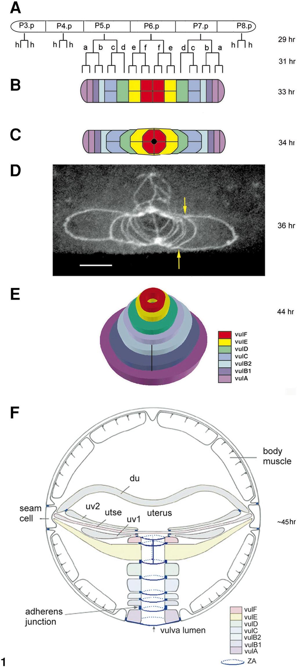
3 Dynamic Ring Formation in Multivulva Morphogenesis 235 genesis of the vulva is summarized in Fig. 1. This process begins as cells from each half of the primordium undergo short-range migrations toward the primordium s midline until meeting with their symmetrical partners from the other half, thus pushing the inner cells dorsally and creating an invaginated stack of seven toroids. This is followed by cell fusion within five of these toroids and attachments of specific toroids to specific epithelial cells and vulval muscle cells. During this time the AC invades the most dorsal ring and then fuses to the utse cell. The last stage involves eversion of the vulva forming a functional tube-shaped organ (Sharma-Kishore et al., 1999). One of the central players in the inductive machinery is the LET-60/RAS protein, which acts as a switch in the determination between vulval and nonvulval fates (Beitel et al., 1990; Han et al., 1990). Loss-of-function (lf) mutations in the let-60 gene give rise to a vulvaless (Vul) phenotype as a result of the 3 sublineage adopted by all the VPCs. Gain-of-function (gf) mutations involve the adoption of 1 /2 /3 sublineages by VPCs that normally adopt only the 3 sublineage and result in a multivulva (Muv) phenotype, in which additional vulva-like structures are generated independently of the presence of the AC (Ferguson et al., 1987; Beitel et al., 1990). RAS, as well as other small GTPases like the Rho family proteins, takes part not only in general proliferation but also in developmental events such as cell migration (reviewed in Sternberg and Han, 1998). In this study we have analyzed a let-60/ras(gf) mutant in order to define the minimal requirements for each and every cellular event during vulva formation. To understand the mutual relationships between the real (functional) and pseudo (additional and nonfunctional) vulvae characteristic to this Muv mutant, we have asked four questions: 1. How does a pseudovulva develop and how does this differ from normal vulva development? 2. Do vulval cells migrate autonomously or attract each other, or is there an external signal that induces migration? 3. Do vulval cells fuse only with homologous cells (e.g., A with A)? 4. Does Ras signaling influence cell fusion and cell migration during vulva formation? First, we have described the complete cellular events taking place during the generation of a Muv mutant. Second, we found out that within each primordium, each half develops autonomously throughout the entire vulvae organogenesis. As the different primordia develop, they compete with each other for vulval cells, resulting in the formation of asymmetrical vulvae. Third, we have observed that the ultimate fate of the VPC s descendants is to produce a structural scaffold which lines the vulval opening and that cells can attain differentiated structural states of absent cells to complete the minimal requirements for vulva formation. Finally, we provide evidence suggesting that RAS may influence the migration and fusion fates of certain cells during vulva formation. MATERIALS AND METHODS General Methods and Strains Methods for the culturing and handling of worms were as described before (Brenner, 1974). Animals were raised at 20 C unless otherwise stated. C. elegans strain N2 was the wild-type FIG. 1. Vulva formation in the wild type (A). The vulva precursor cells (VPCs; P(3-8).p) execute their cellular fate in the mid-l3 stage depending on their signaling. P3.p, P4.p, and P8.p divide once and fuse to the surrounding hypodermal syncytium hyp7. (B) P(5-7).p undergo three rounds of divisions yielding a primordium of 22 cells. Six cell fates established in a palindromic sequence (a f f a) are the precursors of the structural differentiated states (vula-vulf). h, cells fused to hyp7. (C) At the onset of the L4 stage, the cells send unidirectional processes ventrally and laterally toward the midline of the primordium creating an invagination. These short-range migrations take place in a spatial and temporal order starting with the e cells and ending with the external a cells resulting in a stack of seven toroids or rings. Black dot represents the anchor cell (AC). (D) Confocal micrograph of a vulva primordium stained with MH27 mab that reveals apical domains, adherens junctions, or zonula adherens (ZA) of cells (Sharma-Kishore et al., 1999). In each half of the primordium the precursors of vula fused before they started their migrations and the precursors of vulc have fused after they completed their migrations. The yellow arrows point to two filopodia derived from the posterior binucleate a cell. This intermediate is before intratoroidal cell fusion and the central vulf and vule rings on the top are already more dorsal than the external rings. (E) The cells within the rings (except for vulb1 and vulb2) fuse in a temporal order and the gonadal AC fuses to the uterine utse cell resulting in the formation of a tube-shaped L4 vulva. (F) Transverse section of the body of a late L4 stage showing the connections between the vulva and the cells of the uterus (uv1, utse, uv2, and du) and between the vulva and the hypodermis (hyp7 in the ventral side and seam cells on the left and right). The lumen of the vulva shows cylindrical zonula adherens (ZA, in blue; dotted line is posterior) that link the apical domains of the different cells. VulA ring connects to the hypodermis in the ventral side, vulf connects to the uv1 cell of the uterus on the dorsal side, and the apical domains of all the adjacent rings are linked to each other (e.g., vula to vulb1 and vulf to vule). The transverse junctions between the cells of the vulf and vule rings are shown here as vertical lines before they fuse. The same color code for the different cell types and the vulval rings has been maintained for all the figures except for (F). Anterior is to the left except for (F) that is a transverse section in which dorsal is to the top. (B, C) Dorsal views. Nomenclature: a h are the cell fates of the VPC granddaughters. The use of lowercase here and in Figs. 2 and 3 is simply to differentiate the cell fates from the uppercase letters of the panels (see also Fig. 5). The cellular sublineages and the times in hours after hatching at 20 C are from Sulston and Horvitz (1977). The figure is modified from Sharma-Kishore et al. (1999).
4 236 Shemer, Kishore, and Podbilewicz strain (Brenner, 1974). The following strains were used: LGIV: let-60(n1046) (Beitel et al., 1990; Han and Sternberg, 1990). BP74 let-60(n1046) jcis1[mh27-gfp] was obtained by mating males expressing MH27-GFP (Mohler et al., 1998) with let-60(n1046) hermaphrodites and selecting recombinant progeny that expressed the Muv phenotype as well as fluorescent apical junctions of epithelial cells. The jcis1 strain (kindly provided by J. Simske and J. Hardin (Mohler et al., 1998)) contains all known control sequences required to target MH27 to the cellular junctions, prf4 (a plasmid containing the dominant roller mutation rol-6(su1006)) and F35D3 (unc29 DNA) in an N2 background (unc-29 DNA was used to add complexity to the array). The presence of the dominant cuticle mutation rol-6 in the jcis1 strain is unlikely to affect morphogenesis since MH27 antibody was also used in let- 60(n1046) without jcis1. Permeabilization, Fixation, and Immunofluorescence Worms were fixed and permeabilized as described before (Finney and Ruvkun, 1990; Sharma-Kishore et al., 1999). Stages of development were determined by using Nomarski optics or by following the development of the seam cells through fluorescence microscopy (Podbilewicz and White, 1994). Immunofluorescence was carried out as described before (Podbilewicz, 1996; Sharma-Kishore et al., 1999). The mouse monoclonal antibody MH27 (1:300 dilution) was used to stain the adherens junctions of epithelial cells (Francis and Waterston, 1991; Podbilewicz, 1996). Confocal and Fluorescence Microscopy Confocal reconstructions were made by using an MRC-1024, laser confocal scanning microscope (Bio-Rad, Hempstead, UK) with the objective Nikon Plan Apo 60 /1.40 (Podbilewicz, 1996; Sharma-Kishore et al., 1999). Cell identity was revealed using the characteristic staining of the apical junctions of vule and vulf (narrow and tall shape in the dorsoventral axis in 100 and 65% of the vulvae, respectively; see Figs. 1 and 7) and vulb1-vulb2 (short rings compared with the rest) (Sharma-Kishore et al., 1999). Worms were either anesthetized in 0.01% tetramisole, 0.1% tricaine, or paralyzed transiently by lowering the temperature to 8 11 C for min using a temperature control system (Y. Rabin and B. Podbilewicz, unpublished results). Cell lineages were followed as described (Sulston and Horvitz, 1977). RESULTS Cellular Events during Vulva Formation Organogenesis of the C. elegans vulva in wild-type animals can be divided into three phases (Fig. 1): short-range migration forming seven stacked rings, cell fusion, and eversion of the vulva (Sharma-Kishore et al., 1999). To study the development of the let-60(n1046) mutant vulva as well as the pseudovulvae that are characteristic of this gf mutant, we followed vulvae formation in living n1046 worms expressing MH27-GFP (see Materials and Methods). In addition, we have stained worms from different stages of development with an antibody against the adherens junctions and observed three-dimensional confocal section reconstructions (Sharma-Kishore et al., 1999). By analyzing these intermediates it was possible to reveal the mutual influence that the real and pseudovulvae exert on each other and to learn about the minimal cellular events required for the completion of the successive developmental phases. Figure 2 shows the summary of events during vulval formation in let- 60(n1046). Divisions of the Vulva Precursor Cells Reveal Asynchronous Vulval Development of Pseudovulvae in let-60(gf) In wild-type worms, the sublineages of the different VPCs are invariant and dependent on combined signaling pathways (Sulston and Horvitz, 1977; Horvitz and Sulston, 1980; Kimble, 1981; Euling and Ambros, 1996) (Fig. 1). In the let-60(n1046sd) gf mutant it has been shown that the induction signal gives rise to various sublineages resulting in the formation of ectopic pseudovulvae (Beitel et al., 1990). To investigate the morphogenesis derived from these abnormal sublineages in let-60(n1046), we have analyzed living worms and reconstructed intermediates from the mid-l3 stage, h posthatching (Fig. 3). During the first round of the VPC s division in n1046 mutants, the spatial and temporal pattern of the division of P(3-8).p was indistinguishable between the different VPCs. However, during FIG. 2. Multivulva formation in the let-60(n1046) gf mutant. (A C) P(3-8).p adopt different cellular fates according to the signals from the surrounding cells as well as a Let-60/Ras ectopic signal that is a result of a gf mutation (Eisenmann and Kim, 1997). This is one of the observed fates of the VPCs in let-60(n1046) and is represented also in Figs. 4 and 5. (B) All the VPC s daughter cells divide longitudinally. (C) During the terminal round of division the anterior pseudo f cells did not divide in this example. (D) Real and pseudovulvae precursor cells migrate toward their respective primordia midlines as in wild-type animals (Fig. 1) except for the fact that the furthermost cells attached between the different primordia stretch due to competitive attractiveness between equidistant primordia. The f cells in the anterior pseudovulva fuse. (E) The daughters of a in all the primordia and the anterior b1 in the real vulva primordium do not complete their migrations and are therefore absent from the evolving vulvae. As a result, the primordia are asymmetric. In the absence of their symmetrical pairs, two filopodia (arrowheads) migrate toward the midline and turn toward each other until they meet, producing concentric toroids and forming a stack of only five or six rings. (F) The extended cells between the primordia either fuse to hyp7 (as in this example) or remain throughout vulvae formation. Newly formed rings undergo a temporal and spatially ordered process of intratoroidal fusions. The real vulva becomes mature and functional although part of its normal distal components are missing. Specific cells can adopt the structural differentiated states of the absent cells as a result of cell fate transformation. See text for details. (B E) are dorsal views; (A and G) are lateral views. AC, anchor cell (black dot in D and E). A, anterior; P, posterior; R, right; L, left; V, ventral; D, dorsal.
5 Dynamic Ring Formation in Multivulva Morphogenesis 237
 that reveal adherens junctions (Francis and Waterston, 1991; Podbilewicz and White, 1994) or vulvae expressing an MH27-GFP")
6 238 Shemer, Kishore, and Podbilewicz FIG. 3. Heterochronic proliferation of VPCs. Confocal micrographs of mid-l3 to mid-l4 vulvae stained with MH27 mab (A C) that reveal adherens junctions (Francis and Waterston, 1991; Podbilewicz and White, 1994) or vulvae expressing an MH27-GFP construct in their apical junctions. (D). Pictures were obtained by projecting 5 10 serial confocal sections of 0.5 m. (A) Early primordia with the 12 granddaughters of P(5-7).p after two rounds of cell division (a f and f a) as well as the granddaughter cells of P4.p and the daughter cells of P8.p (approximately 31.5 h posthatching). (B) Primordial cells of an asymmetrical real vulva during the terminal round of division (32.5 h). P4.p has undergone only two rounds of divisions yielding four granddaughter cells and the daughter cells of P8.p have fused to hyp7 (h fate; 3 sublineage). (C) Beginning of short-range migrations: the descendants of P3.p and P(5-7).p during the terminal round of division. P4.p has divided only once (34 h). (Inset) A higher magnification of the center of the real vulva primordium showing the great-granddaughters of the VPCs. Arrow points to the filopodia that links two primordia. (D) A real and two pseudovulvae after ring formation. Arrows indicate extended cells between the primordia. These cells do not complete their migrations and will not participate in the formation of the vulvae. In all the pictures anterior is to the left. Bar, 10 m. Estimated times are in hours post hatching and are based on Nomarski optics and stage of development of the seam cells (see A and C, above the vulval cells). the second round of division, the daughters of P3.p, P4.p, and P8.p, which did not fuse to hyp7 (3 sublineage), divided asynchronously (Fig. 3; G. Shemer and B. Podbilewicz, unpublished data). The 12 cells of wild-type animals (a f and f a) undergo a terminal round of division at this stage (see legend to Fig. 1B). In n1046, P(5-7).p and their progeny divided temporally and spatially in a similar way to the wild type but the
7 Dynamic Ring Formation in Multivulva Morphogenesis 239 FIG. 4. Early morphogenesis of vulval cells Apical junctions of the cells were visualized and pictures were obtained as described in Fig. 3. The schematic drawings are dorsal views based on these pictures. (A) Intermediate with all 22 vulval cells plus 13 primordial cells of the anterior pseudovulva and 7 cells that form the primordium of the posterior pseudovulva. (B) The different primordia began to invaginate while the most external cells of the primordia have extended their apical surfaces. (C) In the real vulva, vuld precursors have migrated to surround the inner rings of vule and vulf. In the anterior pseudovulva primordium vuld precursors have migrated toward their inner neighbors and, in the absence of symmetrical pairs, have encompassed vule and vulf. The extended daughter cells of a have fused in all the different primordia. In the real vulva the anterior b1 cell has also extended. (D) In the posterior pseudovulva, vulc precursors have sent extensions (arrows) surrounding vuld that in the absence of vulf served as the inner organizer cell. In the absence of symmetrical pairs, these extensions turned toward each other. Similar filopodia are shown in the anterior pseudovulva and in the central real vulva (arrows). (E) vulc precursors in all the primordia have fused in pairs in a transverse fusion. In the real vulva the vulc ring has formed. In the anterior primordium, these fused cells have sent processes that have encompassed the inner rings. In the posterior primordium, the vulc ring has formed after the apical extensions had met and fused. Closed black circle at the center of the real vulva represents the AC that invades and penetrates the four vulf precursor cells. FIG. 5. Cellular fates and structural differentiated states of the primordial cells of the vulvae. A schematic representation of mid-l4 vulvae ( 38 h) based on fixed and live worms showing the stacks of rings before intratoroidal fusions. The lineage relationships of the different rings are indicated. The cell fates are indicated by the ring precursors (A F) or by cells that will not participate in vulvae formation (H). In the anterior pseudovulva the descendants of P3.p and P4.p have formed a stack of six rings (vule-like and vulf-like, 1 sublineage of P3.p; pseudo-vulb1-vuld, 2 sublineage of P4.p.). In the posterior pseudovulva four rings, half in size of normal rings, have been formed (pseudo-vulb1-vuld, 2 sublineage of P8.p as shown before). The real vulva is composed of only five complete rings and one half-ring (the posterior vulb1). Timing of division between the VPCs is only schematic as division status of the different VPCs is variable. The open rectangles represent cell fusion. Open circles represent nuclei. Free nuclei represent cells that have fused with the hypodermal cell hyp7. F L, longitudinal fusion; F T, transverse fusion.
 and their respective dorsal view diagrams (D F) of pseudovulvae prior to intratoroidal fusion stained with MH27 mab (adherens junctions, green) and")
8 240 Shemer, Kishore, and Podbilewicz FIG. 6. Ring formation in pseudovulvae. Pictures of confocal reconstructions (A C) and their respective dorsal view diagrams (D F) of pseudovulvae prior to intratoroidal fusion stained with MH27 mab (adherens junctions, green) and propidium iodide (nuclei, red). Nuclei of vulval cells were distinguished from the rest of the cells by analysis of each section. The identity of the rings is based on the structural resemblance to known rings in the wild type (see Materials and Methods). (A and D) Pseudovulva containing pseudo vulb2-vulf. The three most ventral rings have each formed from only one half-primordium. vulf is formed from two f cells that fuse between themselves. (B and E) Pseudovulva containing only four rings, each of them formed from only one half-primordium. The d cell has served as the inner organizer cell. (C and F) Pseudovulva containing seven anterior half-rings and five posterior half-rings. This pseudovulva contains 13 nuclei suggesting that the descendants of P3.p and P4.p acted together to obtain this structure not in the conventional way of adopting a 1 or a 2 sublineage. The cells in rings vula-vule have not achieved cell fusion and we would expect them to fuse before the L4 to adult molt. All pictures with ventral down and anterior to the left. All pseudovulvae are from animals in the mid- to late L4 stage. Bar, 10 m. progeny of P3.p, P4.p, and P8.p showed again, like in the 12 cells stage, a retarded phenotype (Fig. 3B). In 28% of the cases P3.p, P4.p, and P8.p divided three times like P(5-7).p yielding 7 or 8 great granddaughter cells (2 or 1 sublineage, respectively) depending on their pattern of division (e.g., P3.p in Fig. 3C). However, in 72% of the cases the extra cells divided only once or twice while P(5-7).p had divided three times (e.g., P4.p in Fig. 3B). The extra cells that escaped the 3 sublineage (fusion to hyp7) were either linked or not linked to the real vulva primordium regardless of their division status (e.g., Figs. 3A 3C). This phenomenon was also observed in later stages of development leading us to conclude that the transformed VPCs can divide at a slower rate than the descendants of P(5-7).p and that some transformed VPCs divide only once like in the wild type but escape fusion to hyp7. Short-Range Migrations: Competition between Migrating Cells In wild-type C. elegans the next stage of vulva development is characterized by short-range migrations that involve the extension of apical domains between equivalent cells from the two sides of the vulva primordium toward the center (Figs. 1C and 1D). To analyze cell migration in let-60(n1046), we have investigated living worms and reconstructions of migration intermediates (33 36 h post hatching). We found that in each primordium the cells migrated toward their primordium s midline but at the onset of the migrations, the apical domains of the most external cells elongated abnormally forming apical extensions between the different primordia (Figs. 2E, 3C, and 3D, arrows). These apical extensions were observed in all of the worms investigated from the beginning of the short-range migrations until later stages. The elongated cells either fused later to hyp7 (Fig. 2F) or remained throughout the entire vulvae formation (Fig. 3D). One interpretation of the apical extensions is that they represent competition between the different primordia during cell migration. To understand how this competition affects the resulting real and pseudovulvae, we characterized the primordia of intermediates from the early and mid-l4 stage, after the completion of these short-range migrations. The precursors of vule-vulb2 migrated normally in all the real vulvae primordia. However, the A cells (and sometimes also the B1 cell) had not completed their
9 Dynamic Ring Formation in Multivulva Morphogenesis 241 TABLE 1 Asymmetry between the Two Halves of Real Vulvae Ring status a short-range migrations or had not migrated at all. In both cases the result was the absence of these cells from the mid-l4 primordia (Table 1, Figs. 4, 5, and 7C). Only in 1 case of 42 did all of the half-rings precursors seem to have completed their migrations. In all of the cases analyzed where there were no extra cells near the real half-vulvae (when the P8.p cells executed the 3 sublineage), the shortrange migrations were not slowed down. This is consistent with the competition between the extra cells that did not fuse to hyp7 and the real vulva cells (Fig. 4). In Fig. 4, pseudovulvae were either formed from the descendants of one VPC (P8.p) or two VPCs (P3.p and P4.p). The migration pattern of the pseudoprimordia resembled that of the real primordium. Both involved sending apical processes from the outer cells toward the inner cells with the concomitant remodeling of adherens junctions with hyp7. In summary, we found that real vulva cells and pseudovulvae cells of n1046 migrated toward the vulva (or pseudovulvae) midline in the same manner as in the wildtype animals except for the most distal cells attached between the primordia, that did not migrate or did not complete their migration. Autonomy of the Two Halves of the Vulva Primordium Results in Asymmetry in Size, Orientation, and Cells Content % of total semivulvae (n 42) No vula 40% (17) No vula no vulb1 29% (12) Incomplete migration of vula primordial cells 29% (12) All rings exist 2% (1) a Half-primordia were counted and the state of the migration of their precursors was determined. Half-primordia that lacked specific rings were calculated as percentage of the total half-primordia observed. In parentheses are numbers of half-primordia. Two halves derived from anterior and posterior cells form each ring in the wild-type vulva (Figs. 1D and 7A). To investigate whether cells attract each other or alternatively vulval cells behave autonomously, we have analyzed the formation of natural semivulvae composed of half-rings in let-60(n1046). When looking on real vulvae in mid-l4 reconstructions and living worms expressing MH27-GFP, equal number of precursor cells in both halves was obtained only in cases where both halves were subjected to equal competition with nearby pseudovulvae (Figs. 7D and 7E). We observed that in most cases both halves showed A P asymmetry with an unequal number of vula and/or vulb1 precursor cells (Figs. 2, 4, 5, 7B, and 7C). This asymmetry was not confined to the contents of the rings but also to their size and orientation. In 54% of these primordia, one half was larger than the other half (Fig. 7B). In addition, in 71% of the vulvae inspected (n 24), the apical domains were oriented toward the anterior side of the worm (arrowheads; Figs. 7D and 7E) regardless of the presence of a posterior pseudovulva primordium. In summary, the asymmetry that we found in let- 60(n1046) suggested that each half of the vulva developed autonomously. In the presence of nearby pseudovulvae, precursor cells of the two halves migrated unequally ending with an inclined invaginated primordium comprised of a different set of cells with additional structural asymmetries. Ring Formation: Half-Vulvae Form Self Complete Rings In wild-type C. elegans anterior and posterior pairs coordinately migrate to form rings (Fig. 1). To further study a natural situation of formation of half-rings in semivulvae and to analyze the process of invagination by ring formation step by step, we have examined pseudovulvae of n1046 in fixed and living worms. The pseudovulvae we investigated can be divided into three main structural subclasses: (1) disorganized structures without any identified structural pattern; (2) pseudovulvae that developed a structure recognizable as those usually obtained by the 1 sublineage (pseudo vule vulf) or 2 sublineage (pseudo vula-vuld: Figs. 6B, 6E, and 7F) or structures that are partially primary and partially secondary (Figs. 6A and 6D); and (3) complex structures with more rings than expected from the sublineage (Figs. 6C and 6F). In 2 sublineage-derived structures, the cells sent two unidirectional extensions from outer to inner cells as the pseudo D cell served as the midline (Fig. 4C; posterior pseudovulva). In the absence of symmetrical pairs, the extensions moved toward each other until they met thus forming complete rings (Figs. 4D and 4E). Because only one cell (D) served as the midline (compared with four F cells in wild-type animals) the rings were half in size in their A P axis of the normal ones (Figs. 2, 4, and 5: posterior pseudoprimordium; Figs. 6B, 6E, and 7F). When primordia of pseudovulvae adopted a more complex structural shape (partially vule-vulf-like and partially vula-vuld-like) that contained four or two pseudo F s, the outer cells formed complete and full-sized rings similar to the real vulva and to the vulva of wild-type animals. Here, also, the cells have sent apical extensions that turned toward each other in the absence of symmetrical pairs (Figs. 2, 4, and 5, anterior pseudoprimordium; and Figs. 6A and 6D). Thus, the size and shape of the A D-derived rings is determined by the dimensions of the dorsal rings that formed first (vulf or vuld). In summary, the ring asymmetry and the formation of

10 242 Shemer, Kishore, and Podbilewicz
11 Dynamic Ring Formation in Multivulva Morphogenesis 243 self rings indicate that cell migration and cell fusion are autonomous processes. Transformations of Rings and Fusion Fates between the Vulval Toroids Figure 5 shows the formation of incomplete stacks of rings due to the competition between primordia in let- 60(n1046). We propose that the ultimate cell fate of the VPCs descendants is a structural differentiated state expressed as the different rings. A to F represent the cell fate of ring formation and H represents the cell fate of fusion to hyp7. Intratoroidal Fusions: Transformations of Cell Fusion Fates In the wild-type, after all the primordial cells complete their migrations and during invasion of the AC followed by its fusion to the utse uterine cell, there is an ordered process of fusion within the rings starting with vuld and ending with vule with the exception of vulb1 and vulb2 (Sharma- Kishore et al., 1999). As a result, a stack of seven rings is built (Fig. 1) and this organ is attached to the surrounding epithelia (through processes from vule), to hyp7 (through vula), to vulval muscles (Sharma-Kishore et al., 1999), and to the uterus (Newman and Sternberg, 1996; Newman et al., 1996). Figure 7A shows a wild-type vulva after ring formation and before intratoroidal fusion. In the wild-type vulva the order of the intratoroidal cell fusions are vuld, vula, vulc, vulf, and vule (Sharma-Kishore et al., 1999). In n1046, the precursor cells of vula and vulc (A A and C C, respectively) fused between themselves on each side of the primordium before (vula) and during (vulc) the migrations as occurs during early fusion events in the wild type (compare Figs. 1C and 1D with 4B 4E). To investigate the intratoroidal fusions phase in n1046, we have analyzed reconstructed larvae and living worms from the mid-l4 stage. We found that even after abnormal migrations, the process of intratoroidal fusions did take place. Figures 7C 7E show reconstructed vulvae in different stages of intratoroidal fusions. Like in the wild type, this phase started only after all the cells that do participate in the formation of the vulvae had completed their migrations (e.g., Figs. 7C and 7E). This suggests that the temporal order of events is independent of the components of the primordia but is dependent on the completion of the migration process by the participating cells. In n1046, the two cells of vuld were the first to fuse longitudinally (Fig. 7C). In the absence of vula from the primordia, the vulc cells fused next (Fig. 7D) and the normal order of events proceeded. In one reconstructed vulva lacking vula, the two cells forming vulb2 fused adopting the fate of vula even though in the wild-type they do not fuse (Fig. 7E). The transverse intra-toroidal fusion (F IT ) in vulf sometimes were found early (e.g., Figs. 7B and 7C right diagrams) and in other cases they occured after the longitudinal intratoroidal fusion (F IL ) as in the wild-type (e.g., Figs. 7D and 7E). As for the pseudovulvae, we observed intratoroidal fusion of pseudo vulc (vuld-like is composed of only one cell in this case) at least in one reconstructed intermediate (Fig. 7F). Thus, where A cells fuse to hyp7, the precursor cells of vulb accomplish a proper interface with hyp7 allowing the base of the vulva to attach to the hypodermis (e.g., Figs. 2F, 5, 6A, and 7D). To summarize, in the absence of specific cells in n1046 the fusions within the existing rings took place after the participating cells of the primordium had completed their migrations. The fusions followed the same order as in the wild-type vulva with the exception of vula that did not participate in vulva formation and the transverse fusions (F IT ) of vulf that were variable. In one case we detected substitution of the A cell fate by the descendants of the B cell not only in forming the most ventral ring (vulb1), but also in undergoing intratoroidal fusion (vulb2) (Fig. 7E). FIG. 7. Intratoroidal fusion in real and pseudovulvae. Confocal reconstructions of vulvae from the mid- to late L4 stage stained as in Fig. 6. Left column, confocal reconstructions. Middle column, lateral view diagrams. Right column, dorsal view diagrams. (A) Wild-type vulva. (B D) Real vulvae of n1046. (E) Pseudovulva of n1046. (A) Lateral view of a wild-type vulva where no intraring cell fusions have occurred. (B) Primordium containing six rings and one posterior half-ring. VulF fused transversely and the rings have not yet undergone intratoroidal fusion. Arrows point to midline where junctions between anterior and posterior vulval cells are. (C) Primordium in which VulF fused transversely and the vuld ring has undergone intratoroidal fusion as expressed by the disappearance of the boundaries between the cells comprising this ring. In the anterior half the vula precursor cells have not completed their migration while in the posterior half these cells as well as vulb1 precursors have not migrated at all and are absent from the primordium. (D) Primordium from later stages containing only five rings in which vulc has also undergone intratoroidal fusion. Arrowhead points to the apex of the stack of rings showing the orientation of the vulva toward the anterior side (left). (E) Primordium in which vulb2 has undergone intratoroidal fusion (in addition to vuld) in the absence of the a cells that have not completed their migration. vulb2 does not fuse in the wild type. Arrowhead dorsal of the stack of rings points to the asymmetric orientation of the vulva toward the anterior side. (F) Primordium of a posterior pseudovulva containing five complete rings in which the pseudo vuld cell serves as the most dorsal ring. Pseudo vulc has undergone intratoroidal fusion. In the mutant let-60(n1046) the order of intratoroidal longitudinal fusions (F IL ) are unfused (n 6), vuld (n 12), vulc (n 5), and vulf and vule (n 1). The order of transverse intratoroidal fusions (F IT ) for vulf is variable since F IT have been found associated with different stages of F IL fusions (n 6). In the wild type the order is vuld, vula, vulc, vulf, and vule. For the two most dorsal rings, F IT and F IL events are coupled in the wild type but not in the mutant. All pictures with ventral down and anterior to the left. Bar, 10 m. ac, anchor cell (black dot).
12 244 Shemer, Kishore, and Podbilewicz Last Phase of Vulva Formation During this phase that involves vulva eversion followed by lumen closure, we have not seen any difference between the development of the real vulva of the mutant and the wild-type vulva. As for the pseudovulvae, this phase did not involve closure of the lumen since no lumen was opened in the absence of the AC. DISCUSSION We have done a step-by-step analysis of vulvae formation in a ras/let-60 Muv mutant through investigation of reconstructed intermediates from different stages of development as well as using a GFP-tagged adherens junction marker enabling us to better understand the minimal requirements for each of the developmental cellular events. The sequence of events leading to the real and pseudovulvae in this mutant is summarized in Fig. 2. We have also described the mutual relationships between the different vulvae and arrived to three major conclusions: (1) The different primordia compete between themselves for vulval cells. (2) Each half-primordium develops autonomously. (3) The ultimate fate of each vulval cell is to become one of the seven structural cell types that comprise the vulva. FIG. 8. A working model for the cell behaviors that drive invagination of the vulva. (A) During the L3 stage the AC induces three of the six multipotential VPCs to adopt vulval fates with the involvement of RTK-LET-60/RAS and Wnt-BAR-1 signaling pathways (Eisenmann et al., 1998; Han et al., 1990; Lackner et al., 1994). LIN-12 is a receptor from the LIN-12- Notch family that is required for lateral signaling (Greenwald et al., 1983) and LIN-15 induces cell fusion or inhibits vulval fates (Herman and Hedgecock, 1990; Thomas and Horvitz, 1999). (B) Based on our work on vulva morphogenesis in wild-type and ras/let-60 hermaphrodites we propose that additional signaling pathways are involved in stereotyped autonomous cell migration and cell fusion processes during ring formation in the late L3 and early L4 stages. (C) After the seven rings are formed intratoroidal fusions occur in a regulated way and the AC fuses to the utse uterine cells. It is unknown how this process is controlled. The seven rings show a pyramidal shape before and during intratoroidal cell fusions. (D) Morphological changes determine the seven structural differentiated states (vula-vulf) and the shape of the stacked rings become cylindrical instead of pyramidal. This transverse view also shows the structural differentiated state of vule that undergoes a major expansion after the intratoroidal fusion step linking two basolateral domains to the lateral hypodermal seams cells (see also Fig. 1F). RAS and other signaling pathways as well as specific fusogenic molecules may be involved in the different steps shown here (?). Fusing membranes (dotted lines). Nuclei (black circles). The Primordial Cells of the Most Dorsal Ring Have a Key Role in the Migration Stage On the onset of vulva formation, the primordial cells have apparently divided into groups according to their future structural differentiated state (forming a real or a pseudovulva). The pattern of morphological events leading to vulva formation was similar between these groups (Figs. 2 and 3). Two events are crucial for the development of the vulvae. The first event is the determination of a primordium s midline, toward which the outer cells migrate. Our results suggest that the boundaries between the primordial cells of the most dorsal ring vulf may be the default midline as the outer cells migrated toward these boundaries not only in the wild-type vulva or the real vulva in let- 60(n1046), but also in pseudovulvae that contained pseudovulf (Figs. 4, 6A, 6C, 6D, and 6F). In other cases, the precursors of the most dorsal ring acted similar to the pseudo vulf and served as a midline (e.g., Figs. 6B, 6E, and 7F). The second event is the trigger that starts the shortrange migrations. Evidence for the existence of this trigger came from the putative competition between the primordia in this Muv mutant. Although each group of cells acted as an independent primordium, the presence of nearby groups affected them and the most external cells did not complete their migration. It is possible that these cells were subjected to signals from both midlines and could not decide where to migrate to, suggesting that cells that serve as a midline are critical in determining the normal temporal and spatial order in which the cells migrate. These results suggest that the primordial cells that form the most dorsal ring (vulf in
13 Dynamic Ring Formation in Multivulva Morphogenesis 245 the real vulva) play a key role in the onset and throughout the short-range migrations stage. When the F and E cells are absent then D cells become the organizer. We propose that there is a hierarchy A to F and each cell knows to migrate toward a neighbor that is higher in the sequence (A, B1, B2, C, D, E, F). Each Half-Vulva Develops Autonomously Vulvae formation in the ras/let-60 mutant described here has enabled us to follow the behavior of many half-vulvae during each and every stage of development. Our results establish that each half-vulva develops autonomously throughout vulva organogenesis. First, due to the competition between the different primordia (see above), each half of the real vulva developed differently leading to asymmetrical vulvae (e.g., Figs. 4, 7B, and 7C). Second, in pseudovulvae that lacked half of the primordial cells, the existing cells started migrating, turned toward themselves after the midline had been passed, and fused intratoroidally forming complete rings (Figs. 4 and 7F). Thus, the correct sequence of events is independent of the interactions between the symmetrical halves. Each half-primordium develops independently over a limited time range and for a defined set of cell migration and cell fusion events. For example, in cases when one halfprimordium contributes E/F fates and the other halfprimordium contributes A/D fates, the E/F become the organizer and set the center. D cells then migrate and surround vule/vulf rather than competing for the center (G. Shemer and B. Podbilewicz, unpublished results). Previous work on the wild-type vulva has shown that each half-vulva can develop autonomously (Sharma-Kishore et al., 1999). However, this report was based largely on only one observation of an EM reconstructed late L4 vulva that had undergone laser microsurgery killing the posterior half of the vulva and on cell lineaging evidence from early stages of vulva formation, prior to organogenesis. Viewing the results presented here, we suggest that key players in the regulation of ring formation are the primordial cells of the most dorsal ring and the surrounding hypodermal syncytium. In addition, the developmental pattern of half-pseudovulvae implies that the cells are intrinsically activated resulting in their ability to migrate until they find self, stop migrating, and undergo intratoroidal fusion even in the absence of their symmetrical pairs (Fig. 4). However, when two half-primordia produce sets of homologous cells, the homologues will eventually recognize each other and extend arms toward each other in a somewhat coordinated fashion, even if the final product ends up asymmetric. Thus, the two halves interact and maintain a hierarchy of cell fates and when one half is missing cell migration, cell fusion and ring formation occur autonomously without changing the F to A center to periphery and dorsoventral hierarchy of interactions. Cellular Fate versus Structural Differentiated State Previous work has described in detail the cellular sublineages of the VPCs (Sternberg and Horvitz, 1989; Greenwald, 1997). The VPCs undergo a stereotyped sequence of cell divisions (1, 2, and 3 sublineages) that result in seven cell fates (A, B, C, D, E, F, and H; Figs. 1A, 2B and 5). Thus, the cellular sublineage is followed by seven cell fates of which A F fates will form vulval rings (Sharma-Kishore et al., 1999). In the wild type the tube-shaped vulva is formed from a stack of seven rings (seven structural differentiated states), some of them multinucleated (Fig. 1) (Sharma- Kishore et al., 1999). However, in let-60(n1046), although some of the VPCs have not changed in their cellular sublineage (P(5-7).p), their structural differentiated state was completely different (Figs. 2, 5, 6C, and 7E). We propose to distinguish between the cellular fates and structural differentiated states by the following nomenclature: A F (cell fates) for precursors of vula-vulf (structural differentiated states), respectively, and H (cell fusion fate) for cells that will not participate in vulva formation and may fuse later to the surrounding hypodermis. Inhibiting Proliferation and Promoting Fusion of VPCs May Be Separate Processes The descendants of P3.p, P4.p, and P8.p do not always proliferate at the same rate as those of P(5-7).p (Fig. 3; G. Shemer and B. Podbilewicz, unpublished data). In fact, we observed cases where one of the VPCs divided only once and the two daughter cells remained undivided until vulva formation had been completed. According to the 3 sublineage (H cell fate), a single division of a VPC should result in fusion to hyp7. However, these cells have escaped this fate, suggesting that fusion to hyp7 is not a default process following proliferation arrest of VPCs daughter cells but rather that cell fusion and proliferation are two separate processes. Previously, it has been shown that the 3 cellular fate appears to be the result of an intracellular signaling system between hyp7 and the VPCs and that two classes of genes are needed to inhibit vulval cell fates (Ferguson and Horvitz, 1989; Herman and Hedgecock, 1990; Hedgecock and Herman, 1995; Thomas and Horvitz, 1999). It is still not well understood how these pathways actually work. The ectopic activity of RAS/LET-60 in n1046 may bypass the fusion promotion pathway leading to nonproliferating cells that escape fusion. A Novel Potential Role for RAS in Vulva Organogenesis The function of the RAS protein is not limited to its central role in proliferation. It has been previously shown that RAS is not essential for general proliferation in C. elegans and Drosophila (Hou et al., 1995; Yochem et al., 1997). However, RAS and other small-related GTPases have been shown to play key roles in axon guidance (Zipkin et al., 1997), spicule formation, germ cell development (Yochem et al., 1997), and sex myoblast migration (Clark et al., 1992) in C. elegans, eye development (Wassarman et al., 1995), embryonic patterning (Schnorr and Berg, 1996), and
14 246 Shemer, Kishore, and Podbilewicz wing vein formation (Roch et al., 1998) in Drosophila and motility, as well as invasiveness of various mitogenic cells in mammals (Keely et al., 1999). In the vulva the only evidence for RAS participation in the actual morphogenesis is the EGF reciprocal signaling that is important for establishing the uterine vulva connection (Chang et al., 1999). Our results imply that RAS may have a potential role during the entire organogenesis of the vulva in cell fusion and cell migration. First, in the background of a ras (gf) mutation, at least some of the cells (the daughters of A in the real vulva) have acquired the ability to fuse to the surrounding hypodermis. The early inductive function of RAS could not contribute to this phenomenon since the lineage of these cells was normal. Rather, this mutation affects the cell fate of A. Second, in the let-60/ras (gf) mutant there are abnormal filopodial extensions of A- and B-derived cells (e.g., Figs. 2E, 3C, 3D, and 4). Third, the structural differentiated state transformation described may be related to the RAS pathway since RAS is normally expressed in all the primordial cells after the execution of the sublineage and throughout vulva formation (Dent and Han, 1998). Based on these results we propose that RAS and other signal transduction processes are involved in late cellular events (e.g., filopodial extensions and cell fusion) required to form a stack of rings that drive invagination (Fig. 8). It has been shown that FUS3, a member of the RAS-MAP kinase pathway, is involved in the fusion of yeast cells during mating (Elion et al., 1990). Cell fusion is also critical in fertilization (Yanagimachi, 1988), formation of epithelia in C. elegans (Podbilewicz and White, 1994), embryonic development in sea urchin and in leech (Hodor and Ettensohn, 1998; Isaksen et al., 1999), and formation of muscles (Doberstein et al., 1997), bones (Jee and Nolan, 1963), and placenta (Pierce and Midgley, 1963). Our working hypothesis is that membrane fusion during development is a universal strategy to accomplish pattern formation (Podbilewicz, 2000). Ring Formation as a Morphogenetic Force in Invagination Epithelial invagination plays a key role during developmental events such as gastrulation and organogenesis. There are various theories on the mechanisms and the morphogenetic forces that drive epithelial invagination such as apical constrictions in the ventral furrow in Drosophila as well as in the primordia of the neural crest (Schoenwolf and Smith, 1990), changes in cell adhesion properties of the basal and apical domains of the folding cells (Gustafson and Wolpert, 1963), secretion of extracellular matrix (Lane et al., 1993), and oriented cell division during gastrulation (Hertzel and Clark, 1992). One of the candidates to be a morphogenetic force during invagination is the formation of bottle cells in sea urchin, Drosophila, and Xenopus embryos (Shih and Keller, 1994). These cells are apparently essential for primary invagination on the onset of gastrulation (Kimberly and Hardin, 1998). Vulva formation in C. elegans has been studied in order to investigate the essence of epithelial invaginations in this animal and mutations that perturb vulva invaginations have been cloned (Herman et al., 1999; Herman and Horvitz, 1999). Here, we have shown that epithelial invagination during vulva formation in C. elegans can be executed through a novel mechanism. Ring formation appears to be the morphogenetic force that drives the invagination of the vulval cells. Invagination is achieved when cells forming rings migrate laterally and ventrally thus disconnecting neighboring cells from the hypodermis. We propose that sequential migration and ring formation is followed by interactions between the migrating cells and between the toroidal cells and the surrounding hypodermis. When ring formation is aberrant, invagination is not complete. It is noteworthy that the bottle cells in sea urchin are also set in a ring but that the folding of these cells is quite different from the process of ring formation in C. elegans. ACKNOWLEDGMENTS We thank J. White, D. Gershon, T. Gattegno, and M. Glickman for valuable discussions and two anonymous reviewers for their critical and constructive comments on the manuscript. We thank B. Mohler, J. Simske, and J. Hardin for strain jcis1. Some nematode strains used in this work were provided by the Caenorhabditis Genetics Center, which is funded by the NIH National Center for Research Resources. This work was supported by the Jacob and Rosaline Cohn Academic Lectureship and by grants from the Israel Science Foundation, the Israel Cancer Research Fund (U.S.A.), and the Binational Science Foundation (Israel). REFERENCES Beitel, G. J., Clark, S. G., and Horvitz, H. R. (1990). Caenorhabditis elegans ras gene let-60 acts as a switch in the pathway of vulval induction. Nature 348, Brenner, S. (1974). The genetics of Caenorhabditis elegans. Genetics 77, Chang, C., Newman, A. P., and Sternberg, P. W. (1999). Reciprocal EGF signaling back to the uterus from the induced C. elegans vulva coordinates morphogenesis of epithelia. Curr. Biol. 9, Clark, S. G., Stern, M. J., and Horvitz, H. R. (1992). C. elegans cell-signalling gene sem-5 encodes a protein with SH2 and SH3 domains. Nature 356, Dent, J. A., and Han, M. (1998). Post-embryonic expression pattern of C. elegans let-60 ras reporter constructs. Mech. Dev. 72, Doberstein, S. K., Fetter, R. D., Mehta, A. Y., and Goodman, C. S. (1997). Genetic analysis of myoblast fusion: blown fuse is required for progression beyond the prefusion complex. J. Cell Biol. 136, Eisenmann, D. M., and Kim, S. K. (1997). Mechanism of activation of the Caenorhabditis elegans ras homologue let-60 by a novel, temperature-sensitive, gain-of-function mutation. Genetics 146,
Chapter 11. Development: Differentiation and Determination
 KAP Biology Dept Kenyon College Differential gene expression and development Mechanisms of cellular determination Induction Pattern formation Chapter 11. Development: Differentiation and Determination
Cell fate specification by Ras-mediated cell signalling in C. elegans. Teresa Tiensuu
 Cell fate specification by Ras-mediated cell signalling in C. elegans Teresa Tiensuu Umeå Centre for Molecular Pathogenesis Umeå University Umeå Sweden 2003 Copyright Teresa Tiensuu 2003 ISBN 91-7305-525-5
Predictive Modeling of Signaling Crosstalk... Model execution and model checking can be used to test a biological hypothesis
 Model execution and model checking can be used to test a biological hypothesis The model is an explanation of a biological mechanism and its agreement with experimental results is used to either validate
Mutations in cye-1, a Caenorhabditis elegans cyclin E homolog, reveal coordination between cell-cycle control and vulval development
 Development 127, 4049-4060 (2000) Printed in Great Britain The Company of Biologists Limited 2000 DEV8754 4049 Mutations in cye-1, a Caenorhabditis elegans cyclin E homolog, reveal coordination between
Adam J. Schindler, L. Ryan Baugh, David R. Sherwood* Abstract. Introduction
 Identification of Late Larval Stage Developmental Checkpoints in Caenorhabditis elegans Regulated by Insulin/IGF and Steroid Hormone Signaling Pathways Adam J. Schindler, L. Ryan Baugh, David R. Sherwood*
Chapter 18 Lecture. Concepts of Genetics. Tenth Edition. Developmental Genetics
 Chapter 18 Lecture Concepts of Genetics Tenth Edition Developmental Genetics Chapter Contents 18.1 Differentiated States Develop from Coordinated Programs of Gene Expression 18.2 Evolutionary Conservation
SUPPLEMENTARY MATERIAL
 1 SUPPLEMENTARY MATERIAL TABLE OF CONTENTS: Overview of VPC fate specification Biological sources of information for the Core Model Model structure: Objects GUI objects Internal objects Allele representation
elegans vulva precursor cells
 Development 126, 1947-1956 (1999) Printed in Great Britain The Company of Biologists Limited 1999 DEV5288 1947 Cell cycle-dependent sequencing of cell fate decisions in Caenorhabditis elegans vulva precursor
The β-catenin homolog BAR-1 and LET-60 Ras coordinately regulate the Hox gene lin-39 during Caenorhabditis elegans vulval development
 Development 125, 3667-3680 (1998) Printed in Great Britain The Company of Biologists Limited 1998 DEV5223 3667 The β-catenin homolog BAR-1 and LET-60 Ras coordinately regulate the Hox gene lin-39 during
CHAPTER 2. Wnt and EGF pathways act together to induce C. elegans. male hook development
 II-1 CHAPTER 2 Wnt and EGF pathways act together to induce C. elegans male hook development Adeline Seah *, Hui Yu *, Michael A. Herman, Edwin L. Ferguson, H. Robert Horvitz and Paul W. Sternberg *Equal
Caenorhabditis elegans
 Caenorhabditis elegans Why C. elegans? Sea urchins have told us much about embryogenesis. They are suited well for study in the lab; however, they do not tell us much about the genetics involved in embryogenesis.
Nature Biotechnology: doi: /nbt Supplementary Figure 1. Overexpression of YFP::GPR-1 in the germline.
 Supplementary Figure 1 Overexpression of YFP::GPR-1 in the germline. The pie-1 promoter and 3 utr were used to express yfp::gpr-1 in the germline. Expression levels from the yfp::gpr-1(cai 1.0)-expressing
Early Development in Invertebrates
 Developmental Biology Biology 4361 Early Development in Invertebrates October 25, 2006 Early Development Overview Cleavage rapid cell divisions divisions of fertilized egg into many cells Gastrulation
Investigating C. elegans development through mosaic analysis
 Primer 4761 Investigating C. elegans development through mosaic analysis John Yochem and Robert K. Herman Department of Genetics, Cell Biology and Development, University of Minnesota, 6-160 Jackson Hall,
Multiple levels of regulation specify the polarity of an asymmetric cell division in C. elegans
 Development 127, 4587-4598 (2) Printed in Great Britain The Company of Biologists Limited 2 DEV4424 4587 Multiple levels of regulation specify the polarity of an asymmetric cell division in C. elegans
GENETIC CONTROL OF CELL INTERACTIONS IN NEMATODE DEVELOPMENT
 Annu. Rev. Genet. 1991. 25. 411-36 Copyright 199l by Annual Reviews Inc. All rights reserved GENETIC CONTROL OF CELL INTERACTIONS IN NEMATODE DEVELOPMENT E. J. Lambie and Judith Kimble Laboratory of Molecular
Developmental Biology
 Developmental Biology 327 (2009) 419 432 Contents lists available at ScienceDirect Developmental Biology jour nal homepage: www. elsevier. com/ developmentalbiology Wnt and EGF pathways act together to
Chapter 2 Organogenesis of the C. elegans Vulva and Control of Cell Fusion
 Chapter 2 Organogenesis of the C. elegans Vulva and Control of Cell Fusion Nathan Weinstein and Benjamin Podbilewicz Abstract The vulva of Caenorhabditis elegans is widely used as a paradigm for the study
Axis Specification in Drosophila
 Developmental Biology Biology 4361 Axis Specification in Drosophila November 2, 2006 Axis Specification in Drosophila Fertilization Superficial cleavage Gastrulation Drosophila body plan Oocyte formation
Wnt and EGF Pathways Act Together to Induce C. elegans Male Hook Development
 University of Nebraska - Lincoln DigitalCommons@University of Nebraska - Lincoln Faculty Publications in the Biological Sciences Papers in the Biological Sciences 3-2009 Wnt and EGF Pathways Act Together
Chapter 4 Evaluating a potential interaction between deltex and git in Drosophila: genetic interaction, gene overexpression and cell biology assays.
 Evaluating a potential interaction between deltex and git in Drosophila: genetic interaction, gene overexpression and cell biology assays. The data described in chapter 3 presented evidence that endogenous
Supplementary Materials for
 www.sciencesignaling.org/cgi/content/full/6/301/ra98/dc1 Supplementary Materials for Regulation of Epithelial Morphogenesis by the G Protein Coupled Receptor Mist and Its Ligand Fog Alyssa J. Manning,
Developmental Biology Lecture Outlines
 Developmental Biology Lecture Outlines Lecture 01: Introduction Course content Developmental Biology Obsolete hypotheses Current theory Lecture 02: Gametogenesis Spermatozoa Spermatozoon function Spermatozoon
1. What are the three general areas of the developing vertebrate limb? 2. What embryonic regions contribute to the developing limb bud?
 Study Questions - Lecture 17 & 18 1. What are the three general areas of the developing vertebrate limb? The three general areas of the developing vertebrate limb are the proximal stylopod, zeugopod, and
The Caenorhabditis elegans APC-related gene apr-1 is required for epithelial cell migration and Hox gene expression
 The Caenorhabditis elegans APC-related gene apr-1 is required for epithelial cell migration and Hox gene expression Erika Fröhli Hoier, 1 William A. Mohler, 2 Stuart K. Kim, 3 and Alex Hajnal 1,4 1 Division
Conservation and diversification of Wnt signaling function during the evolution of nematode vulva development
 Conservation and diversification of Wnt signaling function during the evolution of nematode vulva development Min Zheng 1, Daniel Messerschmidt 1, Benno Jungblut 1,2 & Ralf J Sommer 1 Cell-fate specification
Axis Specification in Drosophila
 Developmental Biology Biology 4361 Axis Specification in Drosophila November 6, 2007 Axis Specification in Drosophila Fertilization Superficial cleavage Gastrulation Drosophila body plan Oocyte formation
The two steps of vulval induction in Oscheius tipulae CEW1 recruit common regulators including a MEK kinase
 Developmental Biology 265 (2004) 113 126 www.elsevier.com/locate/ydbio The two steps of vulval induction in Oscheius tipulae CEW1 recruit common regulators including a MEK kinase Marie-Laure Dichtel-Danjoy
Wan-Ju Liu 1,2, John S Reece-Hoyes 3, Albertha JM Walhout 3 and David M Eisenmann 1*
 Liu et al. BMC Developmental Biology 2014, 14:17 RESEARCH ARTICLE Open Access Multiple transcription factors directly regulate Hox gene lin-39 expression in ventral hypodermal cells of the C. elegans embryo
Coordinated Lumen Contraction and Expansion during Vulval Tube Morphogenesis in Caenorhabditis elegans
 Article Coordinated Lumen Contraction and Expansion during Vulval Tube Morphogenesis in Caenorhabditis elegans Sarfarazhussain Farooqui, 1,2 Mark W. Pellegrino, 1,3 Ivo Rimann, 1,4 Matthias K. Morf, 1,2
STUDY OF THE ROLE OF EGL-38 PAX IN THE DEVELOPING EGG-LAYING SYSTEM AND GERMLINE CELL SURVIVAL IN CAENORHABDITIS ELEGANS DISSERTATION
 STUDY OF THE ROLE OF EGL-38 PAX IN THE DEVELOPING EGG-LAYING SYSTEM AND GERMLINE CELL SURVIVAL IN CAENORHABDITIS ELEGANS DISSERTATION Presented in Partial Fulfillment of the Requirements for the Degree
Drosophila melanogaster- Morphogen Gradient
 NPTEL Biotechnology - Systems Biology Drosophila melanogaster- Morphogen Gradient Dr. M. Vijayalakshmi School of Chemical and Biotechnology SASTRA University Joint Initiative of IITs and IISc Funded by
LIN-39 and the EGFR/RAS/MAPK pathway regulate C. elegans vulval morphogenesis via the VAB-23 zinc finger protein
 Zurich Open Repository and Archive University of Zurich Main Library Strickhofstrasse 39 CH-8057 Zurich www.zora.uzh.ch Year: 2011 LIN-39 and the EGFR/RAS/MAPK pathway regulate C. elegans vulval morphogenesis
CHAPTER 3. EGF and Wnt signaling during patterning of the C. elegans
 III-1 CHAPTER 3 EGF and Wnt signaling during patterning of the C. elegans Bγ/δ Equivalence Group Adeline Seah and Paul W. Sternberg Abstract III-2 During development, different signaling pathways interact
University of Massachusetts Medical School Wan-Ju Liu University of Maryland
 University of Massachusetts Medical School escholarship@umms Program in Systems Biology Publications and Presentations Program in Systems Biology 5-13-2014 Multiple transcription factors directly regulate
SUPPLEMENTARY INFORMATION
 DOI: 10.1038/ncb3267 Supplementary Figure 1 A group of genes required for formation or orientation of annular F-actin bundles and aecm ridges: RNAi phenotypes and their validation by standard mutations.
Green Fluorescent Protein (GFP) Today s Nobel Prize in Chemistry
 Today s Nobel Prize in Chemistry") In the news: High-throughput sequencing using Solexa/Illumina technology The copy number of each fetal chromosome can be determined by direct sequencing of DNA in cell-free plasma from pregnant women Confession:
In the news: High-throughput sequencing using Solexa/Illumina technology The copy number of each fetal chromosome can be determined by direct sequencing of DNA in cell-free plasma from pregnant women Confession:
Hypodermis to Oppose Ras-Mediated Vulval Induction in C. elegans
 Developmental Cell, Vol. 8, 117 123, January, 2005, Copyright 2005 by Elsevier Inc. DOI 10.1016/j.devcel.2004.11.015 lin-35 Rb Acts in the Major Hypodermis to Oppose Ras-Mediated Vulval Induction in C.
Heterochronic genes control the stage-specific initiation and expression of the dauer larva developmental program in Caenorhabditis elegans
 Heterochronic genes control the stage-specific initiation and expression of the dauer larva developmental program in Caenorhabditis elegans Zhongchi Liu and Victor Ambros Department of Cellular and Developmental
MBios 401/501: Lecture 14.2 Cell Differentiation I. Slide #1. Cell Differentiation
 MBios 401/501: Lecture 14.2 Cell Differentiation I Slide #1 Cell Differentiation Cell Differentiation I -Basic principles of differentiation (p1305-1320) -C-elegans (p1321-1327) Cell Differentiation II
with%dr.%van%buskirk%%%
 with%dr.%van%buskirk%%% How$to$do$well?$ Before$class:$read$the$corresponding$chapter$ Come$to$class$ready$to$par9cipate$in$Top$Hat$ Don t$miss$an$exam!!!!!!!!!!!!!!!!!!!!!!!!!!$ But$I m$not$good$with$science
EFF-1 Is Sufficient to Initiate and Execute Tissue-Specific Cell Fusion in C. elegans
 Current Biology, Vol. 14, 1587 1591, September 7, 2004, 2004 Elsevier Ltd. All rights reserved. DOI 10.1016/j.cub.2004.07.059 EFF-1 Is Sufficient to Initiate and Execute Tissue-Specific Cell Fusion in
Conclusions. The experimental studies presented in this thesis provide the first molecular insights
 C h a p t e r 5 Conclusions 5.1 Summary The experimental studies presented in this thesis provide the first molecular insights into the cellular processes of assembly, and aggregation of neural crest and
Two nested gonadal inductions of the vulva in nematodes
 Development 124, 253-259 (1997) Printed in Great Britain The Company of Biologists Limited 1997 DEV5090 253 Two nested gonadal inductions of the vulva in nematodes Marie-Anne Félix and Paul W. Sternberg
The story of cell fusion: big lessons from little worms
 The story of cell fusion: big lessons from little worms Gidi Shemer* and Benjamin Podbilewicz* Summary The ability of two or more cells to unite to form a new syncytial cell has been utilized in metazoans
EGF Signal Propagation during C. elegans Vulval Development Mediated by ROM-1 Rhomboid
 Open access, freely available online EGF Signal Propagation during C. elegans Vulval Development Mediated by ROM-1 Rhomboid PLoS BIOLOGY Amit Dutt [, Stefano Canevascini [, Erika Froehli-Hoier, Alex Hajnal
Developmental Biology 3230 Midterm Exam 1 March 2006
 Name Developmental Biology 3230 Midterm Exam 1 March 2006 1. (20pts) Regeneration occurs to some degree to most metazoans. When you remove the head of a hydra a new one regenerates. Graph the inhibitor
SUPPLEMENTARY INFORMATION
 doi:10.1038/nature11589 Supplementary Figure 1 Ciona intestinalis and Petromyzon marinus neural crest expression domain comparison. Cartoon shows dorsal views of Ciona mid gastrula (left) and Petromyzon
Symmetry breakage in the development of one-armed gonads in nematodes
 Development 22, 229-22 (996) Printed in Great Britain The Company of Biologists Limited 996 DEV507 229 Symmetry breakage in the development of one-armed gonads in nematodes Marie-Anne Félix and Paul W.
Supporting Online Material for
 www.sciencemag.org/cgi/content/full/1178343/dc1 Supporting Online Material for Starvation Protects Germline Stem Cells and Extends Reproductive Longevity in C. elegans This PDF file includes: Giana Angelo
Caenorhabditis elegans Development
 MOLECULAR REPRODUCTION AND DEVELOPMENT 42523528 (1995) LET-23-Mediated Signal Transduction During Caenorhabditis elegans Development PAUL W. STERNBERG, GIOVANNI LESA, JUNHO LEE, WENDY S. KATZ, CHARLES
Cellular Neurobiology BIPN 140 Fall 2016 Problem Set #8
 Cellular Neurobiology BIPN 140 Fall 2016 Problem Set #8 1. Inductive signaling is a hallmark of vertebrate and mammalian development. In early neural development, there are multiple signaling pathways
Upstream Elements Regulating mir-241 and mir-48 Abstract Introduction
 Upstream Elements Regulating mir-241 and mir-48 Hanna Vollbrecht, Tamar Resnick, and Ann Rougvie University of Minnesota: Twin Cities Undergraduate Research Scholarship 2012-2013 Abstract Caenorhabditis
Homology and the hierarchy of biological systems
 Homology and the hierarchy of biological systems Ralf J. Sommer Summary Homology is the similarity between organisms due to common ancestry. Introduced by Richard Owen in 1843 in a paper entitled Lectures
Genes, Development, and Evolution
 14 Genes, Development, and Evolution Chapter 14 Genes, Development, and Evolution Key Concepts 14.1 Development Involves Distinct but Overlapping Processes 14.2 Changes in Gene Expression Underlie Cell
10/2/2015. Chapter 4. Determination and Differentiation. Neuroanatomical Diversity
 Chapter 4 Determination and Differentiation Neuroanatomical Diversity 1 Neurochemical diversity: another important aspect of neuronal fate Neurotransmitters and their receptors Excitatory Glutamate Acetylcholine
purpose of this Chapter is to highlight some problems that will likely provide new
 119 Chapter 6 Future Directions Besides our contributions discussed in previous chapters to the problem of developmental pattern formation, this work has also brought new questions that remain unanswered.
Principles of Experimental Embryology
 Biology 4361 Developmental Biology Principles of Experimental Embryology September 19, 2006 Major Research Questions How do forces outside the embryo affect its development? (Environmental Developmental
CHARACTERIZATION OF let-765/nsh-1 AND ITS ROLE IN RAS SIGNALLING IN Caenorhabditis elegans
 CHARACTERIZATION OF let-765/nsh-1 AND ITS ROLE IN RAS SIGNALLING IN Caenorhabditis elegans by Carrie Leanne Simms MSc, Simon Fraser University 2004 BSc, Simon Fraser University 1999 THESIS SUBMITTED IN
Motif Gene Ontology Anatomy Ontology
 83.4 465 small molecule metabolic process (2.2e-34) hydrolase activity (3.5e-28) biosynthetic process (5.7e-25) locomotion (8.1e-22) anatomical structure development (3.0e-21) locomotion (8.8e-59) anatomical
Axis Specification in Drosophila
 Developmental Biology Biology 4361 Axis Specification in Drosophila July 9, 2008 Drosophila Development Overview Fertilization Cleavage Gastrulation Drosophila body plan Oocyte formation Genetic control
Mesoderm Induction CBT, 2018 Hand-out CBT March 2018
 Mesoderm Induction CBT, 2018 Hand-out CBT March 2018 Introduction 3. Books This module is based on the following books: - 'Principles of Developement', Lewis Wolpert, et al., fifth edition, 2015 - 'Developmental
C. elegans L1 cell adhesion molecule functions in axon guidance
 C. elegans L1 cell adhesion molecule functions in axon guidance Biorad Lihsia Chen Dept. of Genetics, Cell Biology & Development Developmental Biology Center C. elegans embryogenesis Goldstein lab, UNC-Chapel
MIR-237 is Likely a Developmental Timing Gene that Regulates the L2-to-L3 Transition in C. Elegans
 Marquette University e-publications@marquette Master's Theses (2009 -) Dissertations, Theses, and Professional Projects MIR-237 is Likely a Developmental Timing Gene that Regulates the L2-to-L3 Transition
Drosophila Life Cycle
 Drosophila Life Cycle 1 Early Drosophila Cleavage Nuclei migrate to periphery after 10 nuclear divisions. Cellularization occurs when plasma membrane folds in to divide nuclei into cells. Drosophila Superficial
interactions and are regulated by maternal GLP-1 signaling in Caenorhabditis
 Development 122, 4105-4117 (1996) Printed in Great ritain The Company of iologists Limited 1996 DEV3495 4105 lin-12 and glp-1 are required zygotically for early embryonic cellular interactions and are
sem-4/spalt and egl-17/fgf have a conserved role in sex myoblast specification and migration in P. pacificus and C. elegans
 Developmental Biology 293 (2006) 142 153 www.elsevier.com/locate/ydbio sem-4/spalt and egl-17/fgf have a conserved role in sex myoblast specification and migration in P. pacificus and C. elegans Andreas
Life Sciences For NET & SLET Exams Of UGC-CSIR. Section B and C. Volume-08. Contents A. BASIC CONCEPT OF DEVELOPMENT 1
 Section B and C Volume-08 Contents 5. DEVELOPMENTAL BIOLOGY A. BASIC CONCEPT OF DEVELOPMENT 1 B. GAMETOGENESIS, FERTILIZATION AND EARLY DEVELOPMENT 23 C. MORPHOGENESIS AND ORGANOGENESIS IN ANIMALS 91 0
The gon-1 Gene Is Required for Gonadal Morphogenesis in Caenorhabditis elegans
 Developmental Biology 216, 382 393 (1999) Article ID dbio.1999.9491, available online at http://www.idealibrary.com on The gon-1 Gene Is Required for Gonadal Morphogenesis in Caenorhabditis elegans Robert
Three different fusions led to three basic ideas: 1) If one fuses a cell in mitosis with a cell in any other stage of the cell cycle, the chromosomes
 If one fuses a cell in mitosis with a cell in any other stage of the cell cycle, the chromosomes") Section Notes The cell division cycle presents an interesting system to study because growth and division must be carefully coordinated. For many cells it is important that it reaches the correct size
Section Notes The cell division cycle presents an interesting system to study because growth and division must be carefully coordinated. For many cells it is important that it reaches the correct size
 DOI: 10.1038/ncb2819 Gαi3 / Actin / Acetylated Tubulin Gαi3 / Actin / Acetylated Tubulin a a Gαi3 a Actin Gαi3 WT Gαi3 WT Gαi3 WT b b Gαi3 b Actin Gαi3 KO Gαi3 KO Gαi3 KO # # Figure S1 Loss of protein
Novel heterochronic functions of the Caenorhabditis elegans period-related protein LIN-42
 Developmental Biology 289 (2006) 30 43 www.elsevier.com/locate/ydbio Novel heterochronic functions of the Caenorhabditis elegans period-related protein LIN-42 Jason M. Tennessen, Heather F. Gardner, Mandy
Shavenbaby Couples Patterning to Epidermal Cell Shape Control. Chanut-Delalande H, Fernandes I, Roch F, Payre F, Plaza S (2006) PLoS Biol 4(9): e290
 PLoS Biol 4(9): e290") Shavenbaby Couples Patterning to Epidermal Cell Shape Control. Chanut-Delalande H, Fernandes I, Roch F, Payre F, Plaza S (2006) PLoS Biol 4(9): e290 Question (from Introduction): How does svb control the
Shavenbaby Couples Patterning to Epidermal Cell Shape Control. Chanut-Delalande H, Fernandes I, Roch F, Payre F, Plaza S (2006) PLoS Biol 4(9): e290 Question (from Introduction): How does svb control the
Midterm 1. Average score: 74.4 Median score: 77
 Midterm 1 Average score: 74.4 Median score: 77 NAME: TA (circle one) Jody Westbrook or Jessica Piel Section (circle one) Tue Wed Thur MCB 141 First Midterm Feb. 21, 2008 Only answer 4 of these 5 problems.
SUPPLEMENTARY INFORMATION
 SUPPLEMENTARY INFORMATION doi:10.1038/nature12791 Supplementary Figure 1 (1/3) WWW.NATURE.COM/NATURE 1 RESEARCH SUPPLEMENTARY INFORMATION Supplementary Figure 1 (2/3) 2 WWW.NATURE.COM/NATURE SUPPLEMENTARY
Why Flies? stages of embryogenesis. The Fly in History
 The Fly in History 1859 Darwin 1866 Mendel c. 1890 Driesch, Roux (experimental embryology) 1900 rediscovery of Mendel (birth of genetics) 1910 first mutant (white) (Morgan) 1913 first genetic map (Sturtevant
The Worm, Ceanorhabditis elegans
 1 1 Institute of Biology University of Iceland October, 2005 Lecture outline The problem of phenotype Dear Max Sidney Brenner A Nobel Prize in Medicine Genome sequence Some tools Gene structure Genomic
Cell biology: Death drags down the neighbourhood
 Cell biology: Death drags down the neighbourhood The MIT Faculty has made this article openly available. Please share how this access benefits you. Your story matters. Citation As Published Publisher Vasquez,
The Caenorhabditis elegans Presenilin sel-12 Is Required for Mesodermal Patterning and Muscle Function
 Developmental Biology 251, 178 192 (2002) doi:10.1006/dbio.2002.0782 The Caenorhabditis elegans Presenilin sel-12 Is Required for Mesodermal Patterning and Muscle Function Stefan Eimer, Roland Donhauser,
7.013 Problem Set
 7.013 Problem Set 5-2013 Question 1 During a summer hike you suddenly spot a huge grizzly bear. This emergency situation triggers a fight or flight response through a signaling pathway as shown below.
MCDB 4777/5777 Molecular Neurobiology Lecture 29 Neural Development- In the beginning
 MCDB 4777/5777 Molecular Neurobiology Lecture 29 Neural Development- In the beginning Learning Goals for Lecture 29 4.1 Describe the contributions of early developmental events in the embryo to the formation
Nature Neuroscience: doi: /nn.2717
 Supplementary Fig. 1. Dendrite length is not secondary to body length. Dendrite growth proceeds independently of the rate of body growth and decreases in rate in adults. n 20 on dendrite measurement, n
Form of the worm: genetics of epidermal morphogenesis in
 Reviews C. elegans epidermal morphogenesis Form of the worm: genetics of epidermal morphogenesis in C. elegans Ian D. Chin-Sang chinsang@ biology.ucsc.edu Andrew D. Chisholm chisholm@ biology.ucsc.edu
Development of Drosophila
 Development of Drosophila Hand-out CBT Chapter 2 Wolpert, 5 th edition March 2018 Introduction 6. Introduction Drosophila melanogaster, the fruit fly, is found in all warm countries. In cooler regions,
Deficiency Screen Based on the Monoclonal Antibody MH27 to Identify Genetic Loci Required for Morphogenesis of the Caenorhabditis elegans Embryo
 DEVELOPMENTAL DYNAMICS 210:19 32 (1997) Deficiency Screen Based on the Monoclonal Antibody MH27 to Identify Genetic Loci Required for Morphogenesis of the Caenorhabditis elegans Embryo MICHEL LABOUESSE*
Exam 1 ID#: October 4, 2007
 Biology 4361 Name: KEY Exam 1 ID#: October 4, 2007 Multiple choice (one point each) (1-25) 1. The process of cells forming tissues and organs is called a. morphogenesis. b. differentiation. c. allometry.
Questions in developmental biology. Differentiation Morphogenesis Growth/apoptosis Reproduction Evolution Environmental integration
 Questions in developmental biology Differentiation Morphogenesis Growth/apoptosis Reproduction Evolution Environmental integration Representative cell types of a vertebrate zygote => embryo => adult differentiation
Biology 340 Comparative Embryology Lecture 4 Dr. Stuart Sumida. Overview of Pre-Metazoan. and Protostome Development (Insects)
") Biology 340 Comparative Embryology Lecture 4 Dr. Stuart Sumida Overview of Pre-Metazoan and Protostome Development (Insects) Plants Fungi Animals In1998 fossilized animal embryos were reported from the
Biology 340 Comparative Embryology Lecture 4 Dr. Stuart Sumida Overview of Pre-Metazoan and Protostome Development (Insects) Plants Fungi Animals In1998 fossilized animal embryos were reported from the
9/4/2015 INDUCTION CHAPTER 1. Neurons are similar across phyla Thus, many different model systems are used in developmental neurobiology. Fig 1.
 INDUCTION CHAPTER 1 Neurons are similar across phyla Thus, many different model systems are used in developmental neurobiology Fig 1.1 1 EVOLUTION OF METAZOAN BRAINS GASTRULATION MAKING THE 3 RD GERM LAYER
Developmental processes Differential gene expression Introduction to determination The model organisms used to study developmental processes
 Date Title Topic(s) Learning Outcomes: Sept 28 Oct 3 1. What is developmental biology and why should we care? 2. What is so special about stem cells and gametes? Developmental processes Differential gene
Cell-Cell Communication in Development
 Biology 4361 - Developmental Biology Cell-Cell Communication in Development October 2, 2007 Cell-Cell Communication - Topics Induction and competence Paracrine factors inducer molecules Signal transduction
Caenorhabditis elegans as an Experimental Organism
 Caenorhabditis elegans as an Experimental Organism Andrew Chisholm, University of California, Santa Cruz, California, USA Yishi Jin, University of California, Santa Cruz, California, USA Genetic analysis
The Timing of lin-4 RNA Accumulation Controls the Timing of Postembryonic Developmental Events in Caenorhabditis elegans
 Developmental Biology 210, 87 95 (1999) Article ID dbio.1999.9272, available online at http://www.idealibrary.com on The Timing of lin-4 RNA Accumulation Controls the Timing of Postembryonic Developmental
Segment boundary formation in Drosophila embryos
 Segment boundary formation in Drosophila embryos Development 130, August 2003 Camilla W. Larsen, Elizabeth Hirst, Cyrille Alexandre and Jean Paul Vincent 1. Introduction: - Segment boundary formation:
Structure and Function Analysis of LIN-14, a Temporal Regulator of Postembryonic Developmental Events in Caenorhabditis elegans
 MOLECULAR AND CELLULAR BIOLOGY, Mar. 2000, p. 2285 2295 Vol. 20, No. 6 0270-7306/00/$04.00 0 Copyright 2000, American Society for Microbiology. All Rights Reserved. Structure and Function Analysis of LIN-14,
AP Biology Unit 6 Practice Test 1. A group of cells is assayed for DNA content immediately following mitosis and is found to have an average of 8
 AP Biology Unit 6 Practice Test Name: 1. A group of cells is assayed for DNA content immediately following mitosis and is found to have an average of 8 picograms of DNA per nucleus. How many picograms
7.06 Cell Biology EXAM #3 KEY
 7.06 Cell Biology EXAM #3 KEY May 2, 2006 This is an OPEN BOOK exam, and you are allowed access to books, a calculator, and notes BUT NOT computers or any other types of electronic devices. Please write
Regulation and signaling. Overview. Control of gene expression. Cells need to regulate the amounts of different proteins they express, depending on
 Regulation and signaling Overview Cells need to regulate the amounts of different proteins they express, depending on cell development (skin vs liver cell) cell stage environmental conditions (food, temperature,
16 The Cell Cycle. Chapter Outline The Eukaryotic Cell Cycle Regulators of Cell Cycle Progression The Events of M Phase Meiosis and Fertilization
 The Cell Cycle 16 The Cell Cycle Chapter Outline The Eukaryotic Cell Cycle Regulators of Cell Cycle Progression The Events of M Phase Meiosis and Fertilization Introduction Self-reproduction is perhaps
Cell Cell Communication in Development
 Biology 4361 Developmental Biology Cell Cell Communication in Development June 25, 2008 Cell Cell Communication Concepts Cells in developing organisms develop in the context of their environment, including