SPO71 encodes a developmental stage-specific partner for VPS13 in Saccharomcyes cerevisiae
|
|
|
- Ethel Preston
- 5 years ago
- Views:
Transcription
1 EC Accepts, published online ahead of print on 13 September 2013 Eukaryotic Cell doi: /ec Copyright 2013, American Society for Microbiology. All Rights Reserved SPO71 encodes a developmental stage-specific partner for VPS13 in Saccharomcyes cerevisiae Jae-Sook Park 1, Yuuya Okumura 2, Hiroyuki Tachikawa 2 and Aaron M. Neiman 1 * 1 Department of Biochemistry and Cell biology, Stony Brook University, Stony Brook, New York , USA 2 Department of Applied Biological Chemistry, Graduate School of Agricultural and Life Sciences, University of Tokyo, Yayoi, Bunkyo-ku, Tokyo , Japan running title: SPO71 functions with VPS13 in sporulation *Corresponding Author: Dr. Aaron M. Neiman Room 332 Life Sciences Department of Biochemistry and Cell Biology Stony Brook University Stony Brook, New York, ph fax: aaron.neiman@stonybrook.edu 1
2 Abstract Creation of haploid gametes in yeast, termed spores, requires the de novo formation of membranes within the cytoplasm. These membranes, called prospore membranes, enclose the daughter nuclei generated by meiosis. Proper growth and closure of prospore membranes requires the highly conserved Vps13 protein. Mutation of SPO71, a meiosis-specific gene first identified as defective in spore formation, was found to display defects in membrane morphogenesis very similar to those seen in vps13 cells. Specifically, prospore membranes are smaller than in wild type, they fail to close, and membrane vesicles are present within the prospore membrane lumen. As in vps13, the levels of phophatidylinositol-4-phosphate are reduced in the prospore membranes of spo71 cells. SPO71 is required for the translocation of Vps13 from the endosome to the prospore membrane and ectopic expression of SPO71 in vegetative cells results in mislocalization of Vps13. Finally, the two proteins can be co-precipitated from sporulating cells. We propose that Spo71 is a sporulation-specific partner for Vps13 and that they act in concert to regulate prospore membrane morphogenesis. Downloaded from on March 29, 2019 by guest 2
3 Introduction Starvation of diploid cells of the yeast Saccharomyces cerevisiae induces their differentiation into haploid spores, the equivalent of gametes in metazoans (1, 2). During meiosis, two sequential nuclear divisions produce four haploid nuclei and each nucleus is then packaged within newly formed membranes to generate spores. As is frequently seen during differentiation in higher cells, sporulation requires rearrangement of the secretory pathway (3-5). In Meiosis II, secretory vesicles are redirected from the plasma membrane to the poles of the meiotic spindle (6). At each of the four spindle pole bodies, the vesicles dock and fuse with each other to form small membrane compartments termed prospore membranes (5). As Meiosis II proceeds, these membranes expand and at the end of meiosis each prospore membrane encloses a nucleus to give rise to four immature spores. A spore wall then forms in the lumen of the prospore membrane, resulting in a mature spore (7). Membrane closure is a type of cytokinesis, resulting in the physical separation of daughter cell and mother cell cytoplasms. Membrane closure requires removal of a protein complex that localizes to the lip of the prospore membrane termed the leading edge protein complex (LEP) (8). The LEP is composed of at least three proteins, Ssp1, Ady3, and Don1 (9, 10). Ssp1 is targeted for degradation at the end of meiosis II through the action of the anaphase promoting complex and its meiosis-specific activator Ama1 (11). Degradation of Ssp1 leads to release of Ady3 and Don1 from the lip of the prospore membrane and is a prerequisite for membrane closure (8, 11). An ama1 mutant, therefore, is unable to complete meiotic cytokinesis and shows persistent localization of the leading edge complex proteins at the lip of the prospore membrane. Recently, we described a role for the VPS13 gene in promoting prospore membrane closure (12). VPS13 was originally identified by its role in vesicle traffic between the Golgi and the vacuole (13-15). In vegetative cells, the protein is localized to the endosome, but during sporulation Vps13 relocalizes to the prospore membrane (12). vps13 mutants display a variety of prospore membrane defects, including small prospore membranes, the appearance of intralumenal vesicles within prospore membranes and a failure to complete membrane closure. The small prospore membrane phenotype of vps13 results from reduced levels of phosphatidylinositol-4,5- bisphosphate (PtdIns(4,5)P 2 ) and its precursor phosphatidylinositol-4-phosphate 3
4 (PdtIns(4)P) in the prospore membrane (12). These results suggest that VPS13 is repurposed during sporulation to regulate PtdIns phosphate levels at the prospore membrane. VPS13 is highly conserved and mutations of the human orthologs VPS13A and VPS13B, give rise to the inherited disorders chorea acanthocytosis and Cohen syndrome, respectively (16, 17). Understanding the mechanism by which Vps13 influences PtdIns phosphate levels in yeast may therefore provide insight into the basis for these human diseases. During sporulation, a highly regulated program of gene expression results in the induction of sporulation-specific proteins that can alter the function of constitutive gene products (1). For example, the sporulation-specific SNARE protein Spo20 works in concert with the constitutive SNARE molecules Sso1 and Snc1/2 to promote vesicle fusion at the prospore membrane (5, 18, 19). This work shows that Vps13 function is similarly changed during sporulation by the presence of a sporulation-specific protein encoded by SPO71. SPO71 is required for sporulation and for proper prospore membrane growth (20). Here we report that the spo71 sporulation defect results from a defect in membrane closure. spo71 cells share several other phenotypes with vps13 as well, including small prospore membranes, and intralumenal vesicles. The similarity of phenotypes between vps13 and spo71 suggests they work together to promote membrane morphogenesis. We propose that meiotic induction of SPO71 triggers relocalization of Vps13 to the prospore membrane, where the complex of the two proteins functions to regulate PtdIns phosphate levels. Materials and Methods Yeast strains and media Yeast strains used for this study are listed in Table 1. Unless otherwise mentioned, standard yeast media and genetic techniques were used (21). JSP247 and JSP248 were segregants from a cross of AN117-4B to a strain carrying a GFP tagged VPS13 allele (15). JSP290 and JSP291 were segregants from a cross of JSP247 with AN361-6A. AN361-6A is a segregant from a cross of AN117-4B and a SPO71 gene deletion strain (22). AN363 was generated by mating of AN361-6A with a second segregant from the same cross. Mating of JSP290 to JSP291 generated strain JSP294. To construct JSP163 for the FLIP assay, a spo71 segregant was mated with a strain containing a GFP tagged TEF2 allele (15). After tetrad dissection, segregants 4
5 contain both spo71 ::his5 + TEF2::GFP::his5 + were mated to generate JSP163 strain. This strain was transformed with prs426-rfp-spo prior to the FLIP assay. To generate JSP401, VPS13 was first tagged with 3xHA in haploid strains AN117-4B and AN117-16D (23) by PCR-based gene tagging using primers JSO81 and JSO82, creating JSP391 and JSP392, respectively. All oligonucleotide sequences are listed in Table 2. JSP394 is a segregant from the cross of JSP391 and AN117-16D. A cross of JSP392 and JSP394 generated the diploid strain JSP401. Plasmids Plasmids used in this study are listed in Table 3. To construct prs426-vps13, 500 bp of upstream and downstream sequence were first amplified by PCR using the primers, JSO68 and JSO69, and JSO61 and JSO62, respectively. The oligos used to amplify the upstream region (P VPS13 ) introduce the restriction enzyme sites, SacII and NotI. After SacII and NotI double digestion, this fragment was ligated into SacII/NotIdigested prs426 to generate prs426- P VPS13. The downstream fragment (VPS13-Ter) was then similarly cloned into prs426- P VPS13 using XhoI and KpnI to create prs426- P VPS13 - VPS13-Ter. Because of the large size of the VPS13 coding region (~ 10kb) this sequence was introduced into the plasmid by homologous recombination of overlapping segments in S. cerevisiae (24). Overlapping fragments of the VPS13 coding region were amplified by PCR using the primers: JSO70 and JSO64-r, JSO64 and JSO65-r, JSO65 and JSO66-r, and JSO66 and JSO67. The 5' ends of the primers JSO70 and JSO67 carry 27 and 36 nucleotides, respectively, which allow recombination with the plasmid prs426- P VPS13 - VPS13-Ter. vps13 mutant diploid cells (HI29) were co-transformed with the four PCR products and NotI/XhoI-linearized prs426- P VPS13 - VPS13-Ter. Transformants were screened for rescue of the vps13 sporulation defect and the assembled prs426-vps13 was recovered from a rescued strain. prs424-spo71 was similarly constructed by homologous recombination in S. cerevisiae (24). Overlapping fragments of the SPO71 coding region including 500 bp upstream and downstream were amplified by PCR using the primers, JSO159 and JSO160-r, and JSO160 and JSO161. The 5 ends of the primers JSO159 and JSO161 carry 40 nucleotides that allow recombination with the plasmid prs424 linearized with SacI. The PCR products and linearized plasmid were co-transformed into spo71 mutant diploid cells (AN363) and correctly assembled plasmids were identified by 5
6 complementation of the sporulation defect. prs426-p TEF2 -SPO71-RFP was constructed as prs424-spo71 except that prs426- P TEF2 -RFP was used as the linearized plasmid and the oligos and JSO195 and JSO196 were used in place of JSO159 and JSO161, respectively, to provide homology to the ends of the linearized plasmid. EcoRI/HindIII digested prs426- P TEF2 -RFP and the two PCR products were introduced into spo71 diploid cells. prs426- P TEF2 -SPO71-RFP was recovered from a transformant whose sporulation defect was complemented. To generate prs424-spo71-gfp, the chromosomal copy of SPO71 was first fused to GFP by PCR-mediated transformation using HT290 and HT291 and pfa6a-yegfp-hismx6 as a template (10). SPO71-GFP with the SPO71 promoter was then amplified from genomic DNA of the transformant using HT293 and HT66 and cloned into prs424. Fluorescence microscopy For examination of localization of fluorescence markers to the prospore membrane cells were replica plated from selective medium to SPO plates and incubated overnight at room temperature. Cells were transferred to microscopy slides and sporulating cells at the appropriate stage were identified by the presence of prospore membranes. Two fluorescence microscopes were used, a Zeiss Axioplan2 microscope (Carl Zeiss, Thornwood, NY) with a Zeiss mrm Axiocam and a Zeiss Observer.Z1 microscope with an attached Orca II ERG camera (Hamamatsu, Bridgewater, NJ). Zeiss Axiovision 4.8 software was used to acquire images. For scoring of RFP/GFP colocalization, only cells displaying both GFP and RFP fluorescence were included in the analysis. Transmission electron microscopy Transmission electron microscopy was performed as previously described (12). Briefly, sporulating cells were fixed in glutaraldehyde and stained in potassium permanganate. Acetone was used for dehydration. The dehydrated cells were embedded in Epon. Images were collected with an AMT XR-60 camera (AMT, Danvers, MA) attached to an FEI BioTwin G2 microscope (FEI, Hillsboro, OR). To quantitate the abundance of intralumenal vesicles, every prospore membrane visible in a section was scored for the presence or absence of any intralumenal vesicles. As the presence or 6
7 absence of vesicles varied between sections of the same cell, multiple sections from an individual cell were sometimes scored. Immunoprecipitation 1~3x10 9 sporulating cells were lysed in ice-cold immunoprecipitation (IP) buffer (50mM Hepes ph7.4, 150mM KCl, 1mM EDTA, 0.5% NP-40) containing 10 g/ml zymolyase, 1mM DTT and protease inhibitor cocktail (cat Complete Mini, Roche) using a FastPrep-24 (MP Bio) cell lysis machine. Lysates were cleared by centrifugation for 5 min at 11,000xg and 15 mg of cleared lysate was incubated with anti- HA (cat. H3663, Sigma-Aldrich) at 4 o C for 1 hour. 30 l Dynabeads Protein G (cat D, Life Technologies) was added and incubated another 1 hour. Protein-antibody-bead conjugates were collected by magnet and washed twice with IP buffer containing 1mM DTT. Proteins were eluted by the addition of 2 x SDS sample buffer (50mM Tris-HCl ph6.8, 2% SDS, 10% Glycerol, 0.1mg/ml Bromophenol blue, 0.36M -Mercaptoethanol) followed by boiling for 5 min. For precipitation of the GFP-tagged proteins, 20 mg of the lysate was incubated with anti-gfp (cat , Clontech) and processed as for the anti- HA immunoprecipitation. Proteins were detected by standard Western blot analysis. Since the molecular weights of Vps13-3HA and Spo71-GFP are about 365kDa and 175kDa, respectively, a 3-8% Tris-Acetate gradient gel (Life Technologies) was used. For Western blot analysis, monoclonal anti-gfp or anti-ha antibodies were used at 1:1000 dilutions. Results SPO71 is required for membrane closure during spore formation We have previously described a fluorescence loss in photobleaching (FLIP) assay to identify mutants defective in prospore membrane closure (11). The FLIP assay differentiates whether prospore membranes are open or closed by measuring the fluorescence intensity inside the prospore membrane of a diffusible, cytoplasmic green fluorescent protein (GFP) in response to repetitive photobleaching of the cytoplasm outside of the prospore membrane. If the membrane is closed then fluorescence persists within the prospore membrane but is lost in the cytoplasm of the surrounding mother cell. If closure has not occurred, GFP bleaches evenly both inside and outside of the prospore membrane. Both ama1 and vps13 exhibit closure defects using this assay (11, 12). In addition, a significant closure defect was observed for spo71. In post-meiotic spo71 7
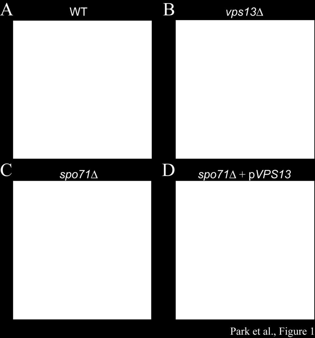
8 cells, 72% of the prospore membranes were still open to the ascal cytoplasm, as opposed to 0% in wild-type cells at a similar stage (Table 4). Prospore membrane closure requires removal of the LEP from the mouth of prospore membrane (8, 11). Don1-GFP is a convenient marker for LEP disassembly as it is localized at the leading edge during prospore membrane growth and relocalizes throughout the spore cytoplasm coincident with membrane closure (8, 11). The previously identified closure mutants ama1 and vps13 differ with respect to removal of the LEP. In ama1 mutants, Don1-GFP persists at the leading edge in the majority of cells, whereas in vps13 mutants a majority of the cells display Don1-GFP redistributed to the cytoplasm (Table 4). To determine if the closure defect of spo71 is upstream or downstream of LEP removal, the distribution of Don1-GFP was monitored in post-meiotic spo71 cells. Don1-GFP relocalizes to the spore cytoplasm in 67% of such cells, though the FLIP results suggest that the majority of prospore membranes are not yet closed (Table 4). Therefore, similar to vps13, the spo71 closure defect occurs after LEP removal (12). Intralumenal vesicles accumulate in spo71 prospore membranes spo71 cells were examined for other vps13 sporulation phenotypes. In addition to the closure defects, vps13 mutants display a unique phenotype, the accumulation of membrane vesicles within the lumen of the prospore membrane (12). Transmission electron microscopy was performed to examine the prospore in spo71 cells. In wild-type cells, the lumenal width between inner and outer prospore membrane is constant (Fig. 1A). In spo71 cells this uniform distance is disrupted by the presence of intralumenal vesicles, just as in vps13 (Fig. 1B and C). These vesicles were visible in 37% of prospore membrane profiles in spo71 (n = 57), lower than that reported for vps13 (75%; (12)), but still significant given that these vesicles were never seen in wildtype cells (n > 100). Expression of VPS13 from a high copy plasmid did not suppress the sporulation defect of spo71 or the formation of these vesicles in the mutant, suggesting that VPS13 does not function downstream of SPO71 (Fig. 1D). Prospore membrane PtdIns-phosphate pools are reduced in spo71 cells Many of the vps13 prospore membrane defects are caused by reduced levels of PtdIns(4)P and PtdIns(4,5)P 2 within the prospore membrane (12). To investigate if these pools are reduced in spo71 cells, the localizations of two lipid-binding sensors -- GFP- 8
9 PH OSH2, a marker for PtdIns(4)P, and GFP-2xPH PLC, a marker for PtdIns(4,5)P 2 -- were monitored during sporulation (25-27). In wild-type cells, both sensors colocalized with the prospore membrane marker Dtr1-RFP in > 70% of the cells (Fig. 2). In spo71 cells, however, only 36% or 38% of the prospore membranes detectable by Dtr1-RFP displayed co-localization with GFP-PH OSH2 or GFP-2xPH PLC, respectively (Fig. 2). These results suggest that the levels of both of these lipids are reduced in the spo71, demonstrating another parallel between the spo71 and vps13 sporulation phenotypes. Spo71-GFP localizes to the prospore membrane during sporulation Vps13 localizes to prospore membranes during meiosis (12). If Spo71 and Vps13 work together directly, then Spo71 should also be found on these membranes. When the chromosomal copy of SPO71 was C-terminally tagged with GFP, no detectable fluorescence signal was seen in sporulating cells. Therefore, a SPO71::GFP fusion under control of the SPO71 promoter was moved to a multi-copy vector. When overexpressed, Spo71-GFP clearly co-localized with the prospore membrane marker RFP-Spo (Fig. 3). This suggests that SPO71 may have rather direct function(s) in prospore membrane assembly. SPO71 regulates Vps13 localization to the prospore membrane during spore formation To determine the genetic requirements for prospore membrane localization of Vps13 and Spo71, localization of each protein was examined in the absence of the other gene. The prospore membrane localization of Spo71-GFP was unchanged in the absence of VPS13 (Fig. 4A). In contrast, Vps13-GFP failed to localize to prospore membranes in spo71 cells (Fig. 4B); only about 4% of prospore membranes contained Vps13-GFP (n>150 in each of three independent experiments). Reintroduction of SPO71 on a plasmid restored prospore membrane localization of Vps13-GFP, confirming that SPO71 is required for proper localization of Vps13 during sporulation (85%, n>230 in each of three independent experiments) (Fig. 4B). Ectopically expressed SPO71 alters Vps13-GFP localization in mitotic cells In mitotic cells, where Spo71 is not present, Vps13 is found on endosomes (15). If SPO71 is a regulator of Vps13 localization during sporulation, expression of SPO71 in mitotic cells might alter Vps13 localization. To test this hypothesis, SPO71::RFP was ectopically expressed under the control of the constitutive TEF2 promoter in mitotic cells 9
10 that also expressed VPS13-GFP. The SPO71::RFP fusion is functional as shown by its ability to rescue the sporulation defect of a spo71 mutant (J.S.P., unpublished observation). In the absence of SPO71::RFP expression, Vps13-GFP foci are observed in the cytoplasm, likely at endosomes as previously reported (Fig. 5). Expression of SPO71::RFP caused a redistribution of the Vps13-GFP signal. Rather than cytoplasmic foci, GFP fluorescence was diffuse in the cytosol with some concentration at the plasma membrane and bud neck (Fig. 5). Vps13-GFP signal at the plasma membrane was not seen in wild-type cells (Fig. 5). Ectopically expressed SPO71 has been reported to localize to the plasma membrane in mitotic cells (20). This suggests that induction of Spo71-RFP might lead to colocalization with Vps13 at the plasma membrane and bud neck. However, we were unable to test this prediction as the RFP fluorescence from our Spo71-RFP was seen uniformly throughout the cell, suggesting that the RFP moiety may be cleaved from Spo71 in vivo. Nonetheless, these results demonstrate expression of SPO71::RFP is sufficient to alter Vps13 localization. Spo71 physically interacts with Vps13 during sporulation To test for a physical interaction between Vps13 and Spo71, coimmunoprecipitation experiments were performed. Extracts made from sporulating cells expressing SPO71::GFP, VPS13::3xHA, or both fusions were used for immunoprecipitations with both anti-ha and anti-gfp antibodies. A Western blot of the anti-ha immunoprecipitates with anti-gfp antibodies revealed that Spo71-GFP precipitated only when co-expressed with Vps13-3xHA (Fig. 6). Reciprocally, pulldown of Spo71-GFP was able to specifically co-precipitate Vps13-3xHA. Thus, consistent with the similar phenotypes of the mutants, Spo71 forms a sporulation-specific complex with Vps13. The anti-ha pull down reveals extensive degradation of Vps13-3xHA in the extracts. By contrast, only the full length Vps13-3xHA protein is seen in the anti-gfp precipitates. As the HA epitopes are fused to the C-terminal end of Vps13, this observation suggests that the N-terminus of Vps13 is essential for interaction with Spo71. Discussion The data presented provide a model for how constitutive secretory pathway functions can be repurposed during differentiation. The Spo71 and Vps13 proteins form a sporulation-specific complex that regulates PtdIns phosphate levels in the prospore 10
11 membrane. While VPS13 is constitutively expressed, SPO71 is sporulation-specific in its expression. Our data suggest that induction of SPO71 when cells progress out of meiotic prophase leads to Spo71 protein binding to Vps13. In turn, interaction with Spo71 leads to release of Vps13 from the endosome and both proteins then localize to the prospore membrane and influence PtdIns phosphate levels. The spo71 prospore membrane phenotypes described are similar to those of vps13 cells, however in all cases -- e.g. membrane size, the percentage of membranes that fail to close, the fraction of membranes displaying intralumenal vesicles -- the phenotype of spo71 is quantitatively less severe than that of vps13. This suggests the possibility that the critical role of Spo71 is simply to recruit VPS13 to the prospore membrane where Vps13 would be responsible for stimulating PtdIns phosphate levels. If so, overexpression of VPS13 might be expected to rescue some of the spo71 phenotypes. As overexpression of VPS13 does not bypass spo71, we prefer a model in which the two proteins act in concert to regulate PtdIns phosphates. An important issue that remains to be resolved is whether Vps13 performs the same molecular function in two different locations, the endosome or the prospore membrane, at different stages of the life cycle, or if the function of Vps13 at the prospore membrane is different than its vegetative function in endosomal-golgi retrograde transport. While the answer to this question will require identifying the precise molecular function of Vps13 in both processes, the fact that SPO71 is required at the prospore membrane but not at the endosome may indicate that Vps13 has two independent functions. Another critical question is how the Vps13/Spo71 complex affects PtdIns phosphate levels. As the levels of these lipids are based, ultimately, on the rate of their synthesis and degradation, Vps13/Spo71 likely act either by inhibition of PtdIns-4-phophate phosphatases or by activation of PtdIns-4 kinases. For the kinases, the likely targets would be either Pik1, responsible for the Golgi pool of PtdIns(4)P in vegetative cells, or Stt4, responsible for the plasma membrane pool (28, 29). A previous study has implicated PIK1 in contributing to PtdIns phosphates in the prospore membrane (30). However, these effects could be indirect as Pik1-generated PtdIns(4)P in the Golgi could be delivered to the prospore membrane via vesicular transport. For example, initial levels PtdIns(4)P might be from a Pik1-generated Golgi pool delivered by vesicular transport, while maintenance of PtdIns(4)P levels might require Stt4 activity and this latter activity 11
12 could be influenced by Vps13/Spo71. Consistent with this possibility, Stt4 localizes to the prospore membrane (J.S.P. and H.T., unpublished observations). To date, however, we have been unable to observe a physical interaction of Vps13 with either Pik1 or Stt4; though it is possible that any effect on enzymatic activity of the kinases may not be mediated through direct binding. VPS13 is highly conserved in eukaryotic organisms, including four paralogs in human cells (31). Mutations in two of these paralogs result in different genetic syndromes (16, 17). It is of interest, therefore, whether the Spo71/Vps13 complex or its function is conserved in higher organisms. While SPO71 is not as highly conserved as VPS13 -- putative SPO71 orthologs can be found in many fungi, but are absent from metazoans -- the protein contains domains present in higher cells. The Spo71 protein contains 2 pleckstrin homology (PH) domains and a third conserved domain, which sequence alignments suggest may be a third, degenerate PH domain (20, 27); A. M. N., unpublished observation). While no direct orthologs of SPO71 are present in metazoans, there are many PH domain-containing proteins. It is possible that one or more of these mammalian PH domain proteins interacts with a Vps13 ortholog similarly to Spo71. Acknowledgements We thank Nancy Hollingsworth and members of the Neiman lab for helpful discussions and comments on the manuscript. We are grateful to Susan Van Horn in the Stony Brook Center for Microscopy for assistance with electron microscopy. This work was supported by the National Institute of Health grant RO1 GM to A.M.N. 12
13 References 1. Neiman AM Sporulation in the budding yeast Saccharomyces cerevisiae. Genetics 189: Neiman AM Ascospore formation in the yeast Saccharomyces cerevisiae. Microbiology and molecular biology reviews 69: Byers B Cytology of the yeast life cycle. Cold Spring Harbor Monograph Archive 11: Moens PB Fine structure of ascospore development in the yeast Saccharomyces cerevisiae. Canadian journal of microbiology 17: Neiman AM Prospore membrane formation defines a developmentally regulated branch of the secretory pathway in yeast. The Journal of cell biology 140: Nakanishi H, Morishita M, Schwartz CL, Coluccio A, Engebrecht JA, Neiman AM Phospholipase D and the SNARE Sso1p are necessary for vesicle fusion during sporulation in yeast. Journal of cell science 119: Lynn RR, Magee PT Development of the spore wall during ascospore formation in Saccharomyces cerevisiae. The Journal of cell biology 44: Maier P, Rathfelder N, Finkbeiner MG, Taxis C, Mazza M, Le Panse S, Haguenauer-Tsapis R, Knop M Cytokinesis in yeast meiosis depends on the regulated removal of Ssp1p from the prospore membrane. The EMBO Journal 26: Moreno-Borchart AC, Strasser K, Finkbeiner MG, Shevchenko A, Shevchenko A, Knop M Prospore membrane formation linked to the leading edge protein (LEP) coat assembly. The EMBO Journal 20: Nickas ME, Neiman AM Ady3p links spindle pole body function to spore wall synthesis in Saccharomyces cerevisiae. Genetics 160: Diamond AE, Park JS, Inoue I, Tachikawa H, Neiman AM The anaphase promoting complex targeting subunit Ama1 links meiotic exit to cytokinesis during sporulation in Saccharomyces cerevisiae. Molecular biology of the cell 20: Park JS, Neiman AM VPS13 regulates membrane morphogenesis during sporulation in Saccharomyces cerevisiae. Journal of cell science 125: Bankaitis VA, Johnson LM, Emr SD Isolation of yeast mutants defective in protein targeting to the vacuole. Proceedings of the National Academy of Sciences 83: Brickner JH, Fuller RS SOI1 encodes a novel, conserved protein that promotes TGN-endosomal cycling of Kex2p and other membrane proteins by modulating the function of two TGN localization signals. The Journal of cell biology 139: Huh WK, Falvo JV, Gerke LC, Carroll AS, Howson RW, Weissman JS, O'Shea EK Global analysis of protein localization in budding yeast. Nature 425: Kolehmainen J, Black G, Saarinen A, Chandler K, Clayton-Smith J, Tr skelin AL, Perveen R, Kivitie-Kallio S, Norio R, Warburg M Cohen syndrome is caused by mutations in a novel gene, COH1, encoding a transmembrane protein with a presumed role in vesicle-mediated sorting and intracellular protein transport. The American Journal of Human Genetics 72:
14 Rampoldi L, Dobson-Stone C, Rubio JP, Danek A, Chalmers RM, Wood NW, Verellen C, Ferrer X, Malandrini A, Fabrizi GM A conserved sorting-associated protein is mutant in chorea-acanthocytosis. Nature genetics 28: Jäntti J, Aalto MK, Öyen M, Sundqvist L, Keränen S, Ronne H Characterization of temperature-sensitive mutations in the yeast syntaxin 1 homologues Sso1p and Sso2p, and evidence of a distinct function for Sso1p in sporulation. Journal of cell science 115: Yang H-J, Nakanishi H, Liu S, McNew JA, Neiman AM Binding interactions control SNARE specificity in vivo. The Journal of cell biology 183: Parodi EM, Baker CS, Tetzlaff C, Villahermosa S, Huang LS SPO71 mediates prospore membrane size and maturation in Saccharomyces cerevisiae. Eukaryotic Cell 11: Rose MD, Fink GR Methods in Yeast Genetics. Cold Spring Harbor Laboratory Press, Cold Spring Harbor, NY. 22. Rabitsch KP, TÛth A, G lov M, Schleiffer A, Schaffner G, Aigner E, Rupp C, Penkner AM, Moreno-Borchart AC, Primig M A screen for genes required for meiosis and spore formation based on whole-genome expression. Current Biology 11: Neiman AM, Katz L, Brennwald PJ Identification of domains required for developmentally regulated SNARE function in Saccharomyces cerevisiae. Genetics 155: Ma H, Kunes S, Schatz PJ, Botstein D Plasmid construction by homologous recombination in yeast. Gene 58: Roy A, Levine TP Multiple pools of phosphatidylinositol 4-phosphate detected using the pleckstrin homology domain of Osh2p. Journal of Biological Chemistry 279: Stefan CJ, Audhya A, Emr SD The yeast synaptojanin-like proteins control the cellular distribution of phosphatidylinositol (4, 5)-bisphosphate. Molecular biology of the cell 13: Yu JW, Mendrola JM, Audhya A, Singh S, Keleti D, DeWald DB, Murray D, Emr SD, Lemmon MA Genome-Wide Analysis of Membrane Targeting by S. cerevisiae Pleckstrin Homology Domains. Molecular cell 13: Audhya A, Emr SD Stt4 PI 4-kinase localizes to the plasma membrane and functions in the Pkc1-mediated MAP kinase cascade. Developmental cell 2: Walch-Solimena C, Novick P The yeast phosphatidylinositol-4-oh kinase Pik1 regulates secretion at the Golgi. Nature Cell Biology 1: Rudge SA, Sciorra VA, Iwamoto M, Zhou C, Strahl T, Morris AJ, Thorner J, Engebrecht JA Roles of phosphoinositides and of Spo14p (phospholipase D)-generated phosphatidic acid during yeast sporulation. Molecular biology of the cell 15: Velayos-Baeza A, Vettori A, Copley RR, Dobson-Stone C, Monaco A Analysis of the human VPS13 gene family. Genomics 84: Nakanishi H, Suda Y, Neiman AM Erv14 family cargo receptors are necessary for ER exit during sporulation in Saccharomyces cerevisiae. Journal of cell science 120:
15 Nakanishi H, De Los Santos P, Neiman AM Positive and negative regulation of a SNARE protein by control of intracellular localization. Molecular biology of the cell 15:
16 Figure Legends Figure 1. Intralumenal vesicle accumulation within the prospore membrane. Transmission electron microscope images of prospore membranes in (A) wild-type (AN120), (B) vps13 (HI29), (C) spo71 (AN363), and (D) spo71 cells overexpressing VPS13. In all images, yellow arrows indicate prospore membranes and red arrows indicate the accumulation of vesicles within the lumen of a prospore membrane. Scale bars = 500 nm. Figure 2. Effect of spo71 on prospore membrane PtdIns(4)P and PtdIns(4,5)P 2 pools. (A) Sporulating wild-type (AN120) and spo71 (AN363) cells expressing both a PtdIns(4)P sensor (GFP-PH OSH2 ) and a prospore membrane marker (Dtr1-RFP). Red arrow indicates a prospore membrane without the GFP marker in the spo71 cell. Scale bar = 1 m. (B) Quantitation of GFP-PH OSH2 / Dtr1-RFP colocalization. Bars indicate standard error of the mean. More than 100 prospore membranes were scored in each of four separate experiments. (C) Sporulating wild-type (AN120) and spo71 (AN363) cells expressing both a PtdIns(4,5)P 2 sensor (GFP-2xPH PLC ) and a prospore membrane marker (Dtr1-RFP). Red arrow indicates a prospore membrane without the GFP marker in the spo71 cell. Scale bar = 1 m. (D) Quantitation of GFP-PH PLC / Dtr1-RFP colocalization. Bars indicate standard error of the mean. More than 100 prospore membranes were scored in each of two separate experiments. Chi-square test indicates that the differences between wild type and spo71 are significant (p< 0.001). Figure 3. Localization of Spo71-GFP in sporulating cells. Wild-type (AN120) cells carrying prs424-spo71-gfp and the prospore membrane marker prs426-rfp-spo were sporulated and fluorescence was examined during prospore membrane growth. Colocalization to the prospore membrane was seen in 100% of the cells examined (n = 25). Scale bar = 1 m. Figure 4. Vps13-GFP localization during sporulation in wild-type and spo71. (A) Wild-type (AN120) and vps13 (HI29) cells carrying prs424-spo71-gfp and prs426- RFP-Spo were sporulated and fluorescence was examined during prospore 16
17 membrane growth. Colocalization to the prospore membrane was seen in 99% of the prospore membranes examined (n > 150). Scale bar = 1 m. (B) VPS13-GFP SPO71 (JSP257) and VPS13-GFP spo71 (JSP294) cells carrying the prospore membrane marker RFP-Spo were sporulated and cells containing prospore membranes (as identified by the RFP-Spo marker) were examined for colocalization of Vps13-GFP. Scale bar = 1 m. Figure 5. Effect of ectopically expressed SPO71 on Vps13-GFP localization in mitotic cells. VPS13-GFP (JSP257) cells with (right panels) or without (left panel) prs426-p TEF2 -SPO71-RFP were examined in mid-log phase growth. The white arrow indicates Vps13-GFP localization to cytoplasmic foci, likely to be endosomes. Red and yellow arrows indicate Vps13-GFP localization to plasma membranes and bud necks, respectively. Scale bars = 1 m. Plasma membrane or bud neck localization of Vps13- GFP was seen in 97% of the SPO71 overexpressing cells in two independent experiments and at least 120 cells were examined in each. Figure 6. Spo71 interacts with Vps13 during sporulation. VPS13 (AN120) and VPS13-3HA (JSP401) carrying prs424-spo71-gfp or an empty vector were sporulated then lysed and immunoprecipitations were performed using anti-ha or anti-gfp antibodies. Precipitates were then probed by Western blot using anti-ha and anti-gfp antibodies. Stars indicate Vps13-HA and Spo71-GFP degradation products. Molecular size markers are given in kilodaltons. 17
18 Table 1: Strains used in this study. Strain Genotype Source AN117-4B MAT ura3 leu2 his3 SK trp1::hisg arg4-nspi lys2 ho ::LYS2 rme1 ::LEU2 (23) AN117-16D MATa ura3 leu2 trp1::hisg his3 SK lys2 ho ::LYS2 (23) AN120 MATa/MAT ura3/ura3 his3 SK/his3 SK trp1::hisg/trp1::hisg ARG4/arg4-NspI lys2/lys2 (23) ho ::LYS2/ho ::LYS2 RME1/rme1 ::LEU2 leu2/leu2 AN361-6A MATa ura3 leu2 trp1::hisg his3 SK lys2 ho ::LYS2 spo71 ::his5 + this study AN363 MATa/MAT ura3/ura3 his3 SK/his3 SK trp1::hisg/trp1::hisg this study lys2/lys2 ho ::LYS2/ho ::LYS2 RME1/rme1 ::LEU2 leu2/leu2 ARG4/arg4-NspI spo71 :: his5 + /spo71 :: his5 + HI29 MATa/MAT ura3/ura3 his3 SK/his3 SK trp1::hisg/trp1::hisg (32) ARG4/arg4-NspI lys2/lys ho ::LYS2/ho ::LYS2 RME1/rme1 ::LEU2 leu2/leu2 vps13 ::his5 + /vps13 ::his5 + JSP163 MATa/MAT ura3/ura3 TRP1/trp1 ARG4/arg4 LEU2/leu2 spo71 ::his5 + /spo71 ::his5 + this study TEF2::GFP::his5 + /TEF2::GFP::his5 + JSP247 MAT ura3 trp1::hisg arg4-nspi lys2 ho ::LYS2 rme1 ::LEU2 leu2 VPS13::GFP::his5 + this study JSP248 MATa ura3 trp1::hisg ARG lys2 ho ::LYS2 leu2 VPS13::GFP::his5 + this study 18
19 JSP257 MATa/MAT ARG4/arg4-NspI LEU2/leu2 ura3/ura3 trp1/trp1 (12) VPS13::GFP::his5 + /VPS13::GFP::his5 + JSP290 MATa ura3 leu2 trp1::hisg ho ::LYS2 arg4-nspi spo71 ::his5 + VPS13::GFP::his5 + this study JSP291 MAT ura3 leu2 trp1::hisg ho ::LYS2 spo71 ::his5 + VPS13::GFP::his5 + this study JSP294 MATa/MAT ura3/ura3 leu2/leu2 trp1::hisg/trp1::hisg ho ::LYS2/ho ::LYS2 this study arg4-nspi/atg4 spo71 ::his5 + /spo71 ::his5 + VPS13::GFP::his5 + /VPS13::GFP::his5 + JSP391 MAT ura3 trp1::hisg arg4-nspi lys2 ho ::LYS2 rme1 ::LEU2 leu2 his3 SK this study VPS13::3HA::his5 + JSP392 MATa ura3 trp1::hisg lys2 ho ::LYS2 leu2 his3 SK VPS13::3HA::his5 + this study JSP394 MAT ura3 trp1::hisg arg4-nspi lys2 ho ::LYS2 leu2 VPS13::3HA::his5 + this study JSP401 MATa/MAT ura3/ura3 trp1::hisg/trp1::hisg ho ::LYS2/ho ::LYS2 leu2/leu2 this study ARG4/arg4-NspI VPS13::3HA::his5 + /VPS13::3HA::his5 + 19
20 Table 2: Oligonucleotides used in this study. JSO CCG CTC GAG TCA CAT ATG AAA GTA TAT AC -3 JSO GGG GTA CCT TTG ATC TTT AAC AGC GCA T -3 JSO TGA GTA TCC TGA GAT AAA ATT TGG A -3 JSO64-r 5 - TCC AAA TTT TAT CTC AGG ATA CTC A -3 JSO CGT CAC CAA TTA AAA TAA ATC TCA G -3 JSO65-r 5 - CTG AGA TTT ATT TTA ATT GGT GAC G -3 JSO GTGTATTAACCTTCTCATCCTCATC -3 JSO66-r 5 - GAT GAG GAT GAG AAG GTT AAT ACA C -3 JSO TAG TGT ACA AAA GCG GGT ATA TAC TTT CAT ATG TGA CTC GAG CGG TCA TAG GAT AGC TTC ACA GTA CTT A -3 JSO TCC CCG CGG GAA GAT GGT AGA ACG CTG TC -3 JSO ATA AGA ATG CGG CCG CTT AAC TGT TCT TAA TTT TCC TTT TT -3 JSO GAA AAA AGG AAA ATT AAG AAC AGT TAA GCG GCC GCA TTC TTA TAT GTT AGA GTC TTT AGC TGC TAA -3 JSO ACA TCG CCA TTG CTG TTA GAG AAT ACA ATA AGT ACT GTG AAG CTA TCC TAC GGA TCC CCG GGT TAA TTA A -3 20
21 JSO GAA TTA TAG CTA CAT AGT GTA CAA AAG CGG GTA TAT ACT TTC ATA TGT GAG AAT TCG AGC TCG TTT AAA C -3 JSO CGG GGG ATC CAC TAG TTC TAG AGC GGC CGC CAC CGC GGT GGA GCT CCC TTT ACG GTT GGA CAG TGG -3 JSO AAC TTG AAT GAA ATT ATA TTC CAA AA -3 JSO160-r 5 - TTT TGG AAT ATA ATT TCA TTC AAG TT -3 JSO CAA GCG CGC AAT TAA CCC TCA CTA AAG GGA ACA AAA GCT GGA GCT CGC TAC AGT TTC GGT CTA AGT TT -3 JSO TGG CGG CCG CTC TAG AAC TAG TGG ATC CCC CGG GCT GCA GGA ATT CAT GGA TTC TAT CGT TAA TGT TGT -3 JSO AGC GCA TGA ACT CCT TGA TGA CGT CCT CGG AGG AGG CCA TAA GCT TCA TAG TTT GAT TCC GTG AAT TTG -3 HT290 5'-AAA GAC TAA GAA GAA CTG CGT CCA CTT CAA ATT CAC GGA ATC AAA CTA TGC GGA TCC CCG GGT TAA TTA A - 3' HT291 5'- AGT ATA CAC TAA ATT TTA TGC AAT AAT AAA AAG AAA GCA TCC CGC CAA ACG AAT TCG AGC TCG TTT AAA C -3' HT293 5'- GAA GAA GGT ACC AAA TGA TGA TGA AAG GAC TG - 3' HT66 5'-GAA GAA TTC AGA TCT ATA TTA CCC TGT TAT CC -3' 21
22 Table 3: Plasmids used in this study Name Gene(s) Expressed Source prs426-rfp-spo P TEF2-RFP-SPO (33) prs424-dtr1-rfp DTR1-RFP (12) prs426-rfp-spo DON1-GFP P TEF2-RFP-SPO , DON1-GFP (12) prs426-p PRC1-GFP-PH OSH2 GFP-PH OSH2 (27) prs426-gfp-2xph PLC GFP-2xPH PLC (26) prs426-vps13 VPS13 this study prs424-spo71 SPO71 this study prs424-spo71-gfp SPO71-GFP this study prs426-p TEF2-SPO71-RFP P TEF2-SPO71-RFP this study 22
23 Table 4. Prospore membrane closure in spo71 cells Membrane closure by FL)P assay Don GFP distribution Wild type a ama1 a vps13 a spo71 c Closed Open )nd b Cytoplasm Leading edge a. a Data are from (11, 12) b. b Indeterminant: Fluorescence loss was intermediate between open and closed patterns. c. c >50 prospore membranes were scored for FLIP assay. More than110 prospore membranes were examined for Don1-GFP distribution experiment. Downloaded from on March 29, 2019 by guest 23
24 633 24

25


26


27


28


29


30
Practical Bioinformatics
 5/2/2017 Dictionaries d i c t i o n a r y = { A : T, T : A, G : C, C : G } d i c t i o n a r y [ G ] d i c t i o n a r y [ N ] = N d i c t i o n a r y. h a s k e y ( C ) Dictionaries g e n e t i c C o
SUPPORTING INFORMATION FOR. SEquence-Enabled Reassembly of β-lactamase (SEER-LAC): a Sensitive Method for the Detection of Double-Stranded DNA
: a Sensitive Method for the Detection of Double-Stranded DNA") SUPPORTING INFORMATION FOR SEquence-Enabled Reassembly of β-lactamase (SEER-LAC): a Sensitive Method for the Detection of Double-Stranded DNA Aik T. Ooi, Cliff I. Stains, Indraneel Ghosh *, David J. Segal
SUPPORTING INFORMATION FOR SEquence-Enabled Reassembly of β-lactamase (SEER-LAC): a Sensitive Method for the Detection of Double-Stranded DNA Aik T. Ooi, Cliff I. Stains, Indraneel Ghosh *, David J. Segal
Clay Carter. Department of Biology. QuickTime and a TIFF (Uncompressed) decompressor are needed to see this picture.
 decompressor are needed to see this picture.") QuickTime and a TIFF (Uncompressed) decompressor are needed to see this picture. Clay Carter Department of Biology QuickTime and a TIFF (LZW) decompressor are needed to see this picture. Ornamental tobacco
QuickTime and a TIFF (Uncompressed) decompressor are needed to see this picture. Clay Carter Department of Biology QuickTime and a TIFF (LZW) decompressor are needed to see this picture. Ornamental tobacco
SUPPLEMENTARY DATA - 1 -
 - 1 - SUPPLEMENTARY DATA Construction of B. subtilis rnpb complementation plasmids For complementation, the B. subtilis rnpb wild-type gene (rnpbwt) under control of its native rnpb promoter and terminator
High throughput near infrared screening discovers DNA-templated silver clusters with peak fluorescence beyond 950 nm
 Electronic Supplementary Material (ESI) for Nanoscale. This journal is The Royal Society of Chemistry 2018 High throughput near infrared screening discovers DNA-templated silver clusters with peak fluorescence
Number-controlled spatial arrangement of gold nanoparticles with
 Electronic Supplementary Material (ESI) for RSC Advances. This journal is The Royal Society of Chemistry 2016 Number-controlled spatial arrangement of gold nanoparticles with DNA dendrimers Ping Chen,*
Supplemental data. Pommerrenig et al. (2011). Plant Cell /tpc
. Plant Cell /tpc") Supplemental Figure 1. Prediction of phloem-specific MTK1 expression in Arabidopsis shoots and roots. The images and the corresponding numbers showing absolute (A) or relative expression levels (B) of
Supplemental Figure 1. Prediction of phloem-specific MTK1 expression in Arabidopsis shoots and roots. The images and the corresponding numbers showing absolute (A) or relative expression levels (B) of
Crick s early Hypothesis Revisited
 Crick s early Hypothesis Revisited Or The Existence of a Universal Coding Frame Ryan Rossi, Jean-Louis Lassez and Axel Bernal UPenn Center for Bioinformatics BIOINFORMATICS The application of computer
Nature Structural & Molecular Biology: doi: /nsmb Supplementary Figure 1
 Supplementary Figure 1 Zn 2+ -binding sites in USP18. (a) The two molecules of USP18 present in the asymmetric unit are shown. Chain A is shown in blue, chain B in green. Bound Zn 2+ ions are shown as
Supplemental Figure 1.
 A wt spoiiiaδ spoiiiahδ bofaδ B C D E spoiiiaδ, bofaδ Supplemental Figure 1. GFP-SpoIVFA is more mislocalized in the absence of both BofA and SpoIIIAH. Sporulation was induced by resuspension in wild-type
SSR ( ) Vol. 48 No ( Microsatellite marker) ( Simple sequence repeat,ssr),
 Vol. 48 No ( Microsatellite marker) ( Simple sequence repeat,ssr),") 48 3 () Vol. 48 No. 3 2009 5 Journal of Xiamen University (Nat ural Science) May 2009 SSR,,,, 3 (, 361005) : SSR. 21 516,410. 60 %96. 7 %. (),(Between2groups linkage method),.,, 11 (),. 12,. (, ), : 0.
48 3 () Vol. 48 No. 3 2009 5 Journal of Xiamen University (Nat ural Science) May 2009 SSR,,,, 3 (, 361005) : SSR. 21 516,410. 60 %96. 7 %. (),(Between2groups linkage method),.,, 11 (),. 12,. (, ), : 0.
Advanced topics in bioinformatics
 Feinberg Graduate School of the Weizmann Institute of Science Advanced topics in bioinformatics Shmuel Pietrokovski & Eitan Rubin Spring 2003 Course WWW site: http://bioinformatics.weizmann.ac.il/courses/atib
Supporting Information for. Initial Biochemical and Functional Evaluation of Murine Calprotectin Reveals Ca(II)-
-") Supporting Information for Initial Biochemical and Functional Evaluation of Murine Calprotectin Reveals Ca(II)- Dependence and Its Ability to Chelate Multiple Nutrient Transition Metal Ions Rose C. Hadley,
Supporting Information for Initial Biochemical and Functional Evaluation of Murine Calprotectin Reveals Ca(II)- Dependence and Its Ability to Chelate Multiple Nutrient Transition Metal Ions Rose C. Hadley,
SEQUENCE ALIGNMENT BACKGROUND: BIOINFORMATICS. Prokaryotes and Eukaryotes. DNA and RNA
 SEQUENCE ALIGNMENT BACKGROUND: BIOINFORMATICS 1 Prokaryotes and Eukaryotes 2 DNA and RNA 3 4 Double helix structure Codons Codons are triplets of bases from the RNA sequence. Each triplet defines an amino-acid.
Characterization of Pathogenic Genes through Condensed Matrix Method, Case Study through Bacterial Zeta Toxin
 International Journal of Genetic Engineering and Biotechnology. ISSN 0974-3073 Volume 2, Number 1 (2011), pp. 109-114 International Research Publication House http://www.irphouse.com Characterization of
Supplementary Information for
 Supplementary Information for Evolutionary conservation of codon optimality reveals hidden signatures of co-translational folding Sebastian Pechmann & Judith Frydman Department of Biology and BioX, Stanford
Electronic supplementary material
 Applied Microbiology and Biotechnology Electronic supplementary material A family of AA9 lytic polysaccharide monooxygenases in Aspergillus nidulans is differentially regulated by multiple substrates and
Supporting Information
 Supporting Information T. Pellegrino 1,2,3,#, R. A. Sperling 1,#, A. P. Alivisatos 2, W. J. Parak 1,2,* 1 Center for Nanoscience, Ludwig Maximilians Universität München, München, Germany 2 Department of
Regulatory Sequence Analysis. Sequence models (Bernoulli and Markov models)
") Regulatory Sequence Analysis Sequence models (Bernoulli and Markov models) 1 Why do we need random models? Any pattern discovery relies on an underlying model to estimate the random expectation. This model
Regulatory Sequence Analysis Sequence models (Bernoulli and Markov models) 1 Why do we need random models? Any pattern discovery relies on an underlying model to estimate the random expectation. This model
NSCI Basic Properties of Life and The Biochemistry of Life on Earth
 NSCI 314 LIFE IN THE COSMOS 4 Basic Properties of Life and The Biochemistry of Life on Earth Dr. Karen Kolehmainen Department of Physics CSUSB http://physics.csusb.edu/~karen/ WHAT IS LIFE? HARD TO DEFINE,
Supplementary Information
 Electronic Supplementary Material (ESI) for RSC Advances. This journal is The Royal Society of Chemistry 2014 Directed self-assembly of genomic sequences into monomeric and polymeric branched DNA structures
evoglow - express N kit Cat. No.: product information broad host range vectors - gram negative bacteria
 evoglow - express N kit broad host range vectors - gram negative bacteria product information Cat. No.: 2.1.020 evocatal GmbH 2 Content: Product Overview... 4 evoglow express N kit... 4 The evoglow Fluorescent
evoglow - express N kit distributed by Cat.#: FP product information broad host range vectors - gram negative bacteria
 evoglow - express N kit broad host range vectors - gram negative bacteria product information distributed by Cat.#: FP-21020 Content: Product Overview... 3 evoglow express N -kit... 3 The evoglow -Fluorescent
Supplemental Table 1. Primers used for cloning and PCR amplification in this study
 Supplemental Table 1. Primers used for cloning and PCR amplification in this study Target Gene Primer sequence NATA1 (At2g393) forward GGG GAC AAG TTT GTA CAA AAA AGC AGG CTT CAT GGC GCC TCC AAC CGC AGC
Table S1. Primers and PCR conditions used in this paper Primers Sequence (5 3 ) Thermal conditions Reference Rhizobacteria 27F 1492R
 Thermal conditions Reference Rhizobacteria 27F 1492R") Table S1. Primers and PCR conditions used in this paper Primers Sequence (5 3 ) Thermal conditions Reference Rhizobacteria 27F 1492R AAC MGG ATT AGA TAC CCK G GGY TAC CTT GTT ACG ACT T Detection of Candidatus
Table S1. Primers and PCR conditions used in this paper Primers Sequence (5 3 ) Thermal conditions Reference Rhizobacteria 27F 1492R AAC MGG ATT AGA TAC CCK G GGY TAC CTT GTT ACG ACT T Detection of Candidatus
ydci GTC TGT TTG AAC GCG GGC GAC TGG GCG CGC AAT TAA CGG TGT GTA GGC TGG AGC TGC TTC
 Table S1. DNA primers used in this study. Name ydci P1ydcIkd3 Sequence GTC TGT TTG AAC GCG GGC GAC TGG GCG CGC AAT TAA CGG TGT GTA GGC TGG AGC TGC TTC Kd3ydcIp2 lacz fusion YdcIendP1 YdcItrgP2 GAC AGC
Building a Multifunctional Aptamer-Based DNA Nanoassembly for Targeted Cancer Therapy
 Supporting Information Building a Multifunctional Aptamer-Based DNA Nanoassembly for Targeted Cancer Therapy Cuichen Wu,, Da Han,, Tao Chen,, Lu Peng, Guizhi Zhu,, Mingxu You,, Liping Qiu,, Kwame Sefah,
TM1 TM2 TM3 TM4 TM5 TM6 TM bp
 a 467 bp 1 482 2 93 3 321 4 7 281 6 21 7 66 8 176 19 12 13 212 113 16 8 b ATG TCA GGA CAT GTA ATG GAG GAA TGT GTA GTT CAC GGT ACG TTA GCG GCA GTA TTG CGT TTA ATG GGC GTA GTG M S G H V M E E C V V H G T
Vesicle Docking to the Spindle Pole Body Is Necessary to Recruit the Exocyst During Membrane Formation in Saccharomyces cerevisiae
 Molecular Biology of the Cell Vol. 21, 3693 3707, November 1, 2010 Vesicle Docking to the Spindle Pole Body Is Necessary to Recruit the Exocyst During Membrane Formation in Saccharomyces cerevisiae Erin
7.06 Problem Set #4, Spring 2005
 7.06 Problem Set #4, Spring 2005 1. You re doing a mutant hunt in S. cerevisiae (budding yeast), looking for temperaturesensitive mutants that are defective in the cell cycle. You discover a mutant strain
Optimization of Immunoblot Protocol for Use with a Yeast Strain Containing the CDC7 Gene Tagged with myc
 OPTIMIZATION OF IMMUNOBLOT PROTOCOL 121 Optimization of Immunoblot Protocol for Use with a Yeast Strain Containing the CDC7 Gene Tagged with myc Jacqueline Bjornton and John Wheeler Faculty Sponsor: Anne
SUPPLEMENTARY INFORMATION
 DOI:.8/NCHEM. Conditionally Fluorescent Molecular Probes for Detecting Single Base Changes in Double-stranded DNA Sherry Xi Chen, David Yu Zhang, Georg Seelig. Analytic framework and probe design.. Design
6.047 / Computational Biology: Genomes, Networks, Evolution Fall 2008
 MIT OpenCourseWare http://ocw.mit.edu 6.047 / 6.878 Computational Biology: Genomes, Networks, Evolution Fall 2008 For information about citing these materials or our Terms of Use, visit: http://ocw.mit.edu/terms.
Protein Threading. Combinatorial optimization approach. Stefan Balev.
 Protein Threading Combinatorial optimization approach Stefan Balev Stefan.Balev@univ-lehavre.fr Laboratoire d informatique du Havre Université du Havre Stefan Balev Cours DEA 30/01/2004 p.1/42 Outline
SUPPLEMENTARY INFORMATION
 SUPPLEMENTARY INFORMATION DOI:.38/NCHEM.246 Optimizing the specificity of nucleic acid hyridization David Yu Zhang, Sherry Xi Chen, and Peng Yin. Analytic framework and proe design 3.. Concentration-adjusted
Evolvable Neural Networks for Time Series Prediction with Adaptive Learning Interval
 Evolvable Neural Networs for Time Series Prediction with Adaptive Learning Interval Dong-Woo Lee *, Seong G. Kong *, and Kwee-Bo Sim ** *Department of Electrical and Computer Engineering, The University
3. Evolution makes sense of homologies. 3. Evolution makes sense of homologies. 3. Evolution makes sense of homologies
 Richard Owen (1848) introduced the term Homology to refer to structural similarities among organisms. To Owen, these similarities indicated that organisms were created following a common plan or archetype.
The Trigram and other Fundamental Philosophies
 The Trigram and other Fundamental Philosophies by Weimin Kwauk July 2012 The following offers a minimal introduction to the trigram and other Chinese fundamental philosophies. A trigram consists of three
The role of the FliD C-terminal domain in pentamer formation and
 The role of the FliD C-terminal domain in pentamer formation and interaction with FliT Hee Jung Kim 1,2,*, Woongjae Yoo 3,*, Kyeong Sik Jin 4, Sangryeol Ryu 3,5 & Hyung Ho Lee 1, 1 Department of Chemistry,
Protein Phosphatase Type 1-Interacting Protein Ysw1 Is Involved in Proper Septin Organization and Prospore Membrane Formation during Sporulation
 EUKARYOTIC CELL, July 2009, p. 1027 1037 Vol. 8, No. 7 1535-9778/09/$08.00 0 doi:10.1128/ec.00095-09 Copyright 2009, American Society for Microbiology. All Rights Reserved. Protein Phosphatase Type 1-Interacting
Re- engineering cellular physiology by rewiring high- level global regulatory genes
 Re- engineering cellular physiology by rewiring high- level global regulatory genes Stephen Fitzgerald 1,2,, Shane C Dillon 1, Tzu- Chiao Chao 2, Heather L Wiencko 3, Karsten Hokamp 3, Andrew DS Cameron
Modelling and Analysis in Bioinformatics. Lecture 1: Genomic k-mer Statistics
 582746 Modelling and Analysis in Bioinformatics Lecture 1: Genomic k-mer Statistics Juha Kärkkäinen 06.09.2016 Outline Course introduction Genomic k-mers 1-Mers 2-Mers 3-Mers k-mers for Larger k Outline
MEIOSIS LAB INTRODUCTION PART I: SIMULATION OF MEIOSIS EVOLUTION. Activity #9
 AP BIOLOGY EVOLUTION Unit 1 Part 7 Chapter 13 Activity #9 NAME DATE PERIOD MEIOSIS LAB INTRODUCTION Meiosis involves two successive nuclear divisions that produce four haploid cells. Meiosis I is the reduction
Cytokinesis in yeast meiosis depends on the regulated removal of Ssp1p from the prospore membrane
 The EMBO Journal (2007) 26, 1843 1852 & 2007 European Molecular Biology Organization All Rights Reserved 0261-4189/07 www.embojournal.org Cytokinesis in yeast meiosis depends on the regulated removal of
Sex-Linked Inheritance in Macaque Monkeys: Implications for Effective Population Size and Dispersal to Sulawesi
 Supporting Information http://www.genetics.org/cgi/content/full/genetics.110.116228/dc1 Sex-Linked Inheritance in Macaque Monkeys: Implications for Effective Population Size and Dispersal to Sulawesi Ben
Supplemental Figure 1. Phenotype of ProRGA:RGAd17 plants under long day
 Supplemental Figure 1. Phenotype of ProRGA:RGAd17 plants under long day conditions. Photo was taken when the wild type plant started to bolt. Scale bar represents 1 cm. Supplemental Figure 2. Flowering
13-3. Synthesis-Secretory pathway: Sort lumenal proteins, Secrete proteins, Sort membrane proteins
 13-3. Synthesis-Secretory pathway: Sort lumenal proteins, Secrete proteins, Sort membrane proteins Molecular sorting: specific budding, vesicular transport, fusion 1. Why is this important? A. Form and
DISCOVERIES OF MACHINERY REGULATING VESICLE TRAFFIC, A MAJOR TRANSPORT SYSTEM IN OUR CELLS. Scientific Background on the Nobel Prize in Medicine 2013
 DISCOVERIES OF MACHINERY REGULATING VESICLE TRAFFIC, A MAJOR TRANSPORT SYSTEM IN OUR CELLS Scientific Background on the Nobel Prize in Medicine 2013 Daniela Scalet 6/12/2013 The Nobel Prize in Medicine
AtTIL-P91V. AtTIL-P92V. AtTIL-P95V. AtTIL-P98V YFP-HPR
 Online Resource 1. Primers used to generate constructs AtTIL-P91V, AtTIL-P92V, AtTIL-P95V and AtTIL-P98V and YFP(HPR) using overlapping PCR. pentr/d- TOPO-AtTIL was used as template to generate the constructs
part 3: analysis of natural selection pressure
 part 3: analysis of natural selection pressure markov models are good phenomenological codon models do have many benefits: o principled framework for statistical inference o avoiding ad hoc corrections
Alternative Modes of Organellar Segregation during Sporulation in Saccharomyces cerevisiae
 EUKARYOTIC CELL, Nov. 2007, p. 2009 2017 Vol. 6, No. 11 1535-9778/07/$08.00 0 doi:10.1128/ec.00238-07 Copyright 2007, American Society for Microbiology. All Rights Reserved. Alternative Modes of Organellar
Evolutionary dynamics of abundant stop codon readthrough in Anopheles and Drosophila
 biorxiv preprint first posted online May. 3, 2016; doi: http://dx.doi.org/10.1101/051557. The copyright holder for this preprint (which was not peer-reviewed) is the author/funder. All rights reserved.
Codon Distribution in Error-Detecting Circular Codes
 life Article Codon Distribution in Error-Detecting Circular Codes Elena Fimmel, * and Lutz Strüngmann Institute for Mathematical Biology, Faculty of Computer Science, Mannheim University of Applied Sciences,
ChemiScreen CaS Calcium Sensor Receptor Stable Cell Line
 PRODUCT DATASHEET ChemiScreen CaS Calcium Sensor Receptor Stable Cell Line CATALOG NUMBER: HTS137C CONTENTS: 2 vials of mycoplasma-free cells, 1 ml per vial. STORAGE: Vials are to be stored in liquid N
Near-instant surface-selective fluorogenic protein quantification using sulfonated
 Electronic Supplementary Material (ESI) for rganic & Biomolecular Chemistry. This journal is The Royal Society of Chemistry 2014 Supplemental nline Materials for ear-instant surface-selective fluorogenic
Supplementary Information
 Supplementary Information Arginine-rhamnosylation as new strategy to activate translation elongation factor P Jürgen Lassak 1,2,*, Eva Keilhauer 3, Max Fürst 1,2, Kristin Wuichet 4, Julia Gödeke 5, Agata
Why do more divergent sequences produce smaller nonsynonymous/synonymous
 Genetics: Early Online, published on June 21, 2013 as 10.1534/genetics.113.152025 Why do more divergent sequences produce smaller nonsynonymous/synonymous rate ratios in pairwise sequence comparisons?
7.06 Problem Set
 7.06 Problem Set 5 -- 2006 1. In the first half of the course, we encountered many examples of proteins that entered the nucleus in response to the activation of a cell-signaling pathway. One example of
The 3 Genomic Numbers Discovery: How Our Genome Single-Stranded DNA Sequence Is Self-Designed as a Numerical Whole
 Applied Mathematics, 2013, 4, 37-53 http://dx.doi.org/10.4236/am.2013.410a2004 Published Online October 2013 (http://www.scirp.org/journal/am) The 3 Genomic Numbers Discovery: How Our Genome Single-Stranded
Supplementary Figure 1. Schematic of split-merger microfluidic device used to add transposase to template drops for fragmentation.
 Supplementary Figure 1. Schematic of split-merger microfluidic device used to add transposase to template drops for fragmentation. Inlets are labelled in blue, outlets are labelled in red, and static channels
Meiosis. Bởi: OpenStaxCollege
 Meiosis Bởi: OpenStaxCollege Sexual reproduction requires fertilization, a union of two cells from two individual organisms. If those two cells each contain one set of chromosomes, then the resulting cell
A Few Terms: When and where do you want your cells to divide?
 Today: - Lab 4 Debrief - Mitosis - Lunch -Meiosis Other: Blood Drive Today! TIME: 11:00am 1:00pm + 2:00pm 5:00pm PLACE: Baxter Events Center Thinking About Mitosis When and where do you want your cells
CELB40060 Membrane Trafficking in Animal Cells. Prof. Jeremy C. Simpson. Lecture 2 COPII and export from the ER
 CELB40060 Membrane Trafficking in Animal Cells Prof. Jeremy C. Simpson Lecture 2 COPII and export from the ER Today s lecture... The COPII coat - localisation and subunits Formation of the COPII coat at
Using algebraic geometry for phylogenetic reconstruction
 Using algebraic geometry for phylogenetic reconstruction Marta Casanellas i Rius (joint work with Jesús Fernández-Sánchez) Departament de Matemàtica Aplicada I Universitat Politècnica de Catalunya IMA
Topic 8 Mitosis & Meiosis Ch.12 & 13. The Eukaryotic Genome. The Eukaryotic Genome. The Eukaryotic Genome
 Topic 8 Mitosis & Meiosis Ch.12 & 13 The Eukaryotic Genome pp. 244-245,268-269 Genome All of the genes in a cell. Eukaryotic cells contain their DNA in long linear pieces. In prokaryotic cells, there is
7.06 Spring 2004 PS 6 KEY 1 of 14
 7.06 Spring 2004 PS 6 KEY 1 of 14 Problem Set 6. Question 1. You are working in a lab that studies hormones and hormone receptors. You are tasked with the job of characterizing a potentially new hormone
Ady3p Links Spindle Pole Body Function to Spore Wall Synthesis in
 Copyright 2002 by the Genetics Society of America Ady3p Links Spindle Pole Body Function to Spore Wall Synthesis in Saccharomyces cerevisiae Mark E. Nickas and Aaron M. Neiman 1 Department of Biochemistry
FliZ Is a Posttranslational Activator of FlhD 4 C 2 -Dependent Flagellar Gene Expression
 JOURNAL OF BACTERIOLOGY, July 2008, p. 4979 4988 Vol. 190, No. 14 0021-9193/08/$08.00 0 doi:10.1128/jb.01996-07 Copyright 2008, American Society for Microbiology. All Rights Reserved. FliZ Is a Posttranslational
Chain-like assembly of gold nanoparticles on artificial DNA templates via Click Chemistry
 Electronic Supporting Information: Chain-like assembly of gold nanoparticles on artificial DNA templates via Click Chemistry Monika Fischler, Alla Sologubenko, Joachim Mayer, Guido Clever, Glenn Burley,
CCHS 2016_2017 Biology Fall Semester Exam Review
 CCHS 2016_2017 Biology Fall Semester Exam Review Biomolecule General Knowledge Macromolecule Monomer (building block) Function Structure 1. What type of biomolecule is hair, skin, and nails? Energy Storage
3.a.2- Cell Cycle and Meiosis
 Big Idea 3: Living systems store, retrieve, transmit and respond to information essential to life processes. 3.a.2- Cell Cycle and Meiosis EU 3.A: Heritable information provides for continuity of life.
ASSAY OF CHROMOSOME MOVEMENT AND PAIRING DURING MEIOSIS IN SACCHAROMYCES CEREVISIAE USING LIVE CELL IMAGING
 ASSAY OF CHROMOSOME MOVEMENT AND PAIRING DURING MEIOSIS IN SACCHAROMYCES CEREVISIAE USING LIVE CELL IMAGING James McGehee Abstract Meiosis is a specialized form of cell division, in which homologous chromosomes
Encoding of Amino Acids and Proteins from a Communications and Information Theoretic Perspective
 Jacobs University Bremen Encoding of Amino Acids and Proteins from a Communications and Information Theoretic Perspective Semester Project II By: Dawit Nigatu Supervisor: Prof. Dr. Werner Henkel Transmission
AP Biology Unit 6 Practice Test 1. A group of cells is assayed for DNA content immediately following mitosis and is found to have an average of 8
 AP Biology Unit 6 Practice Test Name: 1. A group of cells is assayed for DNA content immediately following mitosis and is found to have an average of 8 picograms of DNA per nucleus. How many picograms
Meiosis * OpenStax. This work is produced by OpenStax-CNX and licensed under the Creative Commons Attribution License 3.0.
 OpenStax-CNX module: m45466 1 Meiosis * OpenStax This work is produced by OpenStax-CNX and licensed under the Creative Commons Attribution License 3.0 By the end of this section, you will be able to: Abstract
Chapter 8 Lectures by Gregory Ahearn University of North Florida
 Chapter 8 The Continuity of Life: How Cells Reproduce Lectures by Gregory Ahearn University of North Florida Copyright 2009 Pearson Education, Inc. 8.1 Why Do Cells Divide? Cells reproduce by cell division.
Supplemental data. Vos et al. (2008). The plant TPX2 protein regulates pro-spindle assembly before nuclear envelope breakdown.
. The plant TPX2 protein regulates pro-spindle assembly before nuclear envelope breakdown.") Supplemental data. Vos et al. (2008). The plant TPX2 protein regulates pro-spindle assembly before nuclear envelope breakdown. SUPPLEMENTAL FIGURE 1 ONLINE Xenopus laevis! Xenopus tropicalis! Danio rerio!
Supplemental data. Vos et al. (2008). The plant TPX2 protein regulates pro-spindle assembly before nuclear envelope breakdown. SUPPLEMENTAL FIGURE 1 ONLINE Xenopus laevis! Xenopus tropicalis! Danio rerio!
Ladies and Gentlemen.. The King of Rock and Roll
 Ladies and Gentlemen.. The King of Rock and Roll Learning Objectives: The student is able to construct an explanation, using visual representations or narratives, as to how DNA in chromosomes is transmitted
Identification of a Locus Involved in the Utilization of Iron by Haemophilus influenzae
 INFECrION AND IMMUNITY, OCt. 1994, p. 4515-4525 0019-9567/94/$04.00+0 Copyright 1994, American Society for Microbiology Vol. 62, No. 10 Identification of a Locus Involved in the Utilization of Iron by
Fertilization of sperm and egg produces offspring
 In sexual reproduction Fertilization of sperm and egg produces offspring In asexual reproduction Offspring are produced by a single parent, without the participation of sperm and egg CONNECTIONS BETWEEN
Biology: Life on Earth
 Biology: Life on Earth Eighth Edition Lecture for Chapter 11 The Continuity of Life: Cellular Reproduction Cellular Reproduction Intracellular activity between one cell division to the next is the cell
Bio 102 Practice Problems Cell Cycle and Cell Division
 Bio 102 Practice Problems Cell Cycle and Cell Division Multiple choice: Unless otherwise directed, circle the one best answer: 1. Which one of the following events does NOT occur during prophase of mitosis?
Ch. 13 Meiosis & Sexual Life Cycles
 Introduction Ch. 13 Meiosis & Sexual Life Cycles 2004-05 Living organisms are distinguished by their ability to reproduce their own kind. -Offspring resemble their parents more than they do less closely
Insects act as vectors for a number of important diseases of
 pubs.acs.org/synthbio Novel Synthetic Medea Selfish Genetic Elements Drive Population Replacement in Drosophila; a Theoretical Exploration of Medea- Dependent Population Suppression Omar S. Abari,,# Chun-Hong
Lecture Series 5 Cell Cycle & Cell Division
 Lecture Series 5 Cell Cycle & Cell Division Reading Assignments Read Chapter 18 Cell Cycle & Cell Division Read Chapter 19 pages 651-663 663 only (Benefits of Sex & Meiosis sections these are in Chapter
CCHS 2015_2016 Biology Fall Semester Exam Review
 Biomolecule General Knowledge Macromolecule Monomer (building block) Function Energy Storage Structure 1. What type of biomolecule is hair, skin, and nails? 2. What is the polymer of a nucleotide? 3. Which
Introduction to Molecular Phylogeny
 Introduction to Molecular Phylogeny Starting point: a set of homologous, aligned DNA or protein sequences Result of the process: a tree describing evolutionary relationships between studied sequences =
Learning Objectives LO 3.7 The student can make predictions about natural phenomena occurring during the cell cycle. [See SP 6.4]
![Learning Objectives LO 3.7 The student can make predictions about natural phenomena occurring during the cell cycle. [See SP 6.4]](/thumbs/94/118687827.jpg "Learning Objectives LO 3.7 The student can make predictions about natural phenomena occurring during the cell cycle. [See SP 6.4]") Big Ideas 3.A.2: In eukaryotes, heritable information is passed to the next generation via processes that include the cell cycle and mitosis or meiosis plus fertilization. CHAPTER 13 MEIOSIS AND SEXUAL
Big Ideas 3.A.2: In eukaryotes, heritable information is passed to the next generation via processes that include the cell cycle and mitosis or meiosis plus fertilization. CHAPTER 13 MEIOSIS AND SEXUAL
16 The Cell Cycle. Chapter Outline The Eukaryotic Cell Cycle Regulators of Cell Cycle Progression The Events of M Phase Meiosis and Fertilization
 The Cell Cycle 16 The Cell Cycle Chapter Outline The Eukaryotic Cell Cycle Regulators of Cell Cycle Progression The Events of M Phase Meiosis and Fertilization Introduction Self-reproduction is perhaps
Reading Assignments. A. Systems of Cell Division. Lecture Series 5 Cell Cycle & Cell Division
 Lecture Series 5 Cell Cycle & Cell Division Reading Assignments Read Chapter 18 Cell Cycle & Cell Death Read Chapter 19 Cell Division Read Chapter 20 pages 659-672 672 only (Benefits of Sex & Meiosis sections)
Lecture Series 5 Cell Cycle & Cell Division
 Lecture Series 5 Cell Cycle & Cell Division Reading Assignments Read Chapter 18 Cell Cycle & Cell Death Read Chapter 19 Cell Division Read Chapter 20 pages 659-672 672 only (Benefits of Sex & Meiosis sections)
Timing molecular motion and production with a synthetic transcriptional clock
 Timing molecular motion and production with a synthetic transcriptional clock Elisa Franco,1, Eike Friedrichs 2, Jongmin Kim 3, Ralf Jungmann 2, Richard Murray 1, Erik Winfree 3,4,5, and Friedrich C. Simmel
Pathways and Controls of N 2 O Production in Nitritation Anammox Biomass
 Supporting Information for Pathways and Controls of N 2 O Production in Nitritation Anammox Biomass Chun Ma, Marlene Mark Jensen, Barth F. Smets, Bo Thamdrup, Department of Biology, University of Southern
Cell Division. Mitosis 11/8/2016
 Cell division consists of two phases, nuclear division followed by cytokinesis. Nuclear division divides the genetic material in the nucleus, while cytokinesis divides the cytoplasm. There are two kinds
Three different fusions led to three basic ideas: 1) If one fuses a cell in mitosis with a cell in any other stage of the cell cycle, the chromosomes
 If one fuses a cell in mitosis with a cell in any other stage of the cell cycle, the chromosomes") Section Notes The cell division cycle presents an interesting system to study because growth and division must be carefully coordinated. For many cells it is important that it reaches the correct size
Section Notes The cell division cycle presents an interesting system to study because growth and division must be carefully coordinated. For many cells it is important that it reaches the correct size
7.06 Cell Biology EXAM #3 April 21, 2005
 7.06 Cell Biology EXAM #3 April 21, 2005 This is an open book exam, and you are allowed access to books, a calculator, and notes but not computers or any other types of electronic devices. Please write
E. Incorrect! At telophase II, cells are nearly completed with meiosis, with no cross-over.
 OAT Biology - Problem Drill 06: Mitosis and Meiosis Question No. 1 of 10 1. During meiosis, cross-over between homologous chromosomes occurs at the end of. Question #01 (A) Anaphase II (B) Metaphase I
Signal Transduction. Dr. Chaidir, Apt
 Signal Transduction Dr. Chaidir, Apt Background Complex unicellular organisms existed on Earth for approximately 2.5 billion years before the first multicellular organisms appeared.this long period for
BIOL 502 Population Genetics Spring 2017
 BIOL 502 Population Genetics Spring 2017 Lecture 1 Genomic Variation Arun Sethuraman California State University San Marcos Table of contents 1. What is Population Genetics? 2. Vocabulary Recap 3. Relevance
Supplementary information. Porphyrin-Assisted Docking of a Thermophage Portal Protein into Lipid Bilayers: Nanopore Engineering and Characterization.
 Supplementary information Porphyrin-Assisted Docking of a Thermophage Portal Protein into Lipid Bilayers: Nanopore Engineering and Characterization. Benjamin Cressiot #, Sandra J. Greive #, Wei Si ^#,